Article

Clinical and Pathologic Characteristics of Cytologically Indeterminate Thyroid Nodules with Non-V600E BRAF Alterations

1
Head and Neck Service, Department of Surgery, Memorial Sloan Kettering Cancer Center, New York, NY 10065, USA
2
Department of Pathology, Memorial Sloan Kettering Cancer Center, New York, NY 10065, USA
*
Author to whom correspondence should be addressed.
Cancers 2025, 17(5), 741; https://doi.org/10.3390/cancers17050741
Submission received: 8 January 2025 / Revised: 17 February 2025 / Accepted: 19 February 2025 / Published: 22 February 2025
(This article belongs to the Special Issue 2nd Edition: Molecular Testing for Thyroid Nodules and Cancer)

Simple Summary

Molecular assays are frequently employed as a risk stratification tool for cytologically indeterminate thyroid nodules (ITNs). BRAF V600E mutations have been well studied and are nearly always associated with thyroid cancer. However, the prognostic significance of less prevalent BRAF alterations is unclear. The aim of this retrospective cohort study is to analyze the clinical and histopathological characteristics of non-V600E BRAF alterations to better guide clinical decision-making. Thirty-seven patients with non-V600E BRAF-altered ITNs who underwent surgery were identified. Overall, the malignancy rate was 73%, and no patients in the cohort were found to have local invasion, distant metastatic disease, or recurrence after surgery. Among patients with isolated BRAF mutation (n = 29, 90.6%), 66% of tumors were ATA low-risk cancers, 35% were benign, and none were high-risk cancers. In the appropriate clinical context, thyroid lobectomy or active surveillance can be considered for initial management of non-V600E BRAF-altered ITNs.

Abstract

Background: Molecular assays serve as a potential risk stratification tool for cytologically indeterminate thyroid nodules (ITNs). BRAF V600E mutations are nearly always associated with thyroid cancer. However, the malignancy risk for ITNs with other less common BRAF alterations is less well understood. In this retrospective cohort study, we examine the risk of malignancy (ROM), histopathologic diagnoses, and clinical outcomes for non-V600E BRAF-altered ITNs. Methods: Genomic profiling data obtained from 1034 pre-operative fine-needle aspiration samples from 955 patients were reviewed. Nodules harboring BRAF V600E were excluded. Clinical, radiographic, and histopathologic data were analyzed retrospectively from BRAF-altered ITNs managed surgically at one comprehensive cancer center (2014–2024). Diagnoses were subdivided based on American Thyroid Association (ATA) risk categories. Results: Thirty-seven patients (3.9%) with non-V600E BRAF-altered ITNs were identified (isolated BRAF mutation: n = 29 [78.4%], BRAF + other mutation: n = 3 [8.1%], BRAF fusion: n = 4 [10.8%], BRAF-like gene expression: n = 1 [2.7%]). All BRAF mutations identified in the cohort were class II (RAS-independent, intermediate to high kinase activity). Nodules had a median pre-operative diameter of 1.8 cm (interquartile range [IQR] 1.4–2.5). Patients presented with nodal metastases in 2.7% (n = 1) of cases, and local invasion was not identified in any patients in the cohort. Approximately half of patients (54.1%) were initially treated with a partial thyroidectomy (lobectomy: n = 17 [45.9%], isthmusectomy: n = 3 [8.1%]), and the remaining patients underwent total thyroidectomy (n = 17 [45.9%]). Median post-operative follow-up was 28 months (IQR 17.8–45.5). ROM for BRAF alterations was 73% (95%CI 59–87%; ATA low risk: 64.9%/ATA int risk: 5.4%/ATA high risk: 2.7%). There were no high-risk cancers identified in patients with isolated BRAF mutation (benign: n = 10 [34.5%], ATA low risk: n = 19 [65.5%]), and the most common isolated mutation was K601E (n = 17, 45.9%) which had a 58.8% ROM (all ATA low risk). Patients with isolated BRAF mutations had a significantly lower rate of ATA intermediate or high risk pathology when compared to all other BRAF alterations (0% vs. 37.5%, p = 0.0072). Only three patients were treated with radioactive iodine post-operatively (8.1%), and no completion thyroidectomy procedures were performed in those who did not initially undergo total thyroidectomy. No patients in the cohort were found to have distant metastatic disease or recurrence, and there were no deaths during the follow-up interval. Conclusions: ITNs harboring non-V600E BRAF alterations were rare (3.9% of patients) and typically malignant (73%). Nearly all nodules were benign or ATA low-risk cancers. Only 8% of such nodules were ATA intermediate or high risk cancers. In ITNs with isolated non-V600E BRAF and no other genetic alterations, one-third were non-malignant, and all cancers were ATA low risk. In the appropriate clinical context, thyroid lobectomy or active surveillance can be considered for initial management of non-V600E BRAF-altered ITNs.

1. Introduction

Thyroid nodules are a common finding worldwide, with an estimated prevalence of 60–70% in the general population [1]. Fine-needle aspiration cytology (FNAC) is the diagnostic reference standard for patients with thyroid nodules. However, FNAC yields indeterminate results (Bethesda III/IV) in 15–25% of thyroid nodules [2,3]. Molecular diagnostics have emerged as a potential risk stratification tool to guide clinical decision-making for cytologically indeterminate thyroid nodules (ITNs) [4,5,6,7,8].
BRAF (B-type Raf kinase) gene alterations have been described in many tumor types and are among the most common genetic mutations in thyroid cancer, particularly papillary thyroid carcinoma (PTC). Estimates of BRAF mutation prevalence in PTC range from 29 to 83% [9,10,11,12,13]. BRAF V600E mutations have been studied extensively, as they alone are detected in 57% of all PTC, making them the most common driver mutation [10]. Moreover, some studies have associated BRAF V600E mutations with more aggressive disease and higher cancer-related mortality [14,15].
Despite their low prevalence in ITNs (4.2%), the positive predictive value of BRAF V600E mutations is high—close to all BRAF V600E mutant thyroid nodules represent PTC [16,17,18]. However, the utility and implications for ITNs harboring non-V600E BRAF alterations are less clear, as the literature on these uncommon mutations is limited. Mutations in BRAF are subdivided into three classes based on RAS dependency, kinase activity, and dimerization status [19]. Some prior data have suggested that non-V600E mutations in ITNs may be more likely to represent indolent tumors that are follicular-patterned and, in isolation, do not carry the same risk of malignancy or aggressive behavior [20,21,22].
Because of the associations with BRAF V600E mutations, most clinicians will generally treat thyroid nodules with less common BRAF alterations similarly; however, this may pose a risk of overtreatment if these other nodule genotypes exhibit less aggressive behavior [23]. With more widespread and often reflexive use of molecular diagnostics for ITNs, it is critical to better understand the probability of malignancy and risk profile of ITNs with these alterations. The aim of this study is to analyze the clinical and histopathological characteristics of non-V600E BRAF alterations to better guide decision-making.

2. Materials and Methods
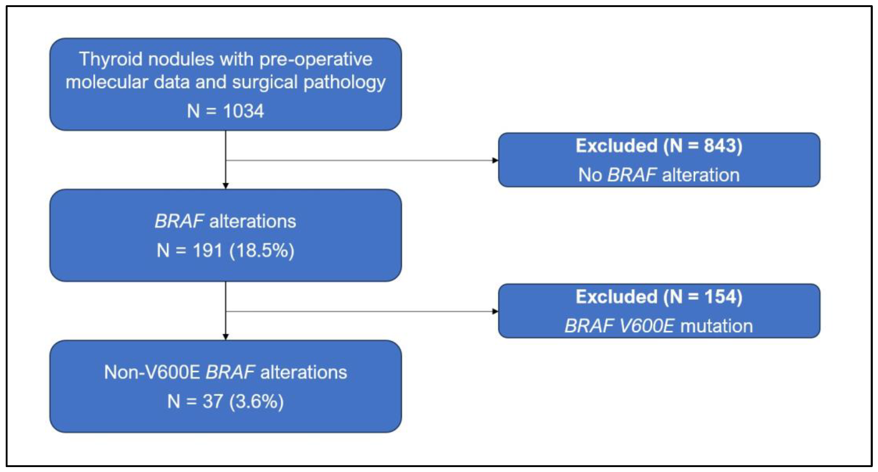
In this retrospective cohort study, genomic profiling data were reviewed from 955 patients who underwent thyroidectomy at one comprehensive cancer center between January 2014 and January 2024. This study was approved by the Institutional Review Board of Memorial Sloan Kettering Cancer Center.
Molecular testing was performed on pre-operative ultrasound-guided FNAC samples from 1034 ITNs. ITNs were classified as Bethesda Category III (Atypia of Undetermined Significance) or Bethesda Category IV [Follicular Neoplasm]) by fellowship-trained cytopathologists. Patients with BRAF gene alterations (n = 193; total including V600E) were identified using DNA and RNA-based sequencing assays (ThyroSeq v2–v3; CBLPath, Rye Brook, NY, USA). ThyroSeq assays are licensed in the US as laboratory tests that are performed in a CLIA (Clinical Laboratory Improvement Amendments)-approved and CAP (College of American Pathologists)-certified environment. These tests have been validated for the detection of mutations down to 5% allelic fraction (a standard threshold for clinical genomic profiling), and for BRAF variants, down to 1% allelic fraction [24]. BRAF mutations were categorized into Class I (RAS-independent constitutively active monomers), Class II (RAS-independent dimers with moderate-to-high kinase activity), or Class III (RAS-dependent with low or no kinase activity). Nodules harboring BRAF V600E mutations (which would fall into class I) were excluded from this study, leaving 37 patients for analysis (Figure 1).
Surgical pathology reports for the BRAF-altered ITNs included in the study were retrospectively examined. All surgical specimens were reviewed by subspecialty head and neck surgical pathologists. Histopathological diagnoses were subsequently stratified into risk categories based on the 2015 American Thyroid Association guidelines. As a quality control measure, findings on histopathology were compared with pre-operative ultrasound, FNAC, and molecular reports by matching nodule size, laterality, and location within the lobe to ensure that the surgical pathology diagnosis rendered corresponded with the nodule that was biopsied and genomically profiled. Incidental malignancies found to be independent from the biopsied nodule were considered separately.
Clinical data from pre-operative, operative, and post-operative timepoints were also retrospectively analyzed for each patient. This included clinical presentation at time of biopsy, operative extent, intraoperative findings, post-operative radioactive iodine (RAI) treatment, and recurrence.
The association between molecular alterations and malignancy was tested using Fisher’s exact test. For all hypothesis testing, significance was set at α < 0.05.

3. Results

3.1. Patient Characteristics

Overall, 37 patients (3.6% of nodules; 19.2% of BRAF alterations) were included in the study (Table 1). The majority of patients were female (n = 27, 73.0%), and the median patient age at the time of diagnosis was 44 years (interquartile range [IQR] 36–58). More than half of the nodules were <2 cm (54.1%) on pre-operative ultrasound with a median nodule diameter of 1.8 cm (IQR 1.4–2.5). High-risk sonographic features were identified in two cases (5.4%; irregular margins [n = 1], suspicious central compartment lymph nodes [n = 1]). One patient (2.7%) presented with both nodal metastasis and gross extrathyroidal extension.

3.2. Cytopathologic and Genetic Findings

Cytopathology for the ITNs included in the study was reported as Bethesda III in 62.2% of patients (n = 23) and Bethesda IV in the remaining cases (n = 14, 37.8%). Genomic profiling revealed non-V600E BRAF mutations in 32 nodules (86.5%), 4 BRAF fusions (10.8%), and 1 case of “BRAF-like gene expression” (2.7%). Among the mutations in the cohort that have been previously characterized, all were class II (n = 25, 67.6%), and the remaining mutations are unclassified (n = 7, 18.9%). K601E was the most common mutation (n = 17), representing more than half (53.1%) of all mutations identified. A single BRAF mutation was detected in 29 nodules, while the remaining three BRAF-mutated nodules (8.1%) harbored co-existing mutations (K601E + EIF1AX p.A113_splice; K601N + KRAS p.G12D; G469 + TERT p.C228T).
BRAF fusions were detected in four ITNs (10.8%). Three of the identified fusions were AGKBRAF (8.1%), and the remaining fusion was AKAP9BRAF (2.7%). Molecular testing revealed BRAF-like gene expression in one of the included nodules (2.7%).

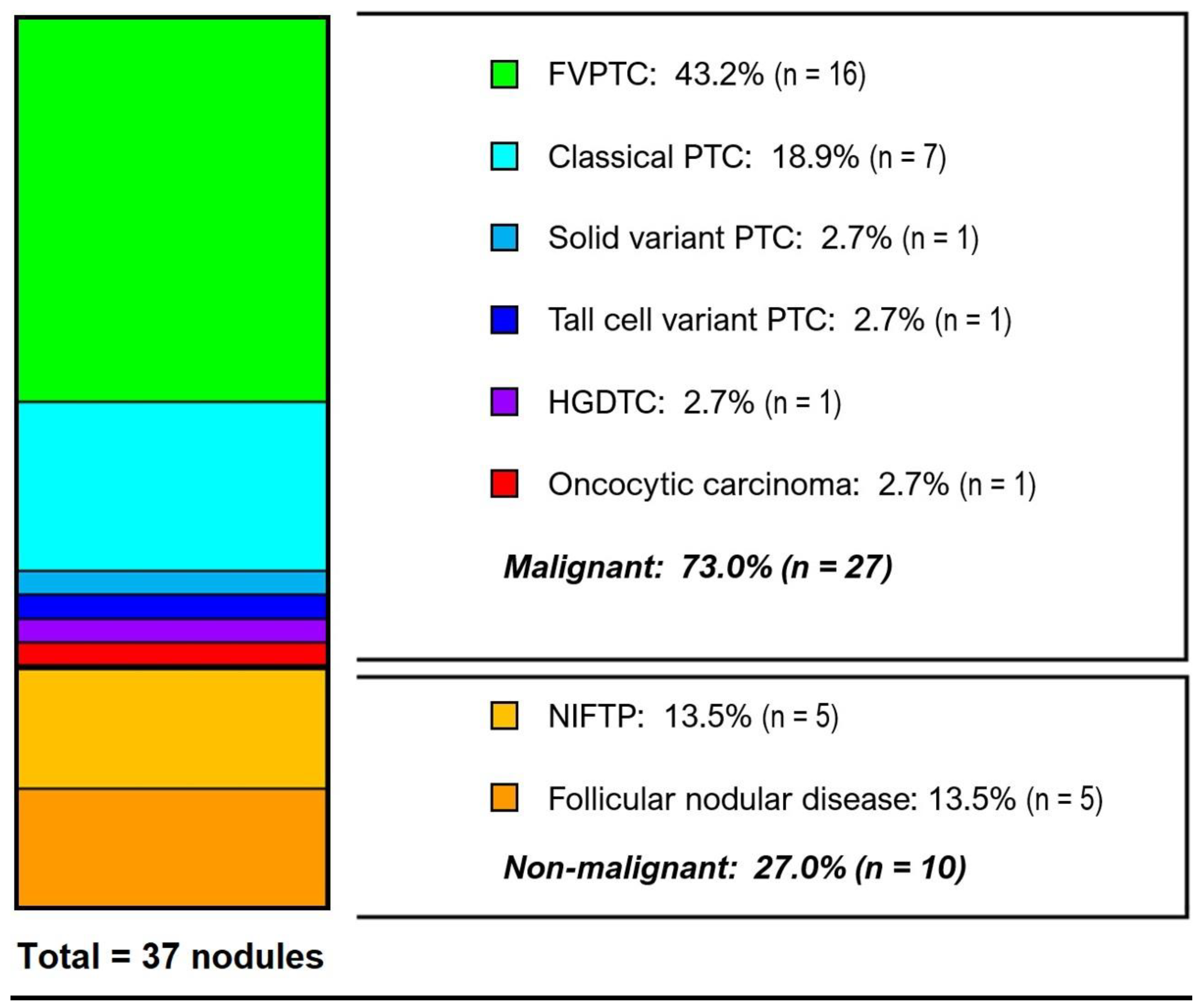
3.3. Histological Diagnoses

Surgical pathology for the cohort is displayed in Figure 2. The overall rate of malignancy (ROM) for BRAF-altered nodules was 73% (95% CI 59–87%, n = 27) with the vast majority of cases being histologically low risk by ATA criteria (n = 24, 88.9%). Papillary carcinoma represented 92.6% of all cancers (n = 25, 67.6%), and the only other histology present was minimally invasive oncocytic carcinoma (n = 1, 2.7%) as well as one high-grade differentiated lesion. Follicular (n = 16, 43.2%) and classical (n = 7, 18.9%) variants (subtypes) of PTC were most common. The histologically benign/non-malignant nodules (n = 10, 27.0%) were evenly split between NIFTP and follicular hyperplasia.
In the entire study population, nearly all nodules (n = 34, 91.9%) were either non-malignant or ATA low-risk cancers. There were no high-risk cancers identified in patients with isolated BRAF mutations (benign: n = 10 [34.5%], ATA low risk: n = 19 [65.5%]). The most common isolated mutation was K601E (n = 17, 45.9%) which had a 58.8% ROM (all ATA low risk). Patients with isolated BRAF mutations had a significantly lower rate of ATA intermediate or high risk pathology when compared to all other BRAF alterations (0% vs. 37.5%, p = 0.0072).
Two patients in the cohort underwent central compartment nodal dissection. Via formal and informal nodal sampling, lymph nodes were present in surgical pathology specimens for 19 patients (51.4%). Nodal metastases were only identified in one patient (5.3%) with AKAP9BRAF fusion. A total of 53 lymph nodes were analyzed from the other 18 patients, and all were negative for malignancy (ETE) and lymph node metastases, and this was the only patient with gross ETE or nodal disease in the cohort.

3.4. Clinical Outcomes

Approximately half of patients (54.1%) were initially treated with a partial thyroidectomy (lobectomy: n = 17 [45.9%], isthmusectomy: n = 3 [8.1%]), and the remaining patients underwent total thyroidectomy (n = 17 [45.9%]). Central compartment neck dissection was performed in only two cases (5.4%). Median postoperative follow-up for the cohort was 27 months (IQR 17.8–45.5). No major operative complications were reported. Only three patients were treated with radioactive iodine post-operatively (8.1%), and no completion thyroidectomy procedures were performed in those who did not initially undergo total thyroidectomy. No patients in the cohort were found to have distant metastatic disease or recurrence, and there were no deaths during the follow-up interval.

4. Discussion

BRAF mutations are the most common genetic alterations in differentiated thyroid cancer. Among these, BRAF V600E is by far the most prevalent, and its clinical implications are well described in the literature [12,14,15,23]. The increased utilization of molecular diagnostics for ITNs has led to the detection of many less common BRAF alterations, the prognostic significance of which is not well understood. In this study, we report a series of non-V600E BRAF alterations as well as the clinical and histopathologic features that distinguish them from BRAF V600E.
Our data indicate that nodules with non-V600E BRAF alterations are typically follicular variant PTC (FV-PTC), and the rate of malignancy (73%) is lower than that of BRAF V600E (>95%) [16,18]. In particular, the most common non-V600E mutation (K601E) had only a 58.8% risk of malignancy and all were ATA low-risk cancers. Overall, the cancers associated with non-V600E BRAF were nearly all low risk, particularly in cases of isolated BRAF mutations. Isolated BRAF mutations, most commonly K601E, were either non-malignant (34.5%) or ATA low risk (65.5%), and no isolated non-V600E BRAF lesion was found to be intermediate or high risk by ATA criteria. Additionally, no distant metastases, recurrences, or deaths occurred in the cohort. These findings appear to support the limited data published on the subject [20,21,22].
Nodal metastases were identified in only one patient (2.7%) in the cohort with an AKAP9BRAF fusion. None of the other patients had clinical or radiographic evidence of nodal disease, and no metastatic disease was found in any of the lymph nodes sampled from these patients (0/53, 0%). These data suggest that those with non-V600E BRAF alterations are at low risk for nodal metastasis, and patients are unlikely to benefit from elective neck dissection. This again contrasts the behavior of BRAF V600E mutations which have been associated with 2–3x increased risk of lymph node metastasis [25,26].
The one ATA high-risk cancer identified in the cohort was found in a nodule harboring an AKAP9BRAF fusion, and histologically this was found to be a tall cell variant PTC with nodal disease and ETE. AKAP9BRAF fusions are known oncogenic fusions, often radiation-induced, that leads to constitutively active BRAF kinase activity and mitogenic signaling via the MAPK pathway [27,28,29]. Interestingly, the patient with this fusion had no history of radiation exposure or any family history of thyroid disease. This patient underwent total thyroidectomy and central neck dissection with post-operative RAI ablation and had an excellent response with no evidence of residual or recurrent disease. The patient’s most recent surveillance testing showed an undetectable thyroglobulin level with negative thyroglobulin antibodies.
The remaining three fusions in the cohort were AGKBRAF, and these were all histologically low-risk PTC (two solid variant, one noninvasive follicular pattern). Additionally, two ATA intermediate-risk cancers were identified. One high-grade differentiated thyroid carcinoma with co-existing G469A and TERT mutations was found. The other was a solid variant PTC with BRAF-like gene expression, and this is likely due to the identification of V600E-related gene products.
Important caveats to this study include its limited sample size, its retrospective nature, and single-center design. Although this is the largest series, to our knowledge, of non-V600E BRAF mutant thyroid nodules with clinicopathologic correlation, further studies will help to better characterize these nodules, particularly those with less common genotypes (e.g., other than K601E). However, in comparison to prior smaller studies, we find that our results are largely congruent: non-V600E BRAF alterations are uncommon (1.1–3.6% of ITNs) and typically represent low-risk PTC (70.3–93.1%) that is most often follicular-patterned [20,21,22].
In comparison with BRAF V600E mutant thyroid nodules, thyroid nodules with non-V600E BRAF mutations have a lower risk of malignancy, and very low risk of aggressive or ATA high-risk malignancy. These data may be helpful in counseling patients and avoiding overtreatment. Given their comparatively indolent nature, isolated non-V600E BRAF mutations without other adverse clinical or radiographic findings can be treated first with lobectomy or active surveillance. We note that in those nodules harboring multiple concomitant mutations, that data are limited, and it is unclear if active surveillance is necessarily as appropriate in all of these cases [30,31].

5. Conclusions

ITNs harboring non-V600E BRAF alterations were usually (73%) malignant, although with lower probability than V600E mutations. Nearly all nodules were benign or ATA low-risk cancers. Only 8% of such nodules were ATA intermediate- or high-risk cancers. In ITNs with isolated non-V600E BRAF and no other genetic alterations, one-third were benign, and all cancers were ATA low risk. In the appropriate clinical context, thyroid lobectomy or active surveillance can be considered for initial management of non-V600E BRAF-altered ITNs.

Author Contributions

R.I.: investigation (lead), formal analysis (lead), writing—original draft (lead), and writing—review and editing (lead); C.E.S.: investigation (supporting); R.A.G.: writing—review and editing (equal); B.X.: writing—review and editing (equal); B.G.: writing—review and editing (equal); R.J.W.: writing—review and editing (equal); B.R.U.: writing—review and editing (equal); L.G.T.M.: conceptualization (lead) and writing—review and editing (equal). All authors have read and agreed to the published version of the manuscript.

Funding

This study was supported by NCI P30 CA008748 (to MSKCC), and Cycle for Survival, Cycle for Survival: Team Fearless4Jen, The Jayme and Peter Flowers Fund, the Sebastian Nativo Fund (to L.G.T.M.).

Institutional Review Board Statement

The study was conducted in accordance with the Declaration of Helsinki and approved by the Institutional Review Board of Memorial Sloan Kettering Cancer Center (IRB 11-195, last approval 11/21/24).

Informed Consent Statement

Patient consent was waived due to the retrospective nature of the study as stated in the final approval by the Institutional Review Board of Memorial Sloan Kettering Cancer Center.

Data Availability Statement

Data presented in this study necessary to replicate results are available on request from the corresponding author. Patient-level data are not publicly available due to the ethics approval agreement.

Conflicts of Interest

L.G.T.M. is listed as an inventor on intellectual property owned by MSK and licensed to PGDx and Tempus, unrelated to this work. Other authors declare no conflicts of interest.

References

  1. Guth, S.; Theune, U.; Aberle, J.; Galach, A.; Bamberger, C.M. Very high prevalence of thyroid nodules detected by high frequency (13 MHz) ultrasound examination. Eur. J. Clin. Investig. 2009, 39, 699–706. [Google Scholar] [CrossRef] [PubMed]
  2. Sangalli, G.; Serio, G.; Zampatti, C.; Bellotti, M.; Lomuscio, G. Fine needle aspiration cytology of the thyroid: A comparison of 5469 cytological and final histological diagnoses. Cytopathology 2006, 17, 245–250. [Google Scholar] [CrossRef] [PubMed]
  3. Cibas, E.S.; Ali, S.Z. The 2017 Bethesda System for Reporting Thyroid Cytopathology. Thyroid 2017, 27, 1341–1346. [Google Scholar] [CrossRef] [PubMed]
  4. Grani, G.; Sponziello, M.; Filetti, S.; Durante, C. Thyroid nodules: Diagnosis and management. Nat. Rev. Endocrinol. 2024, 20, 715–728. [Google Scholar] [CrossRef]
  5. Khan, T.M.; Zeiger, M.A. Thyroid Nodule Molecular Testing: Is It Ready for Prime Time? Front. Endocrinol. 2020, 11, 590128. [Google Scholar] [CrossRef]
  6. Ferraz, C. Molecular testing for thyroid nodules: Where are we now? Rev. Endocr. Metab. Disord. 2024, 25, 149–159. [Google Scholar] [CrossRef]
  7. McMurtry, V.; Canberk, S.; Deftereos, G. Molecular testing in fine-needle aspiration of thyroid nodules. Diagn. Cytopathol. 2023, 51, 36–50. [Google Scholar] [CrossRef]
  8. Alexander, E.K.; Cibas, E.S. Diagnosis of thyroid nodules. Lancet Diabetes Endocrinol. 2022, 10, 533–539. [Google Scholar] [CrossRef]
  9. Giordano, T. Integrated genomic characterization of papillary thyroid carcinoma. Cell 2014, 159, 676–690. [Google Scholar] [CrossRef]
  10. Macerola, E.; Poma, A.M.; Vignali, P.; Basolo, A.; Ugolini, C.; Torregrossa, L.; Ferruccio, S.; Basolo, F. Molecular genetics of follicular-derived thyroid cancer. Cancers 2021, 13, 1139. [Google Scholar] [CrossRef]
  11. Prete, A.; Borges de Souza, P.; Censi, S.; Muzza, M.; Nucci, N.; Sponziello, M. Update on Fundamental Mechanisms of Thyroid Cancer. Front. Endocrinol. 2020, 11, 102. [Google Scholar] [CrossRef] [PubMed]
  12. Xing, M. BRAF mutation in thyroid cancer. Endocr. Relat. Cancer 2005, 12, 245–262. [Google Scholar] [CrossRef] [PubMed]
  13. Nikiforov, Y.E.; Nikiforova, M.N. Molecular genetics and diagnosis of thyroid cancer. Nat. Rev. Endocrinol. 2011, 7, 569–580. [Google Scholar] [CrossRef] [PubMed]
  14. Tabriz, N.; Grone, J.; Uslar, V.; Tannapfel, A.; Weyhe, D. BRAF V600E mutation correlates with aggressive clinico-pathological features but does not influence tumor recurrence in papillary thyroid carcinoma—10-year single-center results. Gland Surg. 2020, 9, 1902–1913. [Google Scholar] [CrossRef]
  15. Xing, M.M.; Alzahrani, A.S.; Carson, K.A.; Viola, D.; Elisei, R.; Bendlova, B.; Yip, L.; Mian, C.; Vianello, F.; Tuttle, M.; et al. Association between BRAF V600E mutation and mortality in patients with papillary thyroid cancer. JAMA 2013, 309, 1493–1501. [Google Scholar] [CrossRef]
  16. Trimboli, P.; Treglia, G.; Condorelli, E.; Romanelli, F.; Crescenzi, A.; Bongiovanni, M.; Giovanella, L. BRAF-mutated carcinomas among thyroid nodules with prior indeterminate FNA report: A systematic review and meta-analysis. Clin. Endocrinol. 2016, 84, 315–320. [Google Scholar] [CrossRef]
  17. Kleiman, D.A.; Sporn, M.J.; Beninato, T.; Crowley, M.J.; Nguyen, A.; Uccelli, A.; Scognamiglio, T.; Zarnegar, R.; Fahey, T.J. Preoperative BRAF(V600E) mutation screening is unlikely to alter initial surgical treatment of patients with indeterminate thyroid nodules: A prospective case series of 960 patients. Cancer 2013, 119, 1495–1502. [Google Scholar] [CrossRef]
  18. Jinih, M.; Foley, N.; Osho, O.; Houlihan, L.; Toor, A.A.; Khan, J.Z.; Achakzai, A.A.; Redmond, H.P. BRAFV600E mutation as a predictor of thyroid malignancy in indeterminate nodules: A systematic review and meta-analysis. Eur. J. Surg. Oncol. 2017, 43, 1219–1227. [Google Scholar] [CrossRef]
  19. Owsley, J.; Stein, M.K.; Porter, J.; In, G.K.; Salem, M.; O’Day, S.; Elliott, A.; Poorman, K.; Gibney, G.; Vanderwalde, A. Prevalence of class I–III BRAF mutations among 114,662 cancer patients in a large genomic database. Exp. Biol. Med. 2021, 246, 31–39. [Google Scholar] [CrossRef]
  20. De Leo, A.; Serban, D.; Maloberti, T.; Sanza, V.; Coluccelli, S.; Altimari, A.; Gruppioni, E.; Chiarucci, F.; Carradini, A.G.; Repaci, A. Expanding the Spectrum of BRAF Non-V600E Mutations in Thyroid Nodules: Evidence-Based Data from a Tertiary Referral Centre. Int. J. Mol. Sci. 2023, 24, 4057. [Google Scholar] [CrossRef]
  21. Murugan, A.K.; Qasem, E.; Al-Hindi, H.; Shi, Y.; Alzahrani, A.S. Classical V600E and other non-hotspot BRAF mutations in adult differentiated thyroid cancer. J. Transl. Med. 2016, 14, 204. [Google Scholar] [CrossRef] [PubMed]
  22. Afkhami, M.; Karunamurthy, A.; Chiosea, S.; Nikiforova, M.N.; Seethala, R.; Nikiforov, Y.E.; Coyne, C. Histopathologic and clinical characterization of thyroid tumors carrying the BRAFK601E mutation. Thyroid 2016, 26, 242–247. [Google Scholar] [CrossRef] [PubMed]
  23. Tufano, R.P.; Teixeira, G.V.; Bishop, J.; Carson, K.A.; Xing, M. BRAF mutation in papillary thyroid cancer and its value in tailoring initial treatment: A systematic review and meta-analysis. Medicine 2012, 91, 274–286. [Google Scholar] [CrossRef]
  24. Nikiforova, M.N.; Lepe, M.; Tolino, L.A.; Miller, M.E.; Ohori, N.P.; Wald, A.I.; Landau, M.S.; Kaya, C.; Malapelle, U.; Bellevi-cine, C.; et al. Thyroid Cytology Smear Slides: An Untapped Re-source for ThyroSeq Testing. Cancer Cytopathol. 2021, 129, 33–42. [Google Scholar] [CrossRef]
  25. Xing, M.; Clark, D.; Guan, H.; Ji, M.; Dackiw, A.; Carson, K.A.; Kim, M.; Tufaro, A.; Ladenson, P.; Zeiger, M.; et al. BRAF mutation testing of thyroid fine-needle aspiration biopsy specimens for preoperative risk stratification in papillary thyroid cancer. J. Clin. Oncol. 2009, 27, 2977–2982. [Google Scholar] [CrossRef]
  26. Howell, G.M.; Nikiforova, M.N.; Carty, S.E.; Armstrong, M.J.; Hodak, S.P.; Stang, M.T.; McCoy, K.L.; Nikiforov, Y.E.; Yip, L. BRAF V600E mutation independently predicts central compartment lymph node metastasis in patients with papillary thyroid cancer. Ann. Surg. Oncol. 2013, 20, 47–52. [Google Scholar] [CrossRef]
  27. Ciampi, R.; Knauf, J.A.; Kerler, R.; Gandhi, M.; Zhu, Z.; Nikiforova, M.N.; Rabes, H.M.; Fagin, J.A.; Nikiforov, Y.E. Oncogenic AKAP9-BRAF fusion is a novel mechanism of MAPK pathway activation in thyroid cancer. J. Clin. Investig. 2005, 115, 94–101. [Google Scholar] [CrossRef]
  28. Lee, J.H.; Lee, E.S.; Kim, Y.S.; Won, N.H.; Chae, Y.S. BRAF mutation and AKAP9 expression in sporadic papillary thyroid carcinomas. Pathology 2006, 38, 201–204. [Google Scholar] [CrossRef]
  29. Yakushina, V.D.; Lerner, L.V.; Lavrov, A.V. Gene fusions in thyroid cancer. Thyroid 2018, 28, 158–167. [Google Scholar] [CrossRef]
  30. Sfreddo, H.J.; Koh, E.S.; Zhao, K.; Swartzwelder, C.E.; Untch, B.R.; Marti, J.L.; Roman, B.R.; Dublin, J.; Wang, R.S.; Xia, R.; et al. RAS-Mutated Cytologically Indeterminate Thyroid Nodules: Prevalence of Malignancy and Behavior Under Active Surveillance. Thyroid 2024, 34, 450–459. [Google Scholar] [CrossRef]
  31. Marcadis, A.R.; Valderrabano, P.; Ho, A.S.; Tepe, J.; Swartzwelder, C.E.; Byrd, S.; Sacks, W.L.; Untch, B.R.; Shaha, A.R.; Xu, B.; et al. Interinstitutional variation in predictive value of the ThyroSeq v2 genomic classifier for cytologically indeterminate thyroid nodules. Surgery 2019, 165, 17–24. [Google Scholar] [CrossRef] [PubMed]
Figure 1. Patient selection process for non-V600E BRAF alteration cohort.
Cancers 17 00741 g001
Figure 2. Histological diagnoses of cohort. The overall rate of malignancy (ROM) for BRAF-altered nodules was 73% (95%CI 59–87%, n = 27). Papillary carcinoma represented 92.6% of all cancers (n = 25, 67.6%). HGDTC, high-grade differentiated thyroid carcinoma; NIFTP, noninvasive follicular thyroid neoplasm with papillary-like nuclear features. Two patients with ATA intermediate-risk and one patient with high-risk DTC were identified in the study cohort (Table 1). One of the intermediate-risk lesions was a high-grade differentiated thyroid carcinoma (HGDTC) with a high mitotic rate associated with co-existing BRAF G469A and TERT mutations. The other was a solid variant of PTC with microscopic invasion of the perithyroidal soft tissues in the nodule with BRAF-like gene expression. The only ATA high-risk lesion in the cohort was a tall cell variant of PTC associated with a AKAP9–BRAF fusion that was found to have extrathyroidal extension (ETE) and lymph node metastases, and this was the only patient with gross ETE or nodal disease in the cohort.
Cancers 17 00741 g002
Table 1. Cohort characteristics of 37 patients with non-V600E BRAF-altered indeterminate thyroid nodules.
Age at Diagnosis (Years) Treatment Type
 Median (IQR) 44 (36–58) Total thyroidectomy17 (45.9%)
 Range19–72 Thyroid lobectomy17 (45.9%)
 Isthmusectomy 3 (8.1%)
Sex
 Female 27 (73.0%)Final surgical pathology
 Male10 (27.0%) Benign/non-malignant10 (27.0%)
 Malignant27 (73.0%)
Nodule size on US (cm)   ATA low risk24 (64.9%)
 Median (IQR) 1.8 (1.4–2.5)  ATA intermediate risk 2 (5.4%)
 <2 20 (54.1%)  ATA high risk 1 (2.7%)
 2–4 16 (43.2%)
 >4 1 (2.7%)Histology
 Papillary carcinoma25 (67.6%)
Bethesda category    Follicular subtype16 (43.2%)
 III23 (62.2%)    Classical subtype 7 (18.9%)
 IV14 (37.8%)    Solid subtype 1 (2.7%)
   Tall cell subtype 1 (2.7%)
Alteration type  NIFTP 5 (13.5%)
BRAF mutation32 (86.5%) Follicular nodular disease 5 (13.5%)
 Class I 0 (0%) Oncocytic carcinoma 1 (2.7%)
 Class II25 (67.6%) HGDTC 1 (2.7%)
   K601E17 (45.9%)
   G469A 4 (10.8%)Gross extrathyroidal extension 1 (2.7%)
   N486_P490del 1 (2.7%)
   Class II + other * 3 (8.1%)Nodal disease 1 (2.7%)
 Class III 0 (0%)
 Unclassified 7 (18.9%)Distant metastases 0 (0%)
BRAF fusion 4 (10.8%)
  AGKBRAF 3 (8.1%)Radioactive iodine 3 (8.1%)
  AKAP9BRAF 1 (2.7%)
BRAF-like gene expression 1 (2.7%)Recurrence 0 (0%)
Duration of follow-up (months) Deaths 0 (0%)
 Median (IQR) 27 (17.8–45.5)
IQR, interquartile range; ATA, American Thyroid Association; HGDTC, high-grade differentiated thyroid carcinoma; NIFTP, noninvasive follicular thyroid neoplasm with papillary-like nuclear features; * K601E + EIF1AX (p.A113_splice); K601N + KRAS (p.G12D); G469 + TERT (p.C228T); T599del (1); T599_V600insP (1); T599_R603delins? (1); A598_T599insV (1); A598dup (1); T488_Q493delinsK (1); exon 2–8 deletion with fusion of exons 1 and 9 (1); Sample positive for “BRAF-like gene expression alterations associated with thyroid cancer” (ThyroSeq v3 GC).
Disclaimer/Publisher’s Note: The statements, opinions and data contained in all publications are solely those of the individual author(s) and contributor(s) and not of MDPI and/or the editor(s). MDPI and/or the editor(s) disclaim responsibility for any injury to people or property resulting from any ideas, methods, instructions or products referred to in the content.

