Article

Minimally Invasive One-Docking, Two-Target, and Three-Port Robotic-Assisted Nephroureterectomy: Redefining Surgical Approach

Department of Urology, Soroka University Medical Center, Faculty of Health Science, Ben-Gurion University of Negev, P.O. Box 151, Beer Sheva 84101, Israel
*
Author to whom correspondence should be addressed.
Cancers 2025, 17(4), 627; https://doi.org/10.3390/cancers17040627
Submission received: 13 January 2025 / Revised: 8 February 2025 / Accepted: 10 February 2025 / Published: 13 February 2025

Simple Summary

This study focuses on robotic radical nephroureterectomy performed for patients with urothelial carcinoma in their kidneys and ureters. The da Vinci Xi platform, which enhances minimally invasive surgical techniques was applied. We aimed to optimize the surgical approach by reducing the number of ports used, and improving visualization during the procedure. The study involved 15 patients operated on by a single surgeon between 2019 and 2024, using a one docking, three-port technique. The median operative time was 133 min, and most surgeries were completed within 150 min. Blood loss was minimal, and complication rate was low. In conclusion, the study highlights that the three-port technique combined with the Xi platform is a safe and effective method for performing robotic radical nephroureterectomy, facilitating quicker procedures and achieving surgical goals.

Abstract

Objective: Nephroureterectomy (NU) is a challenging multi-quadrant surgical procedure that involves intra–infra- and retroperitoneal dissection. The introduction of the da Vinci Xi platform has further improved the minimally invasive approach. With the Xi robotic system, single docking for multi-quadrant dissection is now feasible and increasingly popular. Herein, we redefined the surgical technique by optimizing minimal port usage, maximizing template visualization, and utilizing the Xi platform’s retargeting system, based on our experience with 15 patients. Methods: This single-center cohort study was based on the experience of a single surgeon with 15 patients who underwent robotic radical nephroureterectomy (RRNU) between 2019 and 2024, performed via the one docking, three-port technique. Patient data were retrospectively collected and analyzed from the medical records. Results: The cohort’s median age was 79 years, with male predominance of 80%. The median operative time was 133 min, with 60% of procedures completed within 150 min; longer times (>150 min) were due to additional intraoperative non-robotic procedures. Median blood loss was 100 mL, with two patients requiring intraoperative blood transfusion. Lymph node dissection (LND) in six patients did not significantly affect the overall operative time. The intraoperative and major postoperative complication rates (Clavien-Dindo grade > III) were 13.3%. Conclusions: The use of three robotic ports combined with the Xi platform’s camera-hop feature is a safe and effective technique for multi-quadrant, minimally invasive procedures, particularly in RRNU. This approach facilitates procedural goals and reduces overall operative time.

1. Introduction

Radical nephroureterectomy (NU) with bladder cuff excision (BCE) is the gold-standard treatment for upper tract urothelial carcinoma (UTUC). Current guidelines indicate the importance of the surgical management of UTUC; however, there is no consensus regarding the operative approach [1]. The ‘master–slave’ robotic system was first introduced in the late 1990s, and the first robotic-assisted laparoscopic urology surgery was performed in 2000 [2]. Since then, the da Vinci platform has evolved. In general, the da Vinci robotic platform is superior to the laparoscopic technique owing to its 3D imaging, and the wristed configuration provides a high degree of freedom, allowing accurate and precise movements. Despite these advantages, the da Vinci platform faces limitations, including its high financial cost and economic burden, which restrict its accessibility in many public health institutions worldwide [3].
Although robot-assisted laparoscopic radical NU has already been described, its intraoperative management remains highly versatile and largely depends on surgeon experience and expertise. Standardized radical NU tetrafecta includes negative soft tissue surgical margins, BCE, lymph node dissection (LND) (for high-risk, non-metastatic disease), and no evidence of recurrence within 12 months [4,5]. The key operative goals are to minimize the overall operative time, prevent tumor seeding, and reduce the risk of urine leakage. However, the extensive nature of this surgery, especially when LND is performed, places patients at an increased risk of complications, such as major organ injury, bowel perforation, and significant blood loss. The da Vinci Xi robotic system offers distinct advantages in such complex procedures, particularly in its ability to facilitate multi-quadrant minimally invasive surgery [6]. This article presents our experience with 15 patients who underwent multi-quadrant robotic radical nephroureterectomy (RRNU) using the Xi platform. By utilizing only three ports and the camera-hop feature, we redefined the single-docking RRNU procedure to streamline the procedure and achieve favorable outcomes.

2. Patients and Methods

With the approval of the Institutional Ethics Committee, we retrospectively collected data from patients diagnosed with UTUC who underwent RRNU between 2019 and 2024. Patients who underwent NU for indications other than UTUC were excluded from the study. All patients included in the study had a suspicious lesion on imaging and were diagnosed with UTUC through diagnostic ureteroscopy (Dx URS). Following a multidisciplinary evaluation of all associated risks, they were referred for radical NU (Scheme 1). Notably, updated guidelines now allow a radical surgical approach based solely on imaging or visual appearance, without requiring biopsy-confirmed malignancy [4]. However, our study was conducted before these guideline revisions. The data included preoperative, intraoperative, and postoperative surgical details, with a follow-up period of 12 months to monitor the outcomes. All the procedures were performed by a single surgeon with up to 10 years of experience in robotic surgery. Fifteen patients met the inclusion criteria and were included in this study.

3. Surgical Intervention

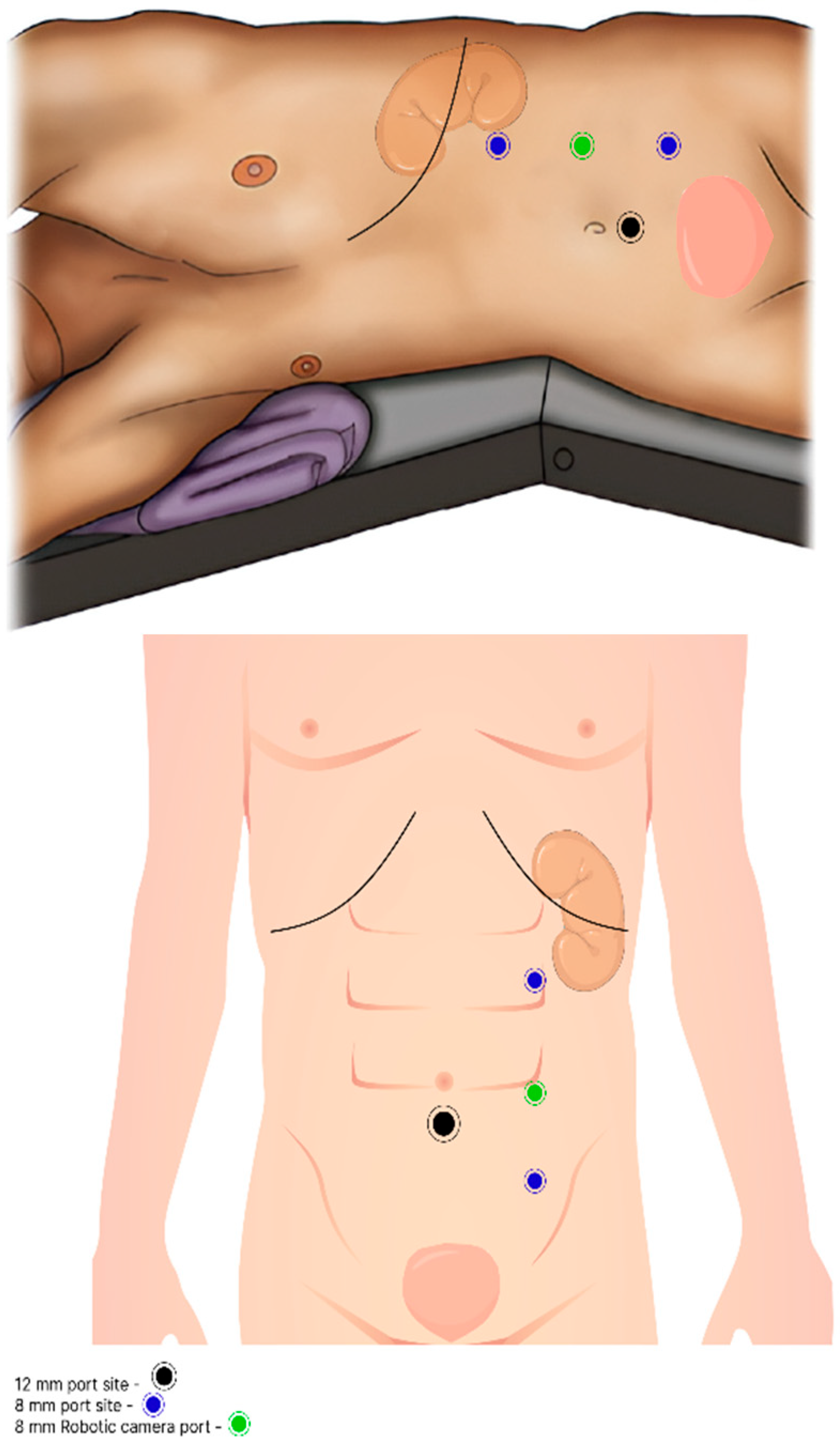
All 15 patients underwent RRNU with BCE by using the Xi da Vinci robotic system. The operator employed a unique intraoperative technique based on single docking for multi-quadrant surgery. After catheter insertion, patients were placed in a modified lateral decubitus position with 30-degree flexion. Trocar placement was planned along the paramedian line, with 8 cm spacing between the trocars. Three 8 mm trocars were used to dock the robotic arms, and a robotic camera was inserted through the middle port. An additional 12 mm trocar was placed in the abdominal midline, inferior to the umbilicus, and 5 cm from both the camera and the third arm trocars, forming a triangular configuration (Figure 1). The camera was initially directed toward the renal area for targeting, enabling optimal robotic arm positioning and rotation for the nephrectomy portion of the procedure.
Once docking was completed, the surgical procedure began with retroperitoneal exposure, facilitated by the dissection of Toldt’s white line. After exposing the kidney, pre-renal tissue was carefully dissected and separated at the superior lateral border. After stapling the main renal blood vessels, caudal dissection was performed at the mid-ureteral level, approximately at the L2–L3 level, parallel to the gonadal vessels. Next, the assistant disconnected the robotic endpoint instruments and redirected the camera toward the pelvis. This step allowed for the retargeting of the robotic camera and reconfiguration of the robotic arms to face the pelvic area. Once the endpoint instruments were reconnected, dissection of the distal ureter was completed, and extravesical BCE was performed.
A Hem-o-lok clip was placed on the distal ureter (depending on the location of the tumor) before complete transection and separation. An inverted U-shaped stitch using a 3-0 V-Loc suture was applied at the ureteral margin to prevent urine leakage during the excision. Following the completion of the BCE procedure, the specimen was placed directly into a sealed laparoscopic bag, and the bladder was sutured with 3-0 V-Loc. In selected cases, LND was performed along the template, extending from the ipsilateral renal vein to the common iliac vessels including the external and internal iliac vessels. To minimize spillage, a separate endo-bag was used for LND specimens. A Jackson–Pratt (JP) drain was placed along the retroperitoneum from the resection bed of the kidney to the pelvis.

4. Results

Fifteen patients were included in the study, with data collected between 2019 and 2024 with a minimal follow-up period of 12 months, except for the last patient who died postoperatively. Table 1 summarizes the patients’ demographic characteristics. The preoperative variables are summarized in Table 2. The median patient age was 79 years with an average age-adjusted Charlson comorbidity index score of 8.

4.1. Operative Details

The median total operative time was 133 min, with 60% of the procedures completed within 150 min. Notably, LND did not significantly prolong the operative time. The median estimated blood loss (EBL) was 100 mL, with two patients requiring intraoperative blood transfusions despite controlled bleeding, attributed to their low preoperative hemoglobin levels. None of the cases required surgical technique conversion for the RRNU portion.

4.2. Lymph Node Dissection and Outcomes

LND was performed in six patients, with two showing positive nodes. Despite the extent of the procedure, no major intra- or postoperative complications were noted.

4.3. Postoperative Outcomes (Table 3)

Complications: The overall major complication rate (Clavien-Dindo grade > III) was 13.33%. One patient succumbed to cardiac arrest on postoperative day (POD) 2. Resuscitation was initiated immediately, followed by an emergency coronary angiography. Unfortunately, the last patient developed irreversible cardiac arrhythmias and did not respond to further resuscitation. The second patient was readmitted on POD 7 with type II myocardial infarction and required angioembolization.
Table 3. Postoperative variables.
Postoperative Lab Values(N = 15)
Hb (g/dL)
Mean11.2
Median11
Range7.5–13.9
Creatinine
Mean1.6
Median1.5
Range1–3.7
eGFR (mL/min/1.73 m2)
Mean48
Median45
Range15–82
Postoperative hospitalization(N = 14)
Length of stay (days)
Mean 5.8
Median5
Range3–10 months
Day of catheter withdrawal
Mean15.5
Median4.5
Range3–150 *
Day of drain withdrawal
Mean 4
Median 3
Range2–14 months
Complications rated by Clavien-Dindo Score (N = 15)
0–1 7 (46.6%)
2–3 months6 (40%)
41 (6.66%)
51 (6.66%)
Complications noted during postoperative period (N = 15)
Fever3
Urosepsis2
Blood transfusion5
Atelectasis 3
Surgical site infection1
Ileus 2
Acute kidney injury9
Type II MI1
Sudden cardiac arrest 1
ICU Observation 1
* One patient remained with indwelling catheter until prostatectomy (TUR-P).
Renal Function: The median postoperative estimated glomerular filtration rate (eGFR) was 45 mL/min/1.73 m2, representing an average decline of 21.87 mL/min/1.73 m2. Three patients with preoperative renal failure developed chronic renal failure (CRF). Among six patients who developed acute kidney injury postoperatively, all maintained an eGFR ≥ 50 mL/min/1.73 m2 at one month.
Hospital Stay: The average hospital stay was five days. Catheters were removed on POD 3 to 5, except in one patient with bladder outlet obstruction who later underwent prostate surgery. Drains were removed on POD 4.

4.4. Oncological Outcomes

A period of 12 months was defined as the minimum timeframe for follow-up. This period was characterized by close monitoring; thus, the majority of metastatic or recurrence cases were identified within this timeframe. Although international guidelines indicate that close follow-up has limited evidence for both high- and low-risk disease following radical NU, our institution continues to adhere to this practice, ensuring diligent monitoring [1]. At 12 months, the overall survival and disease-free survival rates were 78.57% and 42.86%, respectively. Of the two patients who received neoadjuvant therapy, one achieved a pathological complete response (pT0) with no evidence of disease (NED) in the 12-month period. Among the six patients who completed adjuvant therapy, two achieved a complete response. Five patients presented with metastatic disease involving the liver, retroperitoneal lymph nodes, lungs, or bones. Among these, three died from the disease within an average of eight months postoperatively. Two patients continued the second-line adjuvant therapy with avelumab. As shown in Table 4, three patients presented with non-urothelial metastatic disease, with recurrence occurring beyond the 12-month follow-up period defined as our study’s follow-up timeframe.
Bladder Recurrence: Nine patients resumed cystoscopy follow-up; of these, two progressed to metastatic disease. Among the seven non-metastatic patients, three developed bladder recurrence at an average of six months postoperatively, requiring transurethral resection (TURBT) and continued Bacillus Calmette–Guérin (BCG) therapy. One patient refused to undergo BCG instillations.

4.5. Sub-Group Analysis

With regard to Charlson adjusted comorbidity index (CCI): It is important to note that the cohort’s population had a median CCI score of 8. To assess whether this technique is suitable for highly morbid patients, we conducted a sub-analysis using a CCI cut-off point of ≥5. A total of 11 out of the 15 patients met this criterion. Among them, two patients died from metastatic disease, one died from cardiac arrest as mentioned, and two patients are still alive, presenting with metastatic UTUC and receiving adjuvant therapy. The overall survival rate in this sub-analysis group was 72.72%, and the disease-free survival rate was 54.54%.
With regard to age: Regarding age, the cohort’s median age was 79 years. We conducted a sub-group analysis to assess whether the elderly population would benefit from the procedure, using 80 years as the cut-off. A total of seven patients were 80 years or older. Of them, only one patient died from metastatic disease. The remaining six patients survived with no evidence of disease at the 12-month follow-up. The overall survival rate in this subgroup was 85.71%, with the same rate of disease-free survival.

5. Discussion

Early publications on radical NU involved a combination of laparoscopic nephrectomy and robotic distal ureterectomy with BCE [7]. With technological advancements and increased surgical experience, the robotic approach has become the preferred method for radical NU [8]. However, one of the major drawbacks of RRNU compared to open or laparoscopic approaches is its high cost and time consumption [9]. Despite the minimally invasive nature of the procedure, the prolonged operative time limits its advantages in terms of complication rate [10]. Therefore, optimizing even minor factors to reduce procedure time is essential when performing robotic surgery.
Single-docking robotic NU has been described in the literature, although there is no consensus on the optimal number of ports, their placement, or step-by-step procedural methods. Recent advancements have focused on utilizing the camera-hop technique of the Xi robotic system, which involves intraoperative reconfiguration from the cephalad to caudal direction. Although this approach has been shown to be safe, it has not been universally adopted [11,12,13,14].
In our cohort, we employed a relatively under-reported technique for RRNU using only three robotic arm ports and one assistant port: two 8 mm ports for robotic endpoint instruments, one 8 mm port for the camera, and one 12 mm AirSeal assistant port (Figure 1). This minimalist approach allowed for efficient multi-quadrant dissection while minimizing port placement, reducing arm collision, and shortening the overall operative time, thus facilitating a more focused and streamlined surgical process. The intra- and postoperative complications are summarized in Table 5 and Table 6, respectively.
The median operative time for our cohort was 133 min, which is significantly lower than the median operative time of 203 min reported by Pathak et al. in their 90-patient cohort using a four-port RRNU technique [13]. Additionally, intraoperative complications in our cohort were minimal, with only two patients requiring blood transfusions, both with controlled bleeding. Importantly, none of the patients required conversion of the surgical approach due to bowel complications. The postoperative major complication rate, defined as Clavien-Dindo grade > III, was 13.33%, which included one death from sudden cardiac arrest and one patient with type II myocardial infarction. These complications should be considered in the context of our patient population, which primarily consisted of patients with a high burden of comorbidities, as reflected by a median CCI of 8 (Table 1). The CCI is designed to preoperatively identify patients at greater risk of intra- and postoperative complications [15]. In our analysis of the subgroup of patients with a CCI ≥ 5, we observed a total complication rate of 18%.
The inclusion of octogenarian patients in the surgical management of UTUC has been a frequent focus of previous studies. Large cohorts have demonstrated the benefits of minimally invasive RRNU in elderly populations, emphasizing its low complication rates and relatively high overall survival rates [16,17,18].
In our sub-analytical group of patients aged over 80 years, this study corroborates earlier findings, confirming the safety and feasibility of minimally invasive NU. Within this group, we observed a relatively high survival rate of 85.7% and an identical disease remission rate, with a mean follow-up period of 18 months. The complication rate in this subgroup was 14%, comparable to the overall study population.
A review of the literature reveals diverse opinions regarding the factors that most significantly influence surgical outcomes. Wallace et al. reported higher mortality rates among elderly patients; however, their study encompassed 12 different urological procedures rather than focusing solely on NU [19]. In contrast, data from 2,352 patients in the CROES-UTUC registry demonstrated survival benefits associated with radical NU, regardless of age or comorbidities [17].
Our findings suggest that a patient’s morbidity profile, as reflected by the CCI, may have a more significant impact on complication rates than age alone [15]. Nonetheless, further prospective studies are necessary to validate these findings.
It is worth noting, however, that the majority of clinical research supports the superiority of radical NU for UTUC, even in the octogenarian population [16,17,20,21]. Multiple clinical trials have compared minimally invasive approaches to open surgery, all indicating the superiority of the minimally invasive approach, particularly robotic surgery [18,21,22,23,24]. Unfortunately, one of the drawbacks of the robotic approach, as mentioned, is its time consumption, which may limit its use in the fragile elderly population, further underscoring the advantage of our time-efficient surgical technique [25,26].
While several cohorts have described the use of the camera-hop technique with the da Vinci Xi robotic system for NU, to the best of our knowledge, no study has utilized the three-port single-docking technique described in this report. Studies by Nanigian et al. (2006), Lee et al. (2013), Patel et al. (2015), Pathak et al. (2020), and others have reported various robotic approaches; however, they did not employ this specific configuration [9,13,27,28]. Pathak et al. utilized a four-port technique for RRNU and noted several intraoperative complications, including duodenal injuries, emphasizing the advantage of a reduced-port technique in minimizing the risk of such injuries. Additionally, they suggested that the integration of a movable table could further aid in dissection and prevent arm collisions. Arm collision is another challenge that our three-port technique effectively mitigates, particularly when combined with the Xi arm adjustment switch button [13].
One of the largest studies on RRNU, the ROBUUST cohort, analyzed 1118 patients and reported a median operative time of 212.5 min, along with a postoperative complication rate of 14.1%. While these results provide valuable insights, it is important to note that the ROBUUST study did not standardize robotic system use or surgical techniques, as it included data from multiple centers with varying levels of surgeon expertise. This variability makes direct comparison with our cohort challenging [29]. However, when comparing our results to a more homogeneous group—specifically, the study by Veccia and colleagues published in 2022, which analyzed 148 patients from the ROBUUST cohort who underwent single-stage RRNU with the Xi robotic system—the results were more directly comparable [30]. Their study reported a median operative time of 215.5 min (range: 160.5–290.0), with matched estimated blood loss (EBL) and major complication rates. In contrast, our cohort achieved a median operative time of 133 min, which was considerably shorter, while maintaining a similar complication profile. Although our cohort was small, the results align closely with those of the ROBUUST group in terms of overall complication rates and EBL [29]. Furthermore, our reduced operative time was a notable improvement compared with that of the ROBUUST group, underscoring the time efficiency of our three-port approach. These findings suggest that the three-port technique is not only safe, but also highly time-efficient for performing RRNU (Table 4).
Our study specifically excluded benign cases, focusing solely on malignant UTUC to better evaluate the oncological safety of the three-port single-stage robotic approach. Notably, all patients in our cohort had negative surgical margins, including BCE, and none experienced recurrence in the resection bed during the 12-month follow-up period, even among patients with metastasis (disregarding retroperitoneal lymph nodes). This highlights the oncological efficacy of our technique, which is comparable to that of larger and more complex cohorts.
Our study has several strengths. Uniformity in the surgical technique, patient positioning, trocar placement, intraoperative instruments, and stitches ensured consistency and reliability across all cases. The use of a transperitoneal approach and the Xi robotic program further minimized spillage and optimized procedural time, contributing to the overall efficiency of the technique. However, limitations exist, including the retrospective nature of the study, lack of console time data, and variability in non-robotic procedures such as cystoscopy and Mitomycin C (MMC) instillations. In addition, because the procedure was performed by a single surgeon, the results were subject to performance bias. On the other hand, the experience of a single surgeon enhances the reliability and accuracy of the results, strengthening the feasibility and safety of the three-port single-docking technique.

6. Conclusions

RRNU is a complex procedure that demands a high level of expertise, which can be enhanced through advancements in robotic technology, particularly the upgrade from the X to the Xi robotic platform. Our study demonstrated that the three-port, single-docking NU procedure is both feasible and safe, particularly when the retargeting capabilities of the Xi system are utilized. This cohort achieved the defined tetrafecta for NU, with results comparable to those of the largest published case series using four ports. Notably, the three-port, single-docking technique showed superior performance in terms of overall operative time. To further validate these findings, prospective comparisons between three- and four-port single-docking techniques using the Xi da Vinci program are required. Additionally, the integration of artificial intelligence to optimize camera movement and predict shifts in the surgical field may further improve the efficiency and precision of this approach.

Author Contributions

Conceptualization, Y.Z.; Methodology, Y.Z. and M.H.J.; Software, B.H.; Formal analysis, I.V., R.M. and N.A.; Investigation, E.C. and N.A.; Data curation, Y.Z. and E.M.; Writing—original draft, Y.Z.; Writing—review & editing, Y.Z.; Supervision, N.J.M.; Project administration, N.J.M. All authors have read and agreed to the published version of the manuscript.

Funding

The authors received no financial support for the research, authorship, and publication of this article.

Institutional Review Board Statement

The study was conducted in accordance with the Declaration of Helsinki, and approved by the Institutional Ethics Committee of Soroka University Medical Center (protocol code 0069-24-SOR and date of approval 25 March 2024).

Informed Consent Statement

Patient consent was waived by the Institutional Ethics Committee of Soroka University Medical Center.

Data Availability Statement

The datasets generated and/or analyzed during the current study are available from the corresponding author upon reasonable request.

Conflicts of Interest

The authors declare no conflicts of interest.

Abbreviations

BCEBladder cuff excision
BCGBacillus Calmette–Guerin
EBLEstimated blood loss
LNDLymph node dissection
NUNephroureterectomy
MMCMitomycin C
PODPostoperative day
RRNURobotic radical nephroureterectomy
TURBTTransurethral resection of bladder tumor
URS/DxURS ureteroscopy/diagnostic ureteroscopy
UTUCUpper tract urothelial carcinoma

References

  1. Rouprêt, M.; Seisen, T.; Birtle, A.J.; Capoun, O.; Compérat, E.M.; Dominguez-Escrig, J.L.; Gürses Andersson, I.; Liedberg, F.; Mariappan, P.; Hugh Mostafid, A.; et al. European Association of Urology Guidelines on Upper Urinary Tract Urothelial Carcinoma: 2023 Update. Eur. Urol. 2023, 84, 49–64. [Google Scholar] [CrossRef] [PubMed]
  2. Klingler, D.W.; Hemstreet, G.P.; Balaji, K.C. Feasibility of robotic radical nephrectomy—Initial results of single-institution pilot study. Urology 2005, 65, 1086–1089. [Google Scholar] [CrossRef] [PubMed]
  3. Park, S.K.; Olweny, E.O.; Best, S.L.; Tracy, C.R.; Mir, S.A.; Cadeddu, J.A. Patient-reported body image and cosmesis outcomes following kidney surgery: Comparison of laparoendoscopic single-site, laparoscopic, and open surgery. Eur. Urol. 2011, 60, 1097–1104. [Google Scholar] [CrossRef] [PubMed]
  4. Pandolfo, S.D.; Cilio, S.; Aveta, A.; Wu, Z.; Cerrato, C.; Napolitano, L.; Lasorsa, F.; Lucarelli, G.; Verze, P.; Siracusano, S.; et al. Upper Tract Urothelial Cancer: Guideline of Guidelines. Cancers 2024, 16, 1115. [Google Scholar] [CrossRef]
  5. Soria, F.; Pradere, B.; Hurle, R.; D’Andrea, D.; Albisinni, S.; Diamand, R.; Laukhtina, E.; Di Trapani, E.; Aziz, A.; Krajewski, W.; et al. Radical Nephroureterectomy Tetrafecta: A Proposal Reporting Surgical Strategy Quality at Surgery. Eur. Urol. Open Sci. 2022, 42, 1–8. [Google Scholar] [CrossRef]
  6. Ludwig, W.W.; Gorin, M.A.; Pierorazio, P.M.; Allaf, M.E. Frontiers in robot-assisted retroperitoneal oncological surgery. Nat. Rev. Urol. 2017, 14, 731–741. [Google Scholar] [CrossRef]
  7. Lai, W.R.; Lee, B.R. Techniques to resect the distal ureter in robotic/laparoscopic nephroureterectomy. Asian J. Urol. 2016, 3, 120–125. [Google Scholar] [CrossRef]
  8. Saini, S.; Pathak, R.A.; Hemal, A.K. Robotic nephroureterectomy in the management of upper tract urothelial cancer: Inching toward standard of care? Int. Urol. Nephrol. 2022, 54, 1777–1785. [Google Scholar] [CrossRef]
  9. Patel, M.N.; Aboumohamed, A.; Hemal, A. Does transition from the da Vinci Si ® to Xi robotic platform impact single-docking technique for robot-assisted laparoscopic nephroureterectomy? BJU Int. 2015, 116, 990–994. [Google Scholar] [CrossRef]
  10. Yip, W.; Assel, M.J.; Wong, N.C.; Tracey, A.T.; Alvim, R.G.; Nogueira, L.; Almassi, N.; Singla, N.; Clinton, T.N.; Sjoberg, D.D.; et al. Changes in the Perioperative Management and Outcomes of Patients with Upper Tract Urothelial Carcinoma Undergoing Radical Nephroureterectomy at Memorial Sloan Kettering Cancer Center: Over 20 Years of Experience. Urol. Pract. 2024, 11, 356–366. [Google Scholar] [CrossRef]
  11. Darwiche, F.; Swain, S.; Kallingal, G.; Punnen, S.; Manoharan, M.; Parekh, D.J.; Gonzalgo, M.L. Operative technique and early experience for robotic-assisted laparoscopic nephroureterectomy (RALNU) using da Vinci Xi. SpringerPlus 2015, 4, 298. [Google Scholar] [CrossRef] [PubMed]
  12. Morizane, S.; Yumioka, T.; Iwamoto, H.; Hikita, K.; Honda, M.; Takenaka, A. Initial Experience of Robot-Assisted Laparoscopic Nephroureterectomy in Japan: A Useful Technique Using a Vessel Sealing Device for Securing a Good Surgical Field and Efficient Sealing. Asian J. Endosc. Surg. 2022, 15, 458–462. [Google Scholar] [CrossRef]
  13. Pathak, R.A.; Dutta, R.; Williams, J.; Hemal, A.K. Robotic radical nephro-ureterectomy for high-risk upper tract urothelial carcinoma: Step-by-step illustrative video of surgical technique. Urol. Video J. 2020, 8, 100068. [Google Scholar] [CrossRef]
  14. Zargar, H.; Krishnan, J.; Autorino, R.; Akca, O.; Brandao, L.F.; Laydner, H.; Samarasekera, D.; Ko, O.; Haber, G.-P.; Kaouk, J.H.; et al. Robotic Nephroureterectomy: A Simplified Approach Requiring No Patient Repositioning or Robot Redocking. Eur. Urol. 2014, 66, 769–777. [Google Scholar] [CrossRef] [PubMed]
  15. AlAshqar, A.; Ghazarian, M.; Webster, E.M.; Upadhyay, A.; Azodi, M.; Schwartz, P.E.; Ratner, E.; Altwerger, G. Surgical and oncologic outcomes in surgically treated women 80 years and older with endometrioid endometrial cancer as a function of their comorbidities. Gynecol. Oncol. Rep. 2023, 49, 101240. [Google Scholar] [CrossRef]
  16. Koterazawa, S.; Kanno, T.; Kobori, G.; Ito, K.; Nakagawa, H.; Takahashi, T.; Takaoka, N.; Somiya, S.; Nagahama, K.; Ito, M.; et al. Clinical outcomes following laparoscopic radical nephroureterectomy in octogenarians. Int. J. Clin. Oncol. 2023, 28, 155–162. [Google Scholar] [CrossRef]
  17. Teoh, J.Y.C.; Ng, C.F.; Eto, M.; Chiruvella, M.; Capitanio, U.; Esen, T.; Zeng, G.; Lechevallier, E.; Andonian, S.; de la Rosette, J. Radical nephroureterectomy for UTUC conferred survival benefits irrespective of age and comorbidities. World J. Urol. 2022, 40, 2657–2665. [Google Scholar] [CrossRef]
  18. Trecarten, S.; Bhandari, M.; Abdel-Aziz, A.; Noel, O.; Liss, M.; Dursun, F.; Robert, S.; Mansour, A. Open versus minimally invasive nephroureterectomy in octogenarians: An analysis of surgical approach trends, outcomes and survival analysis with propensity matching. In Urologic Oncology: Seminars and Original Investigations; Elsevier: Amsterdam, The Netherlands, 2024; Volume 42. [Google Scholar]
  19. Wallace, B.; Breau, R.H.; Cnossen, S.; Knee, C.; McIsaac, D.; Mallick, R.; Cagiannos, I.; Morash, C.; Lavallée, L.T. Age-stratified perioperative mortality after urological surgeries. Can. Urol. Assoc. J. 2018, 12, 256. [Google Scholar] [CrossRef]
  20. Upfill-Brown, A.; Lenis, A.T.; Faiena, I.; Salmasi, A.H.; Johnson, D.C.; Pooli, A.; Drakaki, A.; Gollapudi, K.; Blumberg, J.; Pantuck, A.J.; et al. Treatment utilization and overall survival in patients receiving radical nephroureterectomy versus endoscopic management for upper tract urothelial carcinoma: Evaluation of updated treatment guidelines. World J. Urol. 2019, 37, 1157–1164. [Google Scholar] [CrossRef]
  21. Ye, J.; Wu, Q.; Liao, X.; Zheng, L.; Wei, Q.; Bao, Y. Advanced Age May Not Be an Absolute Contraindication for Radical Nephroureterectomy in Patients with Upper Tract Urothelial Carcinoma: A Single-Center Case Series and a Systematic Review with Meta-Analyses. J. Clin. Med. 2023, 12, 7273. [Google Scholar] [CrossRef]
  22. Franco, A.; Ditonno, F.; Feng, C.; Manfredi, C.; Sturgis, M.R.; Farooqi, M.; Del Giudice, F.; Coogan, C.; Ferro, M.; Zhang, C.; et al. Minimally Invasive Radical Nephroureterectomy: 5-Year Update of Techniques and Outcomes. Cancers 2023, 15, 4585. [Google Scholar] [CrossRef] [PubMed]
  23. Lee, H.; Kim, H.J.; Lee, S.E.; Hong, S.K.; Byun, S.S. Comparison of oncological and perioperative outcomes of open, laparoscopic, and robotic nephroureterectomy approaches in patients with non-metastatic upper-tract urothelial carcinoma. PLoS ONE 2019, 14, 401. [Google Scholar] [CrossRef] [PubMed]
  24. Rajan, K.; Khalifa, A.; Geraghty, R.; Parmar, K.; KandaSwamy, G.; Gómez Rivas, J.; Somani, B.; Rai, B.P. Oncological Efficacy of Robotic Nephroureterectomy vs. Open and Laparoscopic Nephroureterectomy for Suspected Non-Metastatic UTUC—A Systematic Review and Meta-Analysis. Cancers 2023, 15, 4926. [Google Scholar] [CrossRef] [PubMed]
  25. Maynou, L.; McGuire, A.; Serra-Sastre, V. Efficiency and Productivity Gains of Robotic Surgery: The Case of the English National Health Service. SSRN Electron. J. 2023, 33, 820. [Google Scholar] [CrossRef]
  26. Reddy, K.; Gharde, P.; Tayade, H.; Patil, M.; Reddy, L.S.; Surya, D. Advancements in Robotic Surgery: A Comprehensive Overview of Current Utilizations and Upcoming Frontiers. Cureus 2023, 15, e50415. [Google Scholar] [CrossRef]
  27. Lee, Z.; Cadillo-Chavez, R.; Lee, D.I.; Llukani, E.; Eun, D. The Technique of Single Stage Pure Robotic Nephroureterectomy. J. Endourol. 2013, 27, 189–195. [Google Scholar] [CrossRef]
  28. Nanigian, D.K.; Smith, W.; Ellison, L.M. Robot-Assisted Laparoscopic Nephroureterectomy. J. Endourol. 2006, 20, 463–466. [Google Scholar] [CrossRef]
  29. Ditonno, F.; Franco, A.; Wu, Z.; Wang, L.; Abdollah, F.; Simone, G.; Correa, A.F.; Ferro, M.; Perdonà, S.; Amparore, D.; et al. Robot-assisted nephroureterectomy: Surgical and mid-term oncological outcomes in over 1100 patients (ROBUUST 2.0 collaborative group). BJU Int. 2024, 134, 967–975. [Google Scholar] [CrossRef]
  30. Veccia, A.; Carbonara, U.; Derweesh, I.; Mehrazin, R.; Porter, J.; Abdollah, F.; Mazzone, E.; Sundaram, C.P.; Gonzalgo, M.; Mastroianni, R.; et al. Single-stage Xi® robotic radical nephroureterectomy for upper tract urothelial carcinoma: Surgical technique and outcomes. Minerva Urol. Nephrol. 2022, 74, 233–241. [Google Scholar] [CrossRef]
Scheme 1. Management of upper tract urothelial carcinoma (UTUC) utilized in our cohort. CT-U—computed tomography urography; LND—lymph node dissection; PET-CT—positron emission tomography–computed tomography.
Cancers 17 00627 sch001
Figure 1. Patient positioning and trocar placement (black—for 12 mm trocar; blue—for 8 mm trocar; green—trocar for camera).
Cancers 17 00627 g001
Table 1. Demographic variables.
TotalN = 15
Age, yrs
N 15
Mean (SD) 74.5 (9.63)
Median79
Range(55–86)
BMI
Mean (SD)26.7 (3.55)
Median26.7
Range21.7–33.9
Gender
Male12 (80%)
Female3 (20%)
Race
Ashkenaz Jew 10 (66.67%)
North African descent4 (0.26%)
Other1 (0.06%)
Age-Adjusted Charlson Comorbidity Score
Mean 7.6
Median8
Range(2–14)
Personal history of cancer
Prostate/Breast3
Urothelial carcinoma 2
Other 1
Familial history of cancer
Yes7
No2
N/A6
Environmental exposure
Smoking9
Radiation3
N/A3
Previous abdominal surgeries
Yes7
No6
N/A2
Clinical presentation
Hematuria10
Back/flank pain1
Urine retention1
Other3
N/A—not applicable; SD—standard deviation.
Table 2. Preoperative variables.
TotalN = 15
Hemoglobin
Mean12.04
Median12
Range7.9–15
Creatinine
Mean1.15
Median1
Range0.5–2.2
eGFR (mL/min/1.73 m2)
Mean73.96
Median66.87
Range25.8–168.75
Oncological features
Tumor size
<4 cm6
≥4 cm9
Side
Left7
Right8
Grade
High grade6
Low grade8
Low grade with 5% high grade1
Diagnostic ureteroscopy (Dx URS)
Renal pelvis6
Proximal ureter3
Distal ureter4
Orifice2
Dx URS + TURBT9
Neoadjuvant
Gemcitabine/carboplatin1
ddMVAC1
Additional preoperative evaluation
Renal scan (DTPA)1
PET-CT4
Cystoscopy (at least 1 month interval)3
ddMVAC—dose-dense methotrexate, vinblastine, doxorubicin and cisplatin; DTPA—diethylenetriamine pentaacetate; Dx—diagnostic; PET-CT—positron emission tomography–computed tomography.
Table 4. Twelve-month follow-up.
Total(N = 14)
Overall survival11
Disease-free survival (UTUC only)9
Cystoscopy follow-up9
Bladder instillation2
Long-term renal failure
New postoperative onset 6
Progression of chronic kidney injury3
Dialysis 0
Reoperation
TURBT3
NU/BCE resection nest recurrence0
Imaging follow-up
PET-CT11
Metastatic disease
UC 5
Non-UC 3
Prostate2
Breast1
Oncological treatment
NeoadjuvantN = 2
ddMVAC1
Gemcitabine + carboplatin1
AdjuvantN = 6
First line
Gemcitabine+ cisplatin/carboplatin3
Pembrolizumab +/− EV 3
Response
Complete response2
Partial response2
Progression 2
Second line (avelumab)2
BCE—bladder cuff excision; ddMVAC—dose-dense methotrexate, vinblastine, doxorubicin, and cisplatin; EV—enfortumab vedotin; NU—nephroureterectomy; UTUC—upper tract urothelial carcinoma.
Table 5. Robotic nephroureterectomy—intraoperative variables.
Overall Time (min)
Mean162 min
Median133 min
Range85–337 min
<150 min9 (60%)
≥150 min6 (40%)
Intraoperative additional procedures
Bladder instillation2
Time range for intraoperative instillation112–130 min
Cystoscopy/TURBT3
Time range for TURBT+NU209–307 min
Dx URS1
Time range for Dx URS + cystoscopy + NU 337 min
Lymph nodes dissection6
Time range for LND + NU120–253 min
Intraoperative complications
Bleeding 2 (13.33%)
Blood transfusion
None 13
1 unit1
>1 unit1
EBL
Mean200 mL
Median100 mL
Range100–1000 mL
Conversion of surgical technique0
Dx URS—diagnostic ureteroscopy; EBL—estimated blood loss; LND—lymph node dissection.
Table 6. Pathology results.
Total (N = 15)
Size
<4 cm6
≥4 cm6
Other3
Location
Renal pelvis6
Proximal ureter3
Distal ureter4
Orifice2
Concurrent bladder 8
Side
Right8
Left7
BCE surgical margins
R015
Clinical TNM
TaLG7
TaHG1
TaLG with 5% HG1
T1HG6
Pathological TNM
T03
pT14
pT22
pT3N0M03
pT3N + M+2
pT4N2M1b1
Lymph nodes(N = 6)
Positive2
Negative4
BCE—bladder cuff excision; HG—high grade; LG—low grade; TNM—tumor, node, distant metastasis staging system.
Disclaimer/Publisher’s Note: The statements, opinions and data contained in all publications are solely those of the individual author(s) and contributor(s) and not of MDPI and/or the editor(s). MDPI and/or the editor(s) disclaim responsibility for any injury to people or property resulting from any ideas, methods, instructions or products referred to in the content.

