Review

Pharmacokinetics and Pharmacodynamics of Perfluorooctane Sulfonate (PFOS) and Its Role in the Development and Progression of Prostate, Ovarian and Breast Cancers

by
Uche Okuu Arunsi
1,2,*,†,‡,
Daniel Chukwuebuka Ezirim
2,‡,
Chinonye Courage Arunsi
3,4,
Ahmad Altayyar
5,
Eke Godswill Uche
2,
Favour Chidera Jonathan
2,
Aluba Kalu Opieh
2,
Ifeoma Vivian Anadi
2,
Clinton Ositadinma Ofoegbu
2,
Victor Chukwubuike Nwankwo
6,
Eziuche Amadike Ugbogu
2,
Paschal Emeka Etusim
7 and
Solomon Owumi
8
1
School of Chemistry and Biochemistry, Georgia Institute of Technology, Atlanta, GA 30332-0400, USA
2
Department of Biochemistry, Faculty of Biological Sciences, Abia State University, Uturu PMB 2000, Abia State, Nigeria
3
Department of Nursing Science, Faculty of Health Sciences, Abia State University, Uturu PMB 2000, Abia State, Nigeria
4
Federal Medical Centre, Umuahia 440236, Abia State, Nigeria
5
Naz Coker Ovarian Cancer Research Centre (NOVARC), Nottingham Biodiscovery Institute, School of Medicine, University of Nottingham, Nottingham NG7 2RD, UK
6
Department of Pharmaceutical Sciences, Irma Lerma Ramgel School of Pharmacy, Texas A&M University, College Station, TX 77843, USA
7
Unit of Medical Parasitology and Environmental Entomology, Department of Animal and Environmental Biology, Faculty of Biological Sciences, Abia State University, Uturu PMB 2000, Abia State, Nigeria
8
Cancer Research and Molecular Biology Laboratory, Department of Biochemistry, University of Ibadan, Ibadan 200005, Oyo State, Nigeria
*
Author to whom correspondence should be addressed.
Current address: Georgia Institute of Technology, Atlanta, GA 30332, USA.
These authors contributed equally to this work.
Cancers 2025, 17(21), 3507; https://doi.org/10.3390/cancers17213507
Submission received: 13 September 2025 / Revised: 20 October 2025 / Accepted: 27 October 2025 / Published: 31 October 2025
(This article belongs to the Section Cancer Pathophysiology)

Simple Summary

Perfluorooctane sulfonate (PFOS) is a synthetic chemical found in products like firefighting foams, stain-resistant fabrics, and food packaging. Due to its extreme stability, PFOS persists in the environment and accumulates in living organisms, including humans. PFOS can enter the ecosystems and human bodies, where it has been linked to hormone disruption, DNA damage, and immune and metabolic disturbances. Most concerning is PFOS’s potential to cause cancers of the prostate, breast, and ovary. This review highlights the need for urgent global measures to limit PFOS exposure and safeguard public health.

Abstract

Environmental pollution, driven by industrialization, urbanization, and agricultural practices, has intensified global ecological degradation. Among the most concerning pollutants is PFOS, a synthetic compound known for its chemical stability, environmental persistence, and bioaccumulative potential. Widely utilised in industrial and consumer products, PFOS infiltrates ecosystems and food chains, posing substantial risks to human and animal health. Upon exposure, PFOS disrupts lipid metabolism, damages cellular membranes, and alters signaling pathways through partial metabolism by cytochrome P450 enzymes. Accumulating evidence links PFOS to oxidative stress, mitochondrial dysfunction, endocrine disruption, neurotoxicity, and immunotoxicity. Critically, PFOS contributes to the development and progression of prostate, breast, and ovarian cancers via mechanisms such as hormonal interference, chronic inflammation, and epigenetic modifications. Epidemiological studies further associate elevated PFOS serum levels with increased cancer risk, particularly in occupationally and environmentally exposed populations. This review brings together the latest knowledge on PFOS emissions, mechanistic toxicity, and cancer-causing potential, highlighting the urgent need for focused research and improved regulatory measures to safeguard public health.

1. Introduction

Environmental pollution, caused by human activities such as industrial processes, farming, urbanization, and changes in land use, as well as transport, fossil fuel use, poor waste disposal, mining, and the production of household and consumer goods, has led to widespread environmental damage worldwide. This damage is also influenced by natural factors such as wildfires, volcanic eruptions, soil erosion, desertification, tsunamis, storm surges, natural gas emissions, and ocean and atmospheric circulation patterns. PFOSs represent a class of synthetic perfluorinated compounds widely acknowledged due to their remarkable stability, hydrophobicity, and surfactant properties. PFOS has found extensive application in industrial and consumer sectors, including stain-resistant fabrics, firefighting foams, metal plating, and semiconductor manufacturing [1,2,3]. Owing to its distinctive chemical structure, which is characterized by a fully fluorinated carbon chain in conjunction with a sulfonate functional group, PFOS displays exceptional resistance to degradation [4,5], thereby rendering it a significant concern for environmental and public health. PFOS, classified as a persistent organic pollutant (POP), exhibits substantial ecological persistence, bioaccumulation potential, and capability for long-range transport [6]. It shows resistance to conventional degradation processes such as advanced oxidation methods [7,8,9], reductive degradation methods [10], biodegradation and microbial degradation [11,12], and thermal decomposition and incineration [13,14], leading to extensive contamination of atmospheric, aquatic, terrestrial, and biological systems [4,15]. PFOSs are primarily detected in water bodies such as rivers, estuaries, and oceans, and they are closely associated with anthropogenic activities and natural influences. Their integration into the food chain and aquatic systems has culminated in their presence within food and water supplies, consequently leading to chemical risks associated with dietary exposure (Figure 1). Research findings have established connections between PFOS exposure and conditions such as oxidative stress, mitochondrial dysfunction, endocrine disruption, neurological dysfunction, and immunotoxic effects [16,17,18,19], which raises significant concerns regarding its protracted implications for both human and ecological health. Given its propensity to accumulate in hepatic and plasma tissues [20,21,22], PFOS has been detected globally in various wildlife species, marine organisms, and human populations.
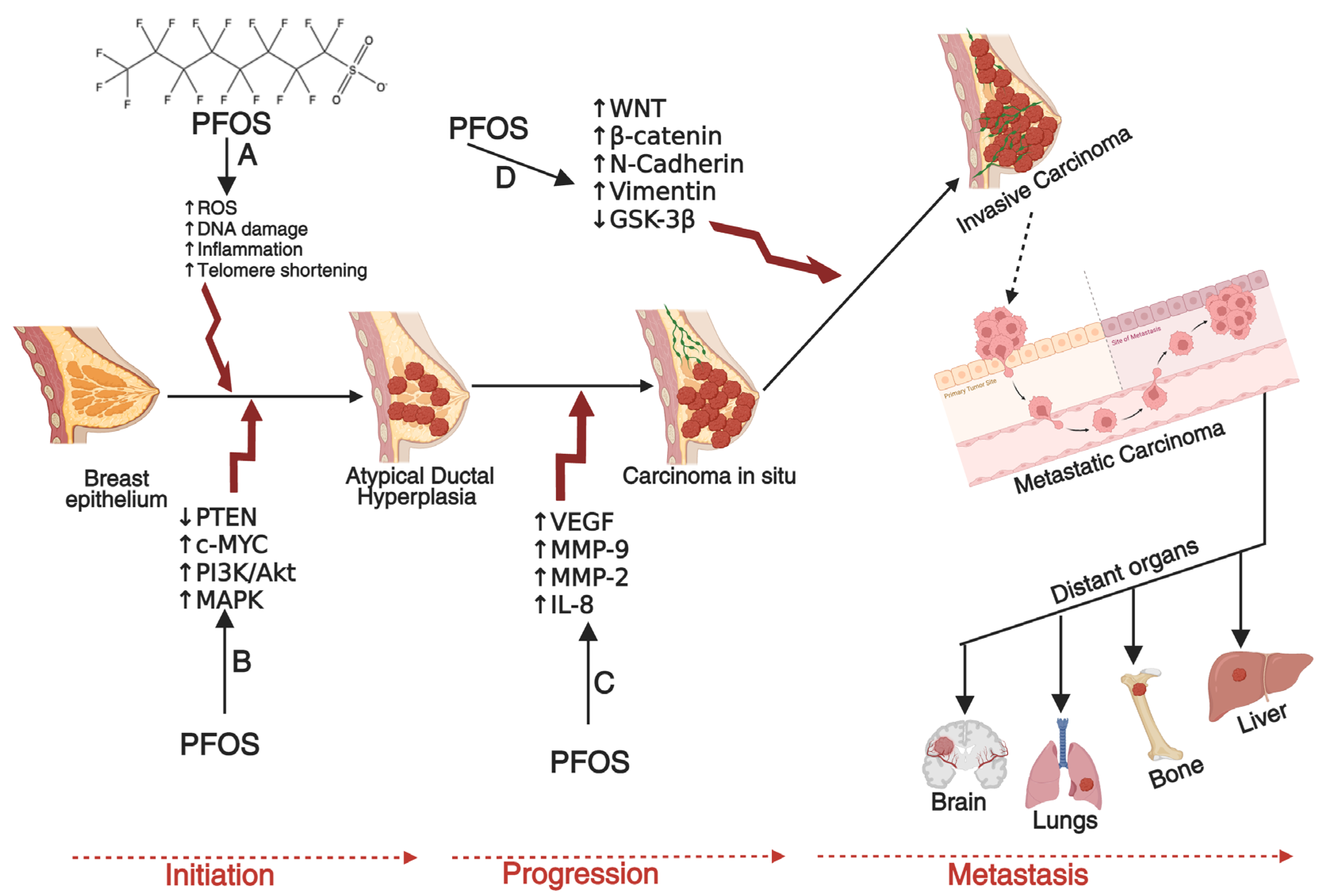
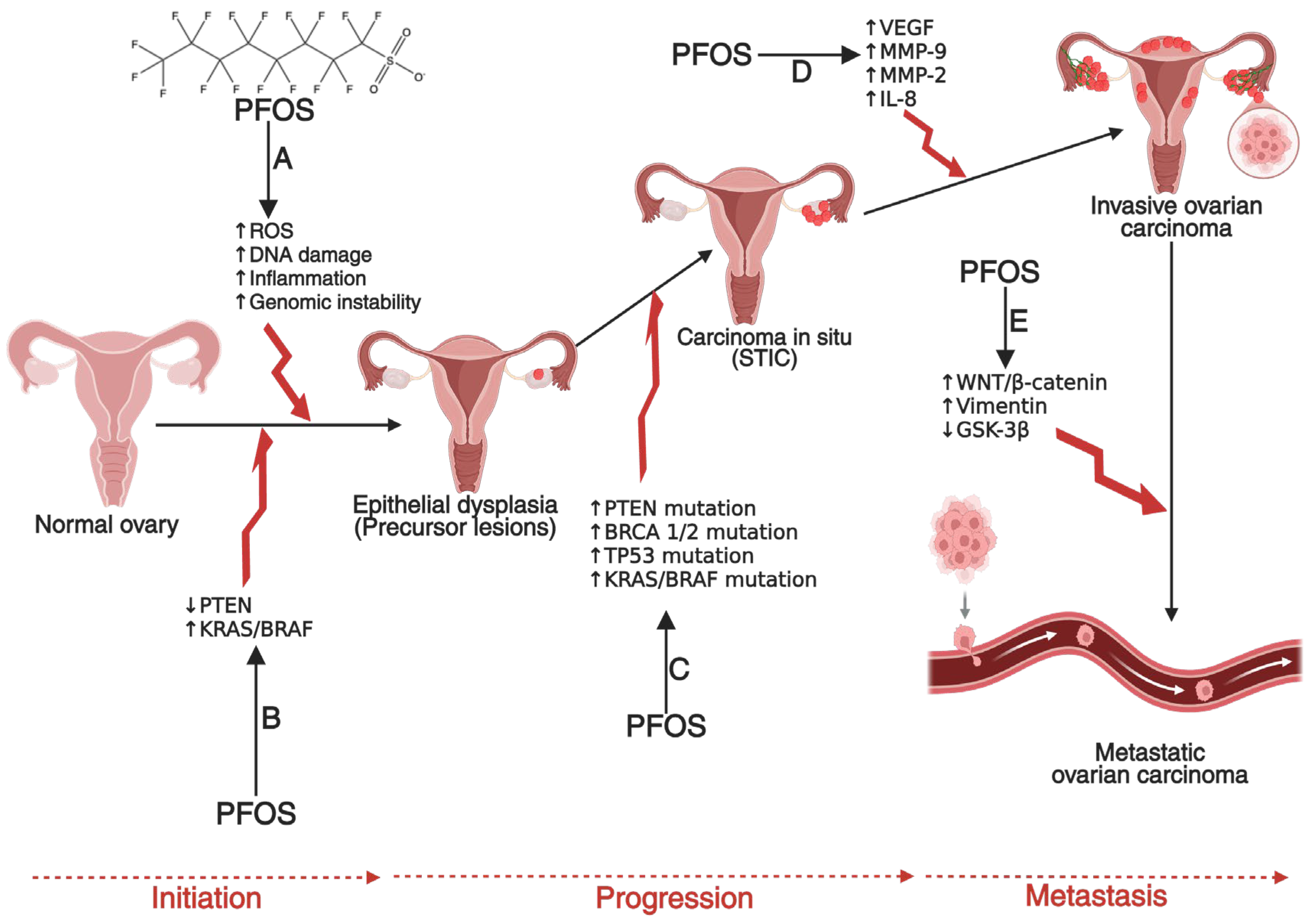
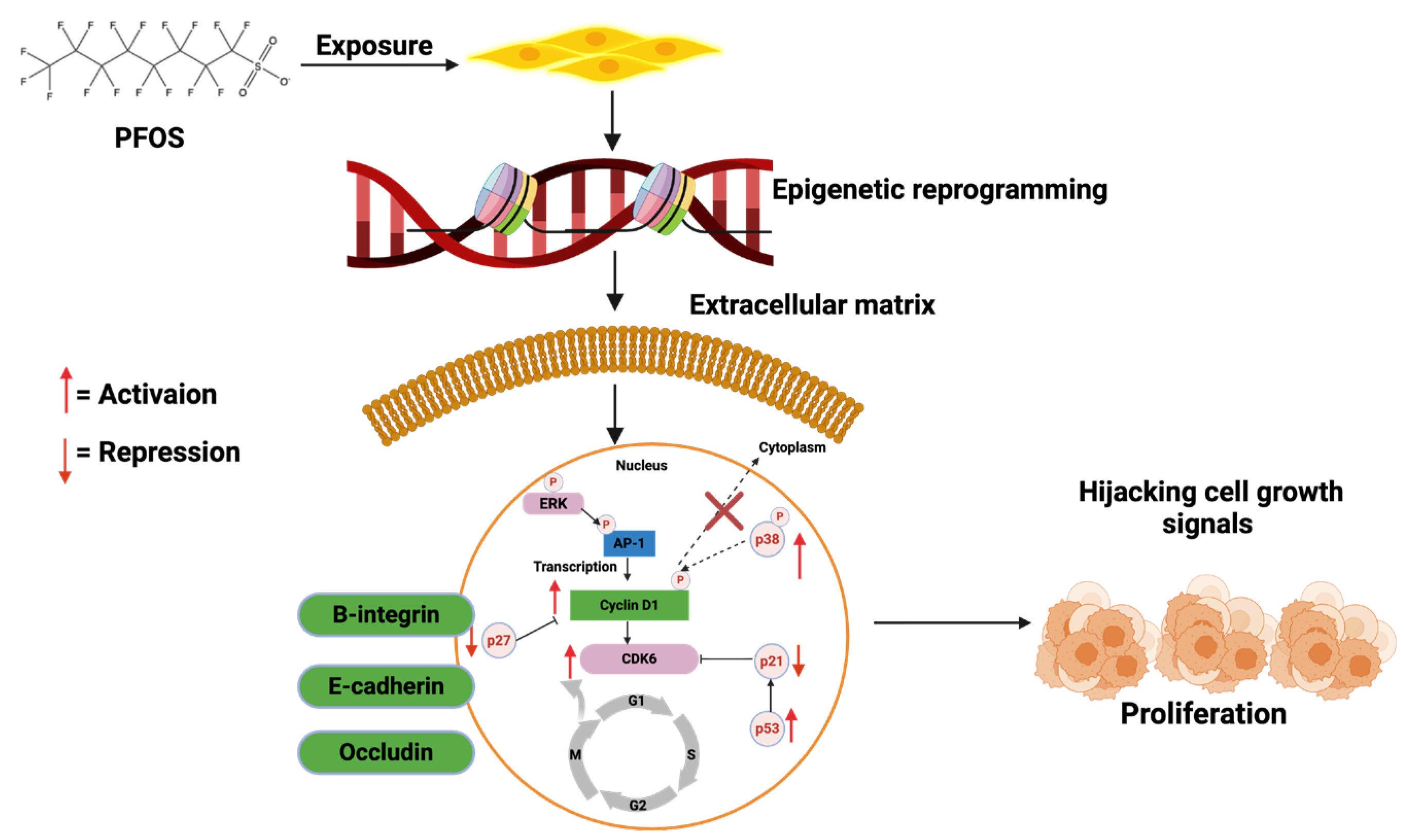
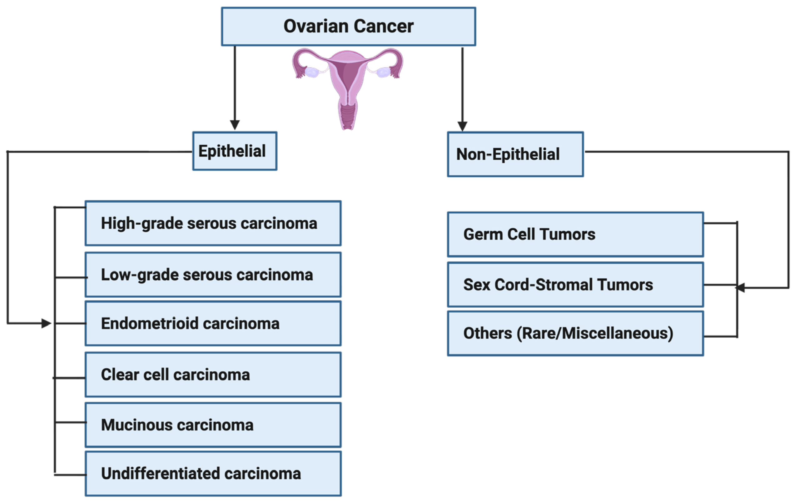
Carcinogenesis is a multi-step process involving genetic mutations, epigenetic modifications, and typical cellular regulatory pathway disruptions [23,24,25]. PFOS has been found to drive these processes through multiple mechanisms. One key mechanism is endocrine disruption, where PFOS mimics or alters the activity of androgen hormones. In breast and ovarian cancers, PFOS has been shown to enhance estrogen receptor (ER) signaling, leading to increased proliferation of mammary gland cells and ovarian epithelial cells, which may contribute to the development of breast and ovarian cancers if left unchecked [26,27,28,29]. Similarly, in prostate cancer (PCa), PFOS has been shown to disrupt androgen receptor (AR) pathways, promoting abnormal cell growth and progression of PCa [30,31,32]. In addition to hormonal interference, PFOS contributes to carcinogenesis by inducing oxidative stress through the persistent generation of reactive oxygen species (ROS) [33,34]. Increased ROS levels can cause DNA damage, impair DNA repair mechanisms, and trigger inflammatory responses that create a tumor-promoting microenvironment [35,36,37,38]. Chronic inflammation plays a critical role in the development and progression of cancers by sustaining cellular proliferation, preventing apoptosis, and facilitating metastasis [39,40,41]. Other mechanisms that promote the development and progression of prostate, breast and ovarian cancers by PFOS include down-regulation of PTEN and GSK-3-β, as well as activation of PI3K/Akt/NF-κB signalling node, c-MYC, WNT1, β-catenin, N-Cadherin, Vimentin, VEGF, MMP-2/9, and pro-inflammatory cytokines and chemokines [42,43]. In PCa, these molecular alterations can disrupt the normal cellular and genetic architecture of prostate epithelial cells, leading to the development of prostatic intraepithelial neoplasia (PIN) in prostate tissues (Figure 2), promoting the transformation of normal breast epithelial cells into malignant phenotypes with enhanced migratory potential to distant organs such as the brain, lungs, bones, and liver (Figure 3), or enhancing the transformation of ovarian epithelial cells to metastatic ovarian cancer (OC) (Figure 4). Furthermore, an upregulation of several metastatic markers, including VEGF, MMPs, and the WNT/β-catenin pathways, will further coordinate the progression of clinical prostate, breast, and ovarian cancers [42,43,44]. Epigenetic modifications are another pathway through which PFOS may drive cancer development. Studies have shown that PFOS exposure alters DNA methylation patterns and histone modifications, leading to the suppression of tumor suppressor genes and the activation of oncogenes [45,46,47]. These genetic and epigenetic changes contribute to uncontrolled cell division and progression of PCa and breast cancer (BCa) [48,49].
Epidemiological studies have provided further support for the link between PFOS exposure and cancers. Research has found that individuals with higher serum PFOS levels have an increased risk of developing prostate, ovarian and breast cancers, particularly in populations with occupational or environmental exposure [32,50,51]. While more research is needed to establish direct causation, the accumulating evidence underscores the need for stricter regulations on PFOS production and use to mitigate potential health risks. Given its widespread environmental presence and long-term persistence in biological systems, PFOS poses a significant public health concern. Understanding the molecular and cellular mechanisms through which PFOS contributes to PCa development and progression is crucial for guiding future research, informing regulatory policies, and implementing strategies to reduce exposure. We are currently examining the toxicological effects of PFOS, concentrating on its emission and environmental distribution, the mechanistic toxicities impacting both human and ecological health, and its potential involvement in the development of prostate, ovarian and breast cancers.

2. Emission and Environmental Distribution of PFOS

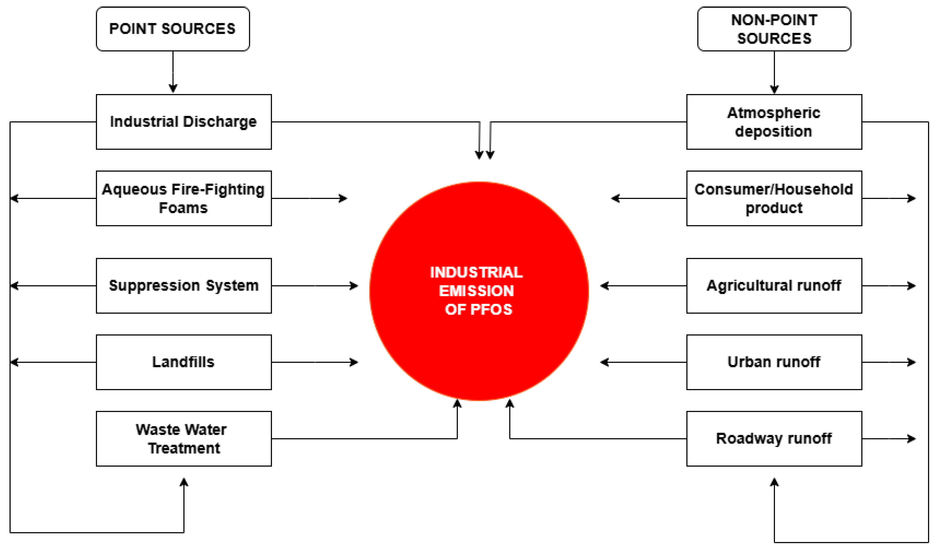
Due to its chemical stability, resistance to degradation, and extensive industrial applications, PFOS can enter the environment through two principal sources (Figure 5): point sources and nonpoint sources [52,53,54,55]. Point sources are defined as identifiable and confined sources of pollution from which PFOS is discharged directly into the air, water, or soil. Because they originate from specific locations, this source type can be easily monitored and regulated. Illustrative examples of point sources of PFOS include industrial discharges, firefighting and fire suppression systems, landfills, and wastewater treatment plants [56,57]. Chemicals containing PFOS have been employed in the treatment of high-quality textiles as finishing agents, often affecting their water, oil, and soil resistance characteristics. The PFOS chemicals utilized are PFOS polymers with a residual PFOS content of 1%. Textile formulations contain, on average, around 27% PFOS polymers. Additionally, back-coating textiles can result in losses, with an emission factor as outlined in the Emission Scenario Document [58]. Recent studies have shown that fluorochemical production plants (FPPs) emit dozens of PFOS [59] based on wastewater analysis. The production of fluoropolymers and their livestock substances is intrinsically related to the use, formation, and emission of PFOS. This has led to high PFOS exposure in people near these FPPs. Companies such as 3M and DuPont, which released significant quantities of PFOS, were cognizant of the toxicity associated with PFOS and the detection of fluorochemicals in workers’ bloodstreams by the 1970s [60]. In the production of aqueous firefighting foams (AFFF), PFOS chemicals have been predominantly employed for fire protection in petrochemical industries, fire departments, and military establishments. The United States Air Force has identified approximately 200 installations where PFOS was used, demonstrating that the United States military ranks among the top consumers of AFFF [61]. In China, the consumption of PFOS-related chemicals is also substantial, amounting to approximately 100 tons annually, with over 50 enterprises engaged in the manufacturing of AFFF [58]. The release of AFFF results in the contamination of soil and water sources with PFOS, particularly in the vicinity of airport firefighting training facilities and fire stations. The significant risk of agricultural contamination caused by PFOS has been monitored and studied [62]. Wastewater treatment plants (WWTPs) are the primary studied point sources of PFOS in surface waters, and they can serve as perennial sources of PFOS to the environment, including biosolids [61]. Biosolids in wastewater treatment plants can be used in agriculture. Biosolids contain large amounts of nitrogen, phosphorus, and organic carbon. Therefore, they are applied as soil amendments [62]. Studies have shown that the extensive application of wastewater biosolids in soil is a pollution pathway of PFOS in agricultural plants [62].
Moreover, nonpoint sources pertain to dispersed and indirect origins of pollution, particularly in contexts where PFOS contamination is observed across vast areas. Nonpoint sources include atmospheric deposition, household and consumer product use, agricultural runoff and urban and roadway runoff [54,63,64]. Such sources present more significant challenges in terms of monitoring and regulation. One notable nonpoint source of PFOS is atmospheric deposition. PFOS and associated perfluorinated compounds are emitted into the atmosphere via industrial emissions, combustion processes, and volatilization from treated surfaces. These airborne contaminants can traverse extensive distances before settling into aquatic systems, soil, and vegetation through precipitation or dry deposition mechanisms [65,66]. D’Ambro [55], in comparing predicted per- and polyfluoroalkyl substances (PFAS) and GenX air concentrations from a fluorochemical manufacturing facility in Eastern North Carolina, United States, found that the area surrounding the facility experiences around 15 days per year of GenX concentration above the inhalation screening level they derived in their study. Household and consumer products represent another primary nonpoint source of PFOS. Many everyday items, including stain-resistant textiles, carpets, nonstick cookware, and food packaging, contain PFOS-based treatments. Over time, the wear and degradation of these products release PFOS into indoor dust, wastewater, and the broader environment. Washing treated fabrics and carpets leads to PFOS contaminating sewage, eventually reaching surface waters through wastewater treatment plants [67,68].
There is substantial evidence indicating that PFOS may accumulate in edible fish species to levels of up to 7 and 170 ng/g wet weight (wwt). Notably, the Minnesota Department of Health has issued advisories concerning fish consumption in areas of the Mississippi River affected by contamination [68]. The land application of biosolids and landfill leachate significantly contributes to nonpoint source contamination by PFOS. Wastewater treatment facilities accept and process effluent from residential, commercial, and industrial origins; however, they do not eliminate PFOS. As a result, the treated wastewater released into fluvial and lacustrine environments acts as a persistent nonpoint source of PFOS within aquatic ecosystems [69]. Likewise, biosolids produced from wastewater treatment are frequently used as agricultural fertilizers. Since biosolids contain residual PFOS, their application can result in PFOS accumulation in the soil, posing a risk of crop contamination and subsequent entry into the food chain [70]. Runoff from contaminated sites is another widespread nonpoint source. Areas with historical PFOS use, such as firefighting training grounds, airports, and military bases, often have residual contamination in soil and groundwater. PFOS can leach from these sites into surrounding water bodies during rainfall, leading to diffuse pollution [71,72,73]. Contamination of marine and freshwater through bioaccumulation also contributes to nonpoint PFOS pollution. PFOS is extremely persistent and accumulates in aquatic organisms, resulting in biomagnification within the food web. Contaminated fish and seafood are secondary nonpoint sources when consumed by humans and other animals, redistributing PFOS throughout ecosystems [74]. Overall, the complexity of nonpoint PFOS sources highlights the challenge of managing environmental contamination. Unlike point sources, which can be specifically regulated, nonpoint sources require a combination of monitoring, pollution mitigation, and policy measures to reduce their impact on human health and ecosystems.

3. Mechanisms of PFOS Toxicokinetics and Dynamics

3.1. Absorption and Distribution of PFOS in Human Tissues and Organs

Humans are exposed to PFOS through various routes such as contaminated drinking water, food intake, consumer goods, and occupational contact [75]. Currently, no known families of enzymes are recognized to metabolize PFOS; therefore, it is classified as a “forever chemical”. Nevertheless, recent research indicates that ingestion of PFOS may inhibit the activity of the CYP540 enzyme family, including enzymes such as CYP1A2, CYP2B6, CYP2C19, CYP2E1, and CYP3A4 [76]. Once in the body, PFOS exhibits unique pharmacokinetic characteristics that set it apart from conventional persistent organic pollutants (POPs) [77]. PFOS has a strong affinity for proteins, especially serum albumin and liver fatty acid-binding proteins. This protein-binding behavior promotes their persistence and accumulation in protein-rich organs [78]. The first study to measure PFOS levels in human internal tissues (excluding blood) was by Olsen et al. [79], who detected PFOS in serum and liver samples from non-exposed individuals. The mean PFOS levels were 18.8 ng/g in liver and 17.7 ng/mL in serum, with a liver-to-serum ratio of 1.3:1. Maestri et al. [80] later analyzed pooled organ samples from seven unexposed individuals in Italy, detecting PFOS across multiple organs, with the highest concentrations in the liver, kidney, and lungs. A German pilot study by Völkel et al. [81] reported mean liver concentrations of 17.9 ng/g of PFOS. These early studies collectively highlighted PFOS accumulation in protein-rich organs, particularly the liver, even among non-occupationally exposed individuals. In a later study, Liu et al. [82] analyzed PFOS in tumor and non-tumor liver tissues from liver cancer patients in China. The mean PFOS levels were comparable between non-tumour liver tissues (22.2 ng/g) and tumor liver tissue (22.4 ng/g) samples, indicating widespread PFOS accumulation in both healthy and malignant liver tissues. In Pavia, Italy, Pirali et al. [83] measured PFOS levels in thyroid tissues from 28 patients who underwent surgery for benign or malignant thyroid disorders. PFOS was detected in all thyroid samples, with median concentrations of 5.3 ng/g (range: 2.1–44.7 ng/g). In addition, Mamsen et al. [84] analyzed PFOS in placenta and fetal tissues from legally terminated pregnancies in Denmark. They found the highest concentrations of 1.3 ng/g in the placenta. They also examined PFOS in embryos, placentas, and multiple fetal organs (liver, lung, heart, CNS, and adipose tissue). PFOS showed the highest median concentrations in both placenta (1.24 ng/g) and fetal tissues (0.83 ng/g). These findings confirmed that PFOS can cross the placenta and accumulate in fetal tissues, raising concerns about prenatal exposure risks.

3.2. PFOS–DNA Interaction Through Non-Covalent Binding

The primary pathways of PFOS toxicity involve non-covalent interactions with DNA and proteins, as demonstrated by both experimental and computational evidence [85,86]. These interactions are subtle yet biologically significant, providing a plausible mechanistic link between PFOS exposure and genomic instability observed in mammalian systems. The interaction of PFOS with DNA is principally influenced by electrostatic forces, hydrogen bonds, and hydrophobic interactions. The sulfonate head group of PFOS possesses a substantial negative charge, enabling it to establish electrostatic associations with cationic regions of DNA-binding proteins or via divalent metal-mediated bridges such as Mg2+ or Ca2+ in proximity to the DNA phosphate backbone. Concurrently, the perfluorinated carbon chain engages in hydrophobic and van der Waals interactions with nucleobases, notably in adenine-thymine (A–T) rich regions, thereby facilitating its insertion into the minor groove of DNA and contributing to the stabilisation of PFOS–DNA complexes [87].
Spectroscopic analyses, including UV–Vis absorption, fluorescence quenching, and circular dichroism, have demonstrated changes in DNA secondary structure upon PFOS binding. These conformational distortions are characterized by a reduction in base stacking and an alteration of the helical twist, indicative of PFOS-induced perturbations in DNA stability and Molecular docking simulations further corroborate these observations, showing that PFOS can localize within DNA grooves, where its fluorinated tail aligns parallel to the helical axis, while the sulfonate group interacts with polar residues, creating a thermodynamically stable but conformationally disruptive complex [87]. Functionally, such interactions can have profound biological consequences. PFOS–DNA binding may hinder the access of DNA polymerases and repair enzymes to their substrates, resulting in replication stress, transcriptional interference, and impaired DNA repair fidelity. These disturbances are consistent with empirical findings that PFOS exposure induces DNA strand breaks, micronuclei formation, and oxidative stress-mediated genotoxicity in mammalian cells. Moreover, epigenetic alterations such as aberrant DNA methylation and histone modification patterns have been reported, suggesting that PFOS not only affects the physical integrity of DNA but also modulates chromatin structure and gene expression [88].

3.3. Disruption of Lipid Metabolism and Mitochondrial Function

The liver is the primary organ for fatty acid metabolism and transport. It plays a central role in regulating lipid homeostasis through various metabolic pathways. Numerous metabolic alterations have been linked to PFOS exposure. It has been reported that PFOS (Figure 6) can activate peroxisome proliferator-activating receptor alpha (PPARα), crucial in regulating lipid metabolism and adipogenesis [89]. PFOS can disrupt the balance of lipid metabolism in the liver, resulting in a series of pathological changes [90] and consequently leading to fatty liver, liver fibrosis, and hepatocellular carcinoma [91]. At near cytotoxic concentrations, PFOS induces the accumulation of triglycerides in HepaRG cells [92] and consistently affects the expression of steatosis marker genes and target genes for PPAR-α [93]. Studies have also revealed that mitochondria function in the hepatocytes [94], oocytes [95,96], and pancreatic β cells [97] can suffer alterations due to PFOS exposure. Hoffman et al. [98] reported that PFOS caused a concentration-dependent reduction in basal, maximal, and ATP-linked respiration in the mitochondria, thereby disrupting placental function. By compromising the bioenergy capacity of trophoblasts, PFOS exposure causes a decline in mitochondrial content and impairs cellular respiration, leading to mitochondrial dysfunction. The mitochondria’s transmembrane potential (ΔΨm) depends on the cellular oxidative phosphorylation. Depolarization of ΔΨm, perhaps caused by mitochondrial swelling, will produce lower ATP [99]. Prenatal exposure to PFOS may have the potential to induce cardiac mitochondrial injury and apoptosis [100].

3.4. Oxidative Stress and Reactive Oxygen Species (ROS) Generation

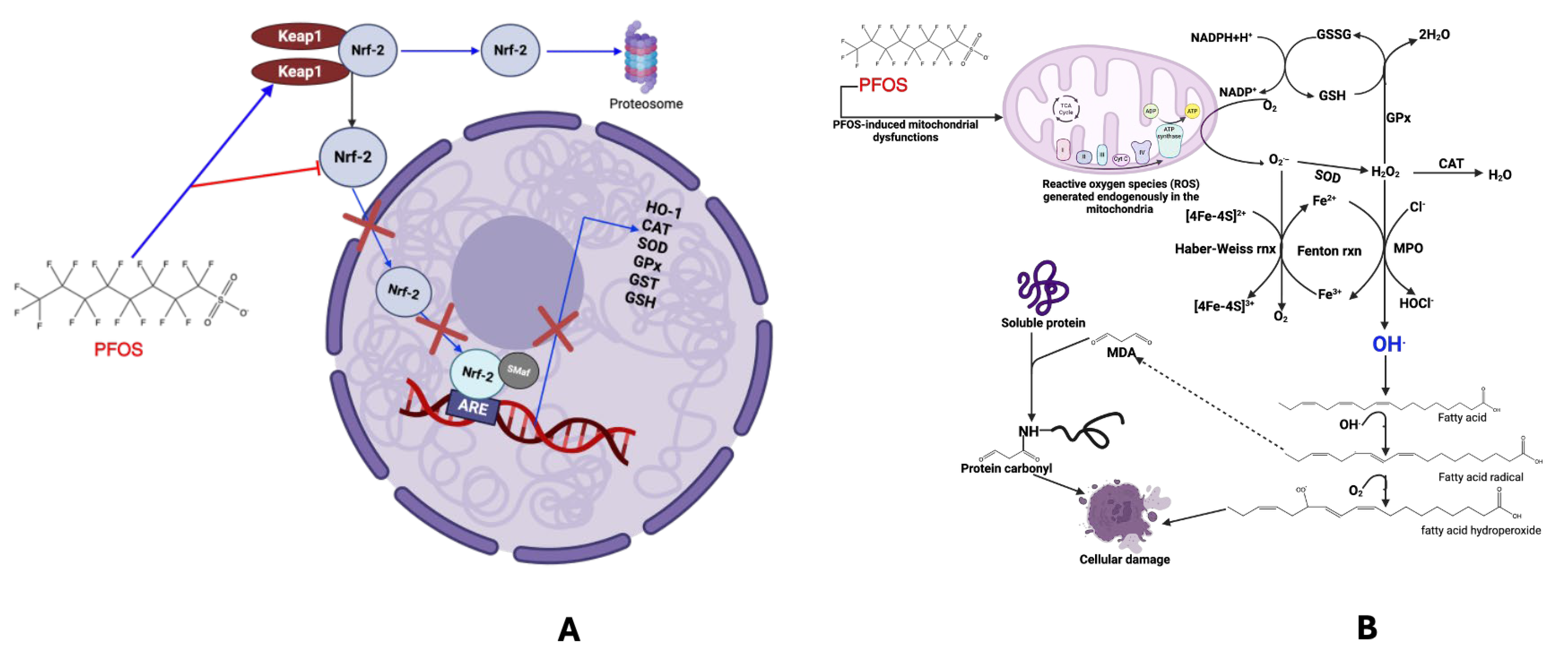
Several studies have revealed that oxidative responses are associated with PFOS-induced cell injury and tissue damage. The Keap-1/Nrf-2 signaling axis has been recognized as one of redox biology’s most intriguing and complex pathways [36,101,102]. Serving as a cellular sensor for oxidative stress, this pathway exemplifies nature’s strategic design in safeguarding cells against the incessant onslaught of ROS. By meticulously regulating the transcriptional activity of Nrf-2, Keap1 functions as a molecular gatekeeper, permitting the stabilization of Nrf-2 solely when the redox equilibrium is disrupted, thereby ensuring a swift yet controlled antioxidant response. Under physiological conditions, the Keap-1/Nrf-2 system is pivotal, activating many cytoprotective genes that coordinate antioxidant defence, detoxification, and metabolic adaptation. Specifically, Nrf-2 moves into the nucleus, where it works together with other transcription factors to turn on genes like heme oxygenase-1 (HO1), catalase (CAT), glutathione peroxidase (GPX), glutathione-s-transferase (GST), and a variety of other helpful protective genes. This response is especially critical in tissues subjected to high oxidative stress, including the lungs, liver, and skin. However, in the presence of toxicants such as PFOS, Keap-1 activation is more favored, leading to the repression of Nrf-2 (Figure 7A). In this circumstance, Nrf-2 is targeted for degradation by the proteasomal system, thereby lowering the GSH, GST, and HO-1 [103]. PFOS induction of oxidative stress is achieved via inhibition of antioxidants (e.g., superoxide dismutase, catalase, GPx4, and GSH) or enhancing the generation of ROS in hepatocytes or endothelial cells will lead to cell death [33,104,105,106,107]. Daenen et al. [108] also reported that oxidative stress produces ROS, causing oxidative damage to nucleic acids, proteins, and lipids, ultimately leading to epithelial cell death and a decline in kidney function (Figure 7B). PFOS induces apoptosis in pulmonary cells via the reactive oxygen species-mediated mitochondria-dependent pathway [109]. Lee et al. [110] and Wen et al. [111] suggested that oxidative stress and mitochondrial function are pivotal in renal tubular cell apoptosis induced by PFOS. In the liver, PFOS exposure can increase ROS production and disrupt the intracellular antioxidant defense system [112]. Production of ROS in excess is cytotoxic, resulting in oxidative damage to hepatocyte biomolecules [113]. By disrupting the electron transport chain and releasing cytotoxic reactive oxygen species (ROS), PFOS can directly inflict damage on the mitochondria of hepatic cells, leading to the production of hydrogen peroxide (H2O2). This compound subsequently diffuses into the lysosome, resulting in the formation of highly reactive hydroxyl radicals [114]. The accumulation of H2O2 and the elevated MDA content induced by PFOS exposure will result in a reduction in SOD and GSH levels [115]. Excess ROS may compromise the integrity of hepatocyte cell membranes, thereby affecting their stability and permeability [116]. ROS can disrupt calcium homeostasis by affecting calcium channels, pumps, and storage organelles, increasing intracellular calcium levels. This dysregulation contributes to cell damage and apoptosis, particularly in hepatocytes [117].

3.5. DNA Damage and Epigenetic Modification

Another PFOS toxicity mechanism is genetic and epigenetic perturbations (Figure 8). Genetic damage, including the breakage of DNA strands, DNA fragmentation, and chromosomal breaks, results from PFOS toxicity [118]. Exposure to PFOS was found to cause direct oxidative damage [119], increase ROS generation, and subsequently lead to DNA damage in liver cancer cell lines (HepG2) [120]. In peripheral blood, Eke and Celik [121] reported that doses of PFOS increase the micronucleus frequency and strongly induce DNA damage. Gonadal development is also impaired by PFOS, leading to a decline in the total count of germ cells, transient arrest in the mitotic cycle, and apoptosis via DNA damage induced by ROS [122]. PFOS exposure distorts proteome structure, aggravates oxidative stress, compromises mitochondrial oxidative phosphorylation, enhances mitochondrial permeability, and promotes DNA damage and apoptosis [18].
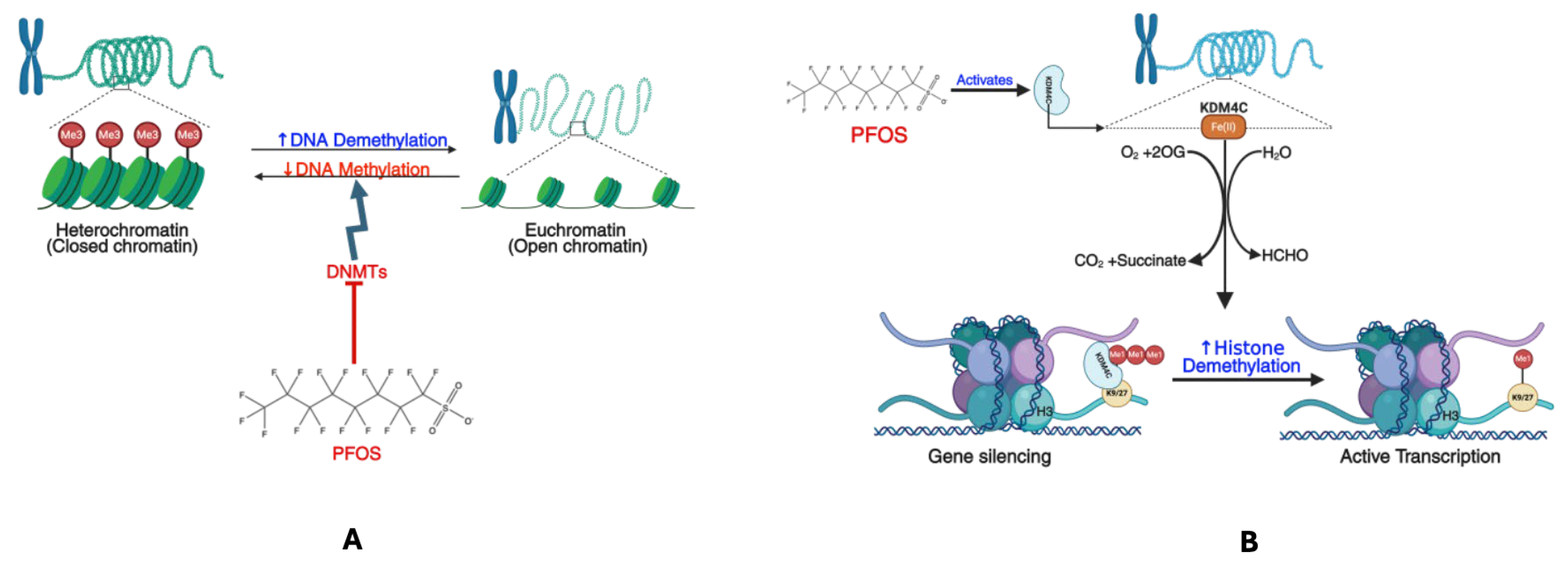
Findings from epigenetic studies demonstrate how PFOS influences gene expression without altering DNA sequence [123]. Different epidemiological studies assessed via Infinium arrays have identified that prenatal PFOS exposure is associated with variations in newborn or childhood blood methylation [99,124,125]. Kim et al. [126] found a significant link between PFOS and epigenetic modification in both adult and newborn cohorts. These modifications can activate oncogenes and/or inhibit tumor suppressor gene expression through DNA methylation, histone modification, and non-coding RNAs [127]. Research suggests that PFOS can contribute to cancer development by altering gene expression and triggering epigenetic changes. This, in turn, affects multiple signaling pathways in various cell types [128]. In the study of Wen et al. [129], exposure to PFOS was found to disrupt the epigenetic signatures, resulting in global DNA hypomethylation and upregulation of histone lysine demethylases, specifically KDM1A (LSD1) and KDM4C (JMJD2C). These alterations contribute to modified gene expression patterns, genomic instability, and increased disease vulnerability. DNA methylation, an essential epigenetic mechanism, entails adding methyl groups (-CH3) to cytosines within CpG dinucleotides, usually leading to gene silencing [130,131,132]. PFOS exposure has been correlated with a worldwide decline in DNA methylation (Figure 8A), which may stem from several mechanisms: the inhibition of DNA methyltransferases (DNMTs), disruption of one-carbon metabolism, and the activation of DNA demethylases [129,133,134,135]. In inhibiting DNMTs, PFOS might diminish the expression and activity of DNMT1, DNMT3A, and DNMT3B, enzymes accountable for maintaining and establishing DNA methylation patterns. This ultimately leads to a gradual loss of methylation across the genome, especially in tumour suppressor genes and repetitive DNA elements, thereby causing increased genomic instability. Regarding one-carbon metabolism disruption, PFOS may hinder S-adenosylmethionine (SAM) production, the primary methyl donor for DNA methylation, and lower SAM levels adversely affect de novo methylation, further facilitating hypomethylation. The activation of DNA demethylases by PFOS is linked to enhanced activity of TET enzymes (Ten-Eleven Translocation proteins), which catalyze the oxidation of 5-methylcytosine (5mC) to 5-hydroxymethylcytosine (5hmC), thereby initiating DNA demethylation. As an outcome of these transformations, oncogenes become abnormally activated, while tumour suppressor genes lose their repressive function, fostering uncontrolled cell proliferation and cancer progression [129,136,137].
KDMs are critical in remodeling chromatin and regulating gene expression [138,139,140]. PFOS has been demonstrated to elevate the expression levels of KDM1A and KDM4C [129], two principal histone demethylases that erase repressive histone marks, consequently fostering a more transcriptionally active chromatin state. KDM1A is a flavin-dependent demethylase that removes methyl groups from H3K4me1/2 (activating mark) and H3K9me1/2 (repressive mark) in a context-dependent manner. The PFOS-induced upregulation of KDM1A (Figure 8B) results in the loss of H3K9me2 repression, permitting the transcriptional activation of oncogenic pathways, altering differentiation programs, promoting stemness-like characteristics in cancer cells, and enhancing proliferation and epithelial–mesenchymal transition (EMT), which are key attributes of tumour progression. KDM4C, a JmjC-domain-containing histone demethylase, specifically demethylates H3K9me3/me2 (marks of heterochromatin), prompting chromatin decondensation and transcriptional activation of oncogenes, which contributes to increased genomic instability as repetitive DNA elements lose heterochromatic repression and increase the expression of MYC and other proliferation-associated genes, invigorating tumour growth. KDM4C has also been shown to promote the activation of HIF1α/VEGFA signaling through the costimulatory factor STAT3 in NSCLC [141]. Thus, the overexpression of KDM1A and KDM4C triggered by PFOS creates a chromatin environment favoring carcinogenesis by promoting oncogenic transcription, diminishing cellular differentiation, and enabling metastatic progression [129]. The concurrent loss of DNA and histone methylation leads to a significant transformation in the epigenetic landscape of cells subjected to PFOS exposure. Ordinarily, DNA methylation and H3K9me3 collaborate to establish and maintain heterochromatin. However, PFOS undermines both marks, resulting in a chromatin state that is more permissive to transcription, thus facilitating oncogenic activation and cell transformation. This observation indicates that PFOS functions as an epigenetic disruptor, potentially increasing the risk of cancer, metabolic disorders, and developmental abnormalities.
Another study employing advanced super-resolution imaging techniques and machine-learning tools elucidated that PFOS can alter the spatial configuration of repressive heterochromatin marks, namely H3-lysine-9-trimethylation (H3K9me3) and H3-lysine-27-trimethylation (H3K27me3), within renal carcinoma cells [142]. Exposure to PFOS does not appear to significantly impact the overall quantities of H3K9me3 or H3K27me3. Instead, it leads to the dispersion and de-clustering of these histone marks. H3K9me3 and H3K27me3 are typically densely clustered in heterochromatic regions, particularly adjacent to the nuclear periphery, to maintain gene silencing. PFOS disrupts this structural organization and renders the repressive domains more diffuse, possibly contributing to aberrant gene expression. Their studies also revealed that PFOS is linked with an increased expression of KDM4A. KDM4A is a histone demethylase responsible for removing methyl groups from H3K9me3 [143]. This enzymatic activity can potentially diminish local concentrations of H3K9me3, thereby weakening chromatin compaction and facilitating a more open euchromatic structure. Although KDM4A does not directly demethylate H3K27me3, the overarching imbalance in chromatin-modifying enzymes may indirectly affect the dynamics of H3K27me3 [142]. These findings suggest that PFOS functions as an epigenetic disruptor, interfering with chromatin modifiers, transcriptional repressors, and nuclear scaffolding proteins. Consequently, this interference may relocate repressive histone marks, undermining their efficacy in gene silencing and promoting the activation of stress, inflammation, or proliferation genes. PFOS did not significantly decrease the expression level of histone demethylase KMT1A [142]. KMT1A (SUV39H1) is a histone methyltransferase (HMT) that catalyses explicitly the trimethylation of histone H3 at lysine 9 (H3K9me3), a characteristic marker of transcriptional repression [144,145,146]. By establishing and preserving heterochromatin, KMT1A is indispensable for genomic stability, gene silencing, and cellular differentiation. KMT1A operates as a chromatin modifier, engaging with heterochromatin protein 1 (HP1) to compact chromatin and inhibit transcription. This repression is crucial for silencing repetitive DNA sequences, maintaining X-chromosome inactivation, and regulating developmental genes [147]. Furthermore, KMT1A collaborates with DNA methyltransferases (DNMTs) and histone deacetylases (HDACs) to reinforce stable gene silencing throughout cellular generations. These alterations, perhaps, are potential drivers of toxicity induced by PFOS.

3.6. Endocrine Disruption

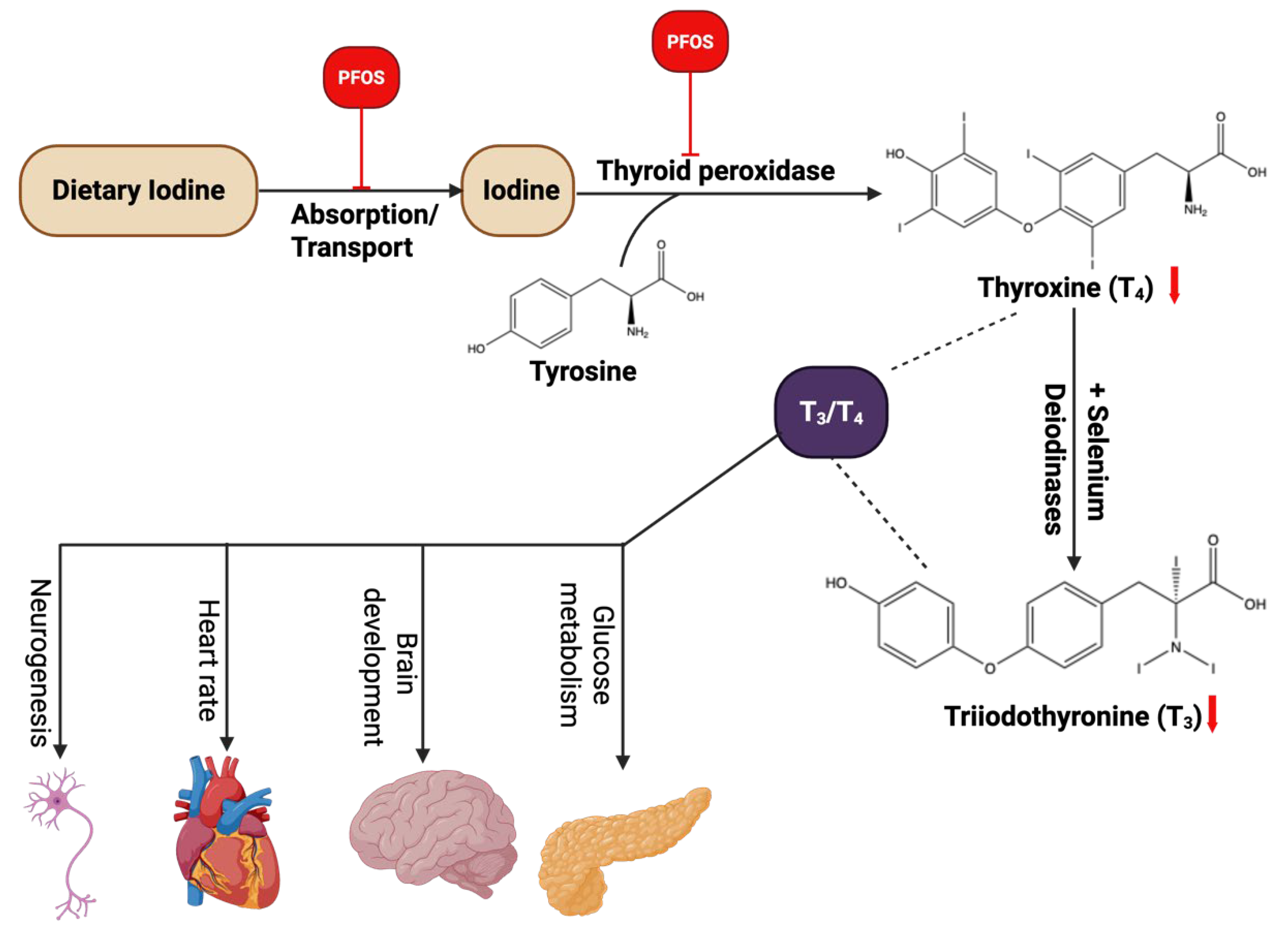
PFOS has been recognized as an endocrine disruptor that may potentially exert adverse effects on multiple hormonal functions, including thyroid health [148]. Previous research has highlighted concerns regarding the impact of PFOS on the thyroid gland, although findings have been inconsistent across in vitro studies and different study populations. Thyroid hormones (TH) are crucial in numerous biochemical processes, such as regulating energy expenditure, growth, and neurodevelopment from fetal development through infancy. Additionally, these hormones continue to influence metabolic processes throughout adulthood [149]. Disruptions to thyroid function caused by PFOS exposure could, therefore, have significant long-term health implications [150]. Thyroid hormones also play a vital role in brain formation during fetal development, guiding key processes such as neurogenesis, neuronal migration, synapse formation, and myelination. Insufficient thyroid hormone levels during critical periods of neurodevelopment can lead to long-term intellectual and behavioural impairments [150]. Beyond early development, thyroid hormones regulate metabolism throughout infancy and adulthood by acting on various organs, including the brain, white and brown adipose tissue, skeletal muscle, liver, and pancreas. Their secretion is controlled by the hypothalamic-pituitary-thyroid (HPT) axis. The hypothalamus initiates thyroid hormone regulation by releasing thyrotropin-releasing hormone (TRH) into the pituitary portal system. This stimulates the secretion of thyroid-stimulating hormone (TSH) by the anterior pituitary. TSH then stimulates the thyroid gland to produce and release thyroid hormones, mainly thyroxine (T4) and triiodothyronine (T3). While the thyroid gland mainly secretes T4, about 80% of circulating T3 is generated through the enzymatic conversion of T4 in peripheral tissues, including the brain. This conversion is facilitated by three types of deiodinase enzymes, which are essential for modulating thyroid hormone activity by controlling the local concentration of active T3 at various biological levels [151]. Excess thyroid hormones, a condition known as hyperthyroidism or thyrotoxicosis, result in a hypermetabolic state characterized by increased basal energy expenditure, weight loss, enhanced fat breakdown, and elevated glucose production [152]. In contrast, insufficient thyroid hormone levels lead to hypothyroidism, a hypometabolic state marked by reduced energy expenditure, weight gain, decreased lipolysis, and lower gluconeogenesis [150].
Recent literature has examined how PFOS disrupts thyroid function, offering insights into the molecular pathways involved in this endocrine interference [153,154,155,156,157]. PFOS exposure can disrupt the biosynthesis and secretion of thyroid hormones at multiple stages (Figure 9). Several mechanisms have been proposed, including impairment of iodine uptake, in which PFOS may compete with iodine or directly inhibit the sodium/iodide symporter (NIS), thus reducing the availability of iodine for thyroid hormone production [155]; interference with thyroglobulin synthesis, wherein PFOS could disrupt the formation of thyroglobulin, a critical precursor in thyroid hormone production [156]; modification of thyroperoxidase (TPO) activity, through which PFOS may alter the activity of TPO, an enzyme essential for the synthesis of thyroid hormones; and disruption of thyroid hormone regulation, in which PFOS could interfere with feedback mechanisms, affect deiodinase enzyme activity, or alter thyroid hormone binding proteins, thereby disrupting the signaling and biological effects of thyroid hormones [157]. These disruptions underscore the potential endocrine-disrupting properties of PFOS and their consequential impacts on thyroid function. According to the Endocrine Society, endocrine-disrupting chemicals (EDCs) are exogenous chemicals or mixtures of chemicals that interfere with any part of hormone action. Considering the endocrine system’s crucial role in controlling various biological and physiological processes, any disruption can lead to the development of several diseases [158].
The thyroid gland and its hormones are essential for maintaining metabolic homeostasis and ensuring proper development during the fetal and childhood stages. Disruptions in thyroid function can lead to significant health issues. Several studies have identified PFOS as an endocrine-disrupting compound capable of interfering with hormonal balance and impairing normal endocrine system activity [149,157]. Experimental research using cultured thyroid cells has shown that PFOS exposure in vitro can lead to thyroid dysfunction [149,157]. PFOS has been observed to accumulate within thyroid cells, and at elevated concentrations, it can exert cytotoxic effects [159]. Moreover, PFOS has been shown to suppress thyroperoxidase (TPO) activity in thyroid tumor cell lines, potentially disrupting the synthesis of thyroid hormones. It may also function as a thyroid hormone receptor (THR) agonist, as demonstrated by luciferase reporter assays involving THRα and THRβ in GH3 cells [160]. Conti et al. [155] reported that PFOS can inhibit iodide uptake in FRTL-5 thyroid cells by interfering with the sodium/iodide symporter (NIS). This inhibitory effect was also observed in non-thyroid cells engineered to express NIS, further supporting the potential of PFAS to impair iodine transport and thyroid function. An in vitro study by Croce et al. [161] evaluated the thyroid-disrupting potential of both long-chain PFAS (such as PFOS and PFOA) and short-chain variants, including PFBS, PFBA, PFPA, and PFPeA. The results showed that while long-chain PFAS caused cytotoxicity and disrupted TSH-dependent cAMP production in rat thyroid cells, short-chain PFAS did not exhibit these effects. This suggests that the impact of PFAS on thyroid function may depend on their molecular structure and chain length. Collectively, these findings indicate that PFOS exposure can impair endocrine function in humans, potentially affecting neurodevelopment, cardiovascular regulation, brain maturation, and glucose metabolism.

3.7. Immunosuppressive Effects and Inflammatory Responses of PFOS

An individual’s health depends on maintaining homeostasis within the immune system. When appropriately balanced, the immune system is capable of recognizing threats, initiating an appropriate response, repairing tissue damage incurred, and subsequently returning to a resting state [162]. Given that the immune system is dispersed across various tissues and organ systems, it is highly vulnerable to toxic substances through multiple exposure pathways. Considering its complex composition of diverse cell types with specialized functions, exposure to toxicants can impair immune function in various ways, potentially leading to immunosuppression, excessive immune activation, or both. Furthermore, disruptions to immune system development, such as those caused by exposure to immunotoxins, tend to yield more severe and enduring consequences than similar disturbances occurring in adulthood. An increasing body of research evidence indicates that PFOS is a significant immunotoxin, thereby raising concerns regarding its impact on immune health [163,164,165]. The most compelling epidemiological evidence of PFOS-related immunotoxicity is the reduction in antibody production following vaccination, especially in children receiving tetanus and diphtheria vaccines. Antibodies play a crucial role in the adaptive immune system, helping to combat infections and mitigate damage from harmful agents. Specifically, Grandjean et al. [166] reported that PFOS negatively impacted the antibody concentrations of study participants of 5 years of age. As PFOS concentrations rose, exposure was linked to a 39% drop in diphtheria antibody levels. A subsequent study involving the same cohort at age 13 confirmed that elevated PFOS levels were associated with reduced antibody levels, indicating a lasting immunotoxic effect [167]. Zhang et al. [168] analyzed multiple epidemiological studies in a systematic review and meta-analysis. They found that exposure to PFOS is linked to lower tetanus antibody levels in children, backing up evidence of immunotoxicity. Animal studies also support the idea that exposure to PFOS can reduce the body’s ability to produce specific antibodies [169].
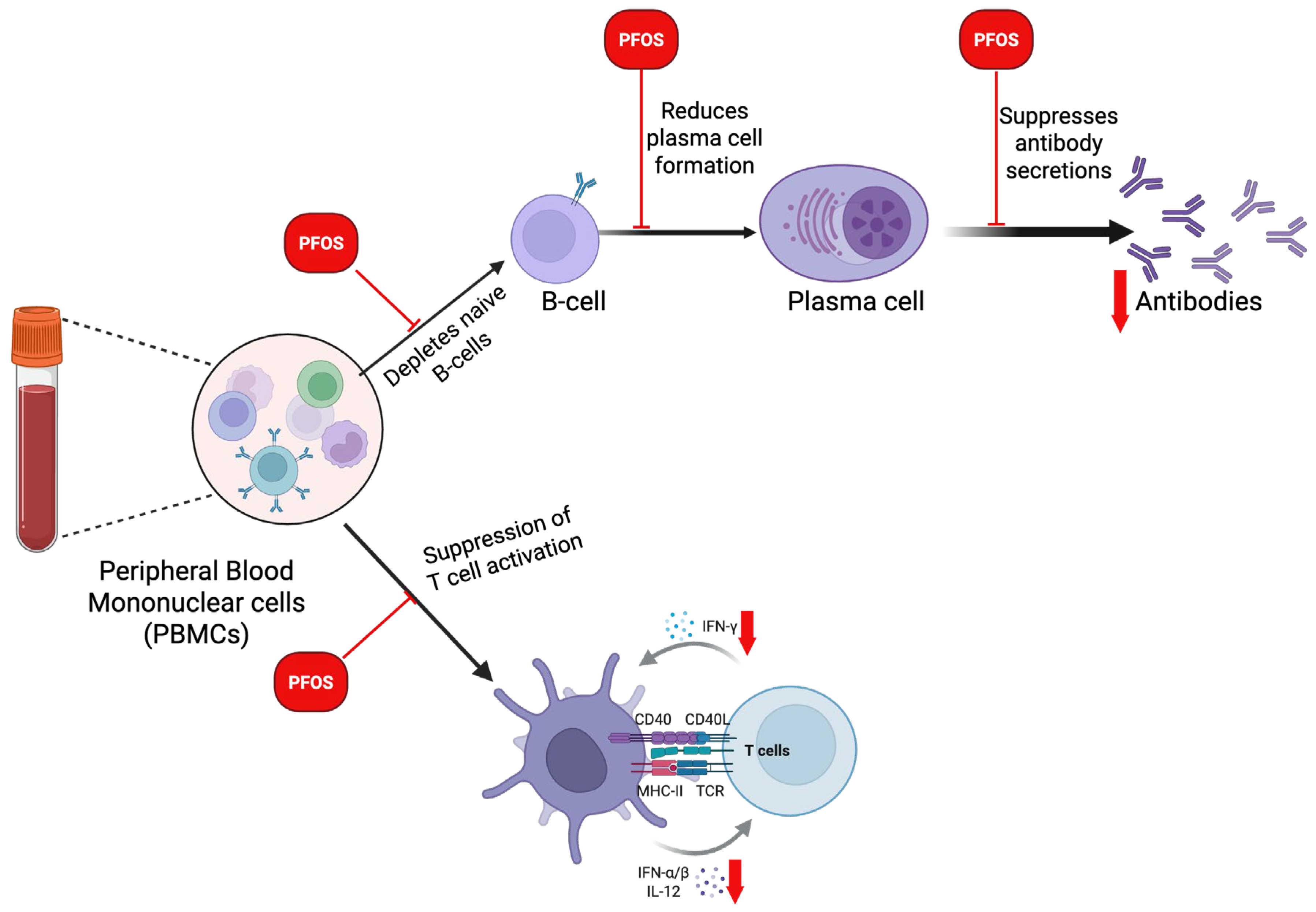
Additionally, research has linked PFOS exposure to an increased risk of respiratory and gastrointestinal infections, particularly in children exposed to the uterus. These findings and recent studies emphasize the immunosuppressive effects of PFOS [170]. These findings reveal that exposure to PFOS, particularly in neonates and children, may impair B-cell function by depleting naïve B cells, disrupting B-cell signaling needed for B-cell differentiation, reducing plasma cell formation, and suppressing the biosynthesis of antibodies essential for mounting an effective humoral immune response against foreign pathogens (Figure 10). An increasing body of evidence indicates that exposure to PFOS has notable immunotoxic effects on T cell populations, resulting in disruptions to their development and functional capacity. Research on animal models has consistently shown that PFOS exposure decreases total T-cell counts, particularly impacting CD4+helper T cells and CD8+cytotoxic T cells. This reduction can be partially attributed to thymic atrophy, as PFOS induces the shrinkage of the thymus, thereby impairing the maturation and output of naïve T cells [171]. Furthermore, beyond mere numerical reductions, PFOS modifies the distributions of T-cell subsets. Such exposure has been linked to a decreased proportion of naïve T cells and an increased prevalence of memory T cells within peripheral lymphoid organs [172]. These shifts may jeopardize the immune system’s capacity to respond effectively to novel antigens.
Additionally, functional impairment of T cells constitutes a notable characteristic of PFOS immunotoxicity. PFOS diminishes the expression of critical activation markers, such as CD69 and CD25, which culminates in reduced T-cell activation following antigenic stimulation [173]. Moreover, PFOS inhibits T-cell proliferation, an essential process for expanding antigen-specific clones during immune responses. The apoptosis of T cells is another mechanism through which PFOS undermines immunity. Elevated rates of apoptosis in T-cells have been documented following PFOS exposure, further diminishing the available pool of functional lymphocytes [174]. At the cytokine level, PFOS disrupts the secretion of crucial signaling molecules such as interleukin-2 (IL-2) and interferon-gamma (IFN-γ), which are vital for T-cell growth, survival, and effector function [175]. This dysregulation of cytokines impairs communication between immune cells and reduces the overall efficacy of immune responses. It is important to note that PFOS alters the equilibrium between T-helper 1 (Th1) and T-helper 2 (Th2) responses. There is a noted suppression of Th1-mediated immunity, characterized by reduced IFN-γ production, alongside a relative promotion of Th2 responses, which may render the host susceptible to allergic conditions and diminish cellular immunity essential for combating viral and intracellular bacterial infections [172]. These findings illustrate that PFOS profoundly disrupts T-cell biology at multiple levels, affecting their development, survival, activation, and cytokine signaling pathways (Figure 10), undermining the host’s adaptive immune defense. Given the central role of T cells in orchestrating immune responses, the dysfunction induced by PFOS in T cells signifies a critical mechanism contributing to the broader immunosuppressive effects observed in exposed populations. Furthermore, PFOS has been found to suppress immune activity in trophoblast cells. Szilagyi et al. [176] found that treatment with PFOS led to a reduction in the expression of chemokines (e.g., CCL2), chemokine receptors (e.g., CCR4), and inflammatory enzymes (e.g., ALOX15) in trophoblasts, which are involved in migration. These inflammatory proteins play a crucial role in establishing optimal blood flow between the placenta and the maternal endometrium.

4. PFOS and Carcinogenesis

4.1. PFOS and the Hallmarks of Carcinogenesis

The Hanahan and Weinberg model describes six fundamental traits referred to as the hallmarks of cancer [177]. The first hallmark discussed is how normal cells require growth factors to divide, but cancer cells find ways to grow without external stimulation. They may produce their own growth factors, overexpress receptors for these signals, or activate signaling pathways downstream of these receptors, such as the Ras-MAPK or PI3K-Akt pathways. This allows them to maintain constant division, even in the absence of normal regulatory cues. For example, mutations in the EGFR or HER2 receptors can cause continuous activation of growth signaling, leading to uncontrolled proliferation [178]. The second hallmark is evading growth suppressors. Healthy cells have tumor suppressor genes, such as p53 and Rb, that act as brakes to prevent unchecked division. Cancer cells bypass these mechanisms by mutating or inactivating these genes. The loss of p53, for instance, removes a key checkpoint that would normally trigger cell-cycle arrest or apoptosis in response to DNA damage. Similarly, inactivation of Rb allows cells to enter the cell cycle even when growth conditions are unfavorable [179]. The third hallmark is resisting cell death, also known as evading apoptosis. Normal cells undergo programmed cell death when they are damaged or no longer needed. Cancer cells, however, develop ways to resist this fate. They often overexpress anti-apoptotic proteins such as Bcl-2 or downregulate pro-apoptotic proteins like Bax. Some also interfere with death receptor signaling (for example, the Fas/FasL pathway), ensuring their survival under conditions that would normally trigger death in normal cells [180]. The fourth hallmark, enabling replicative immortality, refers to the ability of cancer cells to divide indefinitely. Normal cells can only divide a limited number of times because their telomeres, protective caps at the ends of chromosomes, shorten with each division. Cancer cells counter this by reactivating telomerase, the enzyme that rebuilds telomeres, allowing them to escape the natural limit on cellular lifespan and achieve immortality [181]. The fifth hallmark, inducing angiogenesis, describes how tumors stimulate the formation of new blood vessels. As tumors grow beyond a small size, they require their own blood supply to obtain oxygen and nutrients. Cancer cells release signaling molecules such as vascular endothelial growth factor (VEGF) to recruit new blood vessels. These vessels are often irregular and leaky but support the tumor’s continued growth and metastasis [182]. The sixth hallmark, activating invasion and metastasis, explains how cancer spreads from its site of origin to distant organs. Tumor cells acquire the ability to detach from their original tissue, degrade surrounding extracellular matrix using enzymes such as matrix metalloproteinases (MMPs), and migrate through the bloodstream or lymphatic system. A process known as epithelial–mesenchymal transition (EMT) often facilitates this change, giving cancer cells increased mobility and invasiveness [183].
Beyond these original six, Hanahan and Weinberg introduced two new hallmarks that had emerged from research in the previous decades. The first of these is reprogramming energy metabolism. Normal cells primarily use oxidative phosphorylation to produce energy, but cancer cells often rely on glycolysis, even in the presence of oxygen, a phenomenon known as the Warburg effect. This metabolic reprogramming supports rapid cell growth by providing building blocks for biosynthesis rather than just energy. Cancer cells also alter mitochondrial function and nutrient uptake to suit their needs [184]. The second newly added hallmark is evading immune destruction. The immune system can detect and eliminate emerging cancer cells, but tumors evolve strategies to avoid immune surveillance. They may express proteins like PDL1, which bind to immune checkpoints (such as PD-1 on T cells) and inhibit immune attack. They can also recruit immunosuppressive cells like regulatory T cells (Tregs) and myeloid-derived suppressor cells (MDSCs), which weaken the body’s anti-tumor response. This hallmark laid the conceptual foundation for the development of immune checkpoint inhibitors, a major breakthrough in cancer therapy [185]. In addition to these eight hallmarks, the authors identified two enabling characteristics that make it easier for tumors to acquire these malignant traits. The first is genome instability and mutation, which refers to the tendency of cancer cells to accumulate genetic changes. Defects in DNA repair mechanisms, such as mutations in BRCA1 or mismatch repair genes, lead to an increased mutation rate, providing the raw material for evolution within the tumor. The second enabling characteristic is tumor-promoting inflammation. Chronic inflammation in the tumor microenvironment can supply growth factors, survival signals, and pro-angiogenic factors that accelerate tumor progression. Cytokines such as TNF-α and IL-6, produced by immune cells, create a feedback loop that sustains proliferation and metastasis. Hanahan and Weinberg [177] also emphasized the role of the tumor microenvironment (TME), a complex ecosystem consisting of non-cancerous cells, including immune cells, fibroblasts, and blood vessels, that interact with cancer cells. They argued that cancer is not just a cell-autonomous disease but a dynamic ecosystem where tumor and stromal cells co-evolve. The microenvironment provides essential support for growth, angiogenesis, immune evasion, and metastasis [186]. PFOS may act as a multifaceted carcinogen by enhancing several hallmarks of cancer, thereby promoting tumor initiation, progression, and resistance to therapy. Studies have shown that PFOS induces carcinogenesis through multiple mechanisms that align with the hallmarks of cancer, including sustaining proliferative signaling through the activation of PI3K/Akt and MAPK pathways, interfering with p53 signaling, evading apoptosis, orchestrating angiogenesis, reprogramming energy metabolism, suppressing immune function, inducing genomic instability and promoting inflammation in tumors [42,172,187,188].
In 2012, at a workshop organized by the International Agency for Research on Cancer (IARC), participants noted that human carcinogens share one or more characteristics that lead to cancer. This observation led to the development of 10 key characteristics (KCs) of human carcinogens [189]. These 10 key characteristics provide a systematic framework for understanding how chemicals such as PFOS can orchestrate cancer. Instead of focusing on individual mechanisms, the KCs describe common biological hallmarks shared by known human carcinogens. These hallmarks include the ability to damage DNA directly or indirectly, interfere with the mechanisms of DNA repair, alter gene expression through epigenetic changes, induce oxidative stress and inflammation, suppress immune function and disrupt key cellular signaling pathways. Carcinogens may also promote cellular immortality, enhance abnormal cell proliferation, or alter the cellular microenvironment to support tumor growth. This framework helps scientists organize mechanistic data from toxicological, molecular, and epidemiological studies, making it easier to identify potential carcinogens [120]. Experimental evidence suggests that PFOS exhibits several of these characteristics. Although PFOS is not inherently electrophilic and does not undergo metabolic activation to electrophile intermediates, it can indirectly induce cellular damage through redox imbalance. Experimental and in vivo studies have shown that PFOS exposure leads to oxidative stress, which is characterized by increased ROS, decreased glutathione (GSH), and inhibition of antioxidant enzymes such as catalase (CAT) and superoxide dismutase (SOD) [190]. PFOS exposure triggers chronic inflammation, evidenced by elevated IL-6, TNF-α, and IL-1β expression and activation of NF-kB signaling. It also exhibits immunosuppressive effects, reducing antibody response, lymphocyte proliferation, and macrophage activity, which may impair the immune system and promote tumor progression [173]. PFOS interacts with several nuclear receptors, including PPARα, and interferes with estrogen and androgen receptor signaling, thereby modulating lipid metabolism, hormone balance, and cell proliferation [191]. Moreover, PFOS disturbs apoptosis and enhances survival signaling via PI3K/Akt and MAPK pathways, promoting abnormal cell proliferation and resistance to cell death. Collectively, while PFOS does not comply with all IARC KCs, it demonstrates strong alignment with those involving oxidative stress, inflammation, epigenetic alteration, immune suppression, receptor modulation, and dysregulation of cell proliferation and apoptosis. These overlapping pathways suggest that PFOS may promote carcinogenesis through an indirect mechanism of tumor initiation and progression rather than direct genotoxicity.

4.2. Prostate Cancer: Mechanism and Carcinogenesis

PCa is the most prevalent solid cancers that occur in men across Western countries. Globally, PCa ranks as the fourth most common malignancy in men, though its incidence varies across different regions and ethnic groups [192,193]. The lowest rates have been recorded in Asia, such as Tianjin, China, with 1.9 cases per 100,000 men yearly. In contrast, the highest incidence is observed in North America and Scandinavia, particularly among African American men, reaching up to 272 cases per 100,000 annually [194]. The development of PCa is a multi-step process that involves both genetic and biochemical changes. The first stage, “initiation,” involves a genetic alteration that grants the affected cell malignant potential. This is followed by “promotion,” where additional irreversible genetic changes drive abnormal cell growth, leading to tumor progression. Various factors contribute to these changes, including genetic predisposition and environmental influences. There is a crucial interplay between inherited genetic factors and external ecological exposures in the onset and progression of PCa. The prostate is the primary accessory gland in the male reproductive system, playing a vital role in reproductive function. Its epithelial cells are known to produce prostatic fluid, which contains key molecules that support not only the gland’s activity but also crucial reproductive processes, including ejaculation, sperm motility, and capacitation factors essential for male fertility [195,196]. Key components of prostatic fluid include proteins from the kallikrein family, notably prostate-specific antigen (PSA), as well as vital elements such as zinc and intermediates of the Krebs cycle [195,196]. PSA, a cysteine protease synthesized by the epithelial cells of the prostate and secreted into the ductal lumen, plays a crucial role by degrading semenogelins I and II, the proteins responsible for forming the gel-like structure of semen. PSA is subsequently expelled during ejaculation [195,196]. The Reduction in PSA levels may impair the sperm’s ability to fertilize, while elevated PSA levels are commonly utilized as a diagnostic biomarker for PCa [195,196].
PCa exhibits significant clinical variability, ranging from aggressive forms that progress to metastasis to slow-growing, asymptomatic cases with a more indolent course [197,198]. However, the cellular and biological mechanisms driving its high prevalence remain poorly understood. Standard treatment strategies primarily include surgical intervention and radiotherapy. For patients who are not eligible for these options, androgen deprivation therapy is commonly employed as an alternative [199]. Androgen receptor (AR) signaling plays an important role in the transformation of normal prostate epithelial cells into PCa cells. These Androgenic hormones are essential for the normal growth and function of the prostate, acting through the androgen receptor (AR). AR is a ligand-activated transcription factor that belongs to the nuclear receptor superfamily. Specifically, it is classified as NR3C4 (nuclear receptor subfamily 3, group C, member 4) [200].
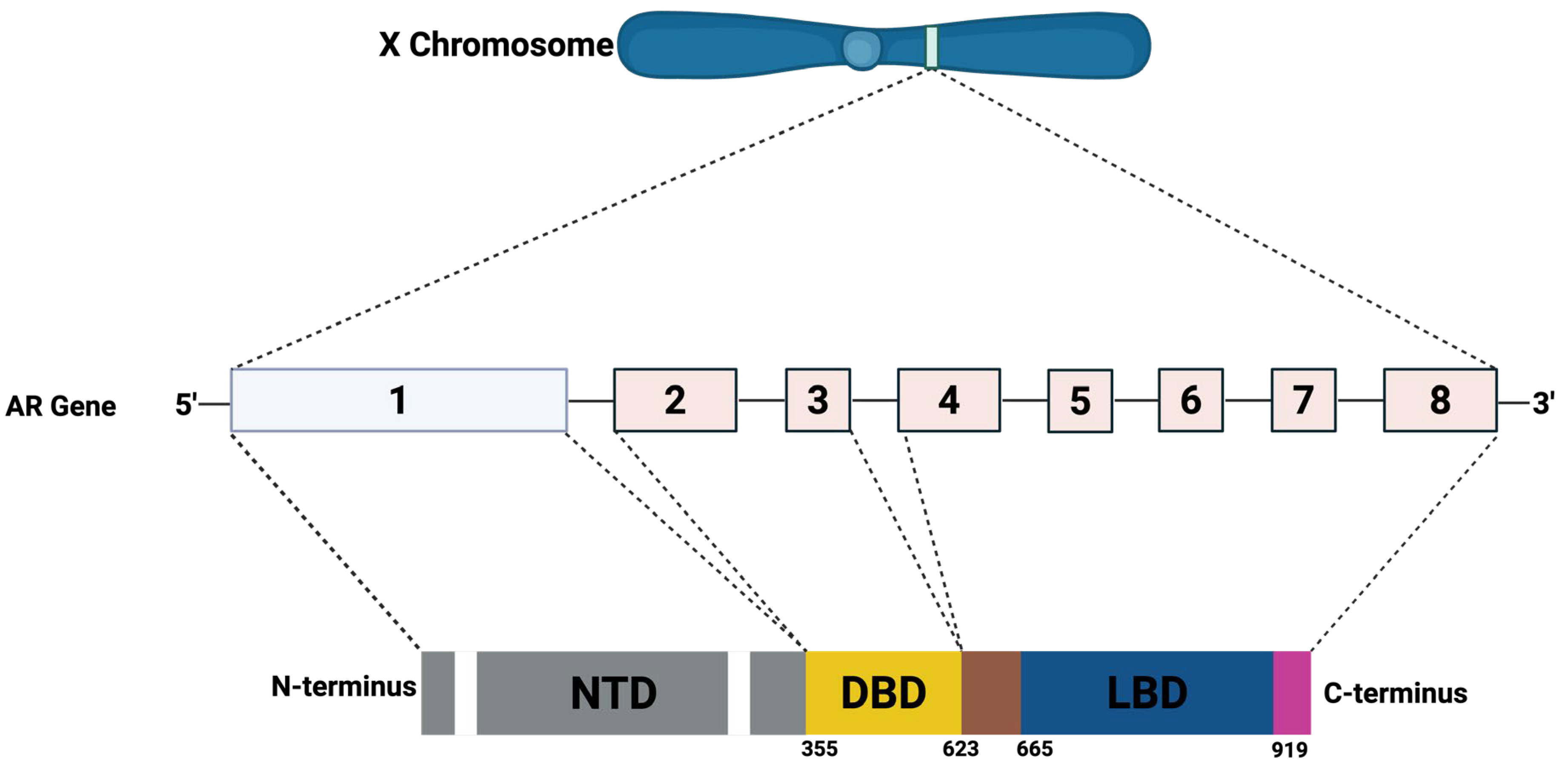
The AR gene is situated on the X chromosome at location Xq11–12 (Figure 11). It comprises 8 exons interspersed with introns that vary in length from approximately 0.7 to 2.6 kilobases. This gene encodes a protein of about 920 amino acids, sharing structural features with other members of the nuclear receptor family. The AR protein is organized into 4 principal functional regions: a flexible N-terminal domain (NTD; residues 1–558), a DNA-binding domain (DBD; residues 558–624), a C-terminal domain (residues 676–919), and a ligand-binding domain (LBD).
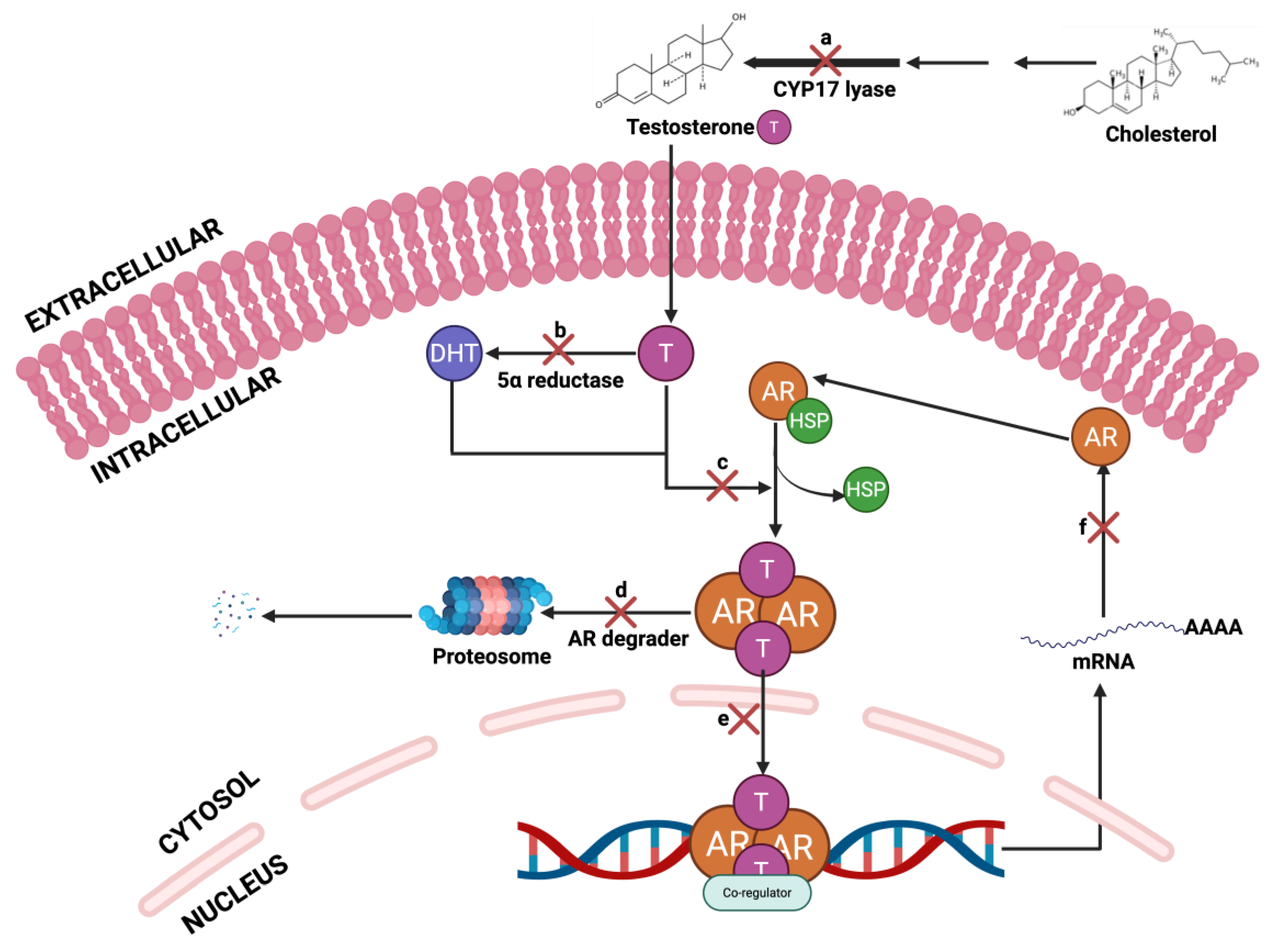
The AR signaling pathway (Figure 12) is a central driver of PCa progression, and therapeutic interventions have been developed to inhibit this axis at multiple molecular checkpoints. The pathway initiates with the biosynthesis of testosterone from cholesterol, catalyzed by CYP17 lyase; inhibition at this step using agents such as abiraterone effectively reduces systemic androgen levels. Testosterone is subsequently converted to the more potent dihydrotestosterone (DHT) via 5α-reductase, a process blocked by inhibitors like finasteride and dutasteride. Upon ligand binding, AR undergoes conformational activation and forms a cytoplasmic complex that can be inhibited by anti-androgens such as enzalutamide, bicalutamide, Seviteronel, TRC253, CR1447, flutamide and darolutamide [201,202,203], which competitively block androgen binding and prevent AR activation. Additionally, AR degraders promote proteasomal degradation of the receptor, further disrupting AR signaling [204,205]. AR degraders, including proteolysis-targeting chimaeras (PROTACs), selective AR degraders (SARDs), and hydrophobic tags (HyT), have been developed to target and treat metastatic PCa [206]. Among AR degraders, PROTACs represent a novel and highly promising therapeutic strategy. PROTACs are bifunctional molecules that simultaneously bind to the AR and an E3 ubiquitin ligase, facilitating the ubiquitination and subsequent proteasomal degradation of the AR protein. Unlike conventional AR antagonists that merely block receptor activity, PROTACs eliminate the receptor entirely, thereby overcoming resistance mechanisms such as AR overexpression, mutations, and splice variants. This targeted degradation approach offers a more sustained suppression of AR signaling and has shown efficacy in preclinical models of castration-resistant PCa (CRPCa). Several AR-targeting degraders are currently under investigation, with early candidates demonstrating potent anti-tumor activity and favorable pharmacokinetic profiles [206,207,208]. The activated AR complex normally translocates into the nucleus and binds to androgen response elements (AREs) on DNA to initiate transcription; this step is also targeted by agents that inhibit nuclear translocation or DNA binding. Finally, AR-mediated transcription of target genes drives tumor growth and survival, and transcriptional inhibitors aim to suppress this terminal output of AR signaling. Collectively, these points of inhibition represent a comprehensive strategy to attenuate AR activity and mitigate PCa progression.
AR blockade, otherwise known as ADT (anti-androgen therapy), has been demonstrated to induce tumor regression in up to 80% of PCa patients [209]. However, over time, PCa often resumes growth despite continued ADT [209]. Resistance to ADT may be caused by several mechanisms linked to androgen signaling, including androgen production within the tumor itself or by the adrenal glands, AR gene amplification or overexpression, the presence of AR mutations, and the development of constitutively active AR splice variants [210].

Evidence of PFOS in Prostate Cancer Development

Wen et al. [211] presented the first direct evidence that PFOS exerts unambiguous biological effects on the human prostate stem/progenitor cell (SPC) population, potentially contributing to an increased carcinogenic risk and tumor progression within the prostate gland. The study utilized a prostasphere model derived from healthy human prostate epithelial cells. The results showed that chronic exposure to PFOS at environmentally relevant concentrations led to a consistent increase in spheroid formation across serial passages, even when the same number of cells was seeded each time. Given that prostaspheres under these culture conditions are predominantly initiated by resident stem cells [212], this sustained increase strongly indicates enhanced symmetric self-renewal of prostate SPCs in the presence of PFOS, facilitating stem cell pool expansion. This is especially significant, considering the established link between cancer risk and the frequency of normal stem cell divisions in various tissues, including the prostate [213,214]. Moreover, PFOS exposure promoted the formation of larger spheroids, indicative of increased proliferation among progenitor cells, which constitute the majority population in prostaspheres [212]. Previous studies have shown that other endocrine-disrupting chemicals (EDCs), such as bisphenol A (BPA), dioxins, and inorganic arsenic, also target prostate SPCs, probably because they express various steroid hormone receptors [215].
The study conducted by Alyssa et al. [216], observed a positive association between PFOS and PCa in men aged ≥70 years. Their finding suggests that older men may be more susceptible to PCa owing to reduced immune function. Also, men with the highest PFOS exposure had about an 18% higher risk of developing PCa compared to those with the lowest exposure. However, when PFOS levels were analyzed as a continuous variable, no clear association with PCa risk was found. But when examined using a non-parametric model, the risk of PCa seemed to increase with PFOS levels up to a certain point (log2 PFOS = 4.2), possibly explaining the null result in the continuous model. Their findings suggest a possible but uncertain link between PFOS exposure and PCa risk; the observed higher risk at high PFOS levels could be real or just due to chance. Previous studies in the general population have also reported no clear or consistent association between PFOS and PCa [216].
Similarly, the study by Wen et al. [211], observed a strong expression of PPARα and RXRα was detected within prostate SPCs, an important finding given that many biological actions of PFOS are mediated through PPARα and its heterodimeric partner RXR [217,218]. These receptor-mediated interactions may initiate signaling cascades that influence SPC behavior and phenotype. Two primary downstream mechanisms were explored: transcriptomic and metabolic alterations induced by PFAS exposure. Single-cell RNA sequencing (scRNA-seq) revealed the emergence of a unique progenitor cell cluster in PFOS-treated spheroids that was absent in controls. Leveraging previously established KRT-based lineage markers [219], the new cluster was identified as an aberrant early-stage luminal progenitor population, a cell type increasingly implicated in prostate carcinogenesis [220]. Transcriptome-wide analysis indicated an upregulation of gene pathways linked to cell proliferation (G2M checkpoint, E2F targets, mitotic spindle) and oncogenic signaling (KRAS, MYC, TNFα via NF-κB, IL-6–JAK–STAT3, and TGF-β). The enrichment of these pathways corresponds with previous findings that associate therapeutic-resistant metastatic PCa with stemness-related signatures, including inflammatory and cytokine-driven signaling [221]. Since PPARs play a central role in metabolic regulation, they also profiled metabolic alterations in SPCs exposed to PFOS. Metabolomic analyses demonstrated elevated glycine and serine biosynthesis and evidence of enhanced aerobic glycolysis (Warburg effect), particularly in PFOS-treated samples.
Metabolic regulation is increasingly recognized as a critical factor in stem cell fate decisions and tumorigenesis [222,223]. Like pluripotent stem cells, cancer stem cells display high glucose uptake and lactate production, known as the Warburg effect [224,225]. Enhanced glycolysis and lactate secretion following PFOS exposure may therefore drive both self-renewal and impaired differentiation. Additionally, recent studies in epidermal pre-malignant stem cells have underscored the importance of serine biosynthesis from glucose as a rate-limiting step in stem cell proliferation, a metabolic phenotype mirrored in PCa stem-like cells [225]. Their Finding of pronounced upregulation of serine and glycine metabolism, particularly following long-term PFOS exposure, raises the possibility that PFOS may promote a pre-malignant state by fueling key biosynthetic pathways. To determine whether these effects translate into enhanced tumorigenic potential, they utilized a xenograft model with RWPE-2 prostate epithelial cells (harboring k-RAS activation) implanted into nude mice. PFOS administration significantly accelerated tumor growth over 40 days. Given that RWPE-2 cells contain a subpopulation with cancer stem-like properties, this finding suggests that PFOS may act not only on differentiated tumor cells but also on the cancer stem cell compartment, further promoting tumor expansion [226].
These findings (Table 1) provide compelling mechanistic evidence that PFOS directly impacts human prostate stem and progenitor cells by promoting self-renewal, increasing proliferative capacity, and disrupting normal differentiation trajectories. These effects are mediated, at least in part, through PFOS-induced alterations in transcriptional and metabolic programs, pushing SPCs toward a pre-malignant phenotype. Furthermore, in vivo evidence from xenograft models supports the role of PFOS in enhancing tumor growth, likely via modulation of the cancer stem cell niche. These mechanistic insights offer a plausible biological framework for the observed epidemiological associations between PFOS exposure and increased PCa risk and underscore the need for further investigation into the long-term health effects of chronic PFOS exposure.

4.3. Breast Cancer: Mechanism and Carcinogenesis

BCa remains one of the most commonly diagnosed malignancies among women globally [192,193,227,228]. The classification of BCa subtypes is primarily based on hormone receptor expression. The first group includes tumors that are positive for estrogen receptor (ER) and/or progesterone receptor (PR). The second group consists of tumors that overexpress human epidermal growth factor receptor 2 (HER2), regardless of ER or PR status. The third group, known as triple-negative breast cancer (TNBC), lacks expression of ER, PR, and HER2 [229]. Advances in gene expression profiling using microarray technologies have further refined BCa classification into intrinsic molecular subtypes. These include: Luminal A, characterized by ER+/PR+ status, high expression of luminal-related genes (e.g., ESR1, GATA3, XBP1, FOXA1), and low proliferation indicated by low Ki-67 levels; Luminal B, which also expresses ER but shows lower levels of luminal genes (e.g., PGR, FOXA1) and higher Ki-67 (>20%); HER2-enriched, defined by HER2 positivity and moderate luminal gene expression, though not all HER2+ tumors fall into this category; Basal-like, which exhibits elevated expression of EGFR and basal cytokeratins with minimal luminal gene expression; and Claudin-low, a subtype that is negative for ER, PR, and HER2, and also shows reduced expression of claudin 3/4/7 and E-cadherin [230].
ERs are key regulators in the development and progression of several hormone-related cancers, including those of the breast, endometrium, and ovaries [231,232,233]. Estrogens, which are small, lipophilic steroid hormones, exist in four main forms: estrone (E1), estradiol (E2), estriol (E3), and estetrol (E4). Among these, E2 is the most biologically active and is often referred to simply as estrogen. E4, produced exclusively during pregnancy in the fetal liver, exhibits tissue-specific effects [234]. Structurally, estrogens share a steroidal framework composed of four fused rings, with the phenolic A-ring playing a crucial role in receptor binding. Like other steroid hormones, estrogens are synthesized in the rough endoplasmic reticulum, beginning with cholesterol as the precursor [235]. The final step in estrogen biosynthesis involves the aromatization of androstenedione to estrone or estradiol, catalyzed by the enzyme aromatase (CYP19A1). This process predominantly occurs in the ovarian granulosa cells, adrenal cortex, and adipose tissue, with smaller contributions from the breast and placenta. E4 synthesis, however, is restricted to the fetal liver during gestation [234]. Once synthesized, E2 circulates in the bloodstream bound to carrier proteins and diffuses across cell membranes to reach target tissues [236]. Its primary mode of action involves binding to nuclear estrogen receptors, forming complexes that regulate gene transcription either directly or through secondary signaling cascades. The biological effects of estrogens are influenced by the genetic and physiological context of the target cells. Although both sexes produce estrogens, their roles and concentrations differ. Beyond reproductive functions, estrogens are involved in metabolic regulation, energy homeostasis, and cardiovascular and neurological health. While they may offer cardioprotective benefits, they are also implicated in increased cardiovascular risk under certain conditions. Dysregulation of estrogen signaling is associated with various pathologies, including autoimmune, metabolic, and degenerative diseases, as well as hormone-sensitive cancers such as BCa [236].
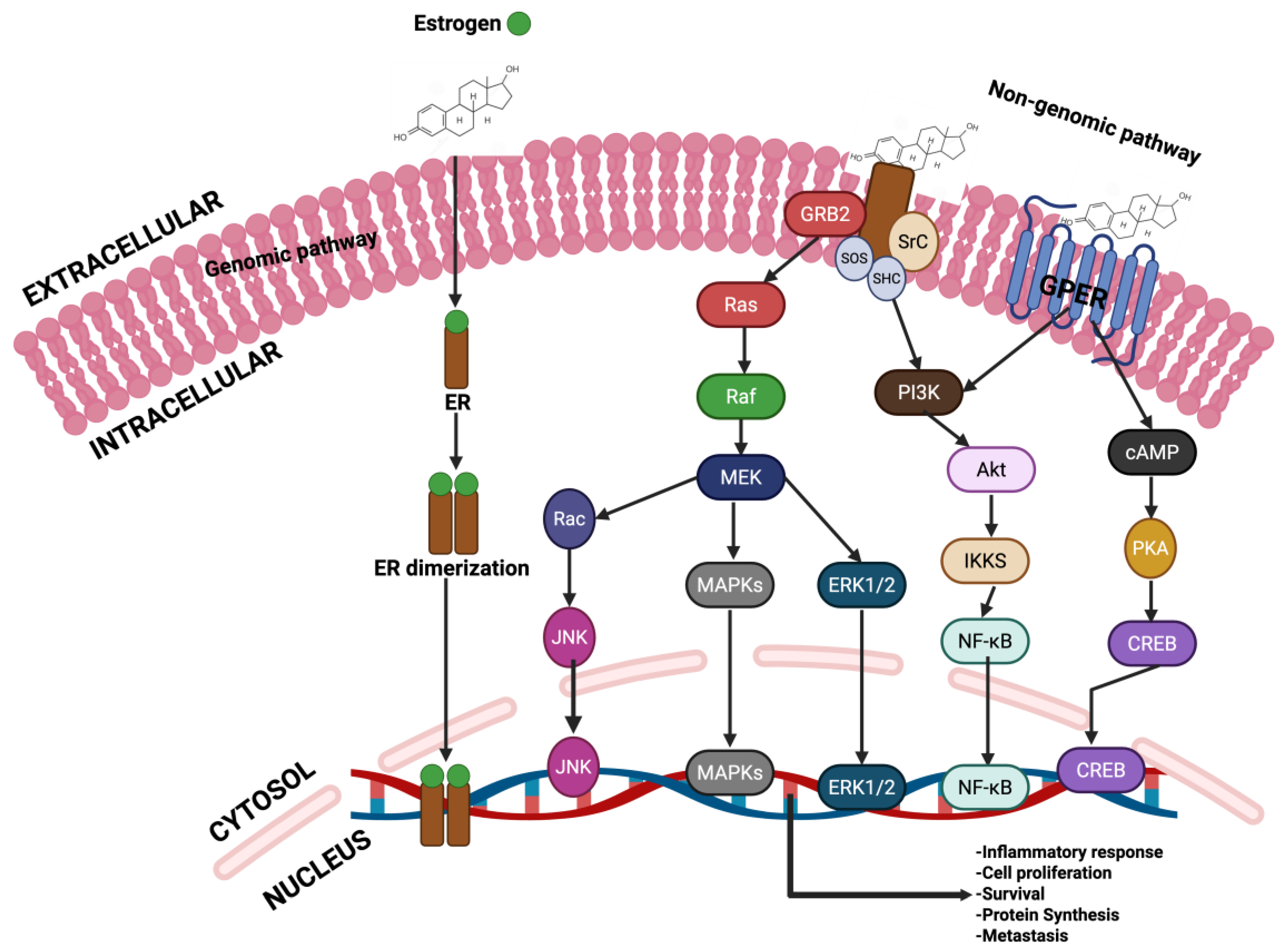
ER signaling operates through both genomic and non-genomic mechanisms (Figure 13). In the genomic pathway, estrogen–ER complexes translocate to the nucleus and modulate gene expression. This can occur indirectly, where the ER interacts with DNA-binding transcription factors (TFs) at their response elements via protein–protein interactions, recruiting coactivators to influence transcription. For instance, ER can engage in non-classical signaling by interacting with transcription factors such as specificity protein 1 (Sp1) and nuclear factor kappa B (NF-κB), even in the absence of estrogen-response elements (EREs) in the target gene promoters [237,238]. In direct genomic signaling, ER binds directly to EREs in the promoter regions of target genes. Ligand binding induces a conformational change in the ER, facilitating its release from the heat shock protein 90 (HSP90) complex, a chaperone that stabilizes unbound ER and prevents its degradation [239]. There is substantial cross-talk between ER and other members of the steroid hormone receptor family, including progesterone receptors (PR), glucocorticoid receptors (GR), and AR, within BCa cells and other hormone-sensitive malignancies such as endometrial cancer [240]. These receptors exhibit overlapping DNA-binding motifs and coordinate their genomic functions through shared recruitment of transcriptional regulators and chromatin-modifying complexes [241]. ER signaling is modulated by the presence and activity of these other receptors. For instance, PR is not only a downstream target of ERα but also interacts directly with ERα, influencing its chromatin-binding landscape and thereby altering gene expression patterns that can impact clinical outcomes. Despite these observations, the exact molecular mechanisms by which PR reshapes ERα activity remain to be fully elucidated. Similarly, AR has been implicated in modulating ER genomic interactions in BCa and its functional role, along with therapeutic targeting strategies, has been investigated across various BCa subtypes [242].
In the context of non-genomic ER signalling, estrogen engages with membrane-associated estrogen receptors (mbERs) or G-protein–coupled estrogen receptor 1 (GPER1), initiating rapid signaling events at the cell surface independent of direct gene transcription [243]. These interactions activate intracellular kinase cascades, notably the PI3K/Akt/IKK/NF-κB and cAMP/PKA/CREB pathways. In addition, the binding of estrogen to ER can activate PI3K/Akt/IKK/NF-κB, Ras/Raf/MEK/MAPKs; MEK/Rac/JNK and MEK/ERK1/2 pathways. Within the MAPK cascade, estrogen binding activates Ras, which subsequently triggers Raf-mediated phosphorylation of MEK, leading to MAPK activation. MAPK then stimulates transcription factors such as c-Jun and c-Fos, promoting expression of downstream genes [244]. Also, mbER-mediated activation of SHC/PI3K/Akt initiates the PI3K–Akt–mTOR pathway, which regulates mTOR signaling. This pathway supports cellular proliferation and survival, and its dysregulation has been linked to the progression of ER-positive BCa and resistance to endocrine therapies [245].
The transcriptional activity of ER is tightly controlled by a network of coregulatory proteins, which include both coactivators and corepressors. These molecules are recruited to the ER via a conserved LxxLL motif within the ligand-binding domain (LBD), where “L” denotes leucine and “x” represents any amino acid [246]. Coregulators often function in concert with enzymatic partners such as methyltransferases, acetyltransferases, deacetylases, kinases, ubiquitin ligases, and ATP-dependent chromatin remodelers, thereby influencing chromatin structure and gene expression [247]. The dynamic balance between coactivators and corepressors is context-dependent and varies across cell types, ultimately shaping the transcriptional output of ER signaling. Corepressors, for example, recruit histone deacetylases (HDACs) to ER-bound chromatin, promoting chromatin condensation and transcriptional repression. By antagonizing coactivator function, corepressors help maintain appropriate levels of nuclear receptor activity. Aberrations in coregulator expression or function are strongly associated with tumorigenesis, cancer progression, and metastasis in malignancies such as breast and colorectal cancer [247].

Evidence of PFOS in Breast Cancer Development

Multiple studies have suggested a potential link between PFOS exposure and BCa development [248,249]. A case–control study conducted on the Greenland Inuit population found an association between elevated serum PFOS levels and an increased risk of BCa [26]. Additionally, PFOS is suspected to exhibit estrogenic properties, with in vitro studies indicating its ability to interact with ERα and ERβ, potentially enhancing ER-dependent transcriptional activity [31,250,251]. However, some studies have reported conflicting findings regarding its estrogenic effects, and the role of PFOS in BCa development remains insufficiently explored [252,253]. The findings by Pierozan et al. [254] found that PFOS led to a sustained rise in cyclin D1 levels, accounting for the increase in cell proliferation. Exposing MCF-10A cells to PFOS triggered the activation of ERK in unexposed daughter cells (Figure 14). ERK pathway activation is triggered by mitogen factors, and it is one of the most commonly mutated genes in various cancers, often resulting in increased cell proliferation [255]. Once activated, ERK translocates from the cytoplasm into the nucleus, where it phosphorylates various nuclear substrates, including transcription factors such as AP-1. AP-1 subsequently binds to the cyclin D1 promoter, thereby enhancing cyclin D1 gene transcription [256]. In another study, PFOS reduced the levels of the CDK inhibitor p21, whose loss is linked to enhanced tumorigenesis and poor prognosis across cancers [257,258]. PFOS also lowered p27 expression in D2 cells, a tumor suppressor frequently downregulated in BCa, where its loss predicts adverse clinical outcomes. Notably, p27 inactivation typically occurs through post-translational mechanisms such as degradation or mislocalization rather than mutation [259,260].
Metastasis remains the primary cause of mortality in BCa, occurring long after the initial tumor develops, with invasion into surrounding tissues as the first step [261,262]. The study of Pierozan et al. [254] indicated that PFOS promotes malignant transformation of MCF-10A cells by reducing the expression levels of key adhesion molecules, including occludin, E-cadherin, and β-integrins, thereby disrupting cell–cell and cell–matrix interactions. Such alterations are consistent with evidence that downregulation of E-cadherin facilitates BCa progression and metastasis, while reduced integrin expression impairs adhesion and drives tumor invasiveness [263,264]. Similarly, reduced occludin expression has been linked to metastatic BCa, while its overexpression inhibits tumour development and increases sensitivity to apoptosis [265,266].
Epigenetic signatures, such as DNA methylation, histone modification, and nucleosome remodelling, are now seen as crucial drivers of carcinogenesis [267,268]. Dysregulated epigenetic control in breast cells can alter gene expression, thereby contributing to the initiation and progression of BCa [269]. Exposure of PFOS to BCa cells increased the global DNA methylation in both MCF-10A cells and their unexposed progeny [254]. Such aberrant methylation patterns, particularly within CpG islands, can silence critical genes including tumor suppressors, DNA repair genes, and regulators of invasion, metastasis, and angiogenesis, thereby sustaining malignant transformation [270,271]. In cancer, CpG island hypermethylation is often coupled with histone H3 and H4 deacetylation, which alters chromatin architecture and disrupts gene expression [268,272]. Active transcription is generally associated with histone modifications such as H3K9ac, H3K27ac, and H3K4me, whereas H3K9 methylation promotes repressive chromatin states [270,273]. In their findings, PFOS decreased H3K9 acetylation, consistent with reduced acetylation observed in aggressive breast tumors and its correlation with poor prognosis [254]. These PFOS-induced histone modifications may contribute to dysregulation of cell-cycle regulators, as both promoter methylation and histone deacetylation are established mechanisms for p21 silencing [274].
Tsai et al. [275] identified a notable correlation between exposure to PFOS and BCa risk in Taiwanese women. Their case study revealed that women aged 50 or younger were particularly susceptible, with a stronger association observed in estrogen receptor (ER)-positive tumors within this age group. These results imply that younger women may be more sensitive to the carcinogenic effects of PFOS compared to older women. Supporting this, a study conducted in Greenland among women of similar ages (50 for cases and 54 for controls) also found a link between PFOS exposure and elevated BCa risk [26]. In contrast, a Danish study involving younger women (average age ~40 years) reported only a weak association between PFAS exposure and BCa [276]. BCa incidence is known to vary across age groups, ethnicities, and regions, likely due to differences in environmental exposures, reproductive patterns, and lifestyle choices [277]. While BCa risk typically increases with age in Western populations [278], this trend was not reflected in the Taiwanese cohort, where younger women showed higher vulnerability.
Previous studies have also suggested that PFOS may alter sex hormone levels, such as follicle-stimulating hormone (FSH) and testosterone, especially during adolescence (ages 12–17), a period marked by hormonal fluctuations [279]. Chronic exposure to PFOS could disrupt hormonal equilibrium, potentially contributing to BCa development [280]. Since their widespread use began in the 1950s, PFOS have been prevalent in industrial processes. Taiwanese environmental studies have detected elevated PFOS levels in rivers near industrial zones, indicating a possible route of exposure for nearby communities. This raises concerns about long-term exposure, particularly among younger individuals, and its implications for BCa risk. Their findings reinforced that women aged ≤50 years were more likely to develop ER-positive tumors in relation to PFOS exposure than those over 50. Similarly, Greenlandic research found a connection between PFOS and BCa risk in women of comparable age, although no significant difference was observed between ER-positive and ER-negative cases [281]. Conversely, a French study reported that low PFOS levels were associated with an increased risk of ER-negative BCa, with the average age at diagnosis being 68.8 years [50]. It is well-established that younger women diagnosed with BCa are more likely to present with ER-negative tumors [282]. While ER-positive BCa is often linked to reproductive factors, ER-negative variants are believed to be influenced more by environmental and lifestyle factors. Additionally, the classification of tumors as ER-positive or ER-negative may be affected by the timing of blood sample collection—whether before or after diagnosis [283]. Interestingly, PFOS concentrations in Taiwanese women were lower than those reported in studies from Greenland and France [50,281]. This discrepancy may be partially explained by age-related differences in tumor biology. Unlike Western trends, younger Taiwanese women (≤50 years) exhibited higher expression of estrogen and progesterone receptors compared to older women (>50 years) [277], a pattern also observed in other East Asian populations. While Wielsøe et al. [281] reported a positive association between PFOS and BCa risk, Bonefeld-Jørgensen et al. [276] did not find a significant link. Notably, Wielsøe et al. [281] documented higher PFOS concentrations than those observed in the current study. In contrast, Barry et al. [284] found that women with the highest PFOS levels had the lowest BCa risk, although their reported concentrations were also higher than those in the present research.
In animal models, mammary fibroadenomas have been observed following two years of PFOS exposure, leading the U.S. EPA to consider PFOS a possible BCa risk factor [285]. However, findings remain inconclusive. Hardisty et al. [286] observed no significant elevation in mammary gland tumors after a two-year dietary exposure to PFOS. Meanwhile, other research reported that perinatal exposure to PFOS in female mice caused persistent abnormalities and delayed mammary gland development [287], a phenomenon that may be mediated by activation of the PPAR-α pathway [288]. In addition, an in vitro study demonstrated that PFOS can enhance the activity of human BCa cells [289]. Although two studies have attempted to identify genes potentially affected by PFOS exposure in humans, the underlying mechanisms remain unclear [290]. Future research is therefore needed to investigate how PFOS may influence BCa pathogenesis in humans. Multiple recognized risk factors for BCa are associated with reproductive history, encompassing early menarche, late menopause, advanced age at first childbirth, breastfeeding practices, and the utilization of exogenous hormones [291]. Other lifestyle factors, including alcohol consumption, obesity, and lack of physical activity, also play a significant role [292]. Additionally, hereditary factors play a role in BCa incidence [293].
These studies are subject to several limitations. First, their cross-sectional nature prevents the establishment of a causal link between PFOS exposure and BCa risk. Second, the relatively small sample sizes contribute to statistical uncertainty, resulting in broad confidence intervals. Additionally, the analysis was restricted by age categorization and lacked sufficient data to thoroughly assess dose–response relationships. A major limitation was the absence of historical exposure records, which is critical given the long latency period associated with BCa development. Although PFOS compounds are known to persist in the body due to their long biological half-lives, relying on a single-time-point measurement may not accurately represent cumulative exposure. Despite these constraints, the studies (Table 1) offer valuable insights, particularly in highlighting age-specific associations between PFOS and BCa risk, which may be influenced by hormonal differences observed between Taiwanese and Western populations.

4.4. Ovarian Cancer: Mechanism and Carcinogenesis

OC is the second most common gynecologic disease in the United States. In 2024. There were about 19,680 diagnosed cases of OV and 12,740 related deaths worldwide [294]. OV is classified into two main histological subtypes (Figure 15): Epithelial ovarian cancers (EOCs) and non-epithelial ovarian cancers (non-EOCs). Approximately 90% of ovarian malignancies are EOCs, while the other 10% are non-EOCs. Figure 15 shows the subtypes of EOCs and non-EOC [295]. OC is broadly classified into epithelial and non-epithelial types. Epithelial ovarian cancers (EOCs), which represent the majority of cases, include high-grade serous carcinoma, low-grade serous carcinoma, endometrioid carcinoma, clear cell carcinoma, mucinous carcinoma, and undifferentiated carcinoma. Non-epithelial ovarian cancers (non-EOCs) include germ cell tumors, sex cord–stromal tumors, and other rare or miscellaneous tumor types. The molecular biology of OC is complex and involves several hormonal, genetic and epigenetic modifications that promote its tumorigenesis and the development of treatment resistance.
Several studies indicated that hormonal factors play a crucial role in the tumorigenesis and progression of different OC subtypes [296,297,298]. For example, it has been shown that hormonal factors, particularly estrogens, play a crucial role in the development and progression of EOCs. Estrogens primarily affect the proliferation, invasion and epithelial-to-mesenchymal transition of cancer cells through their effects on ER including ERα, ERβ, and GPER1. Furthermore, estrogen signaling affects the tumor microenvironment by modulating immune and stromal cells, which further promotes tumor progression and invasion [299]. EOCs, especially the high-grade serous ovarian carcinoma (HGSC) type, tend to be more aggressive and have worse survival rates if diagnosed at advanced stages. It is now known that the fallopian tube epithelium, particularly the fimbrial end rather than the ovarian surface is the primary source of HGSC. This shift is supported by molecular and histopathological evidence identifying precursor lesions such as p53 signatures and serous tubal intraepithelial carcinoma (STIC), which represent early stages in the progression to invasive cancer. Especially among women with BRCA mutations, occult tubal lesions are frequently found during prophylactic surgeries, reinforcing the tubal origin hypothesis [300].
Although several genes and transcription factors are involved in the development and progression of HGSC, the BRCA1 and BRCA2 genes are considered the most significant contributors. Homologous recombination (HR) is one of the DNA repair mechanisms that repairs the double-strand breaks by using an intact homologous DNA molecule as a template. Mutations or epigenetic silencing of BRCA1/2 compromise the HR repair mechanism, leading to genomic instability and tumorigenesis. About 50% of high-grade serous ovarian cancers (HGSOC) exhibit homologous recombination repair (HHR), with approximately 15–20% harboring germline or somatic mutations in BRCA1 or BRCA2. The identification of HRR deficiencies has thus become a crucial factor in guiding precision medicine strategies targeting OC. These HRR-deficient tumors are particularly sensitive to DNA-damaging agents like platinum-based chemotherapies and to targeted therapies such as poly (ADP-ribose) polymerase (PARP) inhibitors. PARP inhibitors (PARPi) such as Olaparib, Niraparib and Rucaparib exploit the concept of synthetic lethality by blocking alternative DNA repair pathways, leading to the accumulation of DNA damage and cell death in HRR-deficient cancer cells. Since its first approval in 2014, PARPi has revolutionized OC care with dramatic improvements in progression-free and overall survival when used as maintenance therapy following platinum-based chemotherapies [301,302]. In recent years, a significant challenge in OC has emerged that has raised our concerns. OC cells show resistance mechanisms to PARPi and chemotherapeutics. This phenomenon has led to a decline in overall survival and progression-free survival rates among patients [301]. Resistance to PARP inhibitors in OC can occur through restoration of homologous recombination (e.g., BRCA reversion mutations), replication fork protection, PARP1 mutations, and increased drug efflux [301,302]. On the other hand, Cisplatin resistance involves enhanced DNA repair, reduced drug accumulation, detoxification by glutathione, and evasion of apoptosis [303].

Evidence of PFOS in Ovarian Cancer Development

Chronic low-dose exposure to PFOS in female mice has been shown to alter steroidogenesis and hormonal regulation, consequently disrupting reproductive function. PFOS lengthens the estrous cycle, especially extending the diestrus phase, and reduces the number of mature ovarian follicles and corpora lutea while increasing follicular atresia. These alterations are accomplished by a drop in the serum levels of important respective hormones, including LH, FSH, P4, E2 and GnRH. Mechanistically, PFOS suppresses the expression of the steroidogenic acute regulatory protein (StAR) gene in the ovary by reducing histone H3K14 acetylation at its promoter, leading to diminished estrogen biosynthesis. The underlying cellular mechanism involves a reduction in histone H3K14 acetylation at the steroidogenic acute regulatory protein, which leads to inhibition of estrogen biosynthesis, impaired follicle development and ovulation, thereby contributing to a reduction in ovarian follicular reserve [304]. PFOS is known to bind to ER, thereby partially blocking and activating their responses [305]. This interference creates a restriction in regulating the genome and therefore increases the risk of OC. The mechanistic pathway involves GPR30 and the Insulin-like growth factor 1 receptor (IGF1R), a growth-promoting factor found in granulosa cells that responds to PFOS in human follicular fluid, thereby promoting the growth of ovarian granulosa tumours [306].
This epigenetic modification results in impaired follicle maturation, disrupted activation of kisspeptin neurons in the anteroventral periventricular nucleus (AVPV), and a blunted LH surge necessary for ovulation. Notably, administration of estrogen or kisspeptin-10 can rescue the impaired LH surge, highlighting the central role of hormonal signaling pathways affected by PFOS. Also, recent findings showed that exposure to PFOS negatively impacts uterine health by dropping the viability of human endometrial stromal cells, causing apoptosis through the upregulation of BAX and the downregulation of BCL-2, interfering with the progression of the cell cycle, and changing receptivity markers like HO, XA10, ITGB3, and FOXO1 [307]. These results imply that PFOS alters the hormonal regulation of reproduction and mitochondrial integrity factors that also play a pivotal role in OC development. The cellular stress, endocrine disruption, and inflammatory environment triggered by PFOS in uterine tissues may extend to ovarian tissue, potentially increasing the consequences of transformation into cancer. Like PFOS, other PFAS, including PFOA, PFHpA, and PFPA, have been found to increase the resistance of OC cells to various treatments, including chemotherapy. Notably, these PFAS at environmentally relevant, sub-cytotoxic concentrations enhanced OC cell survival and increased resistance to the chemotherapy drug carboplatin [308].
Increasing evidence (Table 1) has reported that exposure to PFOS could adversely affect key processes like ovarian folliculogenesis and steroidogenesis, which are needed for normal reproductive health. Epidemiological evidence has reported that exposure to PFOS impacts ovarian steroidogenesis, causes a delayed onset of menarche, disrupts the menstrual cycle, accelerates ovarian ageing, and may cause chronic conditions such as polycystic ovarian syndrome (PCOS) and increases the chances of OC [309]. The study conducted by Knox et al. [310] has reported PFOS exposure to have a significant and negative relationship with serum E2 levels among women aged 42-65 years. Similarly, Zhang et al. [311] suggested that exposure to PFOS may lead to decreased serum E2 and prolactin levels, thereby causing an increase in FSH levels among premature ovarian insufficiency in patients. Their study also revealed higher plasma PFOS concentrations, indicating an increased risk of POI. PFOS has been reported to have weak estrogenic activity, whereas it exerts an anti-estrogenic effect when co-administered with estradiol. The result showed a negative association between PFOS and E2 and a positive association with FSH. PFOS has been reported to cause a decrease in the production of estradiol in women of reproductive age and to alter steroidogenesis. Higher exposure to PFOS has been reported to cause a reduction in the ovarian follicular reserve. The study conducted by Jones et al. [312] found a positive association with OC, revealing an increased risk of serum levels of certain PFOS, which are associated with an increased risk of OC among postmenopausal women. Their study measured the amount of PFOS in blood samples collected before developing OC. After analysing data from 318 OC cases and 472 matched controls, the result showed 47% odds of developing OC.
Table 1. Summary of Research Findings on PFAS and Cancer.
S/NStudy/ModelFindings References
1.Human prostate stem/progenitor cell (SPC) populationPFOS increased carcinogenic risk, tumor progression and spheroid formations in the prostate gland as well as increased the expression of PPARα and RXRα.Wen et al. [211]
2.Epidermal pre-malignant stem cellsPFOS upregulated serine and glycine metabolism and increased the growth of PCa.Imir et al. [226].
3.Case–control studySignificant association between elevated serum PFOS levels and an increased risk of BCaBonefeld-Jorgensen et al. [26].
4.Juvenile rainbow trout (Oncorhynchus mykiss)PFOS could interact with ERα and ERβ, potentially enhancing ER-dependent transcriptional activityBenninghoff et al. [250]
5.H295R cellsPFOS serves as an ER agonist and Thyroid hormone receptor (THR) antagonist, as well as increased estradiol (E2) levels in H295R cells. Du et al. [31]
6.MVLN cellsPFOS significantly induced the ER transactivity while antagonizing the activity of AR.Kjeldsen et al. [251]
7.MCF-10APFOS elevated cyclin D1 and D2 levels, increased the global DNA methylation, as well as reduced the levels of the CDK inhibitor p21, occluding, E-cadherin and β-integrins.Pierozan et al. [254]
8.Case–control studyWomen aged 50 or younger were particularly susceptible to PFOS, with a stronger association observed in estrogen receptor (ER)-positive tumors within this age group.Tsai et al. [275]
9.Case–control studyThere is a strong link between PFOS exposure and elevated BCa risk.Bonefeld-Jorgensen et al. [26].
10.Case–control studyPFOS triggered hormonal fluctuations in adolescents aged 12–17 years.Tsai et al. [275].
11.Case–control studySignificant increase in PFOS levels in cases compared to the controls, indicating the correlation of PFOS with BCa.Wielsøe et al. [281]
12.T47D human BCa cellsPFOS promoted the estrogenic effects of 17β-estradiol in T47D human BCa cells.Sonthithai et al. [289]
13.human endometrial stromal cells (hESCs)PFOS may drive OC by decreasing the expression levels of endometrial tolerance-related proteins Homeobox A10 (HOXA10) and integrin beta 3 (ITGB3), while increasing the expression level of Forkhead box 01 (FOXO1) protein. Ren et al. [307].
14.Age-Related AssociationsPFOS revealed a positive association with PCa in men aged ≥ 70 years.Alyssa et al. [216]
15.Chinese women studyPFOS exposure decreased serum E2 and prolactin levels and increased FSH levels, disrupted ovarian steriodogenesis, and caused premature ovarian insufficiency Zhang et al. [311]
16.OC riskPFOS exposure is associated with OC incidence.Jones et al. [312]
17.Sex HormonesPFOS significantly impacted the serum E2 levels among women aged 42–65Knox et al. [310]

5. Regulatory Actions Against PFOS

Due to the recognized environmental and health hazards posed by PFOS, regulatory bodies, particularly in industrialized nations, have enacted stringent controls on its manufacture and use. In 2009, PFOS was added to the Stockholm Convention on Persistent Organic Pollutants, prompting a global phase-out coordinated by the United Nations Environment Program (UNEP) [313]. Additionally, the U.S. Food and Drug Administration (FDA) withdrew approval for PFOS and several other PFAS in food contact materials. Ongoing investigations by the Agency for Toxic Substances and Disease Registry (ATSDR), under the Centers for Disease Control and Prevention (CDC), are examining human exposure routes and health outcomes linked to PFOS and related PFAS, particularly through drinking water and dietary intake. According to the U.S. Environmental Protection Agency (EPA) and the National Primary Drinking Water Regulation (NPDWR), the enforceable Maximum Contaminant Level (MCL) for PFOS in drinking water is set at 4.0 parts per trillion (ppt), equivalent to 4.0 ng/L [314]. The European Union (EU) has similarly imposed strict limitations under the REACH (Registration, Evaluation, Authorization and Restriction of Chemicals) framework, promoting the development and adoption of safer chemical alternatives [315].
Recently, the International Agency for Research on Cancer (IARC) classified PFOS as a Group 2B substance, indicating it is possibly carcinogenic to humans. This classification is based on evidence linking prolonged PFOS exposure to various cancers, including those of the pancreas, breast, prostate, liver, kidney, ovary, endometrium, bladder, thyroid, and non-Hodgkin lymphoma [32,51,91,128,316,317,318,319]. Although animal studies provide limited carcinogenic data, research by Tessmann et al. [320] demonstrated that PFOS exposure in mice led to the suppression of 3-hydroxy-3-methylglutaryl-CoA synthase-2 and the activation of oncogenic proteins such as c-MYC, FASN, mTOR, and β-catenin—markers associated with intestinal tumorigenesis.
While developed countries have made substantial progress in regulating PFOS and other hazardous chemicals, many developing and underdeveloped nations still lack comprehensive frameworks to mitigate environmental contamination. As highlighted by Wee and Aris, these regions often face systemic challenges, including the absence of standards for physical water parameters (e.g., pH, turbidity), insufficient regulation of chemical contaminants (e.g., disinfectants, heavy metals, nutrients), inadequate monitoring of organic pollutants (e.g., pesticides, petroleum derivatives), and limited capacity to assess microbiological indicators (e.g., coliforms, Escherichia coli) and sensory attributes (e.g., taste, color, odor, texture, temperature, and overall acceptability) [321]. Despite some progress, such as the establishment of water quality standards in Malaysia, India, Bangladesh, the Philippines, China, Turkey, the United Arab Emirates, South Africa, Nigeria, Ghana, and others, PFOS continues to pose exposure risks to both humans and wildlife due to its persistence and widespread distribution in the environment.

6. Conclusions

PFOS, a persistent organic pollutant, presents a substantial risk to environmental and human health due to its chemical stability, bioaccumulative properties, and resistance to degradation. This review has outlined PFOS’s complex toxicological profile, emphasizing its widespread environmental dissemination via both point and nonpoint sources, its intricate pharmacokinetics, and its limited excretion pathways. Mechanistic studies reveal that PFOS disrupts lipid metabolism, induces oxidative stress, impairs mitochondrial function, and interferes with endocrine and immune signaling. These molecular perturbations are implicated in a range of adverse health outcomes, including hepatotoxicity, neurotoxicity, reproductive dysfunction, and immunosuppression. Notably, emerging evidence suggests a potential role for PFOS in carcinogenesis, particularly in hormone-sensitive cancers such as prostate, breast, and ovarian cancers. Proposed mechanisms include endocrine disruption, oxidative DNA damage, chronic inflammation, and epigenetic alterations. Epidemiological and experimental data increasingly support associations between PFOS exposure and elevated cancer risk, especially among populations with occupational or environmental exposure. PFOS has been shown to activate oncogenic pathways, modulate stem cell behavior, and promote tumor growth and metastasis. These findings underscore the need for continued investigation into PFOS’s molecular mechanisms and long-term health effects. Although regulatory measures in several developed countries have curtailed PFOS production and usage, many developing regions lack comprehensive frameworks to monitor and mitigate its impact. Addressing the global burden of PFOS requires coordinated international efforts to enhance surveillance, enforce stringent regulations, and accelerate the development of safer chemical alternatives.

Author Contributions

U.O.A.: Writing—original draft, review & editing, formal analysis, Software supervision, Resources, Project Administration, Conceptualization. D.C.E.: Writing—original draft, review & editing, formal analysis, Software supervision. C.C.A.: Writing—original draft, formal analysis, Software supervision, Resources, Project Administration. A.A.: Writing—original draft, review & editing, Software supervision, Resources, formal analysis. E.G.U.: Writing—original draft, formal analysis. F.C.J.: Writing—original draft, formal analysis. A.K.O.: Writing—original draft, formal analysis. I.V.A.: Writing—original draft, formal analysis. C.O.O.: Writing—original draft, formal analysis. V.C.N.: Writing—review & editing. E.A.U.: Writing—review & editing, Resources, Project Administration. P.E.E.: Writing—review & editing, Resources, Project Administration S.O.: Writing—review & editing, Resources. All authors have read and agreed to the published version of the manuscript.

Funding

This research received no external funding.

Conflicts of Interest

The authors declare no conflicts of interest.

Abbreviations

AVPVAnteroventral Periventricular Nucleus
AFFFAqueous Film Forming Foam
BAXBCL2 Associated X Protein
BCL-2B-Cell Lymphoma 2
BRCA1Breast Cancer 1 Gene
BRCA2Breast Cancer 2 Gene
cAMPCyclic Adenosine Monophosphate
CDKCyclin-Dependent Kinase
CREBcAMP Response Element-Binding Protein
CYPCytochrome P450 (CYP450)
DNADeoxyribonucleic Acid
E2Estradiol
EREstrogen Receptor
ERKExtracellular Signal-Regulated Kinase
FOXO1Forkhead Box Protein O1
FSHFollicle-Stimulating Hormone
FSH.Same as above (Follicle-Stimulating Hormone)
GnRHGonadotropin-Releasing Hormone
GPR30G Protein-Coupled Estrogen Receptor 30 (also known as GPER1)
GSHGlutathione
GSK-3βGlycogen Synthase Kinase 3 Beta
GSTGlutathione S-Transferase
H3K14Histone H3 Lysine 14 (site of acetylation or methylation)
HDACsHistone Deacetylases
HER2Human Epidermal Growth Factor Receptor 2
HOHeme Oxygenase
HO-1Heme Oxygenase-1
IARCInternational Agency for Research on Cancer
IGF1RInsulin-Like Growth Factor 1 Receptor
ITGB3Integrin Beta 3
JNKc-Jun N-terminal Kinase
LBDLigand-Binding Domain
LHLuteinizing Hormone
MAPKsMitogen-Activated Protein Kinases
MCF-10AHuman Non-Tumorigenic Mammary Epithelial Cell Line
MMPMatrix Metalloproteinase
NF-κBNuclear Factor Kappa-Light-Chain-Enhancer of Activated B Cells
NF-κB,Same as above
p38p38 Mitogen-Activated Protein Kinase (MAPK)
P4Progesterone (also known as Pregn-4-ene-3,20-dione)
PARPPoly (ADP-Ribose) Polymerase
PFASPer- and Polyfluoroalkyl Substances
PFHpAPerfluoroheptanoic Acid
PFOAPerfluorooctanoic Acid
PFOSPerfluorooctane Sulfonate
PFPAPerfluoropentanoic Acid
PPARαPeroxisome Proliferator-Activated Receptor Alpha
PINProstatic Intraepithelial Neoplasia
ATPAdenosine Triphosphate
PKAProtein Kinase A
POIPrimary Ovarian Insufficiency
PRProgesterone Receptor
PSAProstate-Specific Antigen
PTENPhosphatase and Tensin Homolog
ROSReactive Oxygen Species
StARSteroidogenic Acute Regulatory Protein
TNBCTriple-Negative Breast Cancer
VEGFVascular Endothelial Growth Factor
WNT1Wingless-Type MMTV Integration Site Family Member 1
XA10Xanthine Dehydrogenase/Aldehyde Oxidase 1 (also known as xanthine oxidase homolog, depending on context)

References

  1. Giesy, J.P.; Kannan, K. Perfluorochemical surfactants in the environment. Environ. Sci. Technol. 2002, 36, 146a–152a. [Google Scholar] [CrossRef] [PubMed]
  2. Wang, T.; Wang, Y.; Liao, C.; Cai, Y.; Jiang, G. Perspectives on the Inclusion of Perfluorooctane Sulfonate into the Stockholm Convention on Persistent Organic Pollutants. Environ. Sci. Technol. 2009, 43, 5171–5175. [Google Scholar] [CrossRef] [PubMed]
  3. Cheng, J.; Huang, L.; Li, Y.; Zhang, Z.; Mu, R.; Liu, C.; Hu, S.; Xiao, Y.; Xu, M. A Review of Treatment Technologies for Perfluorooctane Sulfonate (PFOS) and Perfluorooctanoic Acid (PFOA) in Water. Processes 2023, 11, 2260. [Google Scholar] [CrossRef]
  4. Li, M.; Jin, Y.-T.; Yan, J.-F.; Liu, Z.; Feng, N.-X.; Han, W.; Huang, L.-W.; Li, Q.-K.; Yeung, K.-L.; Zhou, S.-Q.; et al. Exploration of perfluorooctane sulfonate degradation properties and mechanism via electron-transfer dominated radical process. Water Res. 2022, 215, 118259. [Google Scholar] [CrossRef]
  5. Li, M.; Mo, C.-H.; Luo, X.; He, K.-Y.; Yan, J.-F.; Wu, Q.; Yu, P.-F.; Han, W.; Feng, N.-X.; Yeung, K.L.; et al. Exploring key reaction sites and deep degradation mechanism of perfluorooctane sulfonate via peroxymonosulfate activation under electrocoagulation process. Water Res. 2021, 207, 117849. [Google Scholar] [CrossRef]
  6. Chen, F.; Zhou, Y.; Wang, L.; Wang, P.; Wang, T.; Ravindran, B.; Mishra, S.; Chen, S.; Cui, X.; Yang, Y.; et al. Elucidating the degradation mechanisms of perfluorooctanoic acid and perfluorooctane sulfonate in various environmental matrices: A review of green degradation pathways. Environ. Geochem. Health 2024, 46, 349. [Google Scholar] [CrossRef]
  7. Bruton, T.A.; Sedlak, D.L. Treatment of perfluoroalkyl acids by heat-activated persulfate under conditions representative of in situ chemical oxidation. Chemosphere 2018, 206, 457–464. [Google Scholar] [CrossRef]
  8. Wang, L.; Lu, J.; Li, L.; Wang, Y.; Huang, Q. Effects of chloride on electrochemical degradation of perfluorooctanesulfonate by Magnéli phase Ti4O7 and boron doped diamond anodes. Water Res. 2020, 170, 115254. [Google Scholar] [CrossRef]
  9. Schaefer, C.E.; Andaya, C.; Burant, A.; Condee, C.W.; Urtiaga, A.; Strathmann, T.J.; Higgins, C.P. Electrochemical treatment of perfluorooctanoic acid and perfluorooctane sulfonate: Insights into mechanisms and application to groundwater treatment. Chem. Eng. J. 2017, 317, 424–432. [Google Scholar] [CrossRef]
  10. Huang, S.; Jaffé, P.R. Defluorination of Perfluorooctanoic Acid (PFOA) and Perfluorooctane Sulfonate (PFOS) by Acidimicrobium sp. Strain A6. Environ. Sci. Technol. 2019, 53, 11410–11419. [Google Scholar] [CrossRef]
  11. Liang, Y.; Ma, A. Investigating the degradation potential of microbial consortia for perfluorooctane sulfonate through a functional “top-down” screening approach. PLoS ONE 2024, 19, e0303904. [Google Scholar] [CrossRef]
  12. Zhao, S.Y.; Zhou, T.; Wang, B.H.; Liang, T.K.; Liu, L.F. Isolation, Identification, and Biodegradation Behaviors of a Perfluorooctane Sulfonic Acid Precursor (PreFOSs) Degrading Bacterium from Contaminated Soil. Huan Jing Ke Xue 2018, 39, 3321–3328. [Google Scholar]
  13. Yamada, T.; Taylor, P.H.; Buck, R.C.; Kaiser, M.A.; Giraud, R.J. Thermal degradation of fluorotelomer treated articles and related materials. Chemosphere 2005, 61, 974–984. [Google Scholar] [CrossRef]
  14. Taylor, P.H.; Yamada, T.; Striebich, R.C.; Graham, J.L.; Giraud, R.J. Investigation of waste incineration of fluorotelomer-based polymers as a potential source of PFOA in the environment. Chemosphere 2014, 110, 17–22, Erratum in Chemosphere 2022, 298, 134601. [Google Scholar] [CrossRef] [PubMed]
  15. Lindstrom, A.B.; Strynar, M.J.; Libelo, E.L. Polyfluorinated compounds: Past, present, and future. Environ. Sci. Technol. 2011, 45, 7954–7961. [Google Scholar] [CrossRef] [PubMed]
  16. Xiaopeng, C.; Jin, T. Perfluorooctane sulfonate (PFOS) causes aging damage in the liver through the mt-DNA-mediated NLRP3 signaling pathway. Ecotoxicol. Environ. Saf. 2023, 262, 115121. [Google Scholar] [CrossRef] [PubMed]
  17. Tang, L.; Zhu, J.; Zhuge, S.; Yu, J.; Jiang, G. Perfluorooctane sulfonate induces hepatotoxicity through promoting inflammation, cell death and autophagy in a rat model. J. Toxicol. Sci. 2025, 50, 45–55. [Google Scholar] [CrossRef]
  18. Ahmad, M.; Liu, M.; Yang, Z.; Zhang, H.; Nabi, G.; Hao, Y.; Chen, L. Perfluorooctane sulfonate causes DNA damage and apoptosis via oxidative stress in umbilical cord fibroblast cells of Yangtze finless porpoise. Sci. Total Environ. 2025, 958, 178030. [Google Scholar] [CrossRef]
  19. Liu, Y.; Wang, Y.; Ren, T.; Yu, G.; Meng, X.; Feng, L.; Li, F.; Zhang, J.; Wang, C. Unraveling the long-term gastrointestinal impact of perinatal perfluorobutane sulfonate exposure on rat offspring: Intestinal barrier dysfunction and Th17/Treg imbalance. Sci. Total Environ. 2024, 955, 176858. [Google Scholar] [CrossRef]
  20. Wen, Y.; Juhasz, A.; Cui, X. Regulating the absorption and excretion of perfluorooctane sulfonate and its alternatives through influencing enterohepatic circulation. Sci. Total Environ. 2024, 933, 173161. [Google Scholar] [CrossRef]
  21. Ling, J.; Hua, L.; Qin, Y.; Gu, T.; Jiang, S.; Zhao, J. Perfluorooctane sulfonate promotes hepatic lipid accumulation and steatosis in high-fat diet mice through AMP-activated protein kinase/acetyl-CoA carboxylase (AMPK/ACC) pathway. J. Appl. Toxicol. 2023, 43, 312–322. [Google Scholar] [CrossRef]
  22. Gallo, V.; Leonardi, G.; Genser, B.; Lopez-Espinosa, M.J.; Frisbee, S.J.; Karlsson, L.; Ducatman, A.M.; Fletcher, T. Serum perfluorooctanoate (PFOA) and perfluorooctane sulfonate (PFOS) concentrations and liver function biomarkers in a population with elevated PFOA exposure. Environ. Health Perspect. 2012, 120, 655–660. [Google Scholar] [CrossRef]
  23. Wu, T.; Li, Y.; Gong, L.; Lu, J.G.; Du, X.L.; Zhang, W.D.; He, X.L.; Wang, J.Q. Multi-step process of human breast carcinogenesis: A role for BRCA1, BECN1, CCND1, PTEN and UVRAG. Mol. Med. Rep. 2012, 5, 305–312. [Google Scholar] [PubMed]
  24. Narsinh, K.H.; Cui, J.; Papadatos, D.; Sirlin, C.B.; Santillan, C.S. Hepatocarcinogenesis and LI-RADS. Abdom. Radiol. 2018, 43, 158–168. [Google Scholar] [CrossRef] [PubMed]
  25. Masutani, M.; Fujimori, H. Poly(ADP-ribosyl)ation in carcinogenesis. Mol. Asp. Med. 2013, 34, 1202–1216. [Google Scholar] [CrossRef] [PubMed]
  26. Bonefeld-Jorgensen, E.C.; Long, M.; Bossi, R.; Ayotte, P.; Asmund, G.; Krüger, T.; Ghisari, M.; Mulvad, G.; Kern, P.; Nzulumiki, P.; et al. Perfluorinated compounds are related to breast cancer risk in Greenlandic Inuit: A case control study. Environ. Health 2011, 10, 88. [Google Scholar] [CrossRef]
  27. Pierozan, P.; Höglund, A.; Theodoropoulou, E.; Karlsson, O. Perfluorooctanesulfonic acid (PFOS) induced cancer related DNA methylation alterations in human breast cells: A whole genome methylome study. Sci. Total Environ. 2024, 949, 174864. [Google Scholar] [CrossRef]
  28. Pierozan, P.; Karlsson, O. PFOS induces proliferation, cell-cycle progression, and malignant phenotype in human breast epithelial cells. Arch. Toxicol. 2018, 92, 705–716. [Google Scholar] [CrossRef]
  29. Cunat, S.; Hoffmann, P.; Pujol, P. Estrogens and epithelial ovarian cancer. Gynecol. Oncol. 2004, 94, 25–32. [Google Scholar] [CrossRef]
  30. Tachachartvanich, P.; Singam, E.R.A.; Durkin, K.A.; Furlow, J.D.; Smith, M.T.; La Merrill, M.A. In Vitro characterization of the endocrine disrupting effects of per- and poly-fluoroalkyl substances (PFASs) on the human androgen receptor. J. Hazard. Mater. 2022, 429, 128243. [Google Scholar] [CrossRef]
  31. Du, G.; Hu, J.; Huang, H.; Qin, Y.; Han, X.; Wu, D.; Song, L.; Xia, Y.; Wang, X. Perfluorooctane sulfonate (PFOS) affects hormone receptor activity, steroidogenesis, and expression of endocrine-related genes in vitro and in vivo. Environ. Toxicol. Chem. 2013, 32, 353–360. [Google Scholar] [CrossRef] [PubMed]
  32. Chang, V.C.; Rhee, J.; Berndt, S.I.; Moore, S.C.; Freedman, N.D.; Jones, R.R.; Silverman, D.T.; Gierach, G.L.; Hofmann, J.N.; Purdue, M.P. Serum perfluorooctane sulfonate and perfluorooctanoate and risk of postmenopausal breast cancer according to hormone receptor status: An analysis in the Prostate, Lung, Colorectal and Ovarian Cancer Screening Trial. Int. J. Cancer 2023, 153, 775–782. [Google Scholar] [CrossRef] [PubMed]
  33. Qian, Y.; Ducatman, A.; Ward, R.; Leonard, S.; Bukowski, V.; Lan Guo, N.; Shi, X.; Vallyathan, V.; Castranova, V. Perfluorooctane sulfonate (PFOS) induces reactive oxygen species (ROS) production in human microvascular endothelial cells: Role in endothelial permeability. J. Toxicol. Environ. Health A 2010, 73, 819–836. [Google Scholar] [CrossRef] [PubMed]
  34. Xu, N.; Jiang, X.; Liu, Y.; Junaid, M.; Ahmad, M.; Bi, C.; Guo, W.; Jiang Ca Liu, S. Chronic environmental level exposure to perfluorooctane sulfonate overshadows graphene oxide to induce apoptosis through activation of the ROS-p53-caspase pathway in marine medaka Oryzias melastigma. Chemosphere 2024, 365, 143374. [Google Scholar] [CrossRef]
  35. Zhao, M.; Wang, Y.; Li, L.; Liu, S.; Wang, C.; Yuan, Y.; Yang, G.; Chen, Y.; Cheng, J.; Lu, Y.; et al. Mitochondrial ROS promote mitochondrial dysfunction and inflammation in ischemic acute kidney injury by disrupting TFAM-mediated mtDNA maintenance. Theranostics 2021, 11, 1845–1863. [Google Scholar] [CrossRef]
  36. Owumi, S.; Agbarogi, H.; Oluwawibe, B.J.; Otunla, M.T.; Anifowose, M.M.; Arunsi, U.O. Modulation of the Nrf-2 and HO-1 signalling axis is associated with Betaine’s abatement of fluoride-induced hepatorenal toxicities in rats. Naunyn-Schmiedeberg’s Arch. Pharmacol. 2024, 397, 7725–7745. [Google Scholar] [CrossRef]
  37. Cheung, E.C.; Vousden, K.H. The role of ROS in tumour development and progression. Nat. Rev. Cancer 2022, 22, 280–297. [Google Scholar] [CrossRef]
  38. Srinivas, U.S.; Tan, B.W.Q.; Vellayappan, B.A.; Jeyasekharan, A.D. ROS and the DNA damage response in cancer. Redox Biol. 2019, 25, 101084. [Google Scholar] [CrossRef]
  39. Chen, A.; Huang, H.; Fang, S.; Hang, Q. ROS. A “booster” for chronic inflammation and tumor metastasis. Biochim. Biophys. Acta Rev. Cancer 2024, 1879, 189175. [Google Scholar] [CrossRef]
  40. Porter, C.M.; Shrestha, E.; Peiffer, L.B.; Sfanos, K.S. The microbiome in prostate inflammation and prostate cancer. Prostate Cancer Prostatic Dis. 2018, 21, 345–354. [Google Scholar] [CrossRef]
  41. Cole, S.W. Chronic inflammation and breast cancer recurrence. J. Clin. Oncol. 2009, 27, 3418–3419. [Google Scholar] [CrossRef] [PubMed]
  42. Jiajing, C.; Shuqi, Y.; Haoyan, M.; Pingwei, W.; Dongge, L.; Yanping, L.; Qianqian, C.; Saleh, F.; Shuping, R. Perfluorooctane sulfonate causes damage to L-02 cells via Wnt/β-catenin signal path and endoplasmic reticulum stress pathway. Toxicol. Ind. Health 2024, 40, 653–666. [Google Scholar] [CrossRef] [PubMed]
  43. Li, F.; Chen, L.; Peng, B.-X.; Lei, Y.; Li, M.; Guo, L.-H. Perfluorooctane sulfonate promotes the migration of colorectal cancer cells by inducing epithelial-mesenchymal transition. J. Environ. Sci. 2024, 145, 1–12. [Google Scholar] [CrossRef] [PubMed]
  44. Sekhoacha, M.; Riet, K.; Motloung, P.; Gumenku, L.; Adegoke, A.; Mashele, S. Prostate Cancer Review: Genetics, Diagnosis, Treatment Options, and Alternative Approaches. Molecules 2022, 27, 5730. [Google Scholar] [CrossRef]
  45. Maxwell, D.L.; Oluwayiose, O.A.; Houle, E.; Roth, K.; Nowak, K.; Sawant, S.; Paskavitz, A.L.; Liu, W.; Gurdziel, K.; Petriello, M.C.; et al. Mixtures of per- and polyfluoroalkyl substances (PFAS) alter sperm methylation and long-term reprogramming of offspring liver and fat transcriptome. Environ. Int. 2024, 186, 108577. [Google Scholar] [CrossRef]
  46. Wan, Y.J.; Li, Y.Y.; Xia, W.; Chen, J.; Lv, Z.Q.; Zeng, H.C.; Zhang, L.; Yang, W.J.; Chen, T.; Lin, Y.; et al. Alterations in tumor biomarker GSTP gene methylation patterns induced by prenatal exposure to PFOS. Toxicology 2010, 274, 57–64. [Google Scholar] [CrossRef]
  47. van den Dungen, M.W.; Murk, A.J.; Kok, D.E.; Steegenga, W.T. Persistent organic pollutants alter DNA methylation during human adipocyte differentiation. Toxicol. Vitr. 2017, 40, 79–87. [Google Scholar] [CrossRef]
  48. Sarkar, S.; Horn, G.; Moulton, K.; Oza, A.; Byler, S.; Kokolus, S.; Longacre, M. Cancer development, progression, and therapy: An epigenetic overview. Int. J. Mol. Sci. 2013, 14, 21087–21113. [Google Scholar] [CrossRef]
  49. Darwiche, N. Epigenetic mechanisms and the hallmarks of cancer: An intimate affair. Am. J. Cancer Res. 2020, 10, 1954–1978. [Google Scholar]
  50. Mancini, F.R.; Cano-Sancho, G.; Gambaretti, J.; Marchand, P.; Boutron-Ruault, M.C.; Severi, G.; Arveux, P.; Antignac, J.P.; Kvaskoff, M. Perfluorinated alkylated substances serum concentration and breast cancer risk: Evidence from a nested case-control study in the French E3N cohort. Int. J. Cancer 2020, 146, 917–928. [Google Scholar] [CrossRef]
  51. Rhee, J.; Barry, K.H.; Huang, W.Y.; Sampson, J.N.; Hofmann, J.N.; Silverman, D.T.; Calafat, A.M.; Botelho, J.C.; Kato, K.; Purdue, M.P.; et al. A prospective nested case-control study of serum concentrations of per- and polyfluoroalkyl substances and aggressive prostate cancer risk. Environ. Res. 2023, 228, 115718. [Google Scholar] [CrossRef]
  52. Zushi, Y.; Masunaga, S. GIS-based source identification and apportionment of diffuse water pollution: Perfluorinated compound pollution in the Tokyo Bay basin. Chemosphere 2011, 85, 1340–1346. [Google Scholar] [CrossRef]
  53. Chen, H.; Reinhard, M.; Nguyen, T.V.; You, L.; He, Y.; Gin, K.Y. Characterization of occurrence, sources and sinks of perfluoroalkyl and polyfluoroalkyl substances (PFASs) in a tropical urban catchment. Environ. Pollut. 2017, 227, 397–405. [Google Scholar] [CrossRef]
  54. Zhang, Y.Z.; Wang, B.; Wang, W.; Li, W.C.; Huang, J.; Deng, S.B.; Wang, Y.J.; Yu, G. Occurrence and source apportionment of Per- and poly-fluorinated compounds (PFCs) in North Canal Basin, Beijing. Sci. Rep. 2016, 6, 36683. [Google Scholar] [CrossRef]
  55. D’Ambro, E.L.; Murphy, B.N.; Bash, J.O.; Gilliam, R.C.; Pye, H.O.T. Predictions of PFAS regional-scale atmospheric deposition and ambient air exposure. Sci. Total Environ. 2023, 902, 166256. [Google Scholar] [CrossRef]
  56. Koch, A.; Kärrman, A.; Yeung, L.W.Y.; Jonsson, M.; Ahrens, L.; Wang, T. Point source characterization of per- and polyfluoroalkyl substances (PFASs) and extractable organofluorine (EOF) in freshwater and aquatic invertebrates. Environ. Sci. Process Impacts 2019, 21, 1887–1898. [Google Scholar] [CrossRef] [PubMed]
  57. Koch, A.; Yukioka, S.; Tanaka, S.; Yeung, L.W.Y.; Kärrman, A.; Wang, T. Characterization of an AFFF impacted freshwater environment using total fluorine, extractable organofluorine and suspect per- and polyfluoroalkyl substance screening analysis. Chemosphere 2021, 276, 130179. [Google Scholar] [CrossRef]
  58. Xie, S.; Wang, T.; Liu, S.; Jones, K.C.; Sweetman, A.J.; Lu, Y. Industrial source identification and emission estimation of perfluorooctane sulfonate in China. Environ. Int. 2013, 52, 1–8. [Google Scholar] [CrossRef]
  59. Gebbink, W.A.; van Leeuwen, S.P.J. Environmental contamination and human exposure to PFASs near a fluorochemical production plant: Review of historic and current PFOA and GenX contamination in the Netherlands. Environ. Int. 2020, 137, 105583. [Google Scholar] [CrossRef] [PubMed]
  60. Dalmijn, J.; Glüge, J.; Scheringer, M.; Cousins, I.T. Emission inventory of PFASs and other fluorinated organic substances for the fluoropolymer production industry in Europe. Environ. Sci. Process. Impacts 2024, 26, 269–287. [Google Scholar] [CrossRef] [PubMed]
  61. Kurwadkar, S.; Dane, J.; Kanel, S.R.; Nadagouda, M.N.; Cawdrey, R.W.; Ambade, B.; Struckhoff, G.C.; Wilkin, R. Per- and polyfluoroalkyl substances in water and wastewater: A critical review of their global occurrence and distribution. Sci. Total Environ. 2022, 809, 151003. [Google Scholar] [CrossRef]
  62. Zhou, Y.; Zhou, Z.; Lian, Y.; Sun, X.; Wu, Y.; Qiao, L.; Wang, M. Source, transportation, bioaccumulation, distribution and food risk assessment of perfluorinated alkyl substances in vegetables: A review. Food Chem. 2021, 349, 129137. [Google Scholar] [CrossRef] [PubMed]
  63. Sim, W.; Choi, S.; Choo, G.; Yang, M.; Park, J.H.; Oh, J.E. Organophosphate Flame Retardants and Perfluoroalkyl Substances in Drinking Water Treatment Plants from Korea: Occurrence and Human Exposure. Int. J. Environ. Res. Public Health 2021, 18, 2645. [Google Scholar] [CrossRef] [PubMed]
  64. Pico, Y.; Blasco, C.; Farré, M.; Barceló, D. Occurrence of perfluorinated compounds in water and sediment of L’Albufera Natural Park (València, Spain). Environ. Sci. Pollut. Res. Int. 2012, 19, 946–957. [Google Scholar] [CrossRef] [PubMed]
  65. Ahrens, L.; Shoeib, M.; Harner, T.; Lee, S.C.; Guo, R.; Reiner, E.J. Wastewater treatment plant and landfills as sources of polyfluoroalkyl compounds to the atmosphere. Environ. Sci. Technol. 2011, 45, 8098–8105. [Google Scholar] [CrossRef]
  66. Paul, A.G.; Jones, K.C.; Sweetman, A.J. A first global production, emission, and environmental inventory for perfluorooctane sulfonate. Environ. Sci. Technol. 2009, 43, 386–392. [Google Scholar] [CrossRef]
  67. Scheringer, M.; Trier, X.; Cousins, I.T.; de Voogt, P.; Fletcher, T.; Wang, Z.; Webster, T.F. Helsingør statement on poly- and perfluorinated alkyl substances (PFASs). Chemosphere 2014, 114, 337–339. [Google Scholar] [CrossRef]
  68. D’Hollander, W.; de Voogt, P.; De Coen, W.; Bervoets, L. Perfluorinated substances in human food and other sources of human exposure. Rev. Environ. Contam. Toxicol. 2010, 208, 179–215. [Google Scholar]
  69. Mohamed, B.A.; Nicomel, N.R.; Hamid, H.; Li, L.Y. Using circular economy principles in the optimisation of sludge-based activated carbon production for the removal of perfluoroalkyl substances. Sci. Total Environ. 2023, 874, 162392. [Google Scholar] [CrossRef]
  70. Sepulvado, J.G.; Blaine, A.C.; Hundal, L.S.; Higgins, C.P. Occurrence and fate of perfluorochemicals in soil following the land application of municipal biosolids. Environ. Sci. Technol. 2011, 45, 8106–8112. [Google Scholar] [CrossRef]
  71. Houtz, E.F.; Higgins, C.P.; Field, J.A.; Sedlak, D.L. Persistence of perfluoroalkyl acid precursors in AFFF-impacted groundwater and soil. Environ. Sci. Technol. 2013, 47, 8187–8195. [Google Scholar] [CrossRef]
  72. Anderson, R.H.; Long, G.C.; Porter, R.C.; Anderson, J.K. Occurrence of select perfluoroalkyl substances at U.S. Air Force aqueous film-forming foam release sites other than fire-training areas: Field-validation of critical fate and transport properties. Chemosphere 2016, 150, 678–685. [Google Scholar] [CrossRef] [PubMed]
  73. Houtz, E.F.; Sutton, R.; Park, J.S.; Sedlak, M. Poly- and perfluoroalkyl substances in wastewater: Significance of unknown precursors, manufacturing shifts, and likely AFFF impacts. Water Res. 2016, 95, 142–149. [Google Scholar] [CrossRef] [PubMed]
  74. Houde, M.; Martin, J.W.; Letcher, R.J.; Solomon, K.R.; Muir, D.C. Biological monitoring of polyfluoroalkyl substances: A review. Environ. Sci. Technol. 2006, 40, 3463–3473. [Google Scholar] [CrossRef] [PubMed]
  75. Domingo, J.L.; Nadal, M. Human exposure to per-and polyfluoroalkyl substances (PFAS) through drinking water: A review of the recent scientific literature. Environ. Res. 2019, 177, 108648. [Google Scholar] [CrossRef]
  76. Solan, M.E.; Lavado, R. Effects of short-chain per- and polyfluoroalkyl substances (PFAS) on human cytochrome P450 (CYP450) enzymes and human hepatocytes: An in vitro study. Curr. Res. Toxicol. 2023, 5, 100116. [Google Scholar] [CrossRef]
  77. Deepika, D.; Rovira, J.; Sabuz, Ó.; Balaguer, J.; Schuhmacher, M.; Domingo, J.L.; Kumar, V. Framework for risk assessment of PFAS utilizing experimental studies and in-silico models. Environ. Res. 2022, 208, 112722. [Google Scholar] [CrossRef]
  78. Starnes, H.M.; Jackson, T.W.; Rock, K.D.; Belcher, S.M. Quantitative cross-species comparison of serum albumin binding of per-and polyfluoroalkyl substances from five structural classes. Toxicol. Sci. 2024, 199, 132–149. [Google Scholar] [CrossRef]
  79. Olsen, G.W.; Hansen, K.J.; Stevenson, L.A.; Burris, J.M.; Mandel, J.H. Human donor liver and serum concentrations of perfluorooctanesulfonate and other perfluorochemicals. Environ. Sci. Technol. 2003, 37, 888–891. [Google Scholar] [CrossRef]
  80. Maestri, L.; Negri, S.; Ferrari, M.; Ghittori, S.; Fabris, F.; Danesino, P.; Imbriani, M. Determination of perfluorooctanoic acid and perfluorooctanesulfonate in human tissues by liquid chromatography/single quadrupole mass spectrometry. Rapid Commun. Mass Spectrom. Int. J. Devoted Rapid Dissem. Up Minute Res. Mass Spectrom. 2006, 20, 2728–2734. [Google Scholar] [CrossRef]
  81. Völkel, W.; Eisenmenger, W.; Fromme, H. Perfluorinated substances in liver tissue of humans. In Report of a Pilot Study; Department of Environmental Medicine, Bavarian Health and Food Safety Authority: Oberschleissheim, Germany, 2007. [Google Scholar]
  82. Liu, Y.; Lin, N.; Dai, C.; Xu, J.; Zhang, Y.; Xu, M.; Wang, F.; Li, Y.; Chen, D. Occurrence and distribution of per-and polyfluoroalkyl substances (PFASs) in human livers with liver cancer. Environ. Res. 2021, 202, 111775. [Google Scholar] [CrossRef] [PubMed]
  83. Pirali, B.; Negri, S.; Chytiris, S.; Perissi, A.; Villani, L.; La Manna, L.; Cottica, D.; Ferrari, M.; Imbriani, M.; Rotondi, M. Perfluorooctane sulfonate and perfluorooctanoic acid in surgical thyroid specimens of patients with thyroid diseases. Thyroid 2009, 19, 1407–1412. [Google Scholar] [CrossRef] [PubMed]
  84. Mamsen, L.S.; Björvang, R.D.; Mucs, D.; Vinnars, M.-T.; Papadogiannakis, N.; Lindh, C.H.; Andersen, C.Y.; Damdimopoulou, P. Concentrations of perfluoroalkyl substances (PFASs) in human embryonic and fetal organs from first, second, and third trimester pregnancies. Environ. Int. 2019, 124, 482–492. [Google Scholar] [CrossRef] [PubMed]
  85. Li, H.; Yu, B.; Yuan, Y.; Chen, N.; Guo, H.; Zhang, H.; Zhang, Z. Integrated computational analysis of molecular mechanisms underlying perfluorooctane sulfonic acid induced thyroid toxicity. Sci. Rep. 2025, 15, 7920. [Google Scholar] [CrossRef]
  86. Zhang, X.; Chen, L.; Fei, X.-C.; Ma, Y.-S.; Gao, H.-W. Binding of PFOS to serum albumin and DNA. insight into the molecular toxicity of perfluorochemicals. BMC Mol. Biol. 2009, 10, 16. [Google Scholar] [CrossRef]
  87. Arshad, N.; Abbas, N.; Perveen, F.; Mirza, B.; Almuhaini, A.M.; Alkahtani, S. Molecular docking analysis and spectroscopic investigations of zinc(II), nickel(II) N-phthaloyl-β-alanine complexes for DNA binding: Evaluation of antibacterial and antitumor activities. J. Saudi Chem. Soc. 2021, 25, 101323. [Google Scholar] [CrossRef]
  88. Balakrishnan, P.; Thirunavukarasu, K.; Tamizhmani, P.; Michael, A.A.; Velusamy, T. Toxicological Impact of Chronic Chlorpyrifos Exposure: DNA Damage and Epigenetic Alterations Induces Neoplastic Transformation of Liver Cells. Biochem. Biophys. Res. Commun. 2025, 746, 151287. [Google Scholar] [CrossRef]
  89. Behr, A.C.; Plinsch, C.; Braeuning, A.; Buhrke, T. Activation of human nuclear receptors by perfluoroalkylated substances (PFAS). Toxicol. Vitr. 2020, 62, 104700. [Google Scholar] [CrossRef]
  90. Wang, P.; Liu, D.; Yan, S.; Cui, J.; Liang, Y.; Ren, S. Adverse Effects of Perfluorooctane Sulfonate on the Liver and Relevant Mechanisms. Toxics 2022, 10, 265. [Google Scholar] [CrossRef]
  91. Chang, E.T.; Adami, H.O.; Boffetta, P.; Cole, P.; Starr, T.B.; Mandel, J.S. A critical review of perfluorooctanoate and perfluorooctanesulfonate exposure and cancer risk in humans. Crit. Rev. Toxicol. 2014, 44 (Suppl. 1), 1–81. [Google Scholar] [CrossRef]
  92. Sadrabadi, F.; Alarcan, J.; Sprenger, H.; Braeuning, A.; Buhrke, T. Impact of perfluoroalkyl substances (PFAS) and PFAS mixtures on lipid metabolism in differentiated HepaRG cells as a model for human hepatocytes. Arch. Toxicol. 2024, 98, 507–524. [Google Scholar] [CrossRef]
  93. Kersten, S.; Stienstra, R. The role and regulation of the peroxisome proliferator activated receptor alpha in human liver. Biochimie 2017, 136, 75–84. [Google Scholar] [CrossRef]
  94. Wang, J.; Wang, J.; Qiu, T.; Wu, J.; Sun, X.; Jiang, L.; Liu, X.; Yang, G.; Cao, J.; Yao, X. Mitochondrial iron overload mediated by cooperative transfer of plasma membrane ATP5B and TFR2 to mitochondria triggers hepatic insulin resistance under PFOS exposure. Ecotoxicol. Environ. Saf. 2023, 253, 114662. [Google Scholar] [CrossRef]
  95. Wei, K.N.; Wang, X.J.; Zeng, Z.C.; Gu, R.T.; Deng, S.Z.; Jiang, J.; Xu, C.L.; Li, W.; Wang, H.L. Perfluorooctane sulfonate affects mouse oocyte maturation in vitro by promoting oxidative stress and apoptosis induced bymitochondrial dysfunction. Ecotoxicol. Environ. Saf. 2021, 225, 112807. [Google Scholar] [CrossRef] [PubMed]
  96. Jiao, X.; Liu, N.; Xu, Y.; Qiao, H. Perfluorononanoic acid impedes mouse oocyte maturation by inducing mitochondrial dysfunction and oxidative stress. Reprod. Toxicol. 2021, 104, 58–67. [Google Scholar] [CrossRef] [PubMed]
  97. Duan, X.; Sun, W.; Sun, H.; Zhang, L. Perfluorooctane sulfonate continual exposure impairs glucose-stimulated insulin secretion via SIRT1-induced upregulation of UCP2 expression. Environ. Pollut. 2021, 278, 116840. [Google Scholar] [CrossRef] [PubMed]
  98. Hofmann, A.; Mishra, J.S.; Yadav, P.; Dangudubiyyam, S.V.; Blesson, C.S.; Kumar, S. PFOS Impairs Mitochondrial Biogenesis and Dynamics and Reduces Oxygen Consumption in Human Trophoblasts. J. Environ. Sci. Public Health 2023, 7, 164–175. [Google Scholar] [CrossRef]
  99. Liu, Y.; Eliot, M.N.; Papandonatos, G.D.; Kelsey, K.T.; Fore, R.; Langevin, S.; Buckley, J.; Chen, A.; Lanphear, B.P.; Cecil, K.M.; et al. Gestational Perfluoroalkyl Substance Exposure and DNA Methylation at Birth and 12 Years of Age: A Longitudinal Epigenome-Wide Association Study. Environ. Health Perspect. 2022, 130, 37005. [Google Scholar] [CrossRef]
  100. Xia, W.; Wan, Y.; Li, Y.Y.; Zeng, H.; Lv, Z.; Li, G.; Wei, Z.; Xu, S.Q. PFOS prenatal exposure induce mitochondrial injury and gene expression change in hearts of weaned SD rats. Toxicology 2011, 282, 23–29. [Google Scholar] [CrossRef]
  101. Owumi, S.; Otunla, M.; Arunsi, U.; Asogwa, E.; Chimezie, J.; Babalola, J.O.; Altayyar, A.; Owoeye, O. Sub-chronic Berberine supplementation in prepubertal male rats relieved pro-inflammatory stressors and enhanced reproductive functional parameters. Discov. Mol. 2024, 1, 7. [Google Scholar] [CrossRef]
  102. Alsharif, I.A.; Fayed, H.M.; Abdel-Rahman, R.F.; Abd-Elsalam, R.M.; Ogaly, H.A. Miconazole Mitigates Acetic Acid-Induced Experimental Colitis in Rats: Insight into Inflammation, Oxidative Stress and Keap1/Nrf-2 Signaling Crosstalk. Biology 2022, 11, 303. [Google Scholar] [CrossRef]
  103. Ijaz, M.U.; Shahid, H.; Hayat, M.F.; Khan, H.A.; Al-Ghanim, K.A.; Riaz, M.N. The therapeutic potential of isosakuranetin against perfluorooctane sulfonate instigated cardiac toxicity via modulating Nrf-2/Keap-1 pathway, inflammatory, apoptotic, and histological profile. Cell Biochem. Funct. 2024, 42, e4060. [Google Scholar] [CrossRef] [PubMed]
  104. Xu, M.; Liu, G.; Li, M.; Huo, M.; Zong, W.; Liu, R. Probing the Cell Apoptosis Pathway Induced by Perfluorooctanoic Acid and Perfluorooctane Sulfonate at the Subcellular and Molecular Levels. J. Agric. Food Chem. 2020, 68, 633–641. [Google Scholar] [CrossRef] [PubMed]
  105. Tang, L.; Yu, J.; Zhuge, S.; Chen, H.; Zhang, L.; Jiang, G. Oxidative stress and Cx43-mediated apoptosis are involved in PFOS-induced nephrotoxicity. Toxicology 2022, 478, 153283. [Google Scholar] [CrossRef] [PubMed]
  106. Ma, T.; Jiang, Y.; Chen, P.; Xiao, F.; Zhang, J.; Ma, Y.; Chen, T. PFOS and PFOSA induce oxidative stress-mediated cardiac defects in zebrafish via PPARγ and AHR pathways, respectively. Sci. Total Environ. 2024, 951, 175716. [Google Scholar] [CrossRef]
  107. Wang, J.; Wang, Z.; Tang, Y.; Zhao, Y.; Fang, H.; Zhang, Y.; Hou, X.; Tan, H.; Yu, S.; Zhang, H.; et al. PFOS Exposure Promotes Hepatotoxicity in Quails by Exacerbating Oxidative Stress and Inflammation-Induced Apoptosis through Activating TLR4/MyD88/NF-κb Signaling. ACS Omega 2024, 9, 25370–25380. [Google Scholar] [CrossRef]
  108. Daenen, K.; Andries, A.; Mekahli, D.; Van Schepdael, A.; Jouret, F.; Bammens, B. Oxidative stress in chronic kidney disease. Pediatr. Nephrol. 2019, 34, 975–991. [Google Scholar] [CrossRef]
  109. Mao, Z.; Xia, W.; Wang, J.; Chen, T.; Zeng, Q.; Xu, B.; Li, W.; Chen, X.; Xu, S. Perfluorooctane sulfonate induces apoptosis in lung cancer A549 cells through reactive oxygen species-mediated mitochondrion-dependent pathway. J. Appl. Toxicol. 2013, 33, 1268–1276. [Google Scholar] [CrossRef]
  110. Lee, Y.G.; Chou, H.C.; Chen, Y.T.; Tung, S.Y.; Ko, T.L.; Buyandelger, B.; Wen, L.L.; Juan, S.H. L-Carnitine reduces reactive oxygen species/endoplasmic reticulum stress and maintains mitochondrial function during autophagy-mediated cell apoptosis in perfluorooctanesulfonate-treated renal tubular cells. Sci. Rep. 2022, 12, 4673. [Google Scholar] [CrossRef]
  111. Wen, L.L.; Chen, Y.T.; Lee, Y.G.; Ko, T.L.; Chou, H.C.; Juan, S.H. Perfluorooctane sulfonate induces autophagy-associated apoptosis through oxidative stress and the activation of extracellular signal-regulated kinases in renal tubular cells. PLoS ONE 2021, 16, e0245442. [Google Scholar] [CrossRef]
  112. Xing, J.; Wang, G.; Zhao, J.; Wang, E.; Yin, B.; Fang, D.; Zhao, J.; Zhang, H.; Chen, Y.Q.; Chen, W. Toxicity assessment of perfluorooctane sulfonate using acute and subchronic male C57BL/6J mouse models. Environ. Pollut. 2016, 210, 388–396. [Google Scholar] [CrossRef]
  113. Cichoz-Lach, H.; Michalak, A. Oxidative stress as a crucial factor in liver diseases. World J. Gastroenterol. 2014, 20, 8082–8091. [Google Scholar] [CrossRef]
  114. Khansari, M.R.; Yousefsani, B.S.; Kobarfard, F.; Faizi, M.; Pourahmad, J. In vitro toxicity of perfluorooctane sulfonate on rat liver hepatocytes: Probability of distructive binding to CYP 2E1 and involvement of cellular proteolysis. Environ. Sci. Pollut. Res. Int. 2017, 24, 23382–23388. [Google Scholar] [CrossRef]
  115. Renu, K.; Chakraborty, R.; Myakala, H.; Koti, R.; Famurewa, A.C.; Madhyastha, H.; Vellingiri, B.; George, A.; Valsala Gopalakrishnan, A. Molecular mechanism of heavy metals (Lead, Chromium, Arsenic, Mercury, Nickel and Cadmium)—Induced hepatotoxicity—A review. Chemosphere 2021, 271, 129735. [Google Scholar] [CrossRef] [PubMed]
  116. Lv, Z.; Wu, W.; Ge, S.; Jia, R.; Lin, T.; Yuan, Y.; Kuang, H.; Yang, B.; Wu, L.; Wei, J.; et al. Naringin protects against perfluorooctane sulfonate-induced liver injury by modulating NRF2 and NF-kappaB in mice. Int. Immunopharmacol. 2018, 65, 140–147. [Google Scholar] [CrossRef] [PubMed]
  117. Ali, E.S.; Rychkov, G.Y.; Barritt, G.J. TRPM2 Non-Selective Cation Channels in Liver Injury Mediated by Reactive Oxygen Species. Antioxidants 2021, 10, 1243. [Google Scholar] [CrossRef]
  118. Liu, C.; Chang, V.W.; Gin, K.Y.; Nguyen, V.T. Genotoxicity of perfluorinated chemicals (PFCs) to the green mussel (Perna viridis). Sci. Total Environ. 2014, 487, 117–122. [Google Scholar] [CrossRef] [PubMed]
  119. Zhang, Y.H.; Wang, J.; Dong, G.H.; Liu, M.M.; Wang, D.; Zheng, L.; Jin, Y.H. Mechanism of perfluorooctanesulfonate (PFOS)-induced apoptosis in the immunocyte. J. Immunotoxicol. 2013, 10, 49–58. [Google Scholar] [CrossRef]
  120. Wielsøe, M.; Long, M.; Ghisari, M.; Bonefeld-Jørgensen, E.C. Perfluoroalkylated substances (PFAS) affect oxidative stress biomarkers in vitro. Chemosphere 2015, 129, 239–245. [Google Scholar] [CrossRef]
  121. Eke, D.; Çelik, A. Curcumin prevents perfluorooctane sulfonate-induced genotoxicity and oxidative DNA damage in rat peripheral blood. Drug Chem. Toxicol. 2016, 39, 97–103. [Google Scholar] [CrossRef]
  122. Guo, X.; Li, Q.; Shi, J.; Shi, L.; Li, B.; Xu, A.; Zhao, G.; Wu, L. Perfluorooctane sulfonate exposure causes gonadal developmental toxicity in Caenorhabditis elegans through ROS-induced DNA damage. Chemosphere 2016, 155, 115–126. [Google Scholar] [CrossRef] [PubMed]
  123. Berger, S.L.; Kouzarides, T.; Shiekhattar, R.; Shilatifard, A. An operational definition of epigenetics. Genes. Dev. 2009, 23, 781–783. [Google Scholar] [CrossRef] [PubMed]
  124. Perng, W.; Nakiwala, D.; Goodrich, J.M. What Happens In Utero Does Not Stay In Utero: A Review of Evidence for Prenatal Epigenetic Programming by Per- and Polyfluoroalkyl Substances (PFAS) in Infants, Children, and Adolescents. Curr. Environ. Health Rep. 2023, 10, 35–44. [Google Scholar] [CrossRef]
  125. Robinson, S.L.; Zeng, X.; Guan, W.; Sundaram, R.; Mendola, P.; Putnick, D.L.; Waterland, R.A.; Gunasekara, C.J.; Kannan, K.; Gao, C.; et al. Perfluorooctanoic acid (PFOA) or perfluorooctane sulfonate (PFOS) and DNA methylation in newborn dried blood spots in the Upstate KIDS cohort. Environ. Res. 2021, 194, 110668. [Google Scholar] [CrossRef] [PubMed]
  126. Kim, S.; Thapar, I.; Brooks, B.W. Epigenetic changes by per- and polyfluoroalkyl substances (PFAS). Environ. Pollut. 2021, 279, 116929. [Google Scholar] [CrossRef]
  127. Lu, Y.; Chan, Y.T.; Tan, H.Y.; Li, S.; Wang, N.; Feng, Y. Epigenetic regulation in human cancer: The potential role of epi-drug in cancer therapy. Mol. Cancer 2020, 19, 79. [Google Scholar] [CrossRef]
  128. Durham, J.; Tessmann, J.W.; Deng, P.; Hennig, B.; Zaytseva, Y.Y. The role of perfluorooctane sulfonic acid (PFOS) exposure in inflammation of intestinal tissues and intestinal carcinogenesis. Front. Toxicol. 2023, 5, 1244457. [Google Scholar] [CrossRef]
  129. Wen, Y.; Rashid, F.; Fazal, Z.; Singh, R.; Spinella, M.J.; Irudayaraj, J. Nephrotoxicity of perfluorooctane sulfonate (PFOS)-effect on transcription and epigenetic factors. Environ. Epigenetics 2022, 8, dvac010. [Google Scholar] [CrossRef]
  130. Nuñez, J.K.; Chen, J.; Pommier, G.C.; Cogan, J.Z.; Replogle, J.M.; Adriaens, C.; Ramadoss, G.N.; Shi, Q.; Hung, K.L.; Samelson, A.J.; et al. Genome-wide programmable transcriptional memory by CRISPR-based epigenome editing. Cell 2021, 184, 2503–2519.e2517. [Google Scholar] [CrossRef]
  131. Kulis, M.; Esteller, M. DNA methylation and cancer. Adv. Genet. 2010, 70, 27–56. [Google Scholar]
  132. Hervouet, E.; Peixoto, P.; Delage-Mourroux, R.; Boyer-Guittaut, M.; Cartron, P.-F. Specific or not specific recruitment of DNMTs for DNA methylation, an epigenetic dilemma. Clin. Epigenetics 2018, 10, 17. [Google Scholar] [CrossRef]
  133. Tian, J.; Xu, H.; Zhang, Y.; Shi, X.; Wang, W.; Gao, H.; Bi, Y. SAM targeting methylation by the methyl donor, a novel therapeutic strategy for antagonize PFOS transgenerational fertilitty toxicity. Ecotoxicol. Environ. Saf. 2019, 184, 109579. [Google Scholar] [CrossRef] [PubMed]
  134. Chen, Z.; Yang, T.; Walker, D.I.; Thomas, D.C.; Qiu, C.; Chatzi, L.; Alderete, T.L.; Kim, J.S.; Conti, D.V.; Breton, C.V.; et al. Dysregulated lipid and fatty acid metabolism link perfluoroalkyl substances exposure and impaired glucose metabolism in young adults. Environ. Int. 2020, 145, 106091. [Google Scholar] [CrossRef] [PubMed]
  135. Xu, B.; Huang, L.; Jiang, Y.; Xu, Y.; Zhu, M.; Chen, M. Metabolic characterizations of PFOS-induced disruptions in early embryonic development. Ecotoxicol. Environ. Saf. 2025, 293, 118024. [Google Scholar] [CrossRef] [PubMed]
  136. Cheng, X.; Wei, Y.; Zhang, Z.; Wang, F.; He, J.; Wang, R.; Xu, Y.; Keerman, M.; Zhang, S.; Zhang, Y.; et al. Plasma PFOA and PFOS Levels, DNA Methylation, and Blood Lipid Levels: A Pilot Study. Environ. Sci. Technol. 2022, 56, 17039–17051. [Google Scholar] [CrossRef]
  137. Watkins, D.J.; Wellenius, G.A.; Butler, R.A.; Bartell, S.M.; Fletcher, T.; Kelsey, K.T. Associations between serum perfluoroalkyl acids and LINE-1 DNA methylation. Environ. Int. 2014, 63, 71–76. [Google Scholar] [CrossRef]
  138. McGrath, J.; Trojer, P. Targeting histone lysine methylation in cancer. Pharmacol. Ther. 2015, 150, 1–22. [Google Scholar] [CrossRef]
  139. Black, J.C.; Van Rechem, C.; Whetstine, J.R. Histone lysine methylation dynamics: Establishment, regulation, and biological impact. Mol. Cell 2012, 48, 491–507. [Google Scholar] [CrossRef]
  140. Arifuzzaman, S.; Khatun, M.R.; Khatun, R. Emerging of lysine demethylases (KDMs): From pathophysiological insights to novel therapeutic opportunities. Biomed. Pharmacother. 2020, 129, 110392. [Google Scholar] [CrossRef]
  141. Wu, X.; Deng, Y.; Zu, Y.; Yin, J. Histone demethylase KDM4C activates HIF1α/VEGFA signaling through the costimulatory factor STAT3 in NSCLC. Am. J. Cancer Res. 2020, 10, 491–506. [Google Scholar]
  142. Liu, W.; Zhang, X.; Wen, Y.; Anastasio, M.A.; Irudayaraj, J. A machine learning approach to elucidating PFOS-induced alterations of repressive epigenetic marks in kidney cancer cells with single-cell imaging. Environ. Adv. 2023, 11, 100344. [Google Scholar] [CrossRef]
  143. Wilson, C.; Krieg, A.J. KDM4B. A Nail for Every Hammer? Genes 2019, 10, 134. [Google Scholar] [CrossRef] [PubMed]
  144. Yang, Z.; He, L.; Lin, K.; Zhang, Y.; Deng, A.; Liang, Y.; Li, C.; Wen, T. The KMT1A-GATA3-STAT3 Circuit Is a Novel Self-Renewal Signaling of Human Bladder Cancer Stem Cells. Clin. Cancer Res. 2017, 23, 6673–6685. [Google Scholar] [CrossRef] [PubMed]
  145. Yang, D.; Fan, L.; Song, Z.; Fang, S.; Huang, M.; Chen, P. The KMT1A/TIMP3/PI3K/AKT circuit regulates tumor growth in cervical cancer. Reprod. Biol. 2022, 22, 100644. [Google Scholar] [CrossRef] [PubMed]
  146. Wolff, D.W.; Lee, M.H.; Jothi, M.; Mal, M.; Li, F.; Mal, A.K. Camptothecin exhibits topoisomerase1-independent KMT1A suppression and myogenic differentiation in alveolar rhabdomyosarcoma cells. Oncotarget 2018, 9, 25796–25807. [Google Scholar] [CrossRef]
  147. Fuks, F.; Hurd, P.J.; Deplus, R.; Kouzarides, T. The DNA methyltransferases associate with HP1 and the SUV39H1 histone methyltransferase. Nucleic Acids Res. 2003, 31, 2305–2312. [Google Scholar] [CrossRef]
  148. Ghassabian, A.; Bongers-Schokking, J.J.; de Rijke, Y.B.; van Mil, N.; Jaddoe, V.W.; de Muinck Keizer-Schrama, S.M.; Hooijkaas, H.; Hofman, A.; Visser, W.; Roman, G.C.; et al. Maternal thyroid autoimmunity during pregnancy and the risk of attention deficit/hyperactivity problems in children: The Generation R Study. Thyroid 2012, 22, 178–186. [Google Scholar] [CrossRef]
  149. Coperchini, F.; Pignatti, P.; Lacerenza, S.; Negri, S.; Sideri, R.; Testoni, C.; de Martinis, L.; Cottica, D.; Magri, F.; Imbriani, M.; et al. Exposure to perfluorinated compounds: In vitro study on thyroid cells. Environ. Sci. Pollut. Res. 2015, 22, 2287–2294. [Google Scholar] [CrossRef]
  150. Cheng, S.-Y.; Leonard, J.L.; Davis, P.J. Molecular Aspects of Thyroid Hormone Actions. Endocr. Rev. 2010, 31, 139–170. [Google Scholar] [CrossRef]
  151. Oetting, A.; Yen, P.M. New insights into thyroid hormone action. Best Pract. Res. Clin. Endocrinol. Metab. 2007, 21, 193–208. [Google Scholar] [CrossRef]
  152. Mullur, R.; Liu, Y.-Y.; Brent, G.A. Thyroid Hormone Regulation of Metabolism. Physiol. Rev. 2014, 94, 355–382. [Google Scholar] [CrossRef]
  153. Brent, G.A. Graves’ Disease. N. Engl. J. Med. 2008, 358, 2594–2605. [Google Scholar] [CrossRef] [PubMed]
  154. Bali, S.K.; Martin, R.; Almeida, N.M.S.; Saunders, C.; Wilson, A.K. Per- and Polyfluoroalkyl (PFAS) Disruption of Thyroid Hormone Synthesis. ACS Omega 2024, 9, 39554–39563. [Google Scholar] [CrossRef] [PubMed]
  155. Conti, A.; Strazzeri, C.; Rhoden, K.J. Perfluorooctane sulfonic acid, a persistent organic pollutant, inhibits iodide accumulation by thyroid follicular cells in vitro. Mol. Cell Endocrinol. 2020, 515, 110922. [Google Scholar] [CrossRef] [PubMed]
  156. He, L.; Xu, P.; Chen, Y.; Wang, X. Possible mechanisms of toxic effects of per-and polyfluoroalkyl substances exposure on thyroid. J. Environ. Occup. Med. 2023, 40, 1327–1333. [Google Scholar]
  157. Shi, X.; Liu, C.; Wu, G.; Zhou, B. Waterborne exposure to PFOS causes disruption of the hypothalamus-pituitary-thyroid axis in zebrafish larvae. Chemosphere 2009, 77, 1010–1018. [Google Scholar] [CrossRef]
  158. Diamanti-Kandarakis, E.; Bourguignon, J.-P.; Giudice, L.C.; Hauser, R.; Prins, G.S.; Soto, A.M.; Zoeller, R.T.; Gore, A.C. Endocrine-Disrupting Chemicals: An Endocrine Society Scientific Statement. Endocr. Rev. 2009, 30, 293–342. [Google Scholar] [CrossRef]
  159. Song, M.; Kim, Y.-J.; Park, Y.-K.; Ryu, J.-C. Changes in thyroid peroxidase activity in response to various chemicals. J. Environ. Monit. 2012, 14, 2121–2126. [Google Scholar] [CrossRef]
  160. Xin, Y.; Ren, X.-M.; Ruan, T.; Li, C.-H.; Guo, L.-H.; Jiang, G. Chlorinated Polyfluoroalkylether Sulfonates Exhibit Similar Binding Potency and Activity to Thyroid Hormone Transport Proteins and Nuclear Receptors as Perfluorooctanesulfonate. Environ. Sci. Technol. 2018, 52, 9412–9418. [Google Scholar] [CrossRef]
  161. Croce, L.; Coperchini, F.; Tonacchera, M.; Imbriani, M.; Rotondi, M.; Chiovato, L. Effect of long- and short-chain perfluorinated compounds on cultured thyroid cells viability and response to TSH. J. Endocrinol. Investig. 2019, 42, 1329–1335. [Google Scholar] [CrossRef]
  162. DeWitt, J.C.; Germolec, D.R.; Luebke, R.W.; Johnson, V.J. Associating Changes in the Immune System with Clinical Diseases for Interpretation in Risk Assessment. Curr. Protoc. Toxicol. 2016, 67, 1811–18122. [Google Scholar] [CrossRef]
  163. vonderEmbse, A.N.; DeWitt, J.C. Developmental Immunotoxicity (DIT) Testing: Current Recommendations and the Future of DIT Testing. Methods Mol. Biol. 2018, 1803, 47–56. [Google Scholar]
  164. Fang, C.; Huang, Q.; Ye, T.; Chen, Y.; Liu, L.; Kang, M.; Lin, Y.; Shen, H.; Dong, S. Embryonic exposure to PFOS induces immunosuppression in the fish larvae of marine medaka. Ecotoxicol. Environ. Saf. 2013, 92, 104–111. [Google Scholar] [CrossRef]
  165. Pachkowski, B.; Post, G.B.; Stern, A.H. The derivation of a Reference Dose (RfD) for perfluorooctane sulfonate (PFOS) based on immune suppression. Environ. Res. 2019, 171, 452–469. [Google Scholar] [CrossRef]
  166. Grandjean, P.; Andersen, E.W.; Budtz-Jørgensen, E.; Nielsen, F.; Mølbak, K.; Weihe, P.; Heilmann, C. Serum Vaccine Antibody Concentrations in Children Exposed to Perfluorinated Compounds. JAMA 2012, 307, 391–397. [Google Scholar] [CrossRef]
  167. Grandjean, P.; Heilmann, C.; Weihe, P.; Nielsen, F.; Mogensen, U.B.; Budtz-Jørgensen, E. Serum Vaccine Antibody Concentrations in Adolescents Exposed to Perfluorinated Compounds. Environ. Health Perspect. 2017, 125, 077018. [Google Scholar] [CrossRef] [PubMed]
  168. Zhang, X.; Xue, L.; Deji, Z.; Wang, X.; Liu, P.; Lu, J.; Zhou, R.; Huang, Z. Effects of exposure to per- and polyfluoroalkyl substances on vaccine antibodies: A systematic review and meta-analysis based on epidemiological studies. Environ. Pollut. 2022, 306, 119442. [Google Scholar] [CrossRef] [PubMed]
  169. Ehrlich, V.; Bil, W.; Vandebriel, R.; Granum, B.; Luijten, M.; Lindeman, B.; Grandjean, P.; Kaiser, A.M.; Hauzenberger, I.; Hartmann, C.; et al. Consideration of pathways for immunotoxicity of per- and polyfluoroalkyl substances (PFAS). Environ. Health 2023, 22, 19. [Google Scholar] [CrossRef] [PubMed]
  170. Liang, L.; Pan, Y.; Bin, L.; Liu, Y.; Huang, W.; Li, R.; Lai, K.P. Immunotoxicity mechanisms of perfluorinated compounds PFOA and PFOS. Chemosphere 2022, 291, 132892. [Google Scholar] [CrossRef]
  171. Qazi, M.R.; Xia, Z.; Bogdanska, J.; Chang, S.C.; Ehresman, D.J.; Butenhoff, J.L.; Nelson, B.D.; DePierre, J.W.; Abedi-Valugerdi, M. The atrophy and changes in the cellular compositions of the thymus and spleen observed in mice subjected to short-term exposure to perfluorooctanesulfonate are high-dose phenomena mediated in part by peroxisome proliferator-activated receptor-alpha (PPARalpha). Toxicology 2009, 260, 68–76. [Google Scholar]
  172. Dong, G.H.; Liu, M.M.; Wang, D.; Zheng, L.; Liang, Z.F.; Jin, Y.H. Sub-chronic effect of perfluorooctanesulfonate (PFOS) on the balance of type 1 and type 2 cytokine in adult C57BL6 mice. Arch. Toxicol. 2011, 85, 1235–1244. [Google Scholar] [CrossRef] [PubMed]
  173. Dong, G.H.; Zhang, Y.H.; Zheng, L.; Liu, W.; Jin, Y.H.; He, Q.C. Chronic effects of perfluorooctanesulfonate exposure on immunotoxicity in adult male C57BL/6 mice. Arch. Toxicol. 2009, 83, 805–815. [Google Scholar] [CrossRef] [PubMed]
  174. Peden-Adams, M.M.; Keller, J.M.; Eudaly, J.G.; Berger, J.; Gilkeson, G.S.; Keil, D.E. Suppression of humoral immunity in mice following exposure to perfluorooctane sulfonate. Toxicol. Sci. 2008, 104, 144–154. [Google Scholar] [CrossRef] [PubMed]
  175. Zheng, L.; Dong, G.H.; Jin, Y.H.; He, Q.C. Immunotoxic changes associated with a 7-day oral exposure to perfluorooctanesulfonate (PFOS) in adult male C57BL/6 mice. Arch. Toxicol. 2009, 83, 679–689. [Google Scholar] [CrossRef]
  176. Szilagyi, J.T.; Freedman, A.N.; Kepper, S.L.; Keshava, A.M.; Bangma, J.T.; Fry, R.C. Per- and Polyfluoroalkyl Substances Differentially Inhibit Placental Trophoblast Migration and Invasion In Vitro. Toxicol. Sci. 2020, 175, 210–219. [Google Scholar] [CrossRef]
  177. Hanahan, D.; Weinberg, R.A. Hallmarks of Cancer: The Next Generation. Cell 2011, 144, 646–674. [Google Scholar] [CrossRef]
  178. Bhowmick, N.A.; Neilson, E.G.; Moses, H.L. Stromal fibroblasts in cancer initiation and progression. Nature 2004, 432, 332–337. [Google Scholar] [CrossRef]
  179. Lipinski, M.M.; Jacks, T. The retinoblastoma gene family in differentiation and development. Oncogene 1999, 18, 7873–7882. [Google Scholar] [CrossRef]
  180. Adams, J.M.; Cory, S. The Bcl-2 apoptotic switch in cancer development and therapy. Oncogene 2007, 26, 1324–1337. [Google Scholar] [CrossRef]
  181. Blasco, M.A. Telomeres and human disease: Ageing, cancer and beyond. Nat. Rev. Genet. 2005, 6, 611–622. [Google Scholar] [CrossRef]
  182. Hanahan, D.; Folkman, J. Patterns and emerging mechanisms of the angiogenic switch during tumorigenesis. Cell 1996, 86, 353–364. [Google Scholar] [CrossRef] [PubMed]
  183. Cavallaro, U.; Christofori, G. Cell adhesion and signalling by cadherins and Ig-CAMs in cancer. Nat. Rev. Cancer 2004, 4, 118–132. [Google Scholar] [CrossRef] [PubMed]
  184. Warburg, O.H. The Metabolism of Tumours: Investigations from the Kaiser Wilhelm Institute for Biology, Berlin-Dahlem. Translated from the German Edition, with Accounts of Additional Recent Researches, by Frank Dickens; Constable & Company Limited: London, UK, 1931. [Google Scholar]
  185. Vajdic, C.M.; Van Leeuwen, M.T. Cancer incidence and risk factors after solid organ transplantation. Int. J. Cancer 2009, 125, 1747–1754. [Google Scholar] [CrossRef] [PubMed]
  186. Lobo, N.A.; Shimono, Y.; Qian, D.; Clarke, M.F. The Biology of Cancer Stem Cells. Annu. Rev. Cell Dev. Biol. 2007, 23, 675–699. [Google Scholar] [CrossRef]
  187. Liu, R.; Yang, Q.; Fan, S.; He, Y.; Duan, J.; Zhang, S.; Xiong, H.; Zeng, D. PFOS promotes lung adenocarcinoma cell proliferation through PI3K/AKT/NF-κB mediated EMT. Food Chem. Toxicol. 2025, 205, 115714. [Google Scholar] [CrossRef]
  188. Zhang, L.; Louie, A.; Rigutto, G.; Guo, H.; Zhao, Y.; Ahn, S.; Dahlberg, S.; Sholinbeck, M.; Smith, M.T. A systematic evidence map of chronic inflammation and immunosuppression related to per- and polyfluoroalkyl substance (PFAS) exposure. Environ. Res. 2023, 220, 115188. [Google Scholar] [CrossRef]
  189. Smith, M.T.; Guyton, K.Z.; Gibbons, C.F.; Fritz, J.M.; Portier, C.J.; Rusyn, I.; DeMarini, D.M.; Caldwell, J.C.; Kavlock, R.J.; Lambert, P.F.; et al. Key Characteristics of Carcinogens as a Basis for Organizing Data on Mechanisms of Carcinogenesis. Environ. Health Perspect. 2016, 124, 713–721. [Google Scholar] [CrossRef]
  190. Chaudhary, P.; Janmeda, P.; Docea, A.O.; Yeskaliyeva, B.; Abdull Razis, A.F.; Modu, B.; Calina, D.; Sharifi-Rad, J. Oxidative stress, free radicals and antioxidants: Potential crosstalk in the pathophysiology of human diseases. Front. Chem. 2023, 11, 1158198. [Google Scholar] [CrossRef]
  191. Rosen, M.B.; Das, K.P.; Rooney, J.; Abbott, B.; Lau, C.; Corton, J.C. PPARα-independent transcriptional targets of perfluoroalkyl acids revealed by transcript profiling. Toxicology 2017, 387, 95–107. [Google Scholar] [CrossRef]
  192. Arunsi, U.O.; Olugbami, J.O.; Oyelere, A.K. Anticancer Effects of Ascorbic Acid: Not All Sides Fit All. Cancers 2025, 17, 2877. [Google Scholar] [CrossRef]
  193. Bray, F.; Laversanne, M.; Sung, H.; Ferlay, J.; Siegel, R.L.; Soerjomataram, I.; Jemal, A. Global cancer statistics 2022, GLOBOCAN estimates of incidence and mortality worldwide for 36 cancers in 185 countries. CA A Cancer J. Clin. 2024, 74, 229–263. [Google Scholar] [CrossRef] [PubMed]
  194. Quinn, M.; Babb, P. Patterns and trends in prostate cancer incidence, survival, prevalence and mortality. Part I: International comparisons. BJU Int. 2002, 90, 162–173. [Google Scholar] [CrossRef] [PubMed]
  195. Verze, P.; Cai, T.; Lorenzetti, S. The role of the prostate in male fertility, health and disease. Nat. Rev. Urol. 2016, 13, 379–386. [Google Scholar] [CrossRef] [PubMed]
  196. Lorenzetti, S.; Narciso, L.; Marcoccia, D.; Altieri, I. A novel in vitro toxicological approach to identify chemicals with a prostate-mediated effect on male reproduction. J. Biol. Res.-Boll. Della Soc. Ital. Di Biol. Sper. 2011, 84, 36–41. [Google Scholar] [CrossRef]
  197. Rawla, P. Epidemiology of Prostate Cancer. World J. Oncol. 2019, 10, 63–89. [Google Scholar] [CrossRef]
  198. Testa, U.; Castelli, G.; Pelosi, E. Cellular and Molecular Mechanisms Underlying Prostate Cancer Development: Therapeutic Implications. Medicines 2019, 6, 82. [Google Scholar] [CrossRef]
  199. Evans, A.J. Treatment effects in prostate cancer. Mod. Pathol. 2018, 31 (Suppl. S1), 110–121. [Google Scholar] [CrossRef]
  200. van de Wijngaart, D.J.; Dubbink, H.J.; van Royen, M.E.; Trapman, J.; Jenster, G. Androgen receptor coregulators: Recruitment via the coactivator binding groove. Mol. Cell. Endocrinol. 2012, 352, 57–69. [Google Scholar] [CrossRef]
  201. Aurilio, G.; Cimadamore, A.; Mazzucchelli, R.; Lopez-Beltran, A.; Verri, E.; Scarpelli, M.; Massari, F.; Cheng, L.; Santoni, M.; Montironi, R. Androgen Receptor Signaling Pathway in Prostate Cancer: From Genetics to Clinical Applications. Cells 2020, 9, 2653. [Google Scholar] [CrossRef]
  202. Michmerhuizen, A.R.; Spratt, D.E.; Pierce, L.J.; Speers, C.W. ARe we there yet? Understanding androgen receptor signaling in breast cancer. NPJ Breast Cancer 2020, 6, 47. [Google Scholar] [CrossRef]
  203. He, Y.; Xu, W.; Xiao, Y.T.; Huang, H.; Gu, D.; Ren, S. Targeting signaling pathways in prostate cancer: Mechanisms and clinical trials. Signal Transduct. Target. Ther. 2022, 7, 198. [Google Scholar] [CrossRef]
  204. Bhumireddy, A.; Bandaru, N.; Raghurami Reddy, B.; Gore, S.T.; Mukherjee, S.; Balasubramanian, W.R.; Sumanth Kumar, V.; Alapati, K.S.; Venkata Gowri Chandra Sekhar, K.; Nellore, K.; et al. Design, synthesis, and biological evaluation of phenyl thiazole-based AR-V7 degraders. Bioorganic Med. Chem. Lett. 2022, 55, 128448. [Google Scholar] [CrossRef]
  205. Xiao, M.; Ha, S.; Zhu, J.; Tao, W.; Fu, Z.; Wei, H.; Hou, Q.; Luo, G.; Xiang, H. Structure-Activity Relationship (SAR) Studies of Novel Monovalent AR/AR-V7 Dual Degraders with Potent Efficacy against Advanced Prostate Cancer. J. Med. Chem. 2024, 67, 5567–5590. [Google Scholar] [CrossRef]
  206. Ha, S.; Luo, G.; Xiang, H. A Comprehensive Overview of Small-Molecule Androgen Receptor Degraders: Recent Progress and Future Perspectives. J. Med. Chem. 2022, 65, 16128–16154. [Google Scholar] [CrossRef]
  207. Jia, X.; Han, X. Targeting androgen receptor degradation with PROTACs from bench to bedside. Biomed. Pharmacother. 2023, 158, 114112. [Google Scholar] [CrossRef]
  208. Zhao, L.; Han, X.; Lu, J.; McEachern, D.; Wang, S. A highly potent PROTAC androgen receptor (AR) degrader ARD-61 effectively inhibits AR-positive breast cancer cell growth in vitro and tumor growth in vivo. Neoplasia 2020, 22, 522–532. [Google Scholar] [CrossRef] [PubMed]
  209. Rice, M.A.; Malhotra, S.V.; Stoyanova, T. Second-generation antiandrogens: From discovery to standard of care in castration resistant prostate cancer. Front. Oncol. 2019, 9, 801. [Google Scholar] [CrossRef] [PubMed]
  210. Wadosky, K.M.; Koochekpour, S. Molecular mechanisms underlying resistance to androgen deprivation therapy in prostate cancer. Oncotarget 2016, 7, 64447. [Google Scholar] [CrossRef] [PubMed]
  211. Wen, Y.; Hu, M.C.; Makino, K.; Spohn, B.; Bartholomeusz, G.; Yan, D.-H.; Hung, M.-C. HER-2/neu promotes androgen-independent survival and growth of prostate cancer cells through the Akt pathway. Cancer Res. 2000, 60, 6841–6845. [Google Scholar]
  212. Hu, W.Y.; Hu, D.P.; Xie, L.; Li, Y.; Majumdar, S.; Nonn, L.; Hu, H.; Shioda, T.; Prins, G.S. Isolation and functional interrogation of adult human prostate epithelial stem cells at single cell resolution. Stem Cell Res. 2017, 23, 1–12. [Google Scholar] [CrossRef]
  213. Tomasetti, C.; Vogelstein, B. Cancer etiology. Variation in cancer risk among tissues can be explained by the number of stem cell divisions. Science 2015, 347, 78–81. [Google Scholar] [CrossRef]
  214. Tomasetti, C.; Li, L.; Vogelstein, B. Stem cell divisions, somatic mutations, cancer etiology, and cancer prevention. Science 2017, 355, 1330–1334. [Google Scholar] [CrossRef] [PubMed]
  215. Hu, W.Y.; Shi, G.B.; Hu, D.P.; Nelles, J.L.; Prins, G.S. Actions of estrogens and endocrine disrupting chemicals on human prostate stem/progenitor cells and prostate cancer risk. Mol. Cell. Endocrinol. 2012, 354, 63–73. [Google Scholar] [CrossRef] [PubMed]
  216. Troeschel, A.N.; Teras, L.R.; Hodge, J.M.; Rodriguez, J.; Wang, Y.; Daniel, J.; Diver, W.R.; Winquist, A. A case-cohort study of per- and polyfluoroalkyl substance concentrations and incident prostate cancer in the cancer prevention Study-II LifeLink cohort study. Environ. Res. 2024, 259, 119560. [Google Scholar] [CrossRef] [PubMed]
  217. Takacs, M.L.; Abbott, B.D. Activation of mouse and human peroxisome proliferator-activated receptors (alpha, beta/delta, gamma) by perfluorooctanoic acid and perfluorooctane sulfonate. Toxicol. Sci. 2007, 95, 108–117. [Google Scholar] [CrossRef]
  218. Buhrke, T.; Kibellus, A.; Lampen, A. In vitro toxicological characterization of perfluorinated carboxylic acids with different carbon chain lengths. Toxicol. Lett. 2013, 218, 97–104. [Google Scholar] [CrossRef]
  219. Hu, W.Y.; Hu, D.P.; Xie, L.; Nonn, L.; Lu, R.; Abern, M.; Shioda, T.; Prins, G.S. Keratin Profiling by Single-Cell RNA-Sequencing Identifies Human Prostate Stem Cell Lineage Hierarchy and Cancer Stem-Like Cells. Int. J. Mol. Sci. 2021, 22, 8109. [Google Scholar] [CrossRef]
  220. Germann, M.; Wetterwald, A.; Guzmán-Ramirez, N.; van der Pluijm, G.; Culig, Z.; Cecchini, M.G.; Williams, E.D.; Thalmann, G.N. Stem-like cells with luminal progenitor phenotype survive castration in human prostate cancer. Stem Cells 2012, 30, 1076–1086. [Google Scholar] [CrossRef]
  221. Alumkal, J.J.; Sun, D.; Lu, E.; Beer, T.M.; Thomas, G.V.; Latour, E.; Aggarwal, R.; Cetnar, J.; Ryan, C.J.; Tabatabaei, S.; et al. Transcriptional profiling identifies an androgen receptor activity-low, stemness program associated with enzalutamide resistance. Proc. Natl. Acad. Sci. USA 2020, 117, 12315–12323. [Google Scholar] [CrossRef]
  222. Carey, B.W.; Finley, L.W.; Cross, J.R.; Allis, C.D.; Thompson, C.B. Intracellular α-ketoglutarate maintains the pluripotency of embryonic stem cells. Nature 2015, 518, 413–416. [Google Scholar] [CrossRef]
  223. Pavlova, N.N.; Thompson, C.B. The Emerging Hallmarks of Cancer Metabolism. Cell Metab. 2016, 23, 27–47. [Google Scholar] [CrossRef] [PubMed]
  224. Intlekofer, A.M.; Finley, L.W.S. Metabolic signatures of cancer cells and stem cells. Nat. Metab. 2019, 1, 177–188. [Google Scholar] [CrossRef] [PubMed]
  225. Aguilar, E.; Marin de Mas, I.; Zodda, E.; Marin, S.; Morrish, F.; Selivanov, V.; Meca-Cortés, Ó.; Delowar, H.; Pons, M.; Izquierdo, I.; et al. Metabolic Reprogramming and Dependencies Associated with Epithelial Cancer Stem Cells Independent of the Epithelial-Mesenchymal Transition Program. Stem Cells 2016, 34, 1163–1176. [Google Scholar] [CrossRef]
  226. Imir, O.B.; Kaminsky, A.Z.; Zuo, Q.Y.; Liu, Y.J.; Singh, R.; Spinella, M.J.; Irudayaraj, J.; Hu, W.Y.; Prins, G.S.; Madak Erdogan, Z. Per- and Polyfluoroalkyl Substance Exposure Combined with High-Fat Diet Supports Prostate Cancer Progression. Nutrients 2021, 13, 3902. [Google Scholar] [CrossRef] [PubMed]
  227. Sung, H.; Ferlay, J.; Siegel, R.L.; Laversanne, M.; Soerjomataram, I.; Jemal, A.; Bray, F. Global Cancer Statistics 2020, GLOBOCAN Estimates of Incidence and Mortality Worldwide for 36 Cancers in 185 Countries. CA A Cancer J. Clin. 2021, 71, 209–249. [Google Scholar] [CrossRef]
  228. Bray, F.; Ferlay, J.; Soerjomataram, I.; Siegel, R.L.; Torre, L.A.; Jemal, A. Global cancer statistics 2018, GLOBOCAN estimates of incidence and mortality worldwide for 36 cancers in 185 countries. CA A Cancer J. Clin. 2018, 68, 394–424, Erratum in CA A Cancer J. Clin. 2020, 70, 313. [Google Scholar] [CrossRef]
  229. Cao, S.S.; Lu, C.T. Recent perspectives of breast cancer prognosis and predictive factors (Review). Oncol. Lett. 2016, 12, 3674–3678. [Google Scholar] [CrossRef]
  230. Dias, K.; Dvorkin-Gheva, A.; Hallett, R.M.; Wu, Y.; Hassell, J.; Pond, G.R.; Levine, M.; Whelan, T.; Bane, A.L. Claudin-Low Breast Cancer; Clinical & Pathological Characteristics. PLoS ONE 2017, 12, e0168669. [Google Scholar]
  231. Khan, M.Z.I.; Uzair, M.; Nazli, A.; Chen, J.Z. An overview on Estrogen receptors signaling and its ligands in breast cancer. Eur. J. Med. Chem. 2022, 241, 114658. [Google Scholar] [CrossRef]
  232. Acconcia, F.; Fiocchetti, M.; Marino, M. Xenoestrogen regulation of ERα/ERβ balance in hormone-associated cancers. Mol. Cell Endocrinol. 2017, 457, 3–12. [Google Scholar] [CrossRef]
  233. Malainou, C.P.; Stachika, N.; Damianou, A.K.; Anastopoulos, A.; Ploumaki, I.; Triantafyllou, E.; Drougkas, K.; Gomatou, G.; Kotteas, E. Estrogen-Receptor-Low-Positive Breast Cancer: Pathological and Clinical Perspectives. Curr. Oncol. 2023, 30, 9734–9745. [Google Scholar] [CrossRef] [PubMed]
  234. Gallez, A.; Dias Da Silva, I.; Wuidar, V.; Foidart, J.-M.; Péqueux, C. Estetrol and Mammary Gland: Friends or Foes? J. Mammary Gland. Biol. Neoplasia 2021, 26, 297–308. [Google Scholar] [CrossRef]
  235. Fuentes, N.; Silveyra, P. Chapter Three—Estrogen receptor signaling mechanisms. In Advances in Protein Chemistry and Structural Biology; Donev, R., Ed.; Academic Press: Cambridge, MA, USA, 2019; Volume 116, pp. 135–170. [Google Scholar]
  236. Patel, S.; Homaei, A.; Raju, A.B.; Meher, B.R. Estrogen: The necessary evil for human health, and ways to tame it. Biomed. Pharmacother. 2018, 102, 403–411. [Google Scholar] [CrossRef] [PubMed]
  237. Safe, S.; Kim, K. Non-classical genomic estrogen receptor (ER)/specificity protein and ER/activating protein-1 signaling pathways. J. Mol. Endocrinol. 2008, 41, 263–275. [Google Scholar] [CrossRef] [PubMed]
  238. Björnström, L.; Sjöberg, M. Estrogen receptor-dependent activation of AP-1 via non-genomic signalling. Nucl. Recept. 2004, 2, 3. [Google Scholar] [CrossRef]
  239. Powell, E.; Wang, Y.; Shapiro, D.J.; Xu, W. Differential Requirements of Hsp90 and DNA for the Formation of Estrogen Receptor Homodimers and Heterodimers. J. Biol. Chem. 2010, 285, 16125–16134. [Google Scholar] [CrossRef]
  240. West, D.C.; Pan, D.; Tonsing-Carter, E.Y.; Hernandez, K.M.; Pierce, C.F.; Styke, S.C.; Bowie, K.R.; Garcia, T.I.; Kocherginsky, M.; Conzen, S.D. GR and ER Coactivation Alters the Expression of Differentiation Genes and Associates with Improved ER+ Breast Cancer Outcome. Mol. Cancer Res. 2016, 14, 707–719. [Google Scholar] [CrossRef]
  241. Jeong, K.W.; Lee, Y.-H.; Stallcup, M.R. Recruitment of the SWI/SNF chromatin remodeling complex to steroid hormone-regulated promoters by nuclear receptor coactivator flightless-I. J. Biol. Chem. 2009, 284, 29298–29309. [Google Scholar] [CrossRef]
  242. Yang, F.; Ma, Q.; Liu, Z.; Li, W.; Tan, Y.; Jin, C.; Ma, W.; Hu, Y.; Shen, J.; Ohgi, K.A. Glucocorticoid receptor: MegaTrans switching mediates the repression of an ERα-regulated transcriptional program. Mol. Cell 2017, 66, 321–331.e6. [Google Scholar] [CrossRef]
  243. Watters, J.J.; Campbell, J.S.; Cunningham, M.J.; Krebs, E.G.; Dorsa, D.M. Rapid Membrane Effects of Steroids in Neuroblastoma Cells: Effects of Estrogen on Mitogen Activated Protein Kinase Signalling Cascade and c-fos Immediate Early Gene Transcription. Endocrinology 1997, 138, 4030–4033. [Google Scholar] [CrossRef]
  244. Rocca, A.; Braga, L.; Volpe, M.C.; Maiocchi, S.; Generali, D. The Predictive and Prognostic Role of RAS–RAF–MEK–ERK Pathway Alterations in Breast Cancer: Revision of the Literature and Comparison with the Analysis of Cancer Genomic Datasets. Cancers 2022, 14, 5306. [Google Scholar] [CrossRef]
  245. Ciruelos Gil, E.M. Targeting the PI3K/AKT/mTOR pathway in estrogen receptor-positive breast cancer. Cancer Treat. Rev. 2014, 40, 862–871. [Google Scholar] [CrossRef]
  246. Heery, D.M.; Kalkhoven, E.; Hoare, S.; Parker, M.G. A signature motif in transcriptional co-activators mediates binding to nuclear receptors. Nature 1997, 387, 733–736. [Google Scholar] [CrossRef]
  247. Zhang, P.; Torres, K.; Liu, X.; Liu, C.G.; Pollock, R.E. An Overview of Chromatin-Regulating Proteins in Cells. Curr. Protein Pept. Sci. 2016, 17, 401–410. [Google Scholar] [CrossRef]
  248. Olsen, G.W.; Burris, J.M.; Mandel, J.H.; Zobel, L.R. Serum perfluorooctane sulfonate and hepatic and lipid clinical chemistry tests in fluorochemical production employees. J. Occup. Environ. Med. 1999, 41, 799–806. [Google Scholar] [CrossRef]
  249. Olsen Geary, W.; Burris Jean, M.; Ehresman David, J.; Froehlich John, W.; Seacat Andrew, M.; Butenhoff John, L.; Zobel Larry, R. Half-Life of Serum Elimination of Perfluorooctanesulfonate, Perfluorohexanesulfonate, and Perfluorooctanoate in Retired Fluorochemical Production Workers. Environ. Health Perspect. 2007, 115, 1298–1305. [Google Scholar] [CrossRef]
  250. Benninghoff, A.D.; Bisson, W.H.; Koch, D.C.; Ehresman, D.J.; Kolluri, S.K.; Williams, D.E. Estrogen-Like Activity of Perfluoroalkyl Acids In Vivo and Interaction with Human and Rainbow Trout Estrogen Receptors In Vitro. Toxicol. Sci. 2011, 120, 42–58. [Google Scholar] [CrossRef] [PubMed]
  251. Kjeldsen, L.S.; Bonefeld-Jørgensen, E.C. Perfluorinated compounds affect the function of sex hormone receptors. Environ. Sci. Pollut. Res. 2013, 20, 8031–8044. [Google Scholar] [CrossRef] [PubMed]
  252. Maras, M.; Vanparys, C.; Muylle, F.; Robbens, J.; Berger, U.; Barber, J.L.; Blust, R.; De Coen, W. Estrogen-like properties of fluorotelomer alcohols as revealed by mcf-7 breast cancer cell proliferation. Environ. Health Perspect. 2006, 114, 100–105. [Google Scholar] [CrossRef] [PubMed]
  253. Ishibashi, H.; Ishida, H.; Matsuoka, M.; Tominaga, N.; Arizono, K. Estrogenic effects of fluorotelomer alcohols for human estrogen receptor isoforms alpha and beta in vitro. Biol. Pharm. Bull. 2007, 30, 1358–1359. [Google Scholar] [CrossRef]
  254. Pierozan, P.; Cattani, D.; Karlsson, O. Correction to: Perfluorooctane sulfonate (PFOS) and perfluorooctanoic acid (PFOA) induce epigenetic alterations and promote human breast cell carcinogenesis in vitro. Arch. Toxicol. 2020, 94, 3907–3909, Erratum in Arch. Toxicol. 2020, 94, 3907–3909. [Google Scholar] [CrossRef]
  255. Samatar, A.A.; Poulikakos, P.I. Targeting RAS-ERK signalling in cancer: Promises and challenges. Nat. Rev. Drug Discov. 2014, 13, 928–942. [Google Scholar] [CrossRef]
  256. Karin, M. The regulation of AP-1 activity by mitogen-activated protein kinases. Philos. Trans. R. Soc. Lond. B Biol. Sci. 1996, 351, 127–134. [Google Scholar] [CrossRef]
  257. Burton, P.B.; Anderson, C.J.; Corbishly, C.M. Caspase 3 and p27 as predictors of invasive bladder cancer. N. Engl. J. Med. 2000, 343, 1418–1420. [Google Scholar] [CrossRef]
  258. Abbas, T.; Dutta, A. p21 in cancer: Intricate networks and multiple activities. Nat. Rev. Cancer 2009, 9, 400–414. [Google Scholar] [CrossRef]
  259. Alkarain, A.; Jordan, R.; Slingerland, J. p27 deregulation in breast cancer: Prognostic significance and implications for therapy. J. Mammary Gland. Biol. Neoplasia 2004, 9, 67–80. [Google Scholar] [CrossRef] [PubMed]
  260. Belletti, B.; Baldassarre, G. New light on p27(kip1) in breast cancer. Cell Cycle 2012, 11, 3701–3702. [Google Scholar] [CrossRef] [PubMed]
  261. Weigelt, B.; Peterse, J.L.; van’t Veer, L.J. Breast cancer metastasis: Markers and models. Nat. Rev. Cancer 2005, 5, 591–602. [Google Scholar] [CrossRef] [PubMed]
  262. Scully, O.J.; Bay, B.H.; Yip, G.; Yu, Y. Breast cancer metastasis. Cancer Genom. Proteom. 2012, 9, 311–320. [Google Scholar]
  263. Zutter, M.M.; Mazoujian, G.; Santoro, S.A. Decreased expression of integrin adhesive protein receptors in adenocarcinoma of the breast. Am. J. Pathol. 1990, 137, 863–870. [Google Scholar]
  264. Desgrosellier, J.S.; Cheresh, D.A. Integrins in cancer: Biological implications and therapeutic opportunities. Nat. Rev. Cancer 2010, 10, 9–22. [Google Scholar] [CrossRef] [PubMed]
  265. Osanai, M.; Murata, M.; Nishikiori, N.; Chiba, H.; Kojima, T.; Sawada, N. Epigenetic silencing of occludin promotes tumorigenic and metastatic properties of cancer cells via modulations of unique sets of apoptosis-associated genes. Cancer Res. 2006, 66, 9125–9133. [Google Scholar] [CrossRef] [PubMed]
  266. Martin, T.A.; Mansel, R.E.; Jiang, W.G. Loss of occludin leads to the progression of human breast cancer. Int. J. Mol. Med. 2010, 26, 723–734. [Google Scholar] [CrossRef] [PubMed]
  267. Yamashita, K.; Hosoda, K.; Nishizawa, N.; Katoh, H.; Watanabe, M. Epigenetic biomarkers of promoter DNA methylation in the new era of cancer treatment. Cancer Sci. 2018, 109, 3695–3706. [Google Scholar] [CrossRef]
  268. Jovanovic, J.; Rønneberg, J.A.; Tost, J.; Kristensen, V. The epigenetics of breast cancer. Mol. Oncol. 2010, 4, 242–254. [Google Scholar] [CrossRef]
  269. Lo, P.K.; Sukumar, S. Epigenomics and breast cancer. Pharmacogenomics 2008, 9, 1879–1902. [Google Scholar] [CrossRef]
  270. Esteller, M. Cancer epigenomics: DNA methylomes and histone-modification maps. Nat. Rev. Genet. 2007, 8, 286–298. [Google Scholar] [CrossRef]
  271. Radpour, R.; Barekati, Z.; Kohler, C.; Schumacher, M.M.; Grussenmeyer, T.; Jenoe, P.; Hartmann, N.; Moes, S.; Letzkus, M.; Bitzer, J.; et al. Integrated epigenetics of human breast cancer: Synoptic investigation of targeted genes, microRNAs and proteins upon demethylation treatment. PLoS ONE 2011, 6, e27355. [Google Scholar] [CrossRef]
  272. Kondo, Y. Epigenetic cross-talk between DNA methylation and histone modifications in human cancers. Yonsei Med. J. 2009, 50, 455–463. [Google Scholar] [CrossRef]
  273. Baylin, S.B.; Ohm, J.E. Epigenetic gene silencing in cancer—A mechanism for early oncogenic pathway addiction? Nat. Rev. Cancer 2006, 6, 107–116. [Google Scholar] [CrossRef]
  274. Fang, J.Y.; Lu, Y.Y. Effects of histone acetylation and DNA methylation on p21 (WAF1) regulation. World J. Gastroenterol. 2002, 8, 400–405. [Google Scholar] [CrossRef]
  275. Tsai, M.S.; Chang, S.H.; Kuo, W.H.; Kuo, C.H.; Li, S.Y.; Wang, M.Y.; Chang, D.Y.; Lu, Y.S.; Huang, C.S.; Cheng, A.L.; et al. A case-control study of perfluoroalkyl substances and the risk of breast cancer in Taiwanese women. Environ. Int. 2020, 142, 105850. [Google Scholar] [CrossRef]
  276. Bonefeld-Jørgensen, E.C.; Long, M.; Fredslund, S.O.; Bossi, R.; Olsen, J. Breast cancer risk after exposure to perfluorinated compounds in Danish women: A case–control study nested in the Danish National Birth Cohort. Cancer Causes Control 2014, 25, 1439–1448. [Google Scholar] [CrossRef]
  277. Lin, C.Y.; Chen, P.C.; Lo, S.C.; Torng, P.L.; Sung, F.C.; Su, T.C. The association of carotid intima-media thickness with serum Level of perfluorinated chemicals and endothelium-platelet microparticles in adolescents and young adults. Environ. Int. 2016, 94, 292–299. [Google Scholar] [CrossRef]
  278. Ferlay, J.; Soerjomataram, I.; Dikshit, R.; Eser, S.; Mathers, C.; Rebelo, M.; Parkin, D.M.; Forman, D.; Bray, F. Cancer incidence and mortality worldwide: Sources, methods and major patterns in GLOBOCAN 2012. Int. J. Cancer 2015, 136, E359–E386. [Google Scholar] [CrossRef]
  279. Tsai, M.S.; Lin, C.Y.; Lin, C.C.; Chen, M.H.; Hsu, S.H.; Chien, K.L.; Sung, F.C.; Chen, P.C.; Su, T.C. Association between perfluoroalkyl substances and reproductive hormones in adolescents and young adults. Int. J. Hyg. Environ. Health 2015, 218, 437–443. [Google Scholar] [CrossRef] [PubMed]
  280. Folkerd, E.; Dowsett, M. Sex hormones and breast cancer risk and prognosis. Breast 2013, 22, S38–S43. [Google Scholar] [CrossRef] [PubMed]
  281. Wielsøe, M.; Kern, P.; Bonefeld-Jørgensen, E.C. Serum levels of environmental pollutants is a risk factor for breast cancer in Inuit: A case control study. Environ. Health 2017, 16, 56. [Google Scholar] [CrossRef] [PubMed]
  282. Azim, H.A.; Partridge, A.H., Jr. Biology of breast cancer in young women. Breast Cancer Res. 2014, 16, 427. [Google Scholar] [CrossRef]
  283. Kortenkamp, A. Breast cancer, oestrogens and environmental pollutants: A re-evaluation from a mixture perspective. Int. J. Androl. 2006, 29, 193–198. [Google Scholar] [CrossRef]
  284. Barry, V.; Winquist, A.; Steenland, K. Perfluorooctanoic acid (PFOA) exposures and incident cancers among adults living near a chemical plant. Environ. Health Perspect. 2013, 121, 1313–1318. [Google Scholar] [CrossRef] [PubMed]
  285. Choi, J.; Kim, J.Y.; Lee, H.J. Human Evidence of Perfluorooctanoic Acid (PFOA) Exposure on Hepatic Disease: A Systematic Review and Meta-Analysis. Int. J. Environ. Res. Public Health 2022, 19, 11318. [Google Scholar] [CrossRef] [PubMed]
  286. Hardisty, J.F.; Willson, G.A.; Brown, W.R.; McConnell, E.E.; Frame, S.R.; Gaylor, D.W.; Kennedy, G.L.; Butenhoff, J.L. Pathology Working Group review and evaluation of proliferative lesions of mammary gland tissues in female rats fed ammonium perfluorooctanoate (APFO) in the diet for 2 years. Drug Chem. Toxicol. 2010, 33, 131–137. [Google Scholar] [CrossRef] [PubMed]
  287. White, S.S.; Calafat, A.M.; Kuklenyik, Z.; Villanueva, L.; Zehr, R.D.; Helfant, L.; Strynar, M.J.; Lindstrom, A.B.; Thibodeaux, J.R.; Wood, C.; et al. Gestational PFOA exposure of mice is associated with altered mammary gland development in dams and female offspring. Toxicol. Sci. 2007, 96, 133–144. [Google Scholar] [CrossRef]
  288. Yang, Q.; Kurotani, R.; Yamada, A.; Kimura, S.; Gonzalez, F.J. Peroxisome proliferator-activated receptor alpha activation during pregnancy severely impairs mammary lobuloalveolar development in mice. Endocrinology 2006, 147, 4772–4780. [Google Scholar] [CrossRef]
  289. Sonthithai, P.; Suriyo, T.; Thiantanawat, A.; Watcharasit, P.; Ruchirawat, M.; Satayavivad, J. Perfluorinated chemicals, PFOS and PFOA, enhance the estrogenic effects of 17β-estradiol in T47D human breast cancer cells. J. Appl. Toxicol. 2016, 36, 790–801. [Google Scholar] [CrossRef]
  290. Ghisari, M.; Long, M.; Røge, D.M.; Olsen, J.; Bonefeld-Jørgensen, E.C. Polymorphism in xenobiotic and estrogen metabolizing genes, exposure to perfluorinated compounds and subsequent breast cancer risk: A nested case-control study in the Danish National Birth Cohort. Environ. Res. 2017, 154, 325–333. [Google Scholar] [CrossRef]
  291. Lacey, J.V.; Kreimer, A.R., Jr.; Buys, S.S.; Marcus, P.M.; Chang, S.C.; Leitzmann, M.F.; Hoover, R.N.; Prorok, P.C.; Berg, C.D.; Hartge, P. Breast cancer epidemiology according to recognized breast cancer risk factors in the Prostate, Lung, Colorectal and Ovarian (PLCO) Cancer Screening Trial Cohort. BMC Cancer 2009, 9, 84. [Google Scholar] [CrossRef]
  292. Danaei, G.; Vander Hoorn, S.; Lopez, A.D.; Murray, C.J.; Ezzati, M. Causes of cancer in the world: Comparative risk assessment of nine behavioural and environmental risk factors. Lancet 2005, 366, 1784–1793. [Google Scholar] [CrossRef]
  293. Johnson, S.M.; Shaw, J.A.; Walker, R.A. Sporadic breast cancer in young women: Prevalence of loss of heterozygosity at p53, BRCA1 and BRCA2. Int. J. Cancer 2002, 98, 205–209. [Google Scholar] [CrossRef]
  294. Siegel, R.L.; Giaquinto, A.N.; Jemal, A. Cancer statistics, 2024. CA A Cancer J. Clin. 2024, 74, 12–49. [Google Scholar] [CrossRef]
  295. Kurman, R.J.; Shih Ie, M. The Dualistic Model of Ovarian Carcinogenesis: Revisited, Revised, and Expanded. Am. J. Pathol. 2016, 186, 733–747. [Google Scholar] [CrossRef] [PubMed]
  296. Poole, E.M.; Merritt, M.A.; Jordan, S.J.; Yang, H.P.; Hankinson, S.E.; Park, Y.; Rosner, B.; Webb, P.M.; Cramer, D.W.; Wentzensen, N.; et al. Hormonal and reproductive risk factors for epithelial ovarian cancer by tumor aggressiveness. Cancer Epidemiol. Biomark. Prev. 2013, 22, 429–437. [Google Scholar] [CrossRef] [PubMed]
  297. Bodelon, C.; Wentzensen, N.; Schonfeld, S.J.; Visvanathan, K.; Hartge, P.; Park, Y.; Pfeiffer, R.M. Hormonal risk factors and invasive epithelial ovarian cancer risk by parity. Br. J. Cancer 2013, 109, 769–776. [Google Scholar] [CrossRef] [PubMed]
  298. Moorman, P.G.; Calingaert, B.; Palmieri, R.T.; Iversen, E.S.; Bentley, R.C.; Halabi, S.; Berchuck, A.; Schildkraut, J.M. Hormonal risk factors for ovarian cancer in premenopausal and postmenopausal women. Am. J. Pathol. 2008, 167, 1059–1069. [Google Scholar] [CrossRef]
  299. Kozieł, M.J.; Piastowska-Ciesielska, A.W. Estrogens, Estrogen Receptors and Tumor Microenvironment in Ovarian Cancer. Int. J. Mol. Sci. 2023, 24, 14673. [Google Scholar] [CrossRef]
  300. Reade, C.J.; McVey, R.M.; Tone, A.A.; Finlayson, S.J.; McAlpine, J.N.; Fung-Kee-Fung, M.; Ferguson, S.E. The fallopian tube as the origin of high grade serous ovarian cancer: Review of a paradigm shift. J. Obstet. Gynaecol. Can. 2014, 36, 133–140. [Google Scholar] [CrossRef]
  301. Wang, L.; Wang, X.; Zhu, X.; Zhong, L.; Jiang, Q.; Wang, Y.; Tang, Q.; Li, Q.; Zhang, C.; Wang, H.; et al. Drug resistance in ovarian cancer: From mechanism to clinical trial. Mol. Cancer 2024, 23, 66. [Google Scholar] [CrossRef]
  302. Kulkarni, S.; Gajjar, K.; Madhusudan, S. Poly (ADP-ribose) polymerase inhibitor therapy and mechanisms of resistance in epithelial ovarian cancer. Front. Oncol. 2024, 14, 1414112. [Google Scholar] [CrossRef]
  303. Chen, S.-H.; Chang, J.-Y. New Insights into Mechanisms of Cisplatin Resistance: From Tumor Cell to Microenvironment. Int. J. Mol. Sci. 2019, 20, 4136. [Google Scholar] [CrossRef]
  304. Feng, X.; Wang, X.; Cao, X.; Xia, Y.; Zhou, R.; Chen, L. Chronic Exposure of Female Mice to an Environmental Level of Perfluorooctane Sulfonate Suppresses Estrogen Synthesis Through Reduced Histone H3K14 Acetylation of the StAR Promoter Leading to Deficits in Follicular Development and Ovulation. Toxicol. Sci. 2015, 148, 368–379. [Google Scholar] [CrossRef]
  305. Suba, Z. Amplified Crosstalk Between Estrogen Binding and GFR Signaling Mediated Pathways of ER Activation Drives Responses in Tumors Treated with Endocrine Disruptors. Recent Pat. Anti-Cancer Drug Discov. 2018, 13, 428–444. [Google Scholar] [CrossRef] [PubMed]
  306. Gogola-Mruk, J.; Hoffmann-Młodzianowska, M.; Kamińska, K.; Ptak, A. Mixtures of persistent organic pollutants increase ovarian granulosa tumor cell line migration and spheroid invasion by upregulating MMP2 expression and activity via IGF1R. Toxicology 2021, 452, 152715. [Google Scholar] [CrossRef] [PubMed]
  307. Ren, R.; Zhou, X.; Jia, T.; Wang, B.; Liu, A.; Gao, M.; Song, J.; Wang, L.; Jing, Y.; Yu, L.; et al. Developmental exposure to perfluorooctane sulfonate(PFOS) impairs the endometrial receptivity. Sci. Rep. 2025, 15, 1747. [Google Scholar] [CrossRef] [PubMed]
  308. Rickard, B.P.; Tan, X.; Fenton, S.E.; Rizvi, I. Select Per- and Polyfluoroalkyl Substances (PFAS) Induce Resistance to Carboplatin in Ovarian Cancer Cell Lines. Int. J. Mol. Sci. 2022, 23, 5176. [Google Scholar] [CrossRef]
  309. Ding, N.; Harlow, S.D.; Randolph, J.F.; Loch-Caruso, R., Jr.; Park, S.K. Perfluoroalkyl and polyfluoroalkyl substances (PFAS) and their effects on the ovary. Hum. Reprod. Update 2020, 26, 724–752. [Google Scholar] [CrossRef]
  310. Knox, S.S.; Jackson, T.; Javins, B.; Frisbee, S.J.; Shankar, A.; Ducatman, A.M. Implications of early menopause in women exposed to perfluorocarbons. J. Clin. Endocrinol. Metab. 2011, 96, 1747–1753. [Google Scholar] [CrossRef]
  311. Zhang, S.; Tan, R.; Pan, R.; Xiong, J.; Tian, Y.; Wu, J.; Chen, L. Association of Perfluoroalkyl and Polyfluoroalkyl Substances With Premature Ovarian Insufficiency in Chinese Women. J. Clin. Endocrinol. Metab. 2018, 103, 2543–2551. [Google Scholar] [CrossRef]
  312. Jones, R.R.; Madrigal, J.M.; Medgyesi, D.N.; Fisher, J.A.; Calafat, A.M.; Botelho, J.C.; Kato, K.; Albert, P.S.; Silverman, D.T.; Hofmann, J.N.; et al. Serum concentrations of per- and polyfluoroalkyl substances and risk of ovarian cancer. J. Natl. Cancer Inst. 2025, 113, 580–587. [Google Scholar] [CrossRef]
  313. UNEP. UNEP: Stockholm Convention on Persistent Organic Pollutants (POPs); Programme UNE, Ed.; Programme UNE: Genève, Switzerland, 2009. [Google Scholar]
  314. EPA. Our Current Understanding of the Human Health and Environmental Risks of PFAS; Agency UEP, Ed.; Agency UEP: Washington, DC, USA, 2024. [Google Scholar]
  315. ECHA ECA. Perfluorooctane Sulfonic Acid and its Derivatives (PFOS); Agency EC, Ed.; Agency EC: Helsinki, Finland, 2020. [Google Scholar]
  316. Shearer, J.J.; Callahan, C.L.; Calafat, A.M.; Huang, W.Y.; Jones, R.R.; Sabbisetti, V.S.; Freedman, N.D.; Sampson, J.N.; Silverman, D.T.; Purdue, M.P.; et al. Serum Concentrations of Per- and Polyfluoroalkyl Substances and Risk of Renal Cell Carcinoma. J. Natl. Cancer Inst. 2021, 113, 580–587. [Google Scholar] [CrossRef]
  317. Rhee, J.; Chang, V.C.; Cheng, I.; Calafat, A.M.; Botelho, J.C.; Shearer, J.J.; Sampson, J.N.; Setiawan, V.W.; Wilkens, L.R.; Silverman, D.T.; et al. Serum concentrations of per- and polyfluoroalkyl substances and risk of renal cell carcinoma in the Multiethnic Cohort Study. Environ. Int. 2023, 180, 108197. [Google Scholar] [CrossRef]
  318. Purdue, M.P.; Rhee, J.; Denic-Roberts, H.; McGlynn, K.A.; Byrne, C.; Sampson, J.; Botelho, J.C.; Calafat, A.M.; Rusiecki, J. A Nested Case-Control Study of Serum Per- and Polyfluoroalkyl Substances and Testicular Germ Cell Tumors among U.S. Air Force Servicemen. Environ. Health Perspect. 2023, 131, 77007. [Google Scholar] [CrossRef]
  319. Jones, R.R.; Madrigal, J.M.; Troisi, R.; Surcel, H.-M.; Öhman, H.; Kivelä, J.; Kiviranta, H.; Rantakokko, P.; Koponen, J.; Medgyesi, D.N.; et al. Maternal serum concentrations of per- and polyfluoroalkyl substances and childhood acute lymphoblastic leukemia. JNCI J. Natl. Cancer Inst. 2023, 116, 728–736. [Google Scholar] [CrossRef]
  320. Tessmann, J.W.; Deng, P.; Durham, J.; Li, C.; Banerjee, M.; Wang, Q.; Goettl, R.A.; He, D.; Wang, C.; Lee, E.Y.; et al. Perfluorooctanesulfonic acid exposure leads to downregulation of 3-hydroxy-3-methylglutaryl-CoA synthase 2 expression and upregulation of markers associated with intestinal carcinogenesis in mouse intestinal tissues. Chemosphere 2024, 359, 142332. [Google Scholar] [CrossRef]
  321. Wee, S.Y.; Aris, A.Z. Environmental impacts, exposure pathways, and health effects of PFOA and PFOS. Ecotoxicol. Environ. Saf. 2023, 267, 115663. [Google Scholar] [CrossRef]
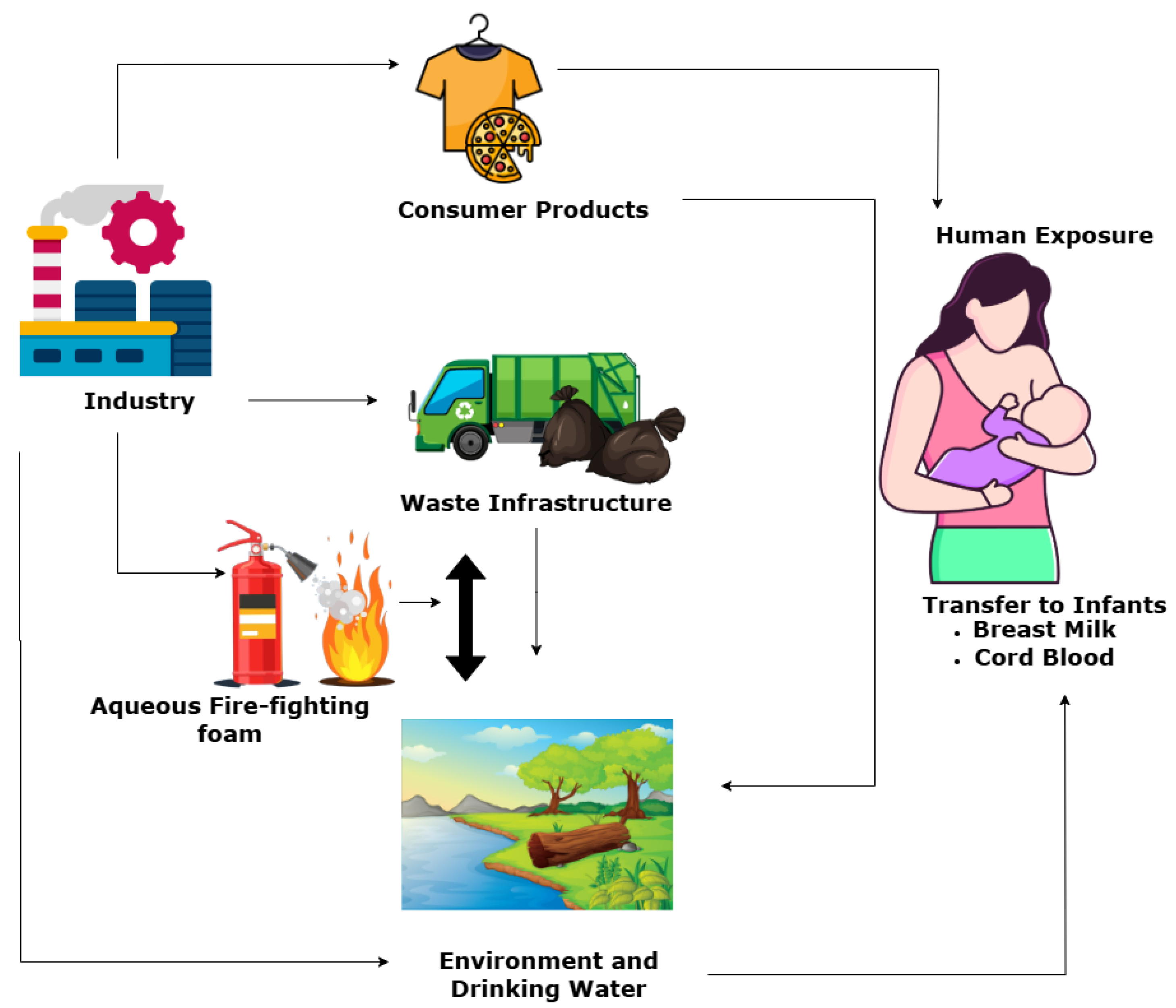
Figure 1. Primary exposure routes of PFOS. PFOS enters the environment through industrial activities, consumer products, aqueous fire-fighting foams, and waste infrastructure. Contaminated water and ecosystems serve as major pathways for human exposure, including maternal transfer via breast milk and cord blood. Arrows indicate the directional flow of PFOS from sources to exposure endpoints. Source: Generated with https://app.diagrams.net/, accessed on 23 April 2025.
Cancers 17 03507 g001
Figure 2. PFOS-mediated induction and progression of PCa. PFOS mediates the transition from normal prostate epithelium to clinical adenocarcinoma. A: The mechanisms of tumor initiation by PFOS involve oxidative stress, DNA damage, inflammation, and telomere shortening, leading to prostatic intraepithelial neoplasia (PIN). B: The formation of PIN can be driven by the activation of other signaling nodes, such as PI3K/Akt/NF-κB. C: Progression is driven by activation of oncogenes such as c-MYC and the repression of PTEN by PFOS. D: PFOS-mediated increase in the expression of IL-8, MMP-2, MMP-9, and VEGF then mediates the transformation of occult adenocarcinoma to clinical adenocarcinoma. E: Other signaling pathways, such as WNT/β-catenin, N-Cadherin and vimentin further facilitate the metastasis of PCa to distant organs Generated with BioRender (https://app.biorender.com, accessed on 28 October 2025).
Cancers 17 03507 g002
Figure 3. PFOS-mediated induction and progression of BCa. PFOS drives the sequential transformation of normal breast epithelial cells into metastatic carcinoma. A: The initiation step involves DNA damage, inflammation, and telomere shortening. B: These processes can deregulate the expression of certain genes, such as PTEN, c-MYC, PI3K/Akt, and MAPK, leading to the formation of atypical ductal hyperplasia. C: The mechanisms of progression are marked by elevated expression of VEGF, MMP-2/9, and IL-8 levels, which promote tumor growth and angiogenesis, leading to the formation of carcinoma in situ. D: PFOS can further trigger the transformation of carcinoma in situ into invasive carcinoma. This process is facilitated by WNT/β-catenin signaling and epithelial–mesenchymal transition markers, enabling dissemination to distant organs such as the lungs, liver, bones, and brain. Generated with BioRender (https://app.biorender.com, accessed on 28 October 2025).
Cancers 17 03507 g003
Figure 4. PFOS-mediated induction and progression of OC. Exposure to PFOS mediates the transformation of normal ovarian tissues into metastatic ovarian carcinoma. A: The Initiation step involves oxidative stress, DNA damage, inflammation, and genomic instability. B: These biological processes can induce dysregulation in the expression of some genes, including PTEN, KRAS and BRAS, thus leading to epithelial dysplasia (precursor lesions). C: The progression step is driven by the accumulations of several mutations, including PTEN, BRCA 1/2, TP53, KRAS, and BRAS, ultimately resulting in the formation of carcinoma in situ (otherwise known as serous tubal intraepithelial carcinoma (STIC)). D: PFOS further drives the activation of several genes, including VEGF, MMP-9/MMP-2, and IL-8. This process is necessary for tumor angiogenesis and invasion, leading to the formation of invasive ovarian carcinoma. E: The metastatic step is facilitated by WNT/β-catenin signaling, vimentin upregulation, and GSK3β inhibition, enabling epithelial–mesenchymal transition and cellular migration. Generated with BioRender (https://app.biorender.com, accessed on 28 October 2025).
Cancers 17 03507 g004
Figure 5. Emission and distribution pathways of PFOS. Point sources include industrial discharge, aqueous fire-fighting foams, suppression systems, landfills, and wastewater treatment facilities. Non-point sources encompass atmospheric deposition, consumer and household products, agricultural runoff, urban runoff, and roadway runoff. Arrows indicate the flow of PFOS from emission sources into the environment. Generated with https://app.diagrams.net/, accessed on 28 April 2025.
Cancers 17 03507 g005
Figure 6. PFOS-mediated disruption of lipid metabolism in the liver. PFOS activates peroxisome proliferator-activated receptor alpha (PPARα), leading to lipid accumulation in hepatic cells. This dysregulation progresses from hepatic steatosis (fatty liver) to inflammation (hepatitis), fibrosis, and ultimately hepatocellular carcinoma. Generated with BioRender (https://app.biorender.com, accessed on 28 April 2025).
Cancers 17 03507 g006
Figure 7. PFOS-mediated induction of oxidative stress. (A): PFOS disrupts the Keap1–Nrf2 signaling axis, promoting the nuclear translocation of Nrf2 and its binding to antioxidant response elements (AREs), which activates transcription of cytoprotective genes. (B): PFOS enhances mitochondrial production of reactive oxygen species (ROS), including superoxide anions (O2), hydrogen peroxide (H2O2), and hydroxyl radicals (OH•), leading to oxidative damage of lipids, proteins, and DNA. These events contribute to cellular dysfunction and apoptosis. Generated with BioRender (https://app.biorender.com, accessed on 4 May 2025).
Cancers 17 03507 g007
Figure 8. PFOS-mediated epigenetic modifications. (A): PFOS disrupts DNA methylation dynamics by inhibiting DNA methyltransferases (DNMTs) and promoting demethylation, leading to the transition from transcriptionally repressive heterochromatin to active euchromatin and aberrant gene expression. (B): PFOS alters histone methylation through the upregulation of histone demethylases, affecting chromatin structure and transcriptional activity. These changes contribute to the activation of oncogenes and suppression of tumor suppressor genes. Generated with BioRender (https://app.biorender.com, accessed on 4 May 2025).
Cancers 17 03507 g008
Figure 9. PFOS-mediated endocrine function disruption. PFOS inhibits iodine uptake, suppresses thyroid peroxidase activity, and disrupts the conversion of thyroxine (T4) to triiodothyronine (T3) by deiodinases. These disruptions lead to reduced levels of T4 and T3, impairing key physiological processes including neurogenesis, brain development, heart rate regulation, and glucose metabolism. Generated with BioRender (https://app.biorender.com, accessed on 15 May 2025).
Cancers 17 03507 g009
Figure 10. PFOS-mediated disruptions of B-cell and T-cell function. PFOS impairs adaptive immune responses by targeting key cellular processes. In B-cells, PFOS depletes naïve B-cell populations, reduces plasma cell differentiation, and suppresses antibody production. In T-cells, PFOS inhibits activation markers (CD3, CD4/CD8, MHC-TCR) and cytokine signaling (e.g., IL-2, IFN-γ), leading to diminished T-cell activation and proliferation. These disruptions collectively contribute to immunosuppression. Generated with BioRender (https://app.biorender.com, accessed on 15 May 2025).
Cancers 17 03507 g010
Figure 11. Structure of the androgen receptor (AR) gene. The AR gene is located on the X chromosome and comprises eight exons. The encoded protein includes three major functional domains: the N-terminal domain (NTD; residues 1–555), DNA-binding domain (DBD; residues 556–623), and ligand-binding domain (LBD; residues 666–919). These domains are essential for AR-mediated transcriptional regulation and play critical roles in PCa development and progression. Generated with BioRender (https://app.biorender.com, accessed on 10 June 2025).
Cancers 17 03507 g011
Figure 12. AR signaling pathway in PCa. The diagram illustrates the molecular cascade beginning with cholesterol conversion to testosterone via CYP17 lyase. Testosterone is further metabolized to dihydrotestosterone (DHT) by 5α-reductase, which binds to cytosolic AR, triggering dissociation from heat shock proteins (HSPs) and enabling nuclear translocation. Inside the nucleus, the AR-DHT complex binds to androgen response elements (AREs) on DNA, initiating transcription of genes that drive PCa progression. Points of therapeutic inhibition are highlighted: (a) CYP17 lyase inhibitors (e.g., abiraterone) block androgen biosynthesis. (b) Androgen deprivation therapy reduces circulating testosterone. (c) 5α-reductase inhibitors (e.g., finasteride, dutasteride) prevent DHT formation. (d) AR inhibition or degradation targets the androgen receptor directly, either by blocking ligand binding, preventing nuclear translocation, or promoting proteasomal degradation. Degraders such as PROTAC act at this level. (e) AR antagonists (e.g., enzalutamide) inhibit receptor activation or promote degradation. (f) Transcriptional inhibitors block AR-mediated gene expression. Generated with BioRender (https://app.biorender.com, accessed on 10 June 2025).
Cancers 17 03507 g012
Figure 13. ER signaling pathway in BCa. The figure illustrates both genomic and non-genomic mechanisms of ER activation. In the genomic pathway, estrogen binds to nuclear ERs, leading to receptor dimerization and translocation into the nucleus, where it regulates gene transcription. In the non-genomic pathway, estrogen interacts with membrane-associated ERs, triggering rapid signaling cascades including Ras/Raf/MEK/ERK, PI3K/Akt, and Src-mediated pathways. These cascades promote cell proliferation, survival, inflammation, protein synthesis, and metastasis through downstream effectors such as JNK, p38 MAPKs, NF-κB, and CREB. Generated with BioRender (https://app.biorender.com, accessed on 22 June 2025).
Cancers 17 03507 g013
Figure 14. Mechanism of PFOS-mediated epigenetic perturbation in BCa. The figure illustrates how PFOS exposure induces epigenetic reprogramming through alterations in DNA methylation (5mC) and histone modifications. These changes disrupt the expression of genes regulating cell adhesion molecules such as β-integrin, E-cadherin, and occludin, compromising extracellular matrix integrity. The resulting epigenetic dysregulation hijacks cell growth signaling pathways such as ERK, AP-1, Cylin D1, and CDK4, promoting uncontrolled proliferation and contributing to BCa progression. Generated with BioRender (https://app.biorender.com, accessed on 29 October 2025).
Cancers 17 03507 g014
Figure 15. Histological subtypes of OC. Generated with BioRender (https://app.biorender.com, accessed on 29 October 2025).
Cancers 17 03507 g015
Disclaimer/Publisher’s Note: The statements, opinions and data contained in all publications are solely those of the individual author(s) and contributor(s) and not of MDPI and/or the editor(s). MDPI and/or the editor(s) disclaim responsibility for any injury to people or property resulting from any ideas, methods, instructions or products referred to in the content.