Review

Optimizing Anti-PD1 Immunotherapy: An Overview of Pharmacokinetics, Biomarkers, and Therapeutic Drug Monitoring

by
Joaquim Faria Monteiro
1,2,*,
Alexandrina Fernandes
1,
Diogo Gavina Tato
1,
Elias Moreira
3,
Ricardo Ribeiro
4,5,
Henrique Reguengo
4,6,
Jorge Gonçalves
1,2 and
Paula Fresco
1,2
1
Laboratório de Farmacologia, Departamento de Ciências do Medicamento, Faculdade de Farmácia, Universidade do Porto, 4050-313 Porto, Portugal
2
UCIBIO, Unidade de Ciências Biomoleculares Aplicadas, Laboratório Associado i4HB, Instituto para a Saúde e Bioeconomia, Faculdade de Farmácia, Universidade do Porto, 4050-313 Porto, Portugal
3
Serviços Farmacêuticos, Unidade Local de Saúde São João, 4200-319 Porto, Portugal
4
Clinical Chemistry, Clinic of Genetics and Pathology, Centro Hospitalar de Santo António, 4099-001 Porto, Portugal
5
Tumour and Microenvironment Interactions Group, i3S Instituto de Investigação e Inovação em Saúde, Universidade do Porto, 4200-135 Porto, Portugal
6
Unit for Multidisciplinary Research in Biomedicine (UMIB), Institute of Biomedical Sciences Abel Salazar (ICBAS), Universidade do Porto, 4050-313 Porto, Portugal
*
Author to whom correspondence should be addressed.
Cancers 2025, 17(19), 3262; https://doi.org/10.3390/cancers17193262
Submission received: 16 September 2025 / Revised: 2 October 2025 / Accepted: 6 October 2025 / Published: 8 October 2025

Abstract

Simple Summary

The purpose of this review is to explore the therapeutic optimization of anti-PD-1 drugs by focusing on pharmacokinetic (PK) principles. The article discusses how interpatient variability and tumor burden affect drug exposure and highlights the PK–pharmacodynamic relationship in terms of exposure-efficacy and exposure-safety. It emphasizes the role of clearance variability, particularly baseline clearance and its dynamic reduction during treatment, as relevant biomarkers for stratifying patients and guiding individualized dosing strategies. The review also examines current dosing approaches, including fixed versus weight-based regimens, and emerging strategies such as therapeutic drug monitoring and PK-guided personalization. By integrating PK data, clearance biomarkers, and clinical outcomes, the review supports a shift toward more adaptive and personalized use of anti-PD1 therapies aimed at maximizing therapeutic benefit while minimizing adverse effects.

Abstract

Anti-PD-1 therapies have transformed cancer treatment by restoring antitumor T cell activity. Despite their broad clinical use, variability in treatment response and immune-related adverse events underscore the need for therapeutic optimization. This article provides an integrative overview of the pharmacokinetics (PKs) of anti-PD-1 antibodies—such as nivolumab, pembrolizumab, and cemiplimab—and examines pharmacokinetic–pharmacodynamic (PK-PD) relationships, highlighting the impact of clearance variability on drug exposure, efficacy, and safety. Baseline clearance and its reduction during therapy, together with interindividual variability, emerge as important dynamic biomarkers with potential applicability across different cancer types for guiding individualized dosing strategies. The review also discusses established biomarkers for anti-PD-1 therapies, including tumor PD-L1 expression and immune cell signatures, and their relevance for patient stratification. The evidence supports a shift from traditional weight-based dosing toward adaptive dosing and therapeutic drug monitoring (TDM), especially in long-term responders and cost-containment contexts. Notably, the inclusion of clearance-based biomarkers—such as baseline clearance and its reduction—into therapeutic models represents a key step toward individualized, dynamic immunotherapy. In conclusion, optimizing anti-PD-1 therapy through PK-PD insights and biomarker integration holds promise for improving outcomes and reducing toxicity. Future research should focus on validating PK-based approaches and developing robust algorithms (machine learning models incorporating clearance, tumor burden, and other validated biomarkers) for tailored cancer treatment.

1. Introduction

While early-stage malignancies often respond favorably to conventional therapies, advanced cancers frequently exhibit resistance, requiring alternative treatment strategies. Cancer immunotherapy has transformed oncology by leveraging the immune system to target and eliminate tumor cells. The discovery of immune checkpoint regulation as a pivotal mechanism of tumor-immune evasion has revolutionized cancer treatment, leading to the development of novel immunotherapeutic approaches.
Immune checkpoints are regulatory proteins found on the surface of cells that help to maintain immune system balance by controlling the strength and duration of immune responses in lymphoid organs and peripheral tissues. This prevents autoimmunity and reduces tissue damage during inflammation. Well-characterized immune checkpoints include cytotoxic T-lymphocyte-associated antigen 4 (CTLA-4) and programmed cell death protein 1 (PD-1) and its ligands PD-L1 or PD-L2.
Concerning the PD-1/PD-L1 axis, PD-1 is a coreceptor that plays a role in the inhibition of T cell function [1,2]. When activated by its ligands, PD-1 switches off the cascades that cause T cell activation, inhibiting T cell function and contributing to T cell exhaustion [3,4,5], which is a state of cellular dysfunction where T cells become progressively less effective at performing their immune functions. PD-1 expression was primarily described on activated T cells [6], and its expression was described afterwards on B cells [7], dendritic cells (DCs) [8], macrophages [9], and natural killer (NK) cells [10].
PD-1 expression is downregulated under basal/resting conditions across multiple cell types, and its expression levels drastically increase within several hours after stimulation [11]. Upregulation of PD-1 and PD-L1 expression occurs during inflammation, acting to help to regulate and restrain the immune system in inflamed tissues [12]. In fact, PD-1 also plays a crucial role in maintaining the balance between effective immunity and self-tolerance, controlling self-reactive clones of immune cells and limiting autoimmunity [13,14].
Immune checkpoint inhibitors (ICIs) are monoclonal antibodies (mAbs) that block inhibitory regulatory pathways, thereby restoring T cell activation. In the context of inflammation, ICIs disable these systems’ capacity to restrain the immune system in inflamed tissues and to regulate peripheral tolerance. In the context of cancer, ICIs restore immune competence by reversing T cell exhaustion and restoring tumor antigen-specific cytotoxic T lymphocyte activity, promoting the elimination of malignant cells [15]. Therefore, these agents have become integral to first- and second-line treatment regimens for refractory tumors, although durable responses and survival benefits are limited to a subset of patients, reflecting ongoing challenges in identifying reliable predictors of response [16].
Key anti-PD-1 mAbs, including nivolumab, pembrolizumab, and cemiplimab have demonstrated clinical benefit across multiple malignancies, notably advanced melanoma, non-small cell lung cancer (NSCLC), and clear cell renal carcinoma [17]. Despite their transformative potential, anti-PD-1 therapies exhibit significant interpatient variability in both efficacy and toxicity. A considerable proportion of patients fail to derive meaningful clinical benefit, and others may develop acquired resistance. Furthermore, immune checkpoint blockade is associated with a variety of immune-related adverse events (irAEs), affecting over 25% of patients to which anti-PD-1 therapies contribute to around 10–15% of cases, with severe reactions in 6–10% and fatal outcomes reported in 0.3–1.3% of patients [18].
Given the high interindividual variability in response and the risk of serious toxicity, optimizing anti-PD-1 therapy requires careful pharmacokinetic (PK) and pharmacodynamic (PD) analysis. Ensuring adequate drug exposure is essential to achieving therapeutic efficacy while minimizing adverse effects. Additionally, understanding resistance mechanisms and identifying reliable biomarkers—such as tumor mutational burden (TMB), PD-L1 expression, the microbiome, hypoxia, interferon-γ signaling, and extracellular matrix composition—are critical to guide personalized treatment approaches [19].
This review aims to explore how the understanding of PK and PD and exploring therapeutic drug monitoring (TDM) can contribute to developing personalized dosing strategies and improving treatment outcomes. To conclude, we will consolidate the current knowledge and evidence base relating to the PKs, biomarkers, and monitoring of anti-PD-1 mAbs, with the ultimate goal of optimizing their clinical use. The review is organized into five sections: (i) the PK of anti-PD-1; (ii) PK-PD relations; (iii) available biomarkers for anti-PD-1 therapies; (iv) exploring PK and TDM to optimize dosing strategies; and (v) perspectives on translating PK insights into clinical practice and future research priorities.

2. Pharmacokinetics of Anti-PD-1

The commercially available anti-PD-1 drugs—pembrolizumab (Keytruda®, Merck Sharp & Dohme B.V., Haarlem, The Netherlands), nivolumab (Opdivo®, Bristol-Myers Squibb Pharma EEIG, Dublin, Ireland), and cemiplimab (Libtayo®, Regeneron Ireland Designated Activity Company, Dublin, Ireland)—are mAbs administered via intravenous infusion, resulting in complete bioavailability. However, alternative routes (e.g., subcutaneous administration) are under investigation to improve patient convenience. The PK of anti-PD-1 mAbs is conditioned by a complex interplay of factors related to the patient but also by tumor-specific factors. These factors include, but are not limited to, lymphatic and interstitial fluid dynamics, transendothelial movement of mAbs, genetic polymorphisms, immunogenicity, immune status, levels of endogenous IgG and albumin, co-morbidities, concomitant medications, and demographic factors such as age, sex, body size, and ethnicity [20], which give particularities to the PK of anti-PD-1 mAbs.
Anti-PD-1 therapies share similar PK profiles (Table 1), characterized by long half-lives, minimal hepatic metabolism, and time-dependent changes in PK, particularly in clearance (CL) mechanisms [21,22]. The temporal variability of CL of these agents warrants thorough consideration in this review. The underlying mechanisms and the factors already identified that contribute to this phenomenon will be comprehensively reviewed.

2.1. Disposition Mechanisms of Anti-PD-1 mAbs

The disposition of anti-PD-1 mAbs is governed by a combination of selective and non-selective mechanisms. Selective elimination occurs through target-mediated drug disposition (TMDD), driven by binding to PD-1 receptors on T cells. Non-selective pathways include interactions with gamma and neonatal Fc receptors, tissue proteolysis within the reticuloendothelial system, and the presence of circulating autoantibodies. In addition, the formation of anti-drug antibodies (ADAs) may alter both clearance and therapeutic efficacy. Ultimately, the PK and PD of anti-PD-1 therapies result from the complex interplay among these mechanisms, which together determine drug exposure, response, and variability across patients.

2.1.1. Target-Mediated Drug Disposition

Anti-PD-1 mAbs are proteolytically degraded by systems that are independent of hepatic enzyme metabolism. Anti-PD-1 mAbs undergo proteolytic degradation intracellularly after a receptor-mediated internalization process occurring in cells express membrane “receptors” with affinity for these mAbs, contributing their CL. PD-1 is a protein found on the surface of cells that may undergo internalization, subsequent ubiquitination, and degradation by the proteasome in activated T cells [26]. PD-1 is the most obvious “receptor” for the anti-PD-1 mAbs and binding of anti-PD-1 mAbs to PD-1 can induce internalization of anti-PD-1/PD-1 complexes, and these complexes can be found in late endosomes [27] where they will be exposed to the proteolytic activity of the lysosomal enzymes. Therefore, the removal of surface PD-1 is both a critical step in the PD as it is the mechanism to remove PD-1 from the membrane, but also a critical step in the CL of the mAbs as it is a mechanism to promote the proteolytic degradation of the anti-PD-1 mAbs. The CL of anti-PD-1 mAbs may be closely linked to their PD effect, as CL can be seen as the result of the ‘heroic sacrifice’ after their PD action. Consequently, the CL of anti-PD-1 mAbs is enhanced when greater PD-1 receptor availability promotes increased binding, internalization, and degradation of the antibody–receptor complex. While internalizable PD-1 receptors are present, a linear CL of mAbs will predominate and elimination will be primarily driven by endocytosis and proteolytic degradation of the complex PD-1/anti-PD-1 mAb. Successful antitumor therapy should reduce the CL of the anti-PD-1 mAbs, since less tumor mass should mean fewer PD-1-positive cells, and, thus, fewer PD-1 receptors to which anti-PD-1 mAbs can bind, and fewer conditions allowing their metabolization.
Not all PD-1 receptors may be internalized. A fraction persists in the membrane during exposure to anti-PD-1 mAbs [27]. Therefore, the presence of PD-1 alone does not guarantee that mAbs will be effective or that the expected reduction in CL will be achieved over time. Several factors can contribute to this resistance to internalization, namely the density of PD-1 receptors or saturation of the endocytic machinery [27], level of PD-1 fucosylation [28] or its location in lipid rafts [29,30]. The impact of these factors on activity and CL may differ according to the mAbs used and may be altered by changes in tumor metabolism during treatment. Therefore, variations in the CL of anti-PD-1 mAbs over time must be interpreted with caution, as they may be influenced by the loss of the receptors’ ability to be internalized, resulting in tumor resistance to treatment.

2.1.2. Non-Selective Mechanisms

Neonatal Fc Receptor
The available anti-PD-1 mAbs possess IgG4 subclass-specific Fc regions that specifically bind to Fc receptors (FcRs), presenting affinity to the neonatal Fc receptor (FcRn) which may also have impact on anti-PD-1 mAbs PK, although with consequences distinct from the binding to the PD-1 receptor.
FcRn is predominantly localized within the intracellular vesicular network, with less than 10% detected at the plasma membrane [31]. This observation should be interpreted with caution, as the relatively low proportion at the cell surface may reflect the receptor’s highly dynamic recycling behavior, analogous to that of the transferrin receptor [31,32]. FcRn protects its natural ligands (such as IgG antibodies and human serum albumin; ALB) from acidic proteolysis in endolysosomes [33,34]. The affinity of ligands to FcRn increases as pH decreases [35] and this pH dependency of FcRn to protect its ligands from proteolysis in the low pH found in the endolysosomes creates optimal conditions to maintain the FcRn/ligand complex protected from lysosomal degradation [36,37,38]. This, in turn, favors a recycling pathway of the anti-PD-1 mAbs back to the plasma membrane and its subsequent release from the cell. During this process, the ligands are preserved, which results in a prolonged serum half-life for ALB and IgG of about 20 days [39]. This capacity of FcRn to preserve ligands from proteolysis makes it a mechanism to promote transcytosis of IgG and ALB across epithelial barriers, both from basolateral to apical surfaces and vice versa in polarized cells [40].
FcRn is present throughout human tissues, with expression in endothelial and immune cells, being particularly expressed in a specialized manner at barrier sites, including the blood–brain barrier, and the placenta and mucosal surfaces [41]. This widespread distribution enables FcRn to serve as a universal recycling system through transcytosis of its natural ligands, IgG antibodies, and ALB, playing a key role in their homeostasis [36,42]. Biological drugs with Fc region are also FcRn substrates [43], including the anti-PD-1 mAbs [44,45]. Interaction of FcRn with anti-PD-1 mAbs increases the volume of distribution, reducing plasma concentration and prolonging their half-life.
The pH dependence of FcRn–ligand interactions have important consequences for the net outcome of FcRn-mediated transcytosis. At physiological pH (7.4), FcRn exhibits negligible binding to its ligands, whereas strong binding occurs at acidic pH values of 6.0–6.5 [32]. Consequently, under normal physiological conditions, the pH gradient between the compartments in which transcytosis may be occurring is negligible, without alterations in FcRn affinity caused by the pH. Consequently, the net transfer of ligands will be determined by their availability in the different compartments. In certain pathological conditions, such as cancer, the pH of the tumor microenvironment can be as low as 5.5 [46,47]. Such pH gradient creates an additional variable because it will change the affinity of FcRn and, by consequence, its capacity to release its ligands (including the anti-PD-1 mAbs) in the tumor microenvironment in comparison to normal tissues. Consequently, there will be a reduction in the conditions for transfer and retention of the drug in the acidic tumor microenvironment, which may result in a decrease in treatment efficacy.
Gamma Fc Receptor
Fcγ receptors (FcγRs) constitute a heterogeneous family of receptors that differ in their affinity for IgG subclasses, cellular distribution, and immunological functions. They are broadly categorized into activating receptors (FcγRI, FcγRIIa, FcγRIIIa) and the inhibitory receptor FcγRIIb [48]. FcγRI binds IgG with high affinity and is predominantly expressed on monocytes, macrophages, and DCs, mediating antibody-dependent cellular cytotoxicity (ADCC) and phagocytosis [48]. FcγRIIa and FcγRIIIa, with intermediate to low affinity, are expressed on macrophages, neutrophils, and NK cells, contributing to cytokine release and cytotoxicity [48]. In contrast, FcγRIIb is the only inhibitory FcγR, expressed on B cells, macrophages, and DCs, acting as a negative regulator of immune activation [49]. In addition to its inhibitory role, FcγRIIb participates in the clearance of antibodies by mediating endocytosis and lysosomal degradation in FcγRIIb-expressing cells, including liver sinusoidal cells and macrophages [49,50].
Although most approved anti-PD-1 antibodies are IgG4 isotypes carrying the S228P mutation, they are designed to minimize effector functions while retaining some binding to FcγRI and FcγRIIb [48]. Importantly, FcγR interactions have also been associated with hyperprogression during PD-1 blockade. Several clinical studies documented higher rates of hyperprogression in patients treated with anti-PD-1 mAbs compared to chemotherapy [51,52,53]; Lo Russo et al. specifically linked this phenomenon to FcγR+ macrophage activity in NSCLC [54] and it may also involve the proliferation of highly suppressive tumor-infiltrating PD-1+ effector Treg cells, which inhibit antitumor immunity and contribute to hyperprogressive disease [55,56].
Beyond these functional effects, FcγR binding may also contribute to the elimination of anti-PD-1 mAbs. Non-specific clearance of anti-PD-1 mAbs occurs through proteolysis in the liver and the reticuloendothelial system, particularly via FcγR-expressing cells such as Kupffer cells, monocytes, and macrophages [57]. Following receptor-mediated endocytosis, antibodies are degraded in lysosomes. Although this pathway is generally nonsaturable and therefore unlikely to markedly alter the pharmacokinetics of anti-PD-1 mAbs when non-specific CL predominates, FcγR-mediated elimination can interact with pharmacodynamic mechanisms and may indirectly accelerate CL, as shown for rituximab [58].
Together, these findings suggest that FcγR binding undermines both the pharmacodynamics and, to some extent, the pharmacokinetics of anti-PD-1 antibodies.
Tissue Proteolysis
Anti-PD-1-mAbs are also substrates of matrix metalloproteinases (MMPs) that may potentially affect their PK and PD. MMP-13 and MMP-2 are the metalloproteinases reported to be involved in anti-PD-1 mAbs proteolysis [59]. Cleavage of anti-PD-1 mAbs formed the fragments F(ab’)2 and Fc. Concerning the F(ab’)2 fragment, although it retains the antigen binding site, it has been reported as less active in inducing T cell proliferation than the respective anti-PD-1 mAbs [60], and inactive in removing PD-1 from the immune synapse and in preventing tumor growth in vivo [61]. Fc fragments at CH2 formed after protease action may retain the capacity to bind to FcRs and to compete with its ligands and, therefore, it is likely that Fc fragments may bind to FcRn and competitively inhibit the binding of other FcRn ligands (including anti-PD-1 mAbs), favoring the lysosomal degradation pathway and decreasing the half-life of anti-PD-1 mAbs [62,63]. Therefore, due to the activity of MMP, it is plausible to observe alterations in the PD-PK of anti-PD-1 mAbs, including reduced therapeutic efficacy due to decreased affinity for PD-1 and a shorter half-life resulting from diminished binding to FcRn.
MMP-13 and MMP-2 are not ubiquitously expressed: MMP-13 is a collagenase that plays a fundamental role in extracellular matrix (ECM) remodeling, particularly in bone biology. MMP-2 is also involved in ECM remodeling and is implicated in tissue repair, wound healing, angiogenesis, embryogenesis, bone remodeling, and tooth development [64]. Both have been found to be overexpressed in various types of cancer: MMP-13 in breast cancer, head and neck carcinomas, chondrosarcomas, and skin carcinomas [65]; MMP-2 in breast cancer [66], ovarian [67], oral [68], prostate [69], bladder [70], colorectal [71] cancers, retinoblastoma [72], and laryngeal squamous cell carcinoma [73]. Expression of both MMP-13 and MMP-2 has been associated with aggressive or invasive tumors and with poor clinical outcomes [74,75], as well as with various cell types present in the tumor microenvironment, including tumor-associated macrophages [76,77,78] and cancer-associated fibroblasts [64,79,80]. As these cell populations increase in the tumor microenvironment, the conditions for the proteolysis of anti-PD-1 mAbs within the tumor microenvironment will also increase, thus favoring the development of resistance to these ICIs.
Autoantibodies
Pathogenic autoantibodies are high-affinity, somatically mutated IgG autoantibodies that are stimulated by inflammation in the target organ [81,82]. These IgG autoantibodies are also FcRn ligands, which means interaction between FcRn and the autoantibodies are the key to removing them from inflamed tissue [83]. Sharing the same transcytosis pathway, FcRn may represent a potential site of interaction between anti-PD-1 mAbs and IgG autoantibodies, which can have clinical implications. In the tumor microenvironment, the pH in areas of inflammation is also in the acidic range [84], so the affinity of FcRn for the ligands remains high, which means there are less conditions to release the ligands carried to the areas of inflammation and less conditions for the ligands formed in the areas of inflammation to bind to FcRn. When anti-PD-1 mAbs are used, these exogenous ligands contribute to saturating FcRn transport in a dose-dependent manner and competing with IgG autoantibodies for transport out of the inflamed zone. This increases the risk of aggravating inflammation and irAEs, as previously reported [85,86,87,88,89].

2.1.3. Anti-Drug Antibodies

The PD and PK of therapeutic proteins such as mAbs may be influenced by antibodies that are formed in response to the drug (anti-drug antibodies-ADAs) which have the potential to alter the efficacy of the treatment. Some ADAs are neutralizing antibodies, which means they block or inactivate the drug’s therapeutic effect; others may simply increase the drug’s clearance from the body (clearing antibodies), reducing drug exposure [90]. In the case where anti-PD-1 mAbs are approved, the risk to produce ADAs seems to be higher with nivolumab (occurrence in about 11% of patients) [91], although without clinical relevance [92]. Nevertheless, the production of ADAs is a factor that should be monitored since its incidence increases when anti-PD-1 mAbs are used in combination therapies [91].

2.2. Pharmacokinetic Models

A putative PK model integrating all these mechanisms and their interrelationships is illustrated in Figure 1. The CL of anti-PD-1 mAbs can be described through a combination of linear and non-linear disposition mechanisms. Linear CL predominates when the PD-1 receptor is saturated, and elimination is primarily driven by proteolytic catabolism in plasma and peripheral tissues. In contrast, TMDD—a mix of linear and non-linear CL—occurs when the receptor is not saturated. In this scenario, elimination is governed by both proteolytic catabolism and receptor-mediated endocytosis from the plasma or interstitial into target cells [93,94]. In addition, the FcRs play a central role in regulating the half-life of anti-PD-1 antibodies through recycling mechanisms that prevent lysosomal degradation and/or elimination mechanisms. Interindividual variability in FcRs function—affected by factors such as systemic inflammation, hypoalbuminemia, or competing endogenous IgG—can influence drug CL and contribute to differences in drug exposure. Furthermore, the development of anti-drug antibodies (ADAs) may enhance antibody CL by forming immune complexes that are rapidly eliminated.
This variability in CL has clinical implications, particularly over time, as it contributes to the progressive increase in drug exposure observed with repeated dosing. This effect is more pronounced in responders compared to non-responders [24,94,95,96], and it will be discussed later.

2.3. Factors Influencing Pharmacokinetics of Anti-PD-1

2.3.1. Albumin and Cachexia

Albumin (ALB) has emerged as a significant covariate influencing the PK of anti-PD-1 mAbs, particularly pembrolizumab and nivolumab. Across multiple studies, lower baseline ALB levels have been consistently associated with increased CL of both agents, underscoring the clinical importance of the patient’s nutritional status for therapeutic antibody disposition. In patients treated with pembrolizumab, those with higher CL exhibited significantly worse overall survival (OS), especially in patients with NSCLC and malignant pleural mesothelioma [97]. This relationship can be partly explained by the finding that patients with lower ALB levels before treatment demonstrate faster elimination of pembrolizumab, while higher ALB levels are associated with reduced CL [97]. Similarly, in patients receiving nivolumab, a strong and clinically meaningful association between ALB and drug CL has been observed. ALB levels exerted a greater than 20% effect on CL, with patients presenting hypoalbuminemia experiencing significantly higher nivolumab elimination [98].
A possible mechanism could be the interaction with FcRn; although ALB and IgG molecules are substrates of FcRn, they bind to distinct sites on the receptor [99,100]. Nevertheless, they may still compete for the overall availability of FcRn [101,102]. Consequently, hypoalbuminemia could increase FcRn availability for IgG molecules, indirectly affecting their homeostasis. An identical interaction may also occur with anti-PD-1 mAbs with potential clinical impact.
Performance status (PS), another indicator of disease severity, has been shown to correlate with ALB, where patients with poorer PS present lower ALB levels, thereby potentially influencing CL indirectly [98]. Several mechanisms are likely involved, being hard to discriminate but, overall, these findings emphasize the need to consider ALB levels and cachexia-related markers when optimizing ICIs dosing.
Cachexia negatively influences patients’ outcome during anti-PD-1 therapy [103] which is associated with reduced systemic concentrations of therapeutic antibodies [104], and elevated CL of anti-PD-1 mAbs parallel with a whole-body protein turnover observed in this population [105], which may contribute to reduced efficacy and poor clinical outcomes. Hypoalbuminemia has been used as a biomarker for cachexia but low ALB cannot explain the elevated CL of anti-PD-1 mAbs since it would be expected that low ALB would provide better conditions for anti-PD-1 mAbs to bind to FcRn, and thus, be spared from target-mediated CL and other forms of non-selective CL. Cachexia has shown to reduce FcRn expression [106]. Thus, from a reduction in the participation of FcRn in anti-PD-1 mAbs homeostasis, an acceleration of the mAbs degradation is expected to occur, with a marked increase in CL.
Low ALB levels may not only signal poorer prognosis but also suggest PK adjustments to maintain therapeutic drug exposure. Moreover, reductions in anti-PD-1 CL during therapy may potentially serve as a surrogate marker for clinical improvement or attenuation of cachexia.

2.3.2. Disease Burden

PK studies of anti-PD-1 mAbs have consistently demonstrated time-varying CL, which is closely associated with disease burden [24,107]. These studies reveal a progressive decline in CL over time, particularly in patients who exhibit a reduction in tumor mass. This indicates that tumor burden is a dynamic and influential covariate in the disposition of anti-PD-1 therapies [24], altering factors that selectively or non-selectively influence the PK of anti-PD-1 mAbs. For example, patients with higher tumor burden and more advanced disease, who are more likely to be cachectic, may exhibit a higher monoclonal antibody CL. Conversely, regression of tumor burden through effective treatment may alleviate cachexia, reduce protein catabolism, and lead to reduced antibody CL [93].
These findings highlight the importance of considering tumor dynamics when interpreting and trying to personalize anti-PD-1 treatment. This will enable us to better interpret the real-world responses to these drugs, develop more accurate biomarkers with a greater predictive capacity of efficacy, failure, and toxicity, and develop treatment algorithms to improve the effectiveness of anti-PD-1 mAbs.

3. Pharmacokinetic–Pharmacodynamic Relations

3.1. Exposure–Efficacy Relations

Dynamic changes in the PK of anti-PD-1 mAbs, particularly time-dependent reductions in CL, have been increasingly recognized as important correlates of clinical efficacy. Patients with clinical response tend to experience a marked decrease in CL over time, indicating increased systemic drug exposure during treatment. This phenomenon is particularly evident in patients achieving complete response, who often show the greatest reductions in CL—around 45% in the case of nivolumab—compared to lower reductions in partial responders (35%), those with stable disease (25%), and patients with progressive disease (20%) [21,24]. For pembrolizumab, longitudinal PK data have shown that patients stratified by quartiles of decreasing CL exhibited significantly improved response rates and overall survival, even if they began treatment in worse clinical conditions such as cachexia [24]. This supports the notion that exposure to anti-PD-1 increases, driven by reduced CL, can be both predictive and prognostic indicators of benefit. Importantly, these PK improvements appear to coincide with improved PS, which itself is a marker of overall patient condition and treatment response. Together, these observations reinforce that changes in drug exposure over time—rather than static early PK values—are more informative of the efficacy of anti-PD-1 agents.
Despite this, earlier pharmacometric studies for pembrolizumab and nivolumab suggested a flat exposure–response (E–R) relationship, especially when analyzing fixed-dose regimens. PD-1 receptor occupancy modeling demonstrated full saturation at clinically relevant doses, with no observed increase in efficacy beyond a certain exposure threshold. This led to the assumption that standard dosing regimens already sit at the plateau of the E–R curve, reducing the utility of TDM or individualized dose titration based on early exposure metrics [24,98,108]. However, more recent data challenge the flat E–R paradigm. Studies such as the one by Desnoyer et al. (2020) presented that CL—both at baseline and in its reduction over time—was the strongest predictor of response and survival in patients treated with anti-PD-1 drugs [21]. This suggests that exposure does matter, especially when considering the dynamic evolution of drug disposition during therapy. Moreover, while initial exposure levels (such as first-dose area under curve (AUC) or serum concentration) may not predict long-term outcomes, the evolving PK profile, particularly decreasing CL, correlates with clinical improvement and may justify exploring alternative dosing strategies tailored to individual patient response. For instance, patients demonstrating substantial reductions in CL might sustain efficacy with extended dosing intervals, which is a strategy that could improve patient convenience and reduce healthcare costs [24,109]. Adding further complexity, the apparent lack of E–R correlation at early timepoints may stem from the immunomodulatory nature of anti-PD-1 drugs. These agents restore T cell function rather than directly attacking tumor cells, and their activity can persist even after serum levels drop below quantifiable levels, due to sustained PD-1 receptor occupancy and immune memory effects [21,110,111,112,113].
These findings advocate for integrating longitudinal PK into clinical assessment, as CL dynamics may provide valuable insights into patient response and offer a pathway toward personalized dosing strategies in immunotherapy.

3.2. Exposure–Safety Relations

While reduced CL of anti-PD-1 mAbs is widely regarded as a favorable PK indicator associated with improved clinical efficacy, this elevated exposure may increase the risk of toxicity, particularly irAEs.
Patients treated with pembrolizumab—for example, those with marked reductions in CL, who are often complete responders—exhibit sustained high plasma drug concentrations, which could predispose them to heightened toxicity [114]. As tumor burden declines and fewer PD-1 receptors are available, TMDD of these drugs decreases. This leads to an accumulation of unbound antibody in circulation, further amplifying systemic exposure, particularly in patients with robust antitumor responses. Such elevated exposures may not always be entirely innocuous. Case reports have linked pembrolizumab to serious and sometimes fatal toxicities, including pericardial effusion [115], myasthenia gravis [116], and nephrotic syndrome [96]. While nephrotic syndrome and hypoalbuminemia generally increase mAbs CL, the resolution of this condition during effective pembrolizumab treatment indicates a complex interplay between disease burden, protein metabolism, and drug disposition. Still, the potential for pembrolizumab to contribute to or worsen renal injury remains a clinically relevant concern, especially given the known association of ICIs with interstitial nephritis. To mitigate toxicity, particularly in patients with marked CL reduction, PK-informed dose adaptation strategies are emerging. For instance, in patients showing >40–50% decrease in CL, extending the dosing interval may preserve efficacy, while lowering cumulative exposure may potentially reduce the risk of irAEs [117,118]. These strategies can enable safer and more personalized treatment regimens. Nonetheless, early dose–toxicity analyses often reported no clear exposure–toxicity relationship for anti-PD-1 agents. For nivolumab, findings have been similarly mixed. Although early exposure measures such as the average concentration after the first dose were not significantly associated with adverse events or treatment discontinuation, baseline CL was strongly predictive of both efficacy and survival [24]. Importantly, while higher exposure due to lower CL has not been consistently linked with increased toxicity risk of anti-PD-1 drugs, rare but severe late-onset irAEs such as thrombocytopenia and autoimmune endocrinopathies have been documented [119]. These may occur independently of serum drug levels, underscoring the complex, delayed, and immune-mediated nature of toxicities associated with PD-1 blockade. In early-phase trials, including those in melanoma, PD-1 occupancy in peripheral lymphocytes was saturated at relatively low doses (≥0.3 mg/kg), with little evidence of a dose-dependent increase in irAE risk [120]. Some exploratory studies even noted negligible differences in irAE incidence across various dosing regimens, including fixed-dose schedules, suggesting that PD-1 blockade achieves a therapeutic ceiling beyond which toxicity does not proportionally escalate [121,122]. However, more profound analyses complicate this narrative. In some patients, particularly those treated with 480 mg Q4W, a modest increase in grade 2 irAEs was noted compared to 3 mg/kg Q2W, when peak concentration was considered as the exposure variable [121]. This suggests that while trough levels may remain within therapeutic range, transient high exposures could still contribute to irAE onset. Furthermore, patients with high baseline levels of pro-inflammatory cytokines such as IFN-γ, IL-2, and TNF-α were more likely to develop irAEs, implying a host-related immunologic predisposition to toxicity [119]. Likewise, increased effector T cell counts prior to the onset of irAEs may indicate a pre-existing state of immune activation that becomes dysregulated upon checkpoint blockade.
In conclusion, while reduced CL and elevated exposure to anti-PD-1 therapies are desirable from an efficacy standpoint, they also carry an increased risk of toxicity in some patients. The absence of a consistent dose–toxicity relationship across the general population may reflect the immunologically driven nature of irAEs, which depend not only on drug exposure but also on host immune status, T cell repertoire, and cytokine milieu. These nuances highlight the need for individualized monitoring strategies that consider both PK parameters and immune profiling to optimize safety while preserving efficacy in anti-PD-1 treatment.

3.3. Clearance–Efficacy and/or Safety Relationships

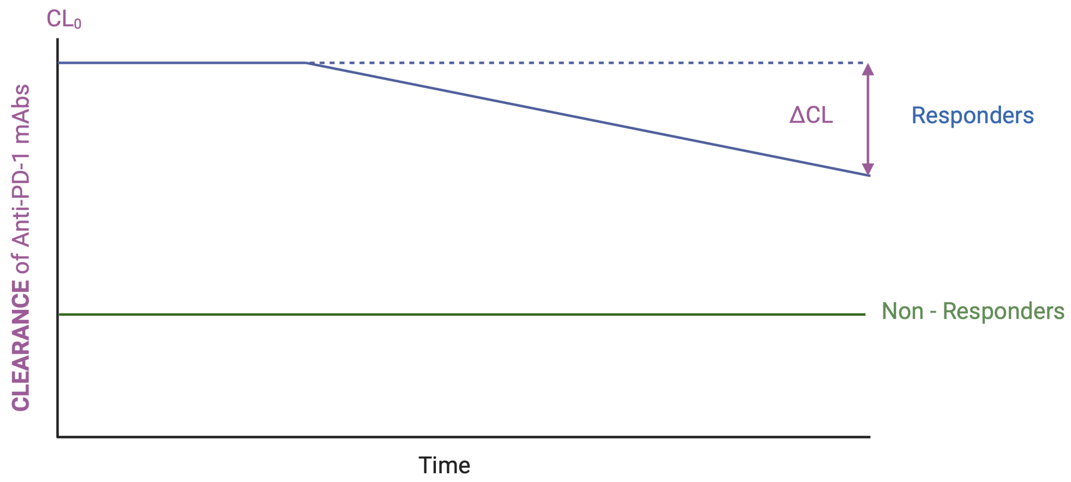
In the PK evaluation of anti-PD-1 mAbs, two CL-based indicators have gained increasing recognition for their prognostic and pharmacodynamic relevance: baseline CL (CL0) and change in CL over time (ΔCL) [123]—Figure 2. CL0 refers to the initial estimate of drug CL typically calculated from plasma concentrations measured after the first or two doses. It reflects the patient’s initial capacity to eliminate the drug and can be influenced by both physiological and pathological factors, including TMDD, systemic inflammation, and cachexia. ΔCL describes the temporal variation in CL during treatment, most often expressed as the percent reduction from baseline. A decreasing ΔCL over time generally indicates an improving physiological or disease state, potentially corresponding to reduced antigen load, decreased inflammation, or resolution of cachexia. Together, CL0 and ΔCL offer valuable insights into the biological status of the patient and can act as dynamic biomarkers for treatment efficacy and outcome.
CL0 has emerged as a particularly strong prognostic marker. Studies of pembrolizumab, nivolumab, and other ICIs confirmed that CL0 is a dose-independent predictor of survival and response [44,124]. Turner et al. demonstrated that patients in the lowest quartile of pembrolizumab CL0 had significantly improved survival compared to those in the highest quartile, regardless of receiving 2 mg/kg or 10 mg/kg, illustrating that higher exposure could not compensate for high baseline clearance [124]. These findings are reinforced by flat E–R relationships observed within clinically relevant dosing ranges. For instance, objective response rate did not increase with pembrolizumab doses from 2 to 10 mg/kg, suggesting the lowest evaluated dose lies near the efficacy plateau [125]. Similarly, no meaningful correlation between drug concentration (Cmin) and efficacy was found for nivolumab over a broad dose range [92,126]. These observations highlight a frequent misinterpretation in pharmacometrics: when only a single dose level is studied, apparent E–R relationships may reflect CL–response (CL–R) correlations because exposure (AUC) and CL are inversely related at a constant dose. Thus, only by evaluating multiple dose levels with balanced patient characteristics can true E–R and CL–R relationships be distinguished [127,128]. Turner et al. emphasized this by showing steep E–R trends within dose strata (2 mg/kg or 10 mg/kg) but there was no relationship between the two groups, despite a 5-fold exposure difference [124]. This underscores the clinical relevance of distinguishing between E–R and CL–R: a true E–R relationship implies that dose escalation might improve outcomes, whereas a CL–R relationship suggests that increasing exposure will not alter response, and efforts should instead focus on understanding the biological basis of elevated clearance.
The mechanisms underlying high CL0 are multifactorial. High target expression and antigen load may drive TMDD, while systemic inflammation and cachexia—commonly present in advanced cancers—contribute to elevated catabolic clearance [44,124], resulting in responders exhibiting higher CL0 values compared to non-responders (see Figure 2). Conversely, a significant decline in ΔCL typically reflects treatment response and improvement in the disease status. Liu et al. quantified this association, showing that patients with CR to nivolumab exhibited a 42.4% reduction in CL, compared to 35.3%, 23.8%, and 20.1% for partial response, stable disease, and progressive disease, respectively [112]. Similar ΔCL–response associations have been observed across multiple ICIs, including pembrolizumab [129] and cemiplimab [130], suggesting a class-wide phenomenon. These observations provide further evidence that changes in CL dynamics are not merely PK artifacts but reflect underlying disease biology.
In contrast to its prognostic value for efficacy, the relationship between anti-PD-1 CL and irAEs remains poorly defined. Some single-center studies found no significant difference in CL between patients experiencing low-grade versus high-grade irAEs [97,131]. These inconsistencies highlight the need for prospective studies exploring the mechanistic underpinnings of CL pathways, including FcRs-mediated processes and their relationship to immune toxicity profiles.
In conclusion, CL0 and ΔCL are robust, exposure-independent PK biomarkers that predict clinical outcomes in patients treated with anti-PD1. Recognition of these CL–R relationships not only inform patient prognosis but also prevents misinterpretation of E–R data and the unnecessary pursuit of higher dosing strategies. Moreover, a deeper understanding of the biological factors driving elevated CL may uncover new therapeutic targets or stratification tools. While the link between CL and safety outcomes remains less clear, it warrants further mechanistic exploration to optimize treatment for vulnerable patient subpopulations.

4. Biomarkers for Anti-PD-1 Therapies

Despite notable clinical successes, immunotherapy with anti-PD-1 agents elicits durable responses in only a subset of cancer patients. Many do not respond to treatment or relapse after an initial treatment response. Identifying and validating biomarkers that can stratify patients likely to benefit from anti-PD-1 therapy remains a critical area of research, as the development, regulatory approval, and clinical adoption of cancer immunotherapies heavily rely on such biomarkers [132,133,134].
A recent meta-analysis using data from 100 peer-reviewed studies and almost 19,000 patients revealed that PD-L1 immunohistochemistry, TMB (neoantigens), and multimodal biomarkers discriminated responders and non-responders better than random assignment [133]. Other biomarkers are related to the immune system (peripheral blood cell populations, cytokine profiles, tumor-infiltrating lymphocytes, microbiome signatures) (see Table 2). Although some biomarkers consistently performed better, heterogeneity in performance across cancer types was observed, meaning that additional research is needed to identify accurate and precise biomarkers for widespread clinical use. Given the complex and dynamic nature of tumor-immune system interactions, the use of a single biomarker is unlikely to consistently yield accurate predictions of therapeutic response. Instead, a multidimensional approach integrating tumor-intrinsic factors and immune-related features—including protein expression, genomic alterations, and transcriptomic profiles—is emerging as necessary to improve predictive accuracy [135]. Prior to clinical implementation, however, candidate biomarkers and associated technologies must undergo rigorous validation to establish their clinical relevance and utility [136]. The Society for Immunotherapy of Cancer’s, recognizing the complexities of this research, has provided expert guidance on biomarker identification, validation, and application in cancer immunotherapy [134,137,138,139,140,141].
Clinically validated biomarkers predictive of response to anti–PD-1 therapy include PD-L1 expression, microsatellite instability, and TMB [158], although many others are being investigated (Table 2).
Anti-PD-1 therapies, like other ICIs, disrupt immune self-tolerance, leading to irAEs that often resemble autoimmune diseases, including rheumatic, dermatologic, gastrointestinal, hepatic, and endocrine disorders [159,160]. Owing to the growing use of anti-PD-1 in oncology, clinicians will increasingly be confronted with common but also with rare irAEs. As such, awareness needs to be raised among professionals regarding the clinical presentation, diagnosis, and management of these toxicities [161]. Ongoing research continues to also investigate biomarkers predictive of anti-PD-1 toxicity to improve patient management (Table 2). The use of machine learning models incorporating validated biomarkers across different conditions may represent a promising strategy to further enhance prediction and guide personalized treatment approaches.

5. Rethinking Dosing Strategies for Anti-PD-1: The Role of TDM

ICIs and, in particular, anti-PD-1 transformed the oncology landscape, offering new therapeutic avenues by enhancing the ability of the host immune system to recognize and attack tumor cells. Unlike conventional anticancer agents that act directly on tumor targets, these drugs exert their effects indirectly through immune modulation, resulting in atypical patterns of tumor response and adverse effects [162]. Despite these advances, durable clinical benefit is achieved in only a subset of patients, highlighting the need for more personalized approaches to maximize efficacy and reduce immune-related toxicity.
Pharmacometrics offers a systematic approach for precision dosing through the integration of PK and PD data, facilitating the optimization of therapeutic regimens. In this context, TDM has been proposed as a tool to guide individualized treatment by ensuring adequate drug exposure at the site of action. However, the unique pharmacological properties of mAbs, such as non-linear PK, TMDD, and immunogenicity, represent challenges to conventional TDM approaches [163]. While TDM is routinely used for small-molecule drugs with narrow therapeutic indices, its application to mAbs in oncology remains limited and controversial. Unlike small molecules, mAbs often lack a well-defined maximum tolerated dose, and their enhanced tolerability can obscure dose–response relationships. Additional complexities arise from delayed PD responses, development of anti-drug antibodies, and evolving immune profiles in cancer patients [164,165].
Initial PK/PD studies with nivolumab suggested a flat exposure–response relationship, with low doses achieving over 70% PD-1 receptor occupancy in circulating T lymphocytes, which is well below approved therapeutic doses [110]. However, this assessment did not account for drug activity in the tumor microenvironment, where therapeutic engagement is most relevant [166]. More recent evidence from patients with NSCLC has demonstrated a correlation between trough concentrations of nivolumab and clinical response, suggesting that PK parameters may indeed influence treatment outcomes [167].
Currently, anti-PD-1 are administered using fixed dosing regimens but given their substantial interindividual PK variability and long half-lives, such strategies may not be optimal for all patients [168]. The observed variability may result in subtherapeutic exposure or heightened toxicity in some individuals, especially in the absence of clear therapeutic windows. This underscores the need for adaptive dosing strategies, including Bayesian- and population-based models, to tailor therapy more effectively [169].
Several population pharmacokinetic (PopPK) models have been developed to characterize the PK of anti-PD-1 mAbs used in the treatment of various cancers. In a recent systematic review, 14 PopPK studies involving anti-PD-1 antibodies, including nivolumab, pembrolizumab, cemiplimab, camrelizumab, and dostarlimab, across a broad range of solid tumors were analyzed. Most models adopted a two-compartment structure with time-varying CL, reflecting PK changes over the course of treatment, which often associated with therapeutic response. Authors concluded that current PopPK models show good predictive performance and provide a robust foundation for the individualization of anti-PD-1 therapy. However, they emphasize the need for further external validation and exploration of additional clinical and biological covariates that may support more precise treatment tailoring [168].
Despite these challenges, TDM has demonstrated clinical utility in other immunologic indications, such as anti-TNF-α therapies [170] and tyrosine kinase inhibitors [171], suggesting potential transferability to oncology. Technological advances in drug quantification—such as ELISA, homogeneous mobility shift assay, electrochemiluminescence immunoassay, and LC-MS/MS—are facilitating more accurate and standardized measurement of mAb concentrations. Still, it is critical to distinguish between free, total, and complexed drug forms, considering the cross-reactivity and specificity of assays used [164].
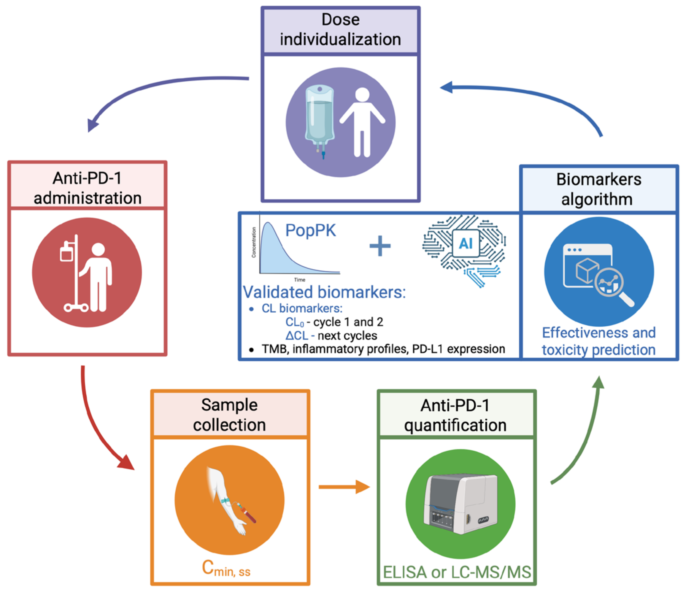
We propose a TDM methodology integrated with clinical evaluation and biomarker analysis, focusing on the longitudinal CL evaluation as a dynamic indicator of both therapeutic effectiveness and potential toxicity. Monitoring should prioritize steady-state trough concentrations (Cmin, ss) which can be quantified using immunoassays such as the Shikari® ELISA kit (BioVision, AbCam, Cambridge, UK). Some authors, namely Sureda et al., proposed a Cmin, ss target of 57 μg/mL for nivolumab, for example [172]. Rather than relying solely on target therapeutic exposure thresholds, TDM should be interpreted in the context of the patient’s evolving clinical response. The incorporation of TDM for anti-PD-1 into routine clinical practice may—as CL decreases, often in association with a favorable clinical response—enable dosing adjustments overtime by reducing the dose or extending the dosing interval, while maintaining adequate drug levels. This adaptive strategy offers the potential for sustained clinical benefit (such as maintaining response and reducing the risk of irAEs) along with economic advantages, supporting a more efficient and personalized use of anti-PD-1 therapies. Ultimately, this strategy aligns with the paradigm of achieving an optimal biologic dose or optimal individualized dose, especially in scenarios where the maximum tolerated dose has not been established or where the saturation of PK/PD parameters has been observed, favoring PK/PD-guided over fixed-dose recommendations.

6. Clinical Implications and Future Directions

The PK variability observed with anti-PD-1 mAbs such as nivolumab, pembrolizumab, and cemiplimab has significant clinical implications. Despite the current regulatory approval of fixed-dose regimens by the FDA and EMA, these strategies may not account for interindividual differences in drug CL and exposure, particularly in long-term responders (patients with ≥6 months of progression-free survival) or patients with altered physiology. As a result, personalized dosing remains underutilized in routine oncology practice.
Emerging PK models, including physiologically based pharmacokinetic (PBPK) simulations and artificial intelligence-driven algorithms, offer promising avenues for individualized treatment. PBPK models can aid in determining CL0 and/or ΔCL, complementing Bayesian estimation of PK parameters. Concurrently, machine learning approaches can integrate real-world patient data to refine dose predictions and improve therapeutic outcomes. However, to date, we have not identified any studies applying these technologies in clinical settings.
To translate these innovations into clinical benefit, future research must prioritize the development of real-time PK monitoring tools, as well as large-scale, prospective trials evaluating the safety and efficacy of CL-guided dosing adjustments. Moreover, the integration of PK insights with TDM and validated biomarkers could enhance the precision of immunotherapy and minimize toxicity, while sustaining efficacy. Integrating advanced PK modeling into clinical workflows will be pivotal for the evolution of precision immuno-oncology—Figure 3. However, these strategies face important limitations, including the need for specialized human resources, adequate computational and laboratory equipment, access to drug monitoring data, and validated algorithms, which are not yet available. These efforts, once addressed, promise to personalize treatment strategies, reduce overtreatment, and improve quality of care in patients receiving anti-PD-1 therapies.
Ultimately, an alternative to overcome T cell exhaustion is the combination of anti–PD-1 agents with novel targets under investigation (e.g., anti-TIM-3, anti-TIGIT) [173,174], or strategies aimed at enhancing anti–PD-1 responses, such as co-stimulatory agonists [175], CTLA-4 inhibitors [176,177], or modulation of the tumor microenvironment with repurposed drugs.

7. Conclusions

Time-dependent reductions in CL of anti-PD-1 antibodies are consistently associated with improved clinical outcomes but also lead to increased drug exposure, potentially increasing the risk of immune-related toxicities. While standard dosing appears safe across broad populations, individual variability, especially in responders, highlights the need for PK-guided dose optimization. Integrating PK monitoring into clinical practice may enhance efficacy while minimizing toxicity, supporting a more personalized and safer use of anti-PD-1 therapies.

Author Contributions

Conceptualization, P.F., J.G., R.R., H.R. and J.F.M.; writing—original draft preparation, A.F., D.G.T., E.M., P.F. and J.F.M.; writing, all authors; supervision, J.F.M., J.G. and P.F. All authors have read and agreed to the published version of the manuscript.

Funding

This research received no external funding.

Institutional Review Board Statement

Not applicable.

Informed Consent Statement

Not applicable.

Conflicts of Interest

The authors declare no conflicts of interest.

Abbreviations

The following abbreviations are used in this manuscript:
ADAsAnti-drug antibodies
ADCCAntibody-dependent cellular citotoxicity
ALBAlbumin
Anti-TIM3Anti-T cell mucin domain 3
Anti-TIGITAnti-T cell immunoreceptor with immunoglobulin and immunoreceptor tyrosine-based inhibitory motif domains
AUCArea under plasmatic concentration versus time curve
BWBody weight
CLClearance
CL0Baseline clearance
CL–RClearance–response
Cmin, ssSteady-state trough concentrations
CTLA4Cytotoxic T- lymphocyte-associated antigen 4
DCsDendritic cells
ELISAEnzyme-linked immunosorbent assay
ECMExtracellular matrix
E–RExposure–Response
FcRnNeonatal Fc receptor
FcRsFc receptors
FcγRGamma Fc receptor
ICIs Immune checkpoint inhibitors
IFN-γGamma-interferon
IgImmunoglobulin
IL-1Interleukin 1
irAEsImmune-related adverse events
LC-MS/MSLiquid chromatography tandem mass spectrometry
mAbsMonoclonal antibodies
MMPMatrix metalloproteinases
NKNatural killer
NSCLCNon-small cell lung cancer
OSOverall survival
PBPeripheral blood
PBPKPhysiologically based pharmacokinetic
PDPharmacodynamics
PD-1Programmed cell death protein 1
PD-L1Programmed death-ligand 1
PKPharmacokinetics
PK–PDPharmacokinetic–Pharmacodynamic
PopPKPopulation pharmacokinetics
PSPerformance status
TDMTherapeutic drug monitoring
TMBTumor mutational burden
TMDDTarget-mediated drug disposition
TNF-αTumor necrosis factor-alpha
VdVolume of distribution
ΔCLChange in clearance over time

References

  1. Baumeister, S.H.; Freeman, G.J.; Dranoff, G.; Sharpe, A.H. Coinhibitory Pathways in Immunotherapy for Cancer. Annu. Rev. Immunol. 2016, 34, 539–573. [Google Scholar] [CrossRef]
  2. Okazaki, T.; Chikuma, S.; Iwai, Y.; Fagarasan, S.; Honjo, T. A rheostat for immune responses: The unique properties of PD-1 and their advantages for clinical application. Nat. Immunol. 2013, 14, 1212–1218. [Google Scholar] [CrossRef] [PubMed]
  3. Barber, D.L.; Wherry, E.J.; Masopust, D.; Zhu, B.; Allison, J.P.; Sharpe, A.H.; Freeman, G.J.; Ahmed, R. Restoring function in exhausted CD8 T cells during chronic viral infection. Nature 2006, 439, 682–687. [Google Scholar] [CrossRef] [PubMed]
  4. Bardhan, K.; Aksoylar, H.-I.; Le Bourgeois, T.; Strauss, L.; Weaver, J.D.; Delcuze, B.; Charest, A.; Patsoukis, N.; Boussiotis, V.A. Publisher Correction: Phosphorylation of PD-1-Y248 is a marker of PD-1-mediated inhibitory function in human T cells. Sci. Rep. 2020, 10, 15905. [Google Scholar] [CrossRef] [PubMed]
  5. Patsoukis, N.; Brown, J.; Petkova, V.; Liu, F.; Li, L.; Boussiotis, V.A. Selective effects of PD-1 on Akt and Ras pathways regulate molecular components of the cell cycle and inhibit T cell proliferation. Sci. Signal. 2012, 5, ra46. [Google Scholar] [CrossRef]
  6. Ishida, Y.; Agata, Y.; Shibahara, K.; Honjo, T. Induced expression of PD-1, a novel member of the immunoglobulin gene superfamily, upon programmed cell death. EMBO J. 1992, 11, 3887–3895. [Google Scholar] [CrossRef]
  7. Nishimura, H.; Minato, N.; Nakano, T.; Honjo, T. Immunological studies on PD-1 deficient mice: Implication of PD-1 as a negative regulator for B cell responses. Int. Immunol. 1998, 10, 1563–1572. [Google Scholar] [CrossRef]
  8. Yao, S.; Wang, S.; Zhu, Y.; Luo, L.; Zhu, G.; Flies, S.; Xu, H.; Ruff, W.; Broadwater, M.; Choi, I.-H.; et al. PD-1 on dendritic cells impedes innate immunity against bacterial infection. Blood 2009, 113, 5811–5818. [Google Scholar] [CrossRef]
  9. Huang, X.; Venet, F.; Wang, Y.L.; Lepape, A.; Yuan, Z.; Chen, Y.; Swan, R.; Kherouf, H.; Monneret, G.; Chung, C.-S.; et al. PD-1 expression by macrophages plays a pathologic role in altering microbial clearance and the innate inflammatory response to sepsis. Proc. Natl. Acad. Sci. USA 2009, 106, 6303–6308. [Google Scholar]
  10. Vari, F.; Arpon, D.; Keane, C.; Hertzberg, M.S.; Talaulikar, D.; Jain, S.; Cui, Q.; Han, E.; Tobin, J.; Bird, R.; et al. Immune evasion via PD-1/PD-L1 on NK cells and monocyte/macrophages is more prominent in Hodgkin lymphoma than DLBCL. Blood 2018, 131, 1809–1819. [Google Scholar] [CrossRef]
  11. Wei, F.; Zhong, S.; Ma, Z.; Kong, H.; Medvec, A.; Ahmed, R.; Freeman, G.J.; Krogsgaard, M.; Riley, J.L. Strength of PD-1 signaling differentially affects T-cell effector functions. Proc. Natl. Acad. Sci. USA 2013, 110, E2480–E2489. [Google Scholar] [CrossRef]
  12. Francisco, L.M.; Sage, P.T.; Sharpe, A.H. The PD-1 pathway in tolerance and autoimmunity. Immunol. Rev. 2010, 236, 219–242. [Google Scholar] [CrossRef]
  13. Mueller, D.L. Mechanisms maintaining peripheral tolerance. Nat. Immunol. 2010, 11, 21–27. [Google Scholar] [CrossRef]
  14. Keir, M.E.; Butte, M.J.; Freeman, G.J.; Sharpe, A.H. PD-1 and its ligands in tolerance and immunity. Annu. Rev. Immunol. 2008, 26, 677–704. [Google Scholar] [CrossRef]
  15. Naimi, A.; Mohammed, R.N.; Raji, A.; Chupradit, S.; Yumashev, A.V.; Suksatan, W.; Shalaby, M.N.; Thangavelu, L.; Kamrava, S.; Shomali, N.; et al. Tumor immunotherapies by immune checkpoint inhibitors (ICIs); the pros and cons. Cell Commun. Signal. 2022, 20, 44. [Google Scholar] [CrossRef]
  16. Lee, H.T.; Lee, S.H.; Heo, Y.S. Molecular Interactions of Antibody Drugs Targeting PD-1, PD-L1, and CTLA-4 in Immuno-Oncology. Molecules 2019, 24, 1190. [Google Scholar] [CrossRef]
  17. Liu, B.; Zhou, H.; Tan, L.; Siu, K.T.H.; Guan, X.Y. Exploring treatment options in cancer: Tumor treatment strategies. Signal Transduct. Target. Ther. 2024, 9, 175. [Google Scholar] [CrossRef] [PubMed]
  18. Darnell, E.P.; Mooradian, M.J.; Baruch, E.N.; Yilmaz, M.; Reynolds, K.L. Immune-Related Adverse Events (irAEs): Diagnosis, Management, and Clinical Pearls. Curr. Oncol. Rep. 2020, 22, 39. [Google Scholar] [CrossRef] [PubMed]
  19. You, W.; Shang, B.; Sun, J.; Liu, X.; Su, L.; Jiang, S. Mechanistic insight of predictive biomarkers for antitumor PD-1/PD-L1 blockade: A paradigm shift towards immunome evaluation (Review). Oncol. Rep. 2020, 44, 424–437. [Google Scholar] [CrossRef] [PubMed]
  20. Wang, N.; Zheng, L.; Li, M.; Hou, X.; Zhang, B.; Chen, J.; Li, S.; Chen, L. Clinical efficacy and safety of individualized pembrolizumab administration based on pharmacokinetic in advanced non-small cell lung cancer: A prospective exploratory clinical trial. Lung Cancer 2023, 178, 183–190. [Google Scholar] [CrossRef]
  21. Desnoyer, A.; Broutin, S.; Delahousse, J.; Maritaz, C.; Blondel, L.; Mir, O.; Chaput, N.; Paci, A. Pharmacokinetic/pharmacodynamic relationship of therapeutic monoclonal antibodies used in oncology: Part 2, immune checkpoint inhibitor antibodies. Eur. J. Cancer 2020, 128, 119–128. [Google Scholar] [CrossRef]
  22. Freshwater, T.; Kondic, A.; Ahamadi, M.; Li, C.H.; de Greef, R.; de Alwis, D.; Stone, J.A. Evaluation of dosing strategy for pembrolizumab for oncology indications. J. Immunother. Cancer 2017, 5, 43. [Google Scholar] [CrossRef]
  23. Ahamadi, M.; Freshwater, T.; Prohn, M.; Li, C.; de Alwis, D.; de Greef, R.; Elassaiss-Schaap, J.; Kondic, A.; Stone, J. Model-Based Characterization of the Pharmacokinetics of Pembrolizumab: A Humanized Anti-PD-1 Monoclonal Antibody in Advanced Solid Tumors. CPT Pharmacomet. Syst. Pharmacol. 2017, 6, 49–57. [Google Scholar] [CrossRef]
  24. Peer, C.J.; Goldstein, D.A.; Goodell, J.C.; Nguyen, R.; Figg, W.D.; Ratain, M.J. Opportunities for using in silico—Based extended dosing regimens for monoclonal antibody immune checkpoint inhibitors. Br. J. Clin. Pharmacol. 2020, 86, 1769–1777. [Google Scholar] [CrossRef] [PubMed]
  25. Nguyen, J.; Epling, D.; Dolphin, N.; Paccaly, A.; Conrado, D.; Davis, J.D.; Al-Huniti, N. Population pharmacokinetics modeling and exposure—Response analyses of cemiplimab in patients with recurrent or metastatic cervical cancer. CPT Pharmacomet. Syst. Pharmacol. 2022, 11, 1458–1471. [Google Scholar] [CrossRef] [PubMed]
  26. Meng, X.; Liu, X.; Guo, X.; Jiang, S.; Chen, T.; Hu, Z.; Liu, H.; Bai, Y.; Xue, M.; Hu, R.; et al. FBXO38 mediates PD-1 ubiquitination and regulates anti-tumour immunity of T cells. Nature 2018, 564, 130–135. [Google Scholar] [CrossRef] [PubMed]
  27. Ben Saad, E.; Oroya, A.; Anto, N.P.; Bachais, M.; Rudd, C.E. PD-1 endocytosis unleashes the cytolytic potential of checkpoint blockade in tumor immunity. Cell Rep. 2024, 43, 114907. [Google Scholar] [CrossRef]
  28. Okada, M.; Chikuma, S.; Kondo, T.; Hibino, S.; Machiyama, H.; Yokosuka, T.; Nakano, M.; Yoshimura, A. Blockage of Core Fucosylation Reduces Cell-Surface Expression of PD-1 and Promotes Anti-tumor Immune Responses of T Cells. Cell Rep. 2017, 20, 1017–1028. [Google Scholar] [CrossRef]
  29. White, D.; Cote-Martin, A.; Bleck, M.; Garaffa, N.; Shaaban, A.; Wu, H.; Liu, D.; Young, D.; Scheer, J.; Lorenz, I.C.; et al. Programmed Cell Death-1 (PD-1) anchoring to the GPI-linked co-receptor CD48 reveals a novel mechanism to modulate PD-1-dependent inhibition of human T cells. Mol. Immunol. 2023, 156, 31–38. [Google Scholar] [CrossRef]
  30. Chen, N.; Li, Z.; Liu, H.; Jiang, A.; Zhang, L.; Yan, S.; He, W.; Yang, J.; Liu, T. Enhancing PD-1 blockade in NSCLC: Reprogramming tumor immune microenvironment with albumin-bound statins targeting lipid rafts and mitochondrial respiration. Bioact. Mater. 2025, 49, 140–153. [Google Scholar] [CrossRef]
  31. D’Hooghe, L.; Chalmers, A.D.; Heywood, S.; Whitley, P. Cell surface dynamics and cellular distribution of endogenous FcRn. PLoS ONE 2017, 12, e0182695. [Google Scholar] [CrossRef] [PubMed]
  32. Rodewald, R.; Kraehenbuhl, J.P. Receptor-mediated transport of IgG. J. Cell Biol. 1984, 99 Pt 2, 159s–164s. [Google Scholar] [CrossRef] [PubMed]
  33. Medesan, C.; Radu, C.; Kim, J.; Ghetie, V.; Ward, E.S. Localization of the site of the IgG molecule that regulates maternofetal transmission in mice. Eur. J. Immunol. 1996, 26, 2533–2536. [Google Scholar] [CrossRef] [PubMed]
  34. Ghetie, V.; Ward, E.S. Transcytosis and catabolism of antibody. Immunol. Res. 2002, 25, 97–114. [Google Scholar] [CrossRef]
  35. Raghavan, M.; Bonagura, V.R.; Morrison, S.L.; Bjorkman, P.J. Analysis of the pH dependence of the neonatal Fc receptor/immunoglobulin G interaction using antibody and receptor variants. Biochemistry 1995, 34, 14649–14657. [Google Scholar] [CrossRef]
  36. Ober, R.J.; Martinez, C.; Vaccaro, C.; Zhou, J.; Ward, E.S. Visualizing the site and dynamics of IgG salvage by the MHC class I-related receptor, FcRn. J. Immunol. 2004, 172, 2021–2029. [Google Scholar] [CrossRef]
  37. Pyzik, M.; Rath, T.; Lencer, W.I.; Baker, K.; Blumberg, R.S. FcRn: The Architect Behind the Immune and Nonimmune Functions of IgG and Albumin. J. Immunol. 2015, 194, 4595–4603. [Google Scholar] [CrossRef]
  38. Pannek, A.; Houghton, F.J.; Verhagen, A.M.; Dower, S.K.; Hinde, E.; Gleeson, P.A. Dynamics of intracellular neonatal Fc receptor-ligand interactions in primary macrophages using biophysical fluorescence techniques. Mol. Biol. Cell 2022, 33, ar6. [Google Scholar] [CrossRef]
  39. Rudnik-Jansen, I.; Howard, K.A. FcRn expression in cancer: Mechanistic basis and therapeutic opportunities. J. Control. Release 2021, 337, 248–257. [Google Scholar] [CrossRef]
  40. Claypool, S.M.; Dickinson, B.L.; Wagner, J.S.; Johansen, F.-E.; Venu, N.; Borawski, J.A.; Lencer, W.I.; Blumberg, R.S. Bidirectional transepithelial IgG transport by a strongly polarized basolateral membrane Fcgamma-receptor. Mol. Biol. Cell 2004, 15, 1746–1759. [Google Scholar] [CrossRef]
  41. Latvala, S.; Jacobsen, B.; Otteneder, M.B.; Herrmann, A.; Kronenberg, S. Distribution of FcRn Across Species and Tissues. J. Histochem. Cytochem. 2017, 65, 321–333. [Google Scholar] [CrossRef]
  42. Chaudhury, C.; Mehnaz, S.; Robinson, J.M.; Hayton, W.L.; Pearl, D.K.; Roopenian, D.C.; Anderson, C.L. The major histocompatibility complex-related Fc receptor for IgG (FcRn) binds albumin and prolongs its lifespan. J. Exp. Med. 2003, 197, 315–322. [Google Scholar] [CrossRef]
  43. Suzuki, T.; Ishii-Watabe, A.; Tada, M.; Kobayashi, T.; Kanayasu-Toyoda, T.; Kawanishi, T.; Yamaguchi, T. Importance of neonatal FcR in regulating the serum half-life of therapeutic proteins containing the Fc domain of human IgG1: A comparative study of the affinity of monoclonal antibodies and Fc-fusion proteins to human neonatal FcR. J. Immunol. 2010, 184, 1968–1976. [Google Scholar] [CrossRef]
  44. Castillo, A.M.M.; Vu, T.T.; Liva, S.G.; Chen, M.; Xie, Z.; Thomas, J.; Remaily, B.; Guo, Y.; Subrayan, U.L.; Costa, T.; et al. Murine cancer cachexia models replicate elevated catabolic pembrolizumab clearance in humans. JCSM Rapid Commun. 2021, 4, 232–244. [Google Scholar] [CrossRef]
  45. Chen, W.; Wang, L.; Ruan, Z.; Lou, H.; Jiang, B. Translate Pharmacokinetics of PD-1/PD-L1 Monoclonal Antibodies from Cynomolgus Monkey to Human: Comparison of Different Approaches. J. Pharm. Sci. 2024, 113, 2915–2921. [Google Scholar] [CrossRef]
  46. Justus, C.R.; Dong, L.; Yang, L.V. Acidic tumor microenvironment and pH-sensing G protein-coupled receptors. Front. Physiol. 2013, 4, 354. [Google Scholar] [CrossRef]
  47. Hosonuma, M.; Yoshimura, K. Association between pH regulation of the tumor microenvironment and immunological state. Front. Oncol. 2023, 13, 1175563. [Google Scholar] [CrossRef] [PubMed]
  48. Chen, X.; Song, X.; Li, K.; Zhang, T. FcγR-Binding Is an Important Functional Attribute for Immune Checkpoint Antibodies in Cancer Immunotherapy. Front. Immunol. 2019, 10, 292. [Google Scholar] [CrossRef] [PubMed]
  49. Smith, K.G.C.; Clatworthy, M.R. FcgammaRIIB in autoimmunity and infection: Evolutionary and therapeutic implications. Nat. Rev. Immunol. 2010, 10, 328–343. [Google Scholar] [CrossRef] [PubMed]
  50. Ganesan, L.P.; Kim, J.; Wu, Y.; Mohanty, S.; Phillips, G.S.; Birmingham, D.J.; Robinson, J.M.; Anderson, C.L. FcγRIIb on liver sinusoidal endothelium clears small immune complexes. J. Immunol. 2012, 189, 4981–4988. [Google Scholar] [CrossRef]
  51. Champiat, S.; Dercle, L.; Ammari, S.; Massard, C.; Hollebecque, A.; Postel-Vinay, S.; Chaput, N.; Eggermont, A.M.; Marabelle, A.; Soria, J.-C.; et al. Hyperprogressive Disease Is a New Pattern of Progression in Cancer Patients Treated by Anti-PD-1/PD-L1. Clin. Cancer Res. 2017, 23, 1920–1928. [Google Scholar] [CrossRef]
  52. Saâda-Bouzid, E.; Defaucheux, C.; Karabajakian, A.; Coloma, V.P.; Servois, V.; Paoletti, X.; Even, C.; Fayette, J.; Guigay, J.; Loirat, D.; et al. Hyperprogression during anti-PD-1/PD-L1 therapy in patients with recurrent and/or metastatic head and neck squamous cell carcinoma. Ann. Oncol. 2017, 28, 1605–1611. [Google Scholar] [CrossRef] [PubMed]
  53. Ferrara, R.; Mezquita, L.; Texier, M.; Lahmar, J.; Audigier-Valette, C.; Tessonnier, L.; Mazieres, J.; Zalcman, G.; Brosseau, S.; Le Moulec, S.; et al. Hyperprogressive Disease in Patients with Advanced Non-Small Cell Lung Cancer Treated With PD-1/PD-L1 Inhibitors or With Single-Agent Chemotherapy. JAMA Oncol. 2018, 4, 1543–1552. [Google Scholar] [CrossRef] [PubMed]
  54. Lo Russo, G.; Moro, M.; Sommariva, M.; Cancila, V.; Boeri, M.; Centonze, G.; Ferro, S.; Ganzinelli, M.; Gasparini, P.; Huber, V.; et al. Antibody-Fc/FcR Interaction on Macrophages as a Mechanism for Hyperprogressive Disease in Non-small Cell Lung Cancer Subsequent to PD-1/PD-L1 Blockade. Clin. Cancer Res. 2019, 25, 989–999. [Google Scholar] [CrossRef] [PubMed]
  55. Kamada, T.; Togashi, Y.; Tay, C.; Ha, D.; Sasaki, A.; Nakamura, Y.; Sato, E.; Fukuoka, S.; Tada, Y.; Tanaka, A.; et al. PD-1+ regulatory T cells amplified by PD-1 blockade promote hyperprogression of cancer. Proc. Natl. Acad. Sci. USA 2019, 116, 9999–10008. [Google Scholar] [CrossRef]
  56. Tan, C.L.; Kuchroo, J.R.; Sage, P.T.; Liang, D.; Francisco, L.M.; Buck, J.; Thaker, Y.R.; Zhang, Q.; McArdel, S.L.; Juneja, V.R.; et al. PD-1 restraint of regulatory T cell suppressive activity is critical for immune tolerance. J. Exp. Med. 2021, 218, e20182232. [Google Scholar] [CrossRef]
  57. Wang, W.; Wang, E.Q.; Balthasar, J.P. Monoclonal antibody pharmacokinetics and pharmacodynamics. Clin. Pharmacol. Ther. 2008, 84, 548–558. [Google Scholar] [CrossRef]
  58. Golay, J.; Semenzato, G.; Rambaldi, A.; Foà, R.; Gaidano, G.; Gamba, E.; Pane, F.; Pinto, A.; Specchia, G.; Zaja, F.; et al. Lessons for the clinic from rituximab pharmacokinetics and pharmacodynamics. mAbs 2013, 5, 826–837. [Google Scholar] [CrossRef]
  59. Gearing, A.J.; Thorpe, S.J.; Miller, K.; Mangan, M.; Varley, P.G.; Dudgeon, T.; Ward, G.; Turner, C.; Thorpe, R. Selective cleavage of human IgG by the matrix metalloproteinases, matrilysin and stromelysin. Immunol. Lett. 2002, 81, 41–48. [Google Scholar] [CrossRef]
  60. Joo, V.; Petrovas, C.; de Leval, L.; Noto, A.; Obeid, M.; Fenwick, C.; Pantaleo, G. A CD64/FcγRI-mediated mechanism hijacks PD-1 from PD-L1/2 interaction and enhances anti-PD-1 functional recovery of exhausted T cells. Front. Immunol. 2023, 14, 1213375. [Google Scholar] [CrossRef]
  61. Hao, L.Y.; Lerrer, S.; Paiola, M.; Moore, E.K.; Gartshteyn, Y.; Song, R.; Goeckeritz, M.; Black, M.J.; Bukhari, S.; Hu, X.; et al. Exclusion of PD-1 from the immune synapse: A novel strategy to modulate T cell function. Mol. Ther. Oncol. 2024, 32, 200839. [Google Scholar] [CrossRef] [PubMed]
  62. Brezski, R.J.; Vafa, O.; Petrone, D.; Tam, S.H.; Powers, G.; Ryan, M.H.; Luongo, J.L.; Oberholtzer, A.; Knight, D.M.; Jordan, R.E. Tumor-associated and microbial proteases compromise host IgG effector functions by a single cleavage proximal to the hinge. Proc. Natl. Acad. Sci. USA 2009, 106, 17864–17869. [Google Scholar] [CrossRef] [PubMed]
  63. Fitzpatrick, E.A.; Wang, J.; Strome, S.E. Strome, Engineering of Fc Multimers as a Protein Therapy for Autoimmune Disease. Front. Immunol. 2020, 11, 496. [Google Scholar] [CrossRef] [PubMed]
  64. Wolosowicz, M.; Prokopiuk, S.; Kaminski, T.W. The Complex Role of Matrix Metalloproteinase-2 (MMP-2) in Health and Disease. Int. J. Mol. Sci. 2024, 25, 13691. [Google Scholar] [CrossRef]
  65. Balbín, M.; Pendás, A.M.; Uría, J.A.; Jiménez, M.G.; Freije, J.P.; López-Otín, C. Expression and regulation of collagenase-3 (MMP-13) in human malignant tumors. APMIS 1999, 107, 45–53. [Google Scholar] [CrossRef]
  66. Azzam, H.S.; Arand, G.; Lippman, M.E.; Thompson, E.W. Association of MMP-2 activation potential with metastatic progression in human breast cancer cell lines independent of MMP-2 production. J. Natl. Cancer Inst. 1993, 85, 1758–1764. [Google Scholar] [CrossRef]
  67. Naylor, M.S.; Stamp, G.W.; Davies, B.D.; Balkwill, F.R. Expression and activity of MMPS and their regulators in ovarian cancer. Int. J. Cancer 1994, 58, 50–56. [Google Scholar] [CrossRef]
  68. Kusukawa, J.; Sasaguri, Y.; Shima, I.; Kameyama, T.; Morimatsu, M. Expression of matrix metalloproteinase-2 related to lymph node metastasis of oral squamous cell carcinoma. A clinicopathologic study. Am. J. Clin. Pathol. 1993, 99, 18–23. [Google Scholar] [CrossRef]
  69. Powell, W.C.; Knox, J.D.; Navre, M.; Grogan, T.M.; Kittelson, J.; Nagle, R.B.; Bowden, G.T. Expression of the metalloproteinase matrilysin in DU-145 cells increases their invasive potential in severe combined immunodeficient mice. Cancer Res. 1993, 53, 417–422. [Google Scholar]
  70. Davies, B.; Waxman, J.; Wasan, H.; Abel, P.; Williams, G.; Krausz, T.; Neal, D.; Thomas, D.; Hanby, A.; Balkwill, F. Levels of matrix metalloproteases in bladder cancer correlate with tumor grade and invasion. Cancer Res. 1993, 53, 5365–5369. [Google Scholar]
  71. Yoshimoto, M.; Itoh, F.; Yamamoto, H.; Hinoda, Y.; Imai, K.; Yachi, A. Expression of MMP-7(PUMP-1) mRNA in human colorectal cancers. Int. J. Cancer 1993, 54, 614–618. [Google Scholar] [CrossRef]
  72. Jones, B.; Moshyedi, P.; Gallo, S.; Tombran-Tink, J.; Arand, G.; Reid, D.A.; Thompson, E.W.; Chader, G.J.; Waldbillig, R.J. Characterization and novel activation of 72-kDa metalloproteinase in retinal interphotoreceptor matrix and Y-79 cell culture medium. Exp. Eye Res. 1994, 59, 257–269. [Google Scholar] [CrossRef]
  73. Liu, W.; Zeng, Z.; Wu, Q.; Hou, J.; Chen, Y. Overexpression of MMP-2 in laryngeal squamous cell carcinoma: A potential indicator for poor prognosis. Otolaryngol. Head Neck Surg. 2005, 132, 395–400. [Google Scholar] [CrossRef] [PubMed]
  74. Chidambaram, D.; Subashini, V.; Nanthanalaxmi, M.; Saranya, I.; Selvamurugan, N. Regulation of matrix metalloproteinase-13 in cancer: Signaling pathways and non-coding RNAs in tumor progression and therapeutic targeting. World J. Clin. Oncol. 2025, 16, 105996. [Google Scholar] [CrossRef] [PubMed]
  75. Reddy, R.A.; Varshini, M.S.; Kumar, R.S. Matrix Metalloproteinase-2 (MMP-2): As an Essential Factor in Cancer Progression. Recent Pat. Anti-Cancer Drug Discov. 2025, 20, 26–44. [Google Scholar] [CrossRef] [PubMed]
  76. Cardoso, A.P.; Pinto, M.L.; Pinto, A.T.; Pinto, M.T.; Monteiro, C.; Oliveira, M.I.; Santos, S.G.; Relvas, J.B.; Seruca, R.; Mantovani, A.; et al. Matrix metalloproteases as maestros for the dual role of LPS- and IL-10-stimulated macrophages in cancer cell behaviour. BMC Cancer 2015, 15, 456. [Google Scholar] [CrossRef]
  77. Xu, F.; Wei, Y.; Tang, Z.; Liu, B.; Dong, J. Tumor-associated macrophages in lung cancer: Friend or foe? (Review). Mol. Med. Rep. 2020, 22, 4107–4115. [Google Scholar]
  78. Wang, S.; Wang, J.; Chen, Z.; Luo, J.; Guo, W.; Sun, L.; Lin, L. Targeting M2-like tumor-associated macrophages is a potential therapeutic approach to overcome antitumor drug resistance. npj Precis. Oncol. 2024, 8, 31. [Google Scholar] [CrossRef]
  79. Nielsen, B.S.; Egeblad, M.; Rank, F.; Askautrud, H.A.; Pennington, C.J.; Pedersen, T.X.; Christensen, I.J.; Edwards, D.R.; Werb, Z.; Lund, L.R. Matrix metalloproteinase 13 is induced in fibroblasts in polyomavirus middle T antigen-driven mammary carcinoma without influencing tumor progression. PLoS ONE 2008, 3, e2959. [Google Scholar] [CrossRef]
  80. Hassona, Y.; Cirillo, N.; Heesom, K.; Parkinson, E.K.; Prime, S.S. Senescent cancer-associated fibroblasts secrete active MMP-2 that promotes keratinocyte dis-cohesion and invasion. Br. J. Cancer 2014, 111, 1230–1237. [Google Scholar] [CrossRef]
  81. Elkon, K.; Casali, P. Nature and functions of autoantibodies. Nat. Clin. Pr. Rheumatol. 2008, 4, 491–498. [Google Scholar] [CrossRef]
  82. Suurmond, J.; Diamond, B. Autoantibodies in systemic autoimmune diseases: Specificity and pathogenicity. J. Clin. Investig. 2015, 125, 2194–2202. [Google Scholar] [CrossRef]
  83. Sesarman, A.; Vidarsson, G.; Sitaru, C. The neonatal Fc receptor as therapeutic target in IgG-mediated autoimmune diseases. Cell. Mol. Life Sci. 2010, 67, 2533–2550. [Google Scholar] [CrossRef]
  84. Hajjar, S.; Zhou, X. pH sensing at the intersection of tissue homeostasis and inflammation. Trends Immunol. 2023, 44, 807–825. [Google Scholar] [CrossRef] [PubMed]
  85. Tardy, M.P.; Gastaud, L.; Boscagli, A.; Peyrade, F.; Gallamini, A.; Thyss, A. Autoimmune hemolytic anemia after nivolumab treatment in Hodgkin lymphoma responsive to immunosuppressive treatment. A case report. Hematol. Oncol. 2016, 35, 875–877. [Google Scholar] [CrossRef] [PubMed]
  86. Ghosn, J.; Vicino, A.; Michielin, O.; Coukos, G.; Kuntzer, T.; Obeid, M. A severe case of neuro-Sjögren’s syndrome induced by pembrolizumab. J. Immunother. Cancer 2018, 6, 110. [Google Scholar] [CrossRef] [PubMed]
  87. Sadik, C.D.; Langan, E.A.; Grätz, V.; Zillikens, D.; Terheyden, P. Checkpoint Inhibition May Trigger the Rare Variant of Anti-LAD-1 IgG-Positive, Anti-BP180 NC16A IgG-Negative Bullous Pemphigoid. Front. Immunol. 2019, 10, 1934. [Google Scholar] [CrossRef]
  88. Pollack, R.; Ashash, A.; Cahn, A.; Rottenberg, Y.; Stern, H.; Dresner-Pollak, R. Immune Checkpoint Inhibitor-induced Thyroid Dysfunction Is Associated with Higher Body Mass Index. J. Clin. Endocrinol. Metab. 2020, 105, e3620–e3627. [Google Scholar] [CrossRef]
  89. Genta, S.; Lajkosz, K.; Yee, N.R.; Spiliopoulou, P.; Heirali, A.; Hansen, A.R.; Siu, L.L.; Saibil, S.; Stayner, L.-A.; Yanekina, M.; et al. Autoimmune PaneLs as PrEdictors of Toxicity in Patients TReated with Immune Checkpoint InhibiTors (ALERT). J. Exp. Clin. Cancer Res. 2023, 42, 276. [Google Scholar] [CrossRef]
  90. Swanson, S.J. What are clinically significant anti-drug antibodies and why is it important to identify them. Front. Immunol. 2024, 15, 1401178. [Google Scholar] [CrossRef]
  91. Galle, P.; Finn, R.S.; Mitchell, C.R.; Ndirangu, K.; Ramji, Z.; Redhead, G.S.; Pinato, D.J. Treatment-emergent antidrug antibodies related to PD-1, PD-L1, or CTLA-4 inhibitors across tumor types: A systematic review. J. Immunother. Cancer 2024, 12, e008266. [Google Scholar] [CrossRef]
  92. Agrawal, S.; Statkevich, P.; Bajaj, G.; Feng, Y.; Saeger, S.; Desai, D.D.; Park, J.; Waxman, I.M.; Roy, A.; Gupta, M. Evaluation of Immunogenicity of Nivolumab Monotherapy and Its Clinical Relevance in Patients with Metastatic Solid Tumors. J. Clin. Pharmacol. 2017, 57, 394–400. [Google Scholar] [CrossRef]
  93. de Vries, F.; Smit, A.A.; Wolbink, G.; de Vries, A.; Loeff, F.C.; Franssen, E.J. Case report: Pharmacokinetics of pembrolizumab in a patient with stage IV non-small cell lung cancer after a single 200 mg administration. Front. Oncol. 2022, 12, 960116. [Google Scholar] [CrossRef] [PubMed]
  94. Leven, C.; Padelli, M.; Carré, J.L.; Bellissant, E.; Misery, L. Immune Checkpoint Inhibitors in Melanoma: A Review of Pharmacokinetics and Exposure-Response Relationships. Clin. Pharmacokinet. 2019, 58, 1393–1405. [Google Scholar] [CrossRef] [PubMed]
  95. Cowles, S.C.; Sheen, A.; Santollani, L.; Lutz, E.A.; Lax, B.M.; Palmeri, J.R.; Freeman, G.J.; Wittrup, K.D. An affinity threshold for maximum efficacy in anti-PD-1 immunotherapy. mAbs 2022, 14, 2088454. [Google Scholar] [CrossRef] [PubMed]
  96. Jansen, A.; Sriram, J.; Pluim, D.; Maas, R.; van Groningen, H.; Piet, B.; ter Heine, R. Therapeutic Exposure and Successful Response to Pembrolizumab in a Patient with Non–Small-Cell Lung Cancer Despite Significant Renal Loss Due to Paraneoplastic Nephrotic Syndrome. Clin. Lung Cancer 2021, 22, e220–e223. [Google Scholar] [CrossRef]
  97. Hurkmans, D.P.; Sassen, S.D.; de Joode, K.; Putter, L.; Basak, E.A.; Wijkhuijs, A.J.; Joerger, M.; Debets, R.; Koch, B.C.; Van der Leest, C.H.; et al. Prospective real-world study on the pharmacokinetics of pembrolizumab in patients with solid tumors. J. Immunother. Cancer 2021, 9, e002344. [Google Scholar] [CrossRef]
  98. Bajaj, G.; Wang, X.; Agrawal, S.; Gupta, M.; Roy, A.; Feng, Y. Model-Based Population Pharmacokinetic Analysis of Nivolumab in Patients with Solid Tumors. CPT Pharmacomet. Syst. Pharmacol. 2017, 6, 58–66. [Google Scholar] [CrossRef]
  99. Chaudhury, C.; Brooks, C.L.; Carter, D.C.; Robinson, J.M.; Anderson, C.L. Albumin binding to FcRn: Distinct from the FcRn-IgG interaction. Biochemistry 2006, 45, 4983–4990. [Google Scholar] [CrossRef]
  100. Oganesyan, V.; Damschroder, M.M.; Cook, K.E.; Li, Q.; Gao, C.; Wu, H.; Dall’ACqua, W.F. Structural insights into neonatal Fc receptor-based recycling mechanisms. J. Biol. Chem. 2014, 289, 7812–7824. [Google Scholar] [CrossRef]
  101. Mezo, A.R.; McDonnell, K.A.; Hehir, C.A.T.; Low, S.C.; Palombella, V.J.; Stattel, J.M.; Kamphaus, G.D.; Fraley, C.; Zhang, Y.; Dumont, J.A.; et al. Reduction of IgG in nonhuman primates by a peptide antagonist of the neonatal Fc receptor FcRn. Proc. Natl. Acad. Sci. USA 2008, 105, 2337–2342. [Google Scholar] [CrossRef]
  102. Sand, K.M.K.; Bern, M.; Nilsen, J.; Noordzij, H.T.; Sandlie, I.; Andersen, J.T. Unraveling the Interaction between FcRn and Albumin: Opportunities for Design of Albumin-Based Therapeutics. Front. Immunol. 2014, 5, 682. [Google Scholar] [CrossRef]
  103. Roch, B.; Coffy, A.; Jean-Baptiste, S.; Palaysi, E.; Daures, J.-P.; Pujol, J.-L.; Bommart, S. Cachexia—Sarcopenia as a determinant of disease control rate and survival in non-small lung cancer patients receiving immune-checkpoint inhibitors. Lung Cancer 2020, 143, 19–26. [Google Scholar] [CrossRef] [PubMed]
  104. Ohuchi, M.; Yagishita, S.; Jo, H.; Akagi, K.; Higashiyama, R.I.; Masuda, K.; Shinno, Y.; Okuma, Y.; Yoshida, T.; Goto, Y.; et al. Early change in the clearance of pembrolizumab reflects the survival and therapeutic response: A population pharmacokinetic analysis in real-world non-small cell lung cancer patients. Lung Cancer 2022, 173, 35–42. [Google Scholar] [CrossRef] [PubMed]
  105. Tohi, M.; Irie, K.; Mizuno, T.; Okuyoshi, H.; Hirabatake, M.; Ikesue, H.; Muroi, N.; Eto, M.; Fukushima, S.; Tomii, K.; et al. Population Pharmacokinetics of Nivolumab in Japanese Patients with Nonsmall Cell Lung Cancer. Ther. Drug Monit. 2023, 45, 110–116. [Google Scholar] [CrossRef] [PubMed]
  106. Vu, T.T.; Kim, K.; Manna, M.; Thomas, J.; Remaily, B.C.; Montgomery, E.J.; Costa, T.; Granchie, L.; Xie, Z.; Guo, Y.; et al. Decoupling FcRn and tumor contributions to elevated immune checkpoint inhibitor clearance in cancer cachexia. Pharmacol. Res. 2024, 199, 107048. [Google Scholar] [CrossRef]
  107. Wang, R.; Zheng, J.; Shao, X.; Ishii, Y.; Roy, A.; Bello, A.; Lee, R.; Zhang, J.; Wind-Rotolo, M.; Feng, Y. Development of a prognostic composite cytokine signature based on the correlation with nivolumab clearance: Translational PK/PD analysis in patients with renal cell carcinoma. J. Immunother. Cancer 2019, 7, 348. [Google Scholar] [CrossRef]
  108. Zhang, J.; Cai, J.; Bello, A.; Roy, A.; Sheng, J. Model-Based Population Pharmacokinetic Analysis of Nivolumab in Chinese Patients with Previously Treated Advanced Solid Tumors, Including Non-Small Cell Lung Cancer. J. Clin. Pharmacol. 2019, 59, 1415–1424. [Google Scholar] [CrossRef]
  109. Higashiyama, R.I.; Yoshida, T.; Yagishita, S.; Ohuchi, M.; Sakiyama, N.; Torasawa, M.; Shirasawa, M.; Masuda, K.; Shinno, Y.; Matsumoto, Y.; et al. Safety Implications of Switching Pembrolizumab Dosage from 200 mg Every 3 Weeks to 400 mg Every 6 Weeks in Patients with Advanced NSCLC. J. Thorac. Oncol. 2022, 17, 1227–1232. [Google Scholar] [CrossRef]
  110. Bellesoeur, A.; Ollier, E.; Allard, M.; Hirsch, L.; Boudou-Rouquette, P.; Arrondeau, J.; Thomas-Schoemann, A.; Tiako, M.; Khoudour, N.; Chapron, J.; et al. Is there an Exposure–Response Relationship for Nivolumab in Real-World NSCLC Patients? Cancers 2019, 11, 1784. [Google Scholar] [CrossRef]
  111. Hamuro, L.; Statkevich, P.; Bello, A.; Roy, A.; Bajaj, G. Nivolumab Clearance Is Stationary in Patients With Resected Melanoma on Adjuvant Therapy: Implications of Disease Status on Time-Varying Clearance. Clin. Pharmacol. Ther. 2019, 106, 1018–1027. [Google Scholar] [CrossRef] [PubMed]
  112. Liu, C.; Yu, J.; Li, H.; Liu, J.; Xu, Y.; Song, P.; Liu, Q.; Zhao, H.; Xu, J.; Maher, V.; et al. Association of time-varying clearance of nivolumab with disease dynamics and its implications on exposure response analysis. Clin. Pharmacol. Ther. 2017, 101, 657–666. [Google Scholar] [CrossRef] [PubMed]
  113. Shchelokov, D.; Demin, O., Jr. Receptor occupancy assessment and interpretation in terms of quantitative systems pharmacology: Nivolumab case study. mAbs 2023, 15, 2156317. [Google Scholar] [CrossRef] [PubMed]
  114. Hamimed, M.; Devillier, R.; Weiller, P.-J.; Marin, C.; Schiano, J.-M.; Belmecheri, N.; Etienne-Grimaldi, M.-C.; Ciccolini, J.; Harbi, S. Life-threatening toxicities upon Pembrolizumab intake: Could pharmacokinetics be the bad guy? Cancer Chemother. Pharmacol. 2024, 93, 627–632. [Google Scholar] [CrossRef]
  115. Atallah-Yunes, S.A.; Kadado, A.J.; Soe, M.H. Pericardial effusion due to pembrolizumab-induced immunotoxicity: A case report and literature review. Curr. Probl. Cancer 2019, 43, 504–510. [Google Scholar] [CrossRef]
  116. Zhao, S.; Zhou, Y.; Sun, W.; Li, Z.; Wang, C. Clinical features, diagnosis, and management of pembrolizumab-induced myasthenia gravis. Clin. Exp. Immunol. 2023, 211, 85–92. [Google Scholar] [CrossRef]
  117. Jiang, M.; Hu, Y.; Lin, G.; Chen, C. Dosing Regimens of Immune Checkpoint Inhibitors: Attempts at Lower Dose, Less Frequency, Shorter Course. Front. Oncol. 2022, 12, 906251. [Google Scholar] [CrossRef]
  118. Colard-Thomas, J.; Manceron, C.; Duflos, C.; Herman, F.; Simon, M.; Maria, A.; Faillie, J.-L.; Viala, M.; Palassin, P. Comparison of clinical safety between standard versus extended interval dosing of immune checkpoint inhibitors: A real-world retrospective cohort study. ESMO Open 2023, 8, 102070. [Google Scholar] [CrossRef]
  119. Zhou, H.; Li, N.; Tang, H.; Chen, H.; Chen, X.; Zhang, L.; Tao, D. Delayed thrombocytopenia as a rare but serious adverse event secondary to immune checkpoint inhibitor: A case report. Ann. Palliat. Med. 2021, 10, 5881–5886. [Google Scholar] [CrossRef]
  120. Feng, Y.; Wang, X.; Bajaj, G.; Agrawal, S.; Bello, A.; Lestini, B.; Finckenstein, F.G.; Park, J.-S.; Roy, A. Nivolumab Exposure-Response Analyses of Efficacy and Safety in Previously Treated Squamous or Nonsquamous Non-Small Cell Lung Cancer. Clin. Cancer Res. 2017, 23, 5394–5405. [Google Scholar] [CrossRef]
  121. Zhao, X.; Shen, J.; Ivaturi, V.; Gopalakrishnan, M.; Feng, Y.; Schmidt, B.; Statkevich, P.; Goodman, V.; Gobburu, J.; Bello, A.; et al. Model-based evaluation of the efficacy and safety of nivolumab once every 4 weeks across multiple tumor types. Ann. Oncol. 2020, 31, 302–309. [Google Scholar] [CrossRef]
  122. Long, G.; Tykodi, S.; Schneider, J.; Garbe, C.; Gravis, G.; Rashford, M.; Agrawal, S.; Grigoryeva, E.; Bello, A.; Roy, A.; et al. Assessment of nivolumab exposure and clinical safety of 480 mg every 4 weeks flat-dosing schedule in patients with cancer. Ann. Oncol. 2018, 29, 2208–2213. [Google Scholar] [CrossRef]
  123. Guo, Y.; Remaily, B.C.; Thomas, J.; Kim, K.; Kulp, S.K.; Mace, T.A.; Ganesan, L.P.; Owen, D.H.; Coss, C.C.; Phelps, M.A. Antibody Drug Clearance: An Underexplored Marker of Outcomes with Checkpoint Inhibitors. Clin. Cancer Res. 2024, 30, 942–958. [Google Scholar] [CrossRef] [PubMed]
  124. Turner, D.C.; Kondic, A.G.; Anderson, K.M.; Robinson, A.G.; Garon, E.B.; Riess, J.W.; Jain, L.; Mayawala, K.; Kang, J.; Ebbinghaus, S.W.; et al. Pembrolizumab Exposure–Response Assessments Challenged by Association of Cancer Cachexia and Catabolic Clearance. Clin. Cancer Res. 2018, 24, 5841–5849. [Google Scholar] [CrossRef] [PubMed]
  125. Chatterjee, M.; Turner, D.C.; Felip, E.; Lena, H.; Cappuzzo, F.; Horn, L.; Garon, E.B.; Hui, R.; Arkenau, H.-T.; Gubens, M.A.; et al. Systematic evaluation of pembrolizumab dosing in patients with advanced non-small-cell lung cancer. Ann. Oncol. 2016, 27, 1291–1298. [Google Scholar] [CrossRef] [PubMed]
  126. Wang, X.; Feng, Y.; Bajaj, G.; Gupta, M.; Agrawal, S.; Yang, A.; Park, J.-S.; Lestini, B.; Roy, A. Quantitative Characterization of the Exposure-Response Relationship for Cancer Immunotherapy: A Case Study of Nivolumab in Patients with Advanced Melanoma. CPT Pharmacomet. Syst. Pharmacol. 2017, 6, 40–48. [Google Scholar] [CrossRef]
  127. Badawi, M.; Coss, C.C.; Phelps, M.A. Letter to the Editor: Exposure–response or clearance–response relationship in immune checkpoint therapy?—A comment on ‘correlation between nivolumab exposure and treatment outcomes in non-small-cell lung cancer’ by Basak et al. Eur. J. Cancer 2019, 114, 25–26. [Google Scholar] [CrossRef]
  128. Dai, H.I.; Vugmeyster, Y.; Mangal, N. Characterizing Exposure–Response Relationship for Therapeutic Monoclonal Antibodies in Immuno-Oncology and Beyond: Challenges, Perspectives, and Prospects. Clin. Pharmacol. Ther. 2020, 108, 1156–1170. [Google Scholar] [CrossRef]
  129. Li, H.; Yu, J.; Liu, C.; Liu, J.; Subramaniam, S.; Zhao, H.; Blumenthal, G.M.; Turner, D.C.; Li, C.; Ahamadi, M.; et al. Time dependent pharmacokinetics of pembrolizumab in patients with solid tumor and its correlation with best overall response. J. Pharmacokinet. Pharmacodyn. 2017, 44, 403–414. [Google Scholar] [CrossRef]
  130. Yang, F.; Paccaly, A.J.; Rippley, R.K.; Davis, J.D.; DiCioccio, A.T. Population pharmacokinetic characteristics of cemiplimab in patients with advanced malignancies. J. Pharmacokinet. Pharmacodyn. 2021, 48, 479–494. [Google Scholar] [CrossRef]
  131. Hurkmans, D.P.; Basak, E.A.; van Dijk, T.; Mercieca, D.; Schreurs, M.W.J.; Wijkhuijs, A.J.M.; Bins, S.; Hoop, E.O.-D.; Debets, R.; Joerger, M.; et al. A prospective cohort study on the pharmacokinetics of nivolumab in metastatic non-small cell lung cancer, melanoma, and renal cell cancer patients. J. Immunother. Cancer 2019, 7, 192. [Google Scholar] [CrossRef]
  132. Topalian, S.L.; Taube, J.M.; Anders, R.A.; Pardoll, D.M. Mechanism-driven biomarkers to guide immune checkpoint blockade in cancer therapy. Nat. Rev. Cancer 2016, 16, 275–287. [Google Scholar] [CrossRef]
  133. Mariam, A.; Kamath, S.; Schveder, K.; Mc Leod, H.; Rotroff, D. Biomarkers for Response to Anti-PD-1/Anti-PD-L1 Immune Checkpoint Inhibitors: A Large Meta-Analysis. Oncology 2023, 37, 210–219. [Google Scholar] [PubMed]
  134. Monette, A.; Warren, S.; Barrett, J.C.; Garnett-Benson, C.; Schalper, K.A.; Taube, J.M.; Topp, B.; Snyder, A. Biomarker development for PD-(L)1 axis inhibition: A consensus view from the SITC Biomarkers Committee. J. Immunother. Cancer 2024, 12, e009427-13. [Google Scholar] [CrossRef] [PubMed]
  135. Cristescu, R.; Mogg, R.; Ayers, M.; Albright, A.; Murphy, E.; Yearley, J.; Sher, X.; Liu, X.Q.; Lu, H.; Nebozhyn, M.; et al. Pan-tumor genomic biomarkers for PD-1 checkpoint blockade-based immunotherapy. Science 2018, 362, eaar3593. [Google Scholar] [CrossRef] [PubMed]
  136. Masucci, G.V.; Cesano, A.; Hawtin, R.; Janetzki, S.; Zhang, J.; Kirsch, I.; Dobbin, K.K.; Alvarez, J.; Robbins, P.B.; Selvan, S.R.; et al. Validation of biomarkers to predict response to immunotherapy in cancer: Volume I—Pre-analytical and analytical validation. J. Immunother. Cancer 2016, 4, 76. [Google Scholar] [CrossRef]
  137. Bedognetti, D.; Balwit, J.M.; Wang, E.; Disis, M.L.; Britten, C.M.; Delogu, L.G.; Tomei, S.; Fox, B.A.; Gajewski, T.F.; Marincola, F.M.; et al. SITC/iSBTc Cancer Immunotherapy Biomarkers Resource Document: Online resources and useful tools—A compass in the land of biomarker discovery. J. Transl. Med. 2011, 9, 155. [Google Scholar] [CrossRef]
  138. Hu-Lieskovan, S.; Bhaumik, S.; Dhodapkar, K.; Grivel, J.-C.J.B.; Gupta, S.; Hanks, B.A.; Janetzki, S.; Kleen, T.O.; Koguchi, Y.; Lund, A.W.; et al. SITC cancer immunotherapy resource document: A compass in the land of biomarker discovery. J. Immunother. Cancer 2020, 8, e000705. [Google Scholar] [CrossRef]
  139. Butterfield, L.H.; Disis, M.L.; Fox, B.A.; Kaufman, D.R.; Khleif, S.N.; Wang, E. SITC 2018 workshop report: Immuno-Oncology Biomarkers: State of the Art. J. Immunother. Cancer 2018, 6, 138. [Google Scholar] [CrossRef]
  140. Butterfield, L.H.; Disis, M.L.; Khleif, S.N.; Balwit, J.M.; Marincola, F.M. Immuno-oncology biomarkers 2010 and beyond: Perspectives from the iSBTc/SITC biomarker task force. J. Transl. Med. 2010, 8, 130. [Google Scholar] [CrossRef]
  141. Yuan, J.; Hegde, P.S.; Clynes, R.; Foukas, P.G.; Harari, A.; Kleen, T.O.; Kvistborg, P.; Maccalli, C.; Maecker, H.T.; Page, D.B.; et al. Novel technologies and emerging biomarkers for personalized cancer immunotherapy. J. Immunother. Cancer 2016, 4, 3. [Google Scholar] [CrossRef]
  142. Sunshine, J.C.; Nguyen, P.L.; Kaunitz, G.J.; Cottrell, T.R.; Berry, S.; Esandrio, J.; Xu, H.; Ogurtsova, A.; Bleich, K.B.; Cornish, T.C.; et al. PD-L1 Expression in Melanoma: A Quantitative Immunohistochemical Antibody Comparison. Clin. Cancer Res. 2017, 23, 4938–4944. [Google Scholar] [CrossRef] [PubMed]
  143. Garon, E.B.; Rizvi, N.A.; Hui, R.; Leighl, N.; Balmanoukian, A.S.; Eder, J.P.; Patnaik, A.; Aggarwal, C.; Gubens, M.; Horn, L.; et al. Pembrolizumab for the treatment of non-small-cell lung cancer. N. Engl. J. Med. 2015, 372, 2018–2028. [Google Scholar] [CrossRef] [PubMed]
  144. Ritterhouse, L.L. Tumor mutational burden. Cancer Cytopathol. 2019, 127, 735–736. [Google Scholar] [CrossRef] [PubMed]
  145. Addeo, A.; Friedlaender, A.; Banna, G.L.; Weiss, G.J. TMB or not TMB as a biomarker: That is the question. Crit. Rev. Oncol. Hematol. 2021, 163, 103374. [Google Scholar] [CrossRef]
  146. Marabelle, A.; Le, D.T.; Ascierto, P.A.; Di Giacomo, A.M.; De Jesus-Acosta, A.; Delord, J.-P.; Geva, R.; Gottfried, M.; Penel, N.; Hansen, A.R.; et al. Efficacy of Pembrolizumab in Patients with Noncolorectal High Microsatellite Instability/Mismatch Repair-Deficient Cancer: Results from the Phase II KEYNOTE-158 Study. J. Clin. Oncol. 2020, 38, 1–10. [Google Scholar] [CrossRef]
  147. Ambrosini, M.; Manca, P.; Nasca, V.; Sciortino, C.; Ghelardi, F.; Seligmann, J.F.; Taieb, J.; Pietrantonio, F. Epidemiology, pathogenesis, biology and evolving management of MSI-H/dMMR cancers. Nat. Rev. Clin. Oncol. 2025, 22, 385–407. [Google Scholar] [CrossRef]
  148. Bhamidipati, D.; Subbiah, V. Tumor-agnostic drug development in dMMR/MSI-H solid tumors. Trends Cancer 2023, 9, 828–839. [Google Scholar] [CrossRef]
  149. Paijens, S.T.; Vledder, A.; de Bruyn, M.; Nijman, H.W. Tumor-infiltrating lymphocytes in the immunotherapy era. Cell. Mol. Immunol. 2021, 18, 842–859. [Google Scholar] [CrossRef]
  150. Derosa, L.; Hellmann, M.D.; Spaziano, M.; Halpenny, D.; Fidelle, M.; Rizvi, H.; Long, N.; Plodkowski, A.J.; Arbour, K.C.; Chaft, J.E.; et al. Negative association of antibiotics on clinical activity of immune checkpoint inhibitors in patients with advanced renal cell and non-small-cell lung cancer. Ann. Oncol. 2018, 29, 1437–1444. [Google Scholar] [CrossRef]
  151. Wang, Z.; Han, J.; Cui, Y.; Fan, K.; Zhou, X. Circulating microRNA-21 as noninvasive predictive biomarker for response in cancer immunotherapy. Med Hypotheses 2013, 81, 41–43. [Google Scholar] [CrossRef]
  152. Capone, M.; Giannarelli, D.; Mallardo, D.; Madonna, G.; Festino, L.; Grimaldi, A.M.; Vanella, V.; Simeone, E.; Paone, M.; Palmieri, G.; et al. Baseline neutrophil-to-lymphocyte ratio (NLR) and derived NLR could predict overall survival in patients with advanced melanoma treated with nivolumab. J. Immunother. Cancer 2018, 6, 74. [Google Scholar] [CrossRef] [PubMed]
  153. Hwang, M.; Canzoniero, J.V.; Rosner, S.; Zhang, G.; White, J.R.; Belcaid, Z.; Cherry, C.; Balan, A.; Pereira, G.; Curry, A.; et al. Peripheral blood immune cell dynamics reflect antitumor immune responses and predict clinical response to immunotherapy. J. Immunother. Cancer 2022, 10, e004688. [Google Scholar] [CrossRef] [PubMed]
  154. Dharmapuri, S.; Özbek, U.; Jethra, H.; Jun, T.; Marron, T.U.; Saeed, A.; Huang, Y.-H.; Muzaffar, M.; Pinter, M.; Balcar, L.; et al. Baseline neutrophil-lymphocyte ratio and platelet-lymphocyte ratio appear predictive of immune treatment related toxicity in hepatocellular carcinoma. World J. Gastrointest. Oncol. 2023, 15, 1900–1912. [Google Scholar] [CrossRef] [PubMed]
  155. Andrews, M.C.; Duong, C.P.M.; Gopalakrishnan, V.; Iebba, V.; Chen, W.-S.; Derosa, L.; Khan, A.W.; Cogdill, A.P.; White, M.G.; Wong, M.C.; et al. Gut microbiota signatures are associated with toxicity to combined CTLA-4 and PD-1 blockade. Nat. Med. 2021, 27, 1432–1441. [Google Scholar] [CrossRef]
  156. Naqash, A.R.; Kihn-Alarcón, A.J.; Stavraka, C.; Kerrigan, K.; Vareki, S.M.; Pinato, D.J.; Puri, S. The role of gut microbiome in modulating response to immune checkpoint inhibitor therapy in cancer. Ann. Transl. Med. 2021, 9, 1034. [Google Scholar] [CrossRef]
  157. Wang, Z.; Li, X.; Chen, J.; Hua, Y.; Zhong, X.; Tang, C.; Gong, S.; Yang, L. The significance of inflammatory markers in prognosticating the effectiveness and safety of immunotherapy in conjunction with chemotherapy during the primary intervention of advanced non-small cell lung carcinoma. Lung Cancer 2024, 192, 107817. [Google Scholar] [CrossRef]
  158. Catalano, M.; Iannone, L.F.; Nesi, G.; Nobili, S.; Mini, E.; Roviello, G. Immunotherapy-related biomarkers: Confirmations and uncertainties. Crit. Rev. Oncol. Hematol. 2023, 192, 104135. [Google Scholar] [CrossRef]
  159. Brahmer, J.R.; Abu-Sbeih, H.; Ascierto, P.A.; Brufsky, J.; Cappelli, L.C.; Cortazar, F.B.; Gerber, D.E.; Hamad, L.; Hansen, E.; Johnson, D.B.; et al. Society for Immunotherapy of Cancer (SITC) clinical practice guideline on immune checkpoint inhibitor-related adverse events. J. Immunother. Cancer 2021, 9, e002435. [Google Scholar] [CrossRef]
  160. Postow, M.A.; Callahan, M.K.; Wolchok, J.D. Immune Checkpoint Blockade in Cancer Therapy. J. Clin. Oncol. 2015, 33, 1974–1982. [Google Scholar] [CrossRef]
  161. Martins, F.; Sofiya, L.; Sykiotis, G.P.; Lamine, F.; Maillard, M.; Fraga, M.; Shabafrouz, K.; Ribi, C.; Cairoli, A.; Guex-Crosier, Y.; et al. Adverse effects of immune-checkpoint inhibitors: Epidemiology, management and surveillance. Nat. Rev. Clin. Oncol. 2019, 16, 563–580. [Google Scholar] [CrossRef]
  162. Lee, L.; Gupta, M.; Sahasranaman, S. Immune Checkpoint inhibitors: An introduction to the next-generation cancer immunotherapy. J. Clin. Pharmacol. 2016, 56, 157–169. [Google Scholar] [CrossRef]
  163. Stroh, M.; Lum, B.L. Should Therapeutic Drug Monitoring for Monoclonal Antibodies Remain the Exception or Become the Norm? Clin. Pharmacol. Ther. 2016, 100, 215–217. [Google Scholar] [CrossRef] [PubMed]
  164. Mould, D.R. Why therapeutic drug monitoring is needed for monoclonal antibodies and how do we implement this? Clin. Pharmacol. Ther. 2016, 99, 351–354. [Google Scholar] [CrossRef] [PubMed]
  165. Imamura, C.K. Therapeutic drug monitoring of monoclonal antibodies: Applicability based on their pharmacokinetic properties. Drug Metab. Pharmacokinet. 2019, 34, 14–18. [Google Scholar] [CrossRef] [PubMed]
  166. Topalian, S.L.; Hodi, F.S.; Brahmer, J.R.; Gettinger, S.N.; Smith, D.C.; McDermott, D.F.; Powderly, J.D.; Carvajal, R.D.; Sosman, J.A.; Atkins, M.B.; et al. Safety, activity, and immune correlates of anti-PD-1 antibody in cancer. N. Engl. J. Med. 2012, 366, 2443–2454. [Google Scholar] [CrossRef]
  167. Basak, E.A.; Koolen, S.L.; Hurkmans, D.P.; Schreurs, M.W.; Bins, S.; de Hoop, E.O.; Wijkhuijs, A.J.; Besten, I.D.; Sleijfer, S.; Debets, R.; et al. Correlation between nivolumab exposure and treatment outcomes in non-small-cell lung cancer. Eur. J. Cancer 2019, 109, 12–20. [Google Scholar] [CrossRef]
  168. Sureda, M.; Calvo, E.; Mata, J.J.; Escudero-Ortiz, V.; Martinez-Navarro, E.; Catalán, A.; Rebollo, J. Dosage of anti-PD-1 monoclonal antibodies: A cardinal open question. Clin. Transl. Oncol. 2021, 23, 1511–1519. [Google Scholar] [CrossRef]
  169. Rodallec, A.; Fanciullino, R.; Benzekry, S.; Ciccolini, J.; on Behalf of the EORTC PAMM Group. Is There Any Room for Pharmacometrics with Immuno-Oncology Drugs? Input from the EORTC-PAMM Course on Preclinical and Early-phase Clinical Pharmacology. Anticancer. Res. 2019, 39, 3419–3422. [Google Scholar] [CrossRef]
  170. Papamichael, K.; Cheifetz, A.S.; Melmed, G.Y.; Irving, P.M.; Casteele, N.V.; Kozuch, P.L.; Raffals, L.E.; Baidoo, L.; Bressler, B.; Devlin, S.M.; et al. Appropriate Therapeutic Drug Monitoring of Biologic Agents for Patients with Inflammatory Bowel Diseases. Clin. Gastroenterol. Hepatol. 2019, 17, 1655–1668.e3. [Google Scholar] [CrossRef]
  171. van der Kleij, M.B.; Guchelaar, N.A.; Mathijssen, R.H.; Versluis, J.; Huitema, A.D.; Koolen, S.L.; Steeghs, N. Therapeutic Drug Monitoring of Kinase Inhibitors in Oncology. Clin. Pharmacokinet. 2023, 62, 1333–1364. [Google Scholar] [CrossRef]
  172. Sureda, M.; Jose Mata, J.; Catalan, A.; Escudero, V.; Martinez-Navarro, E.; Rebollo, J. Therapeutic drug monitoring of nivolumab in routine clinical practice. A pilot study. Farm. Hosp. 2020, 44, 81–86. [Google Scholar]
  173. Hung, A.L.; Maxwell, R.; Theodros, D.; Belcaid, Z.; Mathios, D.; Luksik, A.S.; Kim, E.; Wu, A.; Xia, Y.; Garzon-Muvdi, T.; et al. TIGIT and PD-1 dual checkpoint blockade enhances antitumor immunity and survival in GBM. OncoImmunology 2018, 7, e1466769. [Google Scholar] [CrossRef] [PubMed]
  174. Limagne, E.; Richard, C.; Thibaudin, M.; Fumet, J.-D.; Truntzer, C.; Lagrange, A.; Favier, L.; Coudert, B.; Ghiringhelli, F. Tim-3/galectin-9 pathway and mMDSC control primary and secondary resistances to PD-1 blockade in lung cancer patients. OncoImmunology 2019, 8, e1564505. [Google Scholar] [CrossRef] [PubMed]
  175. Hashimoto, K. CD137 as an Attractive T Cell Co-Stimulatory Target in the TNFRSF for Immuno-Oncology Drug Development. Cancers 2021, 13, 2288. [Google Scholar] [CrossRef] [PubMed]
  176. Zhao, Y.; Ma, Y.; Zang, A.; Cheng, Y.; Zhang, Y.; Wang, X.; Chen, Z.; Qu, S.; He, J.; Chen, C.; et al. First-in-human phase I/Ib study of QL1706 (PSB205), a bifunctional PD1/CTLA4 dual blocker, in patients with advanced solid tumors. J. Hematol. Oncol. 2023, 16, 50. [Google Scholar] [CrossRef]
  177. Spiliopoulou, P.; Kaur, P.; Hammett, T.; Di Conza, G.; Lahn, M. Targeting T regulatory (Treg) cells in immunotherapy-resistant cancers. Cancer Drug Resist. 2024, 7, 2. [Google Scholar] [CrossRef]
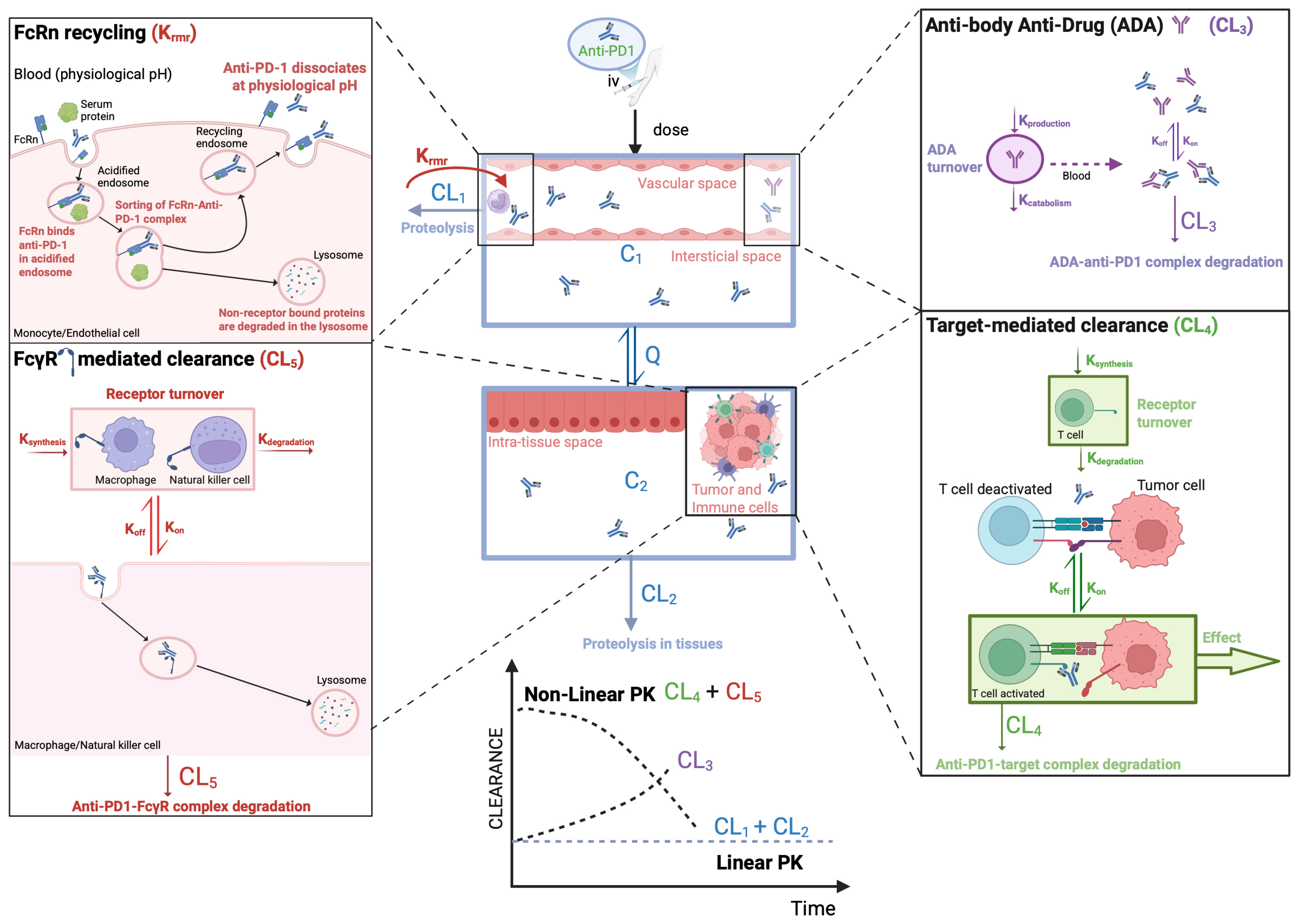
Figure 1. Anti-PD1 pharmacokinetic model to estimate time-dependent changes in clearance (CL). Legends: ADA—antibody anti-drug; Ksynthesis—synthesis constant; Kdegradation—degradation constant, Koff—dissociation constant; Kon—association constant, iv—intravenous. Figure elaborated using BioRender.com.
Cancers 17 03262 g001
Figure 2. Anti-PD1 time-dependent changes in clearance and relation to treatment response. Legends: CL0—basal clearance; ΔCL—change in CL over time. Figure elaborated using BioRender.com.
Cancers 17 03262 g002
Figure 3. CL-guided dose optimization workflow. Legends: AI—artificial intelligence; CL0—basal clearance; ΔCL—change in CL over time; Cmín, ss—steady-state trough concentrations; ELISA—enzyme-linked immunosorbent assay; LC-MS/MS—liquid chromatography tandem mass spectrometry; PopPK—population pharmacokinetic; figure elaborated using BioRender.com.
Cancers 17 03262 g003
Table 1. Anti-PD-1 linear and non-linear pharmacokinetic parameters and models.
PharmacokineticPembrolizumab [21,23]Nivolumab [21,24]Cemiplimab [25]
ModelTwo-compartment model with first-order elimination, zero-order iv infusion rate, and time-varying change in CL
LinearDoses range: 2–10 mg/kg
t1/2β = 22–27.3 days
CL = 0.2–0.22 L/day
Vdss = 6 L
Dose range: 0.1 to 20 mg/kg
t1/2α = 32.5 h, t1/2β = 25 days
CL = 0.226 L/day
Vc = 3.63 L; Vp = 2.78 L
Tmáx = 1–4 h (Normalized to BW of 80 kg)
Dose range: 1 to 10 mg/kg
CL = 0.246 L/day
t1/2β = 19.7 days
Vdss = 5.89 L
Non-linearTime-varying CL = 16–19 weeksTime-varying CL = 12–16 weeks
iv—intravenous; CL—clearance; Vdss—steady-state volume of distribution; BW—body weight; Tmáx—maximum concentration time; t1/2α,β—alfa and beta half-time, respectively.
Table 2. Biomarkers of effectiveness and safety.
BiomarkerCategoryStatusReferences
Effectiveness
PD-L1 expressionProtein (tumor/immune)Approved[142,143,144]
TMBGenomicApproved
(TMB ≥10 mut/1 Mb)
[144,145]
dMMR/MSI-HGenomicApproved[146,147,148]
Tumor-infiltrating lymphocytesHistologicalInvestigational[149]
Gene expression signaturesTranscriptomicInvestigational[135]
Antibiotic useClinical factorsObservational[150]
miRNA signatures (germline)EpigeneticExploratory[151]
Effectiveness and/or Safety
Lymphocyte counts/NLRHematologicInvestigational[152,153,154]
Circulating cytokines and
other inflammatory markers
Soluble proteinsInvestigational[155,156,157]
Clearance monitoring (CL0 and ΔCL)PharmacokineticsInvestigationalThis work * [123]
MSI-H: microsatellite instability; TMB: tumor mutational burden; PD-L1: programmed death-1 ligand; dMMR: DNA mismatch repair; NLR: neutrophil-to-limphocyte ratio; CL0—basal clearance; ΔCL—clearance variation by time; * biomarker proposed in the present review; further validation required.
Disclaimer/Publisher’s Note: The statements, opinions and data contained in all publications are solely those of the individual author(s) and contributor(s) and not of MDPI and/or the editor(s). MDPI and/or the editor(s) disclaim responsibility for any injury to people or property resulting from any ideas, methods, instructions or products referred to in the content.