Systematic Review

Systematic Review and Meta-Analysis of microRNA-7-5p Expression and Biological Significance in Head and Neck Squamous Cell Carcinoma

by
Rikki A. M. Brown
1,2,3,
Michael Phillips
1,2,
Andrew J. Woo
1,2,4,
Omar Kujan
5,
Stephanie Flukes
6,7,
Louise N. Winteringham
1,2,
Larissa C. Dymond
1,2,
Fiona Wheeler
1,2,
Brianna Pollock
1,2,3,
Dianne J. Beveridge
1,2,
Elena Denisenko
1,2 and
Peter J. Leedman
1,2,3,*
1
Harry Perkins Institute of Medical Research, QEII Medical Centre, Nedlands, WA 6009, Australia
2
Centre for Medical Research, The University of Western Australia, Perth, WA 6009, Australia
3
UWA Medical School, University of Western Australia, Perth, WA 6009, Australia
4
School of Medical and Health Sciences, Edith Cowan University, Joondalup, WA 6027, Australia
5
UWA Dental School, University of Western Australia, Perth, WA 6009, Australia
6
Department of Otolaryngology Head and Neck Surgery, Surgical Division, Fiona Stanley Hospital, Perth, WA 6150, Australia
7
Discipline of Surgery, Medical School, University of Western Australia, Stirling Highway, Crawley, WA 6009, Australia
*
Author to whom correspondence should be addressed.
Cancers 2025, 17(19), 3232; https://doi.org/10.3390/cancers17193232
Submission received: 18 July 2025 / Revised: 7 September 2025 / Accepted: 24 September 2025 / Published: 4 October 2025

Simple Summary

This study addresses the complex role of miR-7-5p in head and neck cancer, a disease with limited treatment options and poor survival rates. By integrating patient data, bioinformatics predictions, and laboratory experiments, our study demonstrated that miR-7-5p is frequently upregulated in tumours and linked to worse clinical outcomes. Yet, overwhelming evidence demonstrates the anti-cancer functions of synthetic miR-7-5p mimics. This comprehensive approach enhances our understanding of the role of miR-7-5p in cancer biology and provides a foundation for developing RNA-based therapies in head and neck cancer.

Abstract

Background: Head and neck squamous cell carcinoma (HNSCC) is a prevalent malignancy with poor clinical outcomes. microRNA-7-5p (miR-7-5p) has been described as both a tumour suppressor and an oncomiR depending on the tissue context, but its role in HNSCC remains unclear. This study aimed to clarify the clinical significance and biological function of miR-7-5p in HNSCC by integrating data from multiple sources. Methods: A systematic review of the literature was conducted to identify studies analysing miRNA expression in human head and neck tissues. A meta-analysis of individual patient data from Gene Expression Omnibus (GEO), ArrayExpress, and The Cancer Genome Atlas (TCGA) was performed to assess miR-7-5p expression in tumours and normal tissues, and its associations with clinical parameters and prognostic outcomes. Bioinformatics analyses were used to predict miR-7-5p target genes, classify hub genes, and perform gene ontology enrichment analysis. MicroRNA in situ hybridisation (miRNA ISH) and real-time quantitative PCR (RT-qPCR) were conducted on tissue samples, HNSCC cell lines, and an in vitro model of oral oncogenesis to validate miR-7-5p expression patterns. Results: miR-7-5p was significantly upregulated in tumours compared to normal tissues and associated with larger tumour size, HPV-negative status, poor disease-specific survival, and shorter progression-free intervals. Bioinformatics analysis highlighted miR-7-5p target genes enriched in pathways related to cell growth, survival, and tumourigenesis. Despite evidence supporting the anti-cancer role of exogenous miR-7-5p in preclinical models, the observed endogenous upregulation in tumours suggests that miR-7-5p expression may represent a compensatory or stress-responsive mechanism during tumourigenesis, rather than acting as a primary oncogenic driver. Conclusions: This study provides new insights into the complex role of miR-7-5p in HNSCC, supporting its potential as both a biomarker and a therapeutic target. Understanding the context-specific functions of miR-7-5p is essential for its development as an RNA-based therapeutic in HNSCC.

1. Introduction

Head and neck squamous cell carcinoma (HNSCC) is the sixth most common cancer worldwide, and its incidence and mortality continue to rise. HNSCC develops in the epithelial lining of the oral cavity, sinonasal passages, pharynx, and larynx [1]. The aetiology of HNSCC is linked to both genetic and environmental factors [1]. Males are more likely to be diagnosed with the disease, and other risk factors include family history, diet, smoking, and alcohol abuse. There is also a causal link between viral infections, namely human papillomavirus (HPV) and Epstein–Barr virus (EBV). The HPV status and stage are recognised as major determinants for the prognosis of HNSCC. Although histological evidence presents an evolutionary model of malignant progression, most patients are diagnosed with advanced HNSCC without the detection of a pre-malignant lesion. Furthermore, not all pre-malignant lesions, such as leucoplakia, transform into invasive HNSCC [2]. The lack of effective markers for detecting early-stage disease has led to more cases being diagnosed at advanced stages, which are more difficult to treat [3]. Treatment of HNSCC is typically multimodal, consisting of surgery, radiation, and chemotherapy and requires a careful balance between achieving the best possible cancer control and preserving vital functions such as speech, swallowing, and appearance [4]. Identifying markers that refine the treatment decision-making for HNSCC may improve prognosis, as the 5-year survival rate (~50%) has only modestly improved despite advances in our knowledge. Given the multifactorial causes and the heterogenic nature of the disease, further research into biomarkers that improve early detection and prognostic accuracy will produce better patient outcomes.
MicroRNAs (miRNAs) are small endogenous non-coding RNA molecules that regulate key biological processes by fine-tuning gene expression. They negatively regulate gene expression at the post-transcriptional level by binding to sequences in the 3′-UTR of target genes, inhibiting translation or causing mRNA degradation. Owing to the imperfect complementarity, a single miRNA can target numerous genes, and likewise, a single gene can be targeted by multiple miRNAs [5]. Furthermore, the expression of many genes can be altered indirectly, such as when the expression of a transcription factor is controlled, leading to flow-on effects that control cell fate and biological function [6]. miRNAs can be detected within cells or secreted in extracellular vesicles. Thus, their expression can be evaluated not only from solid tissue samples but also non-invasively in serum or saliva [7].
The development of miRNA-based therapies relies on a detailed understanding of tumour-specific miRNA expression patterns and their relationships to disease biology, prognosis, and treatment response. miRNAs can act as oncogenes or tumour suppressors depending on their expression levels and the cellular context, which includes tissue type, disease stage, and molecular subtype. For instance, some miRNAs may drive initial oncogenic transformation but have no impact on disease progression, or vice versa. miR-7-5p is one such miRNA, described as both a tumour suppressor and an oncomiR in various cancer types, including glioblastoma, breast, lung, and colorectal cancer [8,9]. Its known target genes include key components of pathways frequently dysregulated in cancer, such as EGFR, PI3K/Akt, and RAF1, suggesting that it may play a pivotal role in oncogenic signalling networks [10,11,12,13,14,15]. In HNSCC, the literature is similarly conflicting, with several studies that report miR-7-5p is upregulated in tumour samples and cell lines compared to normal controls [16,17], whereas others describe its downregulation in HNSCC tissue relative to normal [18,19]. This inconsistency raises important questions about the biological role of miR-7-5p in HNSCC, and whether it represents a viable therapeutic target. Notably, exogenous delivery of miR-7-5p has been shown to suppress tumour growth in preclinical HNSCC models [10,19,20], but it remains unclear whether this therapeutic effect is influenced by the endogenous expression status of miR-7-5p. Given the contradictory nature of these observations, our study aims to clarify the expression pattern of miR-7-5p in HNSCC tumour tissues and cell lines compared to normal controls, and to investigate how its expression relates to its biological function in HNSCC.

2. Materials and Methods

2.1. PICO Framework

The study was based on the following PICO framework:
  • P (Population/Problem): Patients with HNSCC
  • I (Intervention/Exposure): Analysis of miR-7-5p expression in patient samples
  • C (Comparison): Expression levels of miR-7-5p in matched normal samples
  • (Outcome): Determination of miR-7-5p’s biological role (tumour suppressor vs. oncomiR) and potential as a therapeutic target in HNSCC

2.2. Search Strategy

The protocol for this systematic review has been registered in the International Prospective Register of Systematic Reviews (PROSPERO: CRD42021283642; available at http://www.crd.york.ac.uk/PROSPERO/display_record.php?ID=CRD42021283642, accessed on 6 October 2021). This study was performed in accordance with Preferred Reporting Items for Systematic Reviews and Meta-analyses (refer to Table S1 for the PRISMA 2020 Checklist). Individual patient data were sought from GEO, ArrayExpress, and dbDEMC databases. A systematic literature search was conducted using Medline via PubMed, Embase, and Web of Science (WOS) to identify studies that may have included raw data in Supplementary Files or reported fold-changes only. The following search terms were used: (“nasal” OR “oral” OR “tongue” OR “mouth” OR “lip” OR “head and neck” OR “laryngeal” OR “larynx” OR “pharyngeal” OR “pharynx”) AND “patients” AND (“cancer” OR “carcinoma”) AND (“microRNA” OR “miRNA” OR “miR”). When the miRNA expression data were not included in the article or submitted to a public repository, efforts were made to contact the corresponding authors to obtain the raw data.

2.3. Selection Criteria

Studies were eligible if they included solid tissue or liquid biopsy samples from patients with HNSCC confirmed by a pathologist and non-cancer controls (normal adjacent tissue or healthy controls). If no non-cancer controls were analysed, the HNSCC cases had to contain matched clinical information to make meaningful comparisons. HNSCC cases were defined as those arising in the mucosal epithelium of the oral cavity, nasal cavity, mouth, lip, pharynx, or larynx. Cancers originating from the oesophagus, salivary gland, thyroid, or other tissues in the head and neck region, such as skin, eyes, and ears were excluded, as these often arise in non-squamous cells and/or are usually classified separately. Samples of oral leucoplakia were not included, as they have the potential to become malignant but do not always transform into invasive carcinoma and therefore cannot be classed as “normal” or “tumour” tissue. Studies that analysed animal samples, primary cell cultures, cell lines, and cell-sorted samples, or data extracted from public repositories (e.g., The Cancer Genome Atlas), were excluded. If original data were not associated with the record, e.g., conference proceedings, abstracts, review articles, case reports, or letters to the editor, and it was not provided by authors upon request, the study was considered ineligible. If the study was non-peer reviewed, lacked sufficient methodology or relevant outcomes, or the article was subsequently retracted, it was excluded. If miR-7-5p was not investigated, miR-7-5p was not detected in >60% of samples, failed quality control, or if the data were uninterpretable, it was not included. For example, undetectable samples had CT values > 35 for RT-qPCR analyses, Probe Signal Intensity values below background and/or an error message for a gene not detected in the raw data. The normalised expression values were obtained from GEO2R or processed gene expression matrices and assessed for quality by two reviewers (R.B. and A.W.) to ensure consensus.

2.4. Data Extraction and Quality Assessment

Research articles identified through the search strategies were compiled in EndNote Reference Managing Software (X8). Smart Groups were used in the screening stage to remove duplicate references and assign eligibility based on the selection criteria. For each record suitable for inclusion, the following information was extracted: first author/contributor, year published, array accession number, country the study was conducted in, patient characteristics (age, gender, risk factors, etc.), specimen characteristics (source, number of participants, control samples, method of preservation, primary tumour site, etc.), assay methods (methods for RNA isolation, miRNA detection, and data normalisation), study design, and reported outcomes (overall survival, disease-free survival, etc.). Data were collated in a standardised data extraction format using Microsoft Excel. For this meta-analysis, normalised gene expression values were used as provided in each dataset, rather than extracting and reprocessing raw data in order to preserve dataset integrity; however, this approach limits direct cross-platform comparability due to differences in normalisation methods and batch effects. The quality of the research articles was evaluated based on the Newcastle–Ottawa Quality Assessment Scale (refer to Table S2). Quality of the arrays was assessed by two reviewers (R.B. and A.W.).

2.5. The Cancer Genome Atlas Data Validation

Data were retrieved for the head and neck squamous cell carcinoma (TCGA-HNSC) cohort via the UCSC Xena browser (https://xenabrowser.net). Mature miRNA expression (Mature accession: MIMAT0000252) was measured using Illumina Hiseq (n = 522) or Illumina GA (n = 33) RNA sequencing platforms. To compare the correlation of miR-7-5p expression with clinicopathological characteristics of the patients, samples were separated into tumour or normal groups, and the level of significance for differences between means was measured using unpaired two-tailed t-test for comparing the whole cohort or via a paired t-test when examining matched tumour–normal samples. Tumour samples were further stratified by high or low expression of miR-7-5p using the median as a cut-off. Expression of miR-7 pre-miRs was extracted from miRCancerDB [21]. Pan-cancer query of miR-7-5p expression, Receiver Operator Characteristic (ROC) analysis, and correlations of miR-7-5p with putative targets were obtained from CancerMIRnome [22]. Expression, contingency and survival analyses were performed using GraphPad Prism software (v8.3.1).

2.6. miRNA and Target Gene Quantitations in Patient Samples

Paired HNSCC tissue and adjacent normal samples were obtained from sixteen patients undergoing surgical resections at the Fiona Stanley Hospital during 2023–2025. Patients provided informed consent, and the study was approved by the Sir Charles Gairdner and Osborne Park Health Care Group Human Research Ethics Committee (RGS0000000919). Snap-frozen tissues were homogenised in lysis buffer using a TissueLyser II (Qiagen, Hilden, Germany), and RNA was isolated using the AllPrep DNA/RNA/miRNA Universal Kit (Qiagen). RNA was quantitated with a Nanodrop ND-1000 spectrophotometer (Thermo Fisher Scientific, Waltham, MA, USA), and the 260/280 and 260/230 ratios of absorbance values were used to assess the purity of RNA. For detection of miRNA expression, RNA was reverse transcribed and real-time quantitative PCR (RT-qPCR) was conducted using TaqMan MicroRNA Assay kits for miR-7-5p (Cat#4440887, ID: 000268) and the U6 snRNA miRNA control (Cat#4427975, ID: 001973) with TaqMan Universal PCR Master Mix (Thermo Fisher Scientific) as per the manufacturer’s instructions. Target gene expression was assessed by reverse transcribing RNA into cDNA using a QuantiTect Reverse Transcription Kit (Qiagen, Cat# 205311), then performing RT-qPCR using SensiMix SYBR Hi-ROX kit (Bioline, Meridian Bioscience, Memphis, TN, USA, Cat# QT605-20) and primers on a QuantStudio 6 Pro System (Thermo Fisher Scientific) using recommended cycling conditions. Validated Quantitect primer assays (Qiagen) were used for EGFR (QT00085701), IRS1 (QT00074144), and IGF1R (QT00005831), and primers for GAPDH (GAPDH-F 5′-GGG GTC ATT GAT GGC AAC AAT A-3′, GAPDH-R 5′-ATG GGG AAG GTG AAG GTC G-3′), RAF1 (RAF1-F 5′-GCA CTG TAG CAC CAA AGT ACC-3′, RAF1-R 5′- CTG GGA CTC CAC TAT CAC CAA TA-3′), and PIK3CD (PIK3CD-F 5′-AAG GAG GAG AAT CAG AGC GTT-3′, PIK3CD-R 5′-GAA GAG CGG CTC ATA CTG GG-3′) were synthesised by Sigma-Aldrich (St. Louis, MO, USA). Relative gene expression was determined using the 2−ΔΔCt method with normalisation to the appropriate endogenous control (U6 for miRNA and GAPDH for target genes), and expression was displayed relative to a calibrator, as described in the figure legends.

2.7. Bioinformatic Analyses

Cross-species conservation was compared using miRBase v22.1 [23], and a circos plot was generated with the Synteny portal [24]. miRNA variants were identified by miRNASNP v3 [25]. Several databases were compared to compile a list of predicted miR-7-5p target genes. miRDB [26], miRTarBase v9.0 [27], TargetScan v7.1 [28], and DIANA-TarBase v8 [29] use alternative detection algorithms, and genes that appeared in >3 databases were considered candidate targets with a high level of confidence. Gene ontology enrichment analysis was performed based on the short-listed candidates [30,31]. GO terms were filtered for redundancy using REVIGO (https://github.com/rajko-horvat/RevigoWeb, accessed on 6 November 2021) [32], and the most representative (dispensability cutoff < 0.05) were plotted using the ggplot2 package in R v4.1.2, as described in Bonnot et al. (2019) [33]. Fold enrichment was used to compare over/under representation of GO terms, and items with FDR < 0.05 from Fisher’s Exact test with Bonferroni correction were considered significant. The STRING v11.5 database (https://string-db.org/) was used to construct a protein–protein interaction (PPI) network to analyse interactions between putative target genes, based on interaction score > 0.7 (high confidence).

2.8. Cell Culture and Drug Treatment

CAL27 and CAL33 cells are immortalised cancer cell lines derived from moderately differentiated squamous cell carcinomas of the tongue and were kindly provided by A/Prof. Pawan Kumar (The Ohio State University) and Prof. Jennifer Grandis (University of Pittsburgh Cancer Centre), respectively. These cells were cultured in Dulbecco’s Modified Eagle’s Medium (DMEM) High Glucose, supplemented with 1X MEM Non-Essential Amino Acids Solution and 10% foetal bovine serum (FBS). Human Oral Keratinocytes (HOK) and Dysplastic Oral Keratinocytes (DOK) were kindly provided by Dr Omar Kujan (University of Western Australia). HOK were cultured in DMEM:F12 supplemented with 10% FBS, 400 ng/mL hydrocortisone (Sigma-Aldrich), and 1% penicillin/streptomycin. DOK were maintained in Advanced DMEM (Invitrogen, Thermo Fisher Scientific, Waltham, MA, USA) with 2% GlutaMAX (Invitrogen, Thermo Fisher Scientific), 3% FBS, 10.3 µM hydrocortisone, and 1% penicillin/streptomycin. All cells were cultured at 37 °C in 5% CO2 for no more than 20 passages (<5 for primary cells) and were confirmed to be free of mycoplasma. RNA was extracted from cultured cells using the RNeasy kit (Qiagen, Cat#74104), following the manufacturer’s instructions, and RT-qPCR was performed as described for patient samples above.
For the in vitro model of oral oncogenesis, HOK and DOK were treated with the chemical carcinogen 4-Nitroquinoline N-oxide (4NQO, Sigma-Aldrich, Cat# N8141) at 0.36 µM (HOK low-dose 4NQO) or 0.72 µM (DOK high-dose 4NQO) concentrations or vehicle control (DMSO) for a total of 7 weeks for HOK and 6 weeks for DOK [34]. The cells were treated every alternate day with media containing the drug for a period of 90 min, then replaced with complete growth medium, and cells were passaged weekly. Cryopreserved vials of these treated cells were provided by Dr Omar Kujan; cells were resurrected, and RNA was harvested when cells reached ~80% confluency. Human periodontal ligament fibroblasts (HPLF) and human oral fibroblasts (HOF), provided by Dr. Omar Kujan, were grown in Fibroblast Growth Medium, all-in-one ready-to-use media (Sigma-Aldrich).
For acute 4NQO treatment, cells were seeded in 6-well plates and allowed to adhere overnight and then cultured in media containing 4NQO at the indicated concentration for 24 h. RNA was extracted using the RNeasy Kit, following the manufacturer’s instructions (Qiagen), and miRNA expression was analysed as described for the patient samples. For UV treatment, cells were seeded in 6-well plates and allowed to adhere overnight. For UV exposure, growth media was removed and set aside, and cells were exposed to UV light using a Stratagene Stratalinker UV 1800 Crosslinker at 100 mJ/cm2, as per the manufacturer’s instructions. Culture plate lids were removed during irradiation. Immediately following UV treatment, the original media was replaced, and cells were returned to the incubator to recover for 2 or 24 h. Total RNA was harvested using the RNeasy kit (Qiagen), according to the manufacturer’s protocol, and RT-qPCR was performed as previously described.
Cell viability was assessed following modulation of miR-7-5p expression using synthetic miRNA mimics or inhibitors (ThermoFisher, Cat# AM17100, AM17000). Cells were seeded in 96-well plates and transfected using Lipofectamine 2000 (ThermoFisher, Cat# 1668019), according to the manufacturer’s instructions. HOK were forward-transfected, whereas oral squamous cell carcinoma lines (CAL27, CAL33) were reverse-transfected. After transfection, cells were incubated under standard culture conditions for 72 h, and viability was measured using a CellTiter AQueous One Solution Cell Proliferation (MTS) Assay (Promega, Madison, WI, USA, Cat# G3580), following the manufacturer’s protocol, and absorbance was read on a Clariostar plate reader (BMG Labtech, Ortenberg, Germany). Each condition was performed in triplicate, and experiments were independently repeated three times. Blank-corrected values were normalised to the lowest concentration of the test agent, and dose–response curves were plotted using GraphPad Prism software (v9.5.1).

2.9. Animal Study and microRNA In-Situ Hybridisation (ISH)

Female 5–6-week-old C57Bl/6 mice (Animal Resources Centre, Perth, Australia) were acclimated for 1 week, then treated with 4NQO (100 µg/mL) or vehicle control (propylene glycol) in the drinking water for a period of 16 weeks, followed by regular drinking water for a total holding period of 24 weeks. Tongues were harvested, fixed in formalin, embedded in paraffin blocks, and sectioned. For ISH, the miRCURY LNA miRNA ISH Optimisation Kit (FFPE) (Qiagen) was used, following the manufacturer’s one-day microRNA ISH protocol. Proteinase K (15 µg/mL) treatment was performed at 37 °C for 10 min, and the digoxigenin (DIG)-labelled LNA probe for miR-7-5p was used at a final concentration of 80 nM (Qiagen), miR-21 at 60 nM, and then the slides were incubated at 55 °C for 60 min. Scramble control and U6 were used as staining controls. Stained slides were imaged with a 3DHistech Scanner (3DHistech, Budapest, Hungary).

2.10. Statistics

The miRNA expression data were analysed to estimate the Standardised Mean Difference between tumour and normal tissues. The meta-analyses were conducted with STATA (v16) using the ipdmetan package [35], Review Manager (RevMan, v5.4.1) for the dichotomous variables, or cancerMIRNome [22] for the ROC analyses of TCGA data. Estimations of the standardised mean difference of miR-7-5p expression in HNSCC patients and controls, and their prognostic effects were presented using forest plots. Either a random- or fixed-effects model was applied to calculate the pooled effect size, as detailed in the text, and 95% confidence intervals were calculated for all studies that included individual patient data. Cochrane’s Q statistic and inconsistency index (I2) test were used to assess heterogeneity, with an I2 ≥ 50% considered a high heterogeneity level. Further subgroup and meta-regression analyses were performed to investigate the source of heterogeneity. Galbraith plots were used to visualise heterogeneity. Funnel plots were used to visualise publication bias. GraphPad Prism was used to generate graphs and analyse datasets and experimental results. p-value < 0.05 was considered statistically significant, with the appropriate statistical test applied, as described in each figure legend.

3. Results

3.1. Study Selection

Based on the search strategy as outlined in Figure 1, a total of 4235 records were identified from Medline via PubMed, Embase, GEO, ArrayExpress, ddDEMC, WOS, and additional references. The published datasets identified in GEO, ArrayExpress, and ddDEMC were matched back to the original citing research article. Approximately 44% were duplicates that were removed from further assessment. Screening of the abstracts from the remaining records excluded a large proportion of studies that either did not investigate HNSCC, were not focused on miRNAs, did not include patient samples, or were analysing public array data. Further refinement excluded retracted studies and those with no associated individual patient data. This included 11 studies not suitable for inclusion in the meta-analysis, as they reported summary fold changes and p-values without standard error or deviations, and therefore an effect size could not be estimated [17,36,37,38,39,40,41,42,43,44,45]. These have been summarised in Table S2, and in all reports, the fold change indicated that miR-7-5p was significantly upregulated in tumour tissues compared to normal controls. To confirm this finding using individual patient data identified through our search strategy, a total of 71 studies were initially considered eligible; however, in 36 of these, miR-7-5p was either not detected or the data were not suitable for analysis, including several studies using serum or saliva samples. Consequently, 35 studies with analysable individual patient data were included in the meta-analyses, all of which were limited to solid tumour/normal samples. Following assessment using the Newcastle-Ottawa Scale, these studies were deemed suitable for inclusion in the analyses (Table S3). A total of 24 studies provided gene expression data for tumour and normal samples [16,46,47,48,49,50,51,52,53,54,55,56,57,58,59,60,61,62,63,64,65,66,67]. An additional 11 studies provided gene expression data for tumour samples only but included sufficient clinical metadata to analyse survival or dichotomous variables (e.g., age, stage, grade, etc.) [68,69,70,71,72,73,74,75,76,77,78].

3.2. Meta-Anlysis of miR-7-5p Expression in HNSCC

To compare tumour and normal tissue samples with individual patient data, a total of twenty-four papers were found (Table 1) and included in the meta-analysis. The included papers were published between 2009 and 2020 and came from eight different countries, including ten from China [16,46,47,48,49,50,51,52,53,54,55,56,57,58,59,60,61,62,63,64,65,66,67]. A broad range of tumour sites, risk factors, and disease stages were represented (Table S4). As summarised in Table 1, the average expression of miR-7-5p for both tumour and normal tissue varied widely (Tumour: 3.29–8478; Normal: 3.52–2569). Although the units of normalised gene expression extracted differed by study (Table S4), the values were not associated with the assessment technique: detection method (p = 0.11), RNA isolation method (p = 0.71), or normalisation of the data (p = 0.22) in univariate analysis. The tumour/normal ratios show that, in the vast majority of the studies, miR-7-5p was upregulated in tumour tissue compared to normal (>1), similar to those that reported fold change and p-value only (Table S2).
The initial meta-analysis using a random-effects model showed a pooled effect size (Hedges’ g) of 0.917 (95% CI of 0.602–1.23, p < 0.001), indicating significantly higher miR-7-5p expression in tumour compared to normal tissues (Figure 2). However, using all 24 studies without differentiation demonstrated a large amount of heterogeneity between studies with respect to miR-7-5p expression (I2 = 76.0, Q = 96.7, p < 0.0001), as illustrated in Figure 2, which warranted further investigation.

3.3. Meta-Regression Analyses

The subgroup meta-analysis summary (Table S5) highlighted key sources of heterogeneity. Tumour site was found to be a significant source of heterogeneity (Q = 16.3, df = 3, p = 0.001). The effect size of studies on tumours of the oral cavity was significantly larger than that of studies on other subsites of the head and neck (coefficient = 1.26, 95% CI of 0.656–1.86; p < 0.001), as detailed in Table S6. There were no notable differences between the reference group (pharynx) and the larynx (p = 0.147) or mixed site categories (p = 0.074). The normalisation method was an additional source of heterogeneity (Q = 43.9, df = 6, p < 0.001) identified by the subgroup analysis (Table S5). Specifically, quantile-normalised studies reported considerably higher tumour-normal differences than the reference approach (variance-stabilising transformation) (coefficient = 1.44, 95% CI of 0.826–2.06; p < 0.001), as summarised in Table S7. There were no discernible effects from other normalisation techniques.

3.4. Sensitivity Analysis: Excluding Quantile-Normalised Studies

Galbraith and funnel plots further supported that quantile normalisation contributed to the observed heterogeneity and potential bias in effect size estimates (Figure 3). To assess the influence of quantile normalisation, a sensitivity meta-analysis was performed excluding those five studies. As shown in Figure 4, the overall effect size decreased to 0.606 (95% CI: 0.343–0.869; p < 0.001), with a substantial reduction in heterogeneity (Q = 43.02, p = 0.001, I2 = 56.2%). Notably, the heterogeneity associated with oral cavity studies dropped markedly (Q = 1.14, p = 0.889, I2 < 0.01%).

3.5. Analysis of miR-7-5p Expression and Clinical Characteristics Based on TCGA Data

TCGA data are a valuable resource for validating meta-analysis findings by providing large-scale, clinically annotated genomic datasets that can help confirm observed expression patterns and explore potential clinical drivers, such as tumour subtype, stage, and patient outcomes. The TCGA HNSCC cohort comprised 554 samples, consisting of 514 tumours, of which 40 had matched normal samples. Table 2 demonstrates that miR-7-5p expression was not significantly correlated with age, sex, clinical stage, tumour grade, invasion status, metastasis, or history of smoking or alcohol use. However, tumours from HPV-negative patients (p < 0.001), those with TP53 mutations (p < 0.01), and tumours classified as larger in size (T3–T4, p < 0.05) had higher miR-7-5p expression. Figure 5 illustrates that miR-7-5p expression was significantly higher in tumour tissues compared to normal tissues, both in unmatched tumour vs. normal comparisons (Figure 5A, p< 0.0001) and in paired tumour–normal samples (Figure 5B, p < 0.001). No significant differences in miR-7-5p expression were observed across different anatomical subsites of the head and neck (Figure 5C, p = 0.431).
Regarding clinical outcomes, miR-7-5p expression did not correlate with overall survival (Figure 5D; HR = 1.18, p = 0.225). However, disease-specific survival was lower in individuals with high miR-7-5p expression (HR = 1.56, p = 0.012). Regardless of treatment status, patients with low miR-7-5p levels had a longer progression-free interval than those with high expression (HR = 1.36, p = 0.031), but there was no discernible difference in disease-free interval (HR = 1.24, p = 0.569).
As miR-7-5p is transcribed from three genomic loci, further examination of TCGA data revealed that miR-7-1 is the main transcript causing increased mature miR-7-5p expression in HNSCC (Figure 5H). This result is consistent with copy number variation (CNV) data (Figure 5I), which demonstrates contemporaneous losses in MIR7-2 and MIR7-3 and frequent copy number gains in MIR7-1.

3.6. Meta-Analysis of Clinical Subgroups and Dichotomous Variables

To evaluate overall effect sizes in the associations found in the TCGA dataset, clinical characteristics were paired with matching data from the systematic review studies. The selection of a fixed-effects model was supported by tests for heterogeneity (I2 < 50%, p > 0.05). With an odds ratio of 0.58 (95% CI: 0.39–0.85, p = 0.005), HPV status was the only variable among the pooled analyses that was significantly correlated with miR-7-5p expression (Table 3).
Although hazard ratios for overall survival and disease-free interval were not significant in the TCGA cohort alone, the disease-free interval may have been underpowered due to smaller sample sizes. To improve precision, these outcomes were pooled with data from the additional studies identified in the systematic review (Table S4). As illustrated in Figure 6, the pooled estimates remained non-significant.

3.7. Bioinformatics Analyses of miR-7-5p in HNSCC

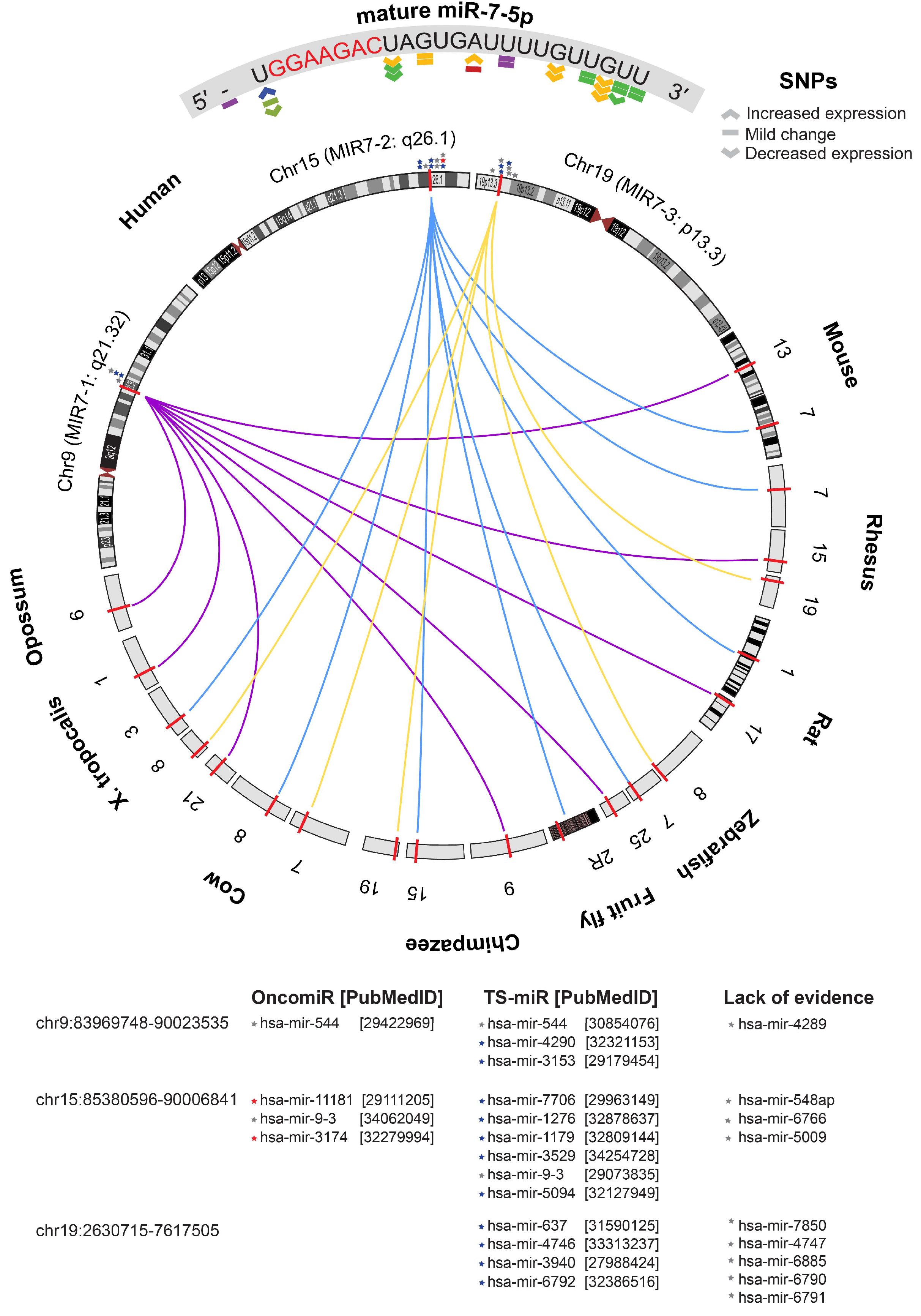
To explore the biological relevance of elevated tumour miR-7-5p expression, we first evaluated evidence that would broadly characterise it as a putative oncomiR or tumour suppressor-miRNA (TS-miR), based on criteria described by Wang et al. (2010) [79]. Relying on the evolutionary conservation theory, the results support a putative tumour suppressor role for miR-7-5p founded upon the following: (1) the mature sequence is highly conserved across species other than humans and primates; (2) there is replication of multiple genes encoding the same mature miR-7-5p sequence; (3) the detection of multiple SNPs that are predicted to mildly change or reduce expression; and (4) the clustering of MIR-7 genes close to other TS-miRs and in genomic regions that are frequently deleted (Figure 7). Such features are characteristic of genes under selective pressure to maintain function and expression, which suggests that miR-7-5p plays a protective role against cancer development and that its loss or downregulation could contribute to tumour progression.
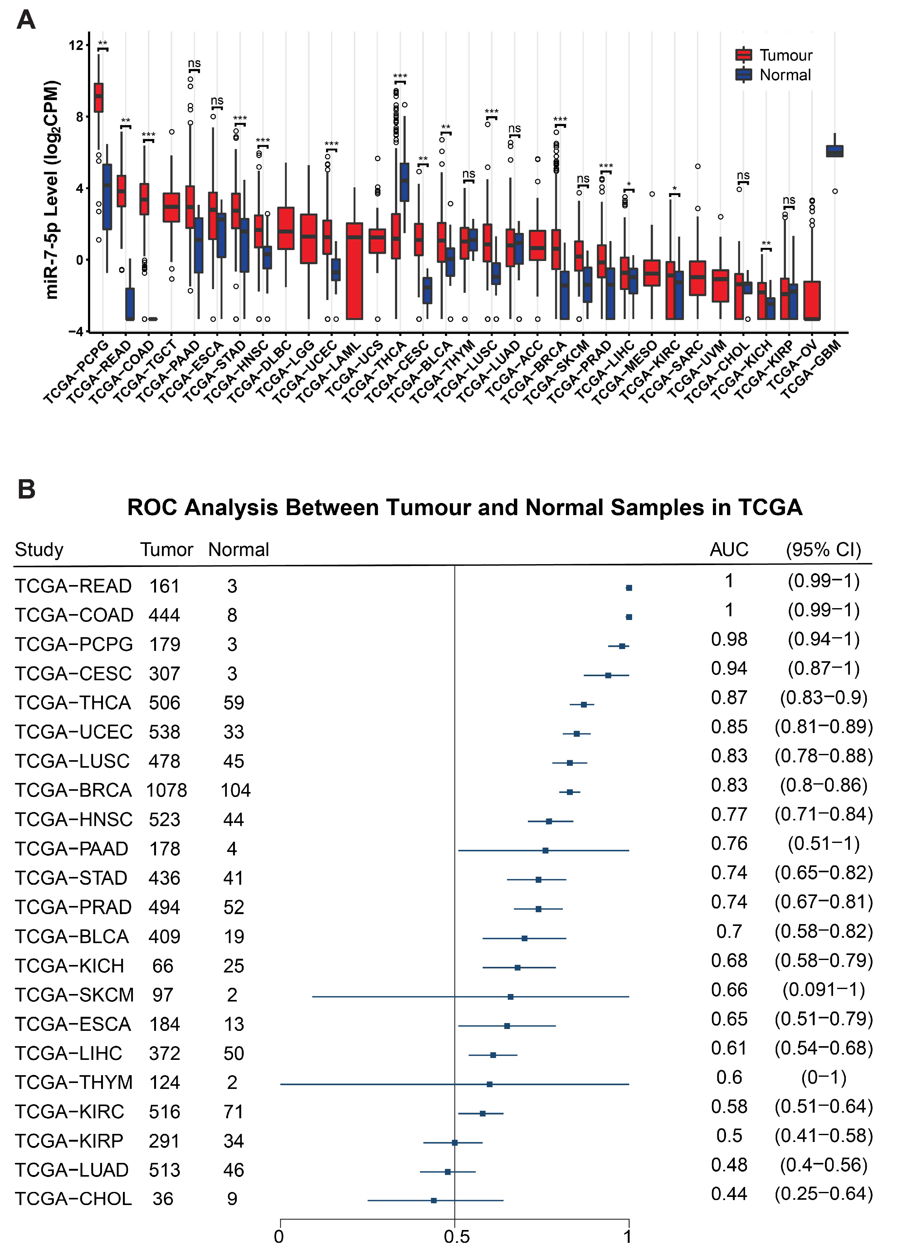
Despite these data supporting innate tumour suppressor features, pan-cancer analysis of TCGA data shows that miR-7-5p is differentially expressed in many tumour types, and, except for thyroid cancer (THCA), it was significantly higher in tumour tissue compared to normal (Figure 8A). Receiver Operator Characteristic (ROC) analyses show that miR-7-5p has potential as a diagnostic biomarker for differentiating tumour from normal for multiple cancer types (Figure 8B).

3.8. Functional Annotation of Candidate Target Genes of miR-7-5p and Construction of a Protein–Protein Interaction (PPI) Network

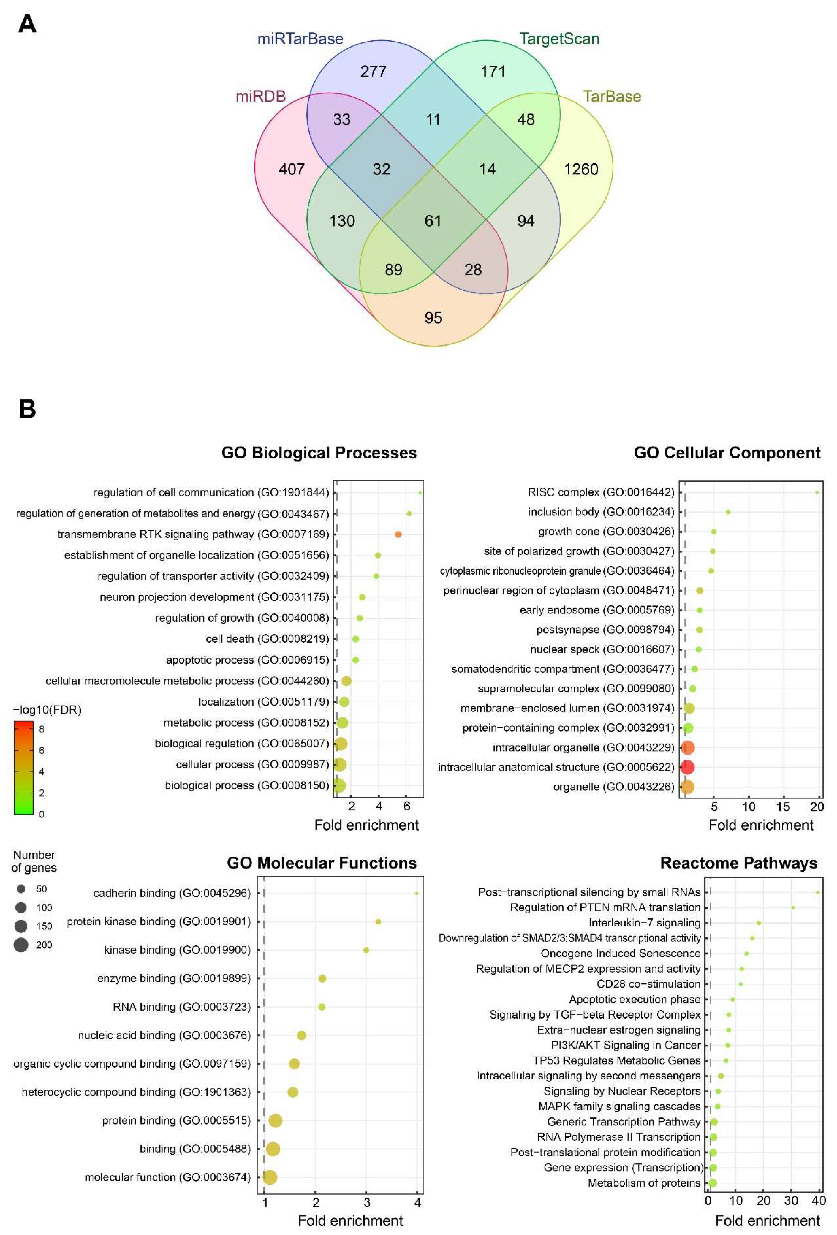
Comparison of four different databases that predict genes with a seed region for miR-7-5p identified a total of 2750 genes, of which 224 were identified in at least three databases (Figure 9A). These 224 genes were mapped to Gene Ontology (GO) terms, which showed an overrepresentation of biological processes pertaining to cell signalling and communication; regulation of cell growth/death, localisation, and metabolism; a diverse range of GO cellular components, with the RISC complex having the highest fold enrichment; and all the GO molecular functions related to binding (Figure 9B). Furthermore, the Reactome pathways identified by PANTHER analysis showed an enrichment for pathways that are associated with tumourigenesis, such as TP53, PI3K/AKT, MAPK, and TGF-β signalling cascades (Figure 9B).
The protein–protein interaction (PPI) analysis constructed a network of 224 nodes and 122 edges, with a PPI enrichment p < 0.001. RAF1, EGFR, PIK3CD, PIK3R3, IGF1R, IRS1, and SNCA had string node degrees ≥ 8, indicating their potential as hub genes. Expression of the hub genes was correlated with miR-7-5p in the TCGA HNSC cohort; with RAF1 and PIK3R3 being the only genes with a significant negative correlation, while the others were positively correlated or the p-value indicated it was not significant (Figure 10B).

3.9. Validation of Meta-Analysis and Biological Investigation

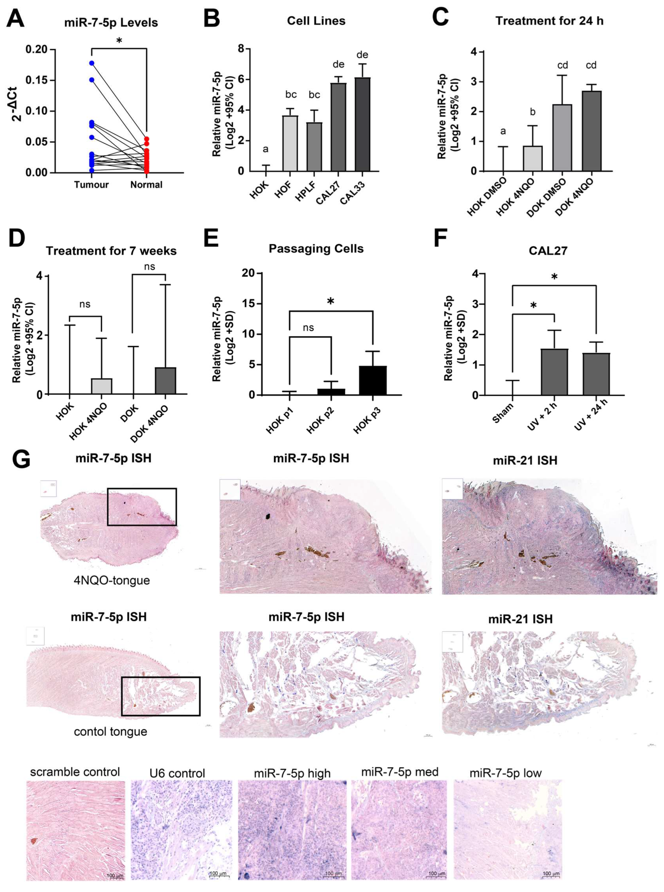
To further investigate the findings of elevated miR-7-5p expression in HNSCC tumours, we evaluated our own clinical cohort of patient samples, cultured cells, and animal tissues. We obtained tumour and normal tissue from sixteen patients (Table S8), with histopathological confirmation of head and neck squamous cell carcinoma. RT-qPCR analyses of these samples showed an elevated expression of miR-7-5p in the tumours compared to matched normal tissues (Figure 11A). Further, examining the expression of five of the hub genes identified in Figure 10A showed a trending correlation in line with Figure 10B; however, unsurprisingly, these did not reach statistical significance due to the small sample size (Figure S1).
The expression of miR-7-5p was compared between several normal oral cell lines (HOK, HOF, and HPLF) and CAL27 and CAL33 oral cancer cell lines. All comparisons were significant except for the pairwise comparisons of the fibroblast cells (HOF v HPLF) and cancer cell lines (CAL27 v CAL33), with miR-7-5p levels being significantly higher in fibroblasts compared to keratinocytes and in the cancer cells compared to normal oral cells (Figure 11B), which is consistent with the data derived from processing bulk tissue samples (Table 1).
To further investigate miR-7-5p expression changes, we used an in vitro model of oral oncogenesis. The HOK and DOK cells from a low passage were treated with the chemical carcinogen 4NQO, which induces cytotoxicity in a manner similar to tobacco products. Interestingly, an acute treatment with 4NQO caused an increase in miR-7-5p expression in normal keratinocytes (Figure 11C), but when the HOK or DOK cells were cultured long-term with pulse treatments of 4NQO for up to 7 weeks, the differences compared to untreated cells were no longer significant (Figure 11D). However, miR-7-5p expression also increased with continued passaging of HOK cells (Figure 11E) and could be linked to subculturing of the cells. Additionally, cell exposure to high-dose UV (100 mJ/cm2) induced miR-7-5p expression, suggesting a connection to the DNA damage response pathway (Figure 11F).
To investigate tissue-wide expression patterns of miR-7-5p, we evaluated its expression in a murine model of 4NQO-induced oral carcinoma using microRNA ISH. The section in Figure 11G shows a prominent, raised, and hyperplastic region with disrupted architecture and loss of normal layering, which is consistent with an invasive squamous cell carcinoma-like lesion. The presence of keratin pearl formation or hyperkeratosis, typical of well-differentiated squamous tumours, is also observed. The chromogenic signal (blue/purple) for miR-7-5p appears granular and cytoplasmic, which is consistent with typical microRNA ISH localisation. Strong epithelial expression of the target miR-7-5p is observed in non-tumour regions, especially in suprabasal layers, with medium heterogenous staining in the tumour bulk and focal stromal positivity, which may correspond to fibroblasts, endothelial cells, or infiltrating immune cells. miR-7-5p signal is evident in the keratinocytes, especially at the tumour-normal interface. Detection of miR-21, a well-established oncomiR, was used as a positive control for tumour expression patterns and also showed concentrated expression in the suprabasal epithelial layers and some regions of the tumour mass, particularly along the invasive front, and sparse signal in the underlying stroma. In contrast, for vehicle-treated mice, although present in the basal and suprabasal epithelial layers, miR-7-5p staining patterns were not diffuse, were absent in the deeper connective tissue and muscle layers and showed very little or no staining in the glands, except for a few positive structures, possibly ducts or nerves, indicating low-level background or physiological expression. This implies that the target miRNA is not ubiquitously expressed in keratinocytes. This pattern may reflect localised expression in certain cell types or an early response to stimuli, such as in response to exposure to the chemical carcinogen 4NQO or catastrophic DNA damage from UV.
We have previously shown that miR-7-5p overexpression in HNSCC cell lines suppresses growth in vitro and in vivo [10]. To further investigate the functional role of miR-7-5p, we manipulated its expression in normal and malignant oral cells (Figure 12). Overexpression of miR-7-5p in human oral keratinocytes (HOK), which express low endogenous levels of miR-7-5p, did not significantly affect cell viability. In contrast, inhibition of miR-7-5p in oral squamous cell carcinoma cell lines (OSCC) CAL27 and CAL33, which have high endogenous miR-7-5p expression, resulted in a modest increase in cell viability compared to the negative control, observed only at the highest inhibitor concentration tested (Figure 12). These results suggest that endogenous miR-7-5p may contribute to limiting OSCC cell growth in some contexts. Importantly, the absence of growth inhibition in normal oral keratinocytes following miR-7-5p overexpression is encouraging for the potential development of RNA-based therapies, as it indicates tumour selectivity without adverse effects on non-malignant cells.

4. Discussion

This study systematically examined the expression and significance of miR-7-5p in HNSCC, integrating publicly available datasets to improve the robustness of findings. The meta-analysis revealed a consistent and significant upregulation of miR-7-5p in tumour tissues compared to normal tissues. This pattern was particularly evident in oral cavity tumours, suggesting site-specific regulation and potential biological relevance. Further, although the current data do not establish strong associations between miR-7-5p expression and clinical outcomes such as survival, its consistently higher expression in tumour tissues across multiple datasets highlights its diagnostic potential for HNSCC and potentially even other cancer types.
Although miRNAs overexpressed in tumours are usually linked to oncogenic activity, the profile of miR-7-5p is more nuanced. Its function as a tumour suppressor is supported by multiple lines of evidence, even though it is highly expressed in tumour tissue. Wang et al. (2010) observed patterns of tumour-associated miRNAs, including differences in their evolutionary conservation, expression, distribution, and target genes [79]. The high conservation of miR-7-5p across many species highlights its evolutionary significance for executing important molecular functions and biological processes, potentially including cancer prevention. Notably, miR-7-5p is encoded by three genes that are processed into the same mature sequence, providing redundancy and fail-safes against loss-of-function mutations. Analysis of miR-7-5p sequences identified SNPs with predicted effects ranging from decreased to mild changes in expression. Additionally, data from the TCGA revealed low overall CNV alterations, with deletions being more common, a feature consistent with other tumour suppressor miRNAs. Furthermore, MIR7 genes cluster together with other putative tumour suppressors such as miR-4290 [80], miR-637 [81], and miR-3940 [82]. Finally, miR-7-5p targets important oncogenic drivers, such as EGFR, RAF1, and PIK3CD, which are key components of the PI3K/Akt and MAPK signalling pathways. Its anti-cancer potential is further supported by functional investigations in various cancer types, including HNSCC, which overwhelmingly demonstrate that exogenous delivery of miR-7-5p inhibits invasion, metastasis, and proliferation [10,19,20,83,84,85,86,87,88].
The paradox of miR-7-5p being both elevated in tumour tissues and exhibiting tumour-suppressive effects is not unique. Comparable differences have been noted with additional miRNAs, such as miR-34a and miR-9-5p [89,90,91]. miR-9-5p, which is encoded near miR-7-2, is another miRNA often associated with anti-cancer activity that is also paradoxically elevated in HNSCC tumours. This has been linked to an EGF-stimulated, c-Myc-driven transcriptional induction, highlighting the complexity of interpreting miRNA expression changes in tumour contexts [90]. EGFR is frequently overexpressed in HNSCC, and as miR-7-5p is a known regulator of EGFR, its upregulation may represent a negative feedback mechanism aimed at restraining excessive EGFR signalling. This suggests that elevated miR-7-5p expression is not necessarily indicative of oncogenic activity but may instead reflect a compensatory response to limit oncogenic signalling through EGFR.
The results herein imply that elevated miR-7-5p expression is unlikely to be a primary driver of oncogenesis, but rather a consequence of compensatory or stress-related cellular responses. We observed upregulation of miR-7-5p in HOK cells following 4NQO treatment and during replicative senescence induced by serial passaging, supporting its role in stress- and senescence-associated processes. Consistent with this, Bian et al. (2021) linked senescent behaviour in inflamed periodontal ligament stem cells to an ANRIL/miR-7-5p/IGF1R axis [92]. Treatment with the chemotherapeutic doxorubicin, which can induce cellular senescence, has been shown to upregulate miR-7-5p expression in prostate cancer cells [93]. Tao et al. (2021) reported that etoposide-induced senescence increased miR-7-5p in sensitive MCF-7 breast cancer cells but decreased levels in the intrinsically resistant A549 lung carcinoma cells [94]. In pancreatic cancer, gemcitabine-resistant cells with a stem-like phenotype showed reduced miR-7-5p, whereas mimic transfection shifted cells from a senescent phenotype toward apoptosis [95]. Similarly, in doxorubicin-resistant lung carcinoma cells, miR-7-5p expression was decreased, and miR-7-5p inhibition enhanced homologous repair, whilst exogenous miR-7-5p induced apoptosis and improved sensitivity to gemcitabine [96]. In hepatocellular carcinoma cells, we have found exogenous miR-7-5p can induce both senescence and apoptosis [15]. In this study, UV treatment increased miR-7-5p expression in OSCC, aligning with reports of elevated secreted miR-7-5p after irradiation [97,98], and inhibiting endogenous miR-7-5p increased cell viability, which is consistent with its important role in determining cell fate. Collectively, these findings suggest that miR-7-5p is induced by UV and chemotherapeutics, which cause DNA damage, potentially mediating senescence to allow repair. However, cells resistant to these agents often exhibit lower miR-7-5p expression and adopt a stem-like state, allowing their persistence despite therapy, and miR-7-5p overexpression in this context can shift cells from senescence to apoptosis. Further supporting the context-dependent nature of miR-7-5p’s regulation, Jung et al. (2012) found that miR-7-5p expression in CAL27 cells varied with physiological conditions such as serum concentration, cell density, and adherence [16]. Taken together, these data point to miR-7-5p acting as an early compensatory signal that is highly dependent on cellular context, including nutrient availability, DNA damage, and oncogenic stress.
From a therapeutic standpoint, the potential benefit of miR-7-5p-based therapeutics is not contraindicated by the endogenous rise of miR-7-5p. Indeed, as preclinical models have shown, supra-physiological delivery of miRNA mimics may enhance its tumour-suppressive effects beyond what can be achieved by native expression alone. For instance, it has been demonstrated that delivering miR-7-5p mimics via transient transfection or lentiviral systems, either as mature sequences or as stem-loop precursors cloned into expression vectors, inhibits cell viability, cell cycle progression, migration, invasion, and the formation of subcutaneous tumours in a variety of HNSCC cell models, while on the other hand, blocking endogenous miR-7-5p frequently has the reverse impact, encouraging carcinogenic behaviours [10,19,20,83,84,85,86,87,88]. Further encouraging is a recent study using miR-7-5p overexpressing transgenic mice that confirmed both the safety and anti-tumour potential of systemic miR-7-5p expression, supporting its candidacy for therapeutic development [93].
Alterations in miR-7-5p expression may be caused by several transcriptional and post-transcriptional mechanisms. Although there is conflicting or lacking evidence regarding their associations with miR-7-5p expression in HNSCC, it has been shown that HOXD10, c-Myc, HNF4α, and FoxP3 all control transcription from MIR7 gene promoters [99,100,101]. Reddy et al. (2008) reported that HOXD10 directly interacts with the MIR7-1 promoter in breast cancer cells [101]. Although HOXD10 expression is elevated in OSCC compared to normal oral keratinocytes and increases further during progression from premalignant lesions to HNSCC, no correlation was observed with miR-7-5p expression [99]. Likewise, c-myc has been reported to stimulate miR-7-5p expression from the MIR7-1 promoter in the CL1-5 lung carcinoma cell line, though this mechanism was described as an inducer of oncogenic function and has not been validated in other cancers [102]. Other transcription factors reported to promote miR-7 expression through interacting with the promoter regions of MIR7 genes include HNF4α with MIR7-2 and FoxP3 with MIR7-1 and MIR7-2 [100]. The TCGA data indicated that pre-miR-7-1 levels were higher than pre-miR-7-2 or pre-miR-7-3, which may implicate involvement of factors that interact with the MIR-7-1 promoter. Furthermore, RNA-binding proteins can impact miR-7-5p maturation and may have additional impact on expression, including HuR/MSI2, SF2/ASF, and QKI isoforms [100]. Thus, altered expression of these regulatory proteins would modify mature miR-7-5p expression.
The correlation between miR-7-5p levels and p53 mutation status remains unclear. A microarray study by Ganci et al. (2013) reported that miR-7-5p was one of the major miRNAs associated with p53 mutations in HNSCC, with higher miR-7-5p expression in p53-mutant tumours compared to matched normal tissue but not in wild-type cases (n = 18) [38]. However, this association could not be validated in TCGA-HNSC data, as although miR-7-5p expression was elevated in p53-mutant compared to wild-type (Figure S2A), both groups displayed elevated levels of miR-7-5p in comparison to matched normal tissue (Figure S2B). Of the datasets reviewed, only one (GEO89000) reported p53 mutation status [68]. In this cohort of tumour samples, stratification by median miR-7-5p expression showed no association with p53 status (OR = 1.18, p = 0.82; Figure S2C). Notably, the TCGA data indicated higher miR-7-5p levels in tumours with missense and in-frame TP53 mutations (Figure S2D). Such mutations can generate stable, altered p53 proteins with gain-of-function activity that may upregulate miR-7-5p, whereas deleterious or splice-site mutations abolish p53 function, and silent mutations leave protein activity unaffected. Mechanistically, two p53 binding sites have been validated in the MIR-7-1 promoter, and functional assays manipulating its expression confirm that p53 directly regulates miR-7-5p transcription [93]. In addition, in p53 wild-type cells, doxorubicin treatment increased both p53 and miR-7-5p expression, and miR-7-5p overexpression further enhanced drug-induced apoptosis. Importantly, miR-7-5p also sensitised p53-null cells to doxorubicin, suggesting it can help overcome p53-related chemotherapy resistance [93]. Together, these findings indicate that while the p53–miR-7-5p regulatory relationship in HNSCC is complex, miR-7-5p-based therapy could be beneficial irrespective of p53 status.
Determining the cellular source of miR-7-5p within the tumour microenvironment remains a key question. According to one study, oral cancer-associated fibroblasts (CAFs) had higher levels of miR-7-5p than normal fibroblasts, which may indicate a stromal role [103]. Our data also showed that normal fibroblasts have greater expression of miR-7-5p than normal oral keratinocytes. However, our analysis of a public dataset (GSE172287) found no significant difference in miR-7-5p expression when comparing normal, dysplastic, and cancer-associated oral fibroblasts (Figure S3) [104]. Ultimately, a limitation of the meta-analysis is that it relies on data derived from the processing of bulk tissue samples, preventing the determination of cell-specific contributions within the microenvironment to the overall gene expression pattern. Another dataset (GSE103322) provides single-cell RNA-sequencing results from 18 patients with oral cavity tumours, with reads mapped to miR-7 precursors [105]. However, only pre-miR-7-1 was detected and in a limited number of cells (Figure S4). Current single-cell RNA-sequencing pipelines often exclude mature miRNAs from the reference transcriptome due to their lack of polyA tails, making them undetectable with standard 3′ or 5′ capture technologies. While pre-miRNA transcripts can sometimes be captured during sequencing, their distance from annotated gene bodies can hinder accurate mapping, often requiring the reconstruction of a reference transcriptome including known pre-miR coordinates. Moreover, this technology struggles to detect even lowly expressed protein-coding genes, making low-abundance pre-miRNAs particularly difficult to capture. These technical limitations underscore the need for improved sequencing strategies and data analysis pipelines to enhance the resolution of miRNA expression at the single-cell level. While the microRNA ISH provided some insight, the technology still requires optimisation to enable spatial mapping of the miR-7-5p transcripts to particular cell types within the tumour. Advances in sequencing technology and analytical tools will be essential to accurately map miR-7-5p expression across cellular compartments.
The heterogeneity in the meta-analysis, even following removal of quantile-normalised data, is considerable. Heterogeneity in gene expression studies arises from multiple layers, including patient-to-patient variability, tumour purity, molecular subtype differences, mutational drivers, stromal and immune cell infiltration, sampling variability, and technical factors such as batch effects and platform differences. In our initial meta-analysis, we observed a high degree of heterogeneity. While the choice of the quantile-normalisation method was identified as a major contributor, subgroup analyses indicated that tumour site also explained part of the variability, with oral cancers showing higher miR-7-5p expression than other head and neck subsites. Univariate analyses found that detection platform, RNA isolation method, and normalisation approach were not significantly associated with differences in expression values, suggesting these factors were not primary drivers of heterogeneity. By contrast, biological variables such as HPV status and TP53 mutation were associated with miR-7-5p expression, underscoring the influence of underlying tumour biology. Although reprocessing all raw data through a standardised pipeline with batch correction could reduce technical heterogeneity, it would not be expected to change the overall conclusion: miR-7-5p is consistently expressed at higher levels in tumours compared to normal tissues, particularly in oral cancers.
While the TCGA data analysis indicated no significant association with overall survival or disease-free interval, it was significant for disease-specific survival and progression-free interval. The inconsistency of the survival data likely reflects differences in endpoint definitions and statistical power: DSS and PFI are more directly linked to tumour biology and early progression, whereas OS is confounded by competing comorbidities and subsequent therapies, and DFI is limited by smaller sample size and patient selection. Thus, the impact of miR-7-5p on cancer progression may be more readily captured in DSS and PFI analyses. While this study integrates a wide range of publicly available data to strengthen the validity of findings, some inherent limitations remain. These include reliance on heterogeneous platforms, variability in sample annotation, and incomplete clinical metadata, which may introduce bias. In particular, tumour suppressor miRNAs like miR-7-5p may be underrepresented due to low or undetectable expression levels and limited assay sensitivity. Some studies may have excluded miR-7-5p due to non-significant results or detection failure. Although platforms such as qPCR require perfect sequence complementarity, microarrays offer broader detection, even when sequence variation exists. By comparing studies using different detection methods, we aimed to mitigate such biases. Despite variability in quantitation and normalisation strategies, the data were analysed assuming methods used across studies were valid and comparable.
Head and neck cancer currently lacks reliable diagnostic biomarkers, highlighting an unmet need for liquid biopsy strategies for early detection and tissue assays to identify lesions with high risk of progression. However, as the selection process resulted in datasets generated from liquid biopsies and pre-cancerous lesions being excluded, we cannot assume that the results of the meta-analysis can be generalised to non-solid tissue samples or used for predicting lesion progression. Balakittnen et al. (2024) identified elevated miR-7-5p in saliva samples of OSCC patients and validated it as one of four signature miRNAs useful for early diagnosis and prediction of OSCC in oral potentially malignant disorders [106]. MacLellan et al. (2012) reported elevated levels of miR-7-5p in the serum of OSCC patients and individuals with high-risk oral lesions compared to non-cancer controls, and expression became reduced post-surgery, suggesting that circulating miR-7-5p may originate from the tumour and could serve as a non-invasive biomarker for disease presence and treatment response [107]. While the meta-analyses excluded pre-cancerous lesions, the Cervigne 2009 [48] cohort included miR-7-5p expression data, which showed low levels in normal oral mucosa and non-progressive lesions, with no significant difference compared to progressive lesions (Figure S5). In contrast, malignant oral squamous cell carcinomas exhibited significantly higher expression compared to both normal and progressive lesions, although expression within the malignant group was variable with several high outliers. When stratified by histopathological grade, miR-7-5p expression remained low across keratosis, mild, moderate, and severe dysplasia, as well as carcinoma in situ, but increased markedly in invasive OSCC, which is consistent with upregulation occurring predominantly at the malignant stage (Figure S5). This points to a potential role of miR-7-5p in later stages of carcinogenesis or tumour maintenance rather than in early dysplasia-to-cancer transition. Future studies may provide further insight into the potential of miR-7-5p as a biomarker for early detection and diagnosis of HNSCC using liquid biopsies and pre-malignant lesions.
The clinical implications of the findings herein underscore the potential of miR-7-5p as both a diagnostic biomarker and therapeutic target in HNSCC. Its consistently elevated expression, particularly in OSCC, across diverse datasets supports its utility as a tumour-associated biomarker, which could aid in early detection and tumour classification, and highlights its value as a candidate for inclusion in molecular diagnostic panels. Moreover, the tumour-suppressive role of miR-7-5p provides a compelling rationale for its development as a microRNA-based therapeutic, particularly for tumours resistant to conventional treatments. The ability of miR-7-5p to modulate key oncogenic pathways positions it as a mechanism-informed alternative or complement to targeted therapy, with the potential to overcome resistance. For example, we have previously found that miR-7-5p acts synergistically with the EGFR inhibitor erlotinib to inhibit the growth of resistant HNSCC cells [10]. Together, these findings support the further preclinical development of miR-7-5p as a therapeutic agent and suggest that miR-7-5p profiling may hold clinical value for patient stratification or therapeutic decision-making in HNSCC. Future work should focus on validating its performance in clinically annotated cohorts, exploring delivery strategies for therapeutic use, and integrating spatial or single-cell transcriptomic approaches to resolve tumour and stromal contributions.

5. Conclusions

In conclusion, our findings relating to miR-7-5p expression in HNSCC are intriguing, as it is paradoxically elevated in HNSCC tumours and shows characteristics of a tumour-suppressor miRNA. By combining various available datasets, this work offers the first meta-analysis of miR-7-5p expression in cancer. Despite the fact that none of the included research particularly addressed miR-7-5p, the analysis as a whole yields stronger findings than any single study could. The need to investigate oral cancers as a separate subtype is highlighted by site-specific trends. The intricacy of its biology is highlighted by evidence that miR-7-5p is elevated in HNSCC tumours, possibly through cell-specific interactions within the tumour microenvironment and through involvement in regulatory networks as a compensatory mechanism to limit oncogenesis. Technologies like spatial and single-cell RNA-sequencing will be crucial for further analysing the dynamic regulation of miR-7-5p in tumours, since miRNAs clearly operate in complex molecular and cellular settings. Nonetheless, the collective evidence supports ongoing efforts to develop miR-7-5p mimics as a novel therapeutic strategy in cancer, while also underscoring the need for further studies to clarify context-specific regulation of expression and functional outcomes.

Supplementary Materials

The following supporting information can be downloaded at: https://www.mdpi.com/article/10.3390/cancers17193232/s1, Table S1: PRISMA 2020 Checklist for reporting a systematic review and meta-analysis of individual participant data; Table S2: Characteristics of studies with reported fold changes only; Table S3: Newcastle–Ottawa Scale (NOS) to assess quality of included studies; Table S4: Characteristics of studies included in the meta-analysis with individual patient data; Table S5: Summary of meta-analysis results: All studies; Table S6: Meta-regression results examining the impact of tumour site on effect size, highlighting oral cavity as a significant contributor to increased expression differences; Table S7: Meta-regression results examining the impact of normalisation method on effect size, highlighting quantile normalisation as a significant contributor to increased expression differences; Table S8: Patient Characteristics of the Perkins Cancer Biobank Cohort; Figure S1: Correlations of target genes with miR-7-5p in the PCB cohort. Figure S2: Association of miR-7-5p expression with p53 status in HNSCC; Figure S3: Public array data (GSE172287) showing miR-7-5p expression levels in oral cancer-associated fibroblasts (CAFs), dysplastic oral fibroblasts (DOFs), and normal oral fibroblasts (NOFs); Figure S4: Expression of the MIR7-1 precursor stemloop structure in various cell types from single-cell RNA-sequencing of human oral cavity tumours; Figure S5: Expression of miR-7-5p in normal, dysplastic, and cancer oral tissues.

Author Contributions

Conceptualisation, R.A.M.B.; methodology, R.A.M.B., M.P., L.C.D., and B.P.; validation, M.P., A.J.W., O.K., E.D., and D.J.B.; formal analysis, R.A.M.B. and M.P.; investigation, R.A.M.B.; resources, S.F., L.N.W., L.C.D., and F.W.; data curation, R.A.M.B. and M.P.; writing—original draft preparation, R.A.M.B.; writing—review and editing, R.A.M.B., M.P., A.J.W., O.K., L.C.D., D.J.B., and P.J.L.; supervision, P.J.L.; project administration, P.J.L.; funding acquisition, P.J.L. All authors have read and agreed to the published version of the manuscript.

Funding

Financial support was received from an Australian Government Research Training Program Scholarship, and an Ad-Hoc PhD Top-Up Scholarship was provided by the Kirkbride Melanoma Centre. This research was funded by a National Health and Medical Research Ideas Grant #2029660A, a Garnett Passe and Rodney Williams Memorial Foundation Conjoint Grant #11407, and a Pipeline Accelerator 2023-24 Voucher Scheme provided by Phenomics Australia. The Perkins In Vitro Node Facility is supported by Phenomics Australia (PA). PA is supported by the Australian Government through the National Collaborative Research Infrastructure Strategy (NCRIS) program.

Institutional Review Board Statement

This research was conducted in compliance with the ethical guidelines and standards of the Australian National Health and Medical Research Council and approved by the Sir Charles Gairdner and Osborne Park Health Care Group Human Research Ethics Committee, approval number RGS0000000919. This study adhered to the ethical principles outlined in the Declaration of Helsinki (2008) and was undertaken in accordance with the Australian Code for the Responsible Conduct of Research (2018). The animal study protocol was reviewed and approved by the by the Perkins Animal Ethics Committee, Harry Perkins Institute of Medical Research (AE#045, approval date 2 May 2016).

Informed Consent Statement

Informed consent was obtained from all subjects involved in the study, and their anonymity and confidentiality were ensured throughout the research process.

Data Availability Statement

This study involved analysis of the public array data. The accession numbers and references are detailed in Tables S3 and S4.

Acknowledgments

This research was conducted using samples and data from The Perkins Cancer Biobank (https://perkins.org.au/research/disease/cancer/cancer-biobank/ (accessed on 1 April 2025)).

Conflicts of Interest

Authors declare no conflicts of interest.

References

  1. Johnson, D.E.; Burtness, B.; Leemans, C.R.; Lui, V.W.Y.; Bauman, J.E.; Grandis, J.R. Head and neck squamous cell carcinoma. Nat. Rev. Dis. Primers 2020, 6, 92. [Google Scholar] [CrossRef]
  2. Kujan, O. Oral Potentially Malignant Disorders: Current Knowledge and Future Directions. Dent. Clin. 2025, 69, 327–346. [Google Scholar]
  3. Lim, I.; Tan, J.; Alam, A.; Idrees, M.; Brenan, P.A.; Coletta, R.D.; Kujan, O. Epigenetics in the diagnosis and prognosis of head and neck cancer: A systematic review. J. Oral Pathol. Med. 2024, 53, 90–106. [Google Scholar] [CrossRef] [PubMed]
  4. Batchelor, C.J.J.; Flukes, S.P. Tracheostomy dependence following “organ preservation” (chemo)radiation protocol for laryngeal and hypopharyngeal cancers. Head Neck 2022, 44, 2779–2785. [Google Scholar] [CrossRef] [PubMed]
  5. O’Brien, J.; Hayder, H.; Zayed, Y.; Peng, C. Overview of microRNA biogenesis, mechanisms of actions, and circulation. Front. Endocrinol. 2018, 9, 402. [Google Scholar] [CrossRef]
  6. Ahmadi, M.; Jafari, R.; Marashi, S.A.; Farazmand, A. Indirect role of microRNAs and transcription factors in the regulation of important cancer genes: A network biology approach. Cell. Mol. Biol. 2015, 61, 100–107. [Google Scholar]
  7. Gattuso, G.; Crimi, S.; Lavoro, A.; Rizzo, R.; Musumarra, G.; Gallo, S.; Facciponte, F.; Paratore, S.; Russo, A.; Bordonaro, R.; et al. Liquid Biopsy and Circulating Biomarkers for the Diagnosis of Precancerous and Cancerous Oral Lesions. Non-Coding RNA 2022, 8, 60. [Google Scholar] [CrossRef]
  8. Kalinowski, F.C.; Brown, R.A.M.; Ganda, C.; Giles, K.M.; Epis, M.R.; Horsham, J.; Leedman, P.J. microRNA-7: A tumor suppressor miRNA with therapeutic potential. Int. J. Biochem. Cell Biol. 2014, 54, 312–317. [Google Scholar] [CrossRef]
  9. Horsham, J.L.; Kalinowski, F.C.; Epis, M.R.; Ganda, C.; Brown, R.A.M.; Leedman, P.J. Clinical potential of microRNA-7 in cancer. J. Clin. Med. 2015, 4, 1668–1687. [Google Scholar] [CrossRef]
  10. Kalinowski, F.C.; Giles, K.M.; Candy, P.A.; Ali, A.; Ganda, C.; Epis, M.R.; Webster, R.J.; Leedman, P.J. Regulation of epidermal growth factor receptor signaling and erlotinib sensitivity in head and neck cancer cells by miR-7. PLoS ONE 2012, 7, e47067. [Google Scholar] [CrossRef]
  11. Webster, R.J.; Giles, K.M.; Price, K.J.; Zhang, P.M.; Mattick, J.S.; Leedman, P.J. Regulation of epidermal growth factor receptor signaling in human cancer cells by microRNA-7. J. Biol. Chem. 2009, 284, 5731–5741. [Google Scholar] [CrossRef]
  12. Giles, K.M.; Brown, R.A.; Epis, M.R.; Kalinowski, F.C.; Leedman, P.J. miRNA-7-5p inhibits melanoma cell migration and invasion. Biochem. Biophys. Res. Commun. 2013, 430, 706–710. [Google Scholar] [CrossRef] [PubMed]
  13. Giles, K.M.; Brown, R.A.; Ganda, C.; Podgorny, M.J.; Candy, P.A.; Wintle, L.C.; Richardson, K.L.; Kalinowski, F.C.; Stuart, L.M.; Epis, M.R.; et al. microRNA-7-5p inhibits melanoma cell proliferation and metastasis by suppressing RelA/NF-κB. Oncotarget 2016, 7, 31663–31680. [Google Scholar] [CrossRef] [PubMed]
  14. Kabir, T.D.; Ganda, C.; Brown, R.M.; Beveridge, D.J.; Richardson, K.L.; Chaturvedi, V.; Candy, P.; Epis, M.; Wintle, L.; Kalinowski, F. A microRNA-7/growth arrest specific 6/TYRO3 axis regulates the growth and invasiveness of sorafenib-resistant cells in human hepatocellular carcinoma. Hepatology 2018, 67, 216–231. [Google Scholar] [CrossRef] [PubMed]
  15. Kabir, T.D.; Beck, S.; Stuart, L.M.; Li, J.; Hou, R.; Margolis, S.; Kim, C.; Choi, Y.S.; Bastow, E.R.; Beveridge, D.J.; et al. Inhibition of the Caveolin-1 pathway promotes apoptosis and overcomes pan-tyrosine kinase inhibitor resistance in hepatocellular carcinoma. Cell Death Dis. 2025, 16, 561. [Google Scholar] [CrossRef]
  16. Jung, H.M.; Phillips, B.L.; Patel, R.S.; Cohen, D.M.; Jakymiw, A.; Kong, W.W.; Cheng, J.Q.; Chan, E.K. Keratinization-associated miR-7 and miR-21 regulate tumor suppressor reversion-inducing cysteine-rich protein with kazal motifs (RECK) in oral cancer. J. Biol. Chem. 2012, 287, 29261–29272. [Google Scholar] [CrossRef]
  17. Schneider, A.; Victoria, B.; Lopez, Y.N.; Suchorska, W.; Barczak, W.; Sobecka, A.; Golusinski, W.; Masternak, M.M.; Golusinski, P. Tissue and serum microRNA profile of oral squamous cell carcinoma patients. Sci. Rep. 2018, 8, 675. [Google Scholar] [CrossRef]
  18. Xu, C.Z.; Jiang, C.; Wu, Q.; Liu, L.; Yan, X.; Shi, R. A feed-forward regulatory loop between HuR and the long noncoding RNA HOTAIR promotes head and neck squamous cell carcinoma progression and metastasis. Cell. Physiol. Biochem. 2016, 40, 1039–1051. [Google Scholar] [CrossRef]
  19. Dou, Z.; Gao, L.; Ren, W.; Zhang, H.; Wang, X.; Li, S.; Zheng, J.; Kong, X.; Chi, P.; Zhi, K. CiRS-7 functions as a ceRNA of RAF-1/PIK3CD to promote metastatic progression of oral squamous cell carcinoma via MAPK/AKT signaling pathways. Exp. Cell Res. 2020, 396, 112290. [Google Scholar] [CrossRef]
  20. Jiang, L.; Liu, X.; Chen, Z.; Jin, Y.; Heidbreder, C.E.; Kolokythas, A.; Wang, A.; Dai, Y.; Zhou, X. MicroRNA-7 targets IGF1R (insulin-like growth factor 1 receptor) in tongue squamous cell carcinoma cells. Biochem. J. 2010, 432, 199–205. [Google Scholar] [CrossRef]
  21. Ahmed, M.; Nguyen, H.; Lai, T.; Kim, D.R. miRCancerdb: A database for correlation analysis between microRNA and gene expression in cancer. BMC Res. Notes 2018, 11, 103. [Google Scholar] [CrossRef]
  22. Li, R.; Qu, H.; Wang, S.; Chater, J.M.; Wang, X.; Cui, Y.; Yu, L.; Zhou, R.; Jia, Q.; Traband, R.; et al. CancerMIRNome: An interactive analysis and visualization database for miRNome profiles of human cancer. Nucleic Acids Res. 2021, 50, D1139–D1146. [Google Scholar] [CrossRef]
  23. Kozomara, A.; Birgaoanu, M.; Griffiths-Jones, S. miRBase: From microRNA sequences to function. Nucleic Acids Res. 2018, 47, D155–D162. [Google Scholar] [CrossRef] [PubMed]
  24. Lee, J.; Hong, W.Y.; Cho, M.; Sim, M.; Lee, D.; Ko, Y.; Kim, J. Synteny Portal: A web-based application portal for synteny block analysis. Nucleic Acids Res. 2016, 44, W35–W40. [Google Scholar] [CrossRef] [PubMed]
  25. Liu, C.-J.; Fu, X.; Xia, M.; Zhang, Q.; Gu, Z.; Guo, A.-Y. miRNASNP-v3: A comprehensive database for SNPs and disease-related variations in miRNAs and miRNA targets. Nucleic Acids Res. 2020, 49, D1276–D1281. [Google Scholar] [CrossRef] [PubMed]
  26. Chen, Y.; Wang, X. miRDB: An online database for prediction of functional microRNA targets. Nucleic Acids Res. 2020, 48, D127–D131. [Google Scholar] [CrossRef]
  27. Huang, H.-Y.; Lin, Y.-C.-D.; Li, J.; Huang, K.-Y.; Shrestha, S.; Hong, H.-C.; Tang, Y.; Chen, Y.-G.; Jin, C.-N.; Yu, Y. miRTarBase 2020: Updates to the experimentally validated microRNA–target interaction database. Nucleic Acids Res. 2020, 48, D148–D154. [Google Scholar] [CrossRef]
  28. Agarwal, V.; Bell, G.W.; Nam, J.-W.; Bartel, D.P. Predicting effective microRNA target sites in mammalian mRNAs. Elife 2015, 4, e05005. [Google Scholar] [CrossRef]
  29. Karagkouni, D.; Paraskevopoulou, M.D.; Chatzopoulos, S.; Vlachos, I.S.; Tastsoglou, S.; Kanellos, I.; Papadimitriou, D.; Kavakiotis, I.; Maniou, S.; Skoufos, G.; et al. DIANA-TarBase v8: A decade-long collection of experimentally supported miRNA–gene interactions. Nucleic Acids Res. 2017, 46, D239–D245. [Google Scholar] [CrossRef]
  30. Gene Ontology, C. The Gene Ontology resource: Enriching a GOld mine. Nucleic Acids Res 2021, 49, D325–D334. [Google Scholar] [CrossRef]
  31. Ashburner, M.; Ball, C.A.; Blake, J.A.; Botstein, D.; Butler, H.; Cherry, J.M.; Davis, A.P.; Dolinski, K.; Dwight, S.S.; Eppig, J.T.; et al. Gene ontology: Tool for the unification of biology. The Gene Ontology Consortium. Nat. Genet. 2000, 25, 25–29. [Google Scholar] [CrossRef]
  32. Supek, F.; Bošnjak, M.; Škunca, N.; Šmuc, T. REVIGO summarizes and visualizes long lists of Gene Ontology terms. PLoS ONE 2011, 6, e21800. [Google Scholar] [CrossRef] [PubMed]
  33. Bonnot, T.; Gillard, M.B.; Nagel, D.H. A simple protocol for informative visualization of enriched Gene Ontology terms. Bio-Protocol 2019, 9, e3429. [Google Scholar] [CrossRef]
  34. Chitturi Suryaprakash, R.T.; Shearston, K.; Farah, C.S.; Fox, S.A.; Iqbal, M.M.; Kadolsky, U.; Zhong, X.; Saxena, A.; Kujan, O. A novel preclinical In vitro 3D model of oral carcinogenesis for biomarker discovery and drug testing. Int. J. Mol. Sci. 2023, 24, 4096. [Google Scholar] [CrossRef] [PubMed]
  35. Fisher, D.J. Two-stage individual participant data meta-analysis and generalized forest plots. Stata J. 2015, 15, 369–396. [Google Scholar] [CrossRef]
  36. Chattopadhyay, E.; Singh, R.; Ray, A.; Roy, R.; De Sarkar, N.; Paul, R.R.; Pal, M.; Aich, R.; Roy, B. Expression deregulation of mir31 and CXCL12 in two types of oral precancers and cancer: Importance in progression of precancer and cancer. Sci. Rep. 2016, 6, 32735. [Google Scholar] [CrossRef]
  37. De Sarkar, N.; Roy, R.; Mitra, J.K.; Ghose, S.; Chakraborty, A.; Paul, R.R.; Mukhopadhyay, I.; Roy, B. A quest for miRNA bio-marker: A track back approach from gingivo buccal cancer to two different types of precancers. PLoS ONE 2014, 9, e104839. [Google Scholar] [CrossRef]
  38. Ganci, F.; Sacconi, A.; Bossel Ben-Moshe, N.; Manciocco, V.; Sperduti, I.; Strigari, L.; Covello, R.; Benevolo, M.; Pescarmona, E.; Domany, E.; et al. Expression of TP53 mutation-associated microRNAs predicts clinical outcome in head and neck squamous cell carcinoma patients. Ann. Oncol. 2013, 24, 3082–3088. [Google Scholar] [CrossRef]
  39. Ganci, F.; Sacconi, A.; Manciocco, V.; Covello, R.; Benevolo, M.; Rollo, F.; Strano, S.; Valsoni, S.; Bicciato, S.; Spriano, G.; et al. Altered peritumoral microRNA expression predicts head and neck cancer patients with a high risk of recurrence. Mod. Pathol. 2017, 30, 1387–1401. [Google Scholar] [CrossRef]
  40. Ganci, F.; Sacconi, A.; Manciocco, V.; Sperduti, I.; Battaglia, P.; Covello, R.; Muti, P.; Strano, S.; Spriano, G.; Fontemaggi, G.; et al. MicroRNA expression as predictor of local recurrence risk in oral squamous cell carcinoma. Head Neck 2016, 38 (Suppl. S1), E189–E197. [Google Scholar] [CrossRef]
  41. Gao, W.; Guo, H.; Niu, M.; Zheng, X.; Zhang, Y.; Xue, X.; Bo, Y.; Guan, X.; Li, Z.; Guo, Y.; et al. circPARD3 drives malignant progression and chemoresistance of laryngeal squamous cell carcinoma by inhibiting autophagy through the PRKCI-Akt-mTOR pathway. Mol. Cancer 2020, 19, 166. [Google Scholar] [CrossRef]
  42. Kikkawa, N.; Hanazawa, T.; Fujimura, L.; Nohata, N.; Suzuki, H.; Chazono, H.; Sakurai, D.; Horiguchi, S.; Okamoto, Y.; Seki, N. miR-489 is a tumour-suppressive miRNA target PTPN11 in hypopharyngeal squamous cell carcinoma (HSCC). Br. J. Cancer 2010, 103, 877–884. [Google Scholar] [CrossRef]
  43. Koshizuka, K.; Nohata, N.; Hanazawa, T.; Kikkawa, N.; Arai, T.; Okato, A.; Fukumoto, I.; Katada, K.; Okamoto, Y.; Seki, N. Deep sequencing-based microRNA expression signatures in head and neck squamous cell carcinoma: Dual strands of pre-miR-150 as antitumor miRNAs. Oncotarget 2017, 8, 30288–30304. [Google Scholar] [CrossRef]
  44. Ramdas, L.; Giri, U.; Ashorn, C.L.; Coombes, K.R.; El-Naggar, A.; Ang, K.K.; Story, M.D. miRNA expression profiles in head and neck squamous cell carcinoma and adjacent normal tissue. Head Neck 2009, 31, 642–654. [Google Scholar] [CrossRef]
  45. Zhou, K.; Liu, M.; Cao, Y. New insight into microRNA functions in cancer: Oncogene–microRNA–tumor suppressor gene network. Front. Mol. Biosci. 2017, 4, 46. [Google Scholar] [CrossRef] [PubMed]
  46. Avissar, M.; McClean, M.D.; Kelsey, K.T.; Marsit, C.J. MicroRNA expression in head and neck cancer associates with alcohol consumption and survival. Carcinogenesis 2009, 30, 2059–2063. [Google Scholar] [CrossRef] [PubMed]
  47. Bruce, J.P.; Hui, A.B.Y.; Shi, W.; Perez-Ordonez, B.; Weinreb, I.; Xu, W.; Haibe-Kains, B.; Waggott, D.M.; Boutros, P.C.; O’Sullivan, B.; et al. Identification of a microRNA signature associated with risk of distant metastasis in nasopharyngeal carcinoma. Oncotarget 2015, 6, 4537–4550. [Google Scholar] [CrossRef] [PubMed]
  48. Cervigne, N.K.; Reis, P.P.; Machado, J.; Sadikovic, B.; Bradley, G.; Galloni, N.N.; Pintilie, M.; Jurisica, I.; Perez-Ordonez, B.; Gilbert, R.; et al. Identification of a microRNA signature associated with progression of leukoplakia to oral carcinoma. Hum. Mol. Genet. 2009, 18, 4818–4829. [Google Scholar] [CrossRef]
  49. Chamorro-Petronacci, C.; Perez-Sayáns, M.; Padín-Iruegas, M.E.; Marichalar-Mendia, X.; Gallas-Torreira, M.; García García, A. Differential expression of snoRNAs in oral squamous cell carcinomas: New potential diagnostic markers. J. Enzym. Inhib. Med. Chem. 2018, 33, 424–427. [Google Scholar] [CrossRef]
  50. Farah, C.S.; Fox, S.A.; Dalley, A.J. Integrated miRNA-mRNA spatial signature for oral squamous cell carcinoma: A prospective profiling study of Narrow Band Imaging guided resection. Sci. Rep. 2018, 8, 823. [Google Scholar] [CrossRef]
  51. Fukumoto, I.; Kinoshita, T.; Hanazawa, T.; Kikkawa, N.; Chiyomaru, T.; Enokida, H.; Yamamoto, N.; Goto, Y.; Nishikawa, R.; Nakagawa, M.; et al. Identification of tumour suppressive microRNA-451a in hypopharyngeal squamous cell carcinoma based on microRNA expression signature. Br. J. Cancer 2014, 111, 386–394. [Google Scholar] [CrossRef]
  52. Lapa, R.M.L.; Barros-Filho, M.C.; Marchi, F.A.; Domingues, M.A.C.; de Carvalho, G.B.; Drigo, S.A.; Kowalski, L.P.; Rogatto, S.R. Integrated miRNA and mRNA expression analysis uncovers drug targets in laryngeal squamous cell carcinoma patients. Oral Oncol. 2019, 93, 76–84. [Google Scholar] [CrossRef] [PubMed]
  53. Li, T.; Chen, J.X.; Fu, X.P.; Yang, S.; Zhang, Z.; Chen Kh, H.; Li, Y. microRNA expression profiling of nasopharyngeal carcinoma. Oncol. Rep. 2011, 25, 1353–1363. [Google Scholar] [CrossRef] [PubMed]
  54. Liu, N.; Chen, N.Y.; Cui, R.X.; Li, W.F.; Li, Y.; Wei, R.R.; Zhang, M.Y.; Sun, Y.; Huang, B.J.; Chen, M.; et al. Prognostic value of a microRNA signature in nasopharyngeal carcinoma: A microRNA expression analysis. Lancet Oncol. 2012, 13, 633–641. [Google Scholar] [CrossRef] [PubMed]
  55. Lyu, X.; Fang, W.; Cai, L.; Zheng, H.; Ye, Y.; Zhang, L.; Li, J.; Peng, H.; Cho, W.C.; Wang, E.; et al. TGFβR2 is a major target of miR-93 in nasopharyngeal carcinoma aggressiveness. Mol. Cancer 2014, 13, 51. [Google Scholar] [CrossRef]
  56. Lyu, K.X.; Li, Y.; Xu, Y.; Yue, H.J.; Wen, Y.H.; Liu, T.S.; Chen, S.Y.; Liu, Q.H.; Yang, W.Q.; Zhu, X.L.; et al. Using RNA sequencing to identify a putative lncRNA-associated ceRNA network in laryngeal squamous cell carcinoma. RNA Biol. 2020, 17, 977–989. [Google Scholar] [CrossRef]
  57. Peng, J.; Feng, Y.; Rinaldi, G.; Levine, P.; Easley, S.; Martinez, E.; Hashmi, S.; Sadeghi, N.; Brindley, P.J.; Mulvenna, J.P.; et al. Profiling miRNAs in nasopharyngeal carcinoma FFPE tissue by microarray and Next Generation Sequencing. Genom. Data 2014, 2, 285–289. [Google Scholar] [CrossRef]
  58. Saito, K.; Inagaki, K.; Kamimoto, T.; Ito, Y.; Sugita, T.; Nakajo, S.; Hirasawa, A.; Iwamaru, A.; Ishikura, T.; Hanaoka, H.; et al. MicroRNA-196a is a putative diagnostic biomarker and therapeutic target for laryngeal cancer. PLoS ONE 2013, 8, e71480. [Google Scholar] [CrossRef]
  59. Severino, P.; Brüggemann, H.; Andreghetto, F.M.; Camps, C.; Klingbeil Mde, F.; de Pereira, W.O.; Soares, R.M.; Moyses, R.; Wünsch-Filho, V.; Mathor, M.B.; et al. MicroRNA expression profile in head and neck cancer: HOX-cluster embedded microRNA-196a and microRNA-10b dysregulation implicated in cell proliferation. BMC Cancer 2013, 13, 533. [Google Scholar] [CrossRef]
  60. Shi, L.J.; Zhang, C.Y.; Zhou, Z.T.; Ma, J.Y.; Liu, Y.; Bao, Z.X.; Jiang, W.W. MicroRNA-155 in oral squamous cell carcinoma: Overexpression, localization, and prognostic potential. Head Neck 2015, 37, 970–976. [Google Scholar] [CrossRef]
  61. Shiah, S.G.; Hsiao, J.R.; Chang, W.M.; Chen, Y.W.; Jin, Y.T.; Wong, T.Y.; Huang, J.S.; Tsai, S.T.; Hsu, Y.M.; Chou, S.T.; et al. Downregulated miR329 and miR410 promote the proliferation and invasion of oral squamous cell carcinoma by targeting Wnt-7b. Cancer Res. 2014, 74, 7560–7572. [Google Scholar] [CrossRef]
  62. Stansfield, J.C.; Rusay, M.; Shan, R.; Kelton, C.; Gaykalova, D.A.; Fertig, E.J.; Califano, J.A.; Ochs, M.F. Toward Signaling-Driven Biomarkers Immune to Normal Tissue Contamination. Cancer Inform. 2016, 15, 15–21. [Google Scholar] [CrossRef]
  63. Wu, Y.; Zhang, Y.; Zheng, X.; Dai, F.; Lu, Y.; Dai, L.; Niu, M.; Guo, H.; Li, W.; Xue, X.; et al. Circular RNA circCORO1C promotes laryngeal squamous cell carcinoma progression by modulating the let-7c-5p/PBX3 axis. Mol. Cancer 2020, 19, 99. [Google Scholar] [CrossRef] [PubMed]
  64. Yang, Y.; Lian, M.; Feng, L.; Wang, R.; Shi, Q.; Zhai, J.; Liu, X.; Fang, J. Array profiling identified dysregulated miRNAs and target genes and pathways in laryngeal squamous cell carcinoma. Int. J. Clin. Exp. Med. 2018, 11, 1973–1983. [Google Scholar]
  65. Zhao, R.; Li, F.-Q.; Tian, L.-L.; Shang, D.-S.; Guo, Y.; Zhang, J.-R.; Liu, M. Comprehensive analysis of the whole coding and non-coding RNA transcriptome expression profiles and construction of the circRNA–lncRNA co-regulated ceRNA network in laryngeal squamous cell carcinoma. Funct. Integr. Genom. 2019, 19, 109–121. [Google Scholar] [CrossRef] [PubMed]
  66. Zheng, X.; Wu, K.; Liao, S.; Pan, Y.; Sun, Y.; Chen, X.; Zhang, Y.; Xia, S.; Hu, Y.; Zhang, J. MicroRNA-transcription factor network analysis reveals miRNAs cooperatively suppress RORA in oral squamous cell carcinoma. Oncogenesis 2018, 7, 79. [Google Scholar] [CrossRef]
  67. Zhuang, Z.; Yu, P.; Xie, N.; Wu, Y.; Liu, H.; Zhang, M.; Tao, Y.; Wang, W.; Yin, H.; Zou, B.; et al. MicroRNA-204-5p is a tumor suppressor and potential therapeutic target in head and neck squamous cell carcinoma. Theranostics 2020, 10, 1433–1453. [Google Scholar] [CrossRef]
  68. Citron, F.; Armenia, J.; Franchin, G.; Polesel, J.; Talamini, R.; D’Andrea, S.; Sulfaro, S.; Croce, C.M.; Klement, W.; Otasek, D.; et al. An integrated approach identifies mediators of local recurrence in head and neck squamous carcinoma. Clin. Cancer Res. 2017, 23, 3769–3780. [Google Scholar] [CrossRef]
  69. De Cecco, L.; Giannoccaro, M.; Marchesi, E.; Bossi, P.; Favales, F.; Locati, L.D.; Licitra, L.; Pilotti, S.; Canevari, S. Integrative miRNA-Gene Expression Analysis Enables Refinement of Associated Biology and Prediction of Response to Cetuximab in Head and Neck Squamous Cell Cancer. Genes 2017, 8, 35. [Google Scholar] [CrossRef]
  70. Hess, A.K.; Muer, A.; Mairinger, F.D.; Weichert, W.; Stenzinger, A.; Hummel, M.; Budach, V.; Tinhofer, I. MiR-200b and miR-155 as predictive biomarkers for the efficacy of chemoradiation in locally advanced head and neck squamous cell carcinoma. Eur. J. Cancer 2017, 77, 3–12. [Google Scholar] [CrossRef]
  71. Hess, J.; Unger, K.; Maihoefer, C.; Schüttrumpf, L.; Wintergerst, L.; Heider, T.; Weber, P.; Marschner, S.; Braselmann, H.; Samaga, D.; et al. A five-microRNA signature predicts survival and disease control of patients with head and neck cancer negative for HPV infection. Clin. Cancer Res. 2019, 25, 1505–1516. [Google Scholar] [CrossRef] [PubMed]
  72. Holt, J.; Walter, V.; Yin, X.; Marron, D.; Wilkerson, M.D.; Choi, H.Y.; Zhao, X.; Jo, H.; Hayes, D.N.; Ko, Y.H. Integrative Analysis of miRNAs Identifies Clinically Relevant Epithelial and Stromal Subtypes of Head and Neck Squamous Cell Carcinoma. Clin. Cancer Res. 2021, 27, 831–842. [Google Scholar] [CrossRef] [PubMed]
  73. Zhang, S. MicroRNAs Related with the Metastasis of Human Tongue Squamous Cell Carcinoma. 2016. Available online: https://www.ncbi.nlm.nih.gov/geo/query/acc.cgi?acc=GSE81821 (accessed on 22 September 2021).
  74. Lian, M.; Tao, Y.; Chen, J.; Shen, X.; Hou, L.; Cao, S.; Fang, J. Variation of PPARG expression in chemotherapy-sensitive patients of hypopharyngeal squamous cell carcinoma. PPAR Res. 2021, 2021, 5525091. [Google Scholar] [CrossRef] [PubMed]
  75. de Jong, M.C.; Ten Hoeve, J.J.; Grénman, R.; Wessels, L.F.; Kerkhoven, R.; Te Riele, H.; van den Brekel, M.W.; Verheij, M.; Begg, A.C. Pretreatment microRNA Expression Impacting on Epithelial-to-Mesenchymal Transition Predicts Intrinsic Radiosensitivity in Head and Neck Cancer Cell Lines and Patients. Clin. Cancer Res. Off. J. Am. Assoc. Cancer Res. 2015, 21, 5630–5638. [Google Scholar] [CrossRef]
  76. Jung, A.C.; Job, S.; Ledrappier, S.; Macabre, C.; Abecassis, J.; de Reyniès, A.; Wasylyk, B. A poor prognosis subtype of HNSCC is consistently observed across methylome, transcriptome, and miRNome analysis. Clin. Cancer Res. Off. J. Am. Assoc. Cancer Res. 2013, 19, 4174–4184. [Google Scholar] [CrossRef]
  77. Yoon, A.J.; Wang, S.; Kutler, D.I.; Carvajal, R.D.; Philipone, E.; Wang, T.; Peters, S.M.; LaRoche, D.; Hernandez, B.Y.; McDowell, B.D.; et al. MicroRNA-based risk scoring system to identify early-stage oral squamous cell carcinoma patients at high-risk for cancer-specific mortality. Head Neck 2020, 42, 1699–1712. [Google Scholar] [CrossRef]
  78. Yoon, A.J.; Wang, S.; Shen, J.; Robine, N.; Philipone, E.; Oster, M.W.; Nam, A.; Santella, R.M. Prognostic value of miR-375 and miR-214-3p in early stage oral squamous cell carcinoma. Am. J. Transl. Res. 2014, 6, 580–592. [Google Scholar]
  79. Wang, D.; Qiu, C.; Zhang, H.; Wang, J.; Cui, Q.; Yin, Y. Human microRNA oncogenes and tumor suppressors show significantly different biological patterns: From functions to targets. PLoS ONE 2010, 5, e13067. [Google Scholar] [CrossRef]
  80. Qian, Y.; Wu, X.; Wang, H.; Hou, G.; Han, X.; Song, W. MicroRNA-4290 suppresses PDK1-mediated glycolysis to enhance the sensitivity of gastric cancer cell to cisplatin. Braz. J. Med. Biol. Res. 2020, 53, e9330. [Google Scholar] [CrossRef]
  81. Huang, W.; Cao, J.; Peng, X. LINC01234 facilitates growth and invasiveness of oral squamous cell carcinoma through regulating the miR-637/NUPR1 axis. Biomed. Pharmacother. 2019, 120, 109507. [Google Scholar] [CrossRef]
  82. Ren, K.; Li, Y.; Lu, H.; Li, Z.; Han, X. miR-3940-5p Functions as a Tumor Suppressor in Non-Small Cell Lung Cancer Cells by Targeting Cyclin D1 and Ubiquitin Specific Peptidase-28. Transl. Oncol. 2017, 10, 80–89. [Google Scholar] [CrossRef]
  83. Feng, S.; Wang, Y.; Zhang, R.; Yang, G.; Liang, Z.; Wang, Z.; Zhang, G. Curcumin exerts its antitumor activity through regulation of miR-7/Skp2/p21 in nasopharyngeal carcinoma cells. Onco Targets Ther. 2017, 10, 2377–2388. [Google Scholar] [CrossRef] [PubMed]
  84. Peng, J.; Liu, F.; Zheng, H.; Wu, Q.; Liu, S. IncRNA ZFAS1 contributes to the radioresistance of nasopharyngeal carcinoma cells by sponging hsa-miR-7-5p to upregulate ENO2. Cell Cycle 2021, 20, 126–141. [Google Scholar] [CrossRef] [PubMed]
  85. Zhang, J.; Hu, H.; Zhao, Y.; Zhao, Y. CDR1as is overexpressed in laryngeal squamous cell carcinoma to promote the tumour’s progression via miR-7 signals. Cell Prolif. 2018, 51, e12521. [Google Scholar] [CrossRef] [PubMed]
  86. Zhong, Q.; Huang, J.; Wei, J.; Wu, R. Circular RNA CDR1as sponges miR-7-5p to enhance E2F3 stability and promote the growth of nasopharyngeal carcinoma. Cancer Cell Int. 2019, 19, 252. [Google Scholar] [CrossRef]
  87. Lee, K.M.; Choi, E.J.; Kim, I.A. microRNA-7 increases radiosensitivity of human cancer cells with activated EGFR-associated signaling. Radiother. Oncol. 2011, 101, 171–176. [Google Scholar] [CrossRef]
  88. Tian, K.; Zheng, L.; Yuan, T.; Chen, X.; Chen, Q.; Xue, X.; Huang, S.; He, W.; Jin, M.; Zhang, Y. The circRNA hsa-circ-0013561 regulates head and neck squamous cell carcinoma development via the miR-7-5p/PDK3 axis. Cancer Cell Int. 2024, 24, 91. [Google Scholar] [CrossRef]
  89. Mockly, S.; Houbron, É.; Seitz, H. A rationalized definition of general tumor suppressor microRNAs excludes miR-34a. Nucleic Acids Res. 2022, 50, 4703–4712. [Google Scholar] [CrossRef]
  90. Citron, F.; Segatto, I.; Musco, L.; Pellarin, I.; Rampioni Vinciguerra, G.L.; Franchin, G.; Fanetti, G.; Miccichè, F.; Giacomarra, V.; Lupato, V.; et al. miR-9 modulates and predicts the response to radiotherapy and EGFR inhibition in HNSCC. EMBO Mol. Med. 2021, 13, e12872. [Google Scholar] [CrossRef]
  91. Sun, L.; Liu, L.; Fu, H.; Wang, Q.; Shi, Y. Association of decreased expression of serum miR-9 with poor prognosis of oral squamous cell carcinoma patients. Med. Sci. Monit. 2016, 22, 289. [Google Scholar] [CrossRef]
  92. Bian, M.; Yu, Y.; Li, Y.; Zhou, Z.; Wu, X.; Ye, X.; Yu, J. Upregulating the Expression of LncRNA ANRIL Promotes Osteogenesis via the miR-7-5p/IGF-1R Axis in the Inflamed Periodontal Ligament Stem Cells. Front. Cell Dev. Biol. 2021, 9, 604400. [Google Scholar] [CrossRef]
  93. Wang, C.; Li, W.; Hu, Q.; Feng, N.; Liu, C.; Shi, N.; Chen, S.; Chen, M.; Guan, H.; You, Z.; et al. Transgenic construction and functional miRNA analysis identify the role of miR-7 in prostate cancer suppression. Oncogene 2022, 41, 4645–4657. [Google Scholar] [CrossRef]
  94. Tao, Y.; Yue, P.; Miao, Y.; Gao, S.; Wang, B.; Leng, S.X.; Meng, X.; Zhang, H. The lncRNA MEG3/miR-16-5p/VGLL4 regulatory axis is involved in etoposide-induced senescence of tumor cells. J. Gene Med. 2021, 23, e3291. [Google Scholar] [CrossRef] [PubMed]
  95. Ye, Z.-Q.; Chen, H.-B.; Zhang, T.-Y.; Chen, Z.; Tian, L.; Gu, D.-N. MicroRNA-7 modulates cellular senescence to relieve gemcitabine resistance by targeting PARP1/NF-κB signaling in pancreatic cancer cells. Oncol. Lett. 2021, 21, 139. [Google Scholar] [CrossRef] [PubMed]
  96. Lai, J.; Yang, H.; Zhu, Y.; Ruan, M.; Huang, Y.; Zhang, Q. MiR-7-5p-mediated downregulation of PARP1 impacts DNA homologous recombination repair and resistance to doxorubicin in small cell lung cancer. BMC Cancer 2019, 19, 602. [Google Scholar] [CrossRef] [PubMed]
  97. Song, M.; Wang, Y.; Shang, Z.-F.; Liu, X.-D.; Xie, D.-F.; Wang, Q.; Guan, H.; Zhou, P.-K. Bystander autophagy mediated by radiation-induced exosomal miR-7-5p in non-targeted human bronchial epithelial cells. Sci. Rep. 2016, 6, 30165. [Google Scholar] [CrossRef]
  98. Cai, S.; Shi, G.S.; Cheng, H.Y.; Zeng, Y.N.; Li, G.; Zhang, M.; Song, M.; Zhou, P.K.; Tian, Y.; Cui, F.M.; et al. Exosomal miR-7 Mediates Bystander Autophagy in Lung after Focal Brain Irradiation in Mice. Int. J. Biol. Sci. 2017, 13, 1287–1296. [Google Scholar] [CrossRef]
  99. Hakami, F.; Darda, L.; Stafford, P.; Woll, P.; Lambert, D.W.; Hunter, K.D. The roles of HOXD10 in the development and progression of head and neck squamous cell carcinoma (HNSCC). Br. J. Cancer 2014, 111, 807–816. [Google Scholar] [CrossRef]
  100. Horsham, J.L.; Ganda, C.; Kalinowski, F.C.; Brown, R.A.; Epis, M.R.; Leedman, P.J. MicroRNA-7: A miRNA with expanding roles in development and disease. Int. J. Biochem. Cell Biol. 2015, 69, 215–224. [Google Scholar] [CrossRef]
  101. Reddy, S.D.N.; Ohshiro, K.; Rayala, S.K.; Kumar, R. MicroRNA-7, a Homeobox D10 Target, Inhibits p21-Activated Kinase 1 and Regulates Its Functions. Cancer Res. 2008, 68, 8195–8200. [Google Scholar] [CrossRef]
  102. Chou, Y.-T.; Lin, H.-H.; Lien, Y.-C.; Wang, Y.-H.; Hong, C.-F.; Kao, Y.-R.; Lin, S.-C.; Chang, Y.-C.; Lin, S.-Y.; Chen, S.-J.; et al. EGFR promotes lung tumorigenesis by activating miR-7 through a Ras/ERK/Myc pathway that targets the Ets2 transcriptional repressor ERF. Cancer Res. 2010, 70, 8822–8831. [Google Scholar] [CrossRef]
  103. Shen, Z.; Qin, X.; Yan, M.; Li, R.; Chen, G.; Zhang, J.; Chen, W. Cancer-associated fibroblasts promote cancer cell growth through a miR-7-RASSF2-PAR-4 axis in the tumor microenvironment. Oncotarget 2017, 8, 1290–1303. [Google Scholar] [CrossRef]
  104. Rajthala, S.; Min, A.; Parajuli, H.; Debnath, K.C.; Ljøkjel, B.; Hoven, K.M.; Kvalheim, A.; Lybak, S.; Neppelberg, E.; Vintermyr, O.K.; et al. Profiling and functional analysis of microRNA deregulation in cancer-associated fibroblasts in oral squamous cell carcinoma depicts an anti-invasive role of microRNA-204 via regulation of their motility. Int. J. Mol. Sci. 2021, 21, 11960. [Google Scholar] [CrossRef]
  105. Yu, X.; Wang, Z.; Zeng, T. Essential gene expression pattern of head and neck squamous cell carcinoma revealed by tumor-specific expression rule based on single-cell RNA sequencing. BBA Mol. Basis Dis. 2020, 7, 165791. [Google Scholar] [CrossRef]
  106. Balakittnen, J.; Ekanayake Weeramange, C.; Wallace, D.F.; Duijf, P.H.G.; Cristino, A.S.; Hartel, G.; Barrero, R.A.; Taheri, T.; Kenny, L.; Vasani, S.; et al. A novel saliva-based miRNA profile to diagnose and predict oral cancer. Int. J. Oral Sci. 2024, 16, 14. [Google Scholar] [CrossRef]
  107. Maclellan, S.A.; Lawson, J.; Baik, J.; Guillaud, M.; Poh, C.F.; Garnis, C. Differential expression of miRNAs in the serum of patients with high-risk oral lesions. Cancer Med. 2012, 1, 268–274. [Google Scholar] [CrossRef]
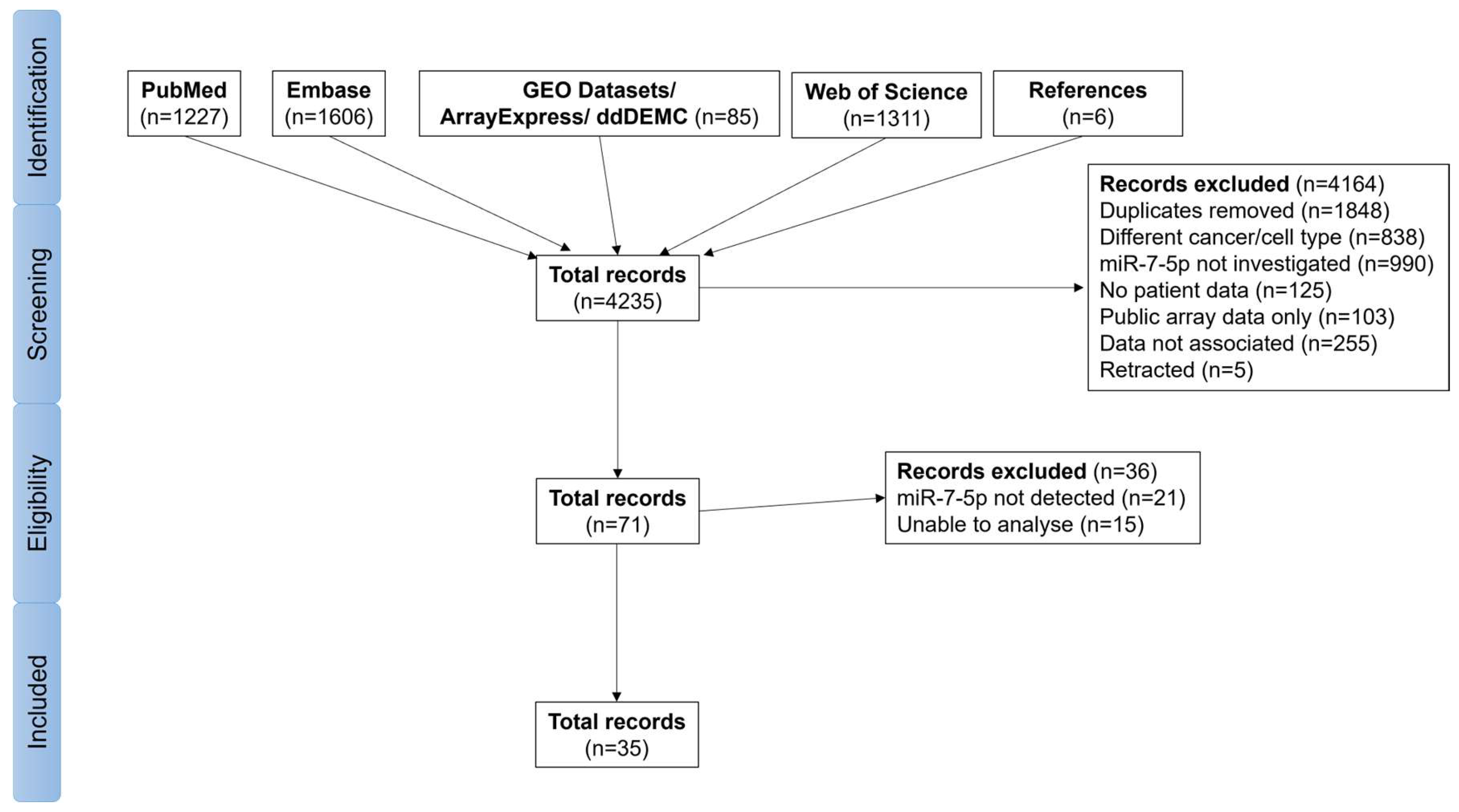
Figure 1. Flow chart for the search strategy.
Cancers 17 03232 g001
Figure 2. Forest plot comparing miR-7-5p levels in HNSCC tumour and normal tissues [16,46,47,48,49,50,51,52,53,54,55,56,57,58,59,60,61,62,63,64,65,66,67]. The diamond for the overall effect summary, located to the right side of the graph, favours miR-7-5p expression being higher in tumour than in normal tissue.
Cancers 17 03232 g002
Figure 3. Evaluation of heterogeneity (A) with Galbraith plots and publication bias (B) using funnel plots, with all 24 studies included or with the quantile-normalisation method excluded.
Cancers 17 03232 g003
Figure 4. Forest plot showing the meta-analysis results by anatomical site following exclusion of quantile-normalised studies [16,46,47,48,49,51,52,53,54,55,56,58,60,62,63,64,65,66]. The diamond for the overall effect summary, located to the right side of the graph, favours miR-7-5p expression being higher in tumour than in normal tissue.
Cancers 17 03232 g004
Figure 5. Analysis of miR-7-5p expression and survival from TCGA data. Expression levels of miR-7 in (A) all HNSCC tumours and normal samples, (B) tumour and normal-adjacent tissues from the same patients, and (C) all HNSCC by primary tumour site. Kaplan–Meier curves of the (D) overall survival, (E) disease-free survival, (F) disease-free interval, and (G) progression-free interval of HNSCC patients based on their tumour miR-7 expression (median cut-off). (H) Expression of miR-7 pre-miRs in the TCGA-HNSCC cohort extracted from miRCancerdb. (I) Copy number variant (CNV) frequency for each of the MIR-7 genes in TCGA-cohort.
Cancers 17 03232 g005
Figure 6. Forest plots for miR-7-5p expression level and overall and disease-free survival [16,18,47,50,52,54,60,70,71,78]. Generic inverse variance (IV), fixed-effects models, 95% confidence intervals for hazard ratios.
Cancers 17 03232 g006
Figure 7. Synteny plot showing cross-species conservation for the MIR7 genes, SNPs detected in the mature miR-7-5p sequence and likely resulting changes in expression, other miRNA genes proximal to MIR7, and publication references suggesting whether they are oncogenic (OncomiR, red star) or tumour suppressors (TS-miR, blue star), or if there is a lack of evidence or conflicting reports for the functional role of the miRNA (grey star).
Cancers 17 03232 g007
Figure 8. Pan-cancer analysis of miR-7-5p levels. (A) Differential expression analysis of mature miR-7-5p levels across different tumour types from the TCGA database. (B) Forest plot summarising the Receiver Operator Characteristic (ROC) analyses for the diagnostic ability of miR-7-5p across different cancer types from the TCGA database. AUC, Area Under Curve; CI, 95% confidence interval, * p < 0.05, ** p < 0.01, *** p < 0.001, ns, not significant, determined by the Wilcoxon rank-sum test.
Cancers 17 03232 g008
Figure 9. Predicted target genes and association with gene ontology terms. (A) Venn diagram of predicted miR-7-5p target genes from multiple databases. (B) Bubble plots of Gene Ontology term enrichment analysis from 224 common miR-7-5p predicted target genes.
Cancers 17 03232 g009
Figure 10. Network of key target genes and their correlation with miR-7-5p levels. (A) Protein–protein interaction network of candidate miR-7-5p target genes. (B) Pearson correlation analysis of miR-7-5p levels and the expression of hub genes in the TCGA-HNSC cohort. Pearson correlation test for significant association.
Cancers 17 03232 g010
Figure 11. miR-7-5p expression in human head and neck tissues and cells. (A) miR-7-5p expression in matched tumour and adjacent normal tissue from head and neck cancer patients (n = 16) was quantified by RT-qPCR. Data are shown as 2−ΔCt values, normalised to U6. Each sample was reverse transcribed once, and qPCR was run in technical triplicate. Statistical analysis: two-tailed paired t-test, * p < 0.05. (B) miR-7-5p expression in normal oral keratinocytes (HOK), fibroblasts (HOF, HPLF), and oral squamous cell carcinoma lines (CAL27, CAL33). RT-qPCR data represent log2-transformed mean ± 95% CI from n = 3 independent wells (biological replicates). RNA was reverse transcribed in duplicate, with a single qPCR per cDNA replicate. Expression was calculated using the 2−ΔΔCt method and normalised to U6. One-way ANOVA with Tukey’s post-hoc test: Groups not sharing a letter differ significantly (p < 0.05). Representative of three independent experiments. (C) miR-7-5p levels in HOK and DOK cells following 24 h treatment with vehicle (DMSO) or 4NQO (0.36 µM for HOK; 0.72 µM for DOK). Data were log2-transformed, normalised to U6, and expressed relative to DMSO-treated HOK. Replicate set-up and analysis as in (B). Representative of three independent experiments. (D) Long-term 4NQO treatment (6–7 weeks) of cells as in (C), with miR-7-5p levels quantified by RT-qPCR. Data are log2-transformed and expressed relative to untreated controls using 2−ΔΔCt (mean ± 95% CI). Unpaired two-tailed t-test, ns p > 0.05. Representative of three independent experiments. (E) Ct values from the three independent experiments in (D) were compared across cell passages. Data represent mean ± SD of n = 3 biological replicates, each with duplicate reverse-transcriptions and single qPCR. One-way ANOVA with Tukey’s post-hoc test; * p < 0.05 between P1 and P3. (F) CAL27 cells were untreated or treated with UV (100 mJ/cm2) and then recovered for 2 h or 24 h prior to RNA extraction. RT-qPCR was performed as in (B) and in three independent experiments. One-way ANOVA with Tukey’s post-hoc test, * p < 0.05. (G) Representative microRNA ISH staining images of a 4NQO-treated mouse tongue or vehicle-treated tongue, stained with miR-7-5p (target), miR-21 (tumour positive control), U6 (positive staining control), scramble control (negative staining control), and representative images of low-high intensity staining of miR-7-5p in mouse tongue tissues.
Cancers 17 03232 g011
Figure 12. miR-7-5p overexpression and inhibition in normal oral keratinocytes and oral squamous cell carcinoma cells, respectively. Cells were transfected with increasing doses (0–100 nM) of miR-7-5p mimic or inhibitor (antimiR-7-5p) and their respective negative controls for 72 h, after which cell viability was assessed using the MTS assay. Unpaired, two-tailed t-test; * p < 0.05. Representative of three independent experiments.
Cancers 17 03232 g012
Table 1. Summary of studies with publicly available miR-7-5p expression data in head and neck tumour and normal tissue.
AuthorCountryTumour SiteAge (Median)Tumour/NormalTumour (Average)Normal (Average)
Avissar, 2009 [46]AmericaOral cavityN.R.1.185.854.94
Bruce, 2015 [47]CanadaPharynx518.235.170.629
Cervigne, 2009 [48]CanadaOral cavityN.R.−1.952.96−1.52
Chamorro, 2018 [49]SpainOral cavity541.1112.411.2
Farah, 2018 [50]AustraliaOral cavity641.186.185.22
Fukumoto, 2014 [51]JapanPharynx645.348014.97
Jung, 2012 [16]AmericaMixed611.67672402
Lapa, 2019 [52]BrazilLarynx623.3739141162
Li, 2011 [53]ChinaPharynx461.402.211.58
Liu, 2012 [54]ChinaPharynx460.98318326
Lyu, 2014 [55]ChinaPharynxN.R.1.01−2.37−2.34
Lyu, 2020 [56]ChinaLarynxN.R.3.790.3790.100
Peng, 2014 [57]AmericaPharynx481.310.5670.432
Saito, 2013 [58]JapanLarynxN.R.0.988.548.67
Severino, 2013 [59]BrazilOral cavity56−2.202.91−1.32
Shi, 2015 [60]ChinaOral cavityN.R.0.94−3.29−3.52
Shiah, 2014 [61]TaiwanOral cavity492.80769275
Stansfield, 2016 [62]AmericaMixed581.343.222.41
TCGAAmericaMixed613.6836.910.0
Wu, 2020 [63]ChinaLarynxN.R.3.3084792569
Yang, 2018 [64]ChinaLarynx563.6475.520.7
Zhao, 2019 [65]ChinaLarynxN.R.1.081.471.35
Zheng, 2018 [66]ChinaOral cavityN.R.1.912.111.11
Zhuang, 2020 [67]ChinaOral cavity501.945.152.65
N.R.: data were not reported.
Table 2. Correlation between miR-7-5p expression levels and clinical features from TCGA-HNSC.
Clinicopathological ParametersFeaturesnMean ± SDp
Unmatched tissueHNSCC5142.113 ± 1.019<0.0001
Normal401.107 ± 0.522
Matched tissueHNSCC402.443 ± 1.173<0.0001
Normal401.107 ± 0.522
SexMale3762.111 ± 1.0340.9435
Female1382.119 ± 0.980
Age≤50 years831.980 ± 0.8670.1932
>50 years4312.139 ± 1.044
T stageT1 or T21821.986 ± 0.9750.0469
T3 or T43162.175 ± 1.049
N StageN02362.106 ± 1.0340.8749
N1–N32742.120 ± 1.014
M StageM04832.103 ± 1.0230.2772
M162.560 ± 1.011
Clinical StageI–II1142.086 ± 0.9320.8222
III–IV3862.110 ± 1.051
Histological GradeG1–G23652.133 ± 0.9880.8491
G3–G41292.113 ± 1.073
AlcoholYes3432.134 ± 1.0340.592
No1622.082 ± 1.004
TobaccoYes3812.152 ± 1.0290.3022
No1202.042 ± 0.970
HPV StatusPositive951.796 ± 0.9990.0007
Negative4172.188 ± 1.010
TP53 StatusMutant3612.258 ± 1.0170.0017
Wild-Type1461.927 ± 1.025
Perineural invasionYes1702.170 ± 1.0780.7071
No1932.130 ± 0.970
Lymphovascular invasionYes1222.162 ± 1.0020.8121
No2272.134 ± 1.050
Table 3. Analysis of miR-7-5p expression level and dichotomous variables using Mantel-Haenszel fixed-effects model.
ComparisonsStudiesParticipantsI2p(Q)Effect Estimatep(Z)
Male vs. Female2014210%0.760.80 [0.62, 1.01]0.07
>50 years vs. <50 years211490%0.691.02 [0.80, 1.30]0.88
Stage III-IV vs. Stage I-II1911870%0.561.05 [0.81, 1.36]0.73
G3–G4 vs. G2–G156227%0.370.79 [0.55, 1.13]0.19
Drinker vs. Non-drinker66970%0.661.09 [0.79, 1.50]0.60
Smoker vs. Non-smoker77060%0.481.25 [0.90, 1.74]0.18
HPV+ vs. HPV−77210%0.720.58 [0.39, 0.85]0.005
p(Q), p-value of the Q test for heterogeneity; p(Z), p-value of the Z test for significance. Values in brackets for effect estimate represent 95% confidence intervals.
Disclaimer/Publisher’s Note: The statements, opinions and data contained in all publications are solely those of the individual author(s) and contributor(s) and not of MDPI and/or the editor(s). MDPI and/or the editor(s) disclaim responsibility for any injury to people or property resulting from any ideas, methods, instructions or products referred to in the content.