Review

Functional Heterogeneity and Context-Dependent Roles of LncRNAs in Breast Cancer

by
Shu Hui Lye
1,
Nunaya Polycarp
1,
Titilayomi Juliet Durojaye
1 and
Trygve O. Tollefsbol
1,2,3,4,5,*
1
Department of Biology, University of Alabama at Birmingham, Birmingham, AL 35294, USA
2
Integrative Center for Aging Research, University of Alabama at Birmingham, Birmingham, AL 35294, USA
3
O’Neal Comprehensive Cancer Center, University of Alabama at Birmingham, Birmingham, AL 35294, USA
4
Nutrition Obesity Research Center, University of Alabama at Birmingham, Birmingham, AL 35294, USA
5
Comprehensive Diabetes Center, University of Alabama at Birmingham, Birmingham, AL 35294, USA
*
Author to whom correspondence should be addressed.
Cancers 2025, 17(19), 3191; https://doi.org/10.3390/cancers17193191
Submission received: 19 August 2025 / Revised: 26 September 2025 / Accepted: 28 September 2025 / Published: 30 September 2025

Abstract

Simple Summary

This review explores the sources of functional diversity/ambiguity and context-dependent roles of long non-coding RNAs (lncRNAs) in breast cancer. LncRNAs can either promote or slow the progression of breast cancer, influenced by factors such as the tumor subtype, various cellular and molecular mechanisms, and the breast cancer tumor microenvironment. Although lncRNAs hold promise as biomarkers and therapeutic targets, their diverse nature poses challenges for clinical translation. Strategies to address this heterogeneity in lncRNAs include enhancing classification systems, utilizing technology like CRISPR/Cas tools, advanced sequencing methods, and machine learning to analyze their behavior, and—in the context of therapeutic development—focusing on strong targets.

Abstract

As with other non-coding RNAs (ncRNAs), the aberrant expression of long non-coding RNAs (lncRNAs) can be associated with different forms of cancers, including breast cancer (BC). Various lncRNAs may either promote or suppress cell proliferation, metastasis, and other related cancer signaling pathways by interacting with other cellular machinery, thus affecting the expression of BC-related genes. However, lncRNAs are characterized by features that are unlike protein-coding genes, which pose unique challenges when it comes to their study and utility. They are highly diverse and may display contradictory functions depending on factors like the BC subtype, isoform diversity, epigenetic regulation, subcellular localization, interactions with various molecular partners, and the tumor microenvironment (TME), which contributes to the intratumoral heterogeneity and phenotypic plasticity. While lncRNAs have potential clinical utility, their functional heterogeneity coupled with a current paucity of knowledge of their functions present challenges for clinical translation. Strategies to address this heterogeneity include improving classification systems, employing CRISPR/Cas tools for functional studies, utilizing single-cell and spatial sequencing technologies, and prioritizing robust targets for therapeutic development. A comprehensive understanding of the lncRNA functional heterogeneity and context-dependent behavior is crucial for advancing BC research and precision medicine. This review discusses the sources of lncRNA heterogeneity, their implications in BC biology, and approaches to resolve knowledge gaps in order to harness lncRNAs for clinical applications.

1. Introduction

Despite progress in screening and treatment options, breast cancer (BC) continues to be among the most common cancers overall, with incidences and mortality rates projected to continue increasing globally. An analysis of estimated global cancer burden data from the GLOBOCAN database reported 2.3 million new BC cases in 2022, with annual incidence rates rising by 1–5% in approximately half of the 185 countries assessed [1]. Like with many cancers, BC is characterized by complex etiologies, consisting of multiple subtypes with various clinical presentations. As such, a comprehensive understanding of this disease is essential for improving early detection strategies and developing effective therapeutic and preventive interventions. An area of interest that may shed light on the regulatory complexities underlying mammary oncogenesis is the study of long non-coding RNAs (lncRNAs), a distinct subset of non-coding RNAs (ncRNAs).

1.1. LncRNAs: A Brief Overview

LncRNAs are commonly defined to be non-coding transcripts of over 200 nucleotides (nt). Due to the existence of other well-characterized ncRNAs of sizes that approach this 200 nt border, recommendations have also recently been made to redefine lncRNAs to include transcripts largely generated by RNA polymerase II that are over 500 nt [2]. The vastly heterogenous nature of lncRNAs is exemplified by efforts by multiple groups at subcategorizing lncRNAs. In addition to length, for instance, lncRNAs may be classified according to their various molecular characteristics, including the sequence elements, function, subcellular localization, association with other components of cellular components, and biological contexts [3]. Recognizing that the function of an lncRNA can vary depending on the tissue, disease stage, or molecular context is pertinent when it comes to characterizing their roles in cancer biology and for identifying which lncRNAs may serve as reliable biomarkers or therapeutic targets.

1.1.1. LncRNAs: Biogenesis

Like messenger RNAs (mRNAs), lncRNAs are often transcribed by RNA polymerase II, 5′ capped (at 7-methylguanosine) and 3′ polyadenylated, and can be alternatively spliced [2]. LncRNA transcription often occurs at nucleosome-depleted regions, which facilitates chromatin accessibility and R-loop formation [4]. According to the GENCODE consortium’s annotation of human lncRNA, the vast majority of lncRNA introns are flanked by canonical splice sites (GT/AG) [5]. However, many lncRNAs lack polyA tails. They are spliced more inefficiently than mRNA due to a weak internal splicing signal and significant distances between their 5′ splice site, 3′ splice site, and junction (branch point) [6,7]. lncRNA transcripts are expressed at relatively low levels and tend to exhibit more limited temporal restriction compared to mRNA [2]. Their exon architecture may differ from that of protein-coding genes, with 42% of lncRNA transcripts containing only two exons, compared to just 6% of protein-coding gene transcripts [5]. Short sequences within lncRNAs may predict where they localize and how they interact with proteins, unlike mRNAs which are largely present in the cytoplasm [8].

1.1.2. LncRNAs: Functions

Compared to other forms of ncRNAs like microRNAs, which are primarily involved in gene silencing an active RNA-induced silencing complex (RISC), lncRNAs appear to be subject to a greater degree of functional ambiguity. By interacting with components of cellular machinery, including but not limited to proteins, other RNA, and DNA, lncRNAs may be involved in the regulation of the cellular function at multiple regulatory levels. This includes their ability to act as dynamic scaffolds, decoys, and guides that engage with diverse molecular partners, including other DNA, RNAs, and proteins [9,10,11,12,13]. For instance, lncRNAs can guide chromatin-modifying complexes to specific genomic loci to epigenetically silence or activate transcription [14,15,16,17]. They function as decoys by sequestering transcription factors or microRNAs (miRNA), thereby preventing them from binding to their native targets [18,19]. Furthermore, their role as scaffolds enables them to bring together multiple proteins to form functional ribonucleoprotein complexes that influence processes such as mRNA splicing, stability, and translation [20]. Through these fundamental mechanisms, scaffolding, decoying, and guiding lncRNAs integrate signals and modulate critical cellular pathways in BC, including proliferation, apoptosis, and metastasis [13,21].
Consequently, efforts have been made to investigate the role of lncRNAs in cancer pathogenesis. Computational analyses have identified lncRNAs to be significantly implicated in cancer regulation. For example, some long intergenic non-coding RNAs (lincRNAs) may act as universal cancer repressors [22]. In BC patients, the aberrant expression of lncRNAs has been reported [23]. The molecular actions of lncRNAs in BC may lead to either the suppression or facilitation of BC cell proliferation, metastasis, the epithelial–mesenchymal transition (EMT), and even resistance to drugs used in the treatment and management of BC, like tamoxifen and adriamycin [24].
Despite the reported roles of lncRNAs as versatile regulators of gene expression, many other studies show contradictory behavior and a lack of fixed functionality. As an example, homeotic transformation and skeletal malformation was reported in mice with the homozygous deletion of the lncRNA HOX Transcript Antisense RNA (HOTAIR) [25]. However, a later study using the same mutant mice crossed to a different genetic background failed to recapitulate the effects of the HOTAIR knockout [26]. Another widely studied lncRNA, X-inactive specific transcript (XIST), is identified as an oncogene that is often overexpressed in many cancers, but may act as a tumor suppressor in some cases [27,28].

1.2. Breast Cancer: Pathophysiology

The etiology of BC is an ongoing area of study. Most BCs manifest as invasive carcinomas, while a smaller but still significant percentage are lobular carcinomas [29]. Only about 10% or fewer BC cases are due to heritable genetic mutations, the majority of which are found in the tumor suppressor genes BRCA1 and BRCA2 [29]. Over 90% of BCs are considered sporadic. Explanations for pathogenesis or risk factors include hormonal imbalances, immune system influences, and lifestyle factors [30,31,32]. Of significance is the role of estrogen in BC, which promotes cell proliferation in mammary epithelial tissue. Estrogen can also lead to oncogenesis by promoting chromosomal translocations and subsequent copy number amplification events, particularly in genomic regions where estrogen receptors (ERs) bind [33]. Additionally, BC cells interact dynamically with the tumor microenvironment (TME), which consists of immune cells, the extracellular matrix (ECM), hypoxic regions, and other players. Exposure to compounds due to the environment or lifestyle, like certain nutrients or carcinogens, for instance, could change the TME by regulating lymphocyte activity or creating hypoxic conditions, affecting BC outcomes [30,34].
In conjunction with the complex molecular dynamics underlying the BC etiology, the heterogeneity of the disease itself is highlighted by its classification into various subtypes. BCs can be classified by their histological grade, Ki-67 proliferative index, and the expression of three receptors: the estrogen receptor (ER), human epidermal growth factor receptor 2 (HER2), and progesterone receptor (PgR). Further molecular classification groups BCs into Luminal A, Luminal B, HER2-enriched, Basal-like, and Normal-like. Luminal A tumors typically show smaller sizes and lower grades and are characterized by favorable long-term patient survival among BC subtypes [35]. Compared to Luminal A, Luminal B tumors have increased proliferative properties and present worse overall survival. HER2-enriched tumors also often present with higher grades and aggressive features, while TNBC presents with genomic instability along with high aggressiveness and limited therapeutic options [35].
LncRNAs may be involved in BC pathophysiology by regulating signaling pathways such as the MAPK cascade and PI3K/PTEN/AKT/mTOR pathway [36]. Large-scale analyses demonstrate the association of lncRNA expression with specific breast cancer subtypes [32]. For example, ER-positive tumors are enriched for lncRNAs linked to estrogen signaling, while ER-negative and Basal-like tumors show lncRNA signatures related to immune activation and NF-κB signaling. Additionally, lncRNAs expressed in fibroblasts contribute to ECM remodeling, particularly in Luminal A tumors, indicating the influence of the TME.

2. Materials and Methods

The sourcing of relevant research and references in this work was carried out via the appraisal of multiple primary empirical articles and review articles. The searches for these articles were primarily carried out using the PubMed database, presently maintained by the United States National Library of Medicine (NLM). Google Search and Google Scholar are also web engines employed for the searches. Keywords utilized in the creation of this work largely include but are not limited to “breast cancer” and “lncRNA”. Clinicaltrials.gov was also utilized, wherein “breast cancer” was the term entered for the “Condition/disease” filter and “lncRNA” for the “Intervention/treatment” filter. No descriptive statistical analyses nor meta-analyses were conducted in the creation of this work.

3. Sources of LncRNA Functional Heterogeneity

3.1. Breast Cancer Subtypes

LncRNA expression patterns exhibit greater subtype specificity than protein-coding genes (PCGs), with 18.27% of lncRNAs showing subtype-specific expression compared to 10.55% of PCGs [37]. Certain lncRNAs appear to have distinct expression patterns in specific BC subtypes: TSIX is unique to Luminal A, PVT1 to Luminal B, SNHG8 and GAS5 to HER2-enriched tumors, and HCP5, SNHG3, and MIR155HG to Basal-like or triple-negative breast cancer (TNBC) subtypes (Table 1) [38,39,40]. These subtype-specific lncRNAs influence critical cancer-related pathways, such as immune modulation, cell cycle progression, and EMT, underscoring their potential in precision medicine. Some lncRNAs, such as HOTAIR, EMX2OS, and MYCNOS, are expressed across all subtypes, indicating a more general role in BC biology or involvement in cellular processes that are common to diverse tumor types (Table 1) [38].
Among research groups who identified multiple lncRNAs specific to certain BC subtypes is Wasson et al., who analyzed the expression of over 12,000 lncRNAs across BC subtypes using TCGA and TANRIC data (Table 1). The authors revealed subtype-specific associations with patient survival and that select lncRNAs promote proliferation in subtype-matched BC cells. Of note, poorer patient survival linked to LINC01269 expression was observed exclusively in the HER2-positive (HER2+) BC subtype [41]. Consistent with this association, reduced cell proliferation was only observed upon LINC01269 knockdown in HER2+ BC cell lines. Targeting LINC01269 together with HER2 antagonists like trastuzumab produces additive effects on reducing cancer cell viability. Also identified by the authors was the lncRNA AL078604.2, which is associated with poor prognoses, specifically in TNBC patients, and only had a negative effect on TNBC cell proliferation upon knockdown [41]. Interestingly, this particular investigation reported that lncRNAs commonly studied in BC like HOTAIR, NEAT1, and MALAT1 were not significantly linked to patient survival outcomes [41].
Hyaluronan synthase 2 antisense RNA 1 (HAS2-AS1) is an lncRNA with low expression in ER+ tumors and high expression in ER- tumors. However, this high expression is correlated with the better survivability of patients with ER- BC, whereas the same is not observed in patients with ER+ BC (Table 1) [42]. Two-dimensional cell culture experiments appear to recapitulate this finding, whereby HAS2-AS1 overexpression decreases the cell viability and invasiveness of ER- cells, but not ER+ cells [42]. Thus, HAS2-AS1 may function as a tumor suppressor specifically in ER- BC. It could also function as a miRNA sponge and behave as an epigenetic regulator [43].
LINC00324, PTPRG-AS1, and SNHG17 are three examples of lncRNAs significantly associated with both ER+ and ER- subtypes and the tumor’s histologic grade (Table 1). Their expression profiles enable risk stratification for relapse-free survival. Specifically, high LINC00324 and low PTPRG-AS1 and SNHG17 expression correlate with favorable clinical outcomes [44]. Of particular interest, PTPRG-AS1 is an antisense lncRNA to PTPRG, a known tumor suppressor in multiple cancers. The upregulation of PTPRG-AS1 may lead to the suppression of PTPRG, potentially contributing to tumor progression [44]. The unsupervised clustering of lncRNA expression aligns closely with PAM50 molecular subtypes, underscoring their utility in refined molecular subtyping [44,45]. For instance, basal-like/triple-negative tumors exhibit distinct lncRNA expression profiles from other subtypes and normal tissue, indicating the potential of lncRNAs for precise subtype classification and personalized treatment.
Another example of an lncRNA relevant to BC is MNX1-AS1, which is significantly upregulated in TNBC tissues and cell lines, correlating with poor survival outcomes (Table 1) [46]. Mechanistically, MNX1-AS1 promotes tumor growth and metastasis by enhancing the phosphorylation of the transcription factor Stat3, thereby activating the JAK/Stat3 signaling pathway crucial for TNBC progression. Silencing MNX1-AS1 in vitro and in vivo reduces tumor proliferation and invasion, underscoring its potential as a TNBC-specific therapeutic target [46].
Further emphasizing the therapeutic relevance of lncRNAs in TNBC, a clinically guided siRNA screening approach identified BCAL8 (breast cancer-associated lncRNA 8) as a functional driver of tumor proliferation (Table 1). BCAL8 may promote cell-cycle progression by upregulating Cyclin E2. In MDA-MB-231 cells, the knockdown of BCAL8 led to reduced cell proliferation in vitro and reduced tumor growth in xenograft models via the inhibition of the G1–S cell-cycle transition [37]. Elevated BCAL8 expression and genomic gain are also associated with poor prognosis in BC.
Table 1. Representative lncRNAs linked to overall and subtype-specific breast cancers, with corresponding expression patterns and prognostic correlations.
BC Subtype(s)Associated LncRNAExpression in BC Samples or CellsBC PrognosisReferences
All BCsBCAL8UpregulatedLinked to poor prognosis[37]
EMX2OSDownregulatedUnknown correlation[38]
MYCNOSUpregulatedUnknown correlation[38]
NEAT1Frequently overexpressed; some mixed resultsFrequently linked to poor prognosis; some mixed results[41,47,48,49,50]
MALAT1Frequently upregulated; some mixed resultsFrequently linked to poor prognosis; some mixed results[41,49,50]
HOTAIRFrequently upregulated; some mixed resultsFrequently linked to poor prognosis; some mixed results[41,50,51,52]
Luminal ATSIXDownregulatedUnknown correlation[38]
Luminal BPVT1UpregulatedUnknown correlation[38]
HER2-enrichedSNHG8DownregulatedUnknown correlation[38]
GAS5DownregulatedLinked to resistance to trastuzumab and lapatinib[38,44]
LINC01269UpregulatedLinked to poor prognosis[41]
ER-HAS2-AS1UpregulatedLinked to better prognosis[42]
ER+HAS2-AS1DownregulatedWeak correlation[42]
ER+ and ER-LINC00324UpregulatedLinked to better prognosis[44]
PTPRG-AS1DownregulatedLinked to better prognosis[44]
SNHG17DownregulatedLinked to better prognosis[44]
Basal-like/TNBC HCP5UpregulatedLinked to better prognosis[38,39]
SNHG3UpregulatedLinked to poor prognosis[38,40]
MIR155HUpregulatedUnknown correlation[38]
AL078604UpregulatedLinked to poor prognosis[41]
MNX1-AS1UpregulatedLinked to poor prognosis[46]

3.2. Isoform Diversity and Alternative Splicing

Alternative splicing (AS) can contribute to the functional heterogeneity of lncRNAs in BC by generating multiple transcript variants (isoforms) with different regulatory capacities, localization patterns, or interaction partners. While lncRNAs are spliced similarly to mRNAs, they are much less efficiently spliced, which may lead to a greater number of spliced lncRNA transcripts per exon [6]. However, increasing the thymine content in the polypyrimidine tract (PPT) of the 3′ splice site has been experimentally shown to enhance lncRNA splicing by creating a stronger binding site for splicing factors, thus facilitating increased lncRNA splicing efficiency [53]. Should splicing hold greater functional significance for lncRNAs over evolutionary time, their splicing efficiency and subsequent function could potentially become altered via PPT mutations, biasing the production of certain lncRNA isoforms. Currently, it is known that lncRNAs transcripts and their variants are often produced by Alternative Last Exons in both humans and mouse models [6].
LncRNAs can themselves influence the AS of other genes (Figure 1). MALAT1, for example, interacts with several different splicing factors and transcriptional regulators. Turco et al. demonstrate that MALAT1 and ID4 work together to control VEGFA splicing, particularly favoring isoforms that lack exon 7 [54]. This interaction additionally results in the back-splicing of the VEGFA exon 7, generating a circular RNA, circ_0076611 which promotes TNBC cell malignancy by facilitating the translation of select mRNAs [54]. Another example is the lncRNA ZNF649-AS1, which is found to promote trastuzumab resistance in HER2-positive BC [55]. ZNF649-AS1 regulates the AS of EXOC7, likely by facilitating its interaction with pre-mRNA processing factor 8 (PRPF8). This leads to PD-L1-mediated immune evasion, establishing a tumor microenvironment (TME) that compromises the trastuzumab effectiveness [55].
LncRNA-PNUTS is an AS product of the PNUTS pre-mRNA, which can give rise to PNUTS-mRNA or lncRNA-PNUTS. The heterogeneous nuclear ribonucleoprotein E1 (hnRNP E1) controls this splicing decision, whereby its absence shifts splicing towards the production of lncRNA-PNUTS. miR-205 preferentially binds to the lncRNA form over the mRNA form of PNUTS, which correlates with the upregulation of ZEB proteins and increased tumorigenesis in mesenchymal breast tumor cells [56].
Some lncRNAs also affect AS by binding to target pre-mRNAs. One such example is PLANE, which is the longest isoform of the lncRNA MELTF-AS1, and was found to be highly expressed in multiple cancer cell lines. PLANE directly binds to and forms an RNA-RNA duplex with the NCOR2 pre-mRNA at intron 45, cooperating with hnRNPM and thereby directing the AS of NCOR2 to generate lncRNAs that promote tumorigenicity [57].
In addition to AS, some studies have reported differential behavior or an abundance of lncRNA isoforms in the context of BC.

3.2.1. HOX Antisense Intergenic RNA (HOTAIR)

Among the most studied lncRNAs is HOTAIR, which is positioned in the HOXC locus on human chromosome 12q13, implicating its role in the regulation of body patterning and development. HOTAIR interacts with proteins, RNA, and DNA, forming scaffolds that enable the formation of gene regulatory complexes [51]. High HOTAIR expression is prevalent in many human cancers [50,51,58]. In BC, its expression is associated with decreasing levels of tumor suppressors p53 and p21, and the induction of the c-Myc oncogenic pathway. In addition, HOTAIR can act as competing endogenous RNA (ceRNA), sequestering miR-331-3p which normally suppresses HER2 expression [51].
HOTAIR is commonly studied in the context of its canonical isoform (HOTAIR-C), which is a 6-exon 2364 nt transcript. Two other isoforms, HOTAIR-N and HOTAIR-U, have also been reported [59,60]. Data from TCGA suggest that HOTAIR-N is the predominant isoform upregulated in breast carcinomas and is associated with poor overall survival [60,61]. Structurally, the HOTAIR-N promoter consists of a strong CpG island containing G-quadruplex (G4) motifs [60]. This CpG island of the HOTAIR-N promoter in BC cells appears to contribute to HOTAIR-N expression control via DNA and histone modifications [60]. Also notable is the fact that unlike observations in patient breast tumor samples, 2D BC cultures exhibit low HOTAIR expression. In Claudin-low TNBC cells cultured in a laminin-rich extracellular matrix (lrECM), HOTAIR-N, but not HOTAIR-C, is robustly upregulated due to the epigenetic activation of its promoter, mediated by canonical ECM signaling pathways [61]. This induction of HOTAIR-N is required for invasive growth in the 3D culture, implicating its role in translating ECM signals into metastasis.

3.2.2. Nuclear Paraspeckle Assembly Transcript 1 (NEAT1)

A highly abundant lncRNA often upregulated in many cancers including BC, NEAT1, localizes with paraspeckles, which are subnuclear bodies involved in gene expression via the sequestration of mRNAs [47,48,50,62,63]. The NEAT1 lncRNA is transcribed from the NEAT1 gene on chromosome 11q13.1, and encodes two major isoforms: the short isoform, NEAT1_1 (3.7 kb), and NEAT1_2 (23 kb) [64]. While NEAT1_1 is involved in gene regulation, NEAT1_2 is reported to be involved in the structural integrity of paraspeckles, subnuclear membraneless organelles [65]. In mice, Neat1_1 is more highly expressed in most tissues and better conserved. Upon the loss of Neat1_1 via the deletion of its polyadenylation signal, Neat1_1 undergoes forced switching to Neat1_2 [66].
Knutsen et al. reported RNA-FISH analyses carried out on formalin-fixed paraffin-embedded (FFPE) needle biopsies using probes specifically targeting NEAT1_2. From there, it was found that NEAT1_2 was more highly expressed in HER2-positive (HER2+) and luminal B subtypes of breast cancers [67]. Interestingly, NEAT1_2 expression is significantly lower in ER+ tumors compared to ER- tumors, with the inverse being true for NEAT1_1 [67].
It has been demonstrated that knocking out NEAT1_1, but not NEAT1_2, had no observable effect on the proliferation of osteosarcoma and colon cancer cells. Additionally, Neat1_1-specific knockout mice do not exhibit the defects observed in Neat1 KO mice, implying NEAT1_1 as nonessential in the context of cancer cell growth regulation [49]. However, a later study showed that when NEAT1_1 was knocked down in the ER+ BC MCF7 cells, the cells exhibited decreased proliferation [68]. In MMTV-PyVT mice, Neat1_1 inactivation significantly delays mammary tumor onset [68].

3.2.3. Metastasis-Associated Lung Adenocarcinoma Transcript 1 (MALAT1)

The MALAT1 lncRNA, which is also known as NEAT2, is transcribed from the MALAT1 gene located in the 11q13 locus. This lncRNA is derived from the tRNA processing of the MALAT1 primary transcript, in which another ncRNA, mascRNA, is formed. Patients with TNBC exhibit high levels of MALAT1 and a poor prognosis [50]. Mechanistically, MALAT1 may promote metastasis by inducing the epithelial–mesenchymal transition (EMT) via PI3K-Akt pathway activation [69].
Meseure et al. identified two MALAT1 isoforms from the dbEST database, namely the full-length transcript (MALAT1-FL) and an alternatively spliced variant (Δsv-MALAT1). Unlike MALAT1-FL, Δsv-MALAT1 expression varied in the BC tumor samples [70]. Patients with tumors expressing low levels of the Δsv-MALAT1 isoform also showed reduced metastasis-free survival (MFS), indicating this particular isoform’s potential as a prognostic marker [70]. Meseure et al., therefore, proposed that Δsv-MALAT1 may function as a molecular decoy, sequestering interaction partners normally bound to FL-MALAT1, which would disrupt its regulatory activity [70].

3.3. Epigenetic Regulation as a Driver of Functional Heterogeneity

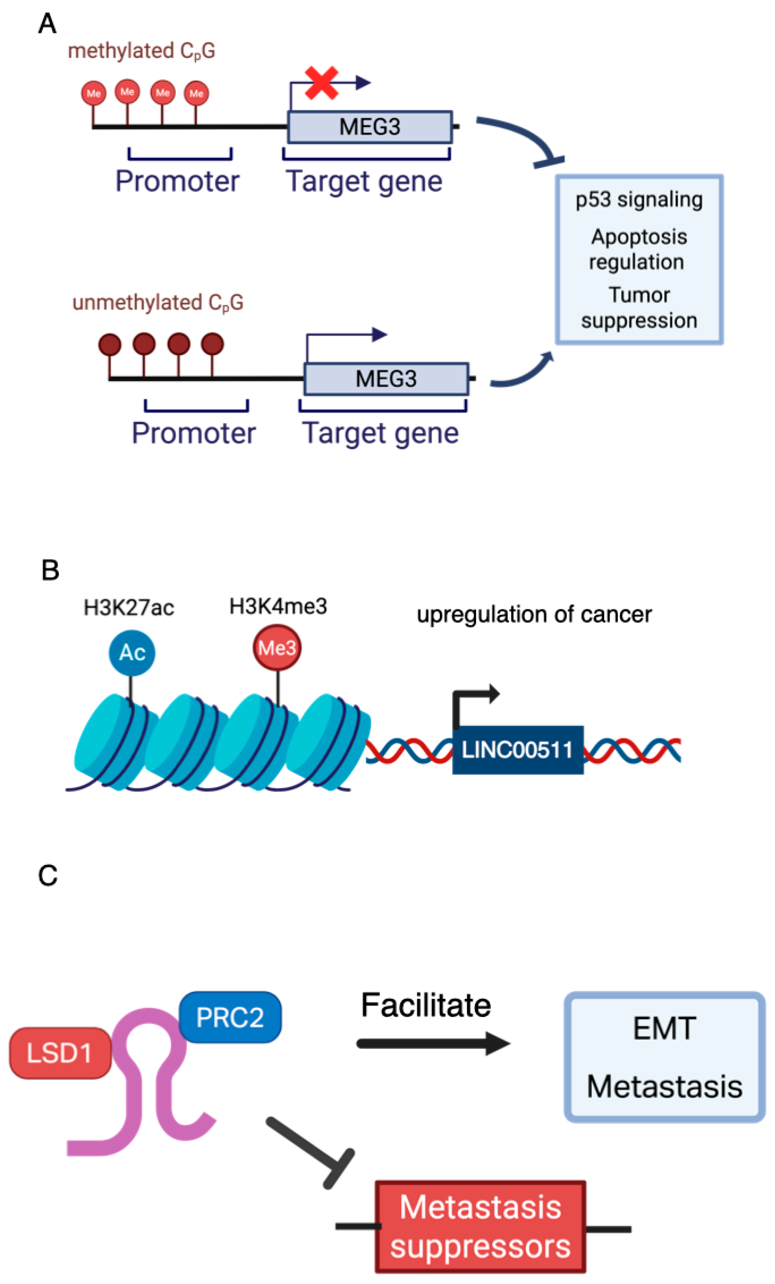
Epigenetic modifications, which are inheritable yet reversible changes in gene expression that do not involve alterations in the DNA sequence, play a crucial role in regulating the activity of lncRNAs in cancer [71]. In BC, DNA methylation, histone modifications, and chromatin remodeling profoundly influence the transcriptional landscape and context-specific functionality of lncRNAs, contributing to intratumoral heterogeneity and phenotypic plasticity [72,73]. Promoter methylation is one of the most studied mechanisms. The tumor suppressor lncRNA MEG3, for example, is frequently silenced in BC due to promoter hypermethylation, which disrupts its role in p53 signaling and apoptosis regulation [74]. Likewise, the reversal of this methylation has been shown to inhibit EMT and suppress tumor progression (Figure 2A) [54].
Histone modifications also regulate lncRNA expression. Superenhancer-associated lncRNAs such as LINC00511 are enriched with H3K27ac and H3K4me3 marks, supporting their upregulation in stem-like cancer cells and linking them to a poor prognosis (Figure 2B) [75]. Chromatin looping further shapes lncRNA behavior by altering isoform expression and promoter–enhancer contacts across BC subtypes.
Importantly, lncRNAs not only respond to epigenetic cues, but actively shape them. For example, HOTAIR functions as a scaffold, recruiting the epigenetic regulators PRC2 and LSD1 to reprogram chromatin and repress metastasis suppressor genes, facilitating EMT and metastasis (Figure 2C) [76]. LncRNAs function as ceRNAs by absorbing miRNAs that regulate EMT-TFs, thereby enhancing the plasticity in response to environmental stress [12].
The dynamic and context-specific roles of lncRNAs emphasize the importance of epigenetic regulation in determining their function. Grasping these interactions is essential for creating subtype-specific biomarkers and developing lncRNA-targeted treatments in precision oncology.

3.4. Subcellular Localization

Unlike mRNAs, which are transported from the nucleus to the cytoplasm for translation, lncRNAs may be largely localized in the nucleus or be shuttled into the cytoplasm. LncRNAs may also be transcribed in the mitochondria by mitochondrial genomes or associated with other organelles including ribosomes, the endoplasmic reticulum, and endosomes [77]. Some of the shuttling of lncRNAs between compartments may be dependent on the cellular context.
The majority of lncRNAs can predominantly be found in the nucleus, where they regulate gene expression through chromatin remodeling, transcriptional control, and RNA processing. An example includes the long Intergenic Non-Protein Coding RNA 839 (LINC00839), which is localized in the nucleus in BC cell lines [78]. The upregulation of LINC00839 is associated with poor prognosis in BC patients in addition to chemoresistance [78]. In vitro evidence indicates that overexpressing LINC00839 enriches c-Myc protein levels via a feedback loop, whereby Myc reinforces LINC00839 upregulation by binding to the LINC00839 promoter region. Downstream events modulate PI3K/AKT signaling, contributing to dysregulated BC cell oncogenecity [78]. As Myc is primarily localized in the nucleus, the nuclear localization of LINC00839 is consistent with its potential involvement in oncogenesis.
Other lncRNAs may not remain localized to one area. H19, which is transcribed from an imprinted locus on chromosome 11p15, can be found in the nucleus, but has a primarily cytoplasmic localization. The high expression of H19 correlates with increased breast tumorigenesis [79]. Mechanistically, H19 can act as an endogenous sponge, which is a feature of many other cytoplasmic lncRNAs. In particular, H19 sequesters microRNAs from the let-7 family, reducing the let-7 availability, which subsequently increases LIN28 [79]. LIN28 itself inhibits the maturation of let-7, creating a double-negative feedback loop with let-7 and a double-positive feedback loop with H19. This regulatory circuitry promotes breast cancer stem cell (BCSC) maintenance [79].

3.5. Interactions with Different Molecular Partners

The functional versatility of long non-coding RNAs (lncRNAs) in BC arises largely from their capacity to engage with diverse molecular partners, including microRNAs (miRNAs), proteins, and other RNA or DNA molecules. These interactions allow lncRNAs to modulate the cellular behavior across multiple regulatory levels, such as transcription, splicing, translation, and chromatin remodeling [12,13].

3.5.1. LncRNA–miRNA Interactions

LncRNAs can function as ceRNAs, which is often described as “miRNA sponging” [80]. ceRNAs can sequester miRNAs through shared miRNA response elements (MREs), preventing these miRNAs from binding to and repressing their target mRNAs [80]. This interaction effectively derepresses miRNA targets, leading to the upregulation of specific genes that can drive oncogenic pathways. The oncogenic lncRNA LINC00511 exemplifies this principle: the direct binding of LINC00511 to tumor-suppressive miR-185-3p liberates the transcription factor E2F1 from miR-185-3p-mediated repression, elevating E2F1 protein levels. This transcriptionally activates key cell cycle regulators, including cyclin-dependent kinase genes CCNA2 and CCNE1, which promote uncontrolled cellular proliferation in BC [80].
The case of the lncRNA PVT1 also highlights the contextual complexity of ceRNA networks: while PVT1 functions as a ceRNA for the miR-200 family, which is critical for maintaining an epithelial phenotype in normal breast tissue, this regulatory interaction is frequently disrupted in breast tumors despite high PVT1 expression [81]. This functional decoupling suggests that the efficacy of a ceRNA is not solely determined by its abundance, but is also influenced by the cellular environment. Potential explanations include post-translational modifications of the core RISC components (e.g., Ago2) that alter the binding affinity [82] or saturation of the ceRNA network by other RNA transcripts with higher affinity for the shared miRNA, effectively outcompeting PVT1 in the tumor context [81].

3.5.2. LncRNA–Protein Interactions

Beyond miRNA interactions, lncRNAs serve as guides and scaffolds for proteins, directing their activity to specific genomic loci or facilitating the assembly of macromolecular complexes [83]. This capacity to act as modular platforms is central to their roles in epigenetic remodeling and the organization of nuclear architecture, processes frequently dysregulated in cancer. HOTAIR, for example, functions as a modular scaffold that simultaneously interacts with distinct histone-modifying complexes via independent domains [17]. Specifically, its 5′ domain binds to the Polycomb Repressive Complex 2 (PRC2), which catalyzes the trimethylation of histone H3 on lysine 27 (H3K27me3) [16]. Concurrently, its 3′ domain recruits the LSD1/CoREST/REST complex, which demethylates histone H3 on lysine 4 (H3K4me2). By co-targeting these two complexes to the promoters of metastasis suppressor genes, such as those within the HOXD cluster, HOTAIR orchestrates a bivalent epigenetic switch that stably silences transcription. This mechanism is a key driver of BC invasion and metastasis, correlating with poor patient prognosis.
As previously discussed, the functional diversity of lncRNAs is exemplified by NEAT1, which exists in two distinct isoforms with divergent roles. NEAT1_2 acts as a critical architectural scaffold, recruiting and organizing essential RNA-binding proteins such as NONO and SFPQ to form the core of these nuclear bodies. Under cellular stress conditions, including hypoxia, a common feature of tumors, paraspeckles rapidly enlarge and sequester various nucleic acids and proteins, including transcription factors and hyperedited mRNAs [84]. This sequestration acts as a post-transcriptional regulatory mechanism, modulating the transcriptome to promote cell survival and suppress apoptosis in BC.

3.5.3. Multiplex and Dynamic Behavior

Many lncRNAs function within multiplex networks, simultaneously interacting with proteins, miRNAs, and chromatin [85,86]. The lncRNA XIST provides an example of multiplexing. While its primary role is to silence one X chromosome in females, its mechanism involves coordinated action across multiple domains. XIST recruits repressive complexes like PRC2 to deposit H3K27me3 across the chromosome, while simultaneously interacting with proteins involved in nuclear compartmentalization, orchestrating structural and epigenetic change [87]. A more direct example relevant to cancer is the lncRNA H19, which has been shown to function as a ceRNA for miRNAs like let-7, while its spliced product can also bind to and modulate the stability of proteins such as p53, demonstrating how a single locus can yield molecules with multiplex functions [88]. Post-transcriptional modifications such as N6-methyladenosine (m6A) methylation can also alter the lncRNA stability, localization, and interaction partners. For example, the m6A modification of the lncRNA LINC01133 has been shown to affect its ability to sponge miRNAs and promote cancer metastasis, adding a dynamic layer of regulation [89,90]. Interpreting these intricate and dynamic interactomes necessitates the application of advanced methodologies.

3.6. Hypoxic Tumor Microenvironments

The BC tumor microenvironment (TME) is the dynamic ecosystem that surrounds and interacts with breast tumor cells. In addition to the malignant epithelial cancer cells, it comprises non-cancerous cell types, including fibroblasts, immune cells, and endothelial cells, as well as various signaling molecules and the extracellular matrix (ECM). Hypoxia is a classic microenvironmental feature that shapes solid tumor progression and heterogeneity. Hypoxia can arise from several factors, including exposure to carcinogens, the tumor cell-driven remodeling of the TME, and elevated metabolic activity in cancer cells. These factors may increase the distance between tumor cells and blood vessels, reducing oxygen diffusion [34]. In response to hypoxic stress, cancer cells can change their behavior, complicating the effectiveness of treatments like chemotherapy [34]. LncRNAs may play an intermediary role between the TME and BC cells by being regulated by hypoxia or by regulating hypoxia signaling [91].
RAMP2-AS1 was identified as a hypoxia-suppressed lncRNA in BC through in silico analyses and in vitro experiments. Unlike many lncRNAs, its upregulation in BC patients is associated with favorable prognostic outcomes. Mechanistically, hypoxia reduces RAMP2-AS1 levels, potentially via a RAMP2-AS1/miR-660-5p/ATM axis in which fewer RAMP2-AS1 molecules are available to sequester miR-660-5p, freeing the latter to bind to the ATM mRNA (Figure 3) [92]. Similarly, N-Myc Downstream Regulated Gene 1-Overlapping 1 (NDRG1-OT1) was identified through the expression profiling of MCF7 BC cells exposed to varying oxygen levels. Its dysregulation under hypoxia suggests a role in the cellular adaptation to low-oxygen stress (Figure 3) [93]. In contrast, RBM5-AS1 is consistently induced under hypoxic conditions and is significantly upregulated in both BC cells and tumor tissues, implying a more active role in tumor adaptation and possible aggressiveness (Figure 3) [94]. Another hypoxia-responsive lncRNA, RPPH1—a component of the RNase P complex—is also upregulated in hypoxic environments (Figure 3) [95]. Its expression is associated with poor prognosis in BC patients, suggesting that distinct hypoxia-regulated lncRNAs may exert opposing effects on tumor progression depending on their molecular targets and mechanisms of action.

3.7. Hormone Signaling

ER and PgR signaling are the primary pathways underpinning hormone-responsive BCs. One component of the TME can be cancer-associated fibroblasts (CAFs), which can remodel the ECM and secrete paracrine factors that can alter estrogen receptor signaling. This influences intratumoral heterogeneity by supporting distinct cancer cell subpopulations with variable hormone dependence [96]. The lncRNA HOTAIR is overexpressed in ER+ BCs and may interact with estrogen signaling pathways by promoting chromatin remodeling and the transcriptional activation of estrogen-responsive genes [97]. Similarly, LINC00472 expression is associated with favorable outcomes in ER+ BC and appears to be regulated by estrogen levels [98]. By modulating these hormone-driven pathways, lncRNAs help maintain heterogeneous BC cell populations with varied hormone sensitivities, facilitating the adaptation of these cells to fluctuating microenvironmental conditions.

4. LncRNAs in the Clinic: Biomarker and Therapeutic Development in Breast Cancer

LncRNAs present a compelling avenue for the development of disease-related biomarkers and targeted intervention strategies. Unlike tissue biopsies, which are invasive and can pose significant discomfort and risks to patients, the extraction of lncRNAs from biofluids—such as blood, plasma, or serum—offers a minimally invasive alternative for cancer diagnostics. Tumor cells can release endogenous lncRNAs into human bodily fluids as extracellular vesicles (EVs), protecting the lncRNAs from degradation [52].
LncRNAs have also gained attention as potential therapeutic targets, especially with the rapid progress in antisense oligonucleotide (ASO) technologies. ASOs and small interfering RNAs (siRNAs) that mediate RNA interference (RNAi) are examples of these approaches, which aim to specifically modulate or silence lncRNAs that contribute to tumor progression, metastasis, or therapy resistance. Specifically, ASOs and siRNA molecules can be delivered to patients to selectively bind and degrade pathogenic lncRNA transcripts [99].
As several computational, in vitro, and in vivo studies have drawn promising correlations between specific lncRNA expression levels and patient prognoses, researchers began touting lncRNAs like HOTAIR, H19, and MALAT1 for their potential as diagnostic biomarkers or therapeutic targets (Table 2) [100]. LncRNAs may also predict patient responses to drug treatments. For instance, a reduction in PRRT3-AS1 is associated with paclitaxel resistance, while the absence of GAS5 is linked to resistance to trastuzumab and lapatinib [101,102].
In a study published by the Zhimin Shao group, an integrated signature consisting of three mRNAs and two lncRNAs was developed using transcriptome arrays to classify TNBC patients based on the disease recurrence risk [103]. The researchers then carried out a randomized phase 3 trial, in which patients classified as “high risk” based on the integrated signature exhibited comparable survival when treated with intensive adjuvant chemotherapy compared to if they were treated with standard adjuvant chemotherapy [104]. This series of studies provides a case for the use of integrated lncRNA signatures to predict patient outcomes and tailor chemotherapy options.
Besides the efforts of some researchers, there are limited data available on the specific utility of lncRNAs in the clinic; much less so in the context of BC [100]. Indeed, the study of lncRNAs is still an area of significant evolution whereby their structure and function are not well-understood. The development of clinical applications for lncRNAs in BC is hinged on a good understanding of lncRNA mechanisms and robust data derived from functional lncRNA studies. To further evolve the field, there is a need to shift focus toward understanding the fundamental mechanisms governing the lncRNA biology in BC. Advancing clinical applications will require multidisciplinary efforts to clarify how lncRNAs contribute to genome organization, nuclear architecture, and regulatory networks.

5. Strategies to Address LncRNA Functional Heterogeneity

5.1. Improving LncRNA Classification Systems

Due to the structural and functional diversity of lncRNAs, coupled with an ongoing evolution of lncRNA research, current lncRNA classification systems will require an increasingly comprehensive framework. To date, the GENCODE system provides among the most extensive and up-to-date source for lncRNA annotations and for understanding how they are classified based on their genomic context, including their relationship to protein-coding genes (e.g., antisense, intronic, and intergenic), exon–intron structure, and transcriptional evidence [5]. The current GENCODE release (version 49) includes 35,899 lncRNAs across reference chromosomes, scaffolds, assembly patches, and alternative loci [105]. In addition to the GENCODE catalog, similar classification efforts by several other groups have also been carried out. Prominent examples include LNCipedia, NONCODE, and LncBook, which also offer detailed annotations of human lncRNAs based on multiple criteria like genomic location, multi-omic data integration, function predictions [106,107,108].
However, the continually evolving landscape of lncRNA research can cause instances of misannotation or inconsistency. GENCODE has continually updated names of transcripts, and some protein-coding genes (PCGs) have been reclassified as lncRNAs over different versions. In one example, an analysis of GENCODE version 24 reports the case of C3orf10, which was originally classified as a PCG, but later annotated as an lncRNA, then back to PGC before being dropped from the database [109]. This dynamicity highlights both the complexity of transcriptome characterization and the challenges in achieving stable, standardized annotations for lncRNA species as more data become available.
There is, hence, a need for the continual and active refinement of lncRNA annotation. Recently, GENCODE has focused on creating their catalog of full-length lncRNAs using Capture Long-read Sequencing (CLS) instead of short-read RNA-seq methods, enabling improved annotation quality (Table 3) [110,111]. This method involves a capture array with probes targeting known and potential lncRNA regions in human and mouse genomes. cDNA libraries were prepared and enriched for complete 5′ to 3′ RNA molecules using CapTrap-Seq. Then, long-read sequencing was carried out on the libraries [111].
Machine learning (ML) and large language models (LLM) can also serve as assets in enhancing lncRNA annotation (Table 3). These methods are suited to addressing the complex semantic nature of lncRNAs by leveraging vast amounts of sequence data to uncover patterns that traditional methods and manual annotation may overlook [117]. For example, TERIUS is an ML tool that implements a two-step filtration process that distinguishes bona fide lncRNAs from false positives, particularly addressing issues with 3′ UTR fragments [118]. An example of an LLM tool is LncRNA-BERT, which uses a model pre-trained on RNA datasets from multiple databases, allowing it to classify lncRNAs effectively without relying on predefined features [119]. Incorporating lncRNAs into multi-omic predictive models has improved molecular classification. ML techniques using lncRNA signatures have demonstrated high precision in differentiating BC subtypes, particularly in distinguishing Basal from HER2-enriched groups. Visualization methods like t-SNE support these findings, highlighting the subtype-specific clustering ability of lncRNAs [120].

5.2. Uncovering LncRNA Functions with CRISPR/Cas Systems

Loss-of-function and gain-of-function experiments are key tools used to elucidate the function of a gene or a protein. Similarly, such experiments can be used to study the various functions of lncRNAs. In the past decade, CRISPR/Cas9 technology has seen a boom, gaining popularity as a tool used for numerous applications ranging from in vitro functional knockout studies to CRISPR-edited CAR-T cells, which can be tailored to target and kill cancer cells. However, while CRISPR/Cas9 genome editing can delete lncRNA genes or regulatory regions, its use is limited in this case because many lncRNA loci overlap or neighbor other genes, risking unintended effects on those genes. Thus, small interfering RNA (siRNA) and antisense oligonucleotides (ASOs) methods remain widely used in the study of the lncRNA function [121]. These methods are not without their disadvantages: RNAi relies on the RNA-induced silencing complex (RISC), which primarily operates in the cytoplasm. CRISPR systems are not bound by this limitation and can operate both in the nucleus and the cytoplasm [122]. ASOs degrade RNA by forming DNA-RNA hybrids recognized by RNase H1, but they require the complex delivery of modified oligonucleotides. In addition, siRNA and ASO methods for functional studies do not give rise to a complete loss of function [122].
CRISPR/Cas9 can be used to target lncRNA genes, creating a double-stranded break (DSB) which is repaired with a single guide RNA (sgRNA) template, which can be customized to induce insertions or deletions of DNA (Table 3). As discussed, this method risks affecting adjacent genes. Hence, CRISPR interference (CRISPRi) or CRISPR activation (CRISPRa) tools can be used instead to repress or activate genes without inducing a DSB (Table 3) [122]. Instead, these tools utilize a dead Cas9 protein (dCas9) to block RNA polymerase II from binding or to recruit transcription activators to a desired region. It should be noted that the dCas9 protein construct could cause unintended alterations of transcriptional contexts by affecting nearby PGCs. While the utilization of these CRISPRi or CRISPRa still induces fewer overall large off-target effects compared to RNAi, these tools are unable to distinguish cis- from trans-acting lncRNA functions [123]. There is another CRISPR system which can directly target lncRNA molecules, which uses Cas13 as a RNA-cleaving enzyme. The CRISPR/Cas13 system enables the study of lncRNA loci that may be located close to each other or to PGCs [124]. While the use of Cas13 constrains the sgRNA design due to its preference for regions containing protospacer flanking sequences, the system has shown similar overall efficiency to RNAi methods with fewer off-target effects [122]. Notably, CRISPR/Cas13 has been used to successfully knock down very long intergenic non-coding RNAs (vlincRNAs), supporting its utility in functional phenotypic studies of lncRNAs across the whole cell (Table 3) [114].
In a study using BC cells, three CRISPR/Cas9 techniques were used to prevent the transcription of the lncRNA LINC00511. Subsequent functional assessments led to the assessment that LINC00511 promotes BC cell proliferation and malignancy [112]. CRISPRa has also been employed to study the function of lncRNA OBSCN-Antisense RNA 1 (OBSCN-AS1) in the context of BC. When activated, OBSCN-AS1 restores OBSCN expression, which suppresses cell migration in TNBC cells [113]. Another study used a CRISPR-Cas13d screen on previously discovered lncRNAs linked to BC to look for lncRNAs that upregulate cell proliferation. The authors identified the lncRNA KILR, which was highlighted as a tumor suppressor that limits proliferation by sequestering the DNA replication factor RPA1 [114].

5.3. Use of Single-Cell and Spatial Sequencing in LncRNA Research

LncRNAs exhibit highly context-specific and cell-type-specific expression patterns, posing challenges for traditional bulk RNA-seq approaches that average out signals across heterogeneous cell populations. Studying lncRNAs in the context of solid cancers adds another layer of complexity because tumors consist of diverse malignant, stromal, and immune cell populations. By enabling the analysis of transcriptional profiles at the resolution of individual cells, single-cell RNA sequencing (scRNA-seq) has emerged as a tool to assist in the detection of heterogeneous and cell-specific lncRNA expression patterns within tumors (Table 3). For example, studies in TNBC xenografts reveal that most lncRNAs are expressed at low but detectable levels, with some lncRNAs such as MALAT1 and NEAT1 widely expressed, while others are restricted to distinct subpopulations [115].
Spatial transcriptomics complements scRNA-seq by preserving the spatial context of gene expression. This enables the preservation of the spatial context of lncRNA expression within BC specimens and potentially mapping lncRNAs to specific tumor niches (Table 3). The utilization of a spatial atlas can enable the identification of circulating biomarker candidates. For example, Xu et al. carried out a spatial transcriptomic analysis, identifying HULC lncRNA abundance in both normal and hepatocellular carcinoma tissue, but at markedly higher levels in the latter [125]. Since HULC is also highly detectable in both liver and blood samples, it is a potential candidate for a non-invasive biomarker for liver cancer [125]. While spatially resolved data on lncRNA targets in BC are limited, similar principles of spatial transcriptomics can be applied to inform biomarker discovery in the context of BC.
Using single-cell and spatial transcriptomics data, Pakrithi et al. identified over 35,000 novel lncRNAs in tissues across 13 types of cancers. The authors note that lncRNAs such as MALAT1 and NORAD are broadly and abundantly expressed across many tissue types, indicating low tissue specificity [116]. Other lncRNAs, like TINCR, are highly expressed only in BC samples, indicating the importance of considering the tissue specificity in lncRNA research [116]. The annotations and visualizations of lncRNA expression profiles for each tissue were made available on SPanC-Lnc—the Spatial and Single-Cell Pan-Cancer Atlas of lncRNAs —an interactive web browser tool [116].

5.4. Prioritizing LncRNA Targets

A certain level of discernment is warranted when prioritizing lncRNA as potential candidates for therapeutic development. While computational analyses of publicly available datasets can be carried out that show significant correlations between lncRNAs and patient survival, observations should be replicable in model systems. As such, functional screens and studies should be conducted across multiple contexts and not limited to single cell lines [126]. To alleviate points of ambiguity present in current lncRNA studies, Ponting and Haerty recommend prioritizing targets that exhibit high sequence conservation, abundance in multiple cell types, specific subcellular localization, and observable interaction with other molecules [126].
Personalized lncRNA expression profiling methods may also be a way to identify potential lncRNA targets relevant to different patients given the variability of lncRNA expression (Table 3). Zhao et al. developed LncRNA Individualization (LncRIndiv), a personalized lncRNA expression profiling method which generates sample-specific lncRNA signatures known as individualized differentially expressed lncRNA (IDElncRNA) [101]. The LncRIndiv pipeline uses lncRNA expression profiles from TCGA to construct a IDElncRNA profile. Expression ranks of lncRNA pairs from TCGA normal samples are used as a reference standard; then, each patient’s tumor sample from TCGA is compared to this reference to detect individualized lncRNA deregulation to generate IDElncRNA profiles. This approach overcomes the limitations of population-level analyses and accounts for the data heterogeneity, normalization issues, and batch effects.

6. Conclusions

Many studies have demonstrated the association of specific lncRNA expression with outcomes ranging from cancer cell behavior in vitro to patient survival and responses to drug therapy. The ease of obtaining lncRNAs through minimally invasive sampling makes them attractive candidates for clinical use in BC, which include applications such as lncRNA-based diagnostics, the direct inhibition of lncRNAs, or their use as therapeutic agents to influence the cancer cell behavior. Despite these promising avenues, challenges remain in effectively translating lncRNA-based therapies into clinical practice due to an incomplete understanding of the lncRNA function and heterogeneity of lncRNA expression. The functional heterogeneity and context dependence of long noncoding RNAs (lncRNAs) is crucial for advancing cancer research. Their diverse modes of action, cell-type specificity, and dynamic responses to environmental or pathological cues can significantly influence the tumor behavior and therapeutic outcomes. Hence, a more comprehensive understanding of lncRNA heterogeneity and their context-dependent behavior is necessary in order to better address the research gaps. This is possible through evolving technologies like CRISPR systems and next-generation sequencing techniques, coupled with persistent efforts with regards to lncRNA classifications. Additionally, therapeutic development efforts in the lncRNA field should prioritize targets supported by rigorous, reproducible evidence from multiple independent studies.

Author Contributions

S.H.L. conceived of, drafted, and edited the manuscript with guidance and additional edits from T.O.T. Subsections and edits were contributed by N.P. and T.J.D., S.H.L., N.P. and T.J.D. prepared figures and tables. All authors have read and agreed to the published version of the manuscript.

Funding

This work was supported by a grant from the National Institute of Health (NCI R01CA178441).

Acknowledgments

We thank the members of Tollefsbol lab for their supportive comments. All authors of this work have consented to this acknowledgement.

Conflicts of Interest

The authors declare no conflicts of interest.

Abbreviations

ASAlternative splicing
ASOAntisense oligonucleotide
BCBreast cancer
BCSCBreast cancer stem cell
CAFCancer-associated fibroblast
ceRNACompeting endogenous RNA
CLIP-seqCrosslinking immunoprecipitation with high-throughput sequencing
CLSCapture long-read sequencing
c-MycCellular myelocytomatosis oncogene
DSBDouble-stranded break
ECMExtracellular matrix
EMTEpithelial–mesenchymal transition
EMT-TFEpithelial–mesenchymal transition transcription factor
EREstrogen receptor
ER+Estrogen receptor-positive
ER-Estrogen receptor-negative
EVExtracellular vesicle
FFPEFormalin-fixed paraffin-embedded
HER2Human epidermal growth factor 2
HER2+Human epidermal growth factor 2-positive
H3K27acHistone H3 lysine 27 acetylation
H3K4me3Histone H3 lysine 4 trimethylation
IDElncRNAIndividualized differentially expressed lncRNA
LLMLarge language model
lncRNALong non-coding ribonucleic acid
lrECMLaminin-rich extracellular matrix
MFSMetastasis-free survival
miRNAMicro-ribonucleic acid
MLMachine learning
mRNAMessenger ribonucleic acid
ntNucleotide
PgRProgesterone receptor
RBPRNA-binding protein
RISCRNA-induced silencing complex
RNA-FISHRNA-fluorescence in situ hybridization
RNAiRNA interference
scRNA-seqSingle-cell RNA sequencing
siRNASmall interfering RNA
TANRICThe Atlas of Noncoding RNAs in Cancer
TCGAThe Cancer Genome Atlas
TNBCTriple-negative breast cancer

References

  1. Kim, J.; Harper, A.; McCormack, V.; Sung, H.; Houssami, N.; Morgan, E.; Mutebi, M.; Garvey, G.; Soerjomataram, I.; Fidler-Benaoudia, M.M. Global Patterns and Trends in Breast Cancer Incidence and Mortality across 185 Countries. Nat. Med. 2025, 31, 1154–1162. [Google Scholar] [CrossRef]
  2. Mattick, J.S.; Amaral, P.P.; Carninci, P.; Carpenter, S.; Chang, H.Y.; Chen, L.-L.; Chen, R.; Dean, C.; Dinger, M.E.; Fitzgerald, K.A.; et al. Long Non-Coding RNAs: Definitions, Functions, Challenges and Recommendations. Nat. Rev. Mol. Cell Biol. 2023, 24, 430–447. [Google Scholar] [CrossRef]
  3. St Laurent, G.; Wahlestedt, C.; Kapranov, P. The Landscape of Long Noncoding RNA Classification. Trends Genet. 2015, 31, 239–251. [Google Scholar] [CrossRef]
  4. Nojima, T.; Proudfoot, N.J. Mechanisms of lncRNA Biogenesis as Revealed by Nascent Transcriptomics. Nat. Rev. Mol. Cell Biol. 2022, 23, 389–406. [Google Scholar] [CrossRef] [PubMed]
  5. Derrien, T.; Johnson, R.; Bussotti, G.; Tanzer, A.; Djebali, S.; Tilgner, H.; Guernec, G.; Martin, D.; Merkel, A.; Knowles, D.G.; et al. The GENCODE v7 Catalog of Human Long Noncoding RNAs: Analysis of Their Gene Structure, Evolution, and Expression. Genome Res. 2012, 22, 1775–1789. [Google Scholar] [CrossRef]
  6. Basu, K.; Dey, A.; Kiran, M. Inefficient Splicing of Long Non-Coding RNAs Is Associated with Higher Transcript Complexity in Human and Mouse. RNA Biol. 2023, 20, 563–572. [Google Scholar] [CrossRef] [PubMed]
  7. Melé, M.; Mattioli, K.; Mallard, W.; Shechner, D.M.; Gerhardinger, C.; Rinn, J.L. Chromatin Environment, Transcriptional Regulation, and Splicing Distinguish lincRNAs and mRNAs. Genome Res. 2017, 27, 27–37. [Google Scholar] [CrossRef]
  8. Constanty, F.; Shkumatava, A. lncRNAs in Development and Differentiation: From Sequence Motifs to Functional Characterization. Development 2021, 148, dev182741. [Google Scholar] [CrossRef]
  9. Fang, Y.; Fullwood, M.J. Roles, Functions, and Mechanisms of Long Non-Coding RNAs in Cancer. Genom. Proteom. Bioinform. 2016, 14, 42–54. [Google Scholar] [CrossRef]
  10. Rinn, J.L.; Chang, H.Y. Genome Regulation by Long Noncoding RNAs. Annu. Rev. Biochem. 2012, 81, 145–166. [Google Scholar] [CrossRef] [PubMed]
  11. Richard, J.L.C.; Eichhorn, P.J.A. Deciphering the Roles of lncRNAs in Breast Development and Disease. Oncotarget 2018, 9, 20179–20212. [Google Scholar] [CrossRef]
  12. Mondal, P.; Meeran, S.M. Long Non-Coding RNAs in Breast Cancer Metastasis. Noncoding RNA Res. 2020, 5, 208–218. [Google Scholar] [CrossRef]
  13. Abdolvand, M.; Chermahini, Z.M.; Bahaloo, S.; Emami, M.H.; Fahim, A.; Rahimi, H.; Amjadi, E.; Maghool, F.; Rohani, F.; Dadkhah, M.; et al. New Long Noncoding RNA Biomarkers and ceRNA Networks on miR-616-3p in Colorectal Cancer: Bioinformatics-Based Study. J. Res. Med. Sci. 2024, 29, 10. [Google Scholar] [CrossRef]
  14. Roberts, T.C.; Morris, K.V.; Weinberg, M.S. Perspectives on the Mechanism of Transcriptional Regulation by Long Non-Coding RNAs. Epigenetics 2014, 9, 13–20. [Google Scholar] [CrossRef]
  15. Gao, N.; Li, Y.; Li, J.; Gao, Z.; Yang, Z.; Li, Y.; Liu, H.; Fan, T. Long Non-Coding RNAs: The Regulatory Mechanisms, Research Strategies, and Future Directions in Cancers. Front. Oncol. 2020, 10, 598817. [Google Scholar] [CrossRef] [PubMed]
  16. Tsai, M.-C.; Manor, O.; Wan, Y.; Mosammaparast, N.; Wang, J.K.; Lan, F.; Shi, Y.; Segal, E.; Chang, H.Y. Long Noncoding RNA as Modular Scaffold of Histone Modification Complexes. Science 2010, 329, 689–693. [Google Scholar] [CrossRef] [PubMed]
  17. Gupta, R.A.; Shah, N.; Wang, K.C.; Kim, J.; Horlings, H.M.; Wong, D.J.; Tsai, M.-C.; Hung, T.; Argani, P.; Rinn, J.L.; et al. Long Non-Coding RNA HOTAIR Reprograms Chromatin State to Promote Cancer Metastasis. Nature 2010, 464, 1071–1076. [Google Scholar] [CrossRef]
  18. Ke, S.; Li, R.; Meng, F.; Fang, M. NKILA Inhibits NF-κB Signaling and Suppresses Tumor Metastasis. Aging 2018, 10, 56–71. [Google Scholar] [CrossRef]
  19. Tay, Y.; Rinn, J.; Pandolfi, P.P. The Multilayered Complexity of ceRNA Crosstalk and Competition. Nature 2014, 505, 344–352. [Google Scholar] [CrossRef] [PubMed]
  20. Gong, C.; Maquat, L.E. lncRNAs Transactivate STAU1-Mediated mRNA Decay by Duplexing with 3′ UTRs via Alu Elements. Nature 2011, 470, 284–288. [Google Scholar] [CrossRef]
  21. Bridges, M.C.; Daulagala, A.C.; Kourtidis, A. LNCcation: LncRNA Localization and Function. J. Cell Biol. 2021, 220, e202009045. [Google Scholar] [CrossRef]
  22. Wang, A.; Hai, R. Noncoding RNAs Serve as the Deadliest Universal Regulators of All Cancers. Cancer Genom. Proteom. 2021, 18, 43–52. [Google Scholar] [CrossRef]
  23. Xu, J.; Wu, K.; Jia, Q.; Ding, X. Roles of miRNA and lncRNA in Triple-Negative Breast Cancer. J. Zhejiang Univ. Sci. B 2020, 21, 673–689. [Google Scholar] [CrossRef]
  24. Su, J.; Deng, L.; Wang, Y.-D. Roles and Mechanisms of Long Non-Coding RNAs in Breast Cancer. Int. J. Mol. Sci. 2022, 24, 89. [Google Scholar] [CrossRef]
  25. Li, L.; Liu, B.; Wapinski, O.L.; Tsai, M.-C.; Qu, K.; Zhang, J.; Carlson, J.C.; Lin, M.; Fang, F.; Gupta, R.A.; et al. Targeted Disruption of Hotair Leads to Homeotic Transformation and Gene Derepression. Cell Rep. 2013, 5, 3–12. [Google Scholar] [CrossRef]
  26. Amândio, A.R.; Necsulea, A.; Joye, E.; Mascrez, B.; Duboule, D. Hotair Is Dispensible for Mouse Development. PLoS Genet. 2016, 12, e1006232. [Google Scholar] [CrossRef]
  27. Yang, J.; Qi, M.; Fei, X.; Wang, X.; Wang, K. Long Non-Coding RNA XIST: A Novel Oncogene in Multiple Cancers. Mol. Med. 2021, 27, 159. [Google Scholar] [CrossRef] [PubMed]
  28. Du, Y.; Weng, X.-D.; Wang, L.; Liu, X.-H.; Zhu, H.-C.; Guo, J.; Ning, J.-Z.; Xiao, C.-C. LncRNA XIST Acts as a Tumor Suppressor in Prostate Cancer through Sponging miR-23a to Modulate RKIP Expression. Oncotarget 2017, 8, 94358–94370. [Google Scholar] [CrossRef] [PubMed]
  29. Xiong, X.; Zheng, L.-W.; Ding, Y.; Chen, Y.-F.; Cai, Y.-W.; Wang, L.-P.; Huang, L.; Liu, C.-C.; Shao, Z.-M.; Yu, K.-D. Breast Cancer: Pathogenesis and Treatments. Signal Transduct. Target. Ther. 2025, 10, 49. [Google Scholar] [CrossRef] [PubMed]
  30. Eren, E.; Das, J.; Tollefsbol, T.O. Polyphenols as Immunomodulators and Epigenetic Modulators: An Analysis of Their Role in the Treatment and Prevention of Breast Cancer. Nutrients 2024, 16, 4143. [Google Scholar] [CrossRef]
  31. Łukasiewicz, S.; Czeczelewski, M.; Forma, A.; Baj, J.; Sitarz, R.; Stanisławek, A. Breast Cancer-Epidemiology, Risk Factors, Classification, Prognostic Markers, and Current Treatment Strategies-An Updated Review. Cancers 2021, 13, 4287. [Google Scholar] [CrossRef] [PubMed]
  32. Bjørklund, S.S.; Aure, M.R.; Hákkinen, J.; Vallon-Christersson, J.; Kumar, S.; Evensen, K.B.; Fleischer, T.; Tost, J.; Sahlberg, K.K.; Mathelier, A.; et al. Subtype and Cell Type Specific Expression of lncRNAs Provide Insight into Breast Cancer. Commun. Biol. 2022, 5, 834. [Google Scholar] [CrossRef]
  33. Lee, J.J.-K.; Jung, Y.L.; Cheong, T.-C.; Espejo Valle-Inclan, J.; Chu, C.; Gulhan, D.C.; Ljungström, V.; Jin, H.; Viswanadham, V.V.; Watson, E.V.; et al. ERα-Associated Translocations Underlie Oncogene Amplifications in Breast Cancer. Nature 2023, 618, 1024–1032. [Google Scholar] [CrossRef]
  34. Chen, Z.; Han, F.; Du, Y.; Shi, H.; Zhou, W. Hypoxic Microenvironment in Cancer: Molecular Mechanisms and Therapeutic Interventions. Signal Transduct. Target. Ther. 2023, 8, 70. [Google Scholar] [CrossRef]
  35. Al-Thoubaity, F.K. Molecular Classification of Breast Cancer: A Retrospective Cohort Study. Ann. Med. Surg. 2020, 49, 44–48. [Google Scholar] [CrossRef]
  36. Al-Noshokaty, T.M.; El-Sayyad, G.S.; Abdelhamid, R.; Mansour, A.; Abdellatif, N.; Alaaeldien, A.; Reda, T.; Gendi, D.; Abdelmaksoud, N.M.; Elshaer, S.S.; et al. Long Non-Coding RNAs and Their Role in Breast Cancer Pathogenesis and Drug Resistance: Navigating the Non-Coding Landscape Review. Exp. Cell Res. 2025, 444, 114365. [Google Scholar] [CrossRef]
  37. Yan, X.; Hu, Z.; Feng, Y.; Hu, X.; Yuan, J.; Zhao, S.D.; Zhang, Y.; Yang, L.; Shan, W.; He, Q.; et al. Comprehensive Genomic Characterization of Long Non-Coding RNAs across Human Cancers. Cancer Cell 2015, 28, 529–540. [Google Scholar] [CrossRef]
  38. Cava, C.; Armaos, A.; Lang, B.; Tartaglia, G.G.; Castiglioni, I. Identification of Long Non-Coding RNAs and RNA Binding Proteins in Breast Cancer Subtypes. Sci. Rep. 2022, 12, 693. [Google Scholar] [CrossRef]
  39. Li, J.; Gao, C.; Liu, C.; Zhou, C.; Ma, X.; Li, H.; Li, J.; Wang, X.; Qi, L.; Yao, Y.; et al. Four lncRNAs Associated with Breast Cancer Prognosis Identified by Coexpression Network Analysis. J. Cell. Physiol. 2019, 234, 14019–14030. [Google Scholar] [CrossRef] [PubMed]
  40. Jiang, Y.; Le, L. Overexpression of lncRNA SNGH3 Predicts Unfavorable Prognosis and Clinical Outcomes in Human Cancers: Evidence from a Meta-Analysis. BioMed Res. Int. 2020, 2020, 7974034. [Google Scholar] [CrossRef] [PubMed]
  41. Wasson, M.-C.D.; Venkatesh, J.; Cahill, H.F.; McLean, M.E.; Dean, C.A.; Marcato, P. LncRNAs Exhibit Subtype-Specific Expression, Survival Associations, and Cancer-Promoting Effects in Breast Cancer. Gene 2024, 901, 148165. [Google Scholar] [CrossRef]
  42. Parnigoni, A.; Caon, I.; Teo, W.X.; Hua, S.H.; Moretto, P.; Bartolini, B.; Viola, M.; Karousou, E.; Yip, G.W.; Götte, M.; et al. The Natural Antisense Transcript HAS2-AS1 Regulates Breast Cancer Cells Aggressiveness Independently from Hyaluronan Metabolism. Matrix Biol. 2022, 109, 140–161. [Google Scholar] [CrossRef] [PubMed]
  43. Parnigoni, A.; Caon, I.; Moretto, P.; Viola, M.; Karousou, E.; Passi, A.; Vigetti, D. The Role of the Multifaceted Long Non-Coding RNAs: A Nuclear-Cytosolic Interplay to Regulate Hyaluronan Metabolism. Matrix Biol. Plus 2021, 11, 100060. [Google Scholar] [CrossRef]
  44. Zhao, W.; Luo, J.; Jiao, S. Comprehensive Characterization of Cancer Subtype Associated Long Non-Coding RNAs and Their Clinical Implications. Sci. Rep. 2014, 4, 6591. [Google Scholar] [CrossRef] [PubMed]
  45. Ferrando, L.; Cirmena, G.; Garuti, A.; Scabini, S.; Grillo, F.; Mastracci, L.; Isnaldi, E.; Marrone, C.; Gonella, R.; Murialdo, R.; et al. Development of a Long Non-Coding RNA Signature for Prediction of Response to Neoadjuvant Chemoradiotherapy in Locally Advanced Rectal Adenocarcinoma. PLoS ONE 2020, 15, e0226595. [Google Scholar] [CrossRef]
  46. Li, J.; Li, Q.; Li, D.; Shen, Z.; Zhang, K.; Bi, Z.; Li, Y. Long Non-Coding RNA MNX1-AS1 Promotes Progression of Triple Negative Breast Cancer by Enhancing Phosphorylation of Stat3. Front. Oncol. 2020, 10, 1108. [Google Scholar] [CrossRef]
  47. Li, S.; Li, J.; Chen, C.; Zhang, R.; Wang, K. Pan-Cancer Analysis of Long Non-Coding RNA NEAT1 in Various Cancers. Genes Dis. 2018, 5, 27–35. [Google Scholar] [CrossRef]
  48. Shin, V.Y.; Chen, J.; Cheuk, I.W.-Y.; Siu, M.-T.; Ho, C.-W.; Wang, X.; Jin, H.; Kwong, A. Long Non-Coding RNA NEAT1 Confers Oncogenic Role in Triple-Negative Breast Cancer through Modulating Chemoresistance and Cancer Stemness. Cell Death Dis. 2019, 10, 270. [Google Scholar] [CrossRef] [PubMed]
  49. Adriaens, C.; Rambow, F.; Bervoets, G.; Silla, T.; Mito, M.; Chiba, T.; Asahara, H.; Hirose, T.; Nakagawa, S.; Jensen, T.H.; et al. The Long Noncoding RNA NEAT1_1 Is Seemingly Dispensable for Normal Tissue Homeostasis and Cancer Cell Growth. RNA 2019, 25, 1681–1695. [Google Scholar] [CrossRef]
  50. Tian, T.; Wang, M.; Lin, S.; Guo, Y.; Dai, Z.; Liu, K.; Yang, P.; Dai, C.; Zhu, Y.; Zheng, Y.; et al. The Impact of lncRNA Dysregulation on Clinicopathology and Survival of Breast Cancer: A Systematic Review and Meta-Analysis. Mol. Ther.-Nucleic Acids 2018, 12, 359–369. [Google Scholar] [CrossRef]
  51. Mozdarani, H.; Ezzatizadeh, V.; Rahbar Parvaneh, R. The Emerging Role of the Long Non-Coding RNA HOTAIR in Breast Cancer Development and Treatment. J. Transl. Med. 2020, 18, 152. [Google Scholar] [CrossRef]
  52. Beylerli, O.; Gareev, I.; Sufianov, A.; Ilyasova, T.; Guang, Y. Long Noncoding RNAs as Promising Biomarkers in Cancer. Noncoding RNA Res. 2022, 7, 66–70. [Google Scholar] [CrossRef] [PubMed]
  53. Krchňáková, Z.; Thakur, P.K.; Krausová, M.; Bieberstein, N.; Haberman, N.; Müller-McNicoll, M.; Staněk, D. Splicing of Long Non-Coding RNAs Primarily Depends on Polypyrimidine Tract and 5′ Splice-Site Sequences Due to Weak Interactions with SR Proteins. Nucleic Acids Res. 2019, 47, 911–928. [Google Scholar] [CrossRef]
  54. Turco, C.; Esposito, G.; Iaiza, A.; Goeman, F.; Benedetti, A.; Gallo, E.; Daralioti, T.; Perracchio, L.; Sacconi, A.; Pasanisi, P.; et al. MALAT1-Dependent Hsa_circ_0076611 Regulates Translation Rate in Triple-Negative Breast Cancer. Commun. Biol. 2022, 5, 598. [Google Scholar] [CrossRef] [PubMed]
  55. Dong, H.; Han, J.; Chen, X.; Sun, H.; Han, M.; Wang, W. LncRNA ZNF649-AS1 Promotes Trastuzumab Resistance and TAM-Dependent PD-L1 Expression in Breast Cancer by Regulating EXOC7 Alternative Splicing. Arch. Biochem. Biophys. 2024, 761, 110128. [Google Scholar] [CrossRef] [PubMed]
  56. Grelet, S.; Link, L.A.; Howley, B.; Obellianne, C.; Palanisamy, V.; Gangaraju, V.K.; Diehl, J.A.; Howe, P.H. A Regulated PNUTS mRNA to lncRNA Splice Switch Mediates EMT and Tumor Progression. Nat. Cell Biol. 2017, 19, 1105–1115. [Google Scholar] [CrossRef]
  57. Teng, L.; Feng, Y.C.; Guo, S.T.; Wang, P.L.; Qi, T.F.; Yue, Y.M.; Wang, S.X.; Zhang, S.N.; Tang, C.X.; La, T.; et al. The Pan-Cancer lncRNA PLANE Regulates an Alternative Splicing Program to Promote Cancer Pathogenesis. Nat. Commun. 2021, 12, 3734. [Google Scholar] [CrossRef]
  58. Xin, X.; Li, Q.; Fang, J.; Zhao, T. LncRNA HOTAIR: A Potential Prognostic Factor and Therapeutic Target in Human Cancers. Front. Oncol. 2021, 11, 679244. [Google Scholar] [CrossRef]
  59. Qu, X.; Alsager, S.; Zhuo, Y.; Shan, B. HOX Transcript Antisense RNA (HOTAIR) in Cancer. Cancer Lett. 2019, 454, 90–97. [Google Scholar] [CrossRef]
  60. Qu, X.; Lin, Z.; Jayawickramarajah, J.; Alsager, J.S.; Schmidt, E.; Nephew, K.P.; Fang, F.; Balasubramanian, S.; Shan, B. G-Quadruplex Is Critical to Epigenetic Activation of the lncRNA HOTAIR in Cancer Cells. iScience 2023, 26, 108559. [Google Scholar] [CrossRef] [PubMed]
  61. Li, M.; Li, X.; Zhuang, Y.; Flemington, E.K.; Lin, Z.; Shan, B. Induction of a Novel Isoform of the lncRNA HOTAIR in Claudin-low Breast Cancer Cells Attached to Extracellular Matrix. Mol. Oncol. 2017, 11, 1698–1710. [Google Scholar] [CrossRef] [PubMed]
  62. Clemson, C.M.; Hutchinson, J.N.; Sara, S.A.; Ensminger, A.W.; Fox, A.H.; Chess, A.; Lawrence, J.B. An Architectural Role for a Nuclear Non-Coding RNA: NEAT1 RNA Is Essential for the Structure of Paraspeckles. Mol. Cell 2009, 33, 717–726. [Google Scholar] [CrossRef]
  63. Fox, A.H.; Lamond, A.I. Paraspeckles. Cold Spring Harb. Perspect. Biol. 2010, 2, a000687. [Google Scholar] [CrossRef]
  64. Yu, X.; Li, Z.; Zheng, H.; Chan, M.T.V.; Wu, W.K.K. NEAT1: A Novel Cancer-related Long Non-coding RNA. Cell Prolif. 2017, 50, e12329. [Google Scholar] [CrossRef] [PubMed]
  65. Hirose, T.; Virnicchi, G.; Tanigawa, A.; Naganuma, T.; Li, R.; Kimura, H.; Yokoi, T.; Nakagawa, S.; Bénard, M.; Fox, A.H.; et al. NEAT1 Long Noncoding RNA Regulates Transcription via Protein Sequestration within Subnuclear Bodies. Mol. Biol. Cell 2014, 25, 169–183. [Google Scholar] [CrossRef]
  66. Isobe, M.; Toya, H.; Mito, M.; Chiba, T.; Asahara, H.; Hirose, T.; Nakagawa, S. Forced Isoform Switching of Neat1_1 to Neat1_2 Leads to the Loss of Neat1_1 and the Hyperformation of Paraspeckles but Does Not Affect the Development and Growth of Mice. RNA 2020, 26, 251–264. [Google Scholar] [CrossRef]
  67. Knutsen, E.; Lellahi, S.M.; Aure, M.R.; Nord, S.; Fismen, S.; Larsen, K.B.; Gabriel, M.T.; Hedberg, A.; Bjørklund, S.S.; Bofin, A.M.; et al. The Expression of the Long NEAT1_2 Isoform Is Associated with Human Epidermal Growth Factor Receptor 2-Positive Breast Cancers. Sci. Rep. 2020, 10, 1277. [Google Scholar] [CrossRef] [PubMed]
  68. Park, M.K.; Zhang, L.; Min, K.-W.; Cho, J.-H.; Yeh, C.-C.; Moon, H.; Hormaechea-Agulla, D.; Mun, H.; Ko, S.; Lee, J.W.; et al. NEAT1 Is Essential for Metabolic Changes That Promote Breast Cancer Growth and Metastasis. Cell Metab. 2021, 33, 2380–2397.e9. [Google Scholar] [CrossRef]
  69. Tsyganov, M.M.; Ibragimova, M.K. MALAT1 Long Non-Coding RNA and Its Role in Breast Carcinogenesis. Acta Naturae 2023, 15, 32–41. [Google Scholar] [CrossRef]
  70. Meseure, D.; Vacher, S.; Lallemand, F.; Alsibai, K.D.; Hatem, R.; Chemlali, W.; Nicolas, A.; De Koning, L.; Pasmant, E.; Callens, C.; et al. Prognostic Value of a Newly Identified MALAT1 Alternatively Spliced Transcript in Breast Cancer. Br. J. Cancer 2016, 114, 1395–1404. [Google Scholar] [CrossRef]
  71. Rodgers, C.D.; Lukowski, Z.L.; Min, J.; Martorana, G.M.; Wilson, M.-K.; Schaefer, J.L.; Levine, M.A.; Meyers, C.A.; Blake, C.R.; Schultz, G.S.; et al. Modulating Ocular Scarring in Glaucoma Filtration Surgery Using the Epigenetic Adjunct Suberoylanilide Hydroxamic Acid. J. Curr. Glaucoma Pract. 2019, 13, 37–41. [Google Scholar] [CrossRef]
  72. Yang, Y.; Wang, Y. Role of Epigenetic Regulation in Plasticity of Tumor Immune Microenvironment. Front. Immunol. 2021, 12, 640369. [Google Scholar] [CrossRef]
  73. Lao, L.; Zeng, W.; Huang, P.; Chen, H.; Jia, Z.; Wang, P.; Huang, D.; Chen, J.; Nie, Y.; Yang, L.; et al. CD8+ T Cell–Dependent Remodeling of the Tumor Microenvironment Overcomes Chemoresistance. Cancer Immunol. Res. 2023, 11, 320–338. [Google Scholar] [CrossRef]
  74. Dong, S.; Ma, M.; Li, M.; Guo, Y.; Zuo, X.; Gu, X.; Zhang, M.; Shi, Y. LncRNA MEG3 Regulates Breast Cancer Proliferation and Apoptosis through miR-141-3p/RBMS3 Axis. Genomics 2021, 113, 1689–1704. [Google Scholar] [CrossRef] [PubMed]
  75. Lin, A.; Li, C.; Xing, Z.; Hu, Q.; Liang, K.; Han, L.; Wang, C.; Hawke, D.H.; Wang, S.; Zhang, Y.; et al. The LINK-A lncRNA Activates Normoxic HIF1\alpha Signalling in Triple-Negative Breast Cancer. Nat. Cell Biol. 2016, 18, 213–224. [Google Scholar] [CrossRef]
  76. Amicone, L.; Marchetti, A.; Cicchini, C. The lncRNA HOTAIR: A Pleiotropic Regulator of Epithelial Cell Plasticity. J. Exp. Clin. Cancer Res. 2023, 42, 147. [Google Scholar] [CrossRef] [PubMed]
  77. Wei, C.; Xu, Y.; Shen, Q.; Li, R.; Xiao, X.; Saw, P.E.; Xu, X. Role of Long Non-Coding RNAs in Cancer: From Subcellular Localization to Nanoparticle-Mediated Targeted Regulation. Mol. Ther.-Nucleic Acids 2023, 33, 774–793. [Google Scholar] [CrossRef] [PubMed]
  78. Chen, Q.; Shen, H.; Zhu, X.; Liu, Y.; Yang, H.; Chen, H.; Xiong, S.; Chi, H.; Xu, W. A Nuclear lncRNA Linc00839 as a Myc Target to Promote Breast Cancer Chemoresistance via PI3K/AKT Signaling Pathway. Cancer Sci. 2020, 111, 3279–3291. [Google Scholar] [CrossRef]
  79. Peng, F.; Li, T.-T.; Wang, K.-L.; Xiao, G.-Q.; Wang, J.-H.; Zhao, H.-D.; Kang, Z.-J.; Fan, W.-J.; Zhu, L.-L.; Li, M.; et al. H19/Let-7/LIN28 Reciprocal Negative Regulatory Circuit Promotes Breast Cancer Stem Cell Maintenance. Cell Death Dis. 2018, 8, e2569. [Google Scholar] [CrossRef]
  80. Lu, G.; Li, Y.; Ma, Y.; Lu, J.; Chen, Y.; Jiang, Q.; Qin, Q.; Zhao, L.; Huang, Q.; Luo, Z.; et al. Long Noncoding RNA LINC00511 Contributes to Breast Cancer Tumourigenesis and Stemness by Inducing the miR-185-3p/E2F1/Nanog Axis. J. Exp. Clin. Cancer Res. 2018, 37, 289. [Google Scholar] [CrossRef]
  81. Conte, F.; Fiscon, G.; Chiara, M.; Colombo, T.; Farina, L.; Paci, P. Role of the Long Non-Coding RNA PVT1 in the Dysregulation of the ceRNA-ceRNA Network in Human Breast Cancer. PLoS ONE 2017, 12, e0171661. [Google Scholar] [CrossRef]
  82. Horman, S.R.; Janas, M.M.; Litterst, C.; Wang, B.; MacRae, I.J.; Sever, M.J.; Morrissey, D.V.; Graves, P.; Luo, B.; Umesalma, S.; et al. Akt-Mediated Phosphorylation of Argonaute 2 Downregulates Cleavage and Upregulates Translational Repression of MicroRNA Targets. Mol. Cell 2013, 50, 356–367. [Google Scholar] [CrossRef] [PubMed]
  83. Wang, K.C.; Chang, H.Y. Molecular Mechanisms of Long Noncoding RNAs. Mol. Cell 2011, 43, 904–914. [Google Scholar] [CrossRef]
  84. Imamura, K.; Imamachi, N.; Akizuki, G.; Kumakura, M.; Kawaguchi, A.; Nagata, K.; Kato, A.; Kawaguchi, Y.; Sato, H.; Yoneda, M.; et al. Long Noncoding RNA NEAT1-Dependent SFPQ Relocation from Promoter Region to Paraspeckle Mediates IL8 Expression upon Immune Stimuli. Mol. Cell 2014, 53, 393–406. [Google Scholar] [CrossRef] [PubMed]
  85. Bhat, S.A.; Ahmad, S.M.; Mumtaz, P.T.; Malik, A.A.; Dar, M.A.; Urwat, U.; Shah, R.A.; Ganai, N.A. Long Non-Coding RNAs: Mechanism of Action and Functional Utility. Noncoding RNA Res. 2016, 1, 43–50. [Google Scholar] [CrossRef]
  86. Tani, H. Biomolecules Interacting with Long Noncoding RNAs. Biology 2025, 14, 442. [Google Scholar] [CrossRef]
  87. Chu, C.; Zhang, Q.C.; da Rocha, S.T.; Flynn, R.A.; Bharadwaj, M.; Calabrese, J.M.; Magnuson, T.; Heard, E.; Chang, H.Y. Systematic Discovery of Xist RNA Binding Proteins. Cell 2015, 161, 404–416. [Google Scholar] [CrossRef]
  88. Kallen, A.N.; Zhou, X.-B.; Xu, J.; Qiao, C.; Ma, J.; Yan, L.; Lu, L.; Liu, C.; Yi, J.-S.; Zhang, H.; et al. The Imprinted H19 lncRNA Antagonizes Let-7 microRNAs. Mol. Cell 2013, 52, 101–112. [Google Scholar] [CrossRef]
  89. Ru, W.; Zhang, X.; Yue, B.; Qi, A.; Shen, X.; Huang, Y.; Lan, X.; Lei, C.; Chen, H. Insight into m6A Methylation from Occurrence to Functions. Open Biol. 2020, 10, 200091. [Google Scholar] [CrossRef] [PubMed]
  90. Li, M.-D.; Shan, B.-J.; Wang, L.; Gao, S.; Hao, L.; Yu, H.-Y.; Pan, Y.-Y. The m6A Modification of LINC01133 Suppresses ER+ Breast Cancer Progression by Modulating IGF2BP2 Protein Stability via a Ubiquitination-Dependent Mechanism. Front. Oncol. 2025, 15, 1608574. [Google Scholar] [CrossRef]
  91. Maldonado, V.; Melendez-Zajgla, J. The Role of Hypoxia-Associated Long Non-Coding RNAs in Breast Cancer. Cells 2022, 11, 1679. [Google Scholar] [CrossRef] [PubMed]
  92. Lou, W.; Xiao, S.; Lin, K. Identification of a Hypoxia-Suppressed lncRNA RAMP2-AS1 in Breast Cancer. Noncoding RNA Res. 2024, 9, 782–795. [Google Scholar] [CrossRef] [PubMed]
  93. Lin, H.-C.; Yeh, C.-C.; Chao, L.-Y.; Tsai, M.-H.; Chen, H.-H.; Chuang, E.Y.; Lai, L.-C. The Hypoxia-Responsive lncRNA NDRG-OT1 Promotes NDRG1 Degradation via Ubiquitin-Mediated Proteolysis in Breast Cancer Cells. Oncotarget 2017, 9, 10470–10482. [Google Scholar] [CrossRef]
  94. Li, X.; Yang, J.; Ni, R.; Chen, J.; Zhou, Y.; Song, H.; Jin, L.; Pan, Y. Hypoxia-Induced lncRNA RBM5-AS1 Promotes Tumorigenesis via Activating Wnt/\beta-Catenin Signaling in Breast Cancer. Cell Death Dis. 2022, 13, 95. [Google Scholar] [CrossRef]
  95. Ning, W.; Yang, J.; Ni, R.; Yin, Q.; Zhang, M.; Zhang, F.; Yang, Y.; Zhang, Y.; Cao, M.; Jin, L.; et al. Hypoxia Induced Cellular and Exosomal RPPH1 Promotes Breast Cancer Angiogenesis and Metastasis through Stabilizing the IGF2BP2/FGFR2 Axis. Oncogene 2025, 44, 147–164. [Google Scholar] [CrossRef]
  96. Diaz Bessone, M.I.; Gattas, M.J.; Laporte, T.; Tanaka, M.; Simian, M. The Tumor Microenvironment as a Regulator of Endocrine Resistance in Breast Cancer. Front. Endocrinol. 2019, 10, 547. [Google Scholar] [CrossRef]
  97. Xue, X.; Yang, Y.A.; Zhang, A.; Fong, K.; Kim, J.; Song, B.; Li, S.; Zhao, J.C.; Yu, J. LncRNA HOTAIR Enhances ER Signaling and Confers Tamoxifen Resistance in Breast Cancer. Oncogene 2016, 35, 2746–2755. [Google Scholar] [CrossRef]
  98. Wang, Z.; Katsaros, D.; Biglia, N.; Shen, Y.; Loo, L.; Yu, X.; Lin, H.; Fu, Y.; Chu, W.-M.; Fei, P.; et al. ER\alpha Upregulates the Expression of Long Non-Coding RNA LINC00472 Which Suppresses the Phosphorylation of NF-\kappaB in Breast Cancer. Breast Cancer Res. Treat. 2019, 175, 353–368. [Google Scholar] [CrossRef]
  99. Zhang, Y.; Xu, Y.; Zhang, Y.; Wang, S.; Zhao, M. The Multiple Functions and Mechanisms of Long Non-Coding RNAs in Regulating Breast Cancer Progression. Front. Pharmacol. 2025, 16, 1559408. [Google Scholar] [CrossRef] [PubMed]
  100. Arriaga-Canon, C.; Contreras-Espinosa, L.; Aguilar-Villanueva, S.; Bargalló-Rocha, E.; García-Gordillo, J.A.; Cabrera-Galeana, P.; Castro-Hernández, C.; Jiménez-Trejo, F.; Herrera, L.A. The Clinical Utility of lncRNAs and Their Application as Molecular Biomarkers in Breast Cancer. Int. J. Mol. Sci. 2023, 24, 7426. [Google Scholar] [CrossRef] [PubMed]
  101. Zhao, Z.; Guo, Y.; Liu, Y.; Sun, L.; Chen, B.; Wang, C.; Chen, T.; Wang, Y.; Li, Y.; Dong, Q.; et al. Individualized lncRNA Differential Expression Profile Reveals Heterogeneity of Breast Cancer. Oncogene 2021, 40, 4604–4614. [Google Scholar] [CrossRef] [PubMed]
  102. Li, W.; Zhai, L.; Wang, H.; Liu, C.; Zhang, J.; Chen, W.; Wei, Q. Downregulation of LncRNA GAS5 Causes Trastuzumab Resistance in Breast Cancer. Oncotarget 2016, 7, 27778–27786. [Google Scholar] [CrossRef]
  103. Jiang, Y.-Z.; Liu, Y.-R.; Xu, X.-E.; Jin, X.; Hu, X.; Yu, K.-D.; Shao, Z.-M. Transcriptome Analysis of Triple-Negative Breast Cancer Reveals an Integrated mRNA-lncRNA Signature with Predictive and Prognostic Value. Cancer Res. 2016, 76, 2105–2114. [Google Scholar] [CrossRef]
  104. He, M.; Jiang, Y.-Z.; Gong, Y.; Fan, L.; Liu, X.-Y.; Liu, Y.; Tang, L.-C.; Mo, M.; Hou, Y.-F.; Di, G.-H.; et al. Intensive Chemotherapy versus Standard Chemotherapy among Patients with High Risk, Operable, Triple Negative Breast Cancer Based on Integrated mRNA-lncRNA Signature (BCTOP-T-A01): Randomised, Multicentre, Phase 3 Trial. BMJ 2024, 387, e079603. [Google Scholar] [CrossRef]
  105. GENCODE—Human Release Statistics. Available online: https://www.gencodegenes.org/human/stats.html (accessed on 27 September 2025).
  106. Volders, P.-J.; Anckaert, J.; Verheggen, K.; Nuytens, J.; Martens, L.; Mestdagh, P.; Vandesompele, J. LNCipedia 5: Towards a Reference Set of Human Long Non-Coding RNAs. Nucleic Acids Res. 2019, 47, D135–D139. [Google Scholar] [CrossRef] [PubMed]
  107. Zhao, L.; Wang, J.; Li, Y.; Song, T.; Wu, Y.; Fang, S.; Bu, D.; Li, H.; Sun, L.; Pei, D.; et al. NONCODEV6: An Updated Database Dedicated to Long Non-Coding RNA Annotation in Both Animals and Plants. Nucleic Acids Res. 2021, 49, D165–D171. [Google Scholar] [CrossRef] [PubMed]
  108. Li, Z.; Liu, L.; Feng, C.; Qin, Y.; Xiao, J.; Zhang, Z.; Ma, L. LncBook 2.0: Integrating Human Long Non-Coding RNAs with Multi-Omics Annotations. Nucleic Acids Res. 2023, 51, D186–D191. [Google Scholar] [CrossRef]
  109. Jalali, S.; Gandhi, S.; Scaria, V. Navigating the Dynamic Landscape of Long Noncoding RNA and Protein-Coding Gene Annotations in GENCODE. Hum. Genom. 2016, 10, 35. [Google Scholar] [CrossRef]
  110. Lagarde, J.; Uszczynska-Ratajczak, B.; Carbonell, S.; Pérez-Lluch, S.; Abad, A.; Davis, C.; Gingeras, T.R.; Frankish, A.; Harrow, J.; Guigó, R.; et al. High-Throughput Annotation of Full-Length Long Noncoding RNAs with Capture Long-Read Sequencing. Nat. Genet. 2017, 49, 1731–1740. [Google Scholar] [CrossRef]
  111. Kaur, G.; Perteghella, T.; Carbonell-Sala, S.; Gonzalez-Martinez, J.; Hunt, T.; Mądry, T.; Jungreis, I.; Arnan, C.; Lagarde, J.; Borsari, B.; et al. GENCODE: Massively Expanding the lncRNA Catalog through Capture Long-Read RNA Sequencing. bioRxiv 2024. [Google Scholar] [CrossRef]
  112. Azadbakht, N.; Doosti, A.; Jami, M.-S. CRISPR/Cas9-Mediated LINC00511 Knockout Strategies, Increased Apoptosis of Breast Cancer Cells via Suppressing Antiapoptotic Genes. Biol. Proced. Online 2022, 24, 8. [Google Scholar] [CrossRef] [PubMed]
  113. Guardia, T.; Zhang, Y.; Thompson, K.N.; Lee, S.J.; Martin, S.S.; Konstantopoulos, K.; Kontrogianni-Konstantopoulos, A. OBSCN Restoration via OBSCN-AS1 Long-Noncoding RNA CRISPR-Targeting Suppresses Metastasis in Triple-Negative Breast Cancer. Proc. Natl. Acad. Sci. USA 2023, 120, e2215553120. [Google Scholar] [CrossRef] [PubMed]
  114. Wang, L.; Bitar, M.; Lu, X.; Jacquelin, S.; Nair, S.; Sivakumaran, H.; Hillman, K.M.; Kaufmann, S.; Ziegman, R.; Casciello, F.; et al. CRISPR-Cas13d Screens Identify KILR, a Breast Cancer Risk-Associated lncRNA That Regulates DNA Replication and Repair. Mol. Cancer 2024, 23, 101. [Google Scholar] [CrossRef]
  115. Pinkney, H.R.; Black, M.A.; Diermeier, S.D. Single-Cell RNA-Seq Reveals Heterogeneous lncRNA Expression in Xenografted Triple-Negative Breast Cancer Cells. Biology 2021, 10, 987. [Google Scholar] [CrossRef] [PubMed]
  116. Prakrithi, P.; Vo, T.; Vu, H.; Xiong, A.; Nguyen, L.; Newman, A.; Whitehall, V.; Gonzalez Cruz, J.L.; Gupta, I.; Nguyen, Q. Unraveling lncRNA Diversity at a Single Cell Resolution and in a Spatial Context across Different Cancer Types. bioRxiv 2024. [Google Scholar] [CrossRef]
  117. Wang, W.; Hou, Z.; Liu, X.; Peng, X. Exploring the Potentials and Challenges of Using Large Language Models for the Analysis of Transcriptional Regulation of Long Non-Coding RNAs. arXiv 2024, arXiv:2411.03522. [Google Scholar] [CrossRef]
  118. Choi, S.-W.; Nam, J.-W. TERIUS: Accurate Prediction of lncRNA via High-Throughput Sequencing Data Representing RNA-Binding Protein Association. BMC Bioinform. 2018, 19, 41. [Google Scholar] [CrossRef]
  119. Romeijn, L.; Cats, D.; Wolstencroft, K.; Mei, H. LncRNA-BERT: An RNA Language Model for Classifying Coding and Long Non-Coding RNA. bioRxiv 2025. [Google Scholar] [CrossRef]
  120. Zhang, S.; Wang, J.; Ghoshal, T.; Wilkins, D.; Mo, Y.-Y.; Chen, Y.; Zhou, Y. lncRNA Gene Signatures for Prediction of Breast Cancer Intrinsic Subtypes and Prognosis. Genes 2018, 9, 65. [Google Scholar] [CrossRef]
  121. Duran, R.C.-D.; Wei, H.; Kim, D.H.; Wu, J.Q. Long Non-Coding RNAs: Important Regulators in the Development, Function, and Disorders of the Central Nervous System. Neuropathol. Appl. Neurobiol. 2019, 45, 538–556. [Google Scholar] [CrossRef]
  122. Zibitt, S.; Hartford, C.C.R.; Lal, A. Interrogating lncRNA Functions via CRISPR/Cas Systems. RNA Biol. 2021, 18, 2097–2106. [Google Scholar] [CrossRef] [PubMed]
  123. Stojic, L.; Lun, A.T.L.; Mangei, J.; Mascalchi, P.; Quarantotti, V.; Barr, A.R.; Bakal, C.; Marioni, J.C.; Gergely, F.; Odom, D.T. Specificity of RNAi, LNA and CRISPRi as Loss-of-Function Methods in Transcriptional Analysis. Nucleic Acids Res. 2018, 46, 5950–5966. [Google Scholar] [CrossRef]
  124. Liang, W.-W.; Müller, S.; Hart, S.K.; Wessels, H.-H.; Méndez-Mancilla, A.; Sookdeo, A.; Choi, O.; Caragine, C.M.; Corman, A.; Lu, L.; et al. Transcriptome-Scale RNA-Targeting CRISPR Screens Reveal Essential lncRNAs in Human Cells. Cell 2024, 187, 7637–7654.e29. [Google Scholar] [CrossRef]
  125. Xu, K.; Jin, X.; Luo, Y.; Zou, H.; Lv, D.; Wang, L.; Fu, L.; Cai, Y.; Shao, T.; Li, Y.; et al. Spatial Transcriptome Analysis of Long Non-Coding RNAs Reveals Tissue Specificity and Functional Roles in Cancer. J. Zhejiang Univ. Sci. B 2023, 24, 15–31. [Google Scholar] [CrossRef] [PubMed]
  126. Ponting, C.P.; Haerty, W. Genome-Wide Analysis of Human Long Noncoding RNAs: A Provocative Review. Annu. Rev. Genom. Hum. Genet. 2022, 23, 153–172. [Google Scholar] [CrossRef] [PubMed]
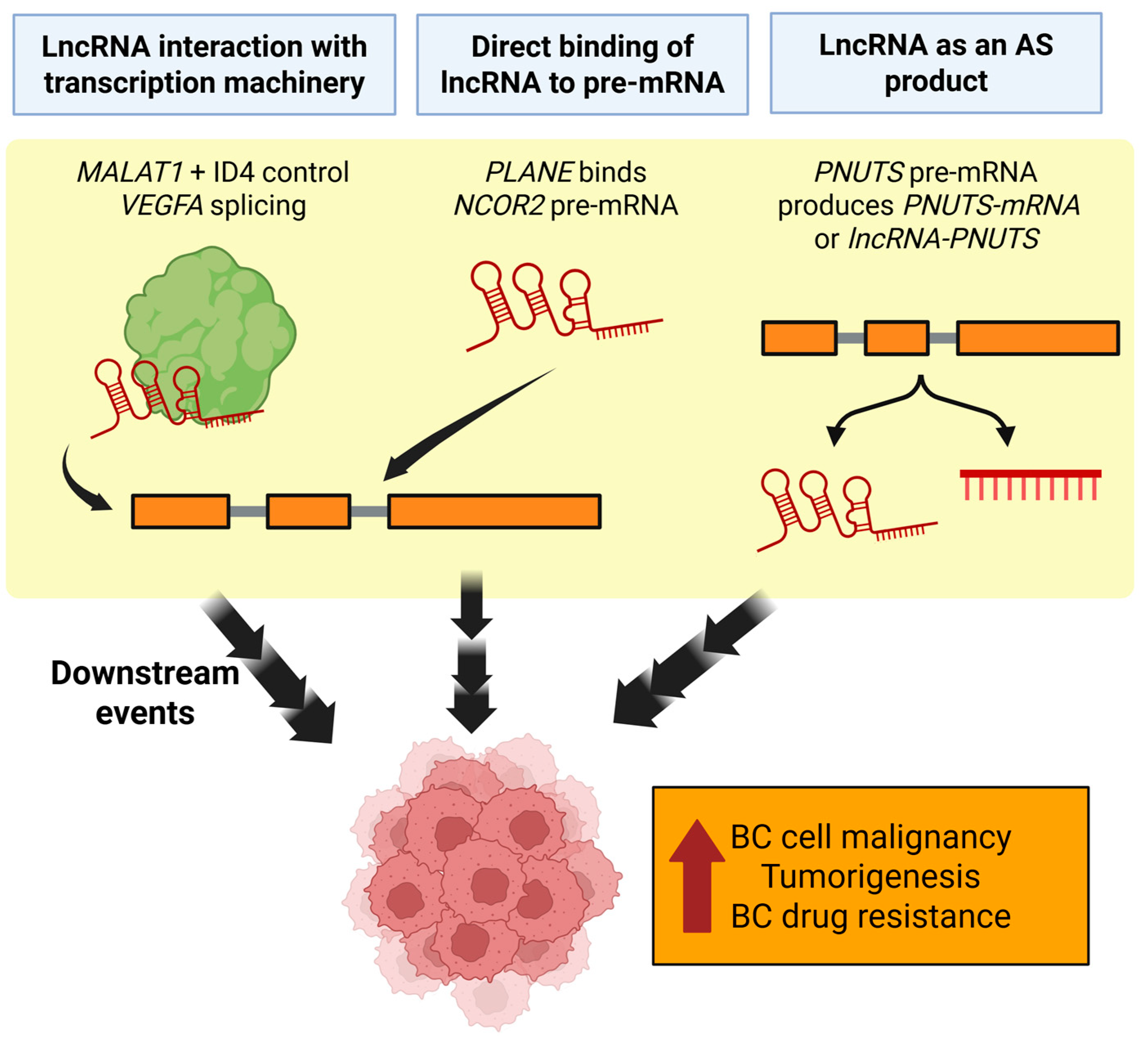
Figure 1. Example mechanisms of breast cancer malignancy regulation by lncRNAs via control of alternative splicing. MALAT1 and PLANE may affect alternative splicing by indirect and direct binding of other breast-cancer related genes, respectively. When PNUTS alternative splicing is biased towards its lncRNA form, tumorigenesis is promoted. AS = Alternative splicing; BC = Breast cancer. Created in https://BioRender.com (Accessed on 19 August 2025).
Cancers 17 03191 g001
Figure 2. Epigenetic regulation mechanisms of lncRNAs in breast cancer. (A) The lncRNA MEG3 is frequently silenced in BC due to promoter methylation. When unmethylated, MEG3 can contribute to p53 signaling. (B) The LINC00511 genomic superenhancer regions are enriched with H3K27ac and H3K4me3, which can lead to upregulation of stem-like cancer cells. (C) HOTAIR recruits PRC2 and LSD1, reprogramming chromatin to silence metastasis suppressor genes and promoting oncogenesis. EMT = Epithelial–mesenchymal transition. Created in https://BioRender.com (Accessed on 19 August 2025).
Cancers 17 03191 g002
Figure 3. Hypoxic conditions influence lncRNA expression and subsequent downstream events related to BC etiology. RAMP-AS1 expression inhibits miR-660-5p from interacting with ATM mRNA, maintaining normal cellular environments. NDRG-OT1, RBM5-AS1, and RPPH1 are examples of lncRNAs that are upregulated in hypoxic conditions. These lncRNAs are involved in mechanisms including stabilization of oncogenic factors or inhibition of certain targets, which promote BC pathogenesis. Created in https://BioRender.com (Accessed on 19 August 2025).
Cancers 17 03191 g003
Table 2. Widely studied lncRNAs with known functions, prognostic relevance, and therapeutic potential.
LncRNAFunctionBC PrognosisTherapeutic PotentialReferences
HOTAIR Scaffolds PRC2/LSD1, drives chromatin remodeling, EMT, and metastasisUpregulation frequently linked to poor prognosis; some mixed resultsASOs/siRNA to disrupt PRC2/LSD1 interaction; HOTAIR-N upregulation as a candidate biomarker[61,76]
NEAT1 Maintains paraspeckle integrity, promotes stress survivalUpregulation frequently linked to poor prognosis; some mixed resultsIsoform-specific targeting; RNA-FISH for patient stratification[63,66,67]
MALAT1 Regulates EMT, metastasis, alternative splicing, PI3K-Akt signalingUpregulation frequently linked to poor prognosis; some mixed results Δsv-MALAT1 as decoy; Δsv-MALAT1 downregulation as a candidate biomarker[49,69,70]
H19 Sponges let-7; supports stemness; confers tamoxifen resistance via autophagy regulationUpregulation frequently linked to poor prognosisH19 upregulation as a candidate biomarker[79,88]
GAS5 Tumor suppressor, modulates drug sensitivity (trastuzumab, lapatinib)Downregulation linked to drug resistance in HER2+ BCRestoration strategies to enhance HER2-targeted therapy[38,102]
Table 3. List of technologies or methods that can be used to resolve knowledge gaps related to lncRNA functional heterogeneity.
Tool/TechniqueOverviewExample Use Case in BCReferences
Personalized lncRNA expression profilingGenerates of sample-specific lncRNA (IDElncRNAs).LncRIndiv constructs a IDElncRNA profile for BRCA to characterize subtype-specfic lncRNAs.[101]
Capture Long-Read Sequencing (CLS)Probes targeting lncRNAs are created, from which cDNA libraries enriching for complete 5′ to 3′ RNA molecules are then made. Long-read sequencing is then carried out on the libraries. Improve lncRNA annotation quality.[111]
CRISPR/Cas9DSB induced by Cas9 is repaired by sgRNA, which can be customized to make specific edits to DNA. Suppression of LINC00511 transcription, which decreases BC cell proliferation rate.[112]
CRISPRaCRISPR system to activate genes by using catalytically inactive Cas9 to recruit transcriptional activators.Activate OBSCN-AS1 to restore OBSCN expression, thus suppressing TNBC cell migration.[113]
CRISPRiCRISPR system to repress gene expression by using catalytically inactive Cas9 to recruit transcriptional repressors.
CRISPR/Cas13dKnock-down lncRNA via cleavage by Cas13.Used in a screen for lncRNAs that can upregulate BC cell proliferation. The tumor suppressor KILR was identified via this method.[114]
scRNA-seqAnalyses of transcriptional profiles at the resolution of individual cells.Together with spatial transcriptomics, TINCR expression was found to be highly expressed in BC samples.[115,116]
Spatial transcriptomicsPreserves the spatial context of gene expression within samples with heterogenous cell populations.Together with scRNA-seq, TINCR expression was found to be highly expressed in BC samples.[116]
Machine learning (ML) methodsThe use of computational models and statistical algorithms to automatically analyze and draw inferences from patterns.Uncover patterns overlooked by traditional annotation. TERIUS distinguishes lncRNAs from false positives by focusing on 3′ UTR fragments.[117,118]
Large Language Models (LLMs)Artificial intelligence system that automatically analyzes and generates human-like language and text based on patterns from large text datasets that it is trained on.Uncover patterns overlooked by traditional annotation.
LncRNA-BERT uses a model trained on existing sequence databases to distinguish mRNA from lncRNA.
[117,119]
Disclaimer/Publisher’s Note: The statements, opinions and data contained in all publications are solely those of the individual author(s) and contributor(s) and not of MDPI and/or the editor(s). MDPI and/or the editor(s) disclaim responsibility for any injury to people or property resulting from any ideas, methods, instructions or products referred to in the content.