Article

Can PSMA-Targeting Radiopharmaceuticals Be Useful for Detecting Brain Metastasis of Various Tumors Using Positron Emission Tomography?

1
Clinic of Nuclear Medicine, University of Health and Sciences Turkey, Istanbul Training and Research Hospital, 34098 Istanbul, Turkey
2
Department of Radiology, University of Health and Sciences Turkey, Istanbul Training and Research Hospital, 34098 Istanbul, Turkey
3
Department of Pathology, University of Health and Sciences Turkey, Istanbul Training and Research Hospital, 34098 Istanbul, Turkey
4
Department of Medical Oncology, University of Health and Sciences Turkey, Istanbul Training and Research Hospital, 34098 Istanbul, Turkey
*
Author to whom correspondence should be addressed.
Cancers 2025, 17(18), 3088; https://doi.org/10.3390/cancers17183088
Submission received: 9 August 2025 / Revised: 17 September 2025 / Accepted: 18 September 2025 / Published: 22 September 2025

Simple Summary

The aim of this prospective study was to investigate the diagnostic value of 68Ga-PSMA-11 PET/CT by comparing 68Ga-PSMA-11 PET/CT, 18F-FDG PET/CT, and MRI findings in patients with brain metastases (BM). Given the absence of PSMA expression in normal brain parenchyma, we aimed to demonstrate the value of 68Ga-PSMA-11 over 18F-FDG in BM imaging. Twenty-four (89%) patients were included in the study for restaging, two (7%) patients for local recurrence assessment, and one (4%) patient for local recurrence and suspicion of additional lesions. The indications for 18F-FDG PET/CT were breast carcinoma for 37% (n:10), followed by lung carcinoma (26% (n:7), colorectal adenocarcinoma for 14% (n:4), squamous cell larynx carcinoma for 7% (n:2), gastric signet ring cell carcinoma for 4% (n:1), pancreatic neuroendocrine tumor grade 3 for 4% (n:1), thyroid papillary carcinoma for 4% (n:1), and malignant melanoma for 4% (n:1). 68Ga-PSMA-11 PET/CT revealed PSMA-positive brain metastases in 26 of 27 patients; only a single patient exhibited PSMA-negative lesion. This patient was followed with a diagnosis of primary larynx squamous carcinoma and had a mass suspected of metastasis. Further tests and an MRI revealed that the lesion in this patient was a hemorrhagic metastasis. In addition to its diagnostic value, we believe our findings may influence PSMA-targeted therapeutic approaches and provide a new perspective on the treatment of patients with BM.

Abstract

Objective: The high expression of prostate-specific membrane antigen (PSMA) associated with neovascularization in non-prostatic malignant tumors and metastatic lesions has been documented in many studies. By taking advantage of the absence of PSMA-related background activity in brain tissue, in recent years, PSMA has been used for the imaging of glial tumors, especially for postoperative follow-up. The aim of this prospective study was to investigate the diagnostic value of 68Ga-PSMA-11 PET/CT by comparing 68Ga-PSMA-11 PET/CT, 18F-FDG PET/CT, and MRI findings in patients with brain metastases (BM). Materials and Method: In this prospective study, 27 cases, 11 female and 16 male, with a mean age of 59.48 ± 12.21 years, were included. Patients diagnosed with BM on 18F-FDG PET/CT or CT/MRI at initial diagnosis or in the follow-up period were included in the study. PET findings of BM lesions obtained from 18F-FDG and 68Ga-PSMA-11 PET/CT imaging, demographic characteristics, histopathological data of the primary foci, and other clinical features were evaluated together. Results: Twenty-four (89%) patients were included in the study for restaging, two (7%) patients for local recurrence assessment, and one (4%) patient for local recurrence and suspicion of additional lesions. The indications for 18F-FDG PET/CT were breast carcinoma for 37% (n:10), followed by lung carcinoma for 26% (n:7), colorectal adenocarcinoma for 14% (n:4), squamous cell larynx carcinoma for 7% (n:2), gastric signet ring cell carcinoma for 4% (n:1), pancreatic NET3 for 4% (n:1), thyroid papillary carcinoma for 4% (n:1), and malignant melanoma for 4% (n:1). In total, 26/27 included patients had PSMA-positive brain metastases but only one patient had PSMA-negative brain metastases with 68Ga-PSMA-11 PET/CT imaging. This patient was followed with a diagnosis of primary larynx squamous carcinoma and had a mass suspected of brain metastases. Further tests and an MRI revealed that the lesion in this patient was a hemorrhagic metastasis. The smallest metastatic focus on 68Ga-PSMA-11 PET/CT imaging was 0.22 cm, also confirmed by MRI (range: 0.22–2.81 cm). The mean ± SD SUVmax of the BM lesions was 17.9 ± 8.6 and 6.8 ± 5.2 on 18F-FDG PET/CT and 68Ga-PSMA-11 PET/CT imaging, respectively. Metastatic foci that could not be detected by 18F-FDG PET/CT imaging were successfully detected with 68Ga-PSMA-11 PET/CT imaging in 11 cases (42%). The distribution and number of metastatic lesions observed on cranial MRI and 68Ga-PSMA-11 PET/CT were compatible with each other for all patients. Immunohistochemical staining was performed in the primary tumor of 10 (38%) cases, and positive IHC staining with PSMA was detected in 5 patients. In addition, positive IHC staining with PSMA was detected in all of the four surgically excised brain metastatic tumor foci. Conclusions: In this study,68Ga-PSMA-11 PET/CT appears to be superior to 18F-FDG in detecting BM from various tumors, largely due to its high expression associated with neovascularization and the absence of PSMA expression in normal brain parenchyma. This lack of physiological uptake in healthy brain tissue provides excellent tumor-to-background contrast, further supporting the advantage of 68Ga-PSMA-11 over 18F-FDG for BM imaging. However, larger studies are required to confirm these findings, particularly through comparisons across tumor types and histopathological subgroups, integrating PSMA immunohistochemistry (IHC) scores with 68Ga-PSMA-11 uptake levels. Beyond its diagnostic potential, our results may also inform PSMA-targeted therapeutic strategies, offering new perspectives for the management of patients with brain metastases from diverse primary tumors.

1. Introduction

Brain metastasis (BM) is much more common than primary central nervous system tumors and is reported in approximately 10% to 30% of patients with metastatic disease. Although survival differs according to the histological type, grade, and molecular subtypes of the primary tumor, BM often have limited treatment response and poor prognosis [1]. BM is frequently observed in lung, breast, renal, and colorectal cancers and malignant melanoma. Treatment options generally include whole-brain radiotherapy, surgical resection, stereotactic radiosurgery, and chemotherapy [2].
Magnetic resonance imaging (MRI) is frequently used and the gold standard in the diagnosis and follow-up of glial tumors. However, it still has limitations for detecting viable tumor localization in previously treated cases and for differential diagnosis of necrosis from recurrence [3]. 18F-fluoro-2-deoxy-glucose (18F-FDG), positron emission tomography/computed tomography (PET/CT), which is widely used in the staging, restaging, and treatment response assessment of many cancers, has a low specificity in BM, due to high physiological radiopharmaceutical uptake. Other disadvantages such as inflammatory uptake secondary to radiation therapy and limited discrimination of low- and high-grade lesions may all cause false negative and positive evaluations [4].
Radio-labeled amino acids have been used in BM imaging for many years in oncology practice [5], and the most widely used one is 11C-methyl-L-methionine (MET), an essential amino acid labeled with the Carbon-11 isotope [6,7]. Due to the short half-life (20 minutes) of carbon-11, it only allows for PET imaging in centers with on-site cyclotrons. In their study, Galldiks et al. elaborated and presented recommendations for the use of PET imaging in the clinical management of patients with BM based on evidence from histology and/or clinically validated studies [8].
Prostate-specific membrane antigen (PSMA) is a type II transmembrane protein and has an important role, especially in high-grade prostate cancer management due to high tumor cell affinity [9]. PSMA does not distribute in the cerebrum, cerebellum, or cerebrospinal fluid. However, it can be expressed at high levels in the vascular endothelium of tumors, and its high expression associated with neovascularization, especially in high-grade gliomas, has been reported in some studies [10,11]. In addition, increased PSMA expression in tumor tissue was significantly associated with poor survival [12]. By utilizing the high tumor-related vascular expression of PSMA, it can successfully be used for the screening of distant organ metastasis not only in prostate cancer but also in different cancer types [13]. Currently, PSMA has been introduced as a promising molecular imaging target to monitor glial tumors, which are the most common primary tumors of the brain, especially in the postoperative period [14]. The aim of this study is to assess the use of 68Ga-PSMA-11 PET/CT in detecting BM, in comparison with 18F-FDG PET/CT, and its potential contribution to MRI, as well as to predict the potential of 177Lu-PSMA-617 treatment with these preliminary findings for the near future.

2. Material and Method

2.1. Patients

This prospective study included patients who were known to have brain metastases and were scheduled to undergo imaging for systemic screening, who were previously treated for brain metastases and suspected of local recurrence, or who had widespread metastases, and who also underwent 18F-FDG PET/CT imaging and subsequently underwent brain MRI due to suspicion of brain metastasis, between February 2021 and March 2022. When the study was designed, the number of participants was planned to be 25–30 and a total of 27 patients were included in the study. All patients who gave consent underwent 68Ga PSMA PET CT imaging.
68Ga-PSMA-11 PET/CT imaging was performed in 27 (11 F, 16 M) patients with a mean age of 59.48 ± 12.21 (range: 34 to 82) years.
This prospective clinical study was approved by the local (Istanbul Training and Research Hospital) ethics committee (approval code 2021/2669). In addition, verbal and written consent was obtained from all included patients, allowing the use of their medical findings for research purposes.

2.2. 18F-FDG PET/CT Imaging

Patients with a blood glucose level of <150 mg/dl after at least 6 hours of fasting before application were included in the study. A standard activity of 3.7–5.2 MBq/kg 18F-FDG was intravenously injected. Approximately 60 minutes following the injection, whole-body PET/CT, including the vertex to the upper thigh area, was performed in the supine position, followed by dedicated brain PET/CT imaging. Imaging was performed with the Siemens mCT Biograph 20 ultraHD LSO PET/CT device (Siemens Molecular Imaging, Hoffmann Estates, IL, USA). For the calculation of SUVmax, regions of interest (ROI), including the highest involvement area in the BM focus, were drawn over PET sections. SUVmax was calculated according to the formula of the maximum activity in the ROI (MBq/ml)/injected 18F-FDG dose (MBq/kg body weight).

2.3. 68Ga-PSMA-11 PET/CT Imaging

All patients were intravenously injected with standard activity of 175 MBq (range 77–350 MBq) 68Ga-PSMA-11. Whole-body and -brain PET/CT imaging was performed in the supine position, including the area from the vertex to the upper part of the thigh 60 minutes after injection. 68Ga-PSMA-11 PET/CT imaging was performed using the same PET/CT system and procedures as the 18F-FDG PET/CT imaging. For the calculation of SUVmax, regions of interest (ROI), including the highest involvement area in the brain metastasis focus, were drawn over PET sections.
The evaluation of PET/CT imaging with both radiopharmaceuticals was performed visually as well as semi-quantitatively using SUVmax of BM lesions calculated for all patients. The obtained PET parameters were comparatively analyzed and correlated with the clinical characteristics of the patients, such as demographics, BM primary focus, and histopathological data (Table 1).

2.4. IHC Staining with PSMA

Immunohistochemistry (IHC) was performed using the prostate-specific membrane antigen (PSMA) (EP192, Epitomics, Inc. Burlingame, CA, USA) Rabbit Monoclonal Primary Antibody (Roche/Cell Marque). The antibody was applied on formalin-fixed, paraffin-embedded (FFPE) tissue sections prepared from both primary tumor lesions and metastatic tumor tissue. Paraffin blocks were cut into 2 µm sections, deparaffinized, and rehydrated through graded alcohols followed by washing in Tris-buffered saline (TBS, Aniara Diagnostica LLC/Medicago AB, Sweden). Tissue sections were subsequently stained on the Ventana Benchmark IHC/ISH automated platform. Slides were incubated for 20 minutes at room temperature with the anti-PSMA rabbit monoclonal antibody (clone EP192) at a dilution of 1:20, and visualization was achieved using the ultraView Universal DAB Detection Kit (Ventana Medical Systems, Roche, Switzerland).

Scoring System

PSMA expression was assessed by two parameters: staining percentage (SP) and staining intensity (SI).
  • SP was visually quantified using a four-tiered system:
    • 0: SP <20% (no expression).
    • 1: SP 20–50% (low expression).
    • 2: SP 50–80% (moderate expression).
    • 3: SP >80% (high expression).
  • SI was also evaluated on a four-tiered scale:
    • 0: No staining.
    • 1: Weak.
    • 2: Moderate.
    • 3: Strong.
  • The final IHC score was calculated by multiplying the staining percentage score by the staining intensity score. Both membranous and cytoplasmic PSMA expression were recorded (Table 2).

2.5. Statistical Analysis

Version 21.0 of the SPSS (Statistical Package for the Social Sciences) program (IBM, Armonk, NY, USA) was used for statistical analysis. Descriptive statistics were expressed as mean ± standard deviation or median (minimum–maximum) for discrete and continuous numerical variables and the number of cases and (%) for categorical variables.

3. Results

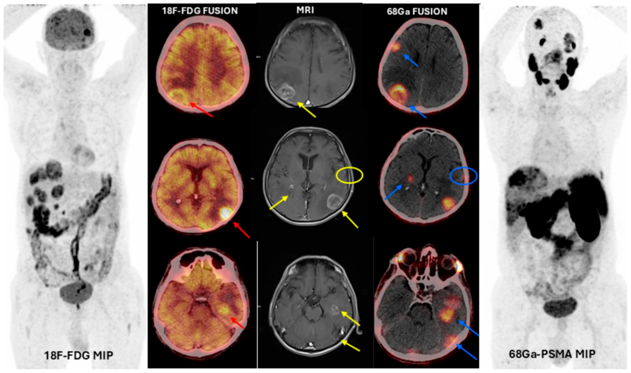
Twenty-four (89%) patients were included in the study for restaging, two (7%) patients for local recurrence assessment, and one (4%) patient for local recurrence and suspicion of additional lesions. The indications for 18F-FDG PET/CT were breast carcinoma (Figure 1) for 37% (n:10), followed by lung carcinoma for 26% (n:7), colorectal adenocarcinoma for 14% (n:4), squamous cell larynx carcinoma for 7% (n:2), gastric signet ring cell carcinoma for 4% (n:1), pancreatic NET3 (Figure 2) for 4% (n:1), papillary thyroid carcinoma for 4% (n:1), and malignant melanoma for 4% (n:1). 68Ga-PSMA-11 PET/CT revealed PSMA-positive brain metastases in 26 of 27 patients; only a single patient exhibited PSMA-negative lesion (Figure 3). This single patient was followed with a diagnosis of primary larynx squamous carcinoma and had a mass suspected of metastasis. Further tests and an MRI revealed that the lesion in this patient was a hemorrhagic metastasis.
The histopathological diagnosis was made by biopsy material obtained on the primary tumor (Table 1). Cranial MRI was performed in all cases within the same week with 68Ga-PSMA-11 PET/CT, and all metastatic lesions were confirmed with MRI. MRI revealed a single metastatic lesion in 15 (55%) patients and multiple metastatic lesions in 12 (45%) patients. Immunohistochemical staining with PSMA was performed in primary tumors of 10 patients, and positive staining was detected in 5 (50%) of them, as well as in all 4 (100%) brain metastatic tumor foci (Table 2).
Table 1. Clinical characteristics of patients *.
NoAgeGenderPrimary Histopathological
Diagnosis
Number of
Metastatic Lesions
Axial Diameter of the Most Prominent
Metastatic Lesion (cm)
18F- FDG SUV Max of
the Most Prominent
Metastatic Lesion
68Ga-PSMA-11 SUVmax of the Most Prominent Metastatic LesionSmallest Axial
Diameter of the
Lesion Detected with 68Ga-PSMA-11 (cm)
Lesions Observed in 68Ga-PSMA-11 PET/CT not Observed in 18F- FDG PET/CT
147FemaleTriple-negative ID Breast Carcinoma14.3411.976.58Not applicableNot applicable
251FemaleTriple-negative ID Breast CarcinomaMultiple1.1323.985.550.71Not applicable
353FemaleHER2 Positive ID Breast CarcinomaMultiple2.4115.6913.830.40Not applicable
465FemaleLuminal B ID Breast Carcinoma32.3017.417.72Not applicableNot applicable
534FemaleLuminal B ID Breast Carcinoma12.7720.1910.77Not applicableNot applicable
641FemaleHER2 Positive ID Breast Carcinoma11.2021.7914.50Not applicableNot applicable
743FemaleLuminal B ID Breast CarcinomaMultiple2.0830.674.490.22+
847FemaleLuminal B ID Breast CarcinomaMultiple0.57No uptake2.110.26+
961MaleSquamous Cell Lung CarcinomaMultiple1.4511.804.380.46+
1056FemaleLung Adenocarcinoma12.34No uptake2.190.39+
1171MaleSquamous Cell Lung Carcinoma13.1910.554.35Not applicableNot applicable
1267MaleSmall Cell Lung CarcinomaMultiple1.279.333.590.31Not applicable
1372MaleColorectal Adenocarcinoma12.005.603.80Not applicableNot applicable
1451MaleColorectal Adenocarcinoma11.60No uptake2.801.60+
1567MaleColorectal AdenocarcinomaMultiple2.4611.726.510.35+
1674MaleColorectal Adenocarcinoma41.3934.878.290.54Not applicable
1768MalePancreatic Neuroendocrine
Tumor Grade 3
Multiple3.0220.2912.441.32+
1847MaleSignet Ring Cell Gastric Carcinoma32.2832.003.77Not applicableNot applicable
1964MaleFollicular Variant of Papillary Thyroid
Carcinoma
11.16No uptake24.951.16+
2063MaleSquamous Cell Lung Carcinoma11.7629.376.891.76Not applicable
2156MaleLung Adenocarcinoma12.81No uptake2.72.81+
2260MaleSquamous Cell Lung Carcinoma +
Squamous Cell Nasopharnygeal
Carcinoma
13.7312.272.23Not applicableNot applicable
2351FemaleTriple-negative ID Breast Carcinoma21.69.554.30.76+
2481FemaleLuminal B ID Breast Carcinoma12.0519.684.18Not applicableNot applicable
2571MaleMalignant MelanomaMultiple3.239.727.080.48+
2663MaleSquamous Cell Larynx Carcinoma +
Squamous Cell Lung Carcinoma
12.18No uptake2.92.18+
2782MaleSquamous Cell Larynx Carcinoma11.7No uptakeNo uptakeNot applicableNot applicable
* ID: Invasive Ductal.
Table 2. Immunohistochemical staining with PSMA in primary tumors and metastatic foci *.
NPrimary Histopathological DiagnosisPrimary Tumor
PSMA Staining Score
Metastatic Tumor
PSMA Staining Score
1.Triple-negative ID Breast CarcinomaSP:11 × 1:1SP:11 × 1:1
SI:1SI:1
2.Triple-negative ID Breast CarcinomaSP:00SP:00
SI:0 SI:0
13.Colorectal AdenocarcinomaSP:11 × 1:1SP:11 × 1:1
SI:1 SI:1
16.HER2 Positive ID Breast CarcinomaSP:11 × 1:1SP:22 × 1:2
SI:1 SI:1
18.Luminal B ID Breast CarcinomaSP:11 × 1:1SP:00
SI:1 SI:0
10.Lung AdenocarcinomaSP:00SP:00
SI:0 SI:0
12.Small Cell Lung CarcinomaSP:00SP:00
SI:0 SI:0
3.HER2 Positive ID Breast CarcinomaSP:00SP:00
SI:0 SI:0
5.Luminal B ID Breast CarcinomaSP:0 SP:00
SI:00SI:0
8.Luminal B ID Breast CarcinomaSP:11 × 1:1SP:22 × 1:2
SI:1 SI:1
* ID: Invasive Ductal; SP: Staining Percentage; SI: Staining Index.
While BM foci were detected in all 26 patients with 68Ga-PSMA-11 PET/CT, only 15 (58%) of them could be detected with 18F-FDG PET/CT. The mean ± SD axial diameter of the largest lesions visualized on 68Ga-PSMA-11 PET/CT was found to be 2.16 ± 0.88 cm (range: 0.57–4.34 cm). The smallest size of metastases that could be visualized was 0.22 cm in axial diameter (Figure 1). Mean ± SD SUVmax of metastatic lesions measured on 18F-FDG PET/CT imaging was 17.9 ± 8.6 (range: 5.6–37.8), and mean ± SD SUVmax measured on 68Ga-PSMA-11 PET/CT imaging was 6.8 ± 5.2 (range: 2.1–24.9). Although the mean SUVmax was higher in 18F-FDG than 68Ga-PSMA-11, no statistically significant difference was found between the two imaging methods (p:0.37).

4. Discussion

The tumor microenvironment plays a crucial role in the prognostic behavior of malignancies. Angiogenesis is one of the most critical pillars of tumor progression, which starts with the stimulation of endothelial cells mediated mainly by vascular endothelial growth factor (VEGF) due to hypoxia and molecular changes [15]. Wernicke et al. reported that PSMA expression was detected in tumor endothelium in all 32 patients with glioblastoma multiforme. Intense PSMA staining was reported in 22 cases (69%) [16]. Tanjore et al. demonstrated the association of neovascular PSMA expression with malignant progression and poor survival after a 10.4-year follow-up of 371 patients with glioblastoma and 52 patients with brain metastatic lung cancer [17]. One of the most common problems in the clinical management of primary brain tumors is the accurate detection of local recurrence during postoperative follow-up. On the other hand, Akgun et al. reported significantly higher PSMA expression in grade IV glial tumors than grade II and III with 68Ga-PSMA-11 PET/CT imaging [14].
In metastatic brain tumors, there is a very heterogeneous distribution for the primary tumor type such that in 15% of patients with BM diagnosis, primary malignant tumor cannot even be detected. Lung cancer, breast cancer, and malignant melanoma have frequently been documented as primaries for patients with BM. In their study, Habbous et al. screened patients with a metastatic brain tumor diagnosis between 2010 and 2018, and reported that BM was found in 25.478 (4.2%) of the 601.678 patients, where, the primary focus was lung cancer in 60%, breast cancer in 11%, and malignant melanoma in 6% [18]. BM is seen throughout the disease in approximately 40–50% of patients with lung cancer, especially small cell lung cancer, and this rate is around 10–20% at the time of initial diagnosis [2]. In breast cancers, approximately 15–20% BM is reported. HER-2 positive breast cancer especially has a high BM rate [19]. In our study, the most common BM focus was breast cancer with a rate of 38%, which id consistent with the published data, followed by lung cancer at 27% and colon cancer at 15%. The reason why our rate of lung cancer cases is lower than the literature may be related to the fact that our hospital is a reference center especially for breast and colorectal cancers. Wernicke et al. reported BM in 14 of 106 patients with breast cancer. In the immunohistochemical staining to investigate the PSMA expression in tumor vascular endothelial cells, positive PSMA expression was observed in 74% (68/92) of primary breast cancers and 100% (n = 14) in BM. With this study, the researchers demonstrated the high expression of PSMA associated with tumor neovascularization [20]. In the study, Nomura et al. investigated the tissues targeted by PSMA immunohistochemically on 19 patients with glioma and 5 with breast cancer, all with a secondary BM diagnosis. At the same time, no PSMA staining was detected in normal brain microvasculature; a significantly increased (p < 0.05) rate of PSMA expression was determined in high angiogenic grade IV glial tumors. In addition, more vital vascular PSMA staining was detected in BM compared to primary breast cancer tumor tissue. Researchers have demonstrated the high expression of PSMA in primary tumor and BM-associated vascular tissues [21].
BM-associated PSMA expression has also been coincidentally documented for different cancer types in the literature. Coincidentally, Hod et al., in their 76-year-old male patient with resected malignant melanoma, observed unexpectedly high PSMA brain involvement in the previous surgical cavity on 68Ga-PSMA-11 PET/CT imaging. After CT and MRI, the diagnosis of local melanoma recurrence was confirmed [22]. Vallejo-Armenta et al. reported significantly increased uptake in metastatic brain tumors, high-grade glial brain tumors, and recurrent gliomas with preoperative [99mTc] Tc-iPSMA SPECT brain imaging in 41 patients diagnosed with BM by MRI. However, they did not detect involvement in low-grade glial brain tumor lesions. Researchers reported that PSMA is expressed at high levels in grade IV glioma and BM vascular endothelium [23]. In support of all these studies, tumor-associated neovascularization of a 47-year-old patient with triple-negative breast carcinoma BM with low 18F-FDG uptake despite high PSMA activity was demonstrated [24]. In the present study, all BM foci that could not be detected with 18F-FDG PET/CT were successfully detected with 68Ga-PSMA-11 PET/CT in 42% of the cases. Wei et al. observed significant tumor regression and decreased PSMA expression in cerebral lesions based on 68Ga-PSMA PET/CT imaging performed after 177Lu-PSMA-617 treatments combined with radiotherapy in two prostate cancer patients with cerebral metastasis [25]. Dall’Armellina S et al. also presented results of PSMA-targeted PET imaging for brain metastases from non-prostate solid tumors, including results from a total of 23 studies [26].
However, false positives associated with 68Ga-PSMA-11 PET/CT imaging should not be ignored. Noto et al. observed focal involvement in the right frontal lobe with suspected BM in a 65-year-old patient diagnosed with prostate adenocarcinoma in 68Ga-PSMA-HBED-CC-PET/CT imaging. Eventually, it was understood that the reason for the cerebral 68Ga-PSMA involvement observed in that patient was recent cerebral infarction [27].
Oh G et al. reported a case of subacute cerebellar infarction mimicking metastasis on prostate-specific membrane antigen (PSMA) PET/CT in a 77-year-old man with prostate cancer [28]. And Jain V et al. also published a case report in which they observed positive uptake on 18 F-PSMA PET/CT due to subacute cerebral infarction [29]. Additionally, Huang Y. et al. observed false-positive findings on a 68Ga-labeled prostate-specific membrane antigen (PSMA) ligand PET/CT scan due to elevated prostate-specific antigen levels in a 56-year-old man with a history of brain abscess [30].
Currently, PSMA-targeted molecular imaging using different radiopharmaceuticals is established mostly for prostate cancer, owing to the higher expression of PSMA in especially aggressive prostate cancer cells. The introduction of theragnostic applications with the use of the PSMA target has recently provided a paradigm change for the metastatic castration-resistant prostate cancer and studies are on the way to assess its use for the earlier periods of the disease. Our study along with aforementioned studies and some others in the literature definitely support the use of the PSMA target for tumors other than prostate cancer, thanks to its overexpression related to neovascularization regardless of the tumor type to the best of our knowledge and, in particular, to detect BM owing to its cerebral biodistribution advantage for healthy brain tissue over the most commonly used oncologic PET radiopharmaceutical 18F-FDG. In this regard, PSMA seems to be a promising target for patients with various tumors with extensive metastasis including BM to provide an alternative systemic treatment through theragnostic applications in the near future.

5. Conclusions

This study demonstrates that the absence of PSMA expression in normal cerebral and cerebellar parenchyma, in contrast to the high physiological background uptake of 18F-FDG, provides 68Ga-PSMA-11 PET/CT with a clear diagnostic advantage in BM imaging. Although PSMA is frequently upregulated in high-grade tumors, this cannot be generalized across all malignancies, underscoring the need for larger studies that systematically correlate PSMA immunohistochemistry (IHC) scores with 68Ga-PSMA-11 uptake across tumor types and histopathological subgroups. Importantly, treatment-related factors such as prior radiotherapy, systemic chemotherapy, and hormone therapy, which may alter receptor expression, should also be addressed in future analyses.
Our findings further highlight the potential of PSMA as a therapeutic target in BM, supporting the development of radionuclide-based strategies capable of exploiting tumor neovascularization while preserving normal brain tissue. To validate this approach and determine its true impact on patient outcomes, well-designed prospective trials incorporating standardized brain MRI alongside 18F-FDG PET/CT and 68Ga-PSMA-11 are warranted. Ultimately, this strategy may contribute to the establishment of innovative therapeutic options for patients with BM from diverse primary tumors.

Author Contributions

E.A.: study design, formal analysis, writing—original draft, and writing—review and editing; N.E.: data analysis, data interpretation, methodology; R.Ş.: literature search, data collection, software; E.B. data collection; Ö.E.F.: data collection; Y.K.: data collection, visualization; A.A.G.: data collection, data interpretation; Y.B.: literature search; T.F.Ç.: study design, supervision. All authors have read and agreed to the published version of the manuscript.

Funding

This study was funded by the budget allocated for education within the scope of the scientific research project in our department. There is no grant number.

Institutional Review Board Statement

All procedures performed in studies involving human participants were in accordance with the ethical standards of the institutional research committee and with the 1964 Helsinki declaration and its later amendments or comparable ethical standards. This prospective clinical study was approved by the local ethics committee (Istanbul Training and Research Hospital Ethics Committee approval code 2021/2669).

Informed Consent Statement

Informed consent was obtained from all participants included in the study.

Data Availability Statement

The data presented in this study are available in this article.

Conflicts of Interest

The authors declare no conflicts of interest.

References

  1. Doron, H.; Pukrop, T.; Erez, N. A Blazing Landscape: Neuroinflammation Shapes Brain Metastasis. Cancer Res. 2019, 79, 423–436. [Google Scholar] [CrossRef]
  2. Fokas, E.; Steinbach, J.P.; Rödel, C. Biology of brain metastases and novel targeted therapies: Time to translate the research. Biochim. Biophys. Acta 2013, 1835, 61–75. [Google Scholar] [CrossRef]
  3. Han, X.; Xue, X.; Zhou, H.; Zhang, G. A molecular view of the radioresistance of gliomas. Oncotarget 2017, 8, 100931–100941. [Google Scholar] [CrossRef]
  4. Moreau, A.; Febvey, O.; Mognetti, T.; Frappaz, D.; Kryza, D. Contribution of Different Positron Emission Tomography Tracers in Glioma Management: Focus on Glioblastoma. Front. Oncol. 2019, 9, 1134. [Google Scholar] [CrossRef]
  5. Bergström, M.; Collins, V.P.; Ehrin, E.; Ericson, K.; Eriksson, L.; Greitz, T.; Halldin, C.; von Hoist, H.; Långström, B.; Lilja, A.; et al. Discrepancies in Brain Tumor Extent as Shown by Computed Tomography and Positron Emission Tomography Using [68Ga]EDTA, [11C]Glucose, and [11C]Methionine. J. Comput. Assist. Tomogr. 1983, 7, 1062–1066. [Google Scholar] [CrossRef]
  6. Herholz, K.; Langen, K.-J.; Schiepers, C.; Mountz, J.M. Brain tumors. Semin. Nucl. Med. 2012, 42, 356–370. [Google Scholar] [CrossRef]
  7. Galldiks, N.; Langen, K.-J.; Pope, W.B. From the clinician’s point of view—What is the status quo of positron emission tomography in patients with brain tumors? Neuro-Oncology 2015, 17, 1434–1444. [Google Scholar] [CrossRef]
  8. Galldiks, N.; Langen, K.-J.; Albert, N.L.; Chamberlain, M.; Soffietti, R.; Kim, M.M.; Law, I.; Le Rhun, E.; Chang, S.; Schwarting, J.; et al. PET imaging in patients with brain metastasis—Report of the RANO/PET group. Neuro-Oncology 2019, 21, 585–595. [Google Scholar] [CrossRef] [PubMed]
  9. Barbosa, F.d.G.; Queiroz, M.A.; Nunes, R.F.; Costa, L.B.; Zaniboni, E.C.; Marin, J.F.G.; Cerri, G.G.; Buchpiguel, C.A. Nonprostatic diseases on PSMA PET imaging: A spectrum of benign and malignant findings. Cancer Imaging 2020, 20, 23. [Google Scholar] [CrossRef] [PubMed]
  10. Kiess, A.P.; Banerjee, S.R.; Mease, R.C.; Rowe, S.P.; Rao, A.; Foss, C.A.; Chen, Y.; Yang, X.; Cho, S.Y.; Nimmagadda, S.; et al. Pros-tate-specific membrane antigen as a target for cancer imaging and therapy. Q. J. Nucl. Med. Mol. Imaging 2015, 59, 241–268. [Google Scholar] [PubMed]
  11. Demirci, E.; Ocak, M.; Kabasakal, L.; Decristoforo, C.; Talat, Z.; Halaç, M.; Kanmaz, B. 68Ga-PSMA PET/CT imaging of metastatic clear cell renal cell carcinoma. Eur. J. Nucl. Med. 2014, 41, 1461–1462. [Google Scholar] [CrossRef]
  12. Fragomeni, R.A.S.; Amir, T.; Sheikhbahaei, S.; Harvey, S.C.; Javadi, M.S.; Solnes, L.B.; Kiess, A.P.; Allaf, M.E.; Pomper, M.G.; Gorin, M.A.; et al. Imaging of Nonprostate Cancers Using PSMA-Targeted Radiotracers: Rationale, Current State of the Field, and a Call to Arms. J. Nucl. Med. 2018, 59, 871–877. [Google Scholar] [CrossRef]
  13. Sathekge, M.; Modiselle, M.; Vorster, M.; Mokgoro, N.; Nyakale, N.; Mokaleng, B.; Ebenhan, T. 68Ga-PSMA imaging of metastatic breast cancer. Eur. J. Nucl. Med. 2015, 42, 1482–1483. [Google Scholar] [CrossRef]
  14. Akgun, E.; Akgun, M.Y.; Selçuk, H.H.; Uzan, M.; Sayman, H.B. 68Ga PSMA PET/MR in the differentiation of low and high grade gliomas: Is 68Ga PSMA PET/MRI useful to detect brain gliomas? Eur. J. Radiol. 2020, 130, 109199. [Google Scholar] [CrossRef]
  15. Hosonaga, M.; Saya, H.; Arima, Y. Molecular and cellular mechanisms underlying brain metastasis of breast cancer. Cancer Metastasis Rev. 2020, 39, 711–720. [Google Scholar] [CrossRef]
  16. Wernicke, A.G.; Edgar, M.A.; Lavi, E.; Liu, H.; Salerno, P.; Bander, N.H.; Gutin, P.H. Prostate-Specific Membrane Antigen as a Potential Novel Vascular Target for Treatment of Glioblastoma Multiforme. Arch. Pathol. Lab. Med. 2011, 135, 1486–1489. [Google Scholar] [CrossRef]
  17. Ramanathan, J.T.; Lehtipuro, S.; Sihto, H.; Tóvári, J.; Reiniger, L.; Téglási, V.; Moldvay, J.; Nykter, M.; Haapasalo, H.; Le Joncour, V.; et al. Prostate-specific membrane antigen expression in the vasculature of primary lung carcinomas associates with faster metastatic dissemination to the brain. J. Cell. Mol. Med. 2020, 24, 6916–6927. [Google Scholar] [CrossRef]
  18. Habbous, S.; Forster, K.; Darling, G.; Jerzak, K.; Holloway, C.M.B.; Sahgal, A.; Das, S. Incidence and real-world burden of brain metastases from solid tumors and hematologic malignancies in Ontario: A population-based study. Neuro-Oncol. Adv. 2020, 3, vdaa178. [Google Scholar] [CrossRef] [PubMed]
  19. Rkosy, P.; Tóth, J.; Béres, E.; Tóth, D.; Szivos, L.; Nagy, J.; Klekner, A.; Virga, J. Prognosis and Treatment Outcomes of Patients Undergoing Resection of Brain Metastases from Breast Cancer. Anticancer Res. 2020, 40, 1759–1770. [Google Scholar] [CrossRef]
  20. Wernicke, A.G.; Varma, S.; Greenwood, E.A.; Christos, P.J.; Chao, K.S.C.; Liu, H.; Bander, N.H.; Shin, S.J. Prostate-specific membrane antigen expression in tumor-associated vasculature of breast cancers. APMIS 2013, 122, 482–489. [Google Scholar] [CrossRef] [PubMed]
  21. Nomura, N.; Pastorino, S.; Jiang, P.; Lambert, G.; Crawford, J.R.; Gymnopoulos, M.; Piccioni, D.; Juarez, T.; Pingle, S.C.; Ma-kale, M.; et al. Prostate specific membrane antigen (PSMA) expression in primary gliomas and breast cancer brain metastases. Cancer Cell Int. 2014, 14, 26. [Google Scholar] [CrossRef]
  22. Hod, N.M.; Lantsberg, S.; Benkovich, E.; Kazap, D.E.; Preiskel, M.; Levin, D. Incidental Detection of Malignant Melanoma Brain Recurrence on 68Ga–Prostate-Specific Membrane Antigen PET/CT. Clin. Nucl. Med. 2020, 45, 896–899. [Google Scholar] [CrossRef] [PubMed]
  23. Vallejo-Armenta, P.; Soto-Andonaegui, J.; Villanueva-Pérez, R.M.; González-Díaz, J.I.; Contreras-Contreras, K.; Bautista-Wong, C.G.; Sandoval-Bonilla, B.; Nettel-Rueda, B.; Santos-Cuevas, C.; Ferro-Flores, G. [99mTc]Tc-iPSMA SPECT brain imaging as a potential specific diagnosis of metastatic brain tumors and high-grade gliomas. Nucl. Med. Biol. 2021, 96–97, 1–8. [Google Scholar] [CrossRef] [PubMed]
  24. Arslan, E.; Ergül, N.; Karagöz, Y.; Gedik, A.A.; Çermik, T.F. Recurrent Brain Metastasis of Triple Negative Breast Cancer With High Uptake in 68Ga-PSMA-11 PET/CT. Clin. Nucl. Med. 2020, 46, e106–e108. [Google Scholar] [CrossRef] [PubMed]
  25. Wei, X.; Schlenkhoff, C.; Schwarz, B.; Essler, M.; Ahmadzadehfar, H. Combination of 177Lu-PSMA-617 and Exter-nal Radiotherapy for the Treatment of Cerebral Metastases in Patients with Castration-Resistant Metastatic Pros-tate Cancer. Clin. Nucl. Med. 2017, 42, 704–706. [Google Scholar] [CrossRef]
  26. Armellina, S.D.; Aghakhanyan, G.; Rizzo, A.; Fanni, S.C.; Aringhieri, G.; Faggioni, L.; Cioni, D.; Neri, E.; Volterrani, D.; Morbelli, S. PSMA-targeted PET imaging for brain metastases from non-prostatic solid tumors: A systematic review. Front. Oncol. 2025, 15, 1553505. [Google Scholar] [CrossRef]
  27. Noto, B.; Vrachimis, A.; Schäfers, M.; Stegger, L.; Rahbar, K. Subacute Stroke Mimicking Cerebral Metastasis in 68Ga-PSMA-HBED-CC PET/CT. Clin. Nucl. Med. 2016, 41, e449–e451. [Google Scholar] [CrossRef]
  28. Oh, G.; Miles, K. Subacute Cerebellar Infarction With Uptake on 68Ga–Prostate-Specific Membrane Antigen PET/CT. Clin. Nucl. Med. 2018, 43, 134–135. [Google Scholar] [CrossRef]
  29. Jain, V.; McIntosh, L. Subacute Cerebral Infarct: An Unusual Cause of Radiotracer Uptake at 18 F-PSMA PET/CT. Clin. Nucl. Med. 2024, 49, 882–883. [Google Scholar] [CrossRef]
  30. Huang, Y.; Zhang, B.; Yue, D. 68 Ga-PSMA Uptake of Brain Abscesses in a Patient With Prostate Cancer. Clin. Nucl. Med. 2025, 50, e283–e285. [Google Scholar] [CrossRef] [PubMed]
Figure 1. (Patient No: 7): A 43-year-old woman with ID breast carcinoma; Estrogen Receptor 95%, Progesterone Receptor 95%, c-erbB2 (–), Ki67: 45%. Metastatic lesion with an axial diameter of 0.22 cm detected by 68Ga-PSMA-11 PET/CT and MRI (T2-weighted). PET: positron emission tomography; MIP: maximum intensity projection; 18F-FDG: 18F-fluorodeoxyglucose; 68Ga PSMA: Gallium-68-prostate-specific membrane antigen (lesion detected with MRI: yellow arrow, 68Ga PSMA-11 fusion image positive lesion: blue arrow).
Cancers 17 03088 g001
Figure 2. (Patient No: 17): A 68-year-old man, brain metastases of primary pancreatic neuroendocrine tumor grade 3 detected with 68Ga-PSMA-11 PET/CT and MRI (T1-weighted). PET: positron emission tomography; MIP: maximum intensity projection; 18F-FDG: 18F-fluorodeoxyglucose; 68Ga PSMA: Gallium-68-prostate-specific membrane antigen (18F- FDG fusion images positive metastatic foci: red arrows, lesions detected with MRI: yellow arrows and yellow circle, 68Ga PSMA-11 fusion images, positive metastatic foci: blue arrows and blue circle).
Cancers 17 03088 g002
Figure 3. (Patient No: 27): An 82-year-old man, squamous cell larynx carcinoma hemorrhagic metastasis (18F- FDG negative fusion and 68Ga PSMA-11 negative fusion images and MRI).
Cancers 17 03088 g003
Disclaimer/Publisher’s Note: The statements, opinions and data contained in all publications are solely those of the individual author(s) and contributor(s) and not of MDPI and/or the editor(s). MDPI and/or the editor(s) disclaim responsibility for any injury to people or property resulting from any ideas, methods, instructions or products referred to in the content.