Article

Long-Term Outcomes Following Reconstruction of Diaphyseal Defects of the Upper and Lower Extremities Using Diaphyseal Implants: A Retrospective Study with Focus on Fixation Technique

Department of Orthopedics and Tumororthopedics, Albert-Schweitzer-Campus 1, University Hospital Münster, 48149 Muenster, Germany
*
Author to whom correspondence should be addressed.
These authors contributed equally to this work.
Cancers 2025, 17(18), 3059; https://doi.org/10.3390/cancers17183059
Submission received: 7 July 2025 / Revised: 23 August 2025 / Accepted: 15 September 2025 / Published: 19 September 2025
(This article belongs to the Special Issue Advances in Soft Tissue and Bone Sarcoma (2nd Edition))

Simple Summary

Reconstruction using diaphyseal implants has been insufficiently studied with regard to functional outcomes and prosthesis survival. This study included 39 patients who underwent intercalary endoprosthetic reconstruction of the humerus, femur, and tibia. It analyzes the impact of the fixation method (cemented; uncemented; with locking screw; without locking screw) of the diaphyseal implant on clinical outcomes. The event-free probability in the competing risk model was 61% (95% CI 43–74%) after one year and 11% (95% CI 3–28%) after five years. The complication rate in the patient cohort was 54%. Higher body weight and older patient age were associated with lower MSTS scores. Patients requiring rapid remobilization or adjuvant radiation therapy may benefit more from diaphyseal implants compared to biological reconstructions. However, the complication and revision rates of diaphyseal implants are elevated.

Abstract

Background: The reconstruction of diaphyseal bone defects following tumor resection offers various biological and endoprosthetic treatment options. The present study analyzes the impact of the fixation method (cemented; uncemented; with locking screw; without locking screw) of the diaphyseal implant on clinical outcomes. Factors such as patient age and weight as well as tumor type and location are also considered. Methods: This study included 39 patients who underwent intercalary endoprosthetic reconstruction of the humerus (n = 4); femur (n = 29); and tibia (n = 6) between 1998 and 2020. Prosthetic complications, fixation methods and the MSTS score for functional outcome were statistically analyzed using SPSS and R. Results: The event-free probability in the competing risk model was 61% (95% CI 43–74%) after one year and 11% (95% CI 3–28%) after five years. The complication rate in the patient cohort was 54%. Cementless prosthesis fixation was associated with a statistically significant better functional outcome. Additionally, higher body weight and older patient age were associated with lower MSTS scores. Conclusions: Patients requiring rapid remobilization or adjuvant radiation therapy may benefit more from diaphyseal implants compared to biological reconstructions. However, the complication and revision rates of diaphyseal implants are elevated. The chosen fixation method shows a statistically significant influence on functional outcome.

1. Introduction

Bone and soft tissue sarcomas as well as bony metastases of the extremities can be treated reconstructively or ablatively. Until the 1970s, amputation was the gold standard, but nowadays limb-sparing therapy is the treatment of choice. The goal of surgical therapy is complete tumor removal including the best possible oncological, functional, and cosmetic outcomes [1]. Early diagnosis and careful planning are essential for a successful procedure enabling tumor resection while preserving the joints [2]. In cases of complete resection of joint-adjacent osseous tumors, the proximal or distal joint can be removed and replaced with an endoprosthesis.
For diaphyseal defects, biological reconstruction has been used more frequently in the past. Biological reconstruction options include allograft, fibula graft (vascularized or free), combined vascularized fibula with allograft, segmental bone transport/distraction osteosynthesis, extracorporeal devitalized autograft, Masquelet technique (bone cement spacer and “induced-membrane” technique), segmental prosthesis [3]. However, these reconstruction types result in high complication rates including non-union (40%), fracture (29%), infection (14%), and generally longer periods of non-weight bearing and use of orthoses [4,5].
Therefore, diaphyseal implants represent a considerable alternative to biological reconstructions. Since purely diaphyseal implants theoretically allow joint preservation, it raises the question whether this leads to improved function and longer implant survival [6]. However, because diaphyseal malignant tumors are relatively rare and the use of mega-prosthetic diaphyseal replacements is a relatively recent approach, the available literature on clinical outcomes remains limited [7]. Over the past few years, customized 3D printing methods have been successfully used to create diaphyseal implants [8].
Due to a lack of current studies, this study investigates the functional outcome of diaphyseal implants using the MSTS score. It also examines various anchoring techniques. There is still disagreement regarding the use of bone cement and locking screws. Since aseptic prosthetic loosening is one of the most common complications, optimal fixation is crucial [9,10,11].
The study also analyzes the influence of tumor location, stem type, age, weight, and tumor size and resection length on functional outcomes, prosthetic stability and risk factors that may lead to complications. The results are compared with existing literature and evaluated against other reconstructive methods. Complication rates for femur and tibia reconstructions are compared as well as the outcome and survival of personalized (“custom-made”) versus standard (“off-the-shelf”) implants.

2. Materials and Methods

2.1. Patient Cohort

This study included 39 consecutive patients (20 female, 19 male) who were treated with diaphyseal resection due to a primary malignant bone tumor or metastasis followed by reconstruction with a diaphyseal implant at the University Hospital Muenster between 1998 and 2020 (Table 1).
The average age at the time of treatment was 55.4 years (range 10–85). Fourteen patients (35.9%) with a mean age of 48.4 years (range 21–80) were still alive at the time of data collection, while 25 patients (64.1%) had died. The average age at the time of death was 66 years (range 47–86).
The median follow-up duration for all patients was 47.6 months (range 1–145 months).
Postoperatively, 32, 25, and 12 patients survived at 1, 2, and 5 years, respectively. Among surviving patients, the mean follow-up was 70.8 months (range 17–145 months).

2.2. Patient Groups

Patients were assigned to different groups. In addition to the prosthesis location (femur n = 29, tibia n = 6, humerus n = 4), classification was additionally based on the anchoring mechanism (cemented vs. uncemented, screwed vs. unscrewed). A total of 64 stems were cemented, while 12 were implanted without cement. Forty stems were screwed, and 36 were not.
Twenty-eight prostheses were fully cemented. In two cases, the locking mechanism could not be determined due to lack of documentation and absence of digital X-ray images.
Furthermore, a distinction was made based on tumor type, and the cohort was subdivided according to existing internal medical conditions.

2.3. Methodology

Data acquisition was conducted using keyword-based searches in the hospital information system (Orbis, Dedalus HealthCare GmbH, Bonn, Germany) and using the surgical and procedural coding system (OPS). In addition to collecting baseline data, complications and failure rates of the surgical procedures were evaluated, and radiological imaging was analyzed using the PACS viewer (Picture Archiving and Communication System; Figure 1). Prosthesis-related complications were classified according to Henderson et al. [11,12] into five types, distinguishing mechanical from non-mechanical complications.
The complications were worked up using existing medical documentation (including daily notes and surgical records) as well as patient interviews. In addition to digital data collection, patients were contacted during routine follow-up appointments at the tumor orthopedic outpatient clinic. This allowed the functional outcome to be recorded in 14 patients.
While the main endpoint of the study was prosthesis survival and complication rate depending on the anchoring mechanisms, functional outcomes were also assessed using the MSTS score (Musculoskeletal Tumor Society) [13]. Due to the retrospective character of the study, the timing of the assessment varied on a case-by-case basis during follow-up.

2.4. Statistical Methods

Statistical evaluations were performed using SPSS Statistics (IBM, Version 27.0.0.0), R (The R Foundation, Version 4.0.3), and Microsoft Excel (Version 16.53).
Descriptive statistics such as frequencies, means, medians, minimum, maximum, and standard deviation were calculated using SPSS.
Cross-tabulations and Pearson correlation analyses were conducted to investigate relationships such as between prosthesis location (femoral/tibial/humeral) and MSTS scores (0–30 points). Prosthesis survival required a more complex analysis due to two competing endpoints in tumor patients: prosthesis failure and death.
A “competing-risk analysis” using R was employed. In this method, patients whose follow-up was shorter than 60 months were censored.

3. Results

3.1. Surgical Indications and Comorbidities

Within the studied cohort, various underlying pathologies lead to implantation of a diaphyseal endoprosthesis. The primary surgical indication were malignant bone tumors in 24 patients and metastatic disease in 15 patients. In case of soft tissue sarcoma, tumor extension in direct contact to the bone required a diaphyseal segment resection with following reconstruction.
Patients in the primary tumor group exhibited a significantly higher five-year postoperative survival probability compared to those in the metastasis group. According to Gray’s test, the cumulative incidence of death (0 = alive, 1 = deceased) during follow-up significantly differed between the two groups (p < 0.05).
Table 2 and Table 3 provide detailed survival probabilities at 1, 2, and 5 years, stratified by individual diagnoses.
Additional influencing factors on overall patient survival included a variety of pre-existing and comorbid medical conditions. The most common comorbidities were internal medical diseases such as arterial hypertension and diabetes mellitus.

3.2. Event-Free Probability

This study defined two competing endpoints for the observation period: prosthesis failure—either due to revision surgery or prosthesis explantation—and death prior to prosthesis failure. Event-free probability was calculated as the time interval during which neither event occurred.
Despite high complication rates following implantation, prosthesis survival was frequently limited. Contributing factors included aseptic and septic loosening as well as periprosthetic fractures [14].
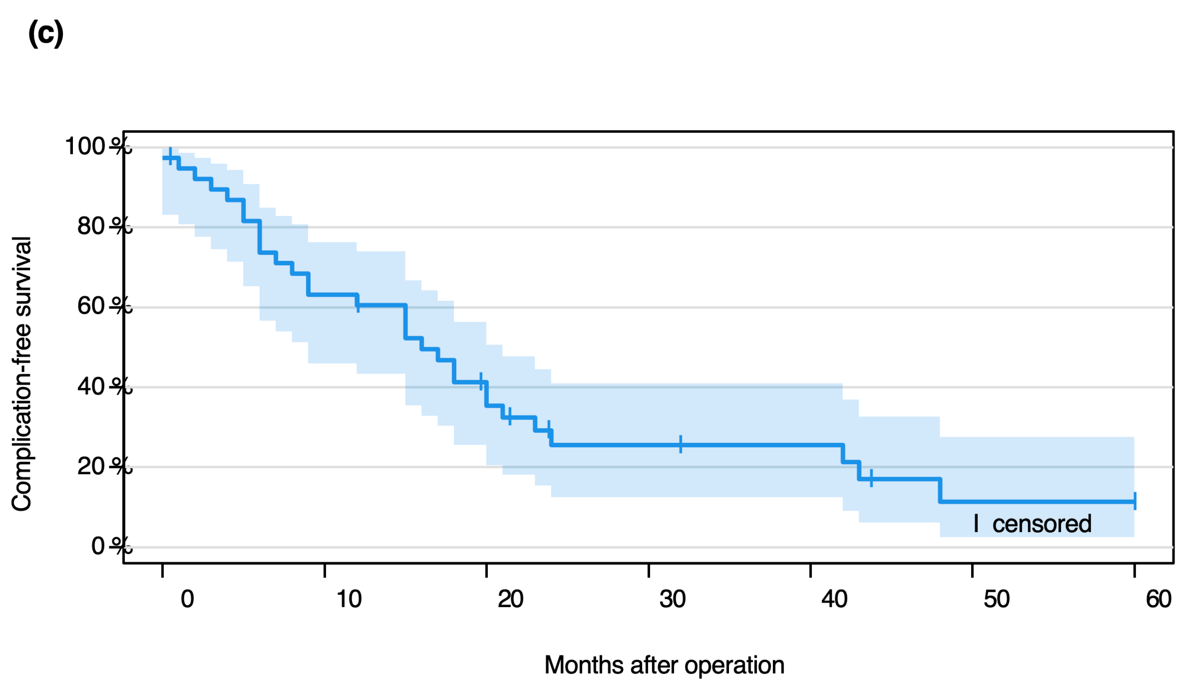
The Event-free probability in the competing risk model, considering “prosthesis failure” and “death” as competing events, was:
  • 61% (95% CI 43–74.0%) at 12 months,
  • 26% (95% CI 13–41%) at 24 months,
  • 11% (95% CI 3–28%) at 60 months.
In 18 of 39 cases (46%), prosthesis explantation was necessary. The median time to explantation was 12 months postoperatively (interquartile range: 5–18 months). In five patients (13%), diaphyseal prosthesis implantation was ultimately followed by limb exarticulation or amputation.

3.3. Implant Complications: Types and Timing

Prosthesis-related complications were classified into five categories per Henderson et al. [12]. Table 4 shows the types, frequencies, anatomical locations, and average time points of complication onset.
Six patients experienced a second complication following the initial revision. Four were cases of recurrent aseptic loosening (Type 2), and two involved local tumor recurrence (Type 5). In one instance of recurrent Type 2 failure, an additional Type 3 complication—a periprosthetic fracture—occurred.
Aseptic loosening had an average onset of approximately 10 months postoperatively. The anatomical distribution included five femoral, two tibial, and one humeral implant (Table 5).
Of the four prosthetic infections (Type 4 complications), two affected femoral and two tibial prostheses. No consistent microbial pattern emerged. In one case, Streptococcus intermedius infection necessitated high-leg exarticulation. Table 6 shows prosthesis failure depending on the underlying diagnosis.

3.4. Risk Factors for Implant Complications

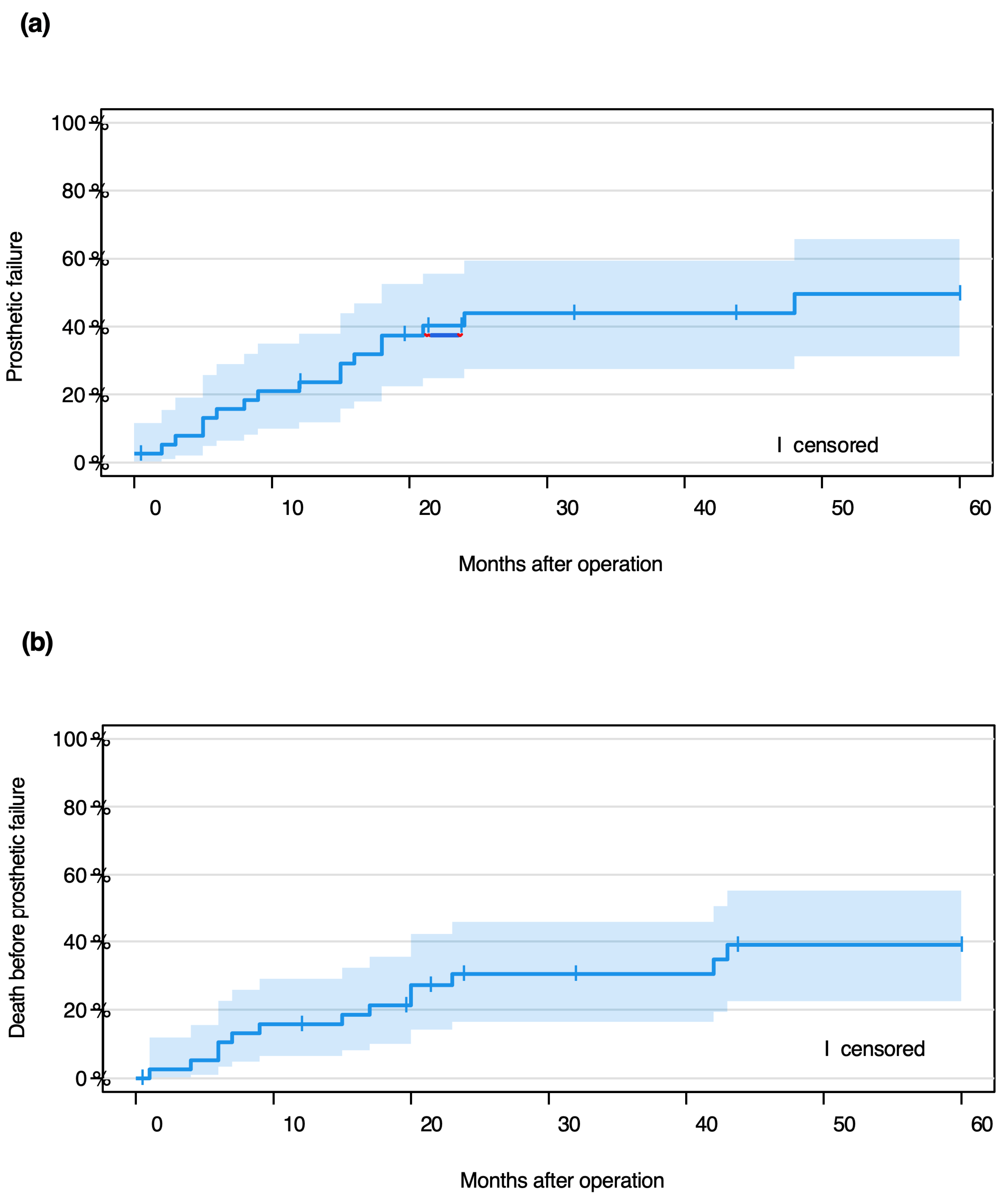
Fine Gray analysis (Figure 2) was used to determine event-free probabilities over one and five years, including both point-wise and log-log transformed confidence intervals. Although certain trends were observed in sub-hazard ratios, no statistically significant risk factors for revision-free survival could be identified. Radiotherapy in particular showed no significant effect on revision free survival. Missing data for variables such as BMI, prior radiation, and chemotherapy limited interpretability in some subgroups (Table 7).
Competing risk analysis showed that neither weight (kg) nor age (years) significantly affected the risk of prosthesis failure. However, increased age was significantly associated with higher risk of death (p < 0.01).
A statistically significant association was found between gender and prosthesis failure within the first postoperative year (Fisher’s Exact Test, p < 0.05), with an odds ratio of 6.9 (95% CI 1.1–42.8), indicating a 6.86-fold increased risk in male patients. This association was not statistically significant at the two- or five-year mark. Gender also significantly influenced prosthesis survival time (p < 0.05). A Cohen’s d of −0.6 indicated a medium effect size, with female patients demonstrating longer prosthesis survival. Although women showed a higher probability of Event-free probability up to five years, the difference was not statistically significant (p = 0.11). Men had a higher average BMI and body weight at surgery: Mean total weight: 80 kg (SD = 23.6), females: 72 kg (SD = 24.0), males: 90 kg (SD = 19.7).
No significant correlation was observed between BMI and survival at 1, 2, or 5 years. Likewise, a higher BMI (≥25) was not significantly associated with mortality during the study (Fisher’s Exact Test, p = 0.14). Odds ratio = 4.000 (95% CI: 0.9–18.2). BMI also had no significant effect on revision-free survival.
The primary focus of this study was on the influence of fixation technique on functional outcome and implant survival. Among 78 stems:
35 were cemented and screwed;
29 were cemented without screws;
5 were screwed but not cemented;
7 were neither cemented nor screwed;
1 case lacked fixation documentation.
Of 39 prostheses, eight failed due to aseptic loosening (20.5%). Loosening affected:
7 of 64 cemented stems (11%);
1 of 12 uncemented stems (8%);
3 of 40 screwed stems (8%);
5 of 36 non-screwed stems (14%).
Loosening occurred equally in proximal and distal stems (4 each; Table 7). No statistically significant correlation was found between fixation type and loosening (Fisher’s Exact Test), although prostheses with cemented, non-screwed fixation showed a higher (but not significant) tendency toward loosening (odds ratio: 3.1, 95% CI: 0.7–13.9).
In total, 21 of 39 prostheses (54%) were affected by Henderson-classified complications. The sample size limited further subgroup analyses.
Among the 78 stems:
14 (18%) were custom-made;
29 (37%) were off-the-shelf.
A significant association was found between stem type and aseptic loosening (p < 0.05), with an odds ratio of 5.4 (95% CI: 1.0–28.9), indicating a higher risk with custom-made stems. Custom stems predominantly had a six-sided solid metal design (13 of 14); one used a “hollow cage” design, which remained complication-free.
The average resection length was 15.4 cm (SD = 4.5), varying by location:
Femur: 16.4 cm (SD = 4.6);
Tibia: 14.8 cm (SD = 3.1);
Humerus: 8.6 cm (SD = 1.0).

3.5. Functional Outcome

The MSTS score was recorded in 25 patients. Across all fixation types, the median MSTS score was 15.0 (IQR: 9.5–25.5).
Cemented: median 14.5 (IQR: 8.0–25.0);
Uncemented: median 27.0 (IQR: 17.0–29.0);
Screwed: median 15.5 (IQR: 10.3–25.3);
Non-screwed: median 14.5 (IQR: 10.3–25.3).
Cemented prostheses had significantly lower MSTS scores (p < 0.05, r = −0.39). Screwing status showed no statistically significant difference.
By location:
Femoral: median 15.5 (IQR: 8.8–25.8);
Tibial: median 17.5 (IQR: 9.0–25.3);
Humeral: 14.0.
A significant moderate negative correlation was found between body weight and MSTS score (Pearson r = −0.4, p < 0.05, CI: −0.7 to −0.03).
Age also negatively correlated with MSTS outcome (Pearson r = −0.6, p < 0.001, CI: −0.817 to −0.303). Patients with metastases were significantly older than those with primary tumors (Cohen’s d = −1.3, p < 0.05).
Gender did not significantly influence MSTS scores.
Patients with primary tumors had significantly higher MSTS scores than those with metastases (p < 0.05, r = 0.4).
Primary tumor: median 20.5 (IQR: 12.5–26.8);
Metastasis: median 8.0 (IQR: 6.5–20.5).
Internal medical comorbidities were associated with significantly lower MSTS scores (p < 0.05, r = 0.47).
Without comorbidities: median 25.0 (IQR: 16.0–28.0);
With comorbidities: median 13.0 (IQR: 8.0–16.5).
Orthopedic, psychological, or neurological comorbidities did not significantly affect functional outcomes.
Radiation, neoadjuvant chemotherapy, and adjuvant therapy showed no significant impact on MSTS scores.

4. Discussion

4.1. Diaphyseal Implants Have Higher Complication Rates Than Biological Reconstructions

Multiple surgical techniques are used for diaphyseal reconstruction post-tumor resection, with no consensus on the ideal method [14,15,16,17]. Lun et al. [17] compared segmental allografts (n = 18) to intercalary prostheses (n = 16) and found significantly lower complication rates in the prosthesis group (19% vs. 67%) along with faster mobilization.
Biological methods like vascularized fibula with allografts offer good integration especially in younger patients, whereas isolated allografts have poor healing due to lack of vascularization [18]. Errani et al. reported a 30% complication rate in combined reconstructions, which is better than isolated allografts [16].
In the literature, complication rates for intercalary prostheses are reported with a range from 14% to 50% [9,15]. Streitbürger et al. described 21 complications in 28 prostheses, representing an even higher rate [14]. Low complication rates were primarily reported by Huang et al. [9]; however, it is important to emphasize that their study involved a small patient group of only 16 patients, of whom 14 had already died within two years. Benevenia et al. reported on a larger cohort of 41 patients [15]. In both studies, complications appear to have occurred relatively early after surgical treatment, as the median follow-up was only 19 and 11 months, respectively. In comparison, the present study shows a complication rate of 54% observed in 39 patients with a median follow-up of 47.6 months.
Overall, the differences between the individual study groups are likely attributable to the varying patient numbers and the differences in follow-up duration. Though prostheses enable quicker recovery, Yao et al. reported good long-term functional outcomes with intercalary allografts (MSTS 26.2) [19].
Biological methods require long unloading periods and provide limited primary stability [17,20]. Nevertheless, biological reconstructions continue to play an important role in the treatment of bone defects. Diaphyseal prostheses are preferred for patients with limited life expectancy or chemotherapy needs due to faster rehabilitation [6, 7].
Although diaphyseal implants are associated with faster recovery, there is no conclusive evidence that they result in higher complication rates than biological options [14,15,16,17,18].

4.2. Cement and Screw Fixation Reduces Aseptic Loosening Risk

Diaphyseal implants allow for cemented or uncemented fixation with optional screw support. Factors like age, bone quality, smoking, and comorbidities influence the choice of procedure. Cementation is advised for compromised bone [15], while younger patients often receive uncemented stems [18].
In our study, cemented stems with screws had a 6% loosening rate, compared to 20% in uncemented stems with screws. However, the uncemented group was small. Benevenia et al. [15] found better MSTS scores (p < 0.01) and fewer complications with cemented stems (21%) than uncemented (33%, p = 0.390). However, it should be mentioned that the follow-up periods in the study of Benevenia et al. and the present study differed significantly (11 vs. 47.6 months). This may affect the rate of aseptic loosening significantly. Contradictory, our uncemented group had better function, likely due to younger age and less adjuvant radiation therapy.
Additionally, antibiotic-loaded cement also helps to prevent infection and recurrence [21]. Though not statistically significant, cemented stems with screws failed less often (5.7%) than cemented stems without screws (17.2%).

4.3. Prosthesis Failure Is More Likely After Primary Tumor Resection, but Not Significantly

The goal of reconstruction is long-term, complication-free function. In our cohort, implant failure rates were: 1-year: 25.0%, 2-year: 52.0%, 5-year: 75.0%.
Survival varies by malignancy. Patients with secondary tumors had significantly higher mortality (Gray’s Test, p < 0.05). Survival in primary tumor patients was higher: 1Y: 87.0%, 2Y: 73.9%, 5Y: 47.1% vs. metastasis patients: 1Y: 73.3%, 2Y: 53.3%, 5Y: 28.6%. These findings align with Pu et al. [22] and Hanna et al. [10].
Patients with soft tissue sarcomas who underwent diaphyseal resection demonstrated higher rates of postoperative prosthetic failure. This may be attributed to inadequate soft tissue coverage of the implant and the high incidence of postoperative radiotherapy (73%).
Despite longer survival in primary tumor patients, prosthetic failure rates at 1, 2, and 5 years showed no significant difference between groups (Fisher Exact Test). One-year revision-free survival was slightly better in primary tumors (65% vs. 53%), but this narrowed by year five (12% vs. 9%).
Thus, tumor type does not significantly affect prosthesis survival, supporting use in both groups.

4.4. Internal Comorbidities Affect Function More than Survival

Patients with internal diseases had a higher mortality rate (73% vs. 53%), but this was not statistically significant (p = 0.31). However, internal diseases significantly worsened functional outcomes (MSTS: 13 vs. 25.), likely due to impaired rehabilitation.
Comorbidities can also impact cancer progression. Stelzl et al. linked diabetes to worse soft tissue sarcoma survival (p < 0.05) [23], possibly due to insulin/IGF-1 effects. Qu et al. identified low HDL-C as a negative prognostic marker [24].
While internal diseases may shorten survival and reduce complication risk due to limited prosthesis lifespan, they also hinder functional outcomes—making faster-recovery implants preferable.

4.5. Overweight Patients Have Worse Function but No Increased Failure Risk

The average BMI in our adult cohort was 28.1, classifying most as overweight [25]. Excess weight increases mechanical load on implants, especially in lower limbs, and may raise the risk of loosening [25,26]. Yet, some long-term studies found no correlation between BMI and prosthetic failure [12,27].
Functionally, we observed a significant inverse relationship between body weight and MSTS score (r = −0.42, p < 0.05; 95% CI: −0.70 to −0.03), consistent with previous findings [28]. However, no significant link was found between BMI and prosthesis survival.
Obesity-associated conditions like depression and fatigue may also impair function [29], but BMI alone shouldn’t contraindicate diaphyseal implants.

4.6. Functional Outcomes in Diaphyseal Implants

We recorded MSTS scores in 25 patients only: 20 femoral (median: 15.5), 4 tibial (17.5), and 1 humeral (14). Limited humeral data restricts conclusions. However, the poorer functional outcomes observed in patients with secondary cancer may be attributed to the gradual deterioration of their general health status. Moreover, long-term survival in this group is significantly reduced, which may prevent the occurrence or detection of late-onset complications. External studies show promising results. Pu et al. reported an MSTS of 28.6 in humeral implants [22], and Zhao et al. found 27.2 (primary) and 26.1 (metastases) over 45 months [30].
Benevenia et al. reported average MSTS scores of 24.9 (humerus), 22.5 (femur), and 23.1 (tibia) [16], with no complications in humeral or tibial implants. Conversely, Mahdal et al. reported a 44% complication rate in humeral implants [31].
Huang et al. reported an MSTS of 25.4 in femoral implants [11]. Despite limited humeral data in our study, the literature supports their functional viability

4.7. Tibial Implants Have Higher Failure Rates than Femoral Ones

Tibial implants had a median MSTS of 17.5 but high complication rates. Of six patients, 33.3% had infections or loosening, and 83.3% experienced prosthesis failure—compared to 37.9% in femoral implants.
Poor soft tissue coverage in the tibia, often addressed via gastrocnemius flaps, may increase infection risk [31,32,33,34]. Given this, primary amputation or biological reconstruction (e.g., vascularized tibia, distraction osteogenesis) may be considered [35,36,37,38,39,40].
Stem modifications like “ultra-short stems” could reduce loosening risk [41], though more robust data is needed.

4.8. Custom-Made Stems Are Linked to Worse Outcomes

In this study, 14 (17.9%) custom-made stems and 29 (37.2%) off-the-shelf stems were analyzed. Statistical analysis revealed that 71.4% of prostheses with a custom-made stem experienced prosthetic failure, compared to 46.8% of those without a custom-made stem. Fisher’s exact test demonstrated a statistically significant association between the implantation of a custom-made stem and the event of aseptic loosening. The odds ratio of 5.4 (95% CI: lower 1.04; upper 28.9) suggests that the risk of aseptic loosening is increased by a factor of 5.4 in custom-made stems compared to standard stems. However, it should be noted that the wide confidence interval limits the interpretability of the odds ratio.
Thirteen of the custom-made stems were so-called hexagonal solid metal stems, while one was a “hollow cage stem” consisting of a lattice structure. Complications occurred exclusively in the hexagonal solid metal stems. It is important to consider the uneven distribution between hollow cage and solid metal stems in this context [15,41].
Anchorage under 5 cm compromises stability [42]. While resection length often predicts failure, we found no such link. “Ultra-short stem” systems introduced in 2017 for short anchorage tibial reconstructions showed good results [41].
3D-printed implants also show promising results: Zhao et al. reported early integration and improved function in tibial reconstructions [43], and Shao et al. demonstrated success in femoral applications [8]. Bischel et al. found conical stems suitable for large defects and hexagonal stems better for distal areas [44]. Nevertheless, studies should consider not only the length of the prosthetic stem but also, in particular, the impact of its diameter

4.9. Study Limitations

This study spans patients from 1998 onward to include long-term outcomes. But prosthesis designs, surgical techniques and adjuvant chemotherapy/radiation protocols have evolved substantially from 1998 to 2020. Therefore, the comparability of cases between 1998 and 2020 is limited. Additionally, the use of custom versus off-the-shelf implants followed clinical judgment, which may confound outcome comparison. The rarity of diaphyseal tumors limited cohort size and statistical power, especially for humeral and tibial cases. Anchorage techniques were unevenly distributed across anatomical sites.
Additionally, the fatal nature of underlying malignancies led to patient loss during follow-up. Mortality-related cohort reduction must be considered when interpreting prosthesis survival rates at 1, 2, and 5 years.
MSTS scores were assessed retrospectively postoperatively as part of the study. In total, follow-up contact could be established in 25 cases only, as some patients had passed away. The timing of this assessment varied on a case-by-case basis.

5. Conclusions

Diaphyseal implants can, in cases without complications, allow for preservation of the affected limb and a rapid return to everyday life. However, this study cohort showed that complication and revision rates are elevated with intercalary prostheses. The following findings can be derived from this study on the long-term outcomes of diaphyseal implants:
The probability of event-free probability after five years is low at 11.4% (95% CI 2.5–27.5%).
-
The fixation method significantly influences the outcome of the implants.
-
The risk of aseptic loosening tends to be lower for cemented and screwin stems than for cemented unscrewed stems. However, this result is not statistically significant.
-
Risk factors that can negatively influence the functional outcome in a statistically significant manner include increased body weight, advanced patient age, and preexisting internal medical conditions.
Therefore, the use of diaphyseal implants should be carefully considered on a case-by-case basis, taking into account the risk factors mentioned above and the experience of the surgeons. To answer the question of the ideal fixation technique and to build a larger database on prosthesis survival times, functional outcomes, and best surgical techniques, further medium- to long-term studies on diaphyseal implants are needed. So far, cemented stems, that are locked with additional screws, seem to be the best option.

Author Contributions

Conceptualization, N.D. and T.B.; methodology, S.K., N.D. and T.B.; software, F.L.; validation, N.D. and T.B.; formal analysis, N.D. and T.B.; investigation, F.L. and S.K.; resources, G.G.; data curation, F.L.; writing—original draft preparation, N.D. and F.L.; writing—review and editing, N.D., T.B., A.M.R., S.K., J.C.T. and M.D.V.; visualization, F.L. and J.C.T.; supervision, N.D. and T.B.; project administration, N.D. All authors have read and agreed to the published version of the manuscript.

Funding

The APC was funded by the Open Access Publishing Funds of the University of Muenster.

Institutional Review Board Statement

The study protocol was approved by the regional ethics committee (University of Muenster; reference no.: 2018-199-f-S). All participants gave written informed consent.

Informed Consent Statement

Informed consent was obtained from all subjects involved in the study.

Data Availability Statement

The datasets used and/or analyzed during the current study are available from the corresponding author on reasonable request.

Acknowledgments

We thank Jana Elisa Burkhardt for the language editing.

Conflicts of Interest

The authors declare no conflicts of interest.

References

  1. Henshaw, R.M. Surgical Advances in Bone and Soft Tissue Sarcoma: 50 Years of Progress. In American Society of Clinical Oncology Educational Book; American Society of Clinical Oncology: Alexandria, VA, USA, 2014; pp. 252–258. [Google Scholar] [CrossRef]
  2. Zekry, K.M.; Yamamoto, N.; Hayashi, K.; Takeuchi, A.; Higuchi, T.; Abe, K.; Taniguchi, Y.; Alkhooly, A.Z.A.A.; Abd-Elfattah, A.S.; Fouly, E.H.; et al. Intercalary frozen autograft for reconstruction of malignant bone and soft tissue tumours. Int. Orthop. 2017, 41, 1481–1487. [Google Scholar] [CrossRef] [PubMed]
  3. Zekry, K.M.; Yamamoto, N.; Hayashi, K.; Takeuchi, A.; Alkhooly, A.Z.A.; Abd-Elfattah, A.S.; Elsaid, A.N.S.; Ahmed, A.R.; Tsuchiya, H. Reconstruction of intercalary bone defect after resection of malignant bone tumor. J. Orthop. Surg. 2019, 27, 1–9. [Google Scholar] [CrossRef] [PubMed]
  4. Hilven, P.H.; Bayliss, L.; Cosker, T.; Dijkstra, P.D.S.; Jutte, P.C.; Lahoda, L.U.; Schaap, G.R.; Bramer, J.A.M.; van Drunen, G.K.; Strackee, S.D.; et al. The vascularized fibular graft for limb salvage after bone tumour surgery: A multicentre study. Bone Jt. J. 2015, 97, 853–861. [Google Scholar] [CrossRef]
  5. Bus, M.P.; Dijkstra, P.D.; Van De Sande, M.A.; Taminiau, A.H.; Schreuder, H.W.; Jutte, P.C.; van der Geest, I.C.M.; Schaap, G.R.; Bramer, J.A.M. Intercalary allograft reconstructions following resection of primary bone tumors: A nationwide multicenter study. J. Bone Jt. Surg. Am. 2014, 96, e26. [Google Scholar] [CrossRef]
  6. Panagopoulos, G.N.; Mavrogenis, A.F.; Mauffrey, C.; Lesenský, J.; Angelini, A.; Megaloikonomos, P.D.; Igoumenou, V.G.; Papanastassiou, J.; Savvidou, O.; Ruggieri, P.; et al. Intercalary reconstructions after bone tumor resections: A review of treatments. Eur. J. Orthop. Surg. Traumatol. 2017, 27, 737–746. [Google Scholar] [CrossRef]
  7. Damron, T.A.; Malhotra, A. Intercalary Diaphyseal Endoprosthetic Reconstruction. J. Am. Acad. Orthop. Surg. Glob. Res. Rev. 2025, 9, e24.00201. [Google Scholar] [CrossRef] [PubMed] [PubMed Central]
  8. Shao, X.; Dou, M.; Yang, Q.; Li, J.; Zhang, A.; Yao, Y.; Chu, Q.; Li, K.; Li, Z. Reconstruction of massive bone defects after femoral tumor resection using two new-designed 3D-printed intercalary prostheses: A clinical analytic study with the cooperative utilization of multiple technologies. BMC Musculoskelet. Disord. 2023, 24, 67. [Google Scholar] [CrossRef] [PubMed] [PubMed Central]
  9. Goebel, L.; Kohn, D.; Orth, P. Endoprosthetic replacement following intercalary resection. Orthopade 2019, 48, 572–581. [Google Scholar] [CrossRef]
  10. Hanna, S.A.; Sewell, M.D.; Aston, W.J.S.; Pollock, R.C.; Skinner, J.A.; Cannon, S.R.; Briggs, T.W.R. Femoral diaphyseal endoprosthetic reconstruction after segmental resection of primary bone tumours. J. Bone Jt. Surg.—Ser. B 2010, 92, 867–874. [Google Scholar] [CrossRef]
  11. Huang, H.C.; Hu, Y.C.; Lun, D.X.; Miao, J.; Wang, F.; Yang, X.G.; Ma, X.L. Outcomes of intercalary prosthetic reconstruction for pathological diaphyseal femoral fractures secondary to metastatic tumors. Orthop. Surg. 2017, 9, 221–228. [Google Scholar] [CrossRef] [PubMed]
  12. Haverkamp, D.; De Man, F.H.R.; De Jong, P.T.; Van Stralen, R.A.; Marti, R.K. Is the long-term outcome of cemented THA jeopardized by patients being overweight? Clin. Orthop. Relat. Res. 2008, 466, 1162–1168. [Google Scholar] [CrossRef]
  13. Henderson, E.R.; Groundland, J.S.; Pala, E.; Dennis, J.A.; Wooten, R.; Cheong, D.; Windhager, R.; Kotz, R.I.; Mercuri, M.; Funovics, P.T.; et al. Failure mode classification for tumor endoprostheses: Retrospective review of five institutions and a literature review. J. Bone Jt. Surg.—Ser. A 2011, 93, 418–429. [Google Scholar] [CrossRef]
  14. Lun, D.X.; Hu, Y.C.; Yang, X.G.; Wang, F.; Xu, Z.W. Short-term outcomes of reconstruction subsequent to intercalary resection of femoral diaphyseal metastatic tumor with pathological fracture: Comparison between segmental allograft and intercalary prosthesis. Oncol. Lett. 2018, 15, 3508–3517. [Google Scholar] [CrossRef]
  15. Gerrand, C.H.; Rankin, K. A system for the functional evaluation of reconstructive procedures after surgical treatment of tumors of the musculoskeletal system. In Classic Papers in Orthopaedics; Springer: London, UK, 1993; Volume 2014, pp. 489–490. [Google Scholar] [CrossRef]
  16. Benevenia, J.; Kirchner, R.; Patterson, F.; Beebe, K.; Wirtz, D.C.; Rivero, S.; Palma, M.; Friedrich, M.J. Outcomes of a Modular Intercalary Endoprosthesis as Treatment for Segmental Defects of the Femur, Tibia, and Humerus. Clin. Orthop. Relat. Res. 2016, 474, 539–548. [Google Scholar] [CrossRef] [PubMed]
  17. Errani, C.; Ceruso, M.; Donati, D.M.; Manfrini, M. Microsurgical reconstruction with vascularized fibula and massive bone allograft for bone tumors. Eur. J. Orthop. Surg. Traumatol. 2019, 29, 307–311. [Google Scholar] [CrossRef] [PubMed]
  18. Streitbürger, A.; Hardes, J.; Nottrott, M.; Guder, W.K. Reconstruction survival of segmental megaendoprostheses: A retrospective analysis of 28 patients treated for intercalary bone defects after musculoskeletal tumor resections. Arch. Orthop. Trauma Surg. 2022, 142, 41–56. [Google Scholar] [CrossRef] [PubMed]
  19. Houdek, M.T.; Wagner, E.R.; Stans, A.A.; Shin, A.Y.; Bishop, A.T.; Sim, F.H.; Moran, S.L. What Is the Outcome of Allograft and Intramedullary Free Fibula (Capanna Technique) in Pediatric and Adolescent Patients With Bone Tumors? Clin. Orthop. Relat. Res. 2016, 474, 660–668. [Google Scholar] [CrossRef]
  20. Yao, W.; Cai, Q.; Wang, J.; Zhang, P.; Wang, X.; Du, X.; Niu, X. Biological reconstruction in the treatment of extremity sarcoma in femur, tibia, and humerus. Medicine 2020, 99, e20715. [Google Scholar] [CrossRef]
  21. Bellan, D.G.; Filho, R.J.-G.; Garcia, J.G.; de Toledo Petrilli, M.; Maia Viola, D.C.; Schoedl, M.F.; Petrilli, A.S. Ewing’S Sarcoma: Epidemiology and Prognosis for Patients Treated At the Pediatric Oncology Institute, Iop-Graacc-Unifesp. Rev. Bras. Ortop. (Engl. Ed.) 2012, 47, 446–450. [Google Scholar] [CrossRef]
  22. Pu, F.; Zhang, Z.; Wang, B.; Liu, J.; Shao, Z. En bloc resection and intercalary prosthesis implantation for the treatment of humeral diaphyseal bone metastases. Int. Orthop. 2021, 45, 281–288. [Google Scholar] [CrossRef]
  23. Stelzl, A.; Aziz, F.; Riedl, J.M.; Posch, F.; Smolle, M.A.; Stojakovic, T.; Terbuch, A.; Pichler, M.; Bergovec, M.; Leithner, A.; et al. Diabetes mellitus is independently associated with adverse clinical outcome in soft tissue sarcoma patients. Sci. Rep. 2020, 10, 12438. [Google Scholar] [CrossRef] [PubMed]
  24. Qu, H.; Guo, W.; Yang, R.; Li, D.; Tang, S.; Yang, Y.; Dong, S.; Zang, J. Reconstruction of segmental bone defect of long bones after tumor resection by devitalized tumor-bearing bone. World J. Surg. Oncol. 2015, 13, 282. [Google Scholar] [CrossRef] [PubMed]
  25. Organization WH. Obesity and Overweight. Available online: https://www.who.int/news-room/fact-sheets/detail/obesity-and-overweight (accessed on 7 July 2025).
  26. Röder, C.; Eggli, S.; Münger, P.; Melloh, M.; Busato, A. Patient characteristics differently affect early cup and stem loosening in THA: A case-control study on 7535 patients. Int. Orthop. 2008, 32, 33–38. [Google Scholar] [CrossRef]
  27. Werner, F.W.; Ayers, D.C.; Maletsky, L.P.; Rullkoetter, P.J. The effect of valgus/varus malalignment on load distribution in total knee replacements. J. Biomech. 2005, 38, 349–355. [Google Scholar] [CrossRef]
  28. McLaughlin, J.R.; Lee, K.R. The outcome of total hip replacement in obese and non-obese patients at 2008, 10- to 18-years. J. Bone Jt. Surg.—Ser. B 2006, 88, 1286–1292. [Google Scholar] [CrossRef]
  29. Yeung, E.; Jackson, M.; Sexton, S.; Walter, W.; Zicat, B. The effect of obesity on the outcome of hip and knee arthroplasty. Int. Orthop. 2011, 35, 929–934. [Google Scholar] [CrossRef]
  30. Zhao, J.; Xu, M.; Zheng, K.; Yu, X. Limb salvage surgery with joint preservation for malignant humeral bone tumors: Operative procedures and clinical application. BMC Surg. 2019, 19, 57. [Google Scholar] [CrossRef]
  31. Mahdal, M.; Pazourek, L.; Apostolopoulos, V.; Krákorová, D.A.; Zambo, I.S.; Tomáš, T. Outcomes of Intercalary Endoprostheses as a Treatment for Metastases in the Femoral and Humeral Diaphysis. Curr. Oncol. 2022, 29, 3519–3530. [Google Scholar] [CrossRef]
  32. Biau, D.; Faure, F.; Katsahian, S.; Jeanrot, C.; Tomeno, B.; Anract, P. Survival of total knee replacement with a megaprosthesis after bone tumor resection. J. Bone Jt. Surg.—Ser. A 2006, 88, 1285–1293. [Google Scholar] [CrossRef][Green Version]
  33. Hennen, J.; Sabo, D.; Martini, A.K.; Bernd, L. Das Manteltransplantat zur Defektrekonstruktion nach Resektion maligner Knochentumoren an der unteren Extremität. Unfallchirurg 2002, 105, 120–127. [Google Scholar] [CrossRef]
  34. Saaiq, M.; Zimri, F.U.K. Clinical Applications and Outcome of Proximally Based Medial Gastrocnemius Muscle Flap. World J. Plast. Surg. 2020, 9, 22–28. [Google Scholar] [CrossRef] [PubMed]
  35. Trieb, K.; Göggel, M.; Dürr, H.R. Proximaler Tibiaersatz mit Transfer des M. Gastrocnemius. Oper. Orthop. Traumatol. 2012, 24, 263–271. [Google Scholar] [CrossRef] [PubMed]
  36. Aponte-Tinao, L.A.; Ayerza, M.A.; Albergo, J.I.; Farfalli, G.L. Do Massive Allograft Reconstructions for Tumors of the Femur and Tibia Survive 10 or More Years after Implantation? Clin. Orthop. Relat. Res. 2020, 478, 517–524. [Google Scholar] [CrossRef]
  37. Grzebień, A.; Chabowski, M.; Malinowski, M.; Uchmanowicz, I.; Milan, M.; Janczak, D. Analysis of selected factors determining quality of life in patients after lower limb amputation—A review article. Pol. J. Surg. 2017, 89, 57–61. [Google Scholar] [CrossRef]
  38. Lesensky, J.; Prince, D.E. Distraction osteogenesis reconstruction of large segmental bone defects after primary tumor resection: Pitfalls and benefits. Eur. J. Orthop. Surg. Traumatol. 2017, 27, 715–727. [Google Scholar] [CrossRef]
  39. Moran, S.L.; Shin, A.Y.; Bishop, A.T. The use of massive bone allograft with intramedullary free fibular flap for limb salvage in a pediatric and adolescent population. Plast. Reconstr. Surg. 2006, 118, 413–419. [Google Scholar] [CrossRef]
  40. Refaat, Y.; Gunnoe, J.; Hornicek, F.J.; Mankin, H.J. Comparison of quality of life after amputation or limb salvage. Clin. Orthop. Relat. Res. 2002, 298–305. [Google Scholar] [CrossRef]
  41. Guder, W.K.; Hardes, J.; Gosheger, G.; Nottrott, M.; Streitbürger, A. Ultra-short stem anchorage in the proximal tibial epiphysis after intercalary tumor resections: Analysis of reconstruction survival in four patients at a mean follow-up of 56 months. Arch. Orthop. Trauma Surg. 2017, 137, 481–488. [Google Scholar] [CrossRef]
  42. Fuchs, B.; Ossendorf, C.; Leerapun, T.; Sim, F.H. Intercalary segmental reconstruction after bone tumor resection. Eur. J. Surg. Oncol. 2008, 34, 1271–1276. [Google Scholar] [CrossRef]
  43. Zhao, D.; Tang, F.; Min, L.; Lu, M.; Wang, J.; Zhang, Y.; Zhao, K.; Zhou, Y.; Luo, Y.; Tu, C. Intercalary reconstruction of the “ultra-critical sized bone defect” by 3D-printed porous prosthesis after resection of tibial malignant tumor. Cancer Manag. Res. 2020, 12, 2503–2512. [Google Scholar] [CrossRef] [PubMed]
  44. Bischel, O.E.; Nadorf, J.; Klein, S.B.; Gantz, S.; Jakubowitz, E.; Kretzer, J.P.; Arnholdt, J.; Seeger, J.B. Modular tumor prostheses: Are current stem designs suitable for distal femoral reconstruction? A biomechanical implant stability analysis in Sawbones. Arch. Orthop. Trauma Surg. 2019, 139, 843–849. [Google Scholar] [CrossRef] [PubMed]
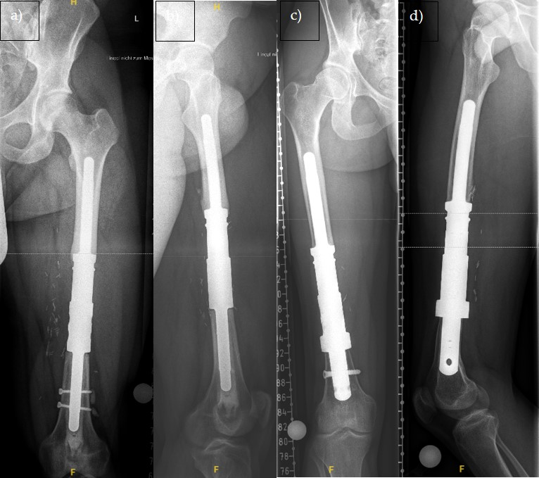
Figure 1. X-ray image in frontal (a) and lateral (b) view of a 34-year-old patient with a left femoral diaphysis implant (standard) and X-ray image in frontal (c) and lateral (d) view of a 26-year-old patient with a left femoral diaphysis implant with ultra short stem (custom made).
Cancers 17 03059 g001
Figure 2. Cumulative incidence function with log-log transformed pointwise confidence intervals: probabilities of prosthetic failure, death before prosthetic failure and complication-free survival. Patients whose follow-up was shorter than 60 months were censored. (a) probability of prosthetic failure; (b) probability of death before prosthetic failure; (c) probability of complication-free survival.
Cancers 17 03059 g002aCancers 17 03059 g002b
Table 1. Overview of the patient cohort.
NoAgeDiagnosisLocaliza tion Bone/Soft TissueSeizeEnneking
Classification
Prior OperationChemoResponseRadiationFU
126ExtraskelettalFemurST5–10IIa-Y2NNED 42
  OS         
263OSFemurBone>10IIb-Y2NDOUC
           76
316EWSFemurBone5–10IIa-Y1NNED 54
432Extraskelettal EWSFemurST5–10na-Y1NNED 24
535Soft Tissue SarcomaFemurST5–10IIb-YNaNNED 76
659Soft Tissue SarcomaFemurST5–10IIb-YNaYAWD 32
759Soft Tissue SarcomaFemurST5–10IIa-YNaYAWD 91
880Soft Tissue SarcomaFemurST5–10IIb-N-YDOD 4
955MFHFemurST>10IIaIntralesional. ResectionN-NDOD 17
1048Soft Tissue SarcomaFemurST5–10IIaIntralesional ResectionN-YDOD 10
1159Soft Tissue SarcomaFemurST5–10IIbIntralesional ResectionYNaYDOUC 6
1253Soft Tissue SarcomaFemurST>10IIaMarginal ResectionN-YNED 97
1341Soft Tissue SarcomaFemurST>10IIaIntralesional ResectionYNaYAWD 104
1452Soft Tissue SarcomaFemurST>10IIb-YNaYDOD 20
1554Soft Tissue SarcomaFemurST>10IIa-YNaYDOUC 42
1669Renal-CAFemurBone5–10Met-N-NAWD 28
  Metastasis         
1762Renal-CAFemurBone<5MetIntramedN-YDOD 80
  Metastasis    Nailing    
1862Renal-CAFemurBone5–10MetIntramedN-NDOD 9
  Metastasis    Nailing    
1958Renal-CA MetastasisFemurBone5–10MetPlate-Osteosynthesis (ORIF)N-NDOD 43
2060Renal-CAFemurBone<5Met-N-YDOD 98
  Metastasis         
2179Renal-CAFemurBone<5Met-N-NDOD 1
  Metastasis         
2262CSFemurBoneNaIIaIntralesio-YNaNDOD 23
       nal Cur-    
       retage    
2372NSCLC Meta-FemurBone<5Met-N-YDOD 6
  stasis         
2485Breast-CaFemurBone<5Met-Na-YDOD 7
  Metastasis         
2582Breast-CaFemurBone5–10Met-N-YDOD 49
  Metastasis         
2617OSFemurBone>10IIb-Y2NNED 44
2710EWSFemurBone5–10IIb-Y2NAWD
           144
2860Soft Tissue SarcomaFemurST>10IIa-N-YDOD 48
2969Renal-CAFemurBone5–10MetIntramedYNaYDOD 17
  Metastasis    Nailing    
3054Soft Tissue SarcomaTibiaBone<5IIa-N-NAWD 6
3144NOSTibiaBone5–10IIb-Y2NDOD 32
3247NOSTibiaBone>10IIb-YNaNDOD
           102
3359Renal-CATibiaBone>10Met-YNaYDOD 73
  Metastasis         
3444AdamantinomaTibiaBone5–10IIb-N-NAWD
           114
3560Colon-CA MetastasisTibiaBone5Met-Na-YDOD 54
3676Soft Tissue SarcomaHumerusST5–10IIb-YNaYNED 50
3768Renal-CAHumerusBoneNaMet-Na-NaDOD 81
  Metastasis         
3864NSCLC Meta-HumerusBone<5Met-N-NDOD 20
  stasis         
3966Breast-CaHumerusBone5–10Met-N-YDOD 15
  Metastasis         
No: Number, Age on the day of surgery (years), MFH: Malignant fibrous, ST: Soft Tissue, Seize in cm, Na: Not available, Met: Metastasized, IIA: Intracompartimental tumor extension, IIB: Extracompartimental extension, Y: yes, N: no, Response: Response to chemotherapy according to Salzer-Kuntschik, FU: Follow-up in months, DOD: death of disease, DOUC: death of unknown cause, NED: no evidence of disease, AWD: alive with disease.
Table 2. Cumulative 1-, 2-, and 5-year patient survival in relation to diagnosis: primary tumor or metastasis.
1-Year Survival2-Year Survival5-Year Survival
Overall82%66%39%
Primary tumor87%74%47%
metastasis73%53%29%
Table 3. Cumulative 1-, 2-, and 5-year survival in relation to diagnosis.
DiagnosisNumber of Patients1-Year Survival2-Year Survival5-Year Survival
Osteosarcoma3100%100%100%
Ewing sarcoma3100%100%100%
Soft tissue sarcoma1680%67%38%
Other primary tumor2100%50%50%
Renal cancer metastasis977.8%66.7%50%
Pulmonary cancer metastasis250%0%0%
Breast cancer metastasis367%33%0%
Colrectal cancer metastasis Metastase1100%100%0%
Table 4. Type of complication according to Henderson et al. [12] and timing of occurrence, categorized by prosthesis location.
Complication According to HendersonTotal Number of Cases (n = 21)Femoral (n = 13)Tibial (n = 5)Humeral (n = 3)Time in Months After Implantation
Mean (SD)
Type 115%0103 (-)
Type 2838%52110 (6)
Type 3210%20017 (2)
Type 4419%2208 (11)
Type 5419%30122 (18)
unclear210%1016 (9)
Table 5. Number of prostheses by location and percentage of aseptic loosening and total prosthesis explantations.
LocalizationNumber of ProthesesAseptic LooseningTotal Prosthesis Explantation
Femoral29517%1138%
Tibial6233%583%
Humeral4125%250%
Table 6. Prosthesis failure during lifetime depending on diagnosis.
DiagnosisNumber of Prostheses
(n = 39)
Prosthesis Failure During LifetimeProsthesis Failure During Lifetime in %
Osteosarcoma3133%
Ewing-Sarcoma3267%
Soft Tissue Sarcoma16956%
Renal Cell Carcinoma Metastasis9333%
Other Primary Tumor2150%
Lung Cancer Metastasis200.0%
Breast Cancer Metastasis3133%
Colon Cancer Metastasis11100%
Table 7. Relative Loosening Rate of Individual Shafts Depending on the Fixation Mechanism.
Fixation MechanismNumber of StemsNumber of Cases with Aseptic LooseningLoosening Rate
cemented+ screwed3526%
cemented+ unscrewed29517%
uncemented + screwed5120%
uncemented + unscrewed700%
No information200%
Disclaimer/Publisher’s Note: The statements, opinions and data contained in all publications are solely those of the individual author(s) and contributor(s) and not of MDPI and/or the editor(s). MDPI and/or the editor(s) disclaim responsibility for any injury to people or property resulting from any ideas, methods, instructions or products referred to in the content.