Article

Validation of the HFA-ICOS Score for Carfilzomib-Induced Cardiotoxicity in Multiple Myeloma: A Real-Life Perspective Study

1
Division of Internal Medicine, University of Torino, 10126 Turin, Italy
2
Division of Internal Medicine, Candiolo Cancer Institute, FPO-IRCCS, 10060 Candiolo, Italy
3
Division of Hematology, University of Torino, 10126 Turin, Italy
*
Author to whom correspondence should be addressed.
Cancers 2025, 17(14), 2353; https://doi.org/10.3390/cancers17142353
Submission received: 29 April 2025 / Revised: 14 June 2025 / Accepted: 8 July 2025 / Published: 15 July 2025
(This article belongs to the Special Issue Cardio-Oncology: An Emerging Paradigm in Modern Medicine: 2nd Edition)

Simple Summary

This study looks to investigate the performance of the HFA-ICOS risk score in predicting adverse cardiovascular events during Carfilzomib in multiple myeloma patients beyond controlled trials. In a clinical setting, 169 patients with multiple myeloma were divided into classes of risk according to the HFA-ICOS score and followed for a mean of 11.2 months. The incidence of events was high (52.7% of the population) during K therapy. By the results, the HFA-ICOS score did not discriminate between patients at low, medium and high risk for events, showing a limited power to predict the risk of events in our population.

Abstract

Background: Despite the inference about the cardiotoxicity induced by Carfilzomib, no validated risk prediction models for adverse cardiovascular events in a real-life population are available. Objectives: The aim of this study was to evaluate the performance of the risk stratification score for Carfilzomib-induced cardiotoxicity of the Heart Failure Association of the European Society of Cardiology and the International Cardio-Oncology Society (HFA-ICOS) in patients with multiple myeloma (MM). Methods: This is a prospective, real-world study including MM patients consecutively enrolled prior to starting Carfilzomib, divided into levels of risk according to the HFA-ICOS proforma. Results: Of 169 patients, 11.8% were classified as ‘low risk’, 38.5% as ‘medium risk’, 45.6% as ‘high risk’ and 4.1% as ‘very high risk’ at baseline. A total of 89 (52.7%) patients experienced one of the following events: 36 (21.3%) had at least one cardiovascular event and 77 (45.6%) had almost one hypertension-related event. No significant differences were observed for the incidence of any cardiovascular events between the different levels of risk (p > 0.05), even considering the HFA-ICOS score as a continuous variable. The integration of the score with the baseline systolic blood pressure and pulse wave velocity enhanced the accuracy of the score (AUC 0.557 vs. 0.736). Conclusions: The HFA-ICOS score did not discriminate between patients at low, medium and high risk, showing a limited discriminatory power in predicting the risk of events in our population. The integration of other parameters in the HFA-ICOS score, such as systolic blood pressure and pulse wave velocity, improved the performance of the score.

1. Introduction

Multiple myeloma (MM) accounts for 1% of neoplastic diseases and is the second most common disease among hematologic malignancies [1], typically affecting elderly populations. In these patients, cardiovascular adverse events (CVAEs) represent the most common complications as a consequence of a high prevalence of coexistent cardiovascular comorbidities, of the increased risk due to MM disease itself, and as a side effect of anti-myeloma treatments [2]. It has been demonstrated that the incidence of CVAEs is higher in patients with MM than in non-MM patients (60.1% vs. 54.5%), and, owing to population aging, the incidence of both MM and cardiovascular diseases is increasing [3]. Carfilzomib (K) therapy, an irreversible second-generation proteasome inhibitor included in different combined treatments, is considered the backbone for patients newly diagnosed and relapsed/refractory with MM, in view of a proven cardiovascular toxicity [4,5,6,7]. A variety of CVAEs were observed during therapy, including heart failure, arrhythmias, acute coronary syndromes, sudden cardiac death and hypertensive events [7,8,9]. Despite the role of prevention and the early identification of patients at higher risk for events being pivotal in patients treated with K, no validated management protocols on cardiovascular risk assessment and follow-up are available. The lack of sufficient data on the main predictors of CVAEs, the heterogeneity of the study protocols and limited data beyond controlled trials in real-life conditions pose a relevant challenge for clinicians. The working group of the Joint Heart Failure Association of the European Society of Cardiology (ESC) and the International Cardio-Oncology Society (HFA-ICOS) have proposed a pretreatment risk assessment tool to stratify cardiovascular risk in cancer patients prior to starting cancer therapies, including K [10,11]. By the application of the score, patients were assigned to separate risk levels prior to starting PI therapies. However, the efficacy of the HFA-ICOS risk assessment tool, specifically for K therapy, has yet to be validated in real-world conditions [12]. The aim of this study was to test the efficacy of the HFA-ICOS tool in a cohort of multiple myeloma patients scheduled for K therapy and prospectively followed, evaluating its role in determining pre-treatment risk for CVAEs.

2. Materials and Methods

The study was conducted according to the guidelines of the Declaration of Helsinki and approved by the Ethics Committee of the ‘Città della Salute e della Scienza’ Hospital of Turin (Protocol Number 0038655). This cohort prospective study was conducted at the third-level Hypertension Unit in collaboration with the Myeloma Unit of ‘Città della Salute e della Scienza’ Hospital in Turin, Italy. Patients affected by MM who had an indication for K therapy regimens were consecutively enrolled. Patients previously treated with K, affected by other hematologic diseases, or with cardiac amyloidosis (assessed by end-organ biopsy or cardiac magnetic resonance imaging) were excluded. Furthermore, patients who did not experience cardiovascular events but still had ongoing K treatment at the end of this study were excluded because their outcome was not defined (event versus no event).

2.1. Baseline Assessment

In accordance with the recommendations [10,11], the assessment of the baseline cardiovascular risk profile in MM patients before starting K included medical history, office blood pressure (BP) measurements, 12-lead ECG, complete blood tests (including natriuretic peptide and troponins when possible), and trans-thoracic echocardiography (TTE) comprehensive of global longitudinal strain assessment (GLS). In addition, the ambulatory blood pressure monitoring (ABPM) and the estimation of arterial stiffness through pulse wave velocity (PWV) were performed. Details of the study methodology have been reported previously [9]. During the first visit, patients with either office or out-of-office blood pressure values in the high/normal range and those with arterial hypertension, according to the ESH recommendations [13], were advised to start anti-hypertensive treatment or to optimize the previous anti-hypertensive therapy in order to obtain blood pressure control. The type of K-based regimen, timing and dosing were decided by the hematologists.

2.2. Application of the HFO-ICOS Score

At baseline, the HFO-ICOS score was calculated considering pre-existing cardiovascular diseases, pre-treatment cardiac biomarkers (if measured), demographic and co-existing medical conditions recognized as cardiovascular risk factors, previous cardiotoxic cancer treatment and lifestyle-related cardiovascular risk factors, as indicated [10,11]. The baseline risk level was derived from the summary of the different variables, as recommended: patients with no risk factors were classified as ‘low risk’, patients with one or more very high risk factors were ‘very high’, and patients with one or more high risk factors were ‘high risk’. Patients with medium risk factors were categorized according to the weight of the medium risk factor as medium 1 or medium 2: patients with one medium 1 risk factor only were ‘low risk’, patients with a single medium 2 risk factor or more than one medium 1 risk factor with points totaling 2–4 were ‘medium risk’, and patients with several medium risk factors with points totaling 5 or more points were ‘high risk’.

2.3. Follow-Up

Follow-up assessment consisted of three- to six-month visits (and/or at the time of any suspected CVAE), including the evaluation performed at the baseline visit, except for the ABPM and PWVs, which were performed if needed. Type and incidence of CVAEs were checked during visits, through periodic review of patients’ electronic reports and by phone interviews. CVAEs were recorded during the active K therapy and graded according to the ESC cardio-oncology recommendation. All types of CVAEs were considered (see Appendix A for definitions). Cardiovascular events considered were acute coronary syndromes, heart failure (HF), arrhythmias, typical chest pain, syncope, sudden death, new onset left ventricular dysfunction—defined as reduction in left ventricular ejection fraction—and/or relative decline of GLS value from baseline, according to the current guidelines on management and prevention of cardiotoxicity [11]. Hypertensive events included a new diagnosis of arterial hypertension or the worsening of known arterial hypertension, uncontrolled hypertension prior to K-infusion, uncontrolled hypertension following K-infusion, masked hypertension, hypertensive urgency, and hypertensive emergency, according to the current guidelines [13]. The events were also classified according to the Common Terminology Criteria for Adverse Events (CTCAE), version 5.0, of the National Cancer Institute. Patients were followed until the end of K therapy.

2.4. Statistical Analysis

The difference in baseline parameters between patients who experienced CVAEs and patients who did not experience CVAEs was investigated by the chi-square test/Fisher’s exact test for categorical variables and by the unpaired t-test/Mann–Whitney test for continuous variables, as appropriate. A two-sided p-value less than 0.05 was used as the level of statistical significance. The univariate binary logistic regressions were used to investigate the parameters and levels of risk of the hazard ratio of each variable for CVAEs. The analysis was performed using dedicated software (R: A Language and Environment for Statistical Computing, software version 4.0.0 for Mac OSX, R Core Team; Vienna, Austria).

3. Results

3.1. Baseline Parameters and Cardiovascular Risk Factors

Between January 2015 and March 2023, 177 of the 210 patients screened for eligibility met the inclusion criteria. Of these, eight patients were still undergoing K therapy and did not experience a CVAE by the end of the study, and they were excluded (Figure 1).
The median age was 70 years (SD 63.0; 74.2), and 77 (45.6%) were male. Among individual risk factors, tobacco use and known arterial hypertension were the most common, with 90 (53%) and 80 (47%) patients, respectively. A large prevalence of pre-existing subclinical organ damage was observed: 30 (18.8%) had left ventricular hypertrophy, 33 (23.1%) had a GLS impairment ≥20%, and 49 (32.2%) had a rise of pulse wave velocity value ≥9 m/s. Baseline characteristics are summarized in Table 1. Furthermore, the population of patients receiving K is confirmed to be a high cardiovascular risk group, characterized by a high prevalence of treated arterial hypertension and comorbidities such as diabetes and dyslipidemia (Table 1). The median duration of K therapy was 16.2 months. The most common regimens were KRD—a combination protocol with lenalidomide and dexamethasone (60 patients; 39.6%)—and KD, a protocol including dexamethasone (54 patients; 35.5%). The K regimens of the enrolled patients are detailed in Table S1.

3.2. Stratification by the HFA-ICOS Score

The assessment of baseline cardiovascular risk before starting K-therapy was based on the application of the HFA-ICOS score [13]. As recommended, the risk level was calculated for each patient from the summary of the baseline risk factors, obtaining four levels of estimated risk for CVAEs: low, medium, high and very high risk (Table 2).
A total of 20 (11.8%) patients were classified as ‘low risk’, 65 (38.5%) as ‘medium risk’, 77 (45.6%) as ‘high risk’ and 7 (4.1%) as ‘very high risk’. In addition, each baseline parameter was compared between the levels of risk (Table 3). Among baseline clinical parameters, the median age, the BMI, known arterial hypertension, previous ischemic heart disease and stroke had a larger prevalence in the levels at higher risk. No differences were found in parameters related to blood pressure profile and in echocardiographic variables, except for the left ventricular mass, which increased at higher levels of risk, as for the pulse wave velocity.

3.3. Incidence of CVAEs During K Therapy

All patients received at least one dose of K and were followed for a mean of 11.2 (5.23; 23.8) months. A total of 89 patients (52.7%) experienced at least one CVAEs. A total of 36 patients (21.3%) had at least one cardiovascular adverse event at a median time of 8.33 (SD 3.43; 20.4) months from the initiation of K therapy: heart failure and arrhythmias had the higher incidence (10 and 12 patients, 5.9% and 7.1%, respectively), and one (0.6%) sudden death occurred after 22 months. Three patients had a new LVEF reduction <40%, two had a new LVEF reduction by ≥10 percentage points to an LVEF of 40–49% and a new relative decline in GLS by >15% from baseline, and six (3.4%) had a new decline in GLS > 15% from baseline value with LVEF > 50% (Table 4). A total of 77 patients (45.6%) experienced at least one hypertension-related event. New onset/worsened hypertension and pre-infusion uncontrolled hypertension had the highest incidence (67 and 52, 39.6% and 30.7%, respectively). Five patients (3%) experienced a hypertension emergency. Despite the high number of observed events, the incidence of severe events (CTACE grade ≥ 3) was low. Among the 169 patients, only five required treatment discontinuation due to cardiovascular toxicity.
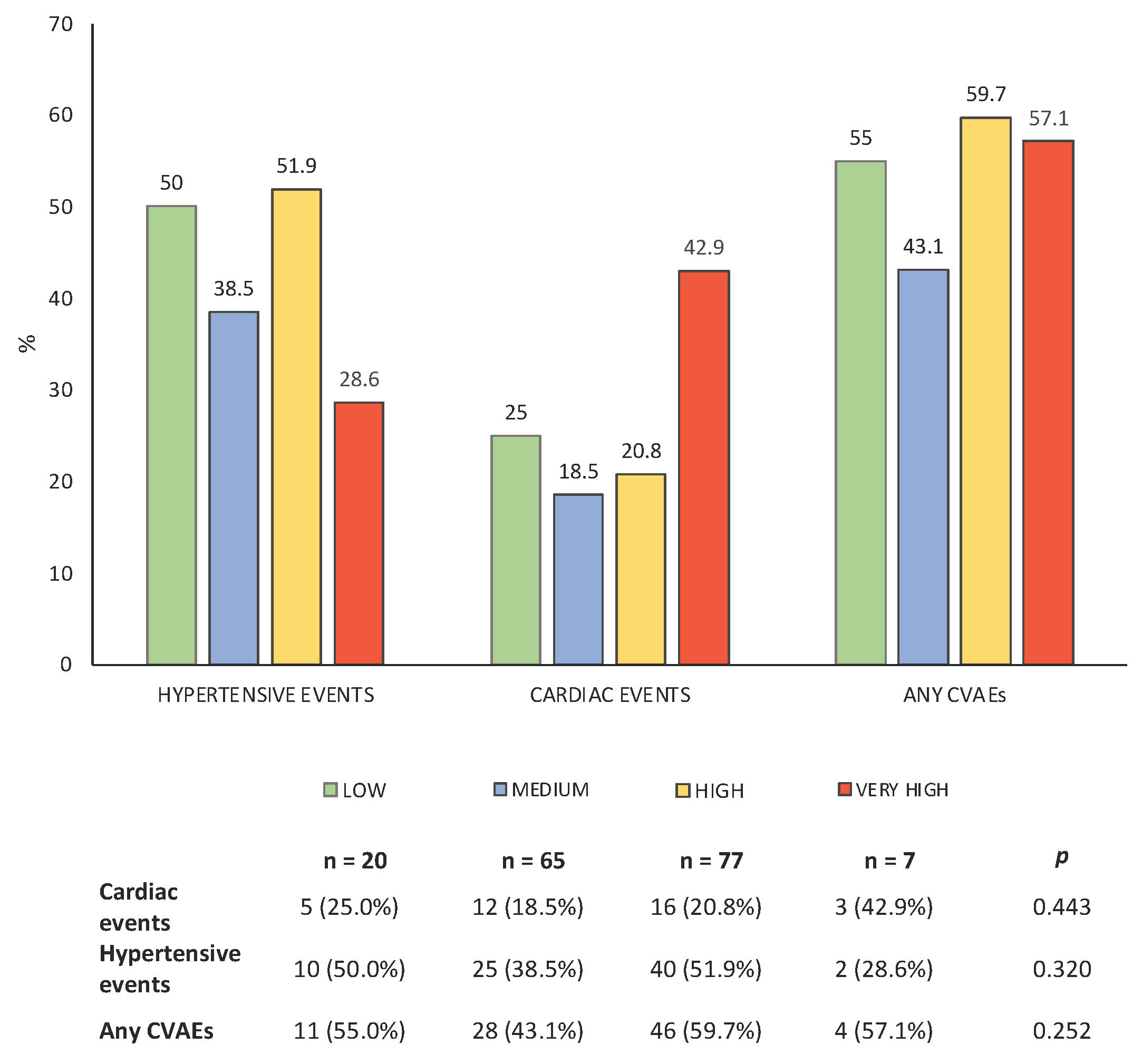
Comparing the incidence of both cardiovascular and hypertensive events among the different levels of risk, no statistically significant differences were found between groups (Figure 2); the incidence of cardiovascular and hypertensive events appears to be comparable across the different risk levels. Of interest, among the patients classified as high risk according to the HFA-ICOS score, none experienced an event of severity grade ≥3 according to the CTCAE scale.

3.4. Accuracy of HFO-ICOS Score and the Role of Other Baseline Parameters as Predictors of CVAEs

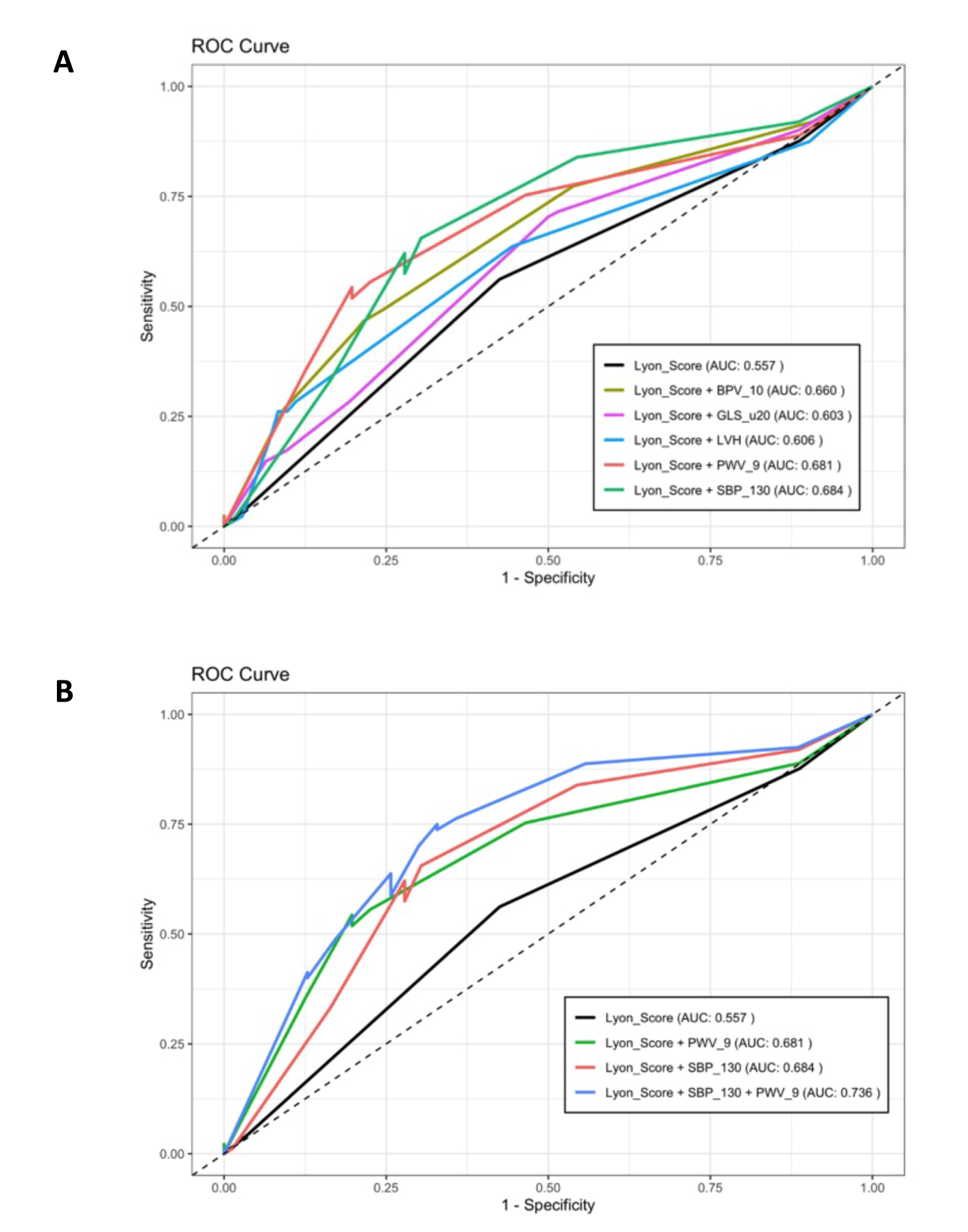
The accuracy of the HFO-ICOS in predicting CVAEs during K therapy in our population showed an area under the curve of only 0.557. However, the accuracy of the score was increased by the integration of other baseline parameters. In particular, the presence at baseline of a systolic blood pressure value ≥130 mmHg and of a pulse wave velocity ≥9 m/s was demonstrated to increase the AUC of the score to 0.684 and 0.681 (p = 0.03; p = 0.038), respectively. Other parameters, such as left ventricular hypertrophy, global longitudinal strain and blood pressure variability, were shown not to increase the accuracy in a statistically significant way (Figure 3A). Conversely, the accuracy of the model further improved by the integration of both parameters, obtaining an AUC of 0.736 (p = 0.002), as shown in Figure 3B.
The role of other baseline parameters in predicting future CVAEs was assessed. Parameters such as office systolic and diastolic blood pressure, 24 h systolic blood pressure, day systolic blood pressure and blood pressure variability were significantly associated with future CVAEs. Similarly, parameters of increased arterial stiffness and left ventricular mass were associated with an increased risk of CVAEs. When using the HFO-ICOS score as a continuous variable, the increase in risk levels estimated at baseline was not a predictor for events (Table 5).

4. Discussion

Cardiotoxicity induced by protocols containing Carfilzomib poses two major concerns: the morbidity and mortality induced by the cardiovascular event itself and the early discontinuation of the first-line chemotherapy for the hematologic disease, leading to a worse outcome. Despite the inference about cardiotoxic burden related to proteasome inhibitors, the lack of sufficient data in real-world clinical settings and the heterogeneity in detecting and managing cardiovascular events represent significant challenges for clinicians [12]. In a context of profound need for definite recommendations in the field, the working group of the Joint Heart Failure Association of the European Society of Cardiology (ESC) and the International Cardio-Oncology Society (HFA-ICOS) published a risk assessment tool to risk stratify oncology patients prior to receiving therapy with proteasome inhibitors [10]. By the cumulative risk derived by the combination of pretreatment patients and therapy-related factors, these guidelines propose a summary score used to classify patients as low, medium, high and at very high risk for cardiovascular events. However, the effectiveness of the HFA-ICOS score is unclear and requires validation in real-life settings. In our study, we have tested, for the first time, the effectiveness of the HFA-ICOS score in predicting cardiovascular adverse events in a cohort of multiple myeloma patients undergoing protocols including K therapy.
From the results, multiple myeloma patients were confirmed to be a population at medium–high cardiovascular risk, exemplified by a substantial proportion of cardiovascular risk factors and of pre-existing subclinical cardiac (18.8% of left ventricular hypertrophy, 23% of GLS impairment ≥−20 %) and vascular organ damage (32% had a rise of pulse wave velocity value ≥−9 m/s). During a median of 11.2 months of follow-up from K starting, the incidence of CVAEs was high: 89 (52.7%) patients experienced at least one event. Adverse cardiovascular events occurred in 21.3% of patients with various degrees of cardiac toxicity; one fatal cardiotoxicity. One of the important features of this study, as in our previous report [7], was the exploration of the rates of mild cancer therapy-related cardiac dysfunction (3.6% of patients), defined as a decrease in global longitudinal strain even when unaccompanied by an LVEF < 50%, according to the latest consensus statement from the International Cardio-Oncology Society [11]. Nearly half of the patients experienced at least one hypertensive event, and 3% had a hypertensive emergency. This incidence was higher than the results observed in preclinical trials [14], in which the rate of CVAEs ranged from 18% to 22% [15,16], but consistent with the findings of studies conducted in curative settings [7], as in Cornell’s study [8], in which the incidence of cardiovascular adverse events during K therapy was observed in about 50% of the population. With regard to hypertensive events, Carfilzomib confirms its role in contributing to blood pressure elevation. This effect may, however, be partially influenced by the concomitant use of dexamethasone in the various treatment regimens. Nonetheless, in the previous study by Astarita et al. [7], which compared the incidence of cardiovascular events between two treatment regimens based on different doses of Carfilzomib—KD (Carfilzomib–dexamethasone, target dose of K: 56 mg/m2) versus KRD (Carfilzomib–lenalidomide–dexamethasone, target dose of K: 27 mg/m2)—both all-type cardiovascular events and hypertensive events occurred more frequently in patients receiving KD compared to those receiving KRD. These findings suggest a primary role of Carfilzomib in the development of such events, even at an equivalent dose of dexamethasone. Nevertheless, although a substantial number of cardiovascular and hypertensive events were observed, the incidence of severe events (CTACE grade ≥3) was low. Among patients who experienced grade ≥3 events, only five required discontinuation of Carfilzomib therapy. In fact, the majority of CVAEs were transient and of low severity. In the remaining cases, treatment with Carfilzomib was continued after either intensification of anti-hypertensive therapy or optimization of cardioprotective management.
By applying the HFA-ICOS risk score at baseline, 11.8% of patients were classified as ‘low risk’, 38.5% as ‘medium risk’, 45.6% as ‘high risk’ and 4.1% as ‘very high risk’ for CVAEs during K therapy. Consequently, about half of the patients were classified as having high or very high risk. Analyzing the characteristics of each group, parameters such as median age, BMI, arterial hypertension, previous ischemic heart disease, stroke, left ventricular mass, and pulse wave velocity were more prevalent in groups at higher risk. However, these parameters, with the exception of the pulse wave velocity, were themselves risk factors considered by the model. In contrast, the groups did not differ for parameters related to office and out-of-office blood pressure profiles or for the other echocardiographic variables. Furthermore, the HFA-ICOS score did not show a good correlation with the incidence of CVAEs during K therapy in our population, even when considered as a continuous variable. During the follow-up, no differences were observed for the incidence of cardiovascular events, hypertensive-related events, or any CVAEs between the low, medium, high and very high risks at baseline (p > 0.05 for all comparisons). As shown in Figure 2, in a population of 169 patients, the score identified only 7 patients as being at very high risk. While it is true that four of these patients (57%) experienced at least one cardiovascular event, the lower-risk groups—which also included a larger number of patients—showed a comparable incidence of events. Therefore, the HFA-ICOS score showed a low sensitivity and did not discriminate between patients at low and high risk for events in a clinical setting. Of interest, among the patients classified as high risk according to the HFA-ICOS score, none experienced an event of severity grade ≥3 according to the CTCAE scale. This finding suggests a limited clinical applicability of the score within our study population. These limits of applicability of the score in our clinical setting could be related to the studies that support the score itself. Indeed, with the exception of the study of Cornell et al. [8], the HFA-ICOS is based on expert opinions, studies based on small sample sizes [17], not including K treatment [18,19] and preclinical studies [16] with significant heterogeneity in the definition of cardiovascular events and limited validation to date. It follows a limited applicability in the real-life world, as highlighted by other clinical studies. In women with HER2+ treated with trastuzumab, two retrospective studies, Suntheralingam et al. [20] and Battisti et al. [21], demonstrated that the HFA-ICOS risk score did not adequately identify patients at low risk for cardiac dysfunction. For overall cardiotoxicity related to cancer treatment, in Cronin’s study [22], the HFA-ICOS showed a low sensitivity with a moderate power in predicting CVAEs in a cohort of women with HER2+ breast cancer. Similarly, Tini et al. [23], in two cohorts of breast cancer women treated with anthracyclines and anti-HER2, concluded that patients classified at medium–high risk using the HFA-ICOS score were not associated with the occurrence of cardiac dysfunction.
Of interest, we investigated the performance of the HFA-ICOS score when integrated with other baseline parameters. In particular, the presence at baseline of a systolic blood pressure value ≥130 mmHg and of a pulse wave velocity ≥9 m/s demonstrated to increase the accuracy of the score, obtaining the maximum accuracy including both the parameters in the model (AUC of 0.736). These findings are consistent with other reports [7] that identified the systolic blood pressure—and, in general, parameters related to in-office and out-of-office blood pressure—and the pulse wave velocity as predictors of CVAEs during K therapy. Consequently, it is conceivable that the inclusion of these variables in the HFA-ICOS score could improve the performance of the score in detecting high-risk patients in real-life settings.
The current study has some limitations. Firstly, the experience of a single center limits the generalizability of the results, which should be confirmed by larger studies with a multicenter design. Moreover, our study lacks some data points and routine follow-up information that are included in the HFA-ICOS score, which introduces potential biases affecting its applicability. In particular, the incidence of venous thrombosis and pulmonary embolism was not assessed, and cardiac biomarkers (troponin and BNP or NT-proBNP) were not routinely measured for all patients. Similarly, we did not include patients affected by cardiac amyloidosis (exclusion criteria), one of the risk factors considered by the HFA-ICOS score. These limitations may lead to incorrect patient stratification as indicated by the score.

5. Conclusions

Our study evaluated for the first time the performance of the HFA-ICOS risk score in predicting cardiovascular adverse events, classified according to the latest consensus statement from the International Cardio-Oncology Society, in a cohort of multiple myeloma patients treated with K therapy in a real-world clinical setting and prospectively followed. The application of the HFA-ICOS risk score at baseline in our population showed limited ability to discriminate patients at low, medium, and high risk for cardiovascular and hypertensive events during K therapy. Indeed, risk groups exhibited a similar incidence and severity of both cardiovascular and hypertensive events. Of interest, the integration of additional parameters into the HFA-ICOS score, such as systolic blood pressure and pulse wave velocity, has been shown to improve the score’s performance. This study represents a first step in understanding the applicability of the score outside controlled clinical trials, and future research on larger, multicenter cohorts is needed to validate these findings.

Supplementary Materials

The following supporting information can be downloaded at: https://www.mdpi.com/article/10.3390/cancers17142353/s1, Table S1. Carfilzomib-based regimens of the population.

Author Contributions

Conceptualization, A.M. and F.V. (Franco Veglio); methodology, A.M.; software, L.A.; formal analysis, L.A. and M.C.; investigation, A.A. and G.M.; resources, A.A., G.M., A.C., C.C., A.P., and G.B.; data curation, A.A., G.M., A.C., C.C., and D.L.; writing—original draft preparation, A.A.; writing—review and editing, A.A. and F.V. (Fabrizio Vallelonga); supervision, A.M., F.V. (Franco Veglio), F.G., and S.B.; project administration, A.M. and F.V. (Franco Veglio). All authors have read and agreed to the published version of the manuscript.

Funding

This research received no external funding.

Institutional Review Board Statement

The study was conducted according to the guidelines of the Declaration of Helsinki and approved by the Ethics Committee of the ‘Città della Salute e della Scienza’ Hospital of Turin (Protocol Number 0038655). All authors have read and approved the submission of the manuscript, have met the criteria for authorship as established by the International Committee of Medical Journal Editors, believe that the paper represents honest work, and are able to verify the validity of the results reported. The study adheres to the STROBE Statement for Observational Studies.

Informed Consent Statement

Written informed consent was obtained from all subjects involved in the study.

Data Availability Statement

Data are contained within the article.

Conflicts of Interest

Alberto Milan received honoraria for advisory board from Amgen and Janssen. Sara Bringhen received honoraria from Bristol-Myers Squibb, Celgene, Amgen and Janssen; advisory boards for Amgen, Karyopharm, Janssen and Celgene; and consultancy fees from Takeda and Janssen. Francesca Gay received honoraria from Amgen, Janseen, Celgene, BMS, Takeda, Abbvie; advisory boards for Amgen, Janseen, Celgene, BMS, Takeda, and Abbvie; and advisory adaptive for Roche Oncopeptides. The other authors declare no conflicts of interest.

Appendix A

Appendix A.1. Cardiovascular Adverse Events Definitions

Acute coronary syndromes, distinguished in ST and non-ST myocardium infarction, were diagnosed in the presence of increased/decreased cardiac biomarkers (with at least one value above the 99th percentile) and at least one of the following: symptoms, ischemic ECG changes, or echocardiographic findings [24]. Heart failure was demonstrated by the presence of at least two of the three following items: symptoms and/or clinical signs, echocardiographic findings, and elevated levels of natriuretic peptides [25]. Arrhythmias included atrial fibrillation or flutter, atrial bigeminy, ventricular tachycardia, ventricular bigeminy/trigeminy, and grade 2 atrioventricular block. Typical chest pain was defined by the presence of chest pain with features suggestive of ischemic cardiac origin (duration, irradiation, onset, etc.), and for this reason subjected to clinical and instrumental assessments (ECG, cardiac troponins and eventually echocardiography) with negative results. Sudden death was defined as an unexpected death during K therapy. Left ventricular dysfunction was defined as (1) a new LVEF reduction to <40%, (2) a new LVEF reduction by 10 percentage points to an LVEF of 40–49% or a new LVEF reduction by 10 percentage points to an LVEF of 40–49% and either a new relative decline in GLS by 15% from baseline or a new rise in cardiac biomarkers, and (3) an LVEF of 50% and a new relative decline in GLS by 15% from baseline and/or a new rise in cardiac biomarkers [11].

Appendix A.2. Hypertensive Adverse Events Definitions

A new diagnosis of arterial hypertension was defined by the presence of blood pressure values of 140/90 mmHg in in-office or out-of-office BP measurements in a subject who was previously normotensive; the worsening of known arterial hypertension was defined as increased BP values of 140/90 mmHg requiring additional anti-hypertensive treatment in a subject with known arterial hypertension. Pre-infusion uncontrolled hypertension was defined by office BP values of 140/90 mmHg within 30 min of prior K infusion, distinguished by limiting or permissive K infusion. Post-infusion uncontrolled hypertension was defined by office BP values of 140/90 mmHg within 30 min of K infusion. Masked hypertension was defined by the presence of arterial hypertension at ABPM when office BP measurements were normal. Hypertensive urgency was demonstrated by symptomatic cases of BP >180/110 mmHg subjected to further assessment that excluded target acute organ damage; hypertensive emergency was defined by symptomatic BP >180/110 mmHg, with evidence of target acute organ damage at further assessment tests [13].

References

  1. Siegel, R.L.; Miller, K.D.; Jemal, A. Cancer statistics, 2020. CA Cancer J. Clin. 2020, 70, 7–30. [Google Scholar] [PubMed]
  2. Bringhen, S.; Milan, A.; D’Agostino, M.; Ferri, C.; Wäsch, R.; Gay, F.; LaRocca, A.; Offidani, M.; Zweegman, S.; Terpos, E.; et al. Prevention, monitoring and treatment of cardiovascular adverse events in myeloma patients receiving carfilzomib A consensus paper by the European Myeloma Network and the Italian Society of Arterial Hypertension. J. Intern. Med. 2019, 286, 63–74. [Google Scholar] [PubMed]
  3. Kistler, K.D.; Kalman, J.; Sahni, G.; Murphy, B.; Werther, W.; Rajangam, K.; Chari, A. Incidence and Risk of Cardiac Events in Patients with Previously Treated Multiple Myeloma Versus Matched Patients Without Multiple Myeloma: An Observational, Retrospective, Cohort Study. Clin. Lymphoma Myeloma Leuk. 2017, 17, 89–96. [Google Scholar]
  4. Georgiopoulos, G.; Makris, N.; Laina, A.; Theodorakakou, F.; Briasoulis, A.; Trougakos, I.P.; Dimopoulos, M.A.; Kastritis, E.; Stamatelopoulos, K. Cardiovascular Toxicity of Proteasome Inhibitors: Underlying Mechanisms and Management Strategies: JACC: CardioOncology State-of-the-Art Review. JACC CardioOncol. 2023, 5, 1–21. [Google Scholar]
  5. Siegel, D.S.; Martin, T.; Wang, M.; Vij, R.; Jakubowiak, A.J.; Lonial, S.; Trudel, S.; Kukreti, V.; Bahlis, N.; Alsina, M.; et al. A phase 2 study of single-agent carfilzomib (PX-171-003-A1) in patients with relapsed and refractory multiple myeloma. Blood 2012, 120, 2817–2825. [Google Scholar] [PubMed]
  6. Chari, A.; Stewart, A.K.; Russell, S.D.; Moreau, P.; Herrmann, J.; Banchs, J.; Hajek, R.; Groarke, J.; Lyon, A.R.; Batty, G.N.; et al. Analysis of carfilzomib cardiovascular safety profile across relapsed and/or refractory multiple myeloma clinical trials. Blood Adv. 2018, 2, 1633–1644. [Google Scholar]
  7. Astarita, A.; Mingrone, G.; Airale, L.; Cesareo, M.; Colomba, A.; Catarinella, C.; Leone, D.; Gay, F.; Bringhen, S.; Veglio, F.; et al. Carfilzomib-Based Regimen and Cardiotoxicity in Multiple Myeloma: Incidence of Cardiovascular Events and Organ Damage in Carfilzomib-Dexamethasone versus Carfilzomib-Lenalidomide-Dexamethasone. A Real-Life Prospective Study. Cancers 2023, 15, 955. [Google Scholar] [CrossRef]
  8. Cornell, R.F.; Ky, B.; Weiss, B.M.; Dahm, C.N.; Gupta, D.K.; Du, L.; Carver, J.R.; Cohen, A.D.; Engelhardt, B.G.; Garfall, A.L.; et al. Prospective Study of Cardiac Events During Proteasome Inhibitor Therapy for Relapsed Multiple Myeloma. J. Clin. Oncol. 2019, 37, 1946–1955. [Google Scholar]
  9. Astarita, A.; Mingrone, G.; Airale, L.; Vallelonga, F.; Covella, M.; Catarinella, C.; Cesareo, M.; Bruno, G.; Leone, D.; Giordana, C.; et al. Multiple Myeloma Patients Undergoing Carfilzomib: Development and Validation of a Risk Score for Cardiovascular Adverse Events Prediction. Cancers 2021, 13, 1631. [Google Scholar] [CrossRef]
  10. Lyon, A.R.; Dent, S.; Stanway, S.; Earl, H.; Brezden-Masley, C.; Cohen-Solal, A.; Tocchetti, C.G.; Moslehi, J.J.; Groarke, J.D.; Bergler-Klein, J.; et al. Baseline cardiovascular risk assessment in cancer patients scheduled to receive cardiotoxic cancer therapies: A position statement and new risk assessment tools from the Cardio-Oncology Study Group of the Heart Failure Association of the European Society of Cardiology in collaboration with the International Cardio-Oncology Society. Eur. J. Heart Fail. 2020, 22, 1945–1960. [Google Scholar]
  11. Lyon, A.R.; López-Fernández, T.; Couch, L.S.; Asteggiano, R.; Aznar, M.C.; Bergler-Klein, J.; Boriani, G.; Cardinale, D.; Cordoba, R.; Cosyns, B.; et al. 2022 ESC Guidelines on cardio-oncology developed in collaboration with the European Hematology Association (EHA), the European Society for Therapeutic Radiology and Oncology (ESTRO) and the International Cardio-Oncology Society (IC-OS). Eur. Heart J. 2022, 43, 4229–4361. [Google Scholar] [PubMed]
  12. Rashed, E.R.; Margulies, K.B. New Cardiotoxicity Risk Assessment Guidelines: Searching for Validation. JACC CardioOncol. 2023, 5, 638–640. [Google Scholar]
  13. McEvoy, J.W.; McCarthy, C.P.; Bruno, R.M.; Brouwers, S.; Canavan, M.D.; Ceconi, C.; Christodorescu, R.M.; Daskalopoulou, S.S.; Ferro, C.J.; Gerdts, E.; et al. 2024 ESC Guidelines for the management of elevated blood pressure and hypertension. Eur. Heart J. 2024, 45, 3912–4018. [Google Scholar] [PubMed]
  14. Waxman, A.J.; Clasen, S.; Hwang, W.-T.; Garfall, A.; Vogl, D.T.; Carver, J.; O’Quinn, R.; Cohen, A.D.; Stadtmauer, E.A.; Ky, B.; et al. Carfilzomib-Associated Cardiovascular Adverse Events. JAMA Oncol. 2018, 4, e174519. [Google Scholar]
  15. Atrash, S.; Tullos, A.; Panozzo, S.; Bhutani, M.; Van Rhee, F.; Barlogie, B.; Usmani, S.Z. Cardiac complications in relapsed and refractory multiple myeloma patients treated with carfilzomib. Blood Cancer J. 2015, 5, e272. [Google Scholar]
  16. Danhof, S.; Schreder, M.; Rasche, L.; Strifler, S.; Einsele, H.; Knop, S. ‘Real-life’ experience of preapproval carfilzomib-based therapy in myeloma—Analysis of cardiac toxicity and predisposing factors. Eur. J. Haematol. 2016, 97, 25–32. [Google Scholar]
  17. Chen, J.H.; Lenihan, D.J.; Phillips, S.E.; Harrell, S.L.; Cornell, R.F. Cardiac events during treatment with proteasome inhibitor therapy for multiple myeloma. Cardiooncology 2017, 3, 4. [Google Scholar]
  18. cGov.UK. Lenalidomide: Risk of Thrombosis and Thromboembolism. February 2011. Available online: https://www.gov.uk/drug-safety-update/lenalidomide-risk-of-thrombosis-and-thromboembolism (accessed on 3 June 2020).
  19. Libourel, E.J.; Sonneveld, P.; van der Holt, B.; de Maat, M.P.; Leebeek, F.W. High incidence of arterial thrombosis in young patients treated for multiple myeloma: Results of a prospective cohort study. Blood 2010, 116, 22–26. [Google Scholar]
  20. Suntheralingam, S.; Fan, C.S.; Calvillo-Argüelles, O.; Abdel-Qadir, H.; Amir, E.; Thavendiranathan, P. Evaluation of Risk Prediction Models to Identify Cancer Therapeutics Related Cardiac Dysfunction in Women with HER2+ Breast Cancer. J. Clin. Med. 2022, 11, 847. [Google Scholar]
  21. Battisti, N.M.L.; Andres, M.S.; Lee, K.A.; Ramalingam, S.; Nash, T.; Mappouridou, S.; Senthivel, N.; Asavisanu, K.; Obeid, M.; Tripodaki, E.S.; et al. Incidence of cardiotoxicity and validation of the Heart Failure Association-International Cardio-Oncology Society risk stratification tool in patients treated with trastuzumab for HER2-positive early breast cancer. Breast Cancer Res. Treat. 2021, 188, 149–163. [Google Scholar]
  22. Cronin, M.; Crowley, A.; Davey, M.G.; Ryan, P.; Abdelshafy, M.; Elkoumy, A.; Elzomor, H.; Arsang-Jang, S.; Ganly, S.; Nash, P.; et al. Heart Failure Association-International Cardio-Oncology Society Risk Score Validation in HER2-Positive Breast Cancer. J. Clin. Med. 2023, 12, 1278. [Google Scholar] [PubMed]
  23. Tini, G.; Cuomo, A.; Battistoni, A.; Sarocchi, M.; Mercurio, V.; Ameri, P.; Volpe, M.; Porto, I.; Tocchetti, C.G.; Spallarossa, P. Baseline cardio-oncologic risk assessment in breast cancer women and occurrence of cardiovascular events: The HFA/ICOS risk tool in real-world practice. Int. J. Cardiol. 2022, 349, 134–137. [Google Scholar] [PubMed]
  24. Byrne, R.A.; Rossello, X.; Coughlan, J.J.; Barbato, E.; Berry, C.; Chieffo, A.; Claeys, M.J.; Dan, G.A.; Dweck, M.R.; Galbraith, M.; et al. 2023 ESC Guidelines for the management of acute coronary syndromes. Eur. Heart J. 2023, 44, 3720–3826. [Google Scholar]
  25. McDonagh, T.A.; Metra, M.; Adamo, M.; Gardner, R.S.; Baumbach, A.; Böhm, M.; Burri, H.; Butler, J.; Čelutkienė, J.; Chioncel, O.; et al. 2023 Focused Update of the 2021 ESC Guidelines for the diagnosis and treatment of acute and chronic heart failure. Eur. Heart J. 2023, 44, 3627–3639. [Google Scholar] [PubMed]
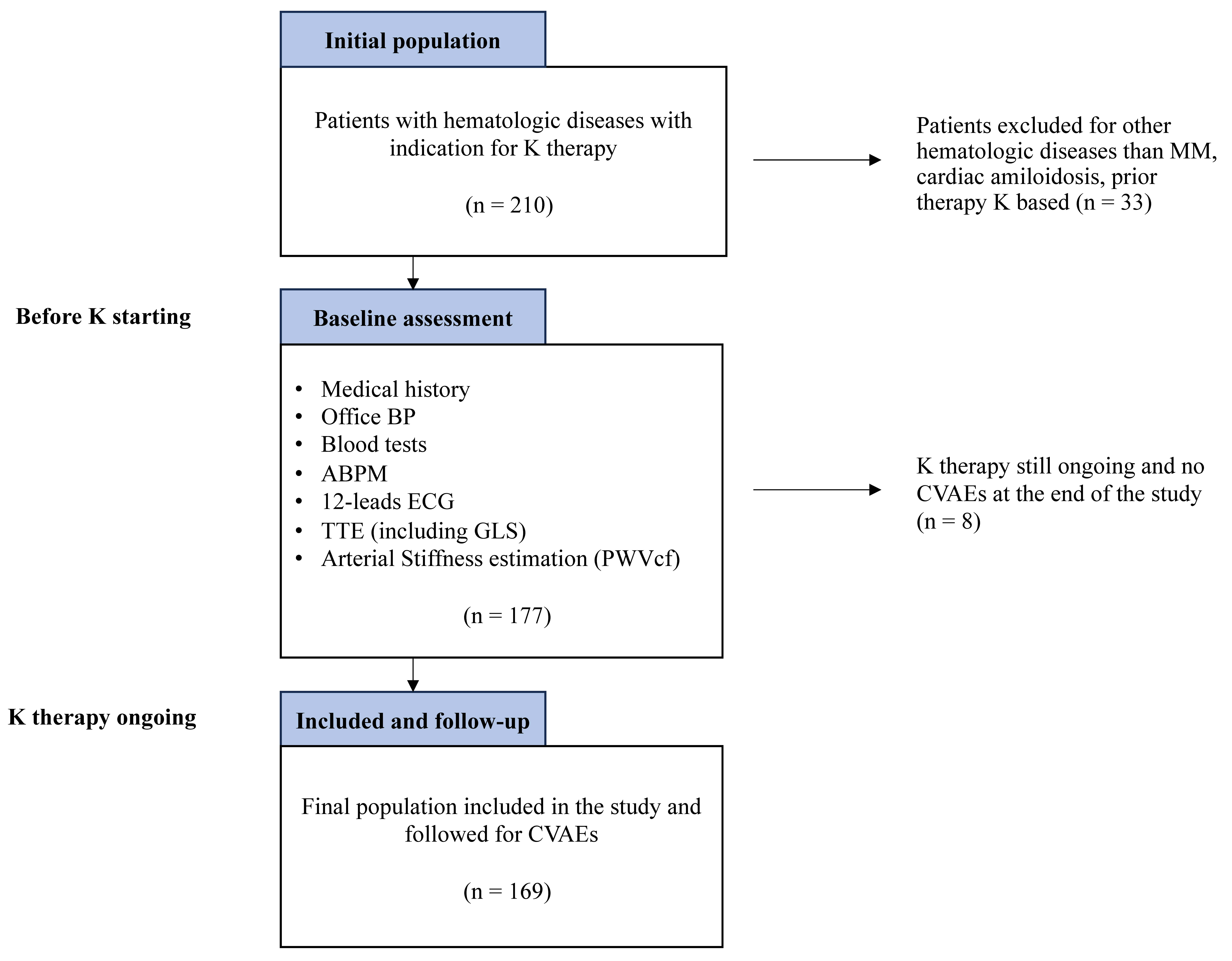
Figure 1. Flowchart of the study protocol.
Cancers 17 02353 g001
Figure 2. Incidence of CVAEs by baseline risk groups of HFO-ICOS score.
Cancers 17 02353 g002
Figure 3. Accuracy of the HFO-ICOS in predicting CVAEs. (A) ROC curve of HFO-ICOS score (black line) and of HFO-ICOS comprehensive of blood pressure variability > 10 (dark green line), global longitudinal strain (GLS) > −20% (pink line), left ventricular hypertrophy (blue line), pulse wave velocity ≥ 9 m/s, and systolic blood pressure (SBP) ≥ 130 mmHg (green line). (B) ROC curve of HFO-ICOS score (black line), HFO-ICOS score comprehensive of pulse wave velocity ≥ 9 m/s (green line), HFO-ICOS score comprehensive of systolic blood pressure (SBP) ≥ 130 mmHg (red line) and HFO-ICOS score comprehensive of both systolic blood pressure (SBP) and pulse wave velocity ≥ 9 m/s (blue line).
Cancers 17 02353 g003
Table 1. Baseline parameters of the population.
ParameterN = 169 (100%)
General
Age, y70.0 (63.0; 74.2)
Male sex77 (45.6%)
BMI, kg/m226.7 (23.9; 30.3)
Individual CV risk factors and comorbidities
Tobacco use (prior/current)90 (53.3)
Obesity (BMI ≥ 30)49 (29)
Known arterial hypertension80 (47.3)
Diabetes mellitus16 (9)
Chronic renal failure (eGFR < 60 mL/m)41 (26.3)
Ischemic heart disease4 (2.3)
Atrial fibrillation5 (2.9)
Dyslipidemia25 (14.8)
Previous stroke3 (1.8)
Office BP values 
SBP, mmHg128 (116; 141)
DBP, mmHg76.0 (70.0; 84.0)
ABPM 1 , mean (SD)
Daytime SBP, mmHg124 (116; 132)
Daytime DBP, mmHg74.0 (69.0; 80.0)
Daytime MBP, mmHg92.0 (86.0; 96.5)
24 h SBP, mmHg120 (112; 129)
24 h DBP, mmHg71.0 (65.0; 76.5)
24 h MBP, mmHg88.0 (82.0; 93.0)
Nighttime SBP, mmHg109 (102; 120)
Nighttime DBP, mmHg62.0 (57.0; 69.0)
Night MBP, mmHg78.0 (73.0; 86.0)
Blood pressure variability9.00 (7.0; 10.0)
Echocardiographic parameters 2
LAVi, ml/m249.0 (38.4; 61.2)
LVMi, g/m286.4 (74.4; 102.0)
LVH30 (18.8)
Diastolic dysfunction9 (6.4)
LVEF, %62.2 (58.3; 65.2)
Stroke volume, mL/m247.4 (39.1; 57.9)
GLS value, %−21.50 (−23.40; −20.10)
GLS value ≥ −20 %33 (23.1)
Arterial stiffness evaluation 3
cfPWV value, m/s8.00 (7.0; 9.0)
cfPWV value ≥ 9 m/s49 (32.2)
Oncological history 
Median MM disease duration, months11.2 (5.23; 23.8)
Line of K therapy2.85 (1; 9)
Mean values estimated in 1 152 patients; 2 165 patients; 3 153 patients. BMI = body mass index; eGFR = estimated glomerular filtration rate; ABPM = ambulatory blood pressure monitoring; SBP = systolic blood pressure; DBP = diastolic blood pressure; MBP = mean blood pressure; LVH = left ventricular hypertrophy; BP = blood pressure; LAVi = left atrium volume index; LVMi = left ventricular mass index; LVH = left ventricular hypertrophy; GLS = global longitudinal strain; cfPWV = carotid–femoral pulse wave velocity; MM = multiple myeloma; SD = standard deviation.
Table 2. Stratification by the HFA-ICOS score of MM patients scheduled for K at baseline.
Patients (n)Score
Previouscardiovascular diseases
Heart failure or cardiomyopathy2 (1.2)Very High
Prior proteasome inhibitor cardiotoxicity0 (0)Very High
Venous thrombosis (DVT or PE)0 (0)Very High
Cardiac amyloidosisnaVery High
Arterial vascular disease (IHD, PCI, CABG, stable angina, TIA, stroke, PVD)7 (4.1)Very High
Prior immunomodulatory CV toxicity0 (0)High
Baseline LVEF < 50% 5 (3)High
Borderline LVEF 50–54% 5 (3)Medium 1
Arrhythmia5 (3)Medium 2
Left ventricular hypertrophy 130 (17.8)Medium 1
Cardiac biomarkers 2 
Elevated baseline troponin0 (0)Medium 2
Elevated baseline BNP or NT-proBNP2 (1.2)High
Demographic and cardiovascular risk factors 
Age ≥ 75 years40 (23.7)High
Age 65–74 years79 (46.7)Medium 1
Arterial hypertension80 (47.3)Medium 1
Diabetes mellitus16 (9.5)Medium 1
Hyperlipidemia25 (14.8)Medium 1
Chronic kidney disease40 (23.7)Medium 1
Family history of thrombophilianaMedium 1
Previous cardiotoxic cancer treatment
Prior anthracycline exposure34 (20.1)High
Prior thoracic spine radiotherapy7 (4.1)Medium 1
Current myeloma treatment 
High-dose of dexamethasone > 160 mg/month0 (0)Medium 1
Lifestyle risk factors  
Current smoker or significant smoking
history
90 (53.3)Medium 1
Obesity (BMI > 30 kg/m2)49 (29)Medium 1
Mean values estimated in 1 176 patients; 2 14 patients. BMI = body mass index; BNP = brain natriuretic peptide; CABG = coronary artery bypass graft; DVT = deep vein thrombosis; IHD = ischemic heart disease; LVEF = left ventricular ejection fraction; NT-proBNP = N-terminal pro-brain natriuretic peptide; PCI = percutaneous coronary intervention; PE = pulmonary embolism; PVD = peripheral vascular disease; TIA = transient ischemic attack; na = not applicable.
Table 3. Comparison of baseline parameters between the levels of risk based on HFO-ICOS score stratification.
ParameterLevels of Risk
1. Low
N = 20
2. Medium
N = 65
3. High
N = 77
4. Very High
N = 7
pComparison
General
Age, y60.0 (54.0; 65.0)69.0 (63.0; 72.0)75.0 (68.0; 77.0)68.0 (66.5; 68.5)<0.001 1 vs. 2/3/4; 2 vs. 3
Male sex8 (40.0)27 (41.5)38 (49.4)4 (57.1)0.677 
BMI, kg/m224.8 (21.8; 27.0)28.1 (25.2; 31.6)26.4 (23.7; 30.1)27.6 (27.3; 31.4)0.0121 vs. 2
Individual CV
risk factors
Tobacco use (prior/current)7 (35.0)37 (56.9)41 (53.2)5 (71.4)0.275 
Obesity (BMI ≥ 30)0 (0.0)22 (33.8)24 (31.2)3 (42.9)0.0051 vs. 2/3/4
Known arterial hypertension2 (10.0)30 (46.2)42 (54.5)6 (85.7)<0.0011 vs. 2/3/4
Diabetes mellitus0 (0)5 (7.7)10 (13.0)1 (14.3)0.253 
Chronic renal failure (eGFR < 60 mL/m)92.9 (77.3; 106)80.7 (64.7; 99.0)67.8 (54.8; 93.5)81.5 (68.3; 89.0)0.0331 vs. 3
Ischemic heart disease0 (0)0 (0)0 (0)4 (57.1)<0.0014 vs. 1/2/3
Atrial fibrillation0 (0)1 (1.5)3 (3.9)1 (14.3)0.277 
Dyslipidaemia0 (0)13 (20)12 (15.6)0 (0)0.099 
Previous stroke0 (0)0 (0)0 (0)3 (42.9)<0.0014 vs. 1/2/3
Office BP values,
mmHg
SBP120 (114; 128)128 (119; 141)130 (119; 142)122 (104; 131)0.081 
DBP74.5 (70.0; 80.0)77.5 (70.8; 87.2)75.0 (69.0; 82.0)79.0 (65.5; 81.5)0.501 
ABPM,
mmHg
Daytime SBP120 (114; 130)126 (118; 133)124 (116; 132)114 (107; 116)0.095 
Daytime DBP74.0 (70.0; 80.5)76.5 (69.0; 82.0)73.0 (68.0; 78.0)66.5 (60.5; 72.5)0.066 
Daytime MBP88.5 (84.8; 96.8)93.0 (88.8; 99.0)91.0 (86.0; 95.5)84.5 (77.0; 92.0)0.104 
24 h SBP114 (107; 124)122 (114; 129)120 (112; 129)108 (104; 115)0.101 
24 h DBP70.0 (65.2; 76.8)73.0 (67.0; 77.5)70.5 (64.8; 75.0)64.5 (58.0; 71.0)0.250 
24 h MBP85.0 (79.5; 92.2)89.0 (84.0; 93.0)89.0 (82.0; 93.0)81.0 (72.8; 90.0)0.334 
Nighttime SBP104 (96.8; 112)110 (102; 121)112 (103; 121)99.5 (95.5; 109)0.093 
Nighttime DBP59.5 (56.2; 66.0)63.5 (59.0; 69.0)63.0 (58.0; 70.0)60.0 (53.5; 70.2)0.484 
Night MBP,73.5 (71.2; 80.8)79.0 (74.0; 86.0)79.0 (74.0; 88.8)74.5 (69.2; 82.8)0.147 
Blood pressure variability8.00 (7.00; 9.00)9.00 (7.50; 11.0)8.50 (7.00; 10.0)8.50 (6.50; 11.2)0.597 
Echocardiographic
parameters
LAVi, mL/m239.0 (33.3; 65.0)49.4 (41.2; 63.0)48.8 (38.3; 58.1)57.7 (49.2; 65.5)0.341 
LVMi, g/m274.5 (66.7; 84.8)82.7 (70.2; 95.7)90.4 (76.9; 104)115 (86.1; 119)0.0021 vs. 3/4
LVH,1 (5.26%)7 (11.5%)18 (24.7%)4 (57.1%)0.0071/2 vs. 4
Diastolic dysfunction0 (0.00%)4 (7.69%)4 (5.97%)1 (16.7%)0.451 
LVEF, %60.2 (57.6; 64.3)62.7 (58.9; 65.6)62.2 (58.6; 64.7)60.8 (55.0; 62.2)0.491 
Stroke volume, mL/m253.0 (39.7; 60.8)50.2 (40.7; 61.5)43.5 (36.1; 53.1)54.9 (53.5; 68.8)0.023 
GLS value, %−21.90 (−23.40; −20.30)−21.60 (−22.70; −19.60)−21.60 (−23.90; −20.25)−21.00 (−21.50; −21.00)0.707 
GLS value ≥ −20%2 (11.8%)15 (28.3%)15 (22.1%)1 (20.0%)0.580 
Arterial stiffness
evaluation
cfPWV value, m/s7.00 (6.00; 7.50)8.00 (7.00; 9.00)9.00 (7.00; 10.0)8.00 (7.00; 9.00)0.0011/2 vs. 3
cfPWV value ≥ 9 m/s2 (10.5%)18 (30.5%)36 (52.2%)2 (40.0%)0.0021 vs. 3
BMI = body mass index; eGFR = estimated glomerular filtration rate; SBP = systolic blood pressure; DBP = diastolic blood pressure; MBP = mean blood pressure; LAVi = left atrium volume index; LVMi = left ventricular mass index; LVH = left ventricular hypertrophy; LVH = left ventricular hypertrophy; GLS = global longitudinal strain; cfPWV = carotid–femoral pulse wave velocity.
Table 4. Incidence of CVAEs during K therapy.
EventsN = 169 (100%)CTCAE
Grade 1–2Grade ≥ 3
Cardiovascular adverse events 136 (21.3)  
ACS (STEMI)2 (1.2)02
ACS (NSTEMI)4 (2.4)04
Angina9 (5.3)90
Heart failure10 (5.9)82
Arrhythmias12 (7.1)111
Sudden death1 (0.6)na0
LVEF impairment5 (3)03
GLS impairment6 (3.6)nana
Hypertensive events 177 (45.6)  
New onset/worsened hypertension67 (39.6)670
Masked hypertension5 (3)50
K pre-infusion hypertension permissive K infusion34 (20.1)277
K pre-infusion hypertension not permissive K infusion18 (10.7)117
K post-infusion hypertension 20 (11.8)137
Hypertensive urgency5 (3)05
Hypertensive emergency0 (0)00
Any CVAEs89 (52.7)  
1 Patients experienced more than one CVAE; hence, the total % amounts to >100. CVAE = cardiovascular adverse event; ACS = acute coronary syndrome; STEMI = ST-elevation myocardial infarction; NSTEMI = non-ST elevation myocardial infarction. CVAEs = cardiovascular adverse events. CTCAE = Common Terminology Criteria for Adverse Events na = not applicable.
Table 5. The role of other baseline parameters in predicting CVAEs during K therapy.
Baseline ParameterBetaOR (SD)Wald TestValore di p
General
Age0.021.02 (0.98–1.06)1.120.264
Male sex0.441.55 (0.84–2.86)1.400.160
BMI0.031.03 (0.96–1.10)0.810.419
Office BP    
SBP0.041.04 (1.02–1.07)3.94<0.001
DBP0.031.03 (1.00–1.06)2.030.042
ABPM
24 h SBP0.031.03 (1.00–1.06)1.970.048
24 h DBP0.021.02 (0.98–1.06)0.920.358
24 h MBP0.021.02 (0.99–1.06)1.150.249
Daytime SBP0.031.03 (1.01–1.06)2.410.016
Daytime DBP0.021.02 (0.99–1.06)1.210.225
Daytime MBP0.031.03 (1.00–1.07)1.850.065
Nighttime SBP0.011.01 (0.98–1.03)0.420.674
Nighttime DBP−0.001.00 (0.96–1.04)−0.160.872
Night MBP0.001.00 (0.97–1.04)0.080.936
Blood pressure
Variability
0.181.19 (1.07–1.35)3.060.002
Echocardiographic parameters
LAVi0.001.00 (0.98–1.02)0.170.865
LVEF0.021.02 (0.97–1.07)0.770.441
LVMi0.021.02 (1.01–1.04)2.690.007
LVH1.193.29 (1.38–8.77)2.550.011
Diastolic dysfunction−0.940.39 (0.08–1.55)−1.290.199
GLS value0.101.10 (0.97–1.26)1.470.142
GLS value ≥ −200.551.73 (0.78–4.02)1.320.188
Arterial stiffness evaluation    
cfPWV0.401.49 (1.23–1.84)3.87<0.001
cfPWV ≥ 9 m/s1.584.84 (2.38–10.32)4.23<0.001
HFA-ICOS score
Level of risk0.241.27 (0.85–1.92)1.150.251
SBP = systolic blood pressure; DBP = diastolic blood pressure; MBP = mean blood pressure; LAVi = left atrium volume index; LVEF = left ventricular ejection fraction; LVMi = left ventricular mass index; LVH = left ventricular hypertrophy; GLS = global longitudinal strain; cfPWV = carotid–femoral pulse wave velocity.
Disclaimer/Publisher’s Note: The statements, opinions and data contained in all publications are solely those of the individual author(s) and contributor(s) and not of MDPI and/or the editor(s). MDPI and/or the editor(s) disclaim responsibility for any injury to people or property resulting from any ideas, methods, instructions or products referred to in the content.