Communication

Fluorescent In Situ Hybridization Testing Allows the Diagnosis of NRG1 Gene Fusions in Lung and Pancreas Cancers with No Other Identified Oncogenic Driver

by
Clara Bastard
1,
Charline Caumont
2,3,4,
Laura Samaison
5,
Isabelle Quintin-Roué
1,
Laurent Doucet
1,
Pascale Marcorelles
1,
Cédric Le Maréchal
6,7,
Jean-Philippe Merlio
2,3,4,
David Cappellen
2,3,4 and
Arnaud Uguen
1,8,*
1
CHU Brest, Service d’Anatomie et Cytologie Pathologiques, F-29200 Brest, France
2
CHU Bordeaux, Department of Tumor Biology, F-33600 Pessac, France
3
BRIC (BoRdeaux Institute of onCology), UMR1312, INSERM, Univ. Bordeaux, F-33000 Bordeaux, France
4
Tumor Bank and Tumor Biology Laboratory, Bordeaux University Hospital, F-33600 Pessac, France
5
Ouest Pathologie, F-29000 Quimper, France
6
CHU Brest, Service de Génétique Moléculaire, F-29200 Brest, France
7
Univ Brest, Inserm, CHU de Brest, UMR1078, F-29200 Brest, France
8
Univ Brest, Inserm, CHU de Brest, LBAI, UMR1227, F-29200 Brest, France
*
Author to whom correspondence should be addressed.
Cancers 2025, 17(14), 2347; https://doi.org/10.3390/cancers17142347
Submission received: 4 June 2025 / Revised: 2 July 2025 / Accepted: 8 July 2025 / Published: 15 July 2025
(This article belongs to the Section Cancer Biomarkers)

Simple Summary

Pancreatic ductal carcinomas and lung adenocarcinomas are aggressive and deadly cancers, among which rare tumors harbor NRG1 fusions making the NRG1-rearranged tumor candidates suitable for specific targeted therapies. Access to therapeutic innovation is conditioned by the diagnosis of NRG1 fusion. In our study, we demonstrate the feasibility of an NRG1 fusion screening using NRG1 fluorescent in situ hybridization in pancreatic and lung tumors lacking other identified oncogenic drivers. In the case of no access to RNA sequencing, NRG1 FISH consists of a valuable tool searching for NRG1 fusions in patients with advanced cancers.

Abstract

Some pancreatic ductal-type (PDADK) and lung adenocarcinomas (LADK) lacking other molecular drivers are reported to harbor NRG1 fusions as potential novel therapeutic targets. We investigated the feasibility of a fluorescent in situ hybridization (FISH)-based diagnosis of NRG1 fusions in a case series of PDADK and LADK lacking other identified oncogenic drivers. First, among a case series of PDADK, KRAS analyses (PCR followed in PCR-negative cases by RNA sequencing—RNAseq) found 27/162 (16.7%) KRAS wild-type cases, among which 1/162 (0.6%) NRG1 fusion was diagnosed using FISH. Secondly, among a case series of LDAK, 191/446 (42.8%) cases had no molecular alterations in EGFR, KRAS, BRAF, HER2, MET, ALK, ROS1 and RET according to NGS and FISH analyses and, among them, 4/446 (0.9%) cases had NRG1 fusions using FISH. Finally, four additional cases out of the two previously mentioned cases series (1 PDADK and 3 LADK) with NRG1 fusions diagnosed by first-line RNAseq were also concluded as NRG1 FISH-positive. The NRG1 FISH tests for the nine NRG1 FISH-positive cases resulted in 50% to 80% of positive tumor nuclei, all with single 3′-NRG1 FISH signals. In our series, of the 22 cases analyzed with both NRG1 FISH (positivity criterion of at least 15% of tumor nuclei with a split between the 5′- and the 3′- parts of the probes and/or isolated single 3′-NRG1 signal) and RNAseq, 17 cases were FISH– RNAseq– and 5 cases were FISH+ RNAseq+ (no FISH+ RNAseq– or FISH– RNAseq+ cases in our study) resulting in 100% sensibility and specificity for the NRG1 FISH test. In the case of no access to RNAseq, NRG1 FISH consists of a valuable tool searching for NRG1 fusions in patients with advanced cancers.

1. Introduction

Among the deadliest cancers are pancreatic ductal adenocarcinoma (PDADK) and non-small cell lung cancers (NSCLC), notably comprising an increasing proportion of lung adenocarcinomas (LADK). Being the fourth leading cause of cancer-related deaths, PDADK is projected to become the second leading cause of death in 2030. Meanwhile, NSCLC remains the leading cause of cancer mortality [1,2]. For PDADK, the only curative treatment is surgery at an early stage but, when diagnosed, only 20% of the patients have resectable tumors and, of those, only 20% survive for 5 years or longer. Most patients with PDADK are resistant to chemotherapy [3,4]. For LADK, about three out of four patients are diagnosed with advanced diseases and require systemic therapies from the first line of treatment. PD-1/PD-L1 immune checkpoint inhibition and targeted therapies against oncogenic drivers, such as EGFR, KRASG12C, MET, BRAFV600E, and HER2 mutants or ALK, ROS1, and RET chimeric proteins have improved the treatment of LADK, but the 5-year survival rate of patients remains low (26% for any stages, from 64% if diagnosed at local stages to 8% for diagnosis at metastatic stages) [5,6]. Thus, it remains crucial to investigate new therapeutic approaches to treat patients with PDADK and NSCLC.
The targeting of tumors with Neuregulin 1 (NRG1) gene fusions could be one promising approach. Indeed, NRG1 fusions drive cancer development through aberrant ERBB receptor-mediated signaling, and ERBB inhibition can inhibit the tumor growth of NRG1-rearranged cancers. Promising therapeutic responses have already been reported in patients with NRG1-rearranged PDADK and LADK [7,8,9,10,11,12]. KRAS wild-type PDADK were reported to frequently harbor NRG1 fusions (until about 6% of PDADK in Jones et al.) and NRG1-rearranged KRAS wild-type PDADK seem particularly frequent in young patients (e.g., 4/17 23.5% of patients <50 years in Heining et al.) [7,8]. NRG1 fusions are also reported in about 0.8% (0.3–2.7%) of patients with LADK, especially those without any known oncogenic mutations, young and non-smokers, and those with a particular subtype of mucinous LADK in which NRG1 fusions are particularly frequent [13,14].
Screening for NRG1 fusions is like searching for a needle in a haystack, and diagnostic strategies can vary from one cancer type to another and from one medical center/healthcare system to another because of various workflow, technical, and medico-economic reasons. RNA sequencing (RNAseq) is now the reference method for searching for gene fusions in cancers, but not every laboratory has implemented it in their routine practice thus far, even for tumors requiring the molecular analysis of numerous genes for therapeutic decisions, such as LADK [15]. Moreover, for some cancers, such as PDADK, molecular testing is not systematically performed, limiting the diagnosis of targetable molecular events, such as NRG1 fusions [16,17].
The use of fluorescence in situ hybridization (FISH) to search for NRG1 fusions has been reported in some studies, from reports on pan-cancer to selected histological subtypes, such as invasive mucinous adenocarcinoma of the lung, with the use of various probes (in-house-developed or commercial probes) [18,19,20]. Nevertheless, reports about NRG1 FISH testing in real-life diagnosis and how it could be used as an alternative or complementary method in the era of RNAseq remain rare. Thus, in this study, we intend to evaluate the value of NRG1 fusion diagnosis using FISH in a case series of PDADK and LADK, with a focus on cancers with no other identified molecular oncogenic driver.

2. Material and Methods

2.1. Cases Selection

The cases included in this study were (1) those with PDADK diagnosed in the CHRU Brest (France) between January 2000 and January 2020 for the PDADK case series of patients and (2) those who had a LADK tumor sample with molecular analyses performed for diagnostic purpose in CHRU Brest between January 2021 and June 2022 for the LADK case series of patients. In addition to these two exhaustive case series, 4 selected additional cases (1 PDADK and 3 LADK) with NRG1 fusions diagnosed later (between 2023 and 2025) using first-line RNAseq were added to our study for method comparison purposes but were not counted in the calculation of NRG1 fusion frequencies. Formalin-fixed paraffin-embedded (FFPE) tumor samples were collected from archives on the basis of initial pathology reports and, for PDADK, slides were digitalized to measure the sample surfaces and the percentages of tumor cells. For LADK, the quantification of tumor cells within the samples had been already performed during theranostic analyses. The present study was conducted in accordance with our national and institutional guidelines. All samples were included in a registered tumor tissue collection, and the present study was conducted in compliance with the Helsinki Declaration and approved by the Institutional Review Board of CHRU Brest (CPP n° AC-2019-3642—DC—2008—214, 11 August 2008).

2.2. Molecular Testing in PDADK

Based on 5 µm FFPE tumor sections, every PDADK of the cases series was first tested for KRAS mutations using the IdyllaTM KRAS mutation test (Biocartis, Mechelen, Belgium) to differentiate KRAS-mutated samples from KRAS wild-type samples. For KRAS wild-type samples, molecular analyses were pursued using RNAseq (Archer® FusionPlex® Lung panel, ArcherDX, Boulder, CO, USA, including NRG1 in the panel of genes analyzed) and NRG1 FISH (ZytoLightSPEC NRG1 Dual Color Break Apart, ZytoVision GmbH, Bremerhaven, Germany). The different analyses (i.e IdyllaTM KRAS testing, RNAseq and FISH ones) were performed following previously reported methodologies [17]. A threshold of at least 15% of tumor nuclei with a positive NRG1 FISH pattern (i.e., a split between the 5′- and the 3′- parts of the probes and/or isolated single 3′-NRG1 signal) was used to define a positive NRG1 FISH result reflecting an NRG1 fusion as used in previous studies [19,21].

2.3. Molecular Testing in LADK

Every LADK sample had initially been tested for EGFR, KRAS, BRAF, HER2, MET mutations and ALK, ROS1, RET fusions, as routinely performed in our daily diagnostic practice using non-RNAseq methods. NRG1 fusion testing was performed on an additional tissue section using NRG1 FISH in every sample, regardless of the molecular alteration that had been diagnosed or undiagnosed in the other genes. The same positivity criteria as those used for PDADK were used to define a positive NRG1 FISH result reflecting an NRG1 fusion.

2.4. Statistical Analyses

Descriptive statistical analyses, area under the receiver operating curve (AUROC) calculations and proportion comparisons (Chi-squared test) were performed using MedCalc Statistical Software version 13.2.2 (MedCalc Software bvba, Ostend, Belgium; 2014). The level of significance was set at p < 0.05. The specificity and sensibility of the NRG1 FISH test were calculated using only the cases tested by both RNAseq and NRG1 FISH, with RNAseq serving as the gold standard method.

3. Results

3.1. Cases Included

Samples from 199 patients with PDADK—112 men (56.6%) and 86 women (43.4%)—with a median age of 68.4 years (36 to 87 years) were included in this study. PDADK tumor samples were either surgical specimens (104, 52.5%) or biopsies (94, 47.5%). Samples between 2000 and 2009 (96, 48.5%) were fixed using acetic formalin, whereas those since 2010 were fixed using neutral buffered formalin 10% (102, 51.5%). Tissue surfaces ranged from 0.7 mm2 to 635.3 mm2 (mean surface area: 216.3 mm2 [95% I.C: 185.4–247.2]) with tumor cell contents of 1% to 100% (mean percentage: 31.3% [95% I.C: 28–34.6]), with 104 (52.5%) samples below the manufacturer’s recommendations for IdyllaTM KRAS mutation testing (i.e., at least 10% of tumor cells content in a tissue area between 50 and 600 mm2 for a 5 µm-thick tissue section).
For LADK, 446 patients were included—251 men (56.3%) and 195 women (43.7%)—with a median age of 66.9 years ranging from 36 to 91 years. LADK tumors samples were mostly (304, 68.2%) lung samples with 142 (31.8%) metastases at different sites (39 lymph nodes, 36 pleural, 20 brain, 12 bone, 12 liver, and 23 other visceral metastases). Every LADK sample was fixed using neutral buffered formalin 10%. Tumor cell contents ranged from 1% to 95% (mean 47.3% [95% I.C:44.7–49.8]) (tissue surfaces used for nucleic acid extraction were not measured for LADK in our routine practice). See Table 1 for the summarized data of patients and tumor samples.

3.2. Molecular Testing in PDADK

KRAS testing using the IdyllaTM KRAS Mutation Test resulted in contributive analyses in 162/199 (81.4%) samples and non contributive analyses in 37/199 (18.6%) samples. The number of invalid results within the samples fixed using acetic formalin from 2000 to 2009 was 37/96 (38.5%), whereas no invalid result was obtained in the 103 samples fixed using neutral buffered formalin (10%) since 2010 (p < 0.0001). Invalid results were obtained in 23/95 (24.2%) biopsy samples and in 14/104 (13.5%) surgical specimens (p = 0.0481). The valid/invalid results of the Idylla tests were also correlated to the tissue surface (AUROC 0.671, p = 0.0012) used for the analysis but not to the percentage of tumor cells within the tissue section (AUROC = 0.522, p = 0.661).
Among samples with contributive IdyllaTM KRAS analyses, a KRAS mutation was diagnosed in 130/162 (80.2%) cases (including 118 mutations in codon 12 and 12 mutations in codon 61) and no mutations were detected in 32/162 (19.7%) cases. RNAseq analyses in the 32 IdyllaTM KRAS-wild type samples reached contributive results in 18/32 (56.3%) samples and diagnosed 5 additional KRAS mutations (2 codon 12 and 3 codon 61 mutations) resulting in a final number of 27/162 (16.7%) KRAS-wild type PDADK samples. For both IdyllaTM and RNAseq, non contributive results were mostly obtained in samples fixed using acetic formalin and/or small biopsy samples. RNAseq analyses also detected a BRAFD594G mutation in one case and two gene fusions in two other cases: an OR2T12-NTRK3 fusion and an ATP1B1-NRG1 fusion.
The PDADK with the ATP1B1-NRG1 fusion (Pancreas #1) had 80% NRG1 FISH-positive nuclei with single 3′-NRG1 signals (whereas among the PDADK samples with no NRG1 fusion, NRG1 FISH results did not exceed 2% of positive nuclei). Thus, the frequency of NRG1-rearranged PDADK was 0.6% (1/162) in our series. A PDADK case out of the series (Pancreas #2) and diagnosed using first-line RNAseq as having a CDH1-NRG1 fusion was also NRG1 FISH-positive with 60% FISH-positive nuclei with single 3′-NRG1 signals. No split and no single 5′ signals were observed in Pancreas #1 and #2.

3.3. Molecular Testing in LADK

Among the 446 LADK samples, EGFR mutations had been diagnosed in 49 cases (11%), KRAS mutations in 159 cases (35.7%), BRAF mutations in 20 cases (4.5%), HER2 mutations in 2 cases (0.4%), MET exon 14 skipping mutations in 13 cases (2.9%), ALK fusions in 12 cases (2.7%), ROS1 fusions in 1 case (0.2%) and RET fusions in 4 cases (0.9%) (double mutations were encountered in five cases: four double KRAS and BRAF mutations and one case with EGFR and BRAF mutations). Moreover, 191 (42.8%) cases had no mutation/gene fusion detected in EGFR, KRAS, BRAF, HER2, MET, ALK, ROS1 or RET genes.
NRG1 FISH tests resulted in percentages of positive nuclei higher than the 15% threshold in four cases of the series (cases Lung #1 to Lung #4, between 50% and 80% of positive nuclei, all with single 3′-NRG1 signals FISH patterns). The vast majority of FISH results were below the positivity threshold (mean 1% of positive nuclei in samples below the 15% threshold ranging from 0.5% to 5%). In this manner, the four cases, Lung #1 to Lung #4, were concluded as bearing NRG1 fusions on the basis of the FISH test, resulting in an NRG1 fusion frequency of 0.9% in our cases series of LADK. Unfortunately, insufficient tumor material provided additional confirmatory RNAseq analyses for these four cases.
Three additional LADK cases out of the series (Lung #5 to Lung #7), diagnosed using first-line RNAseq as having NRG1 fusions (2 CD74-NRG1 and 1 ADAM9-NRG1 fusions), were also NRG1 FISH-positive with 50% to 80% of FISH positive nuclei with single 3′-NRG1 signals. No split and no single 5′ signals were observed in Lung #1 to #7.

3.4. Specificity and Sensitivity of NRG1 FISH Test

Based on the NRG1 FISH results from 22 cases with contributive results using both RNAseq and FISH (19 PDADK and 3 LADK—comprising 17 FISH-negative/RNAseq-negative cases and 5 FISH-positive/RNAseq-positive cases), the NRG1 FISH assay with a 15% positive-nuclei threshold achieved 100% sensitivity and 100% specificity for detecting tumors with NRG1 fusions, with no false positives or false negatives.
Summarized data about the cases with NRG1 fusions appear in Table 2, and some cases are illustrated in Figure 1.

4. Discussion

Strategies to reach the diagnosis (and potentially the therapeutic targeting) of NRG1 fusions can vary between PDADK and LADK.
According to the initial report of 6% NRG1 fusion frequency in PDADK, we expected to diagnose about 12 NRG1 fusions in our case series [8]. The fact that we detected only 1 (0.6%) NRG1-rearranged PDADK is ultimately more concordant with other publications reporting inferior frequencies of NRG1 fusions in about 0.13–0.5% of PDADK, only with a possible overestimation of this frequency in the initial reports and/or also potential variations between different geographical areas and populations [22,23]. According to our results and literature data, to focus the testing for gene fusions, NRG1 and others as NTRK3, on KRAS-wild type PDADK still makes sense. This suggests performing molecular testing in PDADK, either searching for KRAS mutations together with other molecular alterations including genes fusions using RNAseq or with first-line KRAS-focused testing (e.g., using IdyllaTM test) to identify the KRAS-wild type tumors meriting a complementary molecular testing.
RNAseq is particularly useful for analyzing LADK tumor samples given (1) the variety and frequency of targetable gene fusions in patients with LADK, (2), the ability of RNAseq methods to combine the detection of fusions and mutations, and (3) the small size of tumor samples to analyze, often consisting of biopsy specimens, limiting the multiplication of single-target analyses. Nevertheless, in case of no access to RNAseq for technical and economic reasons, easy-to-implement methods such as FISH tests are also logical for searching for gene fusions including NRG1, especially in cases with no other identified oncogenic driver. The 0.9% frequency of FISH-diagnosed NRG1 fusions in our case series of LADK is consistent with the frequencies of NRG1 fusions reported in NSCLC in the literature [13].
NRG1 fusions can involve more than 150 different partners beyond the most common CD74 gene (which accounts for more than 10% of NRG1 fusions). Although an NRG1 FISH test with probes targeting the NRG1 locus cannot identify the fusion partner, it has the advantage of allowing the diagnosis of an NRG1 fusion independent of the partner [24]. In our study, although not every LADK case allowed for confirmatory RNAseq analyses, the strong (100%) sensitivity and specificity of the NRG1 FISH test were encouraging. Notably, we only encountered a single 3′-NRG1 signal FISH-positive pattern that was strongly associated with RNAseq-validated NRG1 fusions in previous studies [18,19,20]. In this manner, in the case of no access to RNAseq, the NRG1 FISH test appears as a robust method for searching for tumors with NRG1 fusions. Nevertheless, efforts should continue to implement the gold-standard RNAseq as a first-line test when analyzing PDADK and LDADK samples, particularly because of its advantages over FISH in multiplex analyses and precisely in identifying fusion genes and their partners.

5. Conclusions

To conclude, the increasing use of RNAseq analyses in solid cancers will keep on improving access to gene fusion diagnosis such as in NRG1. Ancient methods such as NRG1 FISH testing are also valuable due to their excellent sensitivity and specificity (1) in the case of no access to RNAseq or (2) non contributive RNAseq analyses due to the insufficient quality and/or quantity of tumor-extracted RNA.

Author Contributions

Design of the study: A.U., C.B. and P.M.; Case selection: C.B., L.D. and I.Q.-R. Idylla analyses: L.S.; FISH analyses: A.U. and C.B.; RNAseq analyses: C.C., D.C., C.L.M. and J.-P.M. Analysis of data, writing, editing and approving the final manuscript: all the authors. All authors have read and agreed to the published version of the manuscript.

Funding

This research received no external funding.

Institutional Review Board Statement

The present study was conducted in compliance with the Helsinki Declaration and approved by the Institutional Review Board of CHRU Brest, CPP n° AC-2019-3642—DC—2008—214, 11 August 2008).

Informed Consent Statement

Not required for this methodological study with anonymized retrospective pathological data.

Data Availability Statement

Detailled data is unavailable due to privacy or ethical restrictions.

Acknowledgments

The authors would like to acknowledge the staff of Brest University Hospital, and the local tumor tissue biobank BB-0033-00037 (“CRB Santé- Tumorothèque de Brest), the team of the Tumor Bank and Tumor Biology Laboratory of Bordeaux University Hospital, the team of Biocartis, and Omnium group for support.

Conflicts of Interest

Biocartis company has provided IdyllaTM dedicated cartridges for this study but has not taken part in data interpretation or manuscript writing in this work. The authors would like to thank Omnium group for support. The authors declare no funding, employment or any financial or non-financial competing interests in this work.

References

  1. Sung, H.; Ferlay, J.; Siegel, R.L.; Laversanne, M.; Soerjomataram, I.; Jemal, A.; Bray, F. Global Cancer Statistics 2020: GLOBOCAN Estimates of Incidence and Mortality Worldwide for 36 Cancers in 185 Countries. CA Cancer J. Clin. 2021, 71, 209–249. [Google Scholar] [CrossRef]
  2. Rahib, L.; Smith, B.D.; Aizenberg, R.; Rosenzweig, A.B.; Fleshman, J.M.; Matrisian, L.M. Projecting cancer incidence and deaths to 2030: The unexpected burden of thyroid, liver, and pancreas cancers in the United States. Cancer Res. 2014, 74, 2913–2921. [Google Scholar] [CrossRef]
  3. Li, D.; Xie, K.; Wolff, R.; Abbruzzese, J.L. Pancreatic cancer. Lancet 2004, 363, 1049–1057. [Google Scholar] [CrossRef]
  4. Bray, F.; Ferlay, J.; Soerjomataram, I.; Siegel, R.L.; Torre, L.A.; Jemal, A. Global cancer statistics 2018: GLOBOCAN estimates of incidence and mortality worldwide for 36 cancers in 185 countries. CA Cancer J. Clin. 2018, 68, 394–424. [Google Scholar] [CrossRef]
  5. American Cancer Society. Available online: https://www.cancer.org/cancer/lung-cancer/detection-diagnosis-staging/survival-rates.html (accessed on 12 August 2022).
  6. Ettinger, D.S.; Wood, D.E.; Aisner, D.L.; Akerley, W.; Bauman, J.R.; Bharat, A.; Bruno, D.S.; Chang, J.Y.; Chirieac, L.R.; D’Amico, T.A.; et al. NCCN Guidelines Insights: Non-Small Cell Lung Cancer, Version 2.2021. J. Natl. Compr. Cancer Netw. 2021, 19, 254–266. [Google Scholar] [CrossRef] [PubMed]
  7. Heining, C.; Horak, P.; Uhrig, S.; Codo, P.L.; Klink, B.; Hutter, B.; Fröhlich, M.; Bonekamp, D.; Richter, D.; Steiger, K.; et al. NRG1 Fusions in KRAS Wild-Type Pancreatic Cancer. Cancer Discov. 2018, 8, 1087–1095. [Google Scholar] [CrossRef] [PubMed]
  8. Jones, M.R.; Williamson, L.M.; Topham, J.T.; Lee, M.K.C.; Goytain, A.; Ho, J.; Denroche, R.E.; Jang, G.; Pleasance, E.; Shen, Y.; et al. NRG1 Gene Fusions Are Recurrent, Clinically Actionable Gene Rearrangements in KRAS Wild-Type Pancreatic Ductal Adenocarcinoma. Clin. Cancer Res. 2019, 25, 4674–4681. [Google Scholar] [CrossRef] [PubMed]
  9. Jones, M.R.; Lim, H.; Shen, Y.; Pleasance, E.; Ch’ng, C.; Reisle, C.; Leelakumari, S.; Zhao, C.; Yip, S.; Ho, J.; et al. Successful targeting of the NRG1 pathway indicates novel treatment strategy for metastatic cancer. Ann. Oncol. 2017, 28, 3092–3097. [Google Scholar] [CrossRef] [PubMed]
  10. Schram, A.M.; Odintsov, I.; Espinosa-Cotton, M.; Khodos, I.; Sisso, W.J.; Mattar, M.S.; Lui, A.J.W.; Vojnic, M.; Shameem, S.H.; Chauhan, T.; et al. Zenocutuzumab, a HER2xHER3 Bispecific Antibody, Is Effective Therapy for Tumors Driven by NRG1 Gene Rearrangements. Cancer Discov. 2022, 12, 1233–1247. [Google Scholar] [CrossRef]
  11. Cadranel, J.; Liu, S.V.; Duruisseaux, M.; Branden, E.; Goto, Y.; Weinberg, B.A.; Heining, C.; Schlenk, R.F.; Cheema, P.; Jones, M.R.; et al. Therapeutic Potential of Afatinib in NRG1 Fusion-Driven Solid Tumors: A Case Series. Oncologist 2021, 26, 7–16. [Google Scholar] [CrossRef]
  12. Schram, A.M.; Goto, K.; Kim, D.W.; Macarulla, T.; Hollebecque, A.; O’Reilly, E.M.; Ou, S.I.; Rodon, J.; Rha, S.Y.; Nishino, K.; et al. Efficacy of Zenocutuzumab in NRG1 Fusion-Positive Cancer. N. Engl. J. Med. 2025, 392, 566–576. [Google Scholar] [CrossRef] [PubMed]
  13. Stalbovskaya, V.; Wasserman, E.; Fryzek, J.; Bylsma, L.; Sirulnik, L.A. NRG1 fusion-driven cancers: A systematic literature review and meta-analysis. J. Clin. Oncol. 2020, 38, e15605. [Google Scholar] [CrossRef]
  14. Fernandez-Cuesta, L.; Plenker, D.; Osada, H.; Sun, R.; Menon, R.; Leenders, F.; Ortiz-Cuaran, S.; Peifer, M.; Bos, M.; Daßler, J.; et al. CD74-NRG1 fusions in lung adenocarcinoma. Cancer Discov. 2014, 4, 415–422. [Google Scholar] [CrossRef] [PubMed]
  15. Rosas, D.; Raez, L.E.; Russo, A.; Rolfo, C. Neuregulin 1 Gene (NRG1). A Potentially New Targetable Alteration for the Treatment of Lung Cancer. Cancers 2021, 13, 5038. [Google Scholar] [CrossRef]
  16. Tempero, M.A.; Malafa, M.P.; Al-Hawary, M.; Behrman, S.W.; Benson, A.B.; Cardin, D.B.; Chiorean, E.G.; Chung, V.; Czito, B.; Del Chiaro, M.; et al. Pancreatic Adenocarcinoma, Version 2.2021, NCCN Clinical Practice Guidelines in Oncology. J. Natl. Compr. Cancer Netw. 2021, 19, 439–457. [Google Scholar] [CrossRef]
  17. O’Reilly, E.M.; Hechtman, J.F. Tumour response to TRK inhibition in a patient with pancreatic adenocarcinoma harbouring an NTRK gene fusion. Ann. Oncol. 2019, 30, viii36–viii40. [Google Scholar] [CrossRef]
  18. Trombetta, D.; Graziano, P.; Scarpa, A.; Sparaneo, A.; Rossi, G.; Rossi, A.; Di Maio, M.; Antonello, D.; Mafficini, A.; Fabrizio, F.P.; et al. Frequent NRG1 fusions in Caucasian pulmonary mucinous adenocarcinoma predicted by Phospho-ErbB3 expression. Oncotarget 2018, 9, 9661–9671. [Google Scholar] [CrossRef]
  19. Duruisseaux, M.; McLeer-Florin, A.; Antoine, M.; Alavizadeh, S.; Poulot, V.; Lacave, R.; Rabbe, N.; Cadranel, J.; Wislez, M. NRG1 fusion in a French cohort of invasive mucinous lung adenocarcinoma. Cancer Med. 2016, 5, 3579–3585. [Google Scholar] [CrossRef]
  20. Zhang, X.; Li, L.; Gao, F.; Liu, B.; Li, J.; Ren, S.; Peng, S.; Qiu, W.; Pu, X.; Ye, Q. Fluorescent in situ hybridization has limitations in screening NRG1 gene rearrangements. Diagn. Pathol. 2024, 19, 1. [Google Scholar] [CrossRef]
  21. Bocciarelli, C.; Caumont, C.; Samaison, L.; Cariou, M.; Aline-Fardin, A.; Doucet, L.; Roudié, J.; Terris, B.; Merlio, J.P.; Marcorelles, P.; et al. MSI-High RAS-BRAF wild-type colorectal adenocarcinomas with MLH1 loss have a high frequency of targetable oncogenic gene fusions whose diagnoses are feasible using methods easy-to-implement in pathology laboratories. Hum. Pathol. 2021, 114, 99–109. [Google Scholar] [CrossRef]
  22. Jonna, S.; Feldman, R.A.; Swensen, J.; Gatalica, Z.; Korn, W.M.; Borghaei, H.; Ma, P.C.; Nieva, J.J.; Spira, A.I.; Vanderwalde, A.M.; et al. Detection of NRG1 Gene Fusions in Solid Tumors. Clin. Cancer Res. 2019, 25, 4966–4972. [Google Scholar] [CrossRef]
  23. Drilon, A.; Somwar, R.; Mangatt, B.P.; Edgren, H.; Desmeules, P.; Ruusulehto, A.; Smith, R.S.; Delasos, L.; Vojnic, M.; Plodkowski, A.J.; et al. Response to ERBB3-Directed Targeted Therapy in NRG1-Rearranged Cancers. Cancer Discov. 2018, 8, 686–695. [Google Scholar] [CrossRef]
  24. Gupta, B.; Gosa Barrett, L.; Liu, S.V. NRG1 Fusions in NSCLC: Being eNRGy Conscious. Lung Cancer 2024, 15, 143–148. [Google Scholar] [CrossRef]
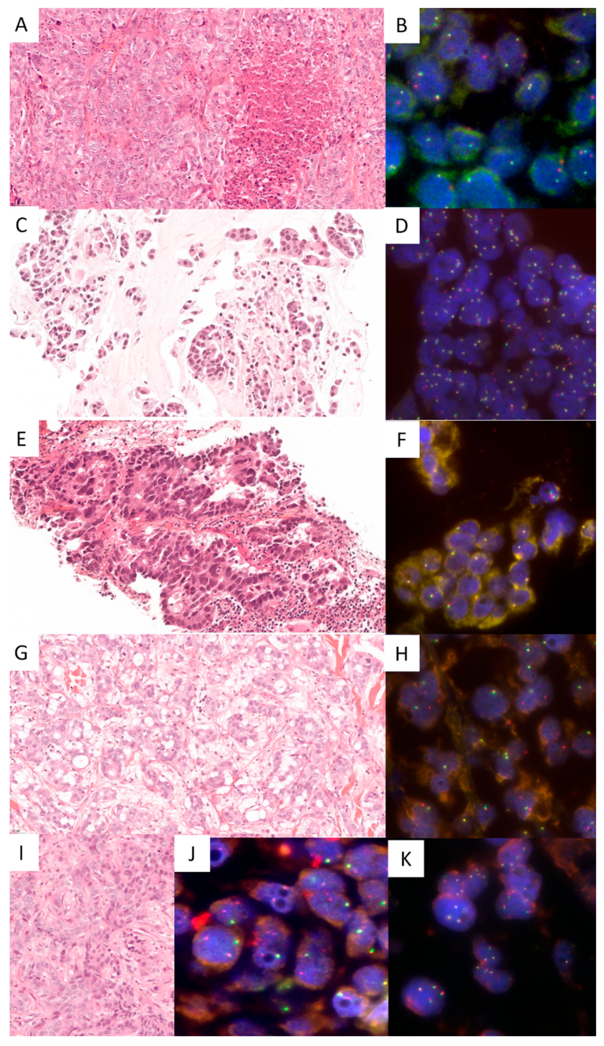
Figure 1. Histopathological and NRG1 fluorescent in situ hybridization (FISH) images of tumors with NRG1 fusions (Lung #1: (A,B); Lung #2: (C,D); Lung #3: (E,F); Lung #4: (G,H); Pancreas #1: (I,J); example of NRG1 FISH-negative result in a RNAseq negative pancreatic tumor: (K); (A,C,E,G,I): hematoxylin eosin saffron, ×20 magnification; (B,D,F,H,J,K): ZytoLight SPEC NRG1 Dual Color Break Apart probe, DAPI counterstaining, ×100 magnification).
Cancers 17 02347 g001
Table 1. Summary of the main features of the two cases series.
Pancreatic Ductal Adenocarcinomasn = 199
Men112 (56.3%)
Women87 (43.7%)
Ages mean 68.4 years (range from 36 to 87 years)
Pancreatic biopsies95 (47.7%)
Pancreatic surgical specimens104 (52.3%)
Lung Adenocarcinomasn = 446
Men251 (56.3%)
Women195 (43.7%)
Agesmean 68.4 years (range from 36 to 87 years)
Lung samples304 (68.2%)
Nodal metastases39 (8.7%)
Other distant metastases103 (23.1%)
Table 2. Summary of clinical, pathological, and molecular data of the cases with NRG1 fusions.
CasesSex (M/F)Age Range (Years)Smoking HabitClinical Stage *Tumor Type and Immunohistochemistry ResultsNRG1 FISH Result **NRG1 RNA Seq Result
Lung #1F82NoIVBLung solid ADK TTF-1−50% single 3′NA
Lung #2M78YesIIALung invasive mucinous ADK TTF-1+70% single 3′NA
Lung #3M71YesIBLung acinar ADK TTF-1+70% single 3′NA
Lung #4M62NAIVBLung acinar ADK TTF-1−80% single 3′NA
Lung #5M85YesIVBLung acinar ADK TTF-1−60% single 3′CD74-NRG1
Lung #6F44NoIIIALung acinar ADK TTF-1+50% single 3′ADAM9-NRG1
Lung #7 M53NoIVBLung papillary ADK80% single 3′CD74-NRG1
Pancreas #1F40NAIVDuctal pancreatic ADK
TTF1−, CK7+,CK20+, CK19+, Bcl10−
80% single 3′ATP1B1-NRG1
Pancreas #2M50NAIVDuctal pancreatic ADK
TTF1−, CK7+,CK20+
60% single 3′CDH1-NRG1
M: male; F: female; * staging according to UICC TNM classification 8th edition; ** no split and no single 5′ signals were observed; FISH: fluorescent in situ hybridization; RNA seq: RNA sequencing; ADK: adenocarcinoma; NA: not available.
Disclaimer/Publisher’s Note: The statements, opinions and data contained in all publications are solely those of the individual author(s) and contributor(s) and not of MDPI and/or the editor(s). MDPI and/or the editor(s) disclaim responsibility for any injury to people or property resulting from any ideas, methods, instructions or products referred to in the content.