Review

Glioblastoma: Overview of Proteomic Investigations and Biobank Approaches for the Development of a Multidisciplinary Translational Network

by
Giusy Ciuffreda
1,†,
Sara Casati
2,3,†,
Francesca Brambilla
1,
Mauro Campello
4,
Valentina De Falco
2,5,
Dario Di Silvestre
1,
Antonio Frigeri
6,7,
Marco Locatelli
8,9,
Lorenzo Magrassi
10,
Andrea Salmaggi
11,
Marco Salvetti
12,13,
Francesco Signorelli
6,7,
Yvan Torrente
8,14,
Giuseppe Emanuele Umana
15,16,
Raffaello Viganò
1 and
Pietro Luigi Mauri
1,2,*
1
Institute of Biomedical Technologies National Research Council (ITB-CNR), 20054 Segrate, Italy
2
Institute of Endotypes in Oncology Metabolism and Immunology “G. Salvatore” (IEOMI-CNR), 80131 Naples, Italy
3
Common Service ELSI, BBMRI.it—Biobanche e Risorse Biomolecolari, 20126 Milan, Italy
4
Department of Neuroscience Great Metropolitan Hospital, 89124 Reggio Calabria, Italy
5
UniCamillus-Saint Camillus, International University of Health Sciences, 00131 Rome, Italy
6
Department of Translational Biomedicine and Neuroscience, School of Medicine, University of Bari Aldo Moro, 70124 Bari, Italy
7
Neurosurgery Unit, University Hospital Policlinico of Bari, School of Medicine, University of Bari Aldo Moro, 70124 Bari, Italy
8
Dino Ferrari Center, Department of Pathophysiology and Transplantation, University of Milan, 20122 Milan, Italy
9
Division of Neurosurgery, Fondazione IRCCS Ca’ Granda Ospedale Maggiore Policlinico, 20122 Milan, Italy
10
Neurosurgery, Dipartimento di Scienze Clinico-Chirurgiche e Pediatriche, Università degli Studi di Pavia, Fondazione IRCCS Policlinico S. Matteo, 27100 Pavia, Italy
11
Department of Neurosciences, Azienda Socio-Sanitaria Territoriale (ASST) di Lecco, Ospedale Alessandro Manzoni, 23900 Lecco, Italy
12
Department of Neuroscience, Mental Health and Sensory Organs (NESMOS), Centre for Experimental Neurological Therapies (CENTERS), Sapienza University of Rome, 00185 Rome, Italy
13
IRCCS Mediterranean Neurological Institute Neuromed, 86077 Pozzilli, Italy
14
Neurology Unit, Fondazione IRCCS Ca’ Granda Ospedale Maggiore Policlinico, 20122 Milan, Italy
15
Department of Medicine and Surgery, University of Enna “Kore”, 94100 Enna, Italy
16
Department of Neurosurgery, Trauma Center, Gamma Knife Center, Cannizzaro Hospital, 95126 Catania, Italy
*
Author to whom correspondence should be addressed.
These authors contributed equally to this work and share first authorship.
Cancers 2025, 17(13), 2151; https://doi.org/10.3390/cancers17132151
Submission received: 19 May 2025 / Revised: 17 June 2025 / Accepted: 24 June 2025 / Published: 26 June 2025
(This article belongs to the Section Cancer Therapy)

Simple Summary

Glioblastoma (GBM) is one of the most aggressive and hardest to treat forms of brain tumor. It is difficult to find effective therapies because of its high genetic and biological complexity. In recent years, several studies have applied innovative proteomic technologies to study the contribution of proteins to GBM to discover biomarkers that can be applied for innovative diagnostic and therapeutic strategies. The present review summarizes recent developments in this area and outlines the importance of bio-preservation in the collection and storage of biological specimens, which are an absolute requirement for translational proteomics studies. Collaboration between hospitals, biobanks, and research centers can accelerate the development of new biomarkers for diagnosis and treatment, with the aim of improving clinical management and finding more effective therapies for GBM patients.

Abstract

Glioblastoma is a highly aggressive, infiltrative brain tumor of the central nervous system (CNS). Its extensive molecular and biochemical heterogenicity hinders the identification of reliable biomarkers and therapeutic targets, thereby making prognosis and existing therapy ineffective. In recent years, breakthroughs in the use of proteomics on a range of biological samples, such as plasma, cerebrospinal fluid (CSF), tissues, brain cells, and exosomes, represent a potential improvement to GBM investigations. Mass spectrometry-based approaches represent an important technique in the characterization of the tumoral proteome, for the identification of differentially expressed proteins, and for studying altered molecular pathways involved in tumor stages. Proteomics studies advance our knowledge about GBM pathogenesis, the discovery of reliable diagnostic and prognostic markers, and therapeutic approaches, also. In this context, for the effective application of proteomics on GBM, it is mandatory to develop a translational network by integrating hospitals, biobanks, and research institutions into a single network, to enable a collaborative approach across disciplines, thereby enabling rapid translation to clinical application of new proteomic insights. Today, high-quality biobanks play a key role in enabling collaborative, ethically compliant research, supporting the effective application of proteomics in glioblastoma studies and the translation of discoveries into clinical practice. This review explores current trends in proteomics and GBM research, highlighting how leveraging biobank infrastructure and fostering institutional cooperation can drive the development of targeted pilot projects to enhance the impact and effectiveness of glioblastoma research.

1. Introduction

Gliomas are tumors arising from glial cells, which provide structural and metabolic support to neurons in the central nervous system, playing a crucial role in maintaining neural function and activity [1]. According to the WHO classification, gliomas are graded from I to IV based on their histopathological characteristics, reflecting increasing malignancy. Among them, glioblastoma multiforme represents the most aggressive and lethal form (WHO grade IV), accounting for 54% of all gliomas and 50.9% of malignant brain and CNS tumors [2]. Typically diagnosed in individuals between 55 and 80 years old [3], recent evidence suggests that GBM can also develop in children, adolescents, and young adults, although its incidence is lower in younger populations and decreases with decreasing age [4]. Despite intensive therapeutic strategies, including surgical resection, adjuvant radiotherapy, and chemotherapy with temozolomide, GBM remains largely incurable, with a median survival of only 15 months and a 5-year survival rate of 7.2% [5,6,7]. GBM is characterized by rapid proliferation, diffuse infiltration into surrounding brain tissue, and a highly heterogeneous molecular landscape, which contributes to its aggressive behavior and therapy resistance [1,8]. Molecular profiling studies, particularly from The Cancer Genome Atlas (TCGA), have identified key genetic alterations underlying GBM pathogenesis, including EGFR amplification, PTEN deletion, TP53 mutations [9], and IDH1/IDH2 alterations, which are associated with distinct molecular subtypes and clinical outcomes [10]. Epigenetically, O6-methylguanine-DNA methyltransferase (MGMT) promoter methylation is a key biomarker influencing the response to temozolomide (TMZ) chemotherapy, as its silencing prevents DNA repair, increasing susceptibility to alkylating agents [11,12,13]. Additionally, widespread DNA hypermethylation and histone modifications contribute to tumor plasticity and therapeutic resistance [14].
GBM is broadly classified into two major subtypes [15]. Primary GBM is most common in elderly patients (>60 years) and is characterized by EGFR amplification (40–50%), with a subset carrying the EGFRvIII mutation, PTEN loss, CDKN2A deletion, and TERT promoter mutations, which drive rapid proliferation and invasion [16,17,18,19,20]. Secondary GBM occurs more frequently in younger patients (<45 years) [21]. This subtype is strongly associated with IDH1/IDH2 mutations (>80%), which induce a distinct metabolic phenotype by altering α-ketoglutarate production and promoting DNA and histone hypermethylation (the glioma CpG island methylator phenotype, G-CIMP) [22]. Unlike primary GBM, EGFR amplification is rare, while TP53 mutations (65–80%), ATRX, and chromosome 19q loss are more prevalent [23]. The biological distinction between GBM subtypes impacts prognosis and therapy, with IDH-mutant (secondary) GBM showing better outcomes and greater chemosensitivity than IDH-wildtype (primary) GBM [24]. These molecular differences emphasize the need for personalized treatment strategies, and emerging proteomic studies aim to identify protein biomarkers that can differentiate these subtypes at a molecular and functional level. In addition to genetic alterations, GBM shows remarkable heterogeneity at the cellular, metabolic, and microenvironmental levels, which significantly contribute to its aggressive phenotype. Glioblastoma stem-like cells (GSCs), expressing stemness markers (SOX2, Nestin, CD133), have been identified as a key driver of tumor recurrence due to their self-renewal capacity and resistance to conventional therapies largely mediated by Wnt, Notch, and Hedgehog signaling pathways [25]. The hypoxic microenvironment of GBM further promotes tumor progression through hypoxia-inducible factor 1-alpha (HIF-1α) activation and vascular endothelial growth factor (VEGF)-driven angiogenesis, enhancing invasion and limiting therapeutic efficacy [26]. Moreover, GBM is characterized by a highly immunosuppressive environment, with mechanisms such as programmed death-ligand 1 (PD-L1) upregulation, transforming growth factor beta (TGF-β) secretion, and the recruitment of regulatory T cells (Tregs) and tumor-associated macrophages (TAMs), which collectively suppress antitumor immune responses and contribute to resistance to immunotherapy [27,28].
Several critical signaling pathways drive GBM pathogenesis and progression, making it one of the most molecularly complex tumors. The phosphoinositide 3-kinase/protein kinase B/mammalian target of rapamycin (PI3K/AKT/Mtor) pathway is frequently activated in GBM due to PTEN loss or EGFR mutations, promoting cell proliferation, survival, and resistance to apoptosis [29]. Similarly, mutations and amplifications in upstream regulators such as EGFR and platelet-derived growth factor receptor (PDGFR) activate the RAS/MAPK pathway, further enhancing tumor growth and invasion. Another hallmark alteration is the inactivation of p53 via mutations or MDM2 amplification, which allows GBM cells to evade apoptosis and sustain proliferation [30]. Furthermore, dysregulated Wnt signaling has been shown to contribute to glioblastoma stem cell maintenance and therapy resistance, reinforcing the tumor’s ability to persist despite aggressive treatment regimens [31,32]. Hypoxic regions within the tumor promote angiogenesis through VEGF signaling, supporting tumor growth and invasive potential [33,34]. Moreover, GBM cells themselves exhibit extreme invasiveness, allowing them to infiltrate surrounding brain tissue, making complete surgical resection nearly impossible [35]. Additionally, matrix metalloproteinases (MMPs) facilitate extracellular matrix degradation, further promoting tumor cell migration [36,37]. Another key challenge in GBM treatment is its ability to disrupt the blood–brain barrier (BBB), promoting tumor infiltration while simultaneously limiting drug delivery, thereby posing a major obstacle to effective pharmacological intervention [38]. This highly dynamic tumor ecosystem poses a significant challenge to treatment, requiring novel approaches that integrate multi-omics technologies to dissect GBM biology at different regulatory levels.
Although genomic and transcriptomic analyses have provided critical insights into the molecular classification of GBM, these approaches primarily capture static genetic alterations and fail to reflect dynamic molecular changes that occur during tumor progression and treatment. Proteomics has emerged as a powerful tool to address this limitation, allowing for the characterization of protein expression patterns, post-translational modifications, and protein–protein interactions that regulate key tumorigenic processes. Among the most relevant proteomic approaches in recent years in the study of brain tumors, especially glioblastoma, mass spectrometry has emerged as a crucial tool for analyzing the tumoral proteome. With advanced technologies like mass spectrometry, proteomic analysis has highlighted significant alterations in GBM tumors compared to normal brain tissue. Mass spectrometry-based proteomics has indeed allowed the identification of protein signatures associated with GBM pathogenesis, highlighting key alterations in metabolic enzymes, membrane proteins, and immune regulators [39]. Recent studies have demonstrated that GBM tumors exhibit significant overexpression of proteins involved in cellular movement, antigen presentation, and cell–cell signaling compared to normal brain tissue [17,39]. Furthermore, proteomic profiling has distinguished IDH1-mutant from IDH1-wildtype GBM by revealing specific metabolic protein alterations, such as increased expression of aldehyde dehydrogenase 1 family member A3 (ALDH1A3) and IDH1-R132H in IDH-mutant tumors, underscoring the relevance of metabolic reprogramming in gliomagenesis [40]. One of the most promising applications of proteomics lies in liquid biopsy, where the detection of extracellular vesicles (EVs), circulating tumor proteins, and cerebrospinal fluid biomarkers offer a minimally invasive strategy for GBM diagnosis and disease monitoring. Several studies have identified tumor-derived EVs carrying proteins such as CD9, tumor susceptibility gene 101 (TSG101), and heat shock protein 70 (HSP70) as potential biomarkers for GBM classification and prognosis [16,41]. These findings indicate that proteomics has the potential not only to enhance understanding of the pathophysiology of GBM, but also to improve therapeutic processes through biomarker validation and personalized medicine. Achievement of therapy resistance will require an integration of proteomic data with genomic, transcriptomic, and metabolomic approaches to identify the perturbed patient specific critical issues.
Notably, proteomics shares with radiomics the ambitious objective of serving as a diagnostic alternative to histological sampling, an approach of particular relevance for tumors located in eloquent regions such as the brainstem.
This review is focused on current trends in the field of proteomics and GBM research to analyze the role of proteomics in the development of key biomarkers, molecular pathways, and novel therapeutics that can enhance clinical management and improve patient health. It is pursued as part of a broader effort to consolidate the Italian translational network of glioblastoma which includes hospitals, biobanks, and research centers. The objective is to incorporate proteomic evaluation into the framework of translational research, using the infrastructure of high-quality biobanks and fostering a cooperative approach that meets European and international standards. In particular, the network provides the tools to facilitate sharing of biospecimens and associated information, their collection and storage standardization, and the development of focused pilot projects, thus helping to integrate and improve the effectiveness of glioblastoma research.
Building on this infrastructure, the future employment of AI techniques may further enhance the interpretation of proteomic data, with the ultimate goal of achieving a cure for glioblastoma [42].

2. GBM Incidence

In terms of GBM incidence, there are substantial variations in the incidence of glioblastoma both at the global level and in specific countries [43,44]. This fact is due to a multitude of combined factors such as genetics, environment, and socioeconomic regions. The National Cancer Institute (NCI, https://www.cancer.gov/, accessed on 24 March 2025) reported that the incidence rate of glioblastoma, approximately 3 to 4 cases per 100,000 people per year, is relatively consistent worldwide. In the United States, the incidence is around 3.3 cases for every 100,000 people (4.07 for males and 2.58 for females), based on a study conducted during the years of 2017 and 2021 [45]. Current statistics from the Istituto Superiore di Sanità (ISS, https://www.iss.it/, Italy, accessed on 24 March 2025) suggest that the incidence of GBM in Italy is approximately 3.5 cases per 100,000 people, a statistic largely supported by the most recent data available [46] (Figure 1).
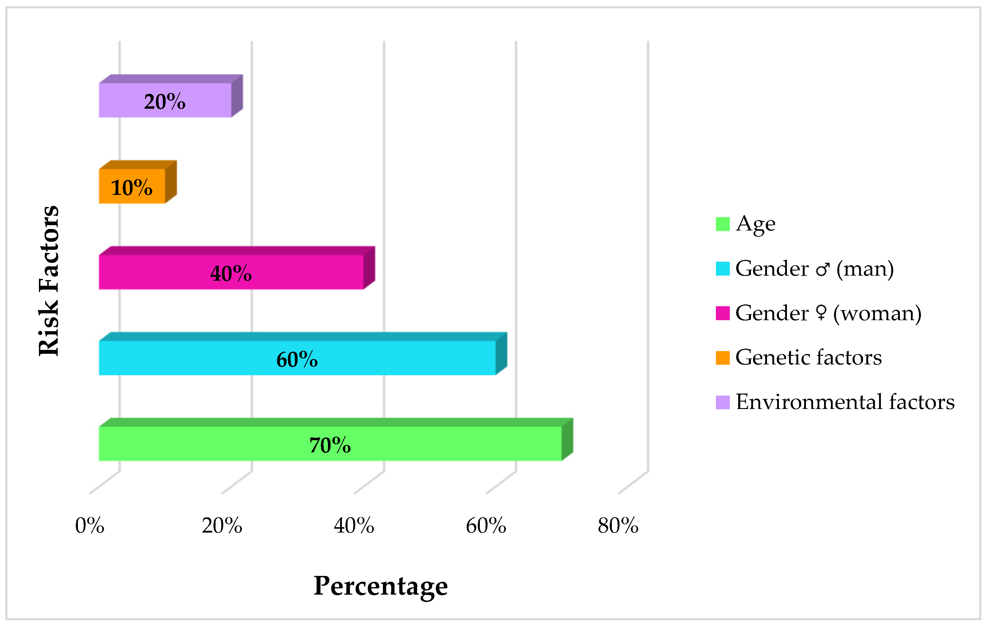
The incidence of glioblastoma has several risk factors associated with it (Figure 2). Firstly, age is an important factor: the incidence is low and increases significantly between the ages of 75 and 84 [47]. This increase is associated with changes in the immune system and inflammatory processes associated with the promotion of tumors in older people. Indeed, aging is accompanied by greater central nervous system inflammation and increased immunosuppressive TGFβ and interleukin 10 (IL-10) factors that impair the immune system and can cause a tumor onset [47].
Moreover, there is also a difference in GBM incidence in relation to sex: men have a greater burden than women (1.6:1) and, possibly, a worse prognosis [48]. These differences make male astrocytes more susceptible to aggressive transformation, characterized by loss of the tumor protein p53 (TP53) tumor suppressor gene and downregulation of RB1 expression, leading to the acquisition of a stem cell-like phenotype with high proliferation and renewal rates [49,50]. In contrast, female cells show higher cyclin-dependent kinase inhibitor 1A (CDKN1A) expression, which helps preserve cell cycle regulation despite TP53 mutations, thus contributing to greater cellular protection [51]. The differences between males and females could also be ascribed to the X chromosome via KDM6A, a tumor suppressor gene which is known to be more expressed in female cells [52].
Environmental aspects, like ionizing radiation from cancer treatments or toxic exposures like diesel smoke and spray paints, are also linked to greater risk, particularly in early age exposure [53,54,55].
In addition, familial history is reported to contribute in 5–10% of glioma cases [56]. The first-degree relatives of glioma patients carry twofold risks of succumbing to brain tumors, particularly if the illness is diagnosed during early years [57]. Inherited Li-Fraumeni syndrome, Turcot syndrome, and Neurofibromatosis type-1 increase risk of GBM and demonstrate how genetic changes in oncogenes and oncosuppressors are plausible mediators of gliomagenesis [47]. Children born to elderly parents or those with certain congenital central nervous system disabilities also have an increased susceptibility to astrocytoma [58,59].
Figure 2. Risk factors for glioblastoma. Stacked bar chart depicting the percentage distribution of age [47], sex [48], environmental exposure [53,54,55], and family history [56] among patients diagnosed with glioblastoma. Note that the risk factors are not mutually exclusive. Data synthesized from studies.
Cancers 17 02151 g002

3. Proteomics Analysis in Glioblastoma Disease

After emphasis on epidemiology and the major risk factors of glioblastoma, it becomes relevant to detail the various types of biological specimens for proteomes of tumors. Concerning biomarker discovery on Glioblastoma, about 8000 articles can be found on PubMed, including 5500 between 2014 and 2024, spanning a wide range of omics approaches for biomarker identification. Omics approaches have allowed the exploration of different aspects of glioblastoma, from the discovery of relevant genetic mutations to the analysis of differential protein profiles and the characterization of altered metabolites.
Genomics studies represent a significant component of the glioblastoma literature (Figure 3) due to their ability to identify genetic mutations, epigenetic alterations, and changes in gene expression that influence tumor progression. The comparison shown in Figure 3 is based on PubMed searches using the keywords ‘Glioblastoma Proteomics Biomarker’, ‘Glioblastoma Genomics Biomarker’, and ‘Glioblastoma Metabolomics Biomarker’.
Among the various omics approaches, proteomics has become increasingly prominent in glioblastoma research. Approximately 80% of proteomics studies on glioblastoma have been published in the last ten years, with the majority utilizing liquid chromatography coupled with high-resolution tandem mass spectrometry (LC-MS/MS), highlighting the growing interest in this technique. In total, 300 articles focused on the application of proteomics were identified, reflecting the growing interest in these approaches. This approach has shown promise for the identification of novel biomarkers and brought about more detailed understanding of the molecular complexity of glioblastoma.
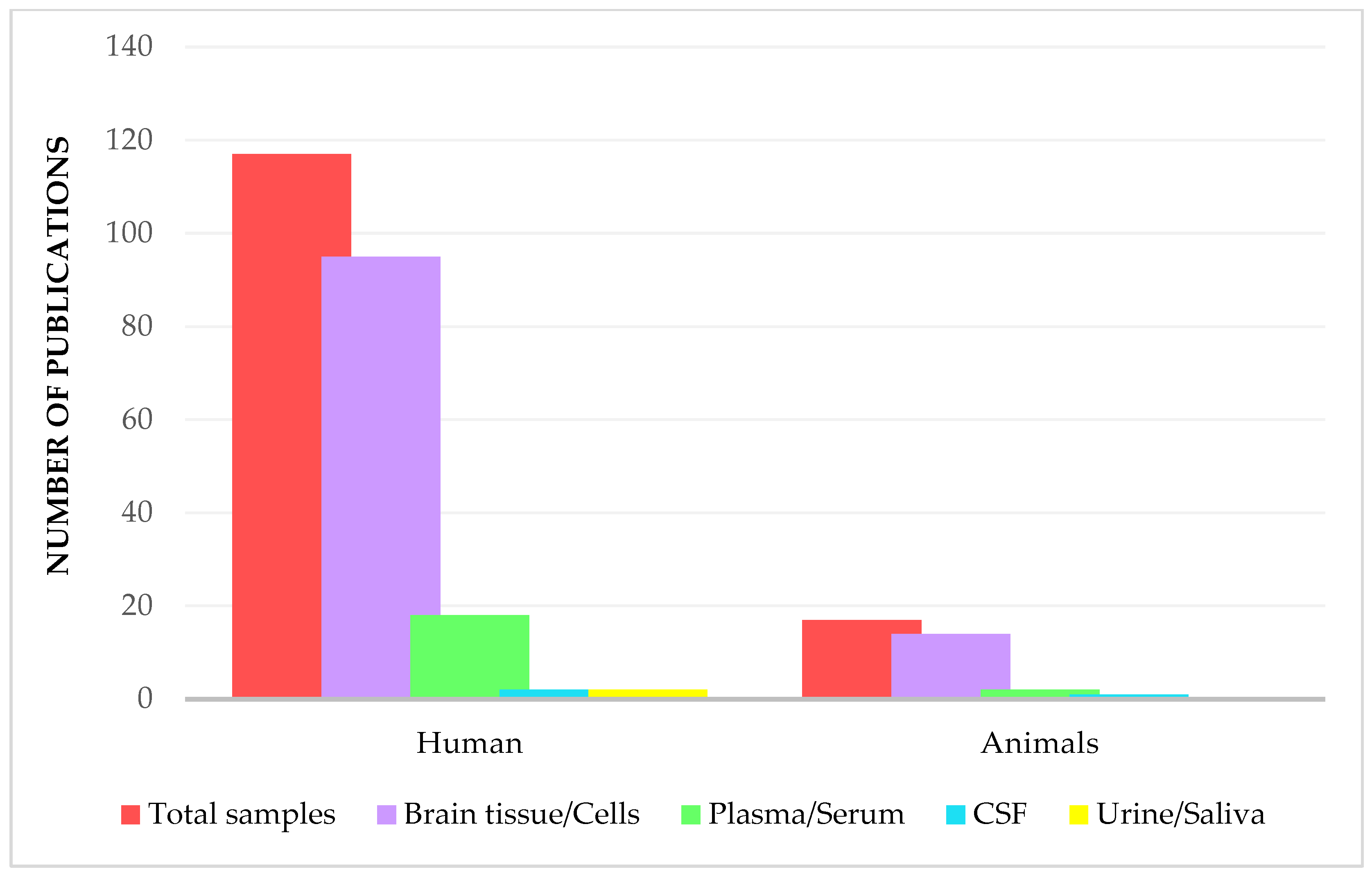
In the following sections we will review the main proteomics studies, according to the type of sample analyzed. Notable proteomics investigations on GBM biomarkers were focused on human samples mainly. Importantly, most studies focus on brain tissue, which is considered the main site for molecular characterization of glioblastoma. However, analysis of biofluids such as CSF, plasma, serum, urine, and saliva offers complementary information on proteomic alterations associated with GBM disease (Figure 4). The distribution illustrated in Figure 4 was derived through a meticulous manual curation of PubMed articles, initially identified using broad and specific keyword combinations, including ‘Glioblastoma’, ‘Proteomics’, ‘Biomarker’, and ‘Glioblastoma Proteomics (mass spectrometry)’. Each study was then carefully classified by sample type and species, based on a thorough review rather than just keyword filtering.
In the following sections, we will explore how various types of samples—from brain tissue to biofluids—contribute to our understanding of GBM.

3.1. Brain Tissue and Related Cell Lines

The proteomics analysis of glioblastoma is mostly applied to the brain tissue samples, which are the primary biological material for the evaluation of the involved proteins for in this highly malignant neoplasm. The differences and types of tissues collected, whether they are fresh or formalin-fixed, influence the precision of the analyses and affect the biomedical detection and the understanding of the molecular pathobiology of the disease. Besides tissue samples, brain cell lines are repeatedly employed as in vitro models for the analysis of proteomes of glioblastoma. The primary cell cultures are an acceptable, albeit incomplete, way to examine the tumor’s molecular mechanisms in an orchestrated environment that is devoid of the actual complexity and heterogenecity of the tumor.
Table 1 presents the bibliographic quotes of the main publications from the last ten years, indicative of the most notable candidate protein biomarkers, type of samples and/or cell culture used, and the proteomics techniques applied. These protein biomarkers are mostly considered candidate biomarkers identified through proteomic analyses; however, their clinical validation status varies across studies.
Of note, the identified proteins may occur in GBM in the form of different proteoforms, thus requiring further studies for their in-depth characterization.
In recent years, LC-MS/MS has become indispensable to study the glioblastoma proteome, allowing the discovery of proteins that are crucial in tumor progression and therapeutic resistance.
Leveraging LC-MS/MS combined with Data-Independent Acquisition (DIA) across multiple studies, researchers have uncovered key proteins and regulatory mechanisms involved in glioblastoma progression and therapeutic resistance. For example, El-Baba et al. [83] pinpointed tumorigenic proteins, including solute carrier family 2 member 1 (SLC2A1), alongside inhibitory genes like phosphatidylethanolamine-binding protein 1 (PEBP1), emphasizing their influence on cell division and nutrient uptake, vital actions in aggressive brain tumors. Naryzhny et al. [85] identified numerous common proteins in various glioblastoma cell lines, such as annexin A1 (ANXA1), ANXA2, and vimentin (VIME), which play a role in several biological processes essential for tumor progression, including exosome formation, cell adhesion, modulation of the immune response, and various metabolic mechanisms. These proteins are also involved in radioresistance and drug resistance, key phenomena for the survival of tumor cells during radiation therapy. In particular, ANXA1 and VIME, involved in invasiveness and cell adhesion, contribute to glioblastoma’s ability to resist the damaging effects of radiotherapy.
Through proteomic profiling, Azzalin et al. [114] demonstrated that glioblastoma cells, specifically the U-87 MG glioblastoma cell line and other glioma cultures, respond to glucose deprivation by upregulating SHC-transforming protein 3 (SHC3), a neuronal adaptor protein with roles in signal transduction and vesicular trafficking. Elevated SHC3 levels enhanced glucose uptake by promoting the translocation of GLUT/SLC2A transporters to the plasma membrane, sustaining the high glycolytic activity typical of glioblastoma. SHC3-mediated inhibition of poly (ADP-ribose) polymerase 1 (PARP1) was linked to altered trafficking and glycosylation of glucose transporters, indicating a regulatory axis that modulates metabolic adaptation under nutrient stress. These findings identify SHC3 and PARP1 as potential metabolic biomarkers in glioblastoma.
Another study by Hu et al. [86] identified differentially expressed proteins between different glioblastoma cell lines, highlighting the role of proteins such as RRAS, protein tyrosine phosphatase receptor type O (PTPRO), and those involved in the phosphoinositide 3-kinase/protein kinase B (PI3K/AKT) and nuclear factor kappa-light-chain-enhancer of activated B cells (NF-κB) pathways, all crucial for regulating cellular processes that promote tumor progression. Moreover, the study showed how the integration of these pathways influences interactions with the extracellular matrix (ECM), a key element in glioblastoma invasiveness [86]. Menezes et al. [87], focused on biomarkers such as Decorin (DCN) and Glypican-1 (GPC-1), both involved in angiogenesis and tumor growth. The study revealed that the inhibition of histone deacetylases (HDAC) alters the so-called AngioMatrix, a set of proteins and molecules that regulate the tumor’s vascular environment, significantly influencing angiogenesis, cell motility, and tumor progression. DCN, in particular, was identified as a key prognostic biomarker, suggesting that its expression could be correlated with the severity and evolution of glioblastoma.
Another crucial aspect concerns resistance to treatments, particularly TMZ, a drug commonly used to treat glioblastoma. In this regard, Yi et al. [91] identified proteins involved in resistance to TMZ, playing roles in various signaling pathways such as actin cytoskeleton regulation, the PI3K-Akt pathway, and focal adhesion and phagosome signaling. Proteins like DEAH-box helicase 9 (DHX9), heterogeneous nuclear ribonucleoprotein R (HNRNPR), and ribosomal protein L3 (RPL3) influence these processes, altering the ability of tumor cells to adapt and survive under treatment. Focal adhesion pathways are crucial in tumor invasiveness, facilitating cell migration and spread, but also determining the response to chemotherapy. Naryzhny et al. [115] have shown that GBM resistant to standard therapies, such as TMZ, transition to a neuronal-like state. This phenotypic change is associated with multilevel activation of the RAS-mitogen-activated protein kinase (RAS-MAPK) signaling pathway, driven by increased or hyperphosphorylation of key components such as RAS, BRAF, and MAPK.
In parallel, Mallawaaratchy et al. [96,97] focused on extracellular vesicles, identifying proteins such as ANXA1 and Integrin beta-1 (ITGB1) as biomarkers in glioblastoma cell invasiveness. These proteins play not only a role in invasiveness, but they are also involved in processes like actin polymerization and endosomal sorting, essential for invadopodia formation, specialized cellular structures involved in tumor cell migration and invasion. In particular, ANXA1 is known for its role in regulating the cell membrane and modulating inflammatory responses, while ITGB1 is involved in cell adhesion and interactions with the extracellular matrix.
Bijnsdorp et al. [101] analyzed the U87WT (wild-type) and U87vIII (mutant) cell lines, revealing key mechanisms underlying tumor aggressiveness and treatment response. The results highlighted different phosphorylation activities and kinase signaling pathways, such as EGFR, CDK1/2/7, GSK3B, AKT1, MAPK1/3, MET, PAK2/4, and PRKCA/B. In particular, EVs from U87vIII cells (mutant for EGFR) carry active EGFR and other kinases, suggesting that EVs may act as signaling vehicles for transmitting signals between tumor cells and the surrounding microenvironment. Xue et al. [70] analyzed glioblastoma tumor tissues, identifying numerous differentially expressed proteins, including key factors such as PP1γ, YAP1, and SOX2, which emerged as particularly relevant for glioma progression. The study revealed that the Hippo signaling pathway, essential for regulating cell growth and proliferation, is activated in glioblastoma. In particular, PP1γ promotes the nuclear translocation of YAP1, a key event for its oncogenic activity and glioma progression and poor prognosis. Jeon et al. [71] explored biomarkers associated with responses to anti-angiogenic therapies in glioblastoma, identifying key proteins such as TMEM173 and FADD, which regulate immune and apoptotic processes. Additionally, ERCC2 and POLD1, proteins involved in DNA repair mechanisms, were associated with poorer prognosis in glioblastoma patients, suggesting that ERCC2 and POLD1 play a crucial role in the response to DNA damage.
An additional advancement in proteomic technology is SWATH-MS (Sequential Window Acquisition of All Theoretical Mass Spectra). Maire et al. [82] identified 104 differentially expressed proteins in extracellular vesicles derived from glioblastoma cells. Among these, CD44, Integrin-β1, and Tetraspanin-14 were highlighted for their involvement in key oncogenic pathways such as PI3K/AKT and MAPK. CD44 promotes cell adhesion and invasiveness, Integrin-β1 regulates migration and focal adhesion, and Tetraspanin-14 contributes to cytoskeletal remodeling and immune response. These proteins also play roles in DNA repair and apoptosis, ultimately supporting tumor survival and progression. In another study, Schulze et al. [89] analyzed U87 and U251 cell lines, identifying biomarkers such as DAB1 and RELN, crucial for cell migration and survival. RELN, overexpressed in the neural subtype of glioblastoma, is associated with better survival but is often silenced by methylation. The interaction between DAB1 and RELN was seen as crucial for limiting glioma progression. DAB1 tumor-suppressive effect suggests that enhancing its expression or activating the RELN-DAB1 pathway could reduce tumor aggressiveness and improve treatment response.
Zheng et al. [65] analyzed radioresistant glioblastoma cell lines (U251, U251R, T98G) and xenografted tumors using quantitative proteomics with Tandem Mass Tag (TMT), identifying 17 upregulated proteins, among which SDC1 and TGM2 were found to be crucial for radioresistance and poor prognosis. Both proteins support mechanisms that increase treatment resistance and worsen prognosis.
Another study by Cosenza-Contrares et al. [60] analyzed initial and recurrent glioblastoma samples, identifying ASAH1 and GPNMB as key regulators. ASAH1 is crucial for sphingosine metabolism and interaction with tumor-associated macrophages, while GPNMB modulates the extracellular matrix, promoting tumor invasiveness. This study highlighted the role of immune pathways and signaling through interleukins [62]. Oh et al. [63] conducted a proteomic analysis on various glioblastoma subtypes, revealing important metabolic vulnerabilities. Among the key proteins, PHGDH stands out for its ability to support cell proliferation under hypoxic conditions.
Jang et al. [69] highlighted biological differences between male and female glioblastoma patients, noting distinctions in tumor mechanisms. In males, EGFR receptor hyperactivation was observed, associated with more aggressive progression, along with specific biomarkers such as COL28A1 and EDNRB; in females, the protein SPP1 (Osteopontin) was crucial in microenvironmental interactions that favor tumor growth.
Finally, Nikitina et al. [116] examined glioma cell lines (DBTRG-05MG), showing that type I interferon and VSV infections influence the cellular response through EGFR and HER2-mediated pathways. Additionally, the EGFR inhibitor gefitinib showed a synergistic effect with interferon signaling, suggesting potential strategies for antiviral and immunological therapies.

3.2. Biofluids

Biofluids and extracellular vesicles have attracted increasing attention as potential sources of biomarkers in GBM. Various biofluids, such as CSF, the fluid fraction obtained through ultrasonic aspiration of brain tumor tissue during neurosurgery (CUSA), serum, and plasma, have been used in proteomic analyses to identify tumor-associated protein markers [117,118,119]. While each of these fluids offers valuable insights into tumor biology, they also come with limitations. For instance, collecting CSF or CUSA fluid is highly invasive and not feasible for routine use. Conversely, peripheral blood is easily accessible but contains a complex mix of molecular components, which can hinder the detection of tumor-specific biomarkers [120]. Table 2 presents a summary of biofluids investigated in proteomic research and the corresponding candidate biomarkers.

3.2.1. Cerebrospinal Fluid

Cerebrospinal fluid is ideal for liquid biopsy in brain tumors because it directly contacts the tumor environment and bypasses the BBB [41], making it less invasive than tissue biopsy [130].
Schmid et al. [121] analyzed the CSF proteome of glioblastoma patients using LC-MS/MS with Data-Dependent Acquisition (DDA) to identify differentially expressed proteins (DEPs). In total, 349 proteins were identified, with more evident alterations in patients with BBB disruption; among these, CHI3L1 and GFAP emerged as promising biomarkers. CHI3L1 was previously associated with neurological diseases such as multiple sclerosis, Alzheimer’s disease [131], and systemic cancers [132]. Additionally, CHI3L1 contributes to tumor aggressiveness by binding to the interleukin-13 receptor (IL13RA2) and inducing a mesenchymal phenotype [133]. GFAP, on the other hand, proved to be a relevant diagnostic biomarker, with higher levels in the CSF of glioblastoma patients compared to other brain tumors and associated with reduced survival [134,135,136].
Of particular interest, CSF proteomic analysis identified two clusters of patients with distinct biological characteristics. The first cluster showed an upregulation of proteins involved in coagulation, such as fibrin (FGA, FGB, FGG) and thrombin (F2), which have previously been associated with the aggressiveness of glioblastoma [121]. This cluster is also characterized by the activation of the “LXR/RXR Activation” pathway, which could represent a therapeutic target. Conversely, the second cluster displays less aggressive features, with pathways associated with synaptogenesis and immunomodulation [41,137,138,139].
A further study by Mikolajewicz et al. [41] detected 755 proteins including GAP43 as a specific biomarker for glioblastoma, while other proteins, such as SERPIN3 and APOE, are involved in pathways related to BBB degradation, angiogenesis, and stemness.
A complementary investigation by Magrassi et al. [122] explored the proteomic composition of cystic fluids from various brain tumors, including secretory meningiomas, cystic schwannomas, and high-grade gliomas. Their study highlights that plasma proteins, such as albumin, haptoglobin, fibrinogen, and transferrin, which leak from disrupted BBB, constitute a significant portion of the cystic fluid proteome and are abundant across all tumor types. Additionally, proteins derived from cerebrospinal fluid, such as prostaglandin D2 synthase, were identified, suggesting a mixed origin of cystic fluid from both plasma and CSF. These findings underscore the potential of cystic fluid proteomics to uncover novel biomarkers and therapeutic targets in brain tumors. Looking ahead, these proteomic signatures could pave the way for minimally invasive diagnostic tools, such as liquid biopsies, which would facilitate earlier detection and personalized treatment strategies.

3.2.2. Plasma

Plasma, in particular, has emerged as a matrix of great interest for proteomic research due to its accessibility and its ability to reflect the physiological and pathological state of the patient. However, the presence of abundant proteins such as albumin and immunoglobulin can obscure low-abundance biomarkers, requiring advanced fractionation and protein depletion techniques to improve the sensitivity of analyses.
The study by Cosenza-Contreras et al. [60] analyzed the proteomic profiles of tumor samples from patients with initial (iGBM) and recurrent (rGBM) glioblastomas. Among the identified proteins, ASAH1, SYNM, and GPNMB were found to be significantly upregulated in rGBM. In particular, ASAH1 was associated with increased neutrophil involvement in the tumor microenvironment, while SYNM was expressed in early tumors compared with healthy controls [27].
Research by Sabbagh et al. [124] placed attention on von Willebrand factor (VWF), identifying it as a possible specific biomarker in plasma EVs of GBM patients, as it may be involved in pro-angiogenic processes and tumor vascularization. The enrichment of VWF in plasma-derived EVs of GBM patients could reflect its participation in the tumor-typical angiogenic and pro-thrombotic response. Finally, the study by Naryzhny et al. [123] investigated plasma proteoforms of haptoglobin (Hp), a key protein in hemoglobin binding, oxidative stress protection, and inflammation control. Hp is an active component of plasma and is involved in numerous processes that are fundamental to homeostasis in the human body. In particular, it binds free hemoglobin (Hb), protecting tissues from oxidative damage and contributing to the regulation of inflammation. Recently, an unprocessed form of Hp, namely zonulin, has attracted attention for its potential role as a biomarker [140]. As an acute phase protein, Hp tends to increase in response to stress conditions. These preliminary results suggest that increased Hp levels could be considered a nonspecific biomarker of GBM [141]. Zonulin, on the other hand, has been identified exclusively in the plasma of GBM patients, suggesting a possible role in tumor progression, particularly in the processes of invasiveness and vascularization. These data suggest that the set of Hp proteoforms could be employed as a panel of biomarkers for GBM: levels of α and β chains could indicate the presence of neoplasia in a nonspecific manner, while zonulin could be a specific marker for GBM [123,140,141,142].

3.2.3. Serum

In a study conducted by Popescu et al. [125] serum samples from 35 patients (14 women and 21 men) with stage IV glioblastoma multiforme were analyzed and compared with 30 healthy controls, identifying CXCL4, S100A8, and S100A9. Although these proteins have previously been associated with various types of cancer [143], they are also known as inflammatory factors [144,145].
Of particular interest is the study conducted by Clavreul et al. [146] which identified the differences in protein abundance in tumor and serum samples from patients with IDH-wildtype glioblastoma, distinguishing between short-term survivors (STS) and long-term survivors (LTS) [147,148,149]. Three tumor proteins (AHSP, FABP7, and TJAP1) were downregulated in the STS group and were not identified in the serum proteome of patients, while 26 serum proteins were upregulated in the STS group; of these, 23 proteins were also identified in the tumor proteome, but were expressed similarly in both the STS and LTS groups. Analysis of the three tumor specific proteins of interest indicated that they were associated with fatty acid transport, Golgi organization, and hemoglobin metabolic processes, respectively. The 26 serum proteins of interest were associated with different biological processes: cellular detoxification of oxidants, cellular homeostasis, regulation of reactive oxygen species metabolism, aging, purine ribonucleotide metabolism, generation of precursor metabolites, VEGFA-VEGFR2 signaling, and IL-18 signaling. In particular, in serum, two proteins were of particular interest: MDH1 and RNH1. MDH1, a central enzyme in metabolic processes such as glycolysis and glutaminolysis, was upregulated in STS patients. This protein could support tumor metabolic reprogramming by reducing oxidative stress and contributing to the Warburg effect [146].
Several studies have isolated Glioma-Associated Stromal Cells (GASCs) from the peritumoral microenvironment of glioblastoma, which had phenotypic and functional properties similar to those of mesenchymal stem cells and CAF [146,150]. These GASCs, which have prognostic value in glioma, can undergo metabolic reprogramming and induce metabolic reprogramming in glioblastoma cells via MDH1.
Kun et al. [127] demonstrated that high expression levels of five driver genes, including RNH1, were associated with poor prognosis in glioblastoma patients. High levels of RNH1 in STS serum could therefore arise from high production in glioblastoma cells to reduce ROS production, as hypothesized for MDH1.

3.2.4. Urine and Saliva

Urine, with its relative simplicity and high sensitivity of analytical techniques, can be an effective “liquid biopsy” for continuous GBM monitoring. Saliva, on the other hand, with its disease-influenced dynamic composition, emerges as an equally promising alternative, particularly for non-invasive monitoring of progression and response to treatment. However, further studies in larger cohorts are needed to validate these approaches and establish their clinical applicability on a wider scale. Urine and saliva can be collected easily, rapidly, and non-invasively, allowing for repeated analysis over time without stressing the patient. A particularly interesting aspect concerns urinary extracellular vesicles (uEV), which contain a wide range of proteins able to reflect tumor burden and GBM progression. The use of advanced mass spectrometry techniques allowed precise quantification of proteins in uEV, improving sensitivity and accuracy compared to traditional methods [128].
An interesting study by Hallal et al. [128] identified two key biomarkers, progranulin (GRN) and prosaposin (PSAP), both associated with tumor recurrence and treatment resistance, through LC-MS/MS with DIA. In particular, proteomic analysis of uEV showed that GRN and PSAP differ in the various stages of the disease (pre-operative, post-operative, and recurrence), highlighting their potential in the surveillance of progression and therapeutic response [117]. Other biomarkers such as ALDOA and S100-A11 were confirmed in uEV and other body fluids, suggesting the potential of urine as a diagnostic source for disease monitoring. uEV also presents proteins associated with treatment resistance, such as ITM2B, which is identified as a significant biomarker for GBM recurrence [151]. Another interesting group of proteins includes the subunits of the TRiC complex (TCP1, CCT2, CCT3, CCT4, CCT6A, CCT7, and CCT8), which were found in significantly increased amounts in preoperative samples from GBM patients [117,118].
Saliva has also emerged as an interesting source of biomarkers for GBM, especially due to the presence of small EVs that reflect the phenotypic composition of tumor cells [152,153,154,155]. A detailed proteomic analysis was performed by Müller et al. [129], which allowed for the identification of 507 proteins in salivary EVs from GBM patients. Of these, 238 proteins were present exclusively in preoperative samples, 215 proteins were detected both before and after treatment, and 54 were found only in postoperative samples. These data suggest a significant heterogeneity in the proteomic content of salivary EVs, which could be used to differentiate disease stages and monitor treatment efficacy [153]. Analysis of DEPs (also referred to as DAPs—differentially abundant proteins) revealed that some proteins, such as ALDOA, 1433E, and TM11B, are associated with unfavorable prognostic outcomes. ALDOA, an enzyme involved in glycolysis, was linked to cellular proliferation and treatment resistance in various cancers, suggesting that its overexpression in GBM cells could indicate a negative prognosis [156,157]. Similarly, the protein 1433E (YWHAE) is crucial for cell cycle regulation and signaling pathways and has been associated with astrocytomas [156,158,159]. The analysis also highlighted the overregulation of proteins such as C3, a complement system component, and PPIA, involved in protein folding and intracellular trafficking, both central to tumor progression [160,161,162]. Unique proteins identified in salivary EVs, including immunoglobulins and proteins related to the TGF-β signaling pathway, strengthen the idea that saliva may contain exclusive biomarkers for GBM. Furthermore, the enriched molecular pathways, such as those associated with the immune system, complement cascade, and iron metabolism, indicate the key role of saliva in assessing tumor progression. These findings suggest that saliva could not only be used to diagnose GBM but also to monitor therapeutic responses and recurrence risk [129,153].

4. Proteomics and Biobanking as Cornerstones of Translational Medicine

To achieve high response rates and maximum reliability of results, the proteomics approach requires high-quality biological samples, collected and stored according to rigorous and standardized protocols. Biobanks offer this guarantee, ensuring the availability of well-characterized materials associated with accurate clinical data and metadata. However, to maximize the effectiveness and impact of research, it is essential to promote the creation of a translational proteomics network that includes biobanks with shared governance and uniform procedures for cataloging and storing samples. This coordinated infrastructure represents even more of a prerequisite for generating reliable and usable data, suitable for multicenter and integrated studies. Samples collected in structured biobanks reduce pre-analytical and technical variables, contributing to greater experimental robustness and reproducibility of results. Process standardization and complete traceability ensure consistency and quality, crucial elements for the validation of biomarkers and the development of new therapies. Furthermore, the desired integration of proteomic data with those from other omic sciences—genomics, metabolomics, transcriptomics—represents a fundamental step to build a solid translational network, capable of producing knowledge applicable to the development of innovative therapies for complex diseases, such as highly invasive tumors like GBMs.
The collection, storage, and use of human biological samples and related data represent the first and fundamental step in the path of translational research. In this context, it is essential to distinguish between different types of biological material storage according to the purpose of use. In a nutshell, a study-oriented collection has a specific purpose within a defined time frame, and a biorepository has a corporate purpose that often does not provide access to third parties; meanwhile, a research biobank guarantees both scientific future purposes and fair access. Below it is reported the definitions of these three main models based on the national and international consensus and regulatory frameworks.
  • “Study-oriented human biological material Collections”—Collection, storage, and use of human biological materials and related data finalized to a specific project, generally oriented by pathology according to research or clinical protocols and participant’s previous expressed consent. In the protocol as in the specific consent, the start and end of the collection and use are declared, at the end of which the samples must be destroyed or biobanked, based on the further consent to biobanking expressed [163].
  • “Biorepository”—A facility that collects, catalogs, and stores samples of biological material, such as urine, blood, tissue, cells, DNA, RNA, and protein, from humans, animals, or plants for laboratory research (https://www.cancer.gov/publications/dictionaries/cancer-terms/def/biorepository, accessed on 3 April 2025). In for-profit contexts, the biorepository is intended for exclusive, corporate use.
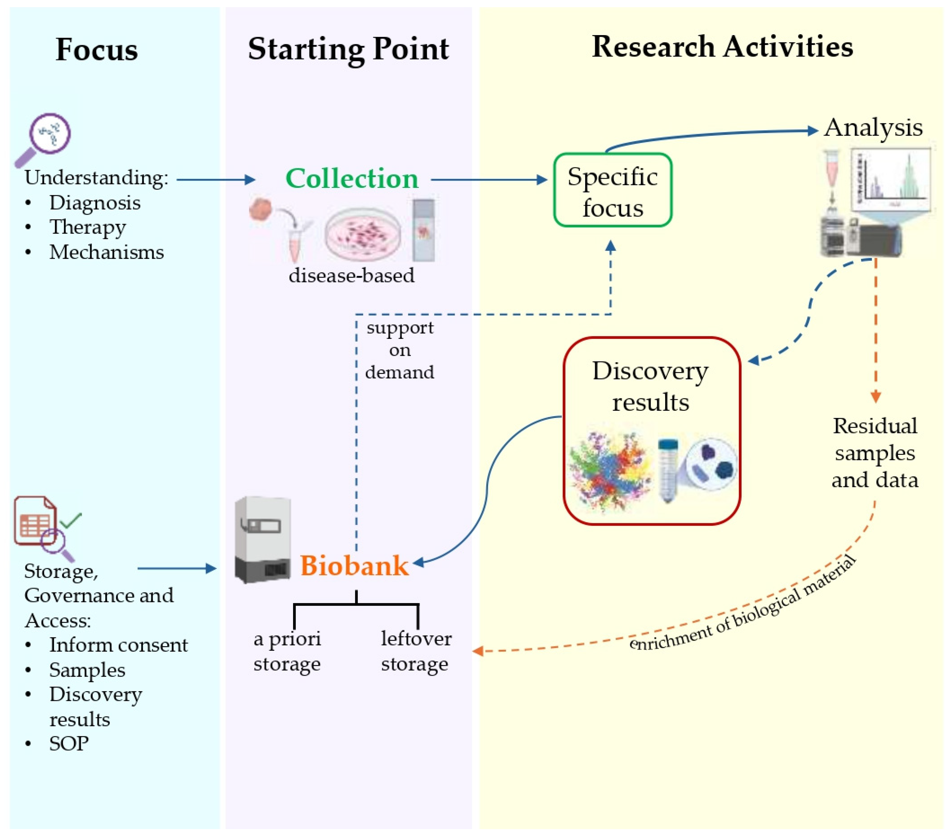
  • “Research Biobank”—A legal entity, or part of a legal entity, formally established at a public or private institution; a non-profit. The research biobank as a service structure, at the service of the scientific communities, is the guarantor of the principles, rights, and processes that constitute biobanking for future research purposes. In full compliance with the informed consent/assent to research biobanking expressed and the rights of the participants involved, the biobank guarantees and manages, according to proven quality standards, the stable and continuous collection, conservation, use, and access of human biological materials, and/or related and derived data, for research. The sharing of biobanked samples/data, as well as results, is the cornerstone of all the activity of a research biobank (Figure 5) [164,165,166,167].
Of note, older collection refers to organized human biological materials originally obtained for different purposes, and subsequently stored and used for research without the explicit consent of the participants, thus not complying with current ethical and legal requirements.
Over the past decades, increasing attention to ethical and legal principles has prompted a progressive shift from the use of historical collections, often lacking informed consent, to the development of structured and compliant approaches to the acquisition and management of biological materials. This evolution reflects the growing need for transparency, standardization, and participant protection, and has laid the foundation for modern biobanking practices based on rigorous scientific, ethical, and procedural frameworks.
Initially, it takes the form of a simple collection process, in which tissues, fluids or other biological materials are obtained from research participants or patients according to standardized protocols. However, this phase does not end with the mere act of collection: it requires a careful acquisition procedure to ensure the integrity of the biological material and its suitability for subsequent analysis.
With increasing complexity, the collection process becomes part of a broader methodological framework, in which validation, preservation, and traceability criteria assume a crucial role. Each sample is accompanied by detailed data, including clinical, molecular, and environmental information, essential to contextualize experimental results and ensure reproducibility.
At a more advanced level, sample collection evolves into biobanking, a highly regulated practice based on ethical, legal, and scientific principles as well as on IT-based anonymization/cryptography and harmonized quality standards.
Research biobanks are recognized as vital components of translational research infrastructure [168]. They play a fundamental role in collecting, processing, storing, using, and distributing biological samples and related data. Biobanks serve as the cornerstone of translational medicine, understood as an inter-disciplinary branch of the biomedical field supported by three main pillars: bench side, bedside, and community [169]. Reliable biological samples are crucial for confirming and validating both basic and preclinical research [170].
Biobanks, as guarantors of the principles, rights and processes, have a strategic position in promoting the reliability and reproducibility of future research data, as well as in supporting responsible research and innovation [171,172].
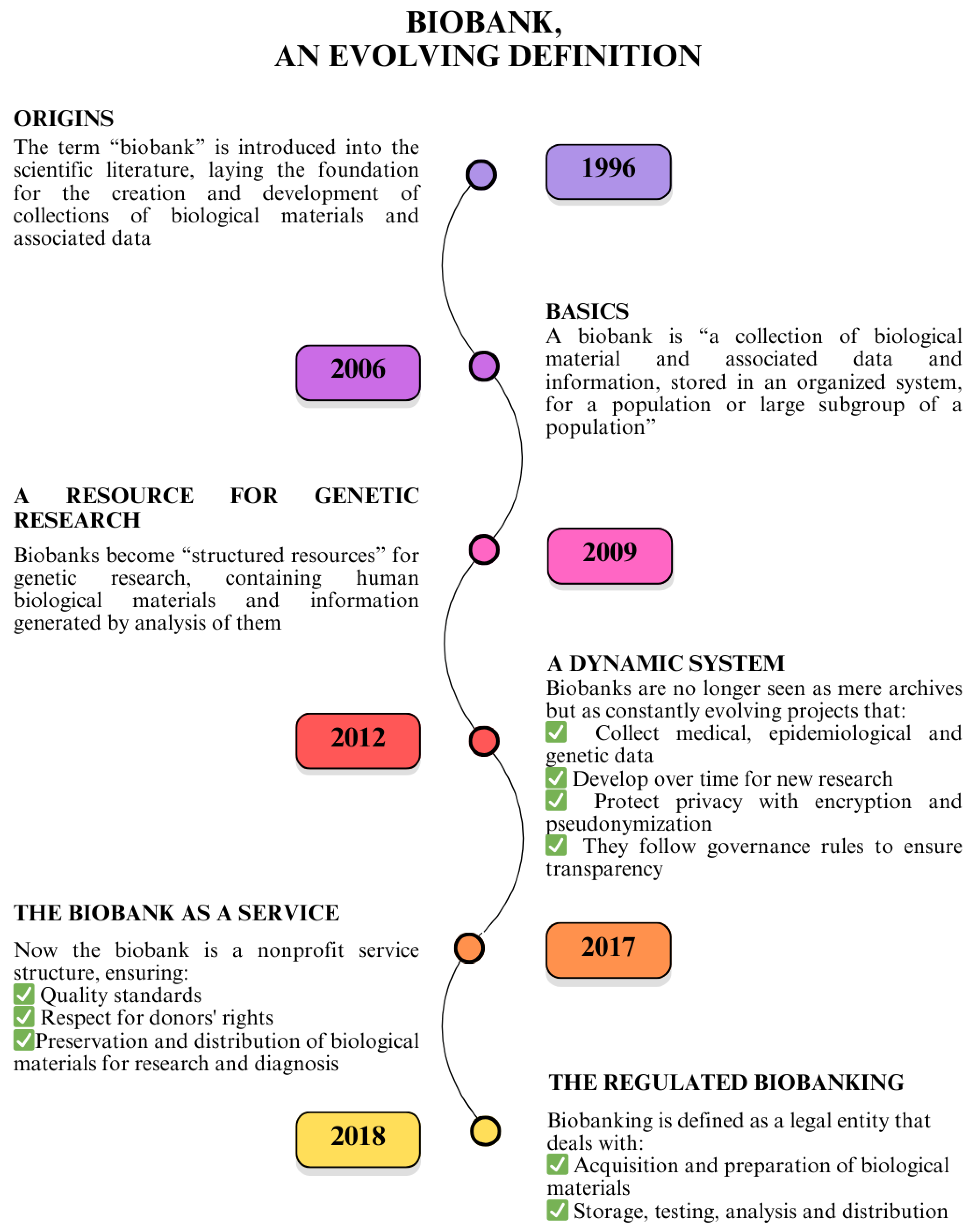
The term “biobank” first appeared in the literature in 1996 [173]. In 2009, the OECD proposed the following consensus definition of the biobank and genetic dataset: “structured resources that can be used for genetic research purposes and that include (a) human biological materials and/or information generated by the analysis thereof; and (b) associated extended information” [174]. This was in conjunction with the first steps of the Pan-European Research Infrastructure for Biobanking and Biomolecular Resources—BBMRI, which in 2013 was formally recognized as an European Research Infrastructure Consortium (ERIC), i.e., a permanent legal entity under European law (www.bbmri-eric.eu) (Figure 6).
Over the past 15 years, the ethical–regulatory framework, along with a global consensus on quality standards for research biobanks, has been consolidated through European and international soft laws, international standards, governmental bodies, and national and international infrastructures; for example, the International Guidelines for Health-related Research Involving Humans of the Council for International Organizations of Medical Sciences (CIOMS)-WHO, Guidelines 11 and 12, and other relevant frameworks [175].
In 2017, the Italian BBMRI community (https://www.bbmri.it/ accessed on 24 March 2025) helped define the role of the biobank as a custodian, as well as a third party, with the following consensus [176], to arrive in 2018 at a global International Organization for Standardization (ISO) standard specifically dedicated to biobanks and biobanking for research, ISO 20387:2018 defines a Biobank as “a legal entity or part of a legal entity that performs Biobanking” and the term Biobanking as “the process of acquisition and preservation, together with some or all of the activities related to the collection, preparation, storage, testing, analysis and distribution of defined biological material and related information and data” [177].
A quality management system (QMS) governs and supervises daily biobanking procedures, ongoing training for operators, corrective actions for non-conformities, personnel safety, and the maintenance of instruments [171]. Biobanks also address ethical, regulatory, and privacy issues [178,179] in an era where the distinctions between ethical principles, research, and clinical care are often challenging to define [180].
Furthermore, only well-preserved frozen biospecimens are ideal for evaluating the genome, transcriptome, and proteome [181].
Finally, for its development, fair and open access is required for researchers to substantial collections of human biological samples, which are well annotated [182], respect FAIR (Findable, Accessible, Interoperable, Reusable)-Health principles [183] in compliance with the ethical–legal–social requirements, which are fundamental to ensuring the accessibility and usability of biological material in the long term.
The BBMRI-ERIC is one of the largest distributed European Research Infrastructures in the “Health and Food” domain as defined by the European Strategy Forum on research Infrastructure (ESFRI, https://www.esfri.eu/, accessed on 24 March 2025). The primary goal of BBMRI-ERIC is to optimize and facilitate pan-European biomedical research by providing access to biobanked samples and related data. This is achieved through sharing and harmonizing good practices and dedicated services, including ethical, legal, and social services, as well as IT and quality services. BBMRI-ERIC offers unique access to samples and data from over 400 biobanks that are formally part of the infrastructure, all of which comply with ethical, legal, and quality standards. Two specific digital tools, “Locator” and “Negotiator,” support this process. The Locator helps researchers find valid samples and related data stored in biobanks, while the Negotiator facilitates interactions between researchers and biobanks when requesting samples and data.
Moreover, it is recommended that Standard Operating Procedures (SOPs) be aligned, wherever possible, with the procedures specified in the WHO/IARC guidelines for biological resource centers dedicated to cancer research. Finally, an economic sustainability plan will be outlined to ensure the long-term continuity of the network.

5. Perspectives for a Glioblastoma Translational Network

In the present overview, we report the state of the art in proteomics for glioblastoma, highlighting a potential increase in its incidence, approximately ±20% over the last 10 years. This trend underscores the need to strengthen a translational research network dedicated to glioblastoma, leveraging innovative approaches such as proteomics.
The multidisciplinary experts involved in this paper represent the preliminary nucleus of a Translational Proteomics Network (TPN), which aims to classify and manage more than one thousand GBM-affected subjects according to standardized biobanking procedures. These samples will be analyzed using state-of-the-art proteomic technologies. The TPN’s primary goals include the identification of early diagnostic biomarkers, potential therapeutic targets for GBM, and tools for effective disease monitoring.
The network should involve hospitals treating glioblastoma patients, as well as biobanking experts who can manage samples and associated data within their own facilities or according to their areas of expertise. Additionally, proteomics can benefit from integration with other omic approaches, such as genomics and metabolomics, to create a comprehensive picture of GBM.
The translational network can take advantage of recent advancements in biobanking, including shared governance, coordination, and standardized cataloging, while addressing ethical, legal, and social implications. These include tools such as informed consents, Material Transfer Agreements (MTAs), Data Transfer Agreements (DTAs), and well-defined policies for the use, access, and return of results.
However, the greatest synergistic potential lies in the integration of clinical data, enabling a complementary convergence between molecular findings (e.g., genes, proteins, and metabolites) and phenotypic characteristics (e.g., clinical and imaging data). This approach reflects the conceptual transition from quantity (molecular expression) to quality (observable phenotype), echoing Hegel’s dialectical principle as expressed in his Science of Logic (1812) [184,185].
To ensure secure data handling, a shared pseudonymization process must be implemented, using persistent unique identifiers and a standardized IT system for managing biobanks within the Glioblastoma Translational Network.
The TPN’s overarching goal is to incorporate proteomic evaluation into the framework of translational research, utilizing the infrastructure of high-quality biobanks while fostering a cooperative approach that aligns with European and international standards. The network also aims to facilitate the sharing of biospecimens and associated data, standardizing their collection and storage, and supporting the development of focused pilot projects that will enhance the effectiveness of GBM research.
Moreover, it is crucial to involve civil society, individuals and companies, in the development of P4 medicine (predictive, preventive, personalized, and participatory medicine) [186]. This approach is further strengthened by advances in systems biology and medicine, which examine health and disease through a global, integrative approach. This contributes to the “One Health” concept, which emphasizes the human–environment interaction [187]. Finally, the recent addition of a fifth ‘P’ (P5 medicine), which addresses psycho-cognitive aspects and the quality of life, underscores the active role of patients in both research and treatment processes [188].

Author Contributions

Conceptualization: G.C., P.L.M.; Formal analysis: G.C., S.C., F.B. and D.D.S.; Investigation: G.C., P.L.M., A.F., Y.T., G.E.U. and A.S.; Data curation: G.C., S.C., V.D.F., F.B. and R.V.; Writing—original draft preparation: G.C., S.C., P.L.M. and V.D.F.; Writing—review and editing: F.B., R.V., Y.T., M.L., A.F., F.S., G.E.U., L.M., M.S., A.S. and M.C.; Visualization G.C. and S.C.; Supervision: P.L.M. and D.D.S.; Funding acquisitions: P.L.M. and S.C. All authors have read and agreed to the published version of the manuscript.

Funding

This research was funded by the Italian Ministry of Health, grant numbers RF-2019-12370396 and GR-2019-12370292 (P.M.). LM was supported by the European Union—Next Generation EU—PNRR M6C2—Investment 2.1 “Enhancement and Strengthening of Biomedical Research in the National Health Service” (PNRR-POC-2022-12376588) and by the Italian Ministry of University and Research for the project “Fluorescent and Gadolinium-based Probes for Targeted Surgery and Neutron Capture Therapy of Glioblastoma”, code 20223ZFB2H_003—CUP F53D23006510006, within the PRIN 2022 program. S.C. was supported by the European Union—Next Generation EU, Italian NRRP project code IR0000031—Strengthening BBMRI.it—CUP B53C22001820006. Views and opinions expressed are those of the author(s) only and do not necessarily reflect those of the European Union or the European Commission. Neither the European Union nor the European Commission can be held responsible for them.

Acknowledgments

The authors thank the Italian node of the ELIXIR infrastructure, coordinated by the National Research Council (CNR), for financial support (ELIXIRxNextGenIT, PNRR, Prot. IR0000010). They also wish to thank the Ministry of University and Research (MUR) for support under the PRIN 2022 program (Prot. 2022LNHZAP; P.M. and M.S.).

Conflicts of Interest

The authors declare no conflicts of interest.

Abbreviations

GBMGlioblastoma Multiforme
CNSCentral Nervous System
CSFCerebrospinal Fluid
TCGAThe Cancer Genome Atlas
TMZTemozolomide
BBBBlood–Brain Barrier
GSCGlioblastoma Stem Cells
NCINational Cancer Institute
ISSIstituto Superiore di Sanità
LC-MS/MSLiquid Chromatography–Tandem Mass Spectrometry
DIAData-Independent Acquisition
ECMExtracellular Matrix
SWATH-MSSequential Window Acquisition of All Theoretical Mass Spectra
TMTTandem Mass Tag
CUSACavitron Ultrasonic Surgical Aspirator
LFQLabel-Free Quantification
DDAData-Dependent Acquisition
DEPsDifferentially Expressed Proteins
iTRAQIsobaric Tags for Relative and Absolute Quantitation
EVsExtracellular Vesicles
GASCsGlioma-Associated Stromal Cells
uEVUrinary Extracellular Vesicles
OECDOrganisation for Economic Co-operation and Development
BBMRIBiobanking and Biomolecular Resources Research Infrastructure
ERICEuropean Research Infrastructure Consortium
CIOMSCouncil for International Organizations of Medical Sciences
ISOInternational Organization for Standardization
QMSQuality Management System
FAIRFindability, Accessibility, Interoperability, and Reusability
ESFRIEuropean Strategy Forum on Research Infrastructures
SOPsStandard Operating Procedures
TPNTranslational Proteomics Network
MTAMaterial Transfer Agreement
DTAData Transfer Agreement

References

  1. Louis, D.N.; Perry, A.; Wesseling, P.; Brat, D.J.; Cree, I.A.; Figarella-Branger, D.; Hawkins, C.; Ng, H.K.; Pfister, S.M.; Reifenberger, G.; et al. The 2021 WHO Classification of Tumors of the Central Nervous System: A Summary. Neuro-Oncology 2021, 23, 1231–1251. [Google Scholar] [CrossRef] [PubMed]
  2. Ostrom, Q.T.; Price, M.; Neff, C.; Cioffi, G.; Waite, K.A.; Kruchko, C.; Barnholtz-Sloan, J.S. CBTRUS Statistical Report: Primary Brain and Other Central Nervous System Tumors Diagnosed in the United States in 2016–2020. Neuro-Oncology 2023, 25, iv1–iv99. [Google Scholar] [CrossRef] [PubMed]
  3. Taylor, O.G.; Brzozowski, J.S.; Skelding, K.A. Glioblastoma Multiforme: An Overview of Emerging Therapeutic Targets. Front. Oncol. 2019, 9, 963. [Google Scholar] [CrossRef]
  4. Perez, A.; Huse, J.T. The Evolving Classification of Diffuse Gliomas: World Health Organization Updates for 2021. Curr. Neurol. Neurosci. Rep. 2021, 21, 67. [Google Scholar] [CrossRef]
  5. Yalamarty, S.S.K.; Filipczak, N.; Li, X.; Subhan, M.A.; Parveen, F.; Ataide, J.A.; Rajmalani, B.A.; Torchilin, V.P. Mechanisms of Resistance and Current Treatment Options for Glioblastoma Multiforme (GBM). Cancers 2023, 15, 2116. [Google Scholar] [CrossRef] [PubMed]
  6. Onishi, S.; Yamasaki, F.; Amatya, V.J.; Takayasu, T.; Yonezawa, U.; Taguchi, A.; Ohba, S.; Takeshima, Y.; Horie, N.; Sugiyama, K. Characteristics and Therapeutic Strategies of Radiation-Induced Glioma: Case Series and Comprehensive Literature Review. J. Neuro-Oncol. 2022, 159, 531–538. [Google Scholar] [CrossRef]
  7. Stupp, R.; Mason, W.P.; van den Bent, M.J.; Weller, M.; Fisher, B.; Taphoorn, M.J.B.; Belanger, K.; Brandes, A.A.; Marosi, C.; Bogdahn, U.; et al. Radiotherapy plus Concomitant and Adjuvant Temozolomide for Glioblastoma. N. Engl. J. Med. 2005, 352, 987–996. [Google Scholar] [CrossRef]
  8. Omuro, A.; DeAngelis, L.M. Glioblastoma and Other Malignant Gliomas. JAMA 2013, 310, 1842–1850. [Google Scholar] [CrossRef]
  9. The Cancer Genome Atlas Research Network. Comprehensive Genomic Characterization Defines Human Glioblastoma Genes and Core Pathways. Nature 2008, 455, 1061–1068. [Google Scholar] [CrossRef]
  10. Brennan, C.W.; Verhaak, R.G.W.; McKenna, A.; Campos, B.; Noushmehr, H.; Salama, S.R.; Zheng, S.; Chakravarty, D.; Sanborn, J.Z.; Berman, S.H.; et al. The Somatic Genomic Landscape of Glioblastoma. Cell 2013, 155, 462–477. [Google Scholar] [CrossRef]
  11. Hegi, M.E.; Diserens, A.-C.; Gorlia, T.; Hamou, M.-F.; de Tribolet, N.; Weller, M.; Kros, J.M.; Hainfellner, J.A.; Mason, W.; Mariani, L.; et al. MGMT Gene Silencing and Benefit from Temozolomide in Glioblastoma. N. Engl. J. Med. 2005, 352, 997–1003. [Google Scholar] [CrossRef] [PubMed]
  12. Weller, M. Where Does O6-methylguanine DNA Methyltransferase Promoter Methylation Assessment Place Temozolomide in the Future Standards of Care for Glioblastoma? Cancer 2018, 124, 1316–1318. [Google Scholar] [CrossRef]
  13. Yu, W.; Zhang, L.; Wei, Q.; Shao, A. O6-Methylguanine-DNA Methyltransferase (MGMT): Challenges and New Opportunities in Glioma Chemotherapy. Front. Oncol. 2020, 9, 1547. [Google Scholar] [CrossRef]
  14. Lu, X.; Maturi, N.P.; Jarvius, M.; Yildirim, I.; Dang, Y.; Zhao, L.; Xie, Y.; Tan, E.-J.; Xing, P.; Larsson, R.; et al. Cell-Lineage Controlled Epigenetic Regulation in Glioblastoma Stem Cells Determines Functionally Distinct Subgroups and Predicts Patient Survival. Nat. Commun. 2022, 13, 2236. [Google Scholar] [CrossRef] [PubMed]
  15. Kleihues, P.; Ohgaki, H. Primary and Secondary Glioblastomas: From Concept to Clinical Diagnosis. Neuro-Oncology 1999, 1, 44–51. [Google Scholar] [CrossRef] [PubMed]
  16. Greco, F.; Anastasi, F.; Pardini, L.F.; Dilillo, M.; Vannini, E.; Baroncelli, L.; Caleo, M.; McDonnell, L.A. Longitudinal Bottom-Up Proteomics of Serum, Serum Extracellular Vesicles, and Cerebrospinal Fluid Reveals Candidate Biomarkers for Early Detection of Glioblastoma in a Murine Model. Molecules 2021, 26, 5992. [Google Scholar] [CrossRef]
  17. Polisetty, R.V.; Gautam, P.; Sharma, R.; Harsha, H.C.; Nair, S.C.; Gupta, M.K.; Uppin, M.S.; Challa, S.; Puligopu, A.K.; Ankathi, P.; et al. LC-MS/MS Analysis of Differentially Expressed Glioblastoma Membrane Proteome Reveals Altered Calcium Signaling and Other Protein Groups of Regulatory Functions. Mol. Cell. Proteom. 2012, 11, 9. [Google Scholar] [CrossRef] [PubMed]
  18. Frattini, V.; Trifonov, V.; Chan, J.M.; Castano, A.; Lia, M.; Abate, F.; Keir, S.T.; Ji, A.X.; Zoppoli, P.; Niola, F.; et al. The Integrated Landscape of Driver Genomic Alterations in Glioblastoma. Nat. Genet. 2013, 45, 1141–1149. [Google Scholar] [CrossRef]
  19. Le Rhun, E.; Preusser, M.; Roth, P.; Reardon, D.A.; van den Bent, M.; Wen, P.; Reifenberger, G.; Weller, M. Molecular Targeted Therapy of Glioblastoma. Cancer Treat. Rev. 2019, 80, 101896. [Google Scholar] [CrossRef]
  20. Kalinina, J.; Peng, J.; Ritchie, J.C.; Van Meir, E.G. Proteomics of Gliomas: Initial Biomarker Discovery and Evolution of Technology. Neuro-Oncology 2011, 13, 926–942. [Google Scholar] [CrossRef]
  21. Watanabe, K.; Tachibana, O.; Sato, K.; Yonekawa, Y.; Kleihues, P.; Ohgaki, H. Overexpression of the EGF Receptor and P53 Mutations Are Mutually Exclusive in the Evolution of Primary and Secondary Glioblastomas. Brain Pathol. 1996, 6, 217–223. [Google Scholar] [CrossRef] [PubMed]
  22. Ohgaki, H.; Dessen, P.; Jourde, B.; Horstmann, S.; Nishikawa, T.; Di Patre, P.L.; Burkhard, C.; Schüler, D.; Probst-Hensch, N.M.; Maiorka, P.C.; et al. Genetic Pathways to Glioblastoma: A Population-Based Study. Cancer Res. 2004, 64, 6892–6899. [Google Scholar] [CrossRef]
  23. Ohgaki, H.; Kleihues, P. Genetic Pathways to Primary and Secondary Glioblastoma. Am. J. Pathol. 2007, 170, 1445–1453. [Google Scholar] [CrossRef] [PubMed]
  24. Fujisawa, H.; Reis, R.M.; Nakamura, M.; Colella, S.; Yonekawa, Y.; Kleihues, P.; Ohgaki, H. Loss of Heterozygosity on Chromosome 10 Is More Extensive in Primary (De Novo) Than in Secondary Glioblastomas. Lab. Investig. 2000, 80, 65–72. [Google Scholar] [CrossRef]
  25. Nakamura, M.; Yang, F.; Fujisawa, H.; Yonekawa, Y.; Kleihues, P.; Ohgaki, H. Loss of Heterozygosity on Chromosome 19 in Secondary Glioblastomas. J. Neuropathol. Exp. Neurol. 2000, 59, 539–543. [Google Scholar] [CrossRef]
  26. Parsons, D.W.; Jones, S.; Zhang, X.; Lin, J.C.-H.; Leary, R.J.; Angenendt, P.; Mankoo, P.; Carter, H.; Siu, I.-M.; Gallia, G.L.; et al. An Integrated Genomic Analysis of Human Glioblastoma Multiforme. Science (1979) 2008, 321, 1807–1812. [Google Scholar] [CrossRef] [PubMed]
  27. Nobusawa, S.; Watanabe, T.; Kleihues, P.; Ohgaki, H. IDH1 Mutations as Molecular Signature and Predictive Factor of Secondary Glioblastomas. Clin. Cancer Res. 2009, 15, 6002–6007. [Google Scholar] [CrossRef]
  28. Dunn, G.P.; Cloughesy, T.F.; Maus, M.V.; Prins, R.M.; Reardon, D.A.; Sonabend, A.M. Emerging Immunotherapies for Malignant Glioma: From Immunogenomics to Cell Therapy. Neuro-Oncology 2020, 22, 1425–1438. [Google Scholar] [CrossRef]
  29. Fan, Q.-W.; Weiss, W.A. Targeting the RTK-PI3K-MTOR Axis in Malignant Glioma: Overcoming Resistance. Curr. Top. Microbiol. Immunol. 2010, 347, 279–296. [Google Scholar] [CrossRef]
  30. Koul, D. PTEN Signaling Pathways in Glioblastoma. Cancer Biol. Ther. 2008, 7, 1321–1325. [Google Scholar] [CrossRef]
  31. Kahlert, U.D.; Suwala, A.K.; Koch, K.; Natsumeda, M.; Orr, B.A.; Hayashi, M.; Maciaczyk, J.; Eberhart, C.G. Pharmacologic Wnt Inhibition Reduces Proliferation, Survival, and Clonogenicity of Glioblastoma Cells. J. Neuropathol. Exp. Neurol. 2015, 74, 889–900. [Google Scholar] [CrossRef]
  32. Lathia, J.D.; Mack, S.C.; Mulkearns-Hubert, E.E.; Valentim, C.L.L.; Rich, J.N. Cancer Stem Cells in Glioblastoma. Genes Dev. 2015, 29, 1203–1217. [Google Scholar] [CrossRef] [PubMed]
  33. Jiang, Y.; Zhou, J.; Zou, D.; Hou, D.; Zhang, H.; Zhao, J.; Li, L.; Hu, J.; Zhang, Y.; Jing, Z. Overexpression of Limb-Bud and Heart (LBH) Promotes Angiogenesis in Human Glioma via VEGFA-Mediated ERK Signalling under Hypoxia. EBioMedicine 2019, 48, 36–48. [Google Scholar] [CrossRef]
  34. Formato, A.; Salbini, M.; Orecchini, E.; Pellegrini, M.; Buccarelli, M.; Vitiani, L.R.; Giannetti, S.; Pallini, R.; D’Alessandris, Q.G.; Lauretti, L.; et al. N-Acetyl-L-Cysteine (NAC) Blunts Axitinib-Related Adverse Effects in Preclinical Models of Glioblastoma. Cancer Med. 2024, 13, e70279. [Google Scholar] [CrossRef]
  35. Bhat, K.P.L.; Balasubramaniyan, V.; Vaillant, B.; Ezhilarasan, R.; Hummelink, K.; Hollingsworth, F.; Wani, K.; Heathcock, L.; James, J.D.; Goodman, L.D.; et al. Mesenchymal Differentiation Mediated by NF-ΚB Promotes Radiation Resistance in Glioblastoma. Cancer Cell 2013, 24, 331–346. [Google Scholar] [CrossRef] [PubMed]
  36. Sarkar, S.; Zemp, F.J.; Senger, D.; Robbins, S.M.; Yong, V.W. ADAM-9 Is a Novel Mediator of Tenascin-C-Stimulated Invasiveness of Brain Tumor–Initiating Cells. Neuro-Oncology 2015, 17, 1095–1105. [Google Scholar] [CrossRef] [PubMed]
  37. Boltman, T.; Meyer, M.; Ekpo, O. Diagnostic and Therapeutic Approaches for Glioblastoma and Neuroblastoma Cancers Using Chlorotoxin Nanoparticles. Cancers 2023, 15, 3388. [Google Scholar] [CrossRef]
  38. Sweeney, M.D.; Zhao, Z.; Montagne, A.; Nelson, A.R.; Zlokovic, B.V. Blood-Brain Barrier: From Physiology to Disease and Back. Physiol. Rev. 2019, 99, 21–78. [Google Scholar] [CrossRef]
  39. Shergalis, A.; Bankhead, A.; Luesakul, U.; Muangsin, N.; Neamati, N. Current Challenges and Opportunities in Treating Glioblastomas. Pharmacol. Rev. 2018, 70, 412–445. [Google Scholar] [CrossRef]
  40. Yan, H.; Parsons, D.W.; Jin, G.; McLendon, R.; Rasheed, B.A.; Yuan, W.; Kos, I.; Batinic-Haberle, I.; Jones, S.; Riggins, G.J.; et al. IDH1 and IDH2 Mutations in Gliomas. N. Engl. J. Med. 2009, 360, 765–773. [Google Scholar] [CrossRef]
  41. Mikolajewicz, N.; Khan, S.; Trifoi, M.; Skakdoub, A.; Ignatchenko, V.; Mansouri, S.; Zuccato, J.; Zacharia, B.E.; Glantz, M.; Zadeh, G.; et al. Leveraging the CSF Proteome toward Minimally-Invasive Diagnostics Surveillance of Brain Malignancies. Neuro-Oncol. Adv. 2022, 4, vdac161. [Google Scholar] [CrossRef]
  42. Mann, M.; Kumar, C.; Zeng, W.F.; Strauss, M.T. Artificial Intelligence for Proteomics and Biomarker Discovery. Cell Syst. 2021, 12, 759–770. [Google Scholar] [CrossRef] [PubMed]
  43. Girardi, F.; Matz, M.; Stiller, C.; You, H.; Gragera, R.M.; Valkov, M.Y.; Bulliard, J.L.; De, P.; Morrison, D.; Wanner, M.; et al. Global Survival Trends for Brain Tumors, by Histology: Analysis of Individual Records for 556,237 Adults Diagnosed in 59 Countries during 2000–2014 (CONCORD-3). Neuro-Oncology 2023, 25, 580–592. [Google Scholar] [CrossRef]
  44. Mousavi, S.E.; Seyedmirzaei, H.; Shahrokhi Nejad, S.; Nejadghaderi, S.A. Epidemiology and Socioeconomic Correlates of Brain and Central Nervous System Cancers in Asia in 2020 and Their Projection to 2040. Sci. Rep. 2024, 14, 21936. [Google Scholar] [CrossRef]
  45. Price, M.; Ballard, C.; Benedetti, J.; Neff, C.; Cioffi, G.; Waite, K.A.; Kruchko, C.; Barnholtz-Sloan, J.S.; Ostrom, Q.T. CBTRUS Statistical Report: Primary Brain and Other Central Nervous System Tumors Diagnosed in the United States in 2017–2021. Neuro-Oncology 2024, 26, vi1–vi85. [Google Scholar] [CrossRef] [PubMed]
  46. PASS. I Numeri Del Cancro in Italia 2024. I Progressi Nelle Aziende Sanitarie per La Salute in Italia, 19 December 2024. [Google Scholar]
  47. Tamimi, A.F.; Juweid, M. Epidemiology and Outcome of Glioblastoma. In Glioblastoma; Exon Publications: Brisbane, Australia, 2017; pp. 143–153. [Google Scholar] [CrossRef]
  48. Ostrom, Q.T.; Gittleman, H.; Xu, J.; Kromer, C.; Wolinsky, Y.; Kruchko, C.; Barnholtz-Sloan, J.S. CBTRUS Statistical Report: Primary Brain and Other Central Nervous System Tumors Diagnosed in the United States in 2009–2013. Neuro-Oncology 2016, 18, v1–v75. [Google Scholar] [CrossRef]
  49. Yang, W.; Warrington, N.M.; Taylor, S.J.; Whitmire, P.; Carrasco, E.; Singleton, K.W.; Wu, N.; Lathia, J.D.; Berens, M.E.; Kim, A.H.; et al. Sex Differences in GBM Revealed by Analysis of Patient Imaging, Transcriptome, and Survival Data. Sci. Transl. Med. 2019, 11, eaao5253. [Google Scholar] [CrossRef] [PubMed]
  50. Sun, T.; Warrington, N.M.; Luo, J.; Brooks, M.D.; Dahiya, S.; Snyder, S.C.; Sengupta, R.; Rubin, J.B. Sexually Dimorphic RB Inactivation Underlies Mesenchymal Glioblastoma Prevalence in Males. J. Clin. Investig. 2014, 124, 4123–4133. [Google Scholar] [CrossRef]
  51. Kfoury, N.; Sun, T.; Yu, K.; Rockwell, N.; Tinkum, K.L.; Qi, Z.; Warrington, N.M.; McDonald, P.; Roy, A.; Weir, S.J.; et al. Cooperative P16 and P21 Action Protects Female Astrocytes from Transformation. Acta Neuropathol. Commun. 2018, 6, 12. [Google Scholar] [CrossRef]
  52. Broestl, L.; Rubin, J.B. Sexual Differentiation Specifies Cellular Responses to DNA Damage. Endocrinology 2021, 162, bqab192. [Google Scholar] [CrossRef]
  53. Hanif, F.; Muzaffar, K.; Perveen, K.; Malhi, S.M.; Simjee, S.U. Glioblastoma Multiforme: A Review of Its Epidemiology and Pathogenesis through Clinical Presentation and Treatment. Asian Pac. J. Cancer Prev. 2017, 18, 3–9. [Google Scholar] [CrossRef] [PubMed]
  54. Cordier, S.; Monfort, C.; Filippini, G.; Preston-Martin, S.; Lubin, F.; Mueller, B.A.; Holly, E.A.; Peris-Bonet, R.; McCredie, M.; Choi, W.; et al. Parental Exposure to Polycyclic Aromatic Hydrocarbons and the Risk of Childhood Brain Tumors: The SEARCH International Childhood Brain Tumor Study. Am. J. Epidemiol. 2004, 159, 1109–1116. [Google Scholar] [CrossRef]
  55. Colopi, A.; Fuda, S.; Santi, S.; Onorato, A.; Cesarini, V.; Salvati, M.; Balistrieri, C.R.; Dolci, S.; Guida, E. Impact of Age and Gender on Glioblastoma Onset, Progression, and Management. Mech. Ageing Dev. 2023, 211, 111801. [Google Scholar] [CrossRef] [PubMed]
  56. Choi, D.J.; Armstrong, G.; Lozzi, B.; Vijayaraghavan, P.; Plon, S.E.; Wong, T.C.; Boerwinkle, E.; Muzny, D.M.; Chen, H.C.; Gibbs, R.A.; et al. The Genomic Landscape of Familial Glioma. Sci. Adv. 2023, 9, eade2675. [Google Scholar] [CrossRef]
  57. Malmer, B.; Henriksson, R.; Grönberg, H. Familial Brain Tumours—Genetics or Environment? A Nationwide Cohort Study of Cancer Risk in Spouses and First-Degree Relatives of Brain Tumour Patients. Int. J. Cancer 2003, 106, 260–263. [Google Scholar] [CrossRef]
  58. Bailey, H.D.; Rios, P.; Lacour, B.; Guerrini-Rousseau, L.; Bertozzi, A.I.; Leblond, P.; Faure-Conter, C.; Pellier, I.; Freycon, C.; Michon, J.; et al. Factors Related to Pregnancy and Birth and the Risk of Childhood Brain Tumours: The ESTELLE and ESCALE Studies (SFCE, France). Int. J. Cancer 2017, 140, 1757–1769. [Google Scholar] [CrossRef] [PubMed]
  59. Fisher, P.G.; Reynolds, P.; Von Behren, J.; Carmichael, S.L.; Rasmussen, S.A.; Shaw, G.M. Cancer in Children with Nonchromosomal Birth Defects. J. Pediatr. 2012, 160, 978–983. [Google Scholar] [CrossRef]
  60. Cosenza-Contreras, M.; Schäfer, A.; Sing, J.; Cook, L.; Stillger, M.N.; Chen, C.Y.; Hidalgo, J.V.; Pinter, N.; Meyer, L.; Werner, T.; et al. Proteometabolomics of Initial and Recurrent Glioblastoma Highlights an Increased Immune Cell Signature with Altered Lipid Metabolism. Neuro-Oncology 2024, 26, 488–502. [Google Scholar] [CrossRef]
  61. Zhang, P.; Guo, Z.; Zhang, Y.; Gao, Z.; Ji, N.; Wang, D.; Zou, L.; Sun, W.; Zhang, L. A Preliminary Quantitative Proteomic Analysis of Glioblastoma Pseudoprogression. Proteome Sci. 2015, 13, 12. [Google Scholar] [CrossRef]
  62. Wang, X.; Zhang, H.; Zhang, M.; Zhang, X.; Mao, W.; Gao, M. Proteogenomic Characterization of Ferroptosis Regulators Reveals Therapeutic Potential in Glioblastoma. BMC Cancer 2023, 23, 415. [Google Scholar] [CrossRef]
  63. Oh, S.; Yeom, J.; Cho, H.J.; Kim, J.H.; Yoon, S.J.; Kim, H.; Sa, J.K.; Ju, S.; Lee, H.; Oh, M.J.; et al. Integrated Pharmaco-Proteogenomics Defines Two Subgroups in Isocitrate Dehydrogenase Wild-Type Glioblastoma with Prognostic and Therapeutic Opportunities. Nat. Commun. 2020, 11, 3288. [Google Scholar] [CrossRef] [PubMed]
  64. Simeone, P.; Trerotola, M.; Urbanella, A.; Lattanzio, R.; Ciavardelli, D.; Di Giuseppe, F.; Eleuterio, E.; Sulpizio, M.; Eusebi, V.; Pession, A.; et al. A Unique Four-Hub Protein Cluster Associates to Glioblastoma Progression. PLoS ONE 2014, 9, e103030. [Google Scholar] [CrossRef]
  65. Zheng, W.; Chen, Q.; Liu, H.; Zeng, L.; Zhou, Y.; Liu, X.; Bai, Y.; Zhang, J.; Pan, Y.; Shao, C. SDC1-Dependent TGM2 Determines Radiosensitivity in Glioblastoma by Coordinating EPG5-Mediated Fusion of Autophagosomes with Lysosomes. Autophagy 2023, 19, 839–857. [Google Scholar] [CrossRef] [PubMed]
  66. Ren, T.; Lin, S.; Wang, Z.; Shang, A. Differential Proteomics Analysis of Low- and High-Grade of Astrocytoma Using ITRAQ Quantification. Onco Targets Ther. 2016, 9, 5883–5895. [Google Scholar] [CrossRef]
  67. Jovčevska, I.; Zupanec, N.; Kočevar, N.; Cesselli, D.; Podergajs, N.; Stokin, C.L.; Myers, M.P.; Muyldermans, S.; Ghassabeh, G.H.; Motaln, H.; et al. TRIM28 and β-Actin Identified via Nanobody-Based Reverse Proteomics Approach as Possible Human Glioblastoma Biomarkers. PLoS ONE 2014, 9, e113688. [Google Scholar] [CrossRef]
  68. Heroux, M.S.; Chesnik, M.A.; Halligan, B.D.; Al-Gizawiy, M.; Connelly, J.M.; Mueller, W.M.; Rand, S.D.; Cochran, E.J.; LaViolette, P.S.; Malkin, M.G.; et al. Comprehensive Characterization of Glioblastoma Tumor Tissues for Biomarker Identification Using Mass Spectrometry-Based Label-Free Quantitative Proteomics. Physiol. Genom. 2014, 46, 467–481. [Google Scholar] [CrossRef]
  69. Jang, B.; Yoon, D.; Lee, J.Y.; Kim, J.; Hong, J.; Koo, H.; Sa, J.K. Integrative Multi-Omics Characterization Reveals Sex Differences in Glioblastoma. Biol. Sex Differ. 2024, 15, 23. [Google Scholar] [CrossRef] [PubMed]
  70. Xue, J.; Sang, W.; Su, L.P.; Gao, H.X.; Cui, W.L.; Abulajiang, G.; Wang, Q.; Zhang, J.; Zhang, W. Proteomics Reveals Protein Phosphatase 1γ as a Biomarker Associated with Hippo Signal Pathway in Glioma. Pathol. Res. Pract. 2020, 216, 153187. [Google Scholar] [CrossRef]
  71. Jeon, H.; Byun, J.; Kang, H.; Kim, K.; Lee, E.; Kim, J.H.; Hong, C.K.; Song, S.W.; Kim, Y.H.; Chong, S.; et al. Proteomic Analysis Predicts Anti-Angiogenic Resistance in Recurred Glioblastoma. J. Transl. Med. 2023, 21, 69. [Google Scholar] [CrossRef]
  72. Rapp, C.; Warta, R.; Stamova, S.; Nowrouzi, A.; Geisenberger, C.; Gal, Z.; Roesch, S.; Dettling, S.; Juenger, S.; Bucur, M.; et al. Identification of T Cell Target Antigens in Glioblastoma Stem-like Cells Using an Integrated Proteomics-Based Approach in Patient Specimens. Acta Neuropathol. 2017, 134, 297–316. [Google Scholar] [CrossRef]
  73. Sethi, M.K.; Downs, M.; Shao, C.; Hackett, W.E.; Phillips, J.J.; Zaia, J. In-Depth Matrisome and Glycoproteomic Analysis of Human Brain Glioblastoma Versus Control Tissue. Mol. Cell. Proteom. 2022, 21, 100216. [Google Scholar] [CrossRef] [PubMed]
  74. Song, Y.-C.; Lu, G.-X.; Zhang, H.-W.; Zhong, X.-M.; Cong, X.-L.; Xue, S.-B.; Kong, R.; Li, D.; Chang, Z.-Y.; Wang, X.-F.; et al. Proteogenomic Characterization and Integrative Analysis of Glioblastoma Multiforme. Oncotarget 2017, 8, 97304–97312. [Google Scholar] [CrossRef] [PubMed]
  75. Wang, J.; Yan, S.; Chen, X.; Wang, A.; Han, Z.; Liu, B.; Shen, H. Identification of Prognostic Biomarkers for Glioblastoma Based on Transcriptome and Proteome Association Analysis. Technol. Cancer Res. Treat. 2022, 21. [Google Scholar] [CrossRef] [PubMed]
  76. Gularyan, S.K.; Gulin, A.A.; Anufrieva, K.S.; Shender, V.O.; Shakhparonov, M.I.; Bastola, S.; Antipova, N.V.; Kovalenko, T.F.; Rubtsov, Y.P.; Latyshev, Y.A.; et al. Investigation of Inter- And Intratumoral Heterogeneity of Glioblastoma Using TOF-SIMS. Mol. Cell. Proteom. 2020, 19, 960–970. [Google Scholar] [CrossRef]
  77. Ait-Belkacem, R.; Berenguer, C.; Villard, C.; Ouafik, L.; Figarella-Branger, D.; Chinot, O.; Lafitte, D. MALDI Imaging and In-Source Decay for Top-down Characterization of Glioblastoma. Proteomics 2014, 14, 1290–1301. [Google Scholar] [CrossRef]
  78. Zhao, R.; Pan, Z.; Li, B.; Zhao, S.; Zhang, S.; Qi, Y.; Qiu, J.; Gao, Z.; Fan, Y.; Guo, Q.; et al. Comprehensive Analysis of the Tumor Immune Microenvironment Landscape in Glioblastoma Reveals Tumor Heterogeneity and Implications for Prognosis and Immunotherapy. Front. Immunol. 2022, 13, 820673. [Google Scholar] [CrossRef]
  79. Bi, B.; Li, F.; Guo, J.; Li, C.; Jing, R.; Lv, X.; Chen, X.; Wang, F.; Azadzoi, K.M.; Wang, L.; et al. Label-Free Quantitative Proteomics Unravels the Importance of RNA Processing in Glioma Malignancy. Neuroscience 2017, 351, 84–95. [Google Scholar] [CrossRef]
  80. Doan, N.B.; Alhajala, H.; Al-Gizawiy, M.M.; Mueller, W.M.; Rand, S.D.; Connelly, J.M.; Cochran, E.J.; Chitambar, C.R.; Clark, P.; Kuo, J.; et al. Acid Ceramidase and Its Inhibitors: A de Novo Drug Target and a New Class of Drugs for Killing Glioblastoma Cancer Stem Cells with High Efficiency. Oncotarget 2017, 8, 112662–112674. [Google Scholar] [CrossRef]
  81. Djuric, U.; Lam, K.H.B.; Kao, J.; Batruch, I.; Jevtic, S.; Papaioannou, M.D.; Diamandis, P. Defining Protein Pattern Differences among Molecular Subtypes of Diffuse Gliomas Using Mass Spectrometry. Mol. Cell. Proteom. 2019, 18, 2029–2043. [Google Scholar] [CrossRef]
  82. Maire, C.L.; Fuh, M.M.; Kaulich, K.; Fita, K.D.; Stevic, I.; Heiland, D.H.; Welsh, J.A.; Jones, J.C.; Görgens, A.; Ricklefs, T.; et al. Genome-Wide Methylation Profiling of Glioblastoma Cell-Derived Extracellular Vesicle DNA Allows Tumor Classification. Neuro-Oncology 2021, 23, 1087–1099. [Google Scholar] [CrossRef]
  83. El-Baba, C.; Ayache, Z.; Goli, M.; Hayar, B.; Kawtharani, Z.; Pisano, C.; Kobeissy, F.; Mechref, Y.; Darwiche, N. The Antitumor Effect of the DNA Polymerase Alpha Inhibitor ST1926 in Glioblastoma: A Proteomics Approach. Int. J. Mol. Sci. 2023, 24, 14069. [Google Scholar] [CrossRef] [PubMed]
  84. Auzmendi-Iriarte, J.; Otaegi-Ugartemendia, M.; Carrasco-Garcia, E.; Azkargorta, M.; Diaz, A.; Saenz-Antoñanzas, A.; Andermatten, J.A.; Garcia-Puga, M.; Garcia, I.; Elua-Pinin, A.; et al. Chaperone-Mediated Autophagy Controls Proteomic and Transcriptomic Pathways to Maintain Glioma Stem Cell Activity. Cancer Res. 2022, 82, 1283–1297. [Google Scholar] [CrossRef]
  85. Naryzhny, S.; Volnitskiy, A.; Kopylov, A.; Zorina, E.; Kamyshinsky, R.; Bairamukov, V.; Garaeva, L.; Shlikht, A.; Shtam, T. Proteome of Glioblastoma-Derived Exosomes as a Source of Biomarkers. Biomedicines 2020, 8, 216. [Google Scholar] [CrossRef]
  86. Hu, Y.; Ye, S.; Li, Q.; Yin, T.; Wu, J.; He, J. Quantitative Proteomics Analysis Indicates That Upregulation of LncRNA HULC Promotes Pathogenesis of Glioblastoma Cells. Onco Targets Ther. 2020, 13, 5927–5938. [Google Scholar] [CrossRef]
  87. Menezes, A.; Julião, G.; Mariath, F.; Ferreira, A.L.; Oliveira-Nunes, M.C.; Gallucci, L.; Evaristo, J.A.M.; Nogueira, F.C.S.; De Abreu Pereira, D.; Carneiro, K. Epigenetic Mechanisms Histone Deacetylase-Dependent Regulate the Glioblastoma Angiogenic Matrisome and Disrupt Endothelial Cell Behavior In Vitro. Mol. Cell. Proteom. 2024, 23, 100722. [Google Scholar] [CrossRef]
  88. Ghosh, D.; Funk, C.C.; Caballero, J.; Shah, N.; Rouleau, K.; Earls, J.C.; Soroceanu, L.; Foltz, G.; Cobbs, C.S.; Price, N.D.; et al. A Cell-Surface Membrane Protein Signature for Glioblastoma. Cell Syst. 2017, 4, 516–529.e7. [Google Scholar] [CrossRef] [PubMed]
  89. Schulze, M.; Violonchi, C.; Swoboda, S.; Welz, T.; Kerkhoff, E.; Hoja, S.; Brüggemann, S.; Simbürger, J.; Reinders, J.; Riemenschneider, M.J. RELN Signaling Modulates Glioblastoma Growth and Substrate-Dependent Migration. Brain Pathol. 2018, 28, 695–709. [Google Scholar] [CrossRef] [PubMed]
  90. Jovčevska, I.; Zupanec, N.; Urlep, Ž.; Vranič, A.; Matos, B.; Stokin, C.L.; Muyldermans, S.; Myers, M.P.; Buzdin, A.A.; Petrov, I.; et al. Differentially Expressed Proteins in Glioblastoma Multiforme Identified with a Nanobody-Based Anti-Proteome Approach and Confirmed by OncoFinder as Possible Tumor-Class Predictive Biomarker Candidates. Oncotarget 2017, 8, 44141–44158. [Google Scholar] [CrossRef]
  91. Yi, G.Z.; Xiang, W.; Feng, W.Y.; Chen, Z.Y.; Li, Y.M.; Deng, S.Z.; Guo, M.L.; Zhao, L.; Sun, X.G.; He, M.Y.; et al. Identification of Key Candidate Proteins and Pathways Associated with Temozolomide Resistance in Glioblastoma Based on Subcellular Proteomics and Bioinformatical Analysis. Biomed. Res. Int. 2018, 2018, 5238760. [Google Scholar] [CrossRef]
  92. Kang, N.; Oh, H.J.; Hong, J.H.; Moon, H.E.; Kim, Y.; Lee, H.J.; Min, H.; Park, H.; Lee, S.H.; Peak, S.H.; et al. Glial Cell Proteome Using Targeted Quantitative Methods for Potential Multi-Diagnostic Biomarkers. Clin. Proteom. 2023, 20, 45. [Google Scholar] [CrossRef]
  93. Haas, T.L.; Sciuto, M.R.; Brunetto, L.; Valvo, C.; Signore, M.; Fiori, M.E.; di Martino, S.; Giannetti, S.; Morgante, L.; Boe, A.; et al. Integrin A7 Is a Functional Marker and Potential Therapeutic Target in Glioblastoma. Cell Stem Cell 2017, 21, 35–50.e9. [Google Scholar] [CrossRef] [PubMed]
  94. Gyuris, A.; Navarrete-Perea, J.; Jo, A.; Cristea, S.; Zhou, S.; Fraser, K.; Wei, Z.; Krichevsky, A.M.; Weissleder, R.; Lee, H.; et al. Physical and Molecular Landscapes of Mouse Glioma Extracellular Vesicles Define Heterogeneity. Cell Rep. 2019, 27, 3972–3987.e6. [Google Scholar] [CrossRef] [PubMed]
  95. Ahmadov, U.; Picard, D.; Bartl, J.; Silginer, M.; Trajkovic-Arsic, M.; Qin, N.; Blümel, L.; Wolter, M.; Lim, J.K.M.; Pauck, D.; et al. The Long Non-Coding RNA HOTAIRM1 Promotes Tumor Aggressiveness and Radiotherapy Resistance in Glioblastoma. Cell Death Dis. 2021, 12, 885. [Google Scholar] [CrossRef]
  96. Mallawaaratchy, D.M.; Buckland, M.E.; McDonald, K.L.; Li, C.C.Y.; Ly, L.; Sykes, E.K.; Christopherson, R.I.; Kaufman, K.L. Membrane Proteome Analysis of Glioblastoma Cell Invasion. J. Neuropathol. Exp. Neurol. 2015, 74, 425–441. [Google Scholar] [CrossRef]
  97. Mallawaaratchy, D.M.; Hallal, S.; Russell, B.; Ly, L.; Ebrahimkhani, S.; Wei, H.; Christopherson, R.I.; Buckland, M.E.; Kaufman, K.L. Comprehensive Proteome Profiling of Glioblastoma-Derived Extracellular Vesicles Identifies Markers for More Aggressive Disease. J. Neuro-Oncol. 2017, 131, 233–244. [Google Scholar] [CrossRef]
  98. Tarasova, I.A.; Tereshkova, A.V.; Lobas, A.A.; Solovyeva, E.M.; Sidorenko, A.S.; Gorshkov, V.; Kjeldsen, F.; Bubis, J.A.; Ivanov, M.V.; Ilina, I.Y.; et al. Comparative Proteomics as a Tool for Identifying Specific Alterations within Interferon Response Pathways in Human Glioblastoma Multiforme Cells. Oncotarget 2017, 9, 1785–1802. [Google Scholar] [CrossRef]
  99. Guffens, L.; Derua, R.; Janssens, V. PME-1 Sensitizes Glioblastoma Cells to Oxidative Stress-Induced Cell Death by Attenuating PP2A-B55α-Mediated Inactivation of MAPKAPK2-RIPK1 Signaling. Cell Death Discov. 2023, 9, 265. [Google Scholar] [CrossRef]
  100. Hvinden, I.C.; Berg, H.E.; Sachse, D.; Skaga, E.; Skottvoll, F.S.; Lundanes, E.; Sandberg, C.J.; Vik-Mo, E.O.; Rise, F.; Wilson, S.R. Nuclear Magnetic Resonance Spectroscopy to Identify Metabolite Biomarkers of Nonresponsiveness to Targeted Therapy in Glioblastoma Tumor Stem Cells. J. Proteome Res. 2019, 18, 2012–2020. [Google Scholar] [CrossRef] [PubMed]
  101. Bijnsdorp, I.V.; Schelfhorst, T.; Luinenburg, M.; Rolfs, F.; Piersma, S.R.; de Haas, R.R.; Pham, T.V.; Jimenez, C.R. Feasibility of Phosphoproteomics to Uncover Oncogenic Signalling in Secreted Extracellular Vesicles Using Glioblastoma-EGFRVIII Cells as a Model. J. Proteom. 2021, 232, 104076. [Google Scholar] [CrossRef]
  102. González-Morales, A.; Zabaleta, A.; Guruceaga, E.; Alonso, M.M.; García-Moure, M.; Fernández-Irigoyen, J.; Santamaría, E. Spatial and Temporal Proteome Dynamics of Glioma Cells during Oncolytic Adenovirus Delta-24-RGD Infection. Oncotarget 2018, 9, 31045–31065. [Google Scholar] [CrossRef]
  103. Nagashima, S.; Maruyama, J.; Honda, K.; Kondoh, Y.; Osada, H.; Nawa, M.; Nakahama, K.I.; Ishigami-Yuasa, M.; Kagechika, H.; Sugimura, H.; et al. CSE1L Promotes Nuclear Accumulation of Transcriptional Coactivator TAZ and Enhances Invasiveness of Human Cancer Cells. J. Biol. Chem. 2021, 297, 100803. [Google Scholar] [CrossRef] [PubMed]
  104. Kohata, T.; Ito, S.; Masuda, T.; Furuta, T.; Nakada, M.; Ohtsuki, S. Laminin Subunit Alpha-4 and Osteopontin Are Glioblastoma-Selective Secreted Proteins That Are Increased in the Cerebrospinal Fluid of Glioblastoma Patients. J. Proteome Res. 2020, 19, 3542–3553. [Google Scholar] [CrossRef] [PubMed]
  105. Choi, D.; Montermini, L.; Kim, D.K.; Meehan, B.; Roth, F.P.; Rak, J. The Impact of Oncogenic Egfrviii on the Proteome of Extracellular Vesicles Released from Glioblastoma Cells. Mol. Cell. Proteom. 2018, 17, 1948–1964. [Google Scholar] [CrossRef] [PubMed]
  106. Li, M.; Ren, T.; Lin, M.; Wang, Z.; Zhang, J. Integrated Proteomic and Metabolomic Profiling the Global Response of Rat Glioma Model by Temozolomide Treatment. J. Proteom. 2020, 211, 103578. [Google Scholar] [CrossRef]
  107. Sangar, V.; Funk, C.C.; Kusebauch, U.; Campbell, D.S.; Moritz, R.L.; Price, N.D. Quantitative Proteomic Analysis Reveals Effects of Epidermal Growth Factor Receptor (EGFR) on Invasion-Promoting Proteins Secreted by Glioblastoma Cells. Mol. Cell. Proteom. 2014, 13, 2618–2631. [Google Scholar] [CrossRef]
  108. Spinelli, C.; Montermini, L.; Meehan, B.; Brisson, A.R.; Tan, S.; Choi, D.; Nakano, I.; Rak, J. Molecular Subtypes and Differentiation Programmes of Glioma Stem Cells as Determinants of Extracellular Vesicle Profiles and Endothelial Cell-Stimulating Activities. J. Extracell. Vesicles 2018, 7, 1490144. [Google Scholar] [CrossRef]
  109. Autelitano, F.; Loyaux, D.; Roudières, S.; Déon, C.; Guette, F.; Fabre, P.; Ping, Q.; Wang, S.; Auvergne, R.; Badarinarayana, V.; et al. Identification of Novel Tumor-Associated Cell Surface Sialoglycoproteins in Human Glioblastoma Tumors Using Quantitative Proteomics. PLoS ONE 2014, 9, e110316. [Google Scholar] [CrossRef] [PubMed]
  110. Turtoi, A.; Blomme, A.; Bianchi, E.; Maris, P.; Vannozzi, R.; Naccarato, A.G.; Delvenne, P.; De Pauw, E.; Bevilacqua, G.; Castronovo, V. Accessibilome of Human Glioblastoma: Collagen-VI-Alpha-1 Is a New Target and a Marker of Poor Outcome. J. Proteome Res. 2014, 13, 5660–5669. [Google Scholar] [CrossRef]
  111. Clavreul, A.; Guette, C.; Faguer, R.; Tétaud, C.; Boissard, A.; Lemaire, L.; Rousseau, A.; Avril, T.; Henry, C.; Coqueret, O.; et al. Glioblastoma-Associated Stromal Cells (GASCs) from Histologically Normal Surgical Margins Have a Myofibroblast Phenotype and Angiogenic Properties. J. Pathol. 2014, 233, 74–88. [Google Scholar] [CrossRef]
  112. Yu, X.; Feng, L.; Liu, D.; Zhang, L.; Wu, B.; Jiang, W.; Han, Z.; Cheng, S. Quantitative Proteomics Reveals the Novel Co-Expression Signatures in Early Brain Development for Prognosis of Glioblastoma Multiforme. Oncotarget 2016, 7, 14161–14171. [Google Scholar] [CrossRef]
  113. Buehler, M.; Yi, X.; Ge, W.; Blattmann, P.; Rushing, E.; Reifenberger, G.; Felsberg, J.; Yeh, C.; Corn, J.E.; Regli, L.; et al. Quantitative Proteomic Landscapes of Primary and Recurrent Glioblastoma Reveal a Protumorigeneic Role for FBXO2-Dependent Glioma-Microenvironment Interactions. Neuro-Oncology 2023, 25, 290–302. [Google Scholar] [CrossRef] [PubMed]
  114. Azzalin, A.; Brambilla, F.; Arbustini, E.; Basello, K.; Speciani, A.; Mauri, P.; Bezzi, P.; Magrassi, L. A New Pathway Promotes Adaptation of Human Glioblastoma Cells to Glucose Starvation. Cells 2020, 9, 1249. [Google Scholar] [CrossRef] [PubMed]
  115. Kim, K.H.; Migliozzi, S.; Koo, H.; Hong, J.H.; Park, S.M.; Kim, S.; Kwon, H.J.; Ha, S.; Garofano, L.; Oh, Y.T.; et al. Integrated Proteogenomic Characterization of Glioblastoma Evolution. Cancer Cell 2024, 42, 358–377.e8. [Google Scholar] [CrossRef] [PubMed]
  116. Nikitina, A.S.; Lipatova, A.V.; Goncharov, A.O.; Kliuchnikova, A.A.; Pyatnitskiy, M.A.; Kuznetsova, K.G.; Hamad, A.; Vorobyev, P.O.; Alekseeva, O.N.; Mahmoud, M.; et al. Multiomic Profiling Identified EGF Receptor Signaling as a Potential Inhibitor of Type I Interferon Response in Models of Oncolytic Therapy by Vesicular Stomatitis Virus. Int. J. Mol. Sci. 2022, 23, 5244. [Google Scholar] [CrossRef]
  117. Hallal, S.; Khani, S.E.; Wei, H.; Lee, M.Y.T.; Sim, H.W.; Sy, J.; Shivalingam, B.; Buckland, M.E.; Alexander-Kaufman, K.L. Deep Sequencing of Small RNAs from Neurosurgical Extracellular Vesicles Substantiates MiR-486-3p as a Circulating Biomarker That Distinguishes Glioblastoma from Lower-Grade Astrocytoma Patients. Int. J. Mol. Sci. 2020, 21, 4954. [Google Scholar] [CrossRef]
  118. Hallal, S.; Azimi, A.; Wei, H.; Ho, N.; Lee, M.Y.T.; Sim, H.W.; Sy, J.; Shivalingam, B.; Buckland, M.E.; Alexander-Kaufman, K.L. A Comprehensive Proteomic SWATH-MS Workflow for Profiling Blood Extracellular Vesicles: A New Avenue for Glioma Tumour Surveillance. Int. J. Mol. Sci. 2020, 21, 4754. [Google Scholar] [CrossRef]
  119. Akers, J.C.; Ramakrishnan, V.; Kim, R.; Skog, J.; Nakano, I.; Pingle, S.; Kalinina, J.; Hua, W.; Kesari, S.; Mao, Y.; et al. MiR-21 in the Extracellular Vesicles (EVs) of Cerebrospinal Fluid (CSF): A Platform for Glioblastoma Biomarker Development. PLoS ONE 2013, 8, e78115. [Google Scholar] [CrossRef]
  120. Ter-Ovanesyan, D.; Norman, M.; Lazarovits, R.; Trieu, W.; Lee, J.H.; Church, G.M.; Walt, D.R. Framework for Rapid Comparison of Extracellular Vesicle Isolation Methods. Elife 2021, 10, e70725. [Google Scholar] [CrossRef]
  121. Schmid, D.; Warnken, U.; Latzer, P.; Hoffmann, D.C.; Roth, J.; Kutschmann, S.; Jaschonek, H.; Rübmann, P.; Foltyn, M.; Vollmuth, P.; et al. Diagnostic Biomarkers from Proteomic Characterization of Cerebrospinal Fluid in Patients with Brain Malignancies. J. Neurochem. 2021, 158, 522–538. [Google Scholar] [CrossRef]
  122. Magrassi, L.; Brambilla, F.; Viganò, R.; Di Silvestre, D.; Benazzi, L.; Bellantoni, G.; Danesino, G.M.; Comincini, S.; Mauri, P. Proteomic Analysis on Sequential Samples of Cystic Fluid Obtained from Human Brain Tumors. Cancers 2023, 15, 4070. [Google Scholar] [CrossRef]
  123. Naryzhny, S.; Ronzhina, N.; Zorina, E.; Kabachenko, F.; Zavialova, M.; Zgoda, V.; Klopov, N.; Legina, O.; Pantina, R. Evaluation of Haptoglobin and Its Proteoforms as Glioblastoma Markers. Int. J. Mol. Sci. 2021, 22, 6533. [Google Scholar] [CrossRef]
  124. Sabbagh, Q.; André-Grégoire, G.; Alves-Nicolau, C.; Dupont, A.; Bidère, N.; Jouglar, E.; Guével, L.; Frénel, J.S.; Gavard, J. The von Willebrand Factor Stamps Plasmatic Extracellular Vesicles from Glioblastoma Patients. Sci. Rep. 2021, 11, 22792. [Google Scholar] [CrossRef] [PubMed]
  125. Popescu, I.D.; Codrici, E.; Albulescu, L.; Mihai, S.; Enciu, A.M.; Albulescu, R.; Tanase, C.P. Potential Serum Biomarkers for Glioblastoma Diagnostic Assessed by Proteomic Approaches. Proteome Sci. 2014, 12, 47. [Google Scholar] [CrossRef] [PubMed]
  126. Clavreul, A.; Guette, C.; Lasla, H.; Rousseau, A.; Blanchet, O.; Henry, C.; Boissard, A.; Cherel, M.; Jézéquel, P.; Guillonneau, F.; et al. Proteomics of Tumor and Serum Samples from Isocitrate Dehydrogenase-Wildtype Glioblastoma Patients: Is the Detoxification of Reactive Oxygen Species Associated with Shorter Survival? Mol. Oncol. 2024, 18, 2783–2800. [Google Scholar] [CrossRef]
  127. Kun, S.; Duan, Q.; Liu, G.; Lu, J.-M. Prognostic Value of DNA Repair Genes Based on Stratification of Glioblastomas. Oncotarget 2017, 8, 58222–58230. [Google Scholar] [CrossRef] [PubMed]
  128. Hallal, S.M.; Tűzesi, Á.; Sida, L.A.; Xian, E.; Madani, D.; Muralidharan, K.; Shivalingam, B.; Buckland, M.E.; Satgunaseelan, L.; Alexander, K.L. Glioblastoma Biomarkers in Urinary Extracellular Vesicles Reveal the Potential for a ‘Liquid Gold’ Biopsy. Br. J. Cancer 2024, 130, 836–851. [Google Scholar] [CrossRef]
  129. Bark, J.M.; Trevisan França de Lima, L.; Zhang, X.; Broszczak, D.; Leo, P.J.; Jeffree, R.L.; Chua, B.; Day, B.W.; Punyadeera, C. Proteome Profiling of Salivary Small Extracellular Vesicles in Glioblastoma Patients. Cancer 2023, 129, 2836–2847. [Google Scholar] [CrossRef] [PubMed]
  130. Sastry, R.A.; Shankar, G.M.; Gerstner, E.R.; Curry, W.T. The Impact of Surgery on Survival after Progression of Glioblastoma: A Retrospective Cohort Analysis of a Contemporary Patient Population. J. Clin. Neurosci. 2018, 53, 41–47. [Google Scholar] [CrossRef]
  131. Craig-Schapiro, R.; Perrin, R.J.; Roe, C.M.; Xiong, C.; Carter, D.; Cairns, N.J.; Mintun, M.A.; Peskind, E.R.; Li, G.; Galasko, D.R.; et al. YKL-40: A Novel Prognostic Fluid Biomarker for Preclinical Alzheimer’s Disease. Biol. Psychiatry 2010, 68, 903–912. [Google Scholar] [CrossRef]
  132. Kušnierová, P.; Zeman, D.; Hradílek, P.; Zapletalová, O.; Stejskal, D. Determination of Chitinase 3-like 1 in Cerebrospinal Fluid in Multiple Sclerosis and Other Neurological Diseases. PLoS ONE 2020, 15, e0233519. [Google Scholar] [CrossRef]
  133. Ku, B.M.; Lee, Y.K.; Ryu, J.; Jeong, J.Y.; Choi, J.; Eun, K.M.; Shin, H.Y.; Kim, D.G.; Hwang, E.M.; Yoo, J.C.; et al. CHI3L1 (YKL-40) Is Expressed in Human Gliomas and Regulates the Invasion, Growth and Survival of Glioma Cells. Int. J. Cancer 2011, 128, 1316–1326. [Google Scholar] [CrossRef] [PubMed]
  134. Tichy, J.; Spechtmeyer, S.; Mittelbronn, M.; Hattingen, E.; Rieger, J.; Senft, C.; Foerch, C. Prospective Evaluation of Serum Glial Fibrillary Acidic Protein (GFAP) as a Diagnostic Marker for Glioblastoma. J. Neuro-Oncol. 2015, 126, 361–369. [Google Scholar] [CrossRef] [PubMed]
  135. Pérez-Larraya, J.G.; Paris, S.; Idbaih, A.; Dehais, C.; Laigle-Donadey, F.; Navarro, S.; Capelle, L.; Mokhtari, K.; Marie, Y.; Sanson, M.; et al. Diagnostic and Prognostic Value of Preoperative Combined GFAP, IGFBP-2, and YKL-40 Plasma Levels in Patients with Glioblastoma. Cancer 2014, 120, 3972–3980. [Google Scholar] [CrossRef]
  136. Kiviniemi, A.; Gardberg, M.; Frantzén, J.; Parkkola, R.; Vuorinen, V.; Pesola, M.; Minn, H. Serum Levels of GFAP and EGFR in Primary and Recurrent High-Grade Gliomas: Correlation to Tumor Volume, Molecular Markers, and Progression-Free Survival. J. Neuro-Oncol. 2015, 124, 237–245. [Google Scholar] [CrossRef] [PubMed]
  137. Piehowski, P.D.; Petyuk, V.A.; Orton, D.J.; Xie, F.; Moore, R.J.; Ramirez-Restrepo, M.; Engel, A.; Lieberman, A.P.; Albin, R.L.; Camp, D.G.; et al. Sources of Technical Variability in Quantitative LC-MS Proteomics: Human Brain Tissue Sample Analysis. J. Proteome Res. 2013, 12, 2128–2137. [Google Scholar] [CrossRef]
  138. Mehta, A.I.; Ross, S.; Lowenthal, M.S.; Fusaro, V.; Fishman, D.A.; Petricoin, E.F.; Liotta, L.A. Biomarker Amplification by Serum Carrier Protein Binding. Dis. Markers 2003, 19, 104879. [Google Scholar] [CrossRef]
  139. Zhang, Y.; Guo, Z.; Zou, L.; Yang, Y.; Zhang, L.; Ji, N.; Shao, C.; Sun, W.; Wang, Y. A Comprehensive Map and Functional Annotation of the Normal Human Cerebrospinal Fluid Proteome. J. Proteom. 2015, 119, 90–99. [Google Scholar] [CrossRef]
  140. Oh, M.K.; Park, H.J.; Lee, J.H.; Bae, H.M.; Kim, I.S. Single Chain Precursor Prohaptoglobin Promotes Angiogenesis by Upregulating Expression of Vascular Endothelial Growth Factor (VEGF) and VEGF Receptor2. FEBS Lett. 2015, 589, 1009–1017. [Google Scholar] [CrossRef]
  141. Skardelly, M.; Armbruster, F.P.; Meixensberger, J.; Hilbig, H. Expression of Zonulin, c-Kit, and Glial Fibrillary Acidic Protein in Human Gliomas. Transl. Oncol. 2009, 2, 117–120. [Google Scholar] [CrossRef]
  142. Díaz-Coránguez, M.; Segovia, J.; López-Ornelas, A.; Puerta-Guardo, H.; Ludert, J.; Chávez, B.; Meraz-Cruz, N.; González-Mariscal, L. Transmigration of Neural Stem Cells across the Blood Brain Barrier Induced by Glioma Cells. PLoS ONE 2013, 8, e60655. [Google Scholar] [CrossRef]
  143. Sandset, P.M. CXCL4-Platelet Factor 4, Heparin-Induced Thrombocytopenia and Cancer. Thromb. Res. 2012, 129 (Suppl. 1), S97–S100. [Google Scholar] [CrossRef] [PubMed]
  144. Baselga, J.; Rothenberg, M.L.; Tabernero, J.; Seoane, J.; Daly, T.; Cleverly, A.; Berry, B.; Rhoades, S.K.; Ray, C.A.; Fill, J.; et al. TGF-β Signalling-Related Markers in Cancer Patients with Bone Metastasis. Biomarkers 2008, 13, 217–236. [Google Scholar] [CrossRef] [PubMed]
  145. Gautam, P.; Nair, S.C.; Gupta, M.K.; Sharma, R.; Polisetty, R.V.; Uppin, M.S.; Sundaram, C.; Puligopu, A.K.; Ankathi, P.; Purohit, A.K.; et al. Proteins with Altered Levels in Plasma from Glioblastoma Patients as Revealed by ITRAQ-Based Quantitative Proteomic Analysis. PLoS ONE 2012, 7, e46153. [Google Scholar] [CrossRef] [PubMed]
  146. Clavreul, A.; Menei, P. Mesenchymal Stromal-Like Cells in the Glioma Microenvironment: What Are These Cells? Cancers 2020, 12, 2628. [Google Scholar] [CrossRef]
  147. Krasny, L.; Huang, P.H. Data-Independent Acquisition Mass Spectrometry (DIA-MS) for Proteomic Applications in Oncology. Mol. Omics 2021, 17, 29–42. [Google Scholar] [CrossRef]
  148. Li, K.W.; Gonzalez-Lozano, M.A.; Koopmans, F.; Smit, A.B. Recent Developments in Data Independent Acquisition (DIA) Mass Spectrometry: Application of Quantitative Analysis of the Brain Proteome. Front. Mol. Neurosci. 2020, 13. [Google Scholar] [CrossRef]
  149. Weke, K.; Kote, S.; Faktor, J.; Al Shboul, S.; Uwugiaren, N.; Brennan, P.M.; Goodlett, D.R.; Hupp, T.R.; Dapic, I. DIA-MS Proteome Analysis of Formalin-Fixed Paraffin-Embedded Glioblastoma Tissues. Anal. Chim. Acta 2022, 1204, 339695. [Google Scholar] [CrossRef]
  150. Bikfalvi, A.; da Costa, C.A.; Avril, T.; Barnier, J.V.; Bauchet, L.; Brisson, L.; Cartron, P.F.; Castel, H.; Chevet, E.; Chneiweiss, H.; et al. Challenges in Glioblastoma Research: Focus on the Tumor Microenvironment. Trends Cancer 2023, 9, 9–27. [Google Scholar] [CrossRef]
  151. Lozada-Delgado, E.L.; Grafals-Ruiz, N.; Miranda-Román, M.A.; Santana-Rivera, Y.; Valiyeva, F.; Rivera-Díaz, M.; Marcos-Martínez, M.J.; Vivas-Mejía, P.E. Targeting MicroRNA-143 Leads to Inhibition of Glioblastoma Tumor Progression. Cancers 2018, 10, 382. [Google Scholar] [CrossRef] [PubMed]
  152. Nonaka, T.; Wong, D.T.W. Saliva-Exosomics in Cancer: Molecular Characterization of Cancer-Derived Exosomes in Saliva. Enzymes 2017, 42, 125–151. [Google Scholar] [CrossRef]
  153. Trevisan França de Lima, L.; Müller Bark, J.; Rasheduzzaman, M.; Ekanayake Weeramange, C.; Punyadeera, C.; Ekanayake Weeramange, C. Saliva as a Matrix for Measurement of Cancer Biomarkers. Cancer Biomark. Clin. Asp. Lab. Determ. 2022, 297–351. [Google Scholar] [CrossRef]
  154. Suma, H.; Prabhu, K.; Shenoy, R.; Annaswamy, R.; Rao, S.; Rao, A. Estimation of Salivary Protein Thiols and Total Antioxidant Power of Saliva in Brain Tumor Patients. J. Cancer Res. Ther. 2010, 6, 278–281. [Google Scholar] [CrossRef]
  155. García-Villaescusa, A.; Morales-Tatay, J.M.; Monleón-Salvadó, D.; González-Darder, J.M.; Bellot-Arcis, C.; Montiel-Company, J.M.; Almerich-Silla, J.M. Using NMR in Saliva to Identify Possible Biomarkers of Glioblastoma and Chronic Periodontitis. PLoS ONE 2018, 13, e0188710. [Google Scholar] [CrossRef] [PubMed]
  156. Sanzey, M.; Abdul Rahim, S.A.; Oudin, A.; Dirkse, A.; Kaoma, T.; Vallar, L.; Herold-Mende, C.; Bjerkvig, R.; Golebiewska, A.; Niclou, S.P. Comprehensive Analysis of Glycolytic Enzymes as Therapeutic Targets in the Treatment of Glioblastoma. PLoS ONE 2015, 10, e0123544. [Google Scholar] [CrossRef] [PubMed]
  157. Sun, J.; He, D.; Fu, Y.; Zhang, R.; Guo, H.; Wang, Z.; Wang, Y.; Gao, T.; Wei, Y.; Guo, Y.; et al. A Novel LncRNA ARST Represses Glioma Progression by Inhibiting ALDOA-Mediated Actin Cytoskeleton Integrity. J. Exp. Clin. Cancer Res. 2021, 40, 187. [Google Scholar] [CrossRef]
  158. Comprehensive Analysis of a Long Non-Coding RNA-Associated Competing Endogenous RNA Network in Glioma. Available online: https://www.spandidos-publications.com/10.3892/ol.2020.11924 (accessed on 9 December 2024).
  159. Huo, J.F.; Chen, X.B. Knockdown of TMPRSS3 Inhibits Cell Proliferation, Migration/Invasion and Induces Apoptosis of Glioma Cells. J. Cell. Biochem. 2019, 120, 7794–7801. [Google Scholar] [CrossRef]
  160. Dunkelberger, J.R.; Song, W.C. Complement and Its Role in Innate and Adaptive Immune Responses. Cell Res. 2009, 20, 34–50. [Google Scholar] [CrossRef]
  161. Bouwens, T.A.M.; Trouw, L.A.; Veerhuis, R.; Dirven, C.M.F.; Lamfers, M.L.M.; Al-Khawaja, H. Complement Activation in Glioblastoma Multiforme Pathophysiology: Evidence from Serum Levels and Presence of Complement Activation Products in Tumor Tissue. J. Neuroimmunol. 2015, 278, 271–276. [Google Scholar] [CrossRef]
  162. Nigro, P.; Pompilio, G.; Capogrossi, M.C. Cyclophilin A: A Key Player for Human Disease. Cell Death Dis. 2013, 4, e888. [Google Scholar] [CrossRef]
  163. World Health Organization (WHO). International Ethical Guidelines for Health-Related Research Involving Humans; Prepared by the Council for International Organizations of Medical Sciences (CIOMS) in Collaboration with the World Health Organization (WHO); Council for International Organizations of Medical Sciences: Geneva, Switzerland, 2016. [Google Scholar]
  164. WMA Declaration of Taipei on Ethical Considerations Regarding Health Databases and Biobanks—WMA—The World Medical Association. Available online: https://www.wma.net/policies-post/wma-declaration-of-taipei-on-ethical-considerations-regarding-health-databases-and-biobanks/ (accessed on 5 May 2025).
  165. Research on Biological Materials of Human Origin (Recommendation CM/Rec(2016)6 and Explanatory Memorandum 2016)—European Sources Online. Available online: https://www.europeansources.info/record/research-on-biological-materials-of-human-origin-recommendation-cm-rec20166-and-explanatory-memorandum-2016/ (accessed on 5 May 2025).
  166. Per Una Buona Pratica Del Biobanking Di Ricerca. Available online: https://www.senato.it/application/xmanager/projects/leg18/attachments/documento_evento_procedura_commissione/files/000/001/414/IORNO_2.pdf (accessed on 5 May 2025).
  167. 2020 Ministero Della Salute Direzione Generale Della Ricerca e Dell’innovazione in Sanità. Available online: http://www.bibliosan.it/bussole_IRCCS/il_materiale_biologico_IRCCS_n_1.pdf (accessed on 5 May 2025).
  168. Quinn, C.M.; Porwal, M.; Meagher, N.S.; Hettiaratchi, A.; Power, C.; Jonnaggadala, J.; McCullough, S.; Macmillan, S.; Tang, K.; Liauw, W.; et al. Moving with the Times: The Health Science Alliance (HSA) Biobank, Pathway to Sustainability. Biomark. Insights 2021, 16, 1. [Google Scholar] [CrossRef]
  169. Cohrs, R.J.; Bidaut, L.; Shahzad, A.; Martin, T.; Ghahramani, P.; Higgins, P.J. VZV Infection of Human Neurons View Project Posterior Cruciate and Meniscofemoral Ligaments View Project Translational Medicine Definition by the European Society for Translational Medicine. New Horiz. Transl. Med. 2015, 2, 86–88. [Google Scholar] [CrossRef]
  170. Annaratone, L.; De Palma, G.; Bonizzi, G.; Sapino, A.; Botti, G.; Berrino, E.; Mannelli, C.; Arcella, P.; Di Martino, S.; Steffan, A.; et al. Basic Principles of Biobanking: From Biological Samples to Precision Medicine for Patients. Virchows Arch. 2021, 479, 233–246. [Google Scholar] [CrossRef]
  171. Grizzle, W.E.; Gunter, E.W.; Sexton, K.C.; Bell, W.C. Quality Management of Biorepositories. Biopreserv. Biobank. 2015, 13, 183–194. [Google Scholar] [CrossRef] [PubMed]
  172. Riegman, P.H.J.; Becker, K.F.; Zatloukal, K.; Pazzagli, M.; Schröder, U.; Oelmuller, U. How Standardization of the Pre-Analytical Phase of Both Research and Diagnostic Biomaterials Can Increase Reproducibility of Biomedical Research and Diagnostics. N. Biotechnol. 2019, 53, 35–40. [Google Scholar] [CrossRef]
  173. Loft, S.; Poulsen, H.E. Cancer Risk and Oxidative DNA Damage in Man. J. Mol. Med. 1996, 74, 297–312. [Google Scholar] [CrossRef] [PubMed]
  174. OECD Legal Instruments. Available online: https://legalinstruments.oecd.org/en/instruments/OECD-LEGAL-0375 (accessed on 8 April 2025).
  175. Dagher, G. Quality Matters: International Standards for Biobanking. Cell Prolif. 2022, 55, e13282. [Google Scholar] [CrossRef]
  176. BBMRI.It. Available online: https://repository.bbmri.it/s/stC8Lc4kPDn2qQt (accessed on 8 April 2025).
  177. ISO 20387:2018(En); Biotechnology—Biobanking—General Requirements for Biobanking. International Organization for Standardization: Geneva, Switzerland, 2018. Available online: https://www.iso.org/obp/ui/en/#iso:std:iso:20387:ed-1:v1:en (accessed on 6 May 2025).
  178. Bledsoe, M.J. Ethical Legal and Social Issues of Biobanking: Past, Present, and Future. Biopreserv. Biobank. 2017, 15, 142–147. [Google Scholar] [CrossRef]
  179. Staunton, C.; Slokenberga, S.; Mascalzoni, D. The GDPR and the Research Exemption: Considerations on the Necessary Safeguards for Research Biobanks. Eur. J. Hum. Genet. 2019, 27, 1159–1167. [Google Scholar] [CrossRef]
  180. Halley, M.C.; Olson, N.W.; Ashley, E.A.; Goldenberg, A.J.; Tabor, H.K. A Just Genomics Needs an ELSI of Translation. Hastings Cent. Rep. 2024, 54, S126–S135. [Google Scholar] [CrossRef]
  181. Shabihkhani, M.; Lucey, G.M.; Wei, B.; Mareninov, S.; Lou, J.J.; Vinters, H.V.; Singer, E.J.; Cloughesy, T.F.; Yong, W.H. The Procurement, Storage, and Quality Assurance of Frozen Blood and Tissue Biospecimens in Pathology, Biorepository, and Biobank Settings. Clin. Biochem. 2014, 47, 258–266. [Google Scholar] [CrossRef]
  182. Müller, H.; Dagher, G.; Loibner, M.; Stumptner, C.; Kungl, P.; Zatloukal, K. Biobanks for Life Sciences and Personalized Medicine: Importance of Standardization, Biosafety, Biosecurity, and Data Management. Curr. Opin. Biotechnol. 2020, 65, 45–51. [Google Scholar] [CrossRef] [PubMed]
  183. Rush, A.; Byrne, J.A.; Watson, P.H. Applying Findable, Accessible, Interoperable, and Reusable Principles to Biospecimens and Biobanks. Biopreservation Biobanking 2024, 22, 550–556. [Google Scholar] [CrossRef]
  184. Carneiro, R.L. The Transition from Quantity to Quality: A Neglected Causal Mechanism in Accounting for Social Evolution. Proc. Natl. Acad. Sci. USA 2000, 97, 12926–12931. [Google Scholar] [CrossRef] [PubMed]
  185. Gutland, C. The Shift from Quantitative to Qualitative Thinking—Problems and Prospects as Viewed from Husserl’s and Hegel’s Philosophy. Front. Psychol. 2023, 14, 1232420. [Google Scholar] [CrossRef] [PubMed]
  186. Hood, L.; Flores, M. A Personal View on Systems Medicine and the Emergence of Proactive P4 Medicine: Predictive, Preventive, Personalized and Participatory. N. Biotechnol. 2012, 29, 613–624. [Google Scholar] [CrossRef]
  187. Pitt, S.J.; Gunn, A. The One Health Concept. Br. J. Biomed. Sci. 2024, 81, 12366. [Google Scholar] [CrossRef]
  188. Gorini, A.; Pravettoni, G. P5 Medicine: A plus for a Personalized Approach to Oncology. Nat. Rev. Clin. Oncol. 2011, 8, 444. [Google Scholar] [CrossRef]
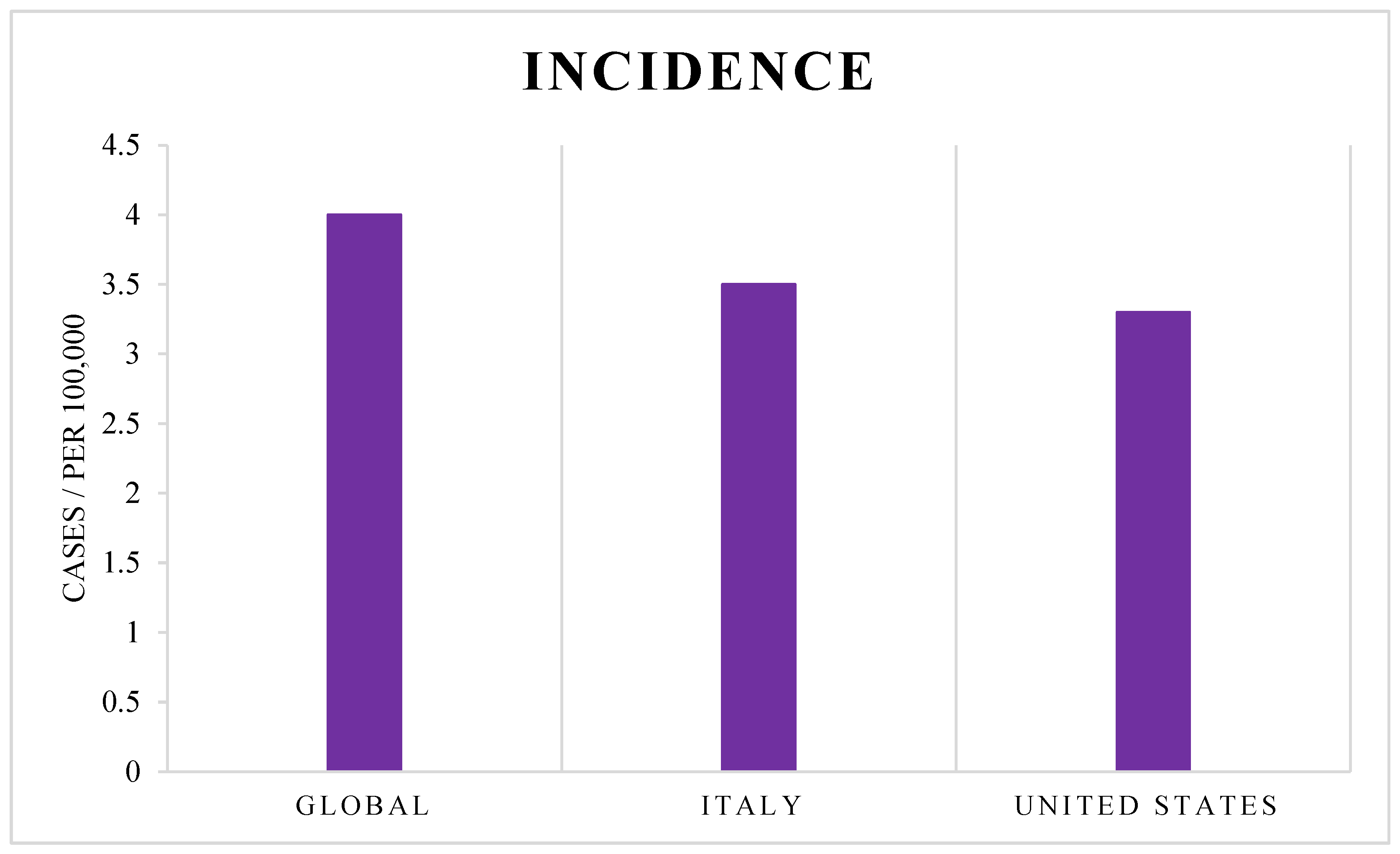
Figure 1. Reported incidence rates of glioblastoma (cases per 100,000 people) globally, in Italy, and in the United States, based on the most recent data from the NCI and ISS.
Cancers 17 02151 g001
Figure 3. Distribution of PubMed articles on glioblastoma genomics, proteomics, and metabolomics. Yellow bars indicate total publications; red bars indicate publications from the last 10 years. Recent studies account for 78% of genomics, 80% of proteomics, and 91% of metabolomics publications.
Cancers 17 02151 g003
Figure 4. PubMed-based distribution of glioblastoma-related articles (2014–2024), grouped by sample type (brain tissue, CSF, plasma, serum, urine, saliva) and categorized as human or animal studies. The histogram emphasizes the prevalence of tissue-based studies and highlights the relative underrepresentation of biofluid-based research.
Cancers 17 02151 g004
Figure 5. Outline of the workflow in biobank-based biomedical research, illustrating the steps from sample collection and storage to analysis, result discovery, and the reuse of residual materials for new studies.
Cancers 17 02151 g005
Figure 6. Evolution of the biobank concept. This infographic illustrates the progressive transformation from simple biological material collections to complex, regulated biobanking infrastructures integrating scientific research, data protection, and public health frameworks.
Cancers 17 02151 g006
Table 1. Proteomics approaches applied to the analysis of brain tissue and cells, with identified candidate biomarkers and associated pathways. References provided in the final column of the table support the methodological approaches described.
SpeciesSample TypeProteomic Approach Biomarkers IdentifiedFunctional RelevanceRef.
HumanTissueLabeling
(TMT; iTRAQ)
ASAH1, GPNMB
MMP9, TIMP1, Fibulins
EGFR, NPM1, RKIP
HNRNPK, ELAVL1, NOVA1
Sphingolipid metabolism and ferroptosis
Immune microenvironment
Tumor progression, migration and angiogenesis
Signaling growth and resistance to therapy
Controlling gene expression in GBM
[60,61,62,63,64,65,66]
No labeling:
LC-MS/MS
(LFQ; DDA; DIA)
YAP1, SOX2, PP1γ
EGFR, FN1, PTEN, BRAF
FN1, TNC, ICAM1, GAGs
HIF1α, IDH1, OXPHOS, Cholesterol, HSPD1, Granzyme A, STAT3, CHI3L1
RPS5, SF3B2, HMGB2
ASAH1, p21-p53-RB, ERCC2, POLD1
Proliferation and survival
Tumor growth and migration
ECM regulation and cell adhesion
Tumor metabolism and hypoxia
Immune response and immunosuppression
RNA processing and splicing
Cell survival, apoptosis, DNA damage
[67,68,69,70,71,72,73,74,75,76,77,78,79,80,81]
CellsNo labeling:
LC-MS/MS
(LFQ; DDA; DIA)
ADAM10, ADAM15, COL6A1, COL1A2, COL6A3,
TIMPs, Fibulin-2/-5/-7
STAT1, STAT2, OAS, IFIT, TRIM25, PME-1, PP2A-B55α, MAPKAPK2, RIPK1
CSE1L, TAZ, Importin α5, WWTR1, RAD51
ECM regulation and tumor progression
IFN signaling
Sensitivity to oxidative stress
Apoptosis, DNA damage
[82,83,84,85,86,87,88,89,90,91,92,93,94,95,96,97,98,99,100,101,102,103,104,105,106,107,108,109,110,111]
AnimalTissueLabeling:
iTRAQ
ILF2, CCT7, CCT4, RPL10A,
MSN, PRPS1, TFRC, APEX1
Early brain development
Primary formation of the neural tube, Regulation of neuronal differentiation,
Synaptic transmission,
Regulation of the nervous system
Regulation of cell survival and tumor proliferation
Mechanisms of drug resistance and chemosensitivity
[112]
CellsNo labeling:
LC-MS/MS
(DDA; DIA)
CaMK2, BCAS1, FBXO2, INF2, PRPS2
CD9, CD81, Nono, Gja1
Tumor growth, microenvironment
Response to hypoxia, glycosaminoglycan biosynthesis
Integrin-mediated signaling pathways, regulation of TGFβ pathways
[94,113]
Table 2. Proteomic approaches applied to the analysis of biofluids, with identified candidate biomarkers and related molecular information. References in the final column support the methodologies and findings reported.
Sample TypeProteomic Approach Biomarkers IdentifiedFunctional RelevanceRef.
Cerebrospinal FluidLC-MS/MS (DDA)CHI3L1 GFAP, GAP43, SERPIN3, APOE, FGA, FGB, FGG, F2Tumor aggressiveness
BBB disruption
Synaptogenesis
Coagulation
Angiogenesis
LXR/RXR Activation pathway
Stemness, Immune modulation
[41,121]
Cystic FluidLC-MS/MS (DDA)Albumin, Haptoglobin, Fibrinogen, Transferrin, Prostaglandin D2 synthase, IgG, IgA, IgM, S100B, GFAPCell adhesion, angiogenesis and cytoskeleton
Acute Inflammatory Response
Immunomodulation
[122]
PlasmaLC-MS/MS (DDA)ASAH1, SYNM, GPNMB, VWF, Hp (α, β chains), zonulinTumor progression, invasiveness and vascularization
Neutrophil involvement
Pro-angiogenic processes and pro-thrombotic response
Oxidative stress protection
Inflammation regulation and homeostasis
[60,123,124]
SerumLC-MS/MS (DIA)CXCL4 (PF4), S100A8, S100A9, MDH1, RNH1, FABP7, TJAP1, AHSPInflammation
Ros metabolism
Nucleotide metabolism
Metabolic reprogramming
Cellular homeostasis
Lipid metabolism and transport
VEGF and IL-18 signaling
[125,126,127]
UrineLC-MS/MS (DIA)GRN, PSAP, ALDOA, S100A11, ITM2B, TCP1, CCT2, CCT3, CCT4, CCT6A, CCT7, CCT8Proteostasis and protein folding
Metabolic reprogramming
Tumor progression
Stress response
[128]
SalivaLC-MS/MS (DIA)ALDOA, 14-3-3ε (YWHAE), TM11B, C3, PPIA, TGF-β-related proteinsCellular proliferation
Cell cycle
Signaling regulation
Complement system activation
Protein folding and trafficking
TGF-β signaling
Immune response
Iron metabolism
[129]
Disclaimer/Publisher’s Note: The statements, opinions and data contained in all publications are solely those of the individual author(s) and contributor(s) and not of MDPI and/or the editor(s). MDPI and/or the editor(s) disclaim responsibility for any injury to people or property resulting from any ideas, methods, instructions or products referred to in the content.