Article

Checkpoint Kinase 1 Inhibitor Combined with Low Dose Hydroxyurea Promotes ATM-Activated NF-κB-Dependent Pro-Inflammatory Chemokine Expression in Melanomas

1
Mater Research Institute, The University of Queensland, Brisbane, QLD 4072, Australia
2
Frazer Institute, Dermatology Research Centre, Faculty of Health, Medicine and Behavioral Sciences, The University of Queensland, Brisbane, QLD 4072, Australia
*
Author to whom correspondence should be addressed.
Cancers 2025, 17(11), 1817; https://doi.org/10.3390/cancers17111817
Submission received: 4 April 2025 / Revised: 25 April 2025 / Accepted: 26 May 2025 / Published: 29 May 2025
(This article belongs to the Section Cancer Drug Development)

Simple Summary

Tumours avoid detection by the body’s immune system through a range of mechanisms. An anti-cancer treatment that can alter this tumour-induced immune avoidance could enhance the ability of the patient’s own immune system to detect and respond appropriately to the tumour. Here, we show that a novel treatment that directly kills melanoma also upregulates chemokines, agents that can attract immune cells into the tumour to enhance an anti-tumour immune response. The mechanism by which this treatment increases these chemokines is also determined and involves a response to damage caused by the treatment that is also responsible for the tumour cell killing by this treatment. This study demonstrates that the new treatment can not only directly and effectively kill melanoma cells but also promote signals that will encourage immune cells to recognise the tumour.

Abstract

Background/Objectives: Melanoma has a rising incidence worldwide. Current treatments are effective, although the development of resistance is common. A novel anti-cancer treatment using checkpoint kinase 1 inhibitor (CHK1i), SRA737, in combination with low-dose hydroxyurea (LDHU), has been demonstrated to effectively kill tumour cells and promote an anti-tumour immune response through the treatment-induced release of pro-inflammatory chemokines and cytokines. These chemokines/cytokines modify the tumour microenvironment from an immunosuppressive to an inflamed state to recruit anti-tumour immune cells. Methods: A panel of human melanoma cell lines was assessed using a panel of chemokines and cytokine expression, and the mechanism of their regulation was investigated. Results: We demonstrate that SRA737 + LDHU upregulates pro-inflammatory chemokines in human melanoma cells in response to SRA737 + LDHU through the ATM-NF-κB signalling pathway. The increased chemokine expression corresponded to the increase in secretion of pro-inflammatory chemokines from tumour cells following SRA737 + LDHU treatment. However, inhibiting NF-κB and ATM did not affect SRA737 + LDHU-induced cell killing. Increased expression of non-NF-κB target genes with SRA737 + LDHU suggests that other transcriptional pathways are also activated and may contribute to the increasing cytokine/chemokine gene expression in response to treatment. Conclusions: SRA737 + LDHU upregulates pro-inflammatory chemokine expression through an ATM-NF-κB-dependent mechanism.

1. Introduction

The tumour microenvironment (TME) contains tumour cells, immune and immunosuppressive cells, blood vessels, extracellular matrix, fibroblasts, and signalling molecules [1]. Anti-tumour immune responses are led by lymphocytes, including cytotoxic CD8+ T cells, CD4+ helper T cells, and natural killer (NK) cells, with the help of pro-inflammatory macrophages (M1) and dendritic cells. T cells and NK cells can be recruited to the TME via chemokines secreted by tumour cells or other immune cells [2,3,4,5], where they are referred to as tumour infiltrating lymphocytes (TILs). Tumour cells can also modify immune cell phenotypes and functions towards immunosuppression and promote tolerance to tumour antigens for immune evasion [1]. The immunosuppressive cells commonly found in the TME include regulatory T cells (Tregs), tumour-associated macrophages (TAMs), and myeloid-derived suppressor cells (MDSCs) [6]. Enhancing the anti-tumour immune response by reducing the immune suppression in the TME is, therefore, essential for the long-term responses to any anti-tumour treatment.
This can occur through multiple mechanisms, including expression of the dendritic cells chemoattractant XCL1, which increases tumour antigenicity to prime T cell responses [7], and secretion of T-cell recruiting chemokines such as CCL2, CCL5, CXCL9, and CXCL10 to attract cytotoxic CD8+ T cells to tumour sites [8].
Many cytokines and chemokines have dual, opposing roles and can have pro-inflammatory or anti-inflammatory functions depending on context and which immune cells are present [2]. Tregs and MDSCs reduce the number and/or activity of TILs in the TME [5]. Cancers recruit Tregs to the TME by expressing CCL5 and CXCL12, differentiating naïve CD4+ T cells, or converting Th17/CD8+ cells into Tregs via TGF-β to evade the immune system [3]. MDSCs are also recruited by cytokines/chemokines such as VEGF, TGF-β, CCL2, and CXCL12 to establish an immunosuppressive TME [9]. Tumour-associated macrophages (TAMs) are attracted to the TME through cytokines and chemokines, including VEGF, CSF-1, TGF-β, CCL2, CCL7, and CCL8. TAMs induce angiogenesis and can contribute to tumour metastasis, and higher densities of TAMs are associated with poorer clinical outcomes [3,10]. An immunosuppressive TME impedes the effectiveness of cancer therapy, making it crucial for novel therapies to modify the TME to a pro-inflammatory state to promote anti-tumour immune responses.
Chemotherapies that induce high levels of DNA damage can promote cytokine/chemokine expression in tumour cells [11]. This is often reported to be due to the activation of cGAS-STING by the presence of cytosolic DNA that is detected by cGAS and converted to cGAMP, which in turn activates STING [12]. STING activates TANK-binding kinase 1 (TBK1), activating IFN regulatory factor 3 (IRF3) and nuclear factor κB (NF-κB) that translocates into the nucleus driving transcription [12,13]. This promotes the expression of pro-inflammatory type-I interferons and cytokines such as tumour necrosis factor (TNF) and interleukin 6 (IL-6) [14]. However, other pathways respond to DNA damage to increase cytokine/chemokine gene expression, including NF-κB [15].
In previous studies, a combination of checkpoint kinase 1 inhibitor (CHK1i) and low-dose hydroxyurea (LDHU) has been shown to have tumour-selective toxicity, targeting tumours with elevated levels of replication stress without significant toxicity in normal tissue [16,17]. This was associated with increased expression of pro-inflammatory cytokines/chemokines in vivo, although the mechanism regulating this was not determined. Here, we investigate the mechanism regulating this treatment-induced chemokine expression in melanomas.

2. Materials and Methods

2.1. Cell Lines

The panel of human melanoma cell lines were originally sourced from Professor Nicholas Hayward QIMR-Berghofer Institute, Brisbane, Queensland, Australia [18]. The cells were cultured in Dulbecco′s Modified Eagle′s Medium (DMEM) (Sigma-Aldrich, St. Louis, MO, USA), 10% heat-inactivated Fetal Bovine Serum (Gibco, Waltham, MA, USA), GlutaMAX (Gibco), 20 mM HEPES (Sigma-Aldrich), and 1 mM Sodium Pyruvate (Sigma-Aldrich) with Antibiotic-Antimycotic (Gibco). Cells were maintained at 37 °C and 5% CO2 and were mycoplasma free. The mutation state of the cell lines is shown in Supplementary Table S2.

2.2. IncuCyte Assays

Cells were seeded in a 96-well plate in 100 µL media. Cells were acclimated for 24–48 h in a 37 °C and 5% CO2 incubator until 70% confluency before treating with 1 µM SRA737 + 0.2 mM hydroxyurea (HU), and 125 nM of SYTOX Green Fluorescence Dye (Thermofisher Scientific, Waltham, MA, USA) was added 30 min prior to imaging to visualise cell death. Cells were imaged for 72 h at 4 h intervals with 10× magnification using Sartorius Incucyte S3 Live Cell Analysis System (Essence Bioscience, Ann Arbor, MI, USA) at 37 °C and 5% CO2. Incucyte Base Analysis Software version 3.1.0 (Essence Bioscience) was used to perform segmentation and cell-by-cell analysis for cell counting and percentage death.

2.3. Immunoblotting

Cells were harvested and washed twice with PBS; then, the cell pellet was stored at −80 °C until required. Cell pellets were lysed in lysis buffer (NETN; 0.5% Nonidet P40, 100 mM NaCl, 20 mM Tris-Cl, and 2 mM EDTA), pH 8, 0.1% sodium dodecyl sulphate (SDS), 25 mM β-glycerophosphate, 25 mM sodium fluoride 0.1 mM sodium orthovanadate, Protease Inhibitor Cocktail (Sigma Aldrich), and 1 mM phenylmethylsulfonyl fluoride. Protein concentrations were normalised. Proteins were resolved on 4–20% SDS-polyacrylamide Mini-PROTEAN TGX (Bio-Rad, South Granville NSW, Australia) precast gels and then transferred onto 0.2 μM polyvinylidene difluoride (PVDF) membrane. Membranes were probed with the antibodies below, then detected using the appropriate horseradish peroxidase labelled secondary antibody and chemiluminescence detection. Primary antibodies were purchased from; phospho-Histone H2A.X (γH2AX) (Ser139/Tyr142) #5438 Cell Signaling Technology (Danvers, MA, USA), phospho-RPA2 Ser4/8 BL-165-5F1Bethyl Laboratories, phospho-ATM (Ser1981) #4526 Cell Signaling Technology, phospho-NF-κB p65 (RelA) (Ser536) #3033Cell Signaling Technology, NF-κB p65 (RelA) #8242Cell Signaling Technology, Histone H3 #9715Cell Signaling Technology, ATM GTX70103 GeneTex (Irvine, CA, USA).

2.4. RT-qPCR

Cells were treated for 24 h, then harvested using cell scrapers; media was removed before adding 1 mL of TRIzol reagent (Invitrogen, Waltham, MA, USA, #15596026). Total RNA was extracted from all cell lines using TRIzol. DNase 1 Kit (Invitrogen #18047019) was used according to the manufacturer’s instructions to remove DNA impurities from extracted RNA. RNA concentration and purity were assessed using Thermo Scientific NanoDrop One/OneC using the absorbance value ratio A260/A280 (1.8–2.0) and A260/A230 (>2.0), respectively. cDNA synthesis was performed using the High-Capacity cDNA Reverse Transcription Kit (Applied Biosystems, Waltham, MA, USA, #4368814), including RNaseOUT recombinant ribonuclease inhibitor (Invitrogen #10777019) according to the manufacturer’s instructions. Reverse transcription was performed using the Eppendorf Mastercycler pro PCR System (Fisher Scientific, Pittsburgh, PA, USA) with 2 µg of RNA. Predesigned KiCqStart primers (MERCK, Bayswater, VIC, Australia) were used for RT-qPCR, listed in Supplementary Table S1. PCR was performed using QuantStudio 7 Flex Real-Time PCR System (Thermo Fisher) in a 384-well plate. Melt curve analysis was performed to confirm specificity. For analysis of results, relative expression (ΔCt) values were normalised against the housekeeping gene YWHAZ (ΔCt = CtTarget Gene—CtYWHAZ), then log-transformed (2−ΔCt) for visual representation as fold change on a log10 y-axis.

2.5. Cytokine/Chemokine Bead Array

LEGENDplex Human Essential Immune Response Panel Cat#740930 (Australian Biosearch, Wangara, WA Australia), was used to assess the concentration of 13 cytokines/chemokines present in cell culture supernatants after treatment with SRA737 + LDHU was performed as per manufacturer’s instructions. Samples were analysed on a CytoFLEX S Flow Cytometer (Beckman Coulter, Lane Cove, Australia). Data were analysed using the provided LEGENDplex Data Analysis Software version 4 (BioLegend, San Diego, CA, USA).

2.6. Immunofluorescence

A2058 cells were seeded on coverslips in a 6-well plate with a density of 3 × 10⁵ cells/well. Cells were acclimated for 24 h until 70% confluency before respective treatments were added. Cells were fixed with 4% paraformaldehyde for 15 min at 37 °C, then washed and permeabilisation with 0.1% Triton X-100, blocked and immunostained for NF-κB p65 (RelA) #8242 Cell Signaling Technology using an Alexa488 labelled secondary antibody #A-11034 ThermoFisher and DAPI. Cells were mounted onto microscope slides and were imaged using an Olympus BX63 (Olympus Life Science, Waltham, MA, USA) microscope at 20× magnification. Cell segmentation and quantification of fluorescence intensity were performed using CellProfiler version 4.2.6 for Windows, www.cellprofiler.org.

2.7. Quantitative Analysis

All graphs and statistical analyses were performed using GraphPad Prism version 10.3.1 for Windows, GraphPad Software, Boston, MA, USA, www.graphpad.com. For statistical methods used, see individual figure legends.

3. Results

Treatment with the combination of 1 mM SRA737 and 0.2 mM HU promoted replication stress, indicated by the phosphorylation of RPA2 at Ser4/8, increased DNA damage indicated by increased γH2AX, and increased cell death (Figure 1A) as we have observed previously using a different CHK1 inhibitor [16]. It also promoted extensive cell killing in all the cell lines used here (Figure 1B; Supplementary Figure S1).
DNA damage has been reported to also promote chemokine expression in many cancer types; in many cases, this is reported to be through the cGAS-STING pathway [19]. However, cGAS-STING is commonly down-regulated in many cancers, as demonstrated in the Cancer Cell Line Encyclopedia (CCLE) dataset, with approximately 35% of all cancer cell lines expressing low levels (<1 TPM) of cGAS and/or STING [20] (Figure 2A) and reported to be commonly inactivated in melanoma and other cancers [17,21]. The lack of typical cGAS-STING-induced type I interferon expression in response to SRA737 + low dose HU treatment in vivo [17] also suggested that this pathway was not responsible for the pro-inflammatory chemokine expression observed in two syngeneic melanoma models with this treatment (Figure 2B). In vitro treatment of a panel of human melanoma cell lines with SRA737 + low dose HU also resulted in increased expression of the pro-inflammatory chemokines CCL2, CCL5, CXCL8, CXCL10, IL6, XCL1, and TNF, and had modest effects on VEGFA and TGFB1 although the magnitude of expression was different for each cell line (Figure 2C). The changes in gene expression correlated with increased levels of the secreted chemokines, although little free TGFβ was detected (Supplementary Figure S2).
It was previously reported that SRA737 + LDHU did not activate the cGAS-STING pathway in melanoma cell lines despite increased cytosolic DNA [17], suggesting other transcriptional pathways were likely to be involved. The majority of chemokines that showed increased expression with SRA737 + LDHU treatment were known NF-κB targets [22]. To investigate the potential involvement of NF-κB, the nuclear localisation of NF-κB RelA/p65 subunit was assessed, and the nuclear/cytoplasmic ratio of RelA was found to be significantly increased in treated cells (Figure 3A,B), indicating NF-κB activation was assessed.
The activation of NF-κB was confirmed using an inhibitor of NF-κB activation BI-605906, which blocked the SRA737 + LDHU-induced increased expression of CCL2, CCL5, CXCL8, CXCL10, IL6, and TNF to at least basal levels, although the NF-κB inhibitor also reduced the basal level of many of these chemokines to undetectable levels. The inhibitor had no effect on VEGFA or TGFB1, neither of which are regulated by NF-κB (Figure 4).
The increased DNA damage suggested the activation of the DNA damage response signalling through Ataxia telangiectasia mutated (ATM), and ATM was activated in response to SRA737 + LDHU in all cell lines (Figure 5A). ATM signalling in response to DNA damage has previously been shown to activate NF-κB [23], and inhibition of ATM using the inhibitor AZD1390 blocked the increase in RelA/p65 Ser536 phosphorylation observed with SRA737 + LDHU treatment to a similar level as the NF-κB inhibitor alone (Figure 5B). It also blocked the SRA737 + LDHU-induced increased expression of the NF-κB-regulated genes but had no effect on either VEGFA or TGFB1 (Figure 5C).
The same effect was observed with a second ATM inhibitor, KU-55933 (Supplementary Figure S3). Although the NF-κB and ATM inhibitors effectively blocked the NF-κB-dependent chemokine expression, they did not affect either cell proliferation as single agents or cell killing by SRA737 + LDHU (Figure 6). This indicates that the NF-κB activation and downstream chemokines expression were not involved in the cell-killing response to treatment.

4. Discussion

This study showed that SRA737 + LDHU treatment of melanoma cells that is sufficient to kill a high proportion of cells also upregulates the expression of pro-inflammatory cytokines and chemokines cells, including CCL2, CCL5, CXCL8, CXCL10, IL-6, TNF, and XCL1, and immunosuppressive VEGFA and TGFB1 before significant cell death occurs. The pro-inflammatory cytokines/chemokines have the potential to enhance the tumour’s immune microenvironment. Increased recruitment of NK cells and macrophages and activation of T cells was observed with this treatment in vivo [17], indicating that this pro-inflammatory program is a functional response. However, the increased expression of immunosuppressive VEGFA and TGFB1 that are not regulated by NF-κB can oppose this pro-inflammatory effect. VEGFA can recruit suppressive immune cells such as TAMs, Tregs, and MDSCs, although it may also increase vascular permeability, thus improving drug delivery of SRA737 + LDHU to tumours in vivo [24]. TGF-β can promote malignancy [25] and immunosuppression by converting CD4+ T cells to Tregs [26]}. Here, we report that TGFB1 is expressed at high levels in all the melanoma cell lines assessed, and SRA737 + LDHU treatment triggered a modest increase. However, there was little evidence of free TGF-β being secreted, suggesting the factors required for full maturation and release of free TGF-β are not present in melanomas [27]. In vivo, there is evidence of increased TGF-β following SRA737 + LDHU treatment, along with an increase in immunosuppressive FoxP3 + Tregs in the TME [17].
This study demonstrates the ability of SRA737 + LDHU to activate the NF-κB signalling pathway and is responsible for the upregulation of pro-inflammatory cytokines and chemokines observed. It is activated by ATM, a DNA damage sensor activated in response to the presence of the level of DNA double-strand breaks induced by replication fork collapse induced by CHK1 inhibition [28]. ATM regulates NEMO to, in turn, release IKKb to activate NF-κB [23]. NF-κB also appears to be responsible for much of the basal expression of CCL2, CCL5, and TNF, possibly a consequence of upregulated NF-κB activity, a common feature of melanoma [29]. CHK1 activated in response to replication stress can also phosphorylate RelA/p65 at Thr505, resulting in the inhibition of NF-κB-dependent anti-apoptotic gene regulation [30], but does not affect pro-inflammatory cytokine expression [31]. The effect of ATM on NF-κB activation is specific as other components involved in DNA damage response, such as DNA-PK and ATR, are not activators of NF-κB [15].
The upregulation of non-NF-κB targets suggests other transcriptional responses occur with SRA737 + LDHU. XCL1 can be regulated by p53 [32], but with p53 commonly defective in melanomas, p63 and p73 may be responsible for regulating its transcription [33]. TGFB1 and VEGFA can be regulated by the Activation Protein 1 (AP-1) complex (c-Jun/c-Fos) [34], which can also regulate the expression of CXCL8 [35] and IL-6 [36]. Since the MAPK pathway, which regulates AP-1 [37], is commonly overactive in melanoma [38], this may explain the high baseline expression of these cytokines/chemokines. The mechanism by which TGFB1 and VEGFA with SRA737 + LDHU treatment is at present not known. However, blocking this pathway could reduce the immunosuppressive effects of these cytokines, enhancing the tumour’s immune microenvironment.

5. Conclusions

In conclusion, we provided evidence that SRA737 + LDHU activates NF-κB in human melanoma cells through the canonical pathway activated by the DNA damage response via ATM. This results in the upregulation of genes encoding for pro-inflammatory cytokines and chemokines, following a consistent general pattern across a variety of human melanoma cell lines independent of the degree of cell killing elicited by SRA737 + LDHU. These cytokines and chemokines can modify the tumour microenvironment from an immunosuppressive state to an inflamed state, promoting an anti-tumour immune response through the recruitment of immune cells to the tumour site.

Supplementary Materials

The following supporting information can be downloaded at: https://www.mdpi.com/article/10.3390/cancers17111817/s1, Figure S1: SRA737 + LDHU induces replication stress and DNA damage; Figure S2: SRA737 + LDHU increases release of cytokines and chemokines from human melanoma cell lines; Figure S3: Inhibition of ATM with SRA737 + LDHU returns expression of NF-κB-target cytokine/chemokine genes to basal levels; Table S1. RT-qPCR Oligonucleotide KiCqStart Primers (MERCK, Sigma-Aldrich); Table S2. Known mutation status of the melanoma cell lines. File S1: Western blot information.

Author Contributions

Conceptualisation, B.G.; methodology, R.B.; validation, N.L.L.-A.G. and N.J.A.R.; formal analysis, N.L.L.-A.G., N.J.A.R., R.B., A.G. and B.G.; investigation, N.L.L.-A.G., N.J.A.R., S.E.O., K.Y.L. and A.G.; resources, S.K.; data curation, N.L.L.-A.G. and N.J.A.R.; writing—original draft preparation, N.L.L.-A.G., N.J.A.R. and B.G.; writing—review and editing, N.L.L.-A.G., Z.Z., S.K. and B.G.; visualisation, N.L.L.-A.G., N.J.A.R. and B.G.; supervision, R.B. and B.G.; project administration, B.G.; funding acquisition, B.G. All authors have read and agreed to the published version of the manuscript.

Funding

This research was funded by Melanoma Research Alliance #827115 and Mater Foundation Smiling for Smiddy.

Institutional Review Board Statement

Not applicable.

Informed Consent Statement

Not applicable.

Data Availability Statement

The original contributions presented in this study are included in the article/Supplementary Materials. Further inquiries can be directed to the corresponding author.

Acknowledgments

The authors thank Jelena Urosevic at Astra Zeneca Cambridge for the AZD1390.

Conflicts of Interest

The authors declare no conflicts of interest.

Abbreviations

ATMAtaxia telangiectasia mutated
CHK1Checkpoint kinase 1
CHK1iCHK1 inhibitor
LDHULow dose hydroxyurea
HUhydroxyurea
IL-6Interleukin 6
IRF3Interferon regulatory factor 3
NF-κBNuclear factor-κB
NKNatural killer
TAMTumour-associated macrophage
TBK1TANK binding kinase 1
TILTumour infiltrating leukocytes
TMETumour microenvironment
TNFTumour necrosis factor
TregT regulatory cells

References

  1. de Visser, K.E.; Joyce, J.A. The evolving tumor microenvironment: From cancer initiation to metastatic outgrowth. Cancer Cell 2023, 41, 374–403. [Google Scholar] [CrossRef]
  2. Propper, D.J.; Balkwill, F.R. Harnessing cytokines and chemokines for cancer therapy. Nat. Rev. Clin. Oncol. 2022, 19, 237–253. [Google Scholar] [CrossRef]
  3. Labani-Motlagh, A.; Ashja-Mahdavi, M.; Loskog, A. The Tumor Microenvironment: A Milieu Hindering and Obstructing Antitumor Immune Responses. Front. Immunol. 2020, 11, 940. [Google Scholar] [CrossRef]
  4. Verdegaal, E.M.; de Miranda, N.F.; Visser, M.; Harryvan, T.; van Buuren, M.M.; Andersen, R.S.; Hadrup, S.R.; van der Minne, C.E.; Schotte, R.; Spits, H.; et al. Neoantigen landscape dynamics during human melanoma-T cell interactions. Nature 2016, 536, 91–95. [Google Scholar] [CrossRef] [PubMed]
  5. Santiago-Sánchez, G.S.; Hodge, J.W.; Fabian, K.P. Tipping the scales: Immunotherapeutic strategies that disrupt immunosuppression and promote immune activation. Front. Immunol. 2022, 13, 993624. [Google Scholar] [CrossRef] [PubMed]
  6. Tie, Y.; Tang, F.; Wei, Y.Q.; Wei, X.W. Immunosuppressive cells in cancer: Mechanisms and potential therapeutic targets. J. Hematol. Oncol. 2022, 15, 61. [Google Scholar] [CrossRef] [PubMed]
  7. Matsuo, K.; Yoshie, O.; Kitahata, K.; Kamei, M.; Hara, Y.; Nakayama, T. Recent Progress in Dendritic Cell-Based Cancer Immunotherapy. Cancers 2021, 13, 2495. [Google Scholar] [CrossRef]
  8. Galon, J.; Bruni, D. Approaches to treat immune hot, altered and cold tumours with combination immunotherapies. Nat. Rev. Drug Discov. 2019, 18, 197–218. [Google Scholar] [CrossRef]
  9. Veglia, F.; Sanseviero, E.; Gabrilovich, D.I. Myeloid-derived suppressor cells in the era of increasing myeloid cell diversity. Nat. Rev. Immunol. 2021, 21, 485–498. [Google Scholar] [CrossRef]
  10. Korneev, K.V.; Atretkhany, K.-S.N.; Drutskaya, M.S.; Grivennikov, S.I.; Kuprash, D.V.; Nedospasov, S.A. TLR-signaling and proinflammatory cytokines as drivers of tumorigenesis. Cytokine 2017, 89, 127–135. [Google Scholar] [CrossRef]
  11. Nastasi, C.; Mannarino, L.; D’Incalci, M. DNA Damage Response and Immune Defense. Int. J. Mol. Sci. 2020, 21, 7504. [Google Scholar] [CrossRef] [PubMed]
  12. Pilger, D.; Seymour, L.W.; Jackson, S.P. Interfaces between cellular responses to DNA damage and cancer immunotherapy. Genes Dev 2021, 35, 602–618. [Google Scholar] [CrossRef]
  13. Barros, E.M.; McIntosh, S.A.; Savage, K.I. The DNA damage induced immune response: Implications for cancer therapy. DNA Repair. 2022, 120, 103409. [Google Scholar] [CrossRef]
  14. Ng, K.W.; Marshall, E.A.; Bell, J.C.; Lam, W.L. cGAS-STING and Cancer: Dichotomous Roles in Tumor Immunity and Development. Trends Immunol. 2017, 19, 30151–30155. [Google Scholar] [CrossRef] [PubMed]
  15. Janssens, S.; Tschopp, J. Signals from within: The DNA-damage-induced NF-kappaB response. Cell Death Differ. 2006, 13, 773–784. [Google Scholar] [CrossRef]
  16. Oo, Z.Y.; Proctor, M.; Stevenson, A.J.; Nazareth, D.; Fernando, M.; Daignault, S.M.; Lanagan, C.; Walpole, S.; Bonazzi, V.; Skalamera, D.; et al. Combined use of subclinical hydroxyurea and CHK1 inhibitor effectively controls melanoma and lung cancer progression, with reduced normal tissue toxicity compared to gemcitabine. Mol. Oncol. 2019, 13, 1503–1518. [Google Scholar] [CrossRef] [PubMed]
  17. Proctor, M.; Gonzalez Cruz, J.L.; Daignault-Mill, S.M.; Veitch, M.; Zeng, B.; Ehmann, A.; Sabdia, M.; Snell, C.; Keane, C.; Dolcetti, R.; et al. Targeting Replication Stress Using CHK1 Inhibitor Promotes Innate and NKT Cell Immune Responses and Tumour Regression. Cancers 2021, 13, 3733. [Google Scholar] [CrossRef] [PubMed]
  18. Johansson, P.; Pavey, S.; Hayward, N. Confirmation of a BRAF mutation-associated gene expression signature in melanoma. Pigment Cell Res. 2007, 20, 216–221. [Google Scholar] [CrossRef]
  19. Li, T.; Chen, Z.J. The cGAS-cGAMP-STING pathway connects DNA damage to inflammation, senescence, and cancer. J. Exp. Med. 2018, 215, 1287–1299. [Google Scholar] [CrossRef]
  20. Ghandi, M.; Huang, F.W.; Jane-Valbuena, J.; Kryukov, G.V.; Lo, C.C.; McDonald, E.R., 3rd; Barretina, J.; Gelfand, E.T.; Bielski, C.M.; Li, H.; et al. Next-generation characterization of the Cancer Cell Line Encyclopedia. Nature 2019, 569, 503–508. [Google Scholar] [CrossRef]
  21. Konno, H.; Yamauchi, S.; Berglund, A.; Putney, R.M.; Mulé, J.J.; Barber, G.N. Suppression of STING signaling through epigenetic silencing and missense mutation impedes DNA damage mediated cytokine production. Oncogene 2018, 37, 2037–2051. [Google Scholar] [CrossRef] [PubMed]
  22. Taniguchi, K.; Karin, M. NF-κB, inflammation, immunity and cancer: Coming of age. Nat. Rev. Immunol. 2018, 18, 309–324. [Google Scholar] [CrossRef] [PubMed]
  23. Miyamoto, S. Nuclear initiated NF-kappaB signaling: NEMO and ATM take center stage. Cell Res. 2011, 21, 116–130. [Google Scholar] [CrossRef]
  24. Klose, R.; Krzywinska, E.; Castells, M.; Gotthardt, D.; Putz, E.M.; Kantari-Mimoun, C.; Chikdene, N.; Meinecke, A.-K.; Schrödter, K.; Helfrich, I.; et al. Targeting VEGF-A in myeloid cells enhances natural killer cell responses to chemotherapy and ameliorates cachexia. Nat. Commun. 2016, 7, 12528. [Google Scholar] [CrossRef] [PubMed]
  25. Cui, W.; Fowlis, D.J.; Bryson, S.; Duffie, E.; Ireland, H.; Balmain, A.; Akhurst, R.J. TGFβ1 Inhibits the Formation of Benign Skin Tumors, but Enhances Progression to Invasive Spindle Carcinomas in Transgenic Mice. Cell 1996, 86, 531–542. [Google Scholar] [CrossRef]
  26. Massagué, J. TGFbeta in Cancer. Cell 2008, 134, 215–230. [Google Scholar] [CrossRef]
  27. Deng, Z.; Fan, T.; Xiao, C.; Tian, H.; Zheng, Y.; Li, C.; He, J. TGF-β signaling in health, disease and therapeutics. Signal Transduct. Target. Ther. 2024, 9, 61. [Google Scholar] [CrossRef]
  28. Ammazzalorso, F.; Pirzio, L.M.; Bignami, M.; Franchitto, A.; Pichierri, P. ATR and ATM differently regulate WRN to prevent DSBs at stalled replication forks and promote replication fork recovery. EMBO J. 2010, 29, 3156–3169. [Google Scholar] [CrossRef]
  29. Ueda, Y.; Richmond, A. NF-kappaB activation in melanoma. Pigment Cell Res 2006, 19, 112–124. [Google Scholar] [CrossRef]
  30. Msaki, A.; Sánchez, A.M.; Koh, L.F.; Barré, B.; Rocha, S.; Perkins, N.D.; Johnson, R.F. The role of RelA (p65) threonine 505 phosphorylation in the regulation of cell growth, survival, and migration. Mol. Biol. Cell 2011, 22, 3032–3040. [Google Scholar] [CrossRef]
  31. Moles, A.; Butterworth, J.A.; Sanchez, A.; Hunter, J.E.; Leslie, J.; Sellier, H.; Tiniakos, D.; Cockell, S.J.; Mann, D.A.; Oakley, F.; et al. A RelA(p65) Thr505 phospho-site mutation reveals an important mechanism regulating NF-κB-dependent liver regeneration and cancer. Oncogene 2016, 35, 4623–4632. [Google Scholar] [CrossRef] [PubMed]
  32. Zhang, Y.; Chen, X.; Fan, Y.; Liu, J.; Yuan, L. XCL1 Aggravates Diabetic Nephropathy-Mediated Renal Glomerular Endothelial Cell Apoptosis and Inflammatory Response via Regulating p53/Nuclear Factor-Kappa B Pathway. Nephron 2021, 146, 84–98. [Google Scholar] [CrossRef] [PubMed]
  33. Dötsch, V.; Bernassola, F.; Coutandin, D.; Candi, E.; Melino, G. p63 and p73, the ancestors of p53. Cold Spring Harb. Perspect. Biol. 2010, 2, a004887. [Google Scholar] [CrossRef] [PubMed]
  34. Jia, J.; Ye, T.; Cui, P.; Hua, Q.; Zeng, H.; Zhao, D. AP-1 transcription factor mediates VEGF-induced endothelial cell migration and proliferation. Microvasc. Res. 2016, 105, 103–108. [Google Scholar] [CrossRef]
  35. Khanjani, S.; Terzidou, V.; Johnson, M.R.; Bennett, P.R. NFκB and AP-1 Drive Human Myometrial IL8 Expression. Mediat. Inflamm. 2012, 2012, 504952. [Google Scholar] [CrossRef]
  36. Hungness, E.S.; Pritts, T.A.; Luo, G.-j.; Sun, X.; Penner, G.C.; Hasselgren, P.-O. The transcription factor activator protein-1 is activated and interleukin-6 production is increased in interleukin-1β-stimulated human enterocytes. Shock 2000, 14, 386–391. [Google Scholar] [CrossRef]
  37. Karin, M.; Liu, Z.-g.; Zandi, E. AP-1 function and regulation. Curr. Opin. Cell Biol. 1997, 9, 240–246. [Google Scholar] [CrossRef]
  38. Inamdar, G.S.; Madhunapantula, S.V.; Robertson, G.P. Targeting the MAPK pathway in melanoma: Why some approaches succeed and other fail. Biochem. Pharmacol. 2010, 80, 624–637. [Google Scholar] [CrossRef]
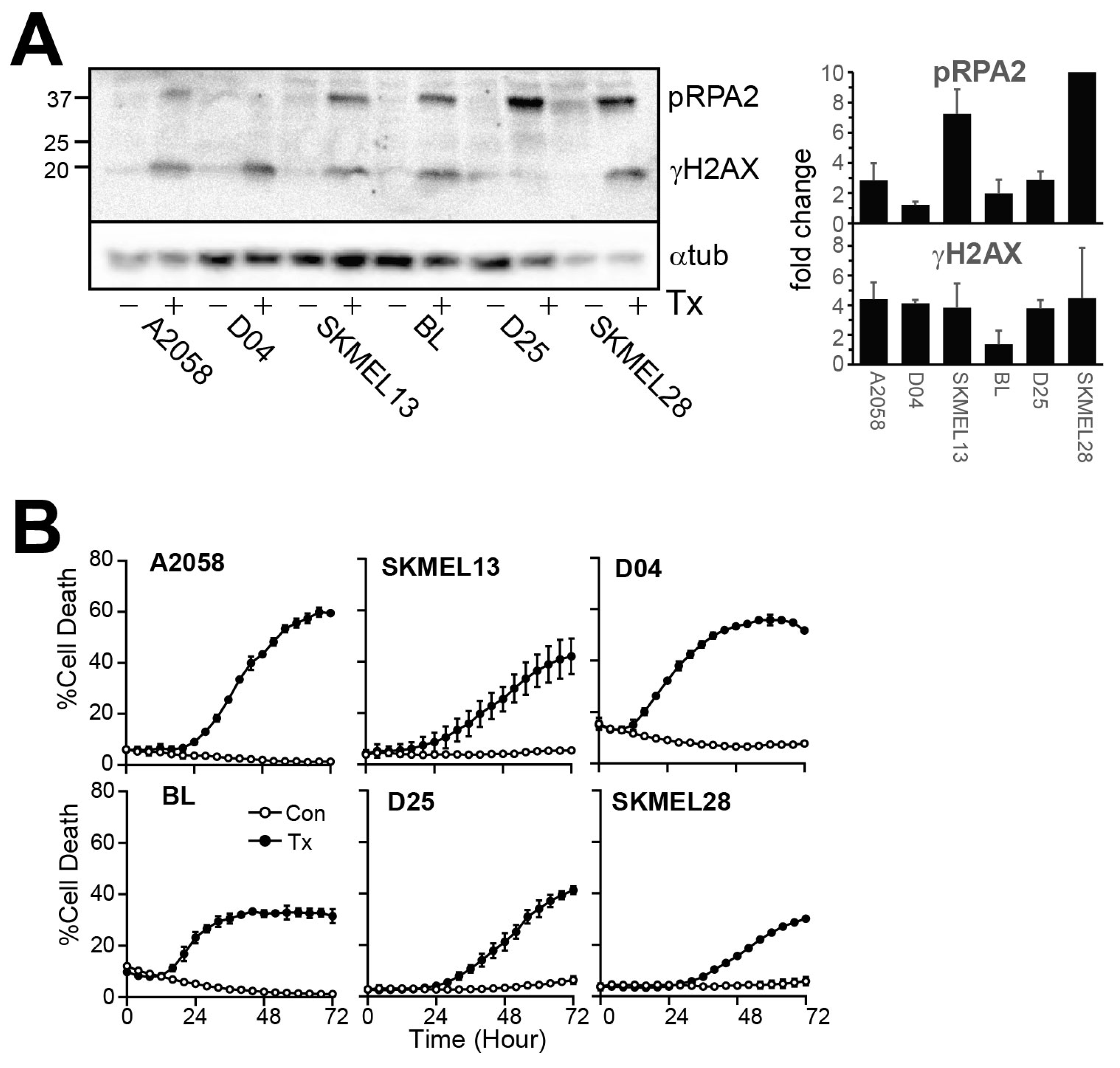
Figure 1. SRA737 + LDHU induces replication stress and DNA damage. (A) Cells were treated with or without 1 µM SRA737 + 0.2 mM HU and harvested at 24 h. Cell lysates were immunoblotted for γH2AX as a marker for DNA damage and RPA2 for replication stress. Histone H3 was used as a loading control. (B) The indicated melanoma cell lines were treated as in A and cells followed using Incucyte live cell imaging using SYTOX Green as a marker of cell death. The % cell death is shown. The data are the mean and SD from three replicates. The uncropped blots are shown in File S1.
Cancers 17 01817 g001
Figure 2. SRA737 + LDHU induces pro-inflammatory chemokine expression. (A) Gene expression data from 1673 human cancer cell lines for CGAS and STING from the CCLE. Melanoma cell lines are highlighted in orange. (B) Gene expression in murine melanoma (YUMMUV1.7 and YUMMUV3.3) was treated with and without SRA737 + LDHU in vivo for the indicated chemokines. The data are from 3–4 individual mice. (C) The melanoma cell lines were treated without or with SRA737 + LDHU (−, +) and harvested at 24 h for RNA extraction, followed by RT-qPCR. Data represents the mean with standard deviation error bars of 3 independent experiments, taking the mean of triplicate wells for each experiment. Multiple paired t-tests (parametric) were performed per gene on ΔCt values, followed by correction for multiple comparisons using the two-stage step-up method of Benjamini, Krieger, and Yekutieli false discovery rate (FDR) at a 5% threshold. Adjusted p-values are represented as * p < 0.05, ** p < 0.01, *** p < 0.001. The colours identify each cell line.
Cancers 17 01817 g002
Figure 3. SRA737 + LDHU activates and drives nuclear localisation of NF-κB. (A) A2058 cells were treated with TNF-α for 1 h or SRA737 + LDHU for 24 h before fixing and labelling. Nuclei were stained with DAPI and RelA. Images were taken using Olympus BX63. The scale bar is 100 µm. (B) CellProfiler was used for cell and nucleus segmentation and quantification of the integrated intensity of RelA of nuclear and cytoplasmic RelA separately. The individual nuclear/cytoplasmic ratio of RelA is shown for 400–800 cells, and the mean is shown for each (light grey bar). One-way ANOVA was performed on ratios with p values were corrected using the Tukey test with a 0.05 (95% CI) threshold. Adjusted p-values are represented as **** p < 0.0001.
Cancers 17 01817 g003
Figure 4. SRA737 + LDHU activates NF-κB-dependent chemokine expression. A2058 and SKMEL13 were treated with 20 µM BI-605906 (NF-κB inhibitor) for 45 min prior to treatment with 1 µM SRA737 + 0.2 mM HU and harvested at 24 h for RNA extraction, followed by RT-qPCR. Data represent the mean with standard deviation error bars and are representative of 3 independent experiments, taking the mean of triplicate wells for each experiment. Two-way ANOVA was performed on ΔCt values with multiple comparisons of biological replicate means of each treatment group. p-values were corrected using the Tukey test with a 0.05 (95% CI) threshold. Adjusted p-values are represented as * p < 0.05, ** p < 0.01, *** p < 0.001, and **** p < 0.0001. The colours identify each cell line.
Cancers 17 01817 g004
Figure 5. SRA737 + LDHU activates ATM, which in turn activates NF-κB-dependent chemokine expression. (A) The indicated melanoma cell lines were treated with 1 µM SRA737 + 0.2 mM HU for 24 h, and cells were harvested for immunoblotting of the indicated proteins. (B) A2058 cells were treated with 100 nM AZD1390 or 20 µM BI-605906, with 1 µM SRA737 + 0.2 mM HU for 24 h, and lysates immunoblotted for the indicated proteins. Histone H3 was used as a loading control. The levels of pRelA relative to the untreated control from 3 independent experiments are shown. (C) A2058 and SKMEL13 were treated with ATM inhibitor 100 nM AZD1390 with 1 µM SRA737 + 0.2 mM HU for 24 h and harvested for RNA extraction, followed by RT-qPCR. Data represent the mean with standard deviation error bars and is representative of 3 independent experiments (n = 3), taking the mean of triplicate wells for each experiment. Two-way ANOVA was performed on ΔCt values with multiple comparisons of biological replicate means of each treatment group. p-values were corrected using the Tukey test with a 0.05 (95% CI) threshold. Adjusted p-values are represented * p < 0.05, ** p < 0.01, and **** p < 0.0001. Only relevant p-values are displayed. The colours identify each cell line. The uncropped blots are shown in File S1.
Cancers 17 01817 g005
Figure 6. Reduction in cell death was not observed with ATM, and NF-κB signalling inhibitors A2058 and SKMEL13 cells were pre-incubated for 45 min with NF-κB signalling inhibitor, BI-605906 (20 μM), or ATM inhibitors KU55933 (10 μM) and AZD1390 (1 μM) with or without 1 μM SRA737+ 0.2 mM HU. Cells were then imaged using IncuCyte for 72 h using SYTOX Green as a marker of cell death. (A) The cell number is shown for the NF-κB and ATM inhibitors. (B) The percent cell death for the cell treated with SRA737 + LDHU and NF-κB and ATM inhibitors.
Cancers 17 01817 g006
Disclaimer/Publisher’s Note: The statements, opinions and data contained in all publications are solely those of the individual author(s) and contributor(s) and not of MDPI and/or the editor(s). MDPI and/or the editor(s) disclaim responsibility for any injury to people or property resulting from any ideas, methods, instructions or products referred to in the content.