Article

Helicobacter pylori Eradication Does Not Adversely Affect the Clinical Course of Gastric Cancer: A Multicenter Study on Screening Endoscopic Examination in Japan

by
So Takahashi
1,2,
Kenta Watanabe
1,*,
Sho Fukuda
1,
Tatsuki Yoshida
1,
Takahiro Dohmen
2,
Junichi Fujiwara
2,
Mari Matsuyama
2,
Shusei Fujimori
3,
Masato Funaoka
3,
Kodai Shirayama
1,3,
Yohei Horikawa
4,
Saki Fushimi
4,
Shu Uchikoshi
1,4,
Kengo Onochi
5,
Ryo Okubo
1,5,
Takao Hoshino
6,
Toru Horii
6,
Taira Kuramitsu
6,
Kotaro Sakaki
1,6,
Toru Ishii
7,
Taiga Komatsu
8,
Yuko Yoshida
8,
Kenji Shirane
9,
Tsuyoshi Ono
10,
Yosuke Shimodaira
1,
Tamotsu Matsuhashi
1 and
Katsunori Iijima
1,*
add Show full author list remove Hide full author list
1
Department of Gastroenterology, Akita University Graduate School of Medicine, 1-1-1 Hondo, Akita 010-8543, Akita, Japan
2
Department of Gastroenterology, Yuri Kumiai General Hospital, 38 Ienoushiro, Kawaguchi, Yurihonjo 015-8511, Akita, Japan
3
Department of Gastroenterology, Yokote Municipal Hospital, 5-31 Negishi, Yokote 013-8602, Akita, Japan
4
Department of Gastroenterology, Hiraka General Hospital, 3-1 Yatsukuchi, Maego, Yokote 013-8610, Akita, Japan
5
Department of Gastroenterology, Omagari Kosei Medical Center, 8-65 Omagaritori, Daisen 014-0027, Akita, Japan
6
Department of Gastroenterology, Akita Kosei Medical Center, 1-1-1 Nishibukuro, Iijima, Akita 011-0948, Akita, Japan
7
Department of Gastroenterology, Akita Red Cross Hospital, 222-1 Nawashirosawa, Kamikitatesaruta, Akita 010-1495, Akita, Japan
8
Department of Gastroenterology, Honjo-Daiichi Hospital, 110 Iwabuchishita, Yurihonjo 015-8567, Akita, Japan
9
Department of Gastroenterology, Shirane Hospital, 5-29 Kyokuhokusakae, Akita 010-0922, Akita, Japan
10
Department of Gastroenterology, Omori Municipal Hospital, 245-205 Sugouta, Omori, Yokote 013-0525, Akita, Japan
*
Authors to whom correspondence should be addressed.
Cancers 2024, 16(4), 733; https://doi.org/10.3390/cancers16040733
Submission received: 6 January 2024 / Revised: 4 February 2024 / Accepted: 8 February 2024 / Published: 9 February 2024
(This article belongs to the Special Issue Research on Early Diagnosis and Treatment of Gastric Cancer)

Abstract

:

Simple Summary

Since gastric cancers (GCs) detected after Helicobacter pylori (HP) eradication present with different morphological characteristics from conventional HP-positive GCs, delayed detection of early-stage GCs may be observed. However, HP eradication is generally considered to be effective in preventing gastric cancer. Due to these contradictory facts, it remains unclear whether the impact of HP eradication on the clinical course of GC is truly beneficial. In this retrospective multicenter study conducted over 5 years, a total of 231 patients with GCs were newly diagnosed and enrolled exclusively through screening endoscopy. Propensity analysis showed that HP eradication was not significantly associated with deeper tumor invasion. HP eradication did not lead to delayed diagnosis of GCs, supporting the recommendation of HP eradication in screening programs to reduce the total number of GC cases.

Abstract

Background: Since gastric cancers (GCs) detected after Helicobacter pylori (HP) eradication present with different morphological characteristics from conventional HP-positive GCs, delayed detection of early-stage GCs may be observed. This study aimed to investigate the clinical impact of HP eradication on diagnosing GC during screening endoscopy. Methods: Eleven health checkup institutions in Japan participated in the present study. All GC cases newly diagnosed by screening endoscopy between January 2016 and December 2020 were included. After propensity score matching, multivariable regression analysis was performed to estimate the effect of HP eradication on deep tumor invasion among HP-eradicated and HP-positive GC cases. Results: A total of 231 patients with GCs (134 HP-eradicated and 97 HP-positive cases) were enrolled. After propensity score matching, there were 81 cases in each group. The distribution of the depth of tumor invasion (pT1a, pT1b1, pT1b2, and pT2) between the HP-eradicated group and HP-positive group was similar (p = 0.82). In the propensity analysis, with HP-positive as the reference value, HP eradication was not significantly associated with T1b–T4-GCs and T1b2–T4-GCs, with odds ratios (95% confidence intervals) of 1.16 (0.48–2.81) and 1.16 (0.42–3.19), respectively. Conclusions: HP eradication does not adversely affect the clinical course of GCs, supporting the recommendation of HP eradication in screening programs to reduce the total number of GC cases without delaying diagnosis.

1. Introduction

Though the incidence and mortality rates of gastric cancer (GC) have been decreasing in Japan, GC continues to be one of the most frequently diagnosed cancers [1]. Helicobacter pylori (HP) is the main cause of GC, and eradication therapy is widely performed in Japan. Although the risk of developing GC decreases with eradication therapy—a recent meta-analysis showed a 46% reduction [2]—it may persist even after a successful HP eradication [3,4,5]. In fact, the proportion of GCs detected after HP eradication has been gradually increasing in Japan [6].
GCs detected after HP eradication present with different morphological characteristics from conventional HP-positive GCs. For example, GCs that developed after HP eradication may appear with a gastritis-like appearance or may be covered with non-neoplastic epithelium [7,8,9,10,11,12], which may hinder the detection of early-stage GC, potentially leading to delayed detection of GC after eradication. However, previous studies included only patients with early-stage GC. Hence, the clinical impact of HP eradication on the entire spectrum of GC, including screening findings of both early-stage and advanced-stage GCs, remains unclear. We considered that the overall impact of HP eradication on the clinical course of GC (e.g., depth or treatment choice) should be examined in an unselected GC cohort by including the entirety of the cases detected.
In the Akita Prefecture, where the incidence of GC is highest in Japan [13,14,15], several facilities have been offering screening endoscopies as part of their health checkup. Using data collected from multiple institutions, this study aimed to examine whether HP eradication is associated with a delayed diagnosis of GC (e.g., GC with deeper tumor invasion) by comparing the clinical stage at diagnosis of HP-positive and HP-eradicated individuals through screening endoscopy.

2. Patients and Methods
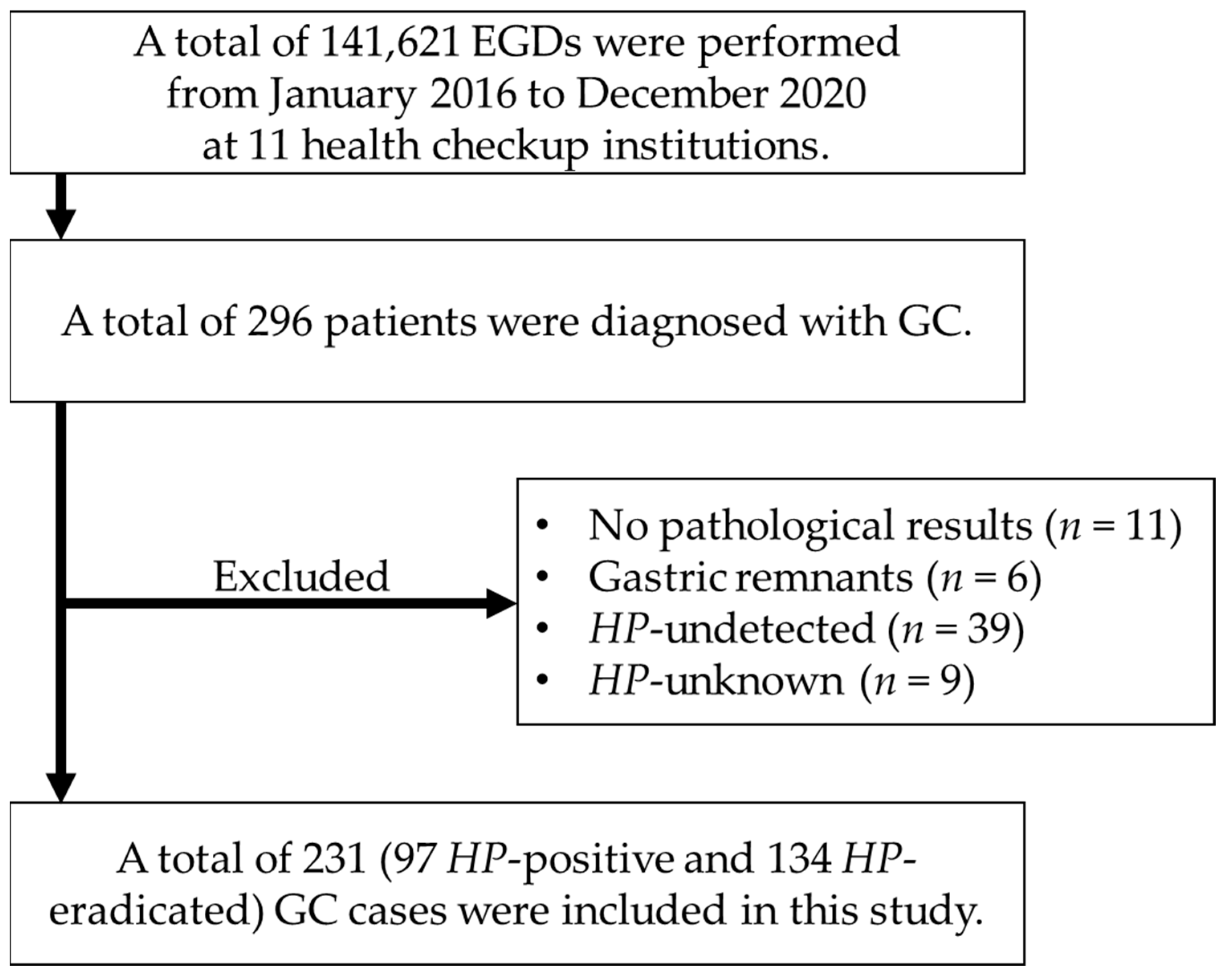
2.1. Patients

A multi-institutional, retrospective observational study involving 11 institutions in Akita Prefecture, Japan that perform screening endoscopies as part of a health checkup was conducted. All patients newly diagnosed with GC through screening endoscopy at these institutions between January 2016 and December 2020 were included in this study. Patients that were either HP-positive or HP-eradicated were defined as eligible participants. Those with GCs that occurred in the remnant stomach or lacked final pathological findings were excluded from this study.

2.2. Definition of HP Infection Status of GC

HP-positive GCs were defined as those with HP infection at the time of diagnosis, and HP-eradicated GCs were defined as those who were diagnosed at least one year after successful HP eradication. A patient was considered to be HP-positive if the patient tested positive in at least one of the following without a history of eradication therapy: A serum antibody test, stool antigen test, 13C-urea breath test, rapid urease test, or histological examination. Successful eradication was judged by a negative 13C-urea breath test [16].

2.3. Data Collection

Medical records were retrospectively reviewed to retrieve data regarding information at the time of GC diagnosis, such as patients’ sex, age, history of endoscopic resection for early-stage GC (primary or metachronous GC), tobacco and alcohol consumption, and the interval between previous and diagnostic endoscopies. In Japan, a two-year interval is recommended for screening endoscopy to detect early-stage GCs [17]. Therefore, the intervals between previous and diagnostic endoscopy examinations were defined as ≤1 year, >1 year to ≤2 years, and >2 years or never undergone. As cancer lesion-related factors, data on the longitudinal location (upper, middle, or lower part of the stomach) of the tumor, macroscopic type (elevated or depressed), tumor size (<20 mm or ≥20 mm), and histological type (differentiated or undifferentiated) were also collected. Data on the depth of tumor invasion were retrieved from the final pathological reports. The review was conducted by more than two experts with >10 years of experience.

2.4. Statistical Analysis

Categorical variables were expressed as the number and proportion (%) and compared using a Chi-square test or Fisher’s exact test, as appropriate. Continuous variables were expressed as the median with interquartile range and compared using a Mann–Whitney U test. Propensity score matching was applied to reduce selection bias and adjust for significant differences in baseline clinical characteristics between the groups. A propensity score was calculated using a logistic regression model incorporating the patients’ age, sex, smoking status, drinking status, and the presence of either a primary or metachronous GC as independent variables. The HP-positive and HP-eradicated groups were matched in a 1:1 ratio using the nearest neighbor method with a caliper width of 0.2 of the standard deviation of the logit of the propensity score. The effect of matching was assessed by an absolute standardized difference (ASD), and a covariate of ASD ≤ 0.1 was considered to be balanced. After propensity score matching, the association between HP eradication and GCs with deep tumor invasion was estimated using logistic regression analysis. Additionally, other factors that can influence the development of GCs with deep tumor invasion were analyzed in another multivariable model. The results of the regression analyses were expressed using odds ratios (ORs) and 95% confidence intervals (CIs). All statistical analyses were conducted using the EZR version 1.63 (Saitama Medical Center, Jichi Medical University, Saitama, Japan) [18], and a p < 0.05 was considered statistically significant.

2.5. Ethical Statement

All procedures were performed in accordance with the Helsinki Declaration of 1964 and later versions. The study protocol was approved by the Ethics Committee of Akita University (ID: 2849) and each participating institute. The need for informed consent was waived by the corresponding Ethics Committees because of the retrospective nature of this study.

3. Results

3.1. Characteristics of Study Population before and after Propensity Score Matching

During the study period, 141,621 screening esophagogastroduodenoscopies (EGDs) were performed in 11 health checkup institutions. From these, 296 (0.21%) patients were diagnosed with GC. The GC detection rate was similar to that reported for health checkup endoscopy in another area of Japan (0.22%) [19]. Among the 296 patients, 11 patients had final pathological results that could not be tracked, 6 patients had GCs arising in the remnant stomach, 39 were HP-undetected cases without a history of eradication, and 9 were HP-unknown cases. These patients were excluded from the study. A total of 231 cases of GC (97 HP-positive and 134 HP-eradicated cases) were included in this study (Figure 1). In terms of tumor invasion depth, there were 168 (72.7%) cases of pT1a, 14 (6.1%) cases of pT1b1, 33 (14.3%) cases of pT1b2, and 16 (6.9%) cases of pT2 or deeper. In terms of treatment, 165 patients (71.4%) underwent endoscopic submucosal dissection (ESD), 64 patients (27.7%) underwent surgery, 1 patient (0.4%) underwent chemotherapy, and 1 patient (0.4%) did not undergo any treatment. Table 1 shows the clinical characteristics of the patients, before and after propensity score matching. Before propensity score matching, the patients’ median age was 65 years (60–70), 199 (86.2%) were male, and 18 (7.8%) had metachronous GC. After propensity score matching, the patients’ median age was 64.5 years (60–70), 143 (88.3%) were male, and 12 (7.4%) had metachronous GC. All five matching factors were considered well-balanced. Both before and after propensity score matching, the interval between previous and diagnostic endoscopic examination was significantly longer in the HP-positive group than in the HP-eradicated group (p < 0.01). Although there was no significant difference in the macroscopic tumor type and tumor size before matching, depressed tumors and tumors measuring <20 mm were significantly more common in the HP-eradicated group after matching (p = 0.05 and p = 0.01, respectively). There was no significant difference in the depth of tumor invasion (pT1a, pT1b1, pT1b2, and pT2 or deeper) between the HP-eradicated group and the HP-positive group both before and after propensity score matching (71.6%, 6.7%, 14.9%, and 6.7% vs. 74.2%, 5.2%, 13.4%, and 7.2%, p = 0.96; 74.1%, 6.2%, 14.8%, and 4.9% vs. 69.1%, 6.2%, 16.0%, and 8.6%, p = 0.82).

3.2. Association between HP Eradication and GCs with Deep Invasion

Among 162 propensity score-matched GC cases, multivariable regression analysis was performed to estimate the association between HP eradication and GCs with pT1b or deeper invasion. As a result, the adjusted OR (95% CI) for HP eradication was 1.16 (0.48–2.81), suggesting no significant association with GCs with pT1b or deeper invasion (Table 2). Similarly, regarding the association of GCs with pT1b2 or deeper invasion, the adjusted OR (95% CI) for HP eradication was 1.16 (0.42–3.19), suggesting no significant association (Table 3). In sensitivity analyses, where HP-eradicated GCs were defined as those who were diagnosed at least two years after successful eradication, the results were largely unchanged, i.e., HP eradication was not significantly associated with deeper tumor invasion (Tables S1–S3).

3.3. Estimating Factors Associated with GCs with Deep Invasion

To estimate the associations between factors other than HP eradication, multivariable regression analysis was performed without propensity score matching. Factors associated with the development of GCs with pT1b or deeper invasion were its location (if in the upper third of the stomach), a tumor size ≥20 mm, and an undifferentiated histological type (Table 4). Similarly, factors associated with the development of GCs with pT1b2 or deeper invasion were being male, its location (if in the upper third of the stomach), a tumor size ≥20 mm, an undifferentiated histological type, or if the interval between previous and diagnostic endoscopy was >2 years or if it was never undergone (Table 5). Again, HP eradication was not associated with GCs with deeper tumor invasion in both multivariable models. Furthermore, a comparison between early-stage and advanced-stage GC revealed that although advanced-stage GC was more likely to have a size ≥20 mm and be of the undifferentiated type, once again HP eradication was not significantly associated with advanced-stage GC (Table S4).

4. Discussion

This study demonstrated that HP eradication does not significantly impact the depth of tumor invasion in GCs detected during screening EGD. Findings clinically significant to the depth of tumor invasion, such as the use of ESD for pT1a–pT1b1, and surgery for pT1b2 or deeper, are directly related to the recommended treatment.
Because healthcare insurance for eradication therapy targeting all HP-positive individuals has been widely implemented in Japan since 2013 [20], a substantial proportion of HP-positive patients have received treatment. With the increased number of HP-eradicated individuals, an increasing number of GCs are being diagnosed in these patient groups, as GCs still emerge even after successful HP eradication [5,21,22]. Consequently, recent studies have reported that the number of diagnosed GCs among patients that are HP-eradicated has already exceeded that in those that are HP-positive [6,23], which holds true in the current study. Therefore, GC detection by EGD in eradicated individuals, compared to traditionally prevalent HP-positive GC, has great clinical implications.
Studies have shown that HP eradication conceals the findings of early cancer lesions, which may lead to a delayed cancer diagnosis [7,8,9,10,24]. Moreover, two previous studies demonstrated that this concern could be a real issue by showing that post-eradicated GC showed a higher proportion of submucosal invasion than cases of HP-positive GC [11,12]. However, these two studies suffered from major selection bias, as the authors admitted, since the studies comprised GC patients who received ESD, excluding cases with early GC beyond the indications for ESD and those with advanced GC. In addition, these studies were performed in high-volume centers, which may have caused a referral bias [11,12]. To minimize this bias, the current study included an entire GC cohort consisting of those diagnosed via screening EGD in multiple health checkup institutes.
Propensity analysis demonstrated that HP eradication did not affect GCs with pT1b or deeper. Meanwhile, consistent with previous reports, we observed some morphological differences in GC between HP-eradicated and HP-positive subjects, e.g., the prevalence of the depressed type and small GCs was higher among patients that are HP-eradicated than in those that are HP-positive, after propensity score matching [8,10,11,25]. Still, HP eradication was not significantly associated with GCs with deep tumor invasion, even after adjusting for such cancer lesion-related factors, suggesting that the effect of HP eradication does not predispose one to develop GC with deep tumor invasion. As studies have demonstrated, HP eradication may make it difficult to diagnose early-stage GC by obscuring the characteristic lesions [7,8,9,10,24]. At the same time, other studies demonstrated that HP eradication decreased the proliferation of GC [7,25], suggesting that the growth of neoplastic lesions was decelerated in HP-eradicated cases [21,26,27,28]. Hence, the potentially delayed diagnosis of GCs after HP eradication due to the difficulty in endoscopic detection will be counterbalanced by the relatively slow growth of the cancer lesions. Consequently, HP eradication may yield no substantial disadvantages for detecting GC at an early stage, as demonstrated in the current study.
In the current study, multivariable regression models without propensity score matching revealed that tumors located in the upper third of the stomach, measuring ≥20 mm, and with an undifferentiated histological type are highly associated with T1b or deeper GCs. In addition to these cancer lesion-related factors, when the subjects of interest were defined as GCs with pT1b2 or deeper, a long-term interval (>two years or never undergone) between previous and diagnostic endoscopic examination was found to be significantly associated with deeper tumor invasion (Table 5). Screening EGD for GC performed every two years, which is the current recommendation in Japan [17,29,30], may be effective in detecting GCs before pT1b2 or deeper invasion, which requires surgical resection.
This study minimized selection bias by including consecutive patients with GCs of different stages diagnosed through screening EGD exclusively from 11 health checkup institutions over 5-year period. Thus, it provides a comprehensive report on the occurrence of GC in the area. Furthermore, this study was conducted in Akita Prefecture, Japan, where the mortality from GC is most rampant, and urgent actions are needed [13,14,15].
Attention should be paid to several limitations associated with this study. First, the retrospective nature of this study may be a limitation of this study, although each GC case was systematically registered at each institute once it was diagnosed at health checkup. Second, morphological and histological changes over time after successful eradication could not be evaluated in this study. Third, despite adopting a multicenter study design over five years, there were only 16 cases of advanced-stage GC, which limited the ability to conduct sufficient subgroup analysis. Finally, some study patients may have been misclassified regarding HP infection status. In this study, possible reinfection after successful eradication may have occurred, although the frequency of reinfection should be very low once the success was confirmed using a 13C-urea breath test [31]. In addition, in patients with a positive serum antibody test, HP may have been spontaneously eliminated due to advanced gastric atrophy, although such cases are supposed to be less common [32].

5. Conclusions

In conclusion, HP eradication does not adversely affect the clinical course of GC patients. Together with the well-established fact that HP eradication reduces the risk of developing GC [2], the findings in this study support the recommendation of HP eradication in screening programs to reduce the total number of GC cases without delaying diagnosis.

Supplementary Materials

The following supporting information can be downloaded at: https://www.mdpi.com/article/10.3390/cancers16040733/s1, Table S1: Baseline characteristics of the study patients before and after propensity score matching, when defining HP-eradicated GCs were defined as those who were diagnosed at least two years after successful eradication; Table S2: Risk of T1b–T4-GC associated with HP eradication, when defining HP-eradicated GCs were defined as those who were diagnosed as at least two years after successful eradication; Table S3: Risk of T1b2–T4-GC associated with HP eradication, when defining HP-eradicated GCs were defined as those who were diagnosed as at least two years after successful eradication; Table S4: Clinical characteristics of the study patients with pT1 and pT2 or deeper GC.

Author Contributions

Conceptualization, K.I.; Methodology, K.W. and K.I.; Formal analysis, K.W.; Investigation, S.T., K.W., S.F. (Sho Fukuda), T.Y., T.D., J.F., M.M., S.F. (Shusei Fujimori), M.F., K.S. (Kodai Shirayama), Y.H., S.F. (Saki Fushimi), S.U., K.O., R.O., T.H. (Takao Hoshino), T.H. (Toru Horii), T.K. (Taira Kuramitsu), K.S. (Kotaro Sakaki), T.I., T.K. (Taiga Komatsu), Y.Y., K.S. (Kenji Shirane), T.O., Y.S. and T.M.; Data curation, S.T. and K.W.; Writing—original draft, S.T. and K.W.; Writing—review & editing, K.W. and K.I.; Supervision, K.I. All authors have read and agreed to the published version of the manuscript.

Funding

This research received no external funding.

Institutional Review Board Statement

This study was conducted according to the guidelines of the Declaration of Helsinki and approved by the Ethics Committees of Akita University (ID: 2849) and each participating institute.

Informed Consent Statement

Patient consent was waived by the corresponding Ethics Committees due to the retrospective nature of this study.

Data Availability Statement

Data presented in this study are available upon reasonable request from the corresponding authors.

Conflicts of Interest

Katsunori Iijima has received lecture fees from Otsuka Pharmaceutical Co., Ltd. and Takeda Pharmaceutical Co., Ltd. All other authors declare no conflicts of interest.

References

  1. Hori, M.; Matsuda, T.; Shibata, A.; Katanoda, K.; Sobue, T.; Nishimoto, H.; Japan Cancer Surveillance Research Group. Cancer incidence and incidence rates in Japan in 2009: A study of 32 population-based cancer registries for the Monitoring of Cancer Incidence in Japan (MCIJ) project. Jpn. J. Clin. Oncol. 2015, 45, 884–891. [Google Scholar] [CrossRef]
  2. Ford, A.C.; Yuan, Y.; Moayyedi, P. Helicobacter pylori eradication therapy to prevent gastric cancer: Systematic review and meta-analysis. Gut 2020, 69, 2113–2121. [Google Scholar] [CrossRef]
  3. Fukase, K.; Kato, M.; Kikuchi, S.; Inoue, K.; Uemura, N.; Okamoto, S.; Terao, S.; Amagai, K.; Hayashi, S.; Asaka, M.; et al. Effect of eradication of Helicobacter pylori on incidence of metachronous gastric carcinoma after endoscopic resection of early gastric cancer: An open-label, randomised controlled trial. Lancet 2008, 372, 392–397. [Google Scholar] [CrossRef]
  4. Majima, A.; Handa, O.; Naito, Y.; Dohi, O.; Okayama, T.; Yoshida, N.; Kamada, K.; Katada, K.; Uchiyama, K.; Ishikawa, T.; et al. Early-Stage Gastric Cancer Can Be Found in Improved Atrophic Mucosa over Time from Successful Helicobacter pylori Eradication. Digestion 2017, 95, 194–200. [Google Scholar] [CrossRef]
  5. Ami, R.; Hatta, W.; Iijima, K.; Koike, T.; Ohkata, H.; Kondo, Y.; Ara, N.; Asanuma, K.; Asano, N.; Imatani, A.; et al. Factors Associated with Metachronous Gastric Cancer Development After Endoscopic Submucosal Dissection for Early Gastric Cancer. J. Clin. Gastroenterol. 2017, 51, 494–499. [Google Scholar] [CrossRef]
  6. Oda, I.; Hoteya, S.; Fujishiro, M. Status of Helicobacter pylori infection and gastric mucosal atrophy in patients with gastric cancer: Analysis based on the Japan Endoscopy Database. Dig. Endosc. 2019, 31, 103. [Google Scholar] [CrossRef] [PubMed]
  7. Ito, M.; Tanaka, S.; Takata, S.; Oka, S.; Imagawa, S.; Ueda, H.; Egi, Y.; Kitadai, Y.; Yasui, W.; Yoshihara, M.; et al. Morphological changes in human gastric tumours after eradication therapy of Helicobacter pylori in a short-term follow-up. Aliment. Pharmacol. Ther. 2005, 21, 559–566. [Google Scholar] [CrossRef]
  8. Ito, M.; Tanaka, S.; Chayama, K. Characteristics and Early Diagnosis of Gastric Cancer Discovered after Helicobacter pylori Eradication. Gut Liver 2021, 15, 338–345. [Google Scholar] [CrossRef]
  9. Kobayashi, M.; Hashimoto, S.; Nishikura, K.; Mizuno, K.; Takeuchi, M.; Sato, Y.; Ajioka, Y.; Aoyagi, Y. Magnifying narrow-band imaging of surface maturation in early differentiated-type gastric cancers after Helicobacter pylori eradication. J. Gastroenterol. 2013, 48, 1332–1342. [Google Scholar] [CrossRef] [PubMed]
  10. Kobayashi, M.; Sato, Y.; Terai, S. Endoscopic surveillance of gastric cancers after Helicobacter pylori eradication. World J. Gastroenterol. 2015, 21, 10553–10562. [Google Scholar] [CrossRef] [PubMed]
  11. Maehata, Y.; Nakamura, S.; Esaki, M.; Ikeda, F.; Moriyama, T.; Hida, R.; Washio, E.; Umeno, J.; Hirahashi, M.; Kitazono, T.; et al. Characteristics of Primary and Metachronous Gastric Cancers Discovered after Helicobacter pylori Eradication: A Multicenter Propensity Score-Matched Study. Gut Liver 2017, 11, 628–634. [Google Scholar] [CrossRef]
  12. Hata, K.; Ito, M.; Boda, T.; Kotachi, T.; Kiso, M.; Masuda, K.; Kurihara, M.; Kuroki, K.; Yorita, N.; Nagasaki, N.; et al. Gastric Cancer with Submucosal Invasion after Successful Helicobacter pylori Eradication: A Propensity Score-Matched Analysis of Patients with Annual Patient Endoscopic Survey. Digestion 2019, 99, 59–65. [Google Scholar] [CrossRef] [PubMed]
  13. Koizumi, S.; Motoyama, S.; Watanabe, N.; Matsuhashi, T.; Iijima, K. Chronological Changes in the Gastric Cancer Subsite in Akita, Japan: The Trends from the Data of a Hospital-Based Registration System. Tohoku J. Exp. Med. 2018, 246, 131–140. [Google Scholar] [CrossRef]
  14. Iijima, K.; Jin, M.; Miura, M.; Watanabe, K.; Watanabe, N.; Shimodaira, Y.; Koizumi, S.; Tobori, F.; Motoyama, S. Disturbance of Gastrointestinal Cancers Diagnoses by the COVID-19 Pandemic in a Depopulated Area of Japan: A Population-Based Study in Akita Prefecture. Tohoku J. Exp. Med. 2022, 257, 65–71. [Google Scholar] [CrossRef] [PubMed]
  15. Iijima, K.; Shimodaira, Y.; Watanabe, K.; Koizumi, S.; Matsuhashi, T.; Jin, M.; Miura, M.; Onochi, K.; Yamai, K.; Fujishima, Y.; et al. A Follow-up Report on the Diagnosis of Gastrointestinal Cancer during the COVID-19 Pandemic in Akita Prefecture, Japan in 2021. Tohoku J. Exp. Med. 2023, 259, 301–306. [Google Scholar] [CrossRef] [PubMed]
  16. Chadwick, G.; Groene, O.; Hoare, J.; Hardwick, R.H.; Riley, S.; Crosby, T.D.; Hanna, G.B.; Cromwell, D.A. A population-based, retrospective, cohort study of esophageal cancer missed at endoscopy. Endoscopy 2014, 46, 553–560. [Google Scholar] [CrossRef] [PubMed]
  17. Hamashima, C.; Fukao, A. Quality assurance manual of endoscopic screening for gastric cancer in Japanese communities. Jpn. J. Clin. Oncol. 2016, 46, 1053–1061. [Google Scholar] [CrossRef]
  18. Kanda, Y. Investigation of the freely available easy-to-use software ‘EZR’ for medical statistics. Bone Marrow Tansplant. 2013, 48, 452–458. [Google Scholar] [CrossRef]
  19. Aida, K.; Yoshikawa, H.; Mochizuki, C.; Mori, A.; Muto, S.; Fukuda, T.; Otsuki, M. Clinicopathological features of gastric cancer detected by endoscopy as part of annual health checkup. J. Gastroenterol. Hepatol. 2008, 23, 632–637. [Google Scholar] [CrossRef]
  20. Asaka, M. A new approach for elimination of gastric cancer deaths in Japan. Int. J. Cancer 2013, 132, 1272–1276. [Google Scholar] [CrossRef]
  21. Yan, L.; Chen, Y.; Chen, F.; Tao, T.; Hu, Z.; Wang, J.; You, J.; Wong, B.C.Y.; Chen, J.; Ye, W. Effect of Helicobacter pylori Eradication on Gastric Cancer Prevention: Updated Report From a Randomized Controlled Trial With 26.5 Years of Follow-up. Gastroenterology 2022, 163, 154–162.e3. [Google Scholar] [CrossRef] [PubMed]
  22. Kwon, Y.H.; Heo, J.; Lee, H.S.; Cho, C.M.; Jeon, S.W. Failure of Helicobacter pylori eradication and age are independent risk factors for recurrent neoplasia after endoscopic resection of early gastric cancer in 283 patients. Aliment. Pharmacol. Ther. 2014, 39, 609–618. [Google Scholar] [CrossRef] [PubMed]
  23. Hatta, W.; Koike, T.; Asonuma, S.; Okata, H.; Uno, K.; Oikawa, T.; Iwai, W.; Yonechi, M.; Fukushi, D.; Kayaba, S.; et al. Smoking history and severe atrophic gastritis assessed by pepsinogen are risk factors for the prevalence of synchronous gastric cancers in patients with gastric endoscopic submucosal dissection: A multicenter prospective cohort study. J. Gastroenterol. 2023, 58, 433–443. [Google Scholar] [CrossRef]
  24. Oka, K.; Iwai, N.; Okuda, T.; Hara, T.; Inada, Y.; Tsuji, T.; Komaki, T.; Sakagami, J.; Naito, Y.; Kagawa, K.; et al. Clinical Features of False-Negative Early Gastric Cancers: A Retrospective Study of Endoscopic Submucosal Dissection Cases. Gastroenterol. Res. Pract. 2021, 2021, 6635704. [Google Scholar] [CrossRef]
  25. Yamamoto, K.; Kato, M.; Takahashi, M.; Haneda, M.; Shinada, K.; Nishida, U.; Yoshida, T.; Sonoda, N.; Ono, S.; Nakagawa, M.; et al. Clinicopathological analysis of early-stage gastric cancers detected after successful eradication of Helicobacter pylori. Helicobacter 2011, 16, 210–216. [Google Scholar] [CrossRef] [PubMed]
  26. Alexandre, L.; Tsilegeridis-Legeris, T.; Lam, S. Clinical and Endoscopic Characteristics Associated with Post-Endoscopy Upper Gastrointestinal Cancers: A Systematic Review and Meta-analysis. Gastroenterology 2022, 162, 1123–1135. [Google Scholar] [CrossRef]
  27. Yasuda, T.; Dohi, O.; Yamada, S.; Ishida, T.; Iwai, N.; Hongo, H.; Terasaki, K.; Tanaka, M.; Yamada, N.; Kamada, K.; et al. Risk and prognostic factors of invasive gastric cancer detection during surveillance endoscopy: Multi-institutional cross-sectional study. Dig. Endosc. 2023, 35, 592–602. [Google Scholar] [CrossRef]
  28. Argueta, E.A.; Moss, S.F. The prevention of gastric cancer by Helicobacter pylori eradication. Curr. Opin. Gastroenterol. 2021, 37, 625–630. [Google Scholar] [CrossRef]
  29. Shin, W.S.; Xie, F.; Chen, B.; Yu, P.; Yu, J.; To, K.F.; Kang, W. Updated Epidemiology of Gastric Cancer in Asia: Decreased Incidence but Still a Big Challenge. Cancers 2023, 15, 2639. [Google Scholar] [CrossRef]
  30. Januszewicz, W.; Turkot, M.H.; Malfertheiner, P.; Regula, J. A Global Perspective on Gastric Cancer Screening: Which Concepts Are Feasible, and When? Cancers 2023, 15, 664. [Google Scholar] [CrossRef]
  31. Take, S.; Mizuno, M.; Ishiki, K.; Imada, T.; Okuno, T.; Yoshida, T.; Yokota, K.; Oguma, K.; Kita, M.; Okada, H.; et al. Reinfection rate of Helicobacter pylori after eradication treatment: A long-term prospective study in Japan. J. Gastroenterol. 2012, 47, 641–646. [Google Scholar] [CrossRef] [PubMed]
  32. Kokkola, A.; Kosunen, T.U.; Puolakkainen, P.; Sipponen, P.; Harkonen, M.; Laxen, F.; Virtamo, J.; Haapiainen, R.; Rautelin, H. Spontaneous disappearance of Helicobacter pylori antibodies in patients with advanced atrophic gastritis. APMIS 2003, 111, 619–624. [Google Scholar] [CrossRef] [PubMed]
Figure 1. Flow diagram of the enrollment of this study. EGD esophagogastroduodenoscopy; GC gastric cancer; HP Helicobacter pylori.
Cancers 16 00733 g001
Table 1. Baseline characteristics of the study patients before and after propensity score matching.
Before Propensity Score MatchingAfter Propensity Score Matching
HP-Eradicated
(n = 134)
HP-Positive
(n = 97)
p ValueASDHP-Eradicated
(n = 81)
HP-Positive
(n = 81)
p ValueASD
Male sex, n (%)115 (85.8)84 (86.6)1.000.0272 (88.9)71 (87.7)1.000.04
Age, years, median (IQR)65 (61–71)64 (59–70)0.310.1364 (59–71)66 (60–70)0.940.03
Metachronous cancer, n (%)11 (8.2)7 (7.2)1.000.046 (7.4)6 (7.4)1.00<0.01
Smoking status, n (%)
Current smoker35 (26.1)28 (28.9)0.840.0822 (27.2)24 (29.6)0.890.07
Past smoker27 (20.1)17 (17.5) 16 (19.8)14 (17.3)
Never smoker67 (50.0)48 (49.5) 43 (53.1)43 (53.1)
Unknown5 (3.7)4 (4.1) 0 (0.0)0 (0.0)
Drinking status, n (%)
Current drinker92 (68.7)68 (70.1)1.000.0460 (74.1)61 (75.3)1.000.07
Past drinker5 (3.7)3 (3.1) 3 (3.7)2 (2.5)
Never drinker30 (22.4)21 (21.6) 18 (22.2)18 (22.2)
Unknown7 (5.2)5 (5.2) 0 (0.0)0 (0.0)
Longitudinal location, n (%)
Upper third28 (20.9)15 (15.5)0.310.1416 (19.8)14 (17.3)0.840.06
Middle or lower third106 (79.1)82 (84.5) 65 (80.2)67 (82.7)
Macroscopic type, n (%)
Elevated type22 (16.4)24 (24.7)0.130.2110 (12.3)21 (25.9)0.050.35
Depressed type112 (83.6)73 (75.3) 71 (87.7)60 (74.1)
Tumor size, n (%)
<20 mm90 (67.2)55 (56.7)0.130.2259 (72.8)43 (53.1)0.010.42
≥20 mm44 (32.8)42 (43.3) 22 (27.2)38 (46.9)
Histological type, n (%)
Differentiated type112 (83.6)83 (85.6)0.720.0671 (87.7)70 (86.4)1.000.04
Undifferentiated type22 (16.4)14 (14.4) 10 (12.3)11 (13.6)
Interval between previous and diagnostic endoscopic examination, n (%)
≤1 year78 (58.2)30 (30.9)<0.010.7345 (55.6)25 (30.9)<0.010.75
>1 year, ≤2 years28 (20.9)15 (15.5) 21 (25.9)14 (17.3)
>2 years or never28 (20.9)52 (53.6) 15 (18.5)42 (51.9)
Depth of tumor invasion, n (%)
T1a96 (71.6)72 (74.2)0.960.0860 (74.1)56 (69.1)0.820.16
T1b19 (6.7)5 (5.2) 5 (6.2)5 (6.2)
T1b220 (14.9)13 (13.4) 12 (14.8)13 (16.0)
T2 or deeper9 (6.7)7 (7.2) 4 (4.9)7 (8.6)
ASD absolute standardized difference; HP Helicobacter pylori; IQR interquartile range.
Table 2. Risk of T1b–T4-GC associated with HP eradication.
Adjusted OR a95% CIp Value
Longitudinal locationUpper third2.010.78–5.130.15
Middle or lower thirdreference
Macroscopic typeDepressed type2.640.77–9.080.12
Elevated typereference
Tumor size≥20 mm5.132.30–11.40<0.01
Histological typeUndifferentiated type3.231.11–9.370.03
Differentiated typereference
Interval between previous and diagnostic endoscopic examination≤1 yearreference
>1 year, ≤2 years1.320.45–3.900.62
>2 years or never1.760.68–4.580.25
HP infection statusHP-eradicated1.160.48–2.810.74
HP-positivereference
CI confidence interval; GC gastric cancer; HP Helicobacter pylori; OR odds ratio; a Adjusted by longitudinal location, macroscopic type, tumor size, histological type, interval between previous and diagnostic endoscopic examination, and HP infection status.
Table 3. Risk of T1b2–T4-GC associated with HP eradication.
Adjusted OR a95% CIp Value
Longitudinal locationUpper third3.161.10–9.050.03
Middle or lower thirdreference
Macroscopic typeDepressed type11.101.30–94.000.03
Elevated typereference
Tumor size≥20 mm7.152.81–18.20<0.01
Histological typeUndifferentiated type2.980.96–9.310.06
Differentiated typereference
Interval between previous and diagnostic endoscopic examination≤1 yearreference
>1 year, ≤2 years2.550.72–9.080.15
>2 years or never2.710.88–8.330.08
HP infection statusHP-eradicated1.160.42–3.190.78
HP-positivereference
CI confidence interval; GC gastric cancer; HP Helicobacter pylori; OR odds ratio; a Adjusted by longitudinal location, macroscopic type, tumor size, histological type, interval between previous and diagnostic endoscopic examination, and HP infection status.
Table 4. Estimating factors associated with T1b–T4-GC.
Adjusted OR a95% CIp Value
SexMale1.580.54–4.630.40
Age 0.980.94–1.030.43
Metachronous cancer 0.190.02–1.600.13
Longitudinal locationUpper third2.361.06–5.250.04
Middle or lower thirdreference
Macroscopic typeDepressed type1.270.49–3.290.63
Elevated typereference
Tumor size≥20 mm5.882.95–11.70<0.01
Histological typeUndifferentiated type3.351.43–7.86<0.01
Differentiated typereference
Interval between previous and diagnostic endoscopic examination≤1 yearreference
>1 year, ≤2 years1.160.44–3.050.77
>2 years or never1.810.81–4.070.15
HP infection statusHP-eradicated1.630.77–3.440.20
HP-positivereference
CI confidence interval; GC gastric cancer; HP Helicobacter pylori; OR odds ratio; a Adjusted by sex, age, metachronous cancer, longitudinal location, macroscopic type, tumor size, histological type, interval between previous and diagnostic endoscopic examination, and HP infection status.
Table 5. Estimating factors associated with T1b2–T4-GC.
Adjusted OR a95% CIp Value
SexMale4.751.07–21.200.04
Age 0.970.92–1.020.29
Metachronous cancer 0.310.03–2.770.29
Longitudinal locationUpper third2.701.12–6.470.03
Middle or lower thirdreference
Macroscopic typeDepressed type1.800.57–5.710.32
Elevated typereference
Tumor size≥20 mm7.923.56–17.60<0.01
Histological typeUndifferentiated type3.711.48–9.33<0.01
Differentiated typereference
Interval between previous and diagnostic endoscopic examination≤1 yearreference
>1 year, ≤2 years1.840.63–5.360.27
>2 years or never2.761.09–6.960.03
HP infection statusHP-eradicated1.740.75–4.030.20
HP-positivereference
CI confidence interval; GC gastric cancer; HP Helicobacter pylori; OR odds ratio; a Adjusted by sex, age, metachronous cancer, longitudinal location, macroscopic type, tumor size, histological type, interval between previous and diagnostic endoscopic examination, and HP infection status.
Disclaimer/Publisher’s Note: The statements, opinions and data contained in all publications are solely those of the individual author(s) and contributor(s) and not of MDPI and/or the editor(s). MDPI and/or the editor(s) disclaim responsibility for any injury to people or property resulting from any ideas, methods, instructions or products referred to in the content.

