Article

New Insights into Mucosa-Associated Microbiota in Paired Tumor and Non-Tumor Adjacent Mucosal Tissues in Colorectal Cancer Patients

by
Adriana González
1,
Asier Fullaondo
1,
David Navarro
2,
Javier Rodríguez
3,
Cristina Tirnauca
4 and
Adrian Odriozola
1,*
1
Department of Genetics, Physical Anthropology and Animal Physiology, University of the Basque Country UPV/EHU, 48940 Bilbao, Spain
2
Danagen-Bioted S.L., 08915 Barcelona, Spain
3
Department of Oncology, Clínica Universidad de Navarra, 31008 Pamplona, Spain
4
Department of Mathematics, Statistics and Computer Science, University of Cantabria, 39005 Santander, Spain
*
Author to whom correspondence should be addressed.
Cancers 2024, 16(23), 4008; https://doi.org/10.3390/cancers16234008
Submission received: 21 October 2024 / Revised: 24 November 2024 / Accepted: 27 November 2024 / Published: 29 November 2024
(This article belongs to the Special Issue Oncogenetics of Colorectal Cancer)

Simple Summary

Colorectal cancer (CRC) is a major burden of disease worldwide. Increasing scientific evidence highlights the role of the gut microbiota in the initiation, development and treatment of CRC. Currently, the analysis of CRC-associated gut microbiota has several limitations that hinder its implementation in precision medicine, including selection of sample type, sequencing platform and taxonomic classification. This article aims to address these constraints to provide data on CRC-associated microbiota and facilitate the implementation of its analysis in personalized medicine. To this end, mucosa-associated microbiota from paired tumor and non-tumor adjacent tissue samples from 65 CRC patients was analyzed through V3–V4 region of 16S rRNA gene amplification, MinION sequencing and NCBI taxonomic classification. Results consistent with available evidence have been obtained. Moreover, to our knowledge, this is the first study that identifies the possible association between a higher relative abundance of Streptococcus periodonticum and a lower relative abundance of Corynebacterium with CRC.

Abstract

Background/Objective: Colorectal cancer (CRC) is one of the most common cancers worldwide. Increasing scientific evidence supports the idea that gut microbiota dysbiosis accompanies colorectal tumorigenesis, and these changes could be causative. Implementing gut microbiota analysis in clinical practice is limited by sample type, sequencing platform and taxonomic classification. This article aims to address these limitations, providing new insights into the microbiota associated with CRC pathogenesis and implementing its analyses in personalized medicine. Methods: To that aim, we evaluate differences in the bacterial composition of 130 paired tumor and non-tumor adjacent tissues from a cohort of CRC patients from the Biobank of the University of Navarra, Spain. The V3–V4 region of the 16S rRNA gene was amplified, sequenced using the MinION platform, and taxonomically classified using the NCBI database. Results: To our knowledge, this is the first study to report an increased relative abundance of Streptococcus periodonticum and a decreased relative abundance of Corynebacterium associated with CRC. Genera such as Fusobacterium, Leptotrichia and Streptococcus showed higher relative abundances in tumor than in non-tumor tissues, as previously described in the literature. Specifically, we identified higher levels of Fusobacterium animalis, Fusobacterium nucleatum, Fusobacterium polymorphum and S. periodonticum in tumor tissues. In contrast, genera such as Bacteroides and Corynebacterium showed lower relative abundances in tumor tissues. There were also differences at the taxonomic level between tumor locations. Conclusions: These results, consistent with previous studies, further support the hypothesis that Leptotrichia and Fusobacterium contribute to CRC progression, with F. nucleatum and F. animalis proposed as key CRC pathogenic taxa. Overall, these results contribute to a better understanding of the CRC-associated microbiota, addressing critical barriers to its implementation in personalized medicine.

1. Introduction

Colorectal cancer (CRC) is one of the most common cancers and a major global burden of disease. In 2022, more than 1.9 million new cases and more than 900,000 deaths were estimated worldwide due to this disease. Despite advances in prevention, diagnosis and treatment, by 2045, the incidence and mortality of CRC are projected to increase by 70.5% and 83.4%, respectively [1].
Increasing scientific evidence has shown that human gut microbiota plays a critical role in CRC pathogenesis [2,3]. The gut microbiota, the community of microorganisms symbiotically inhabiting our gut, can be modulated by several lifestyle factors, among which diet stands out [4]. It is estimated that a 70% reduction in CRC incidence could be achieved by following a healthy and balanced diet [5,6]. Scientific evidence suggests that the key link between CRC and diet may lie in the gut microbiota [7,8].
Gut microbiota composition imbalance, known as dysbiosis, contributes to the start and progression of CRC. Previous works have identified CRC key pathogens, such as Fusobacterium nucleatum [9,10], Streptococcus gallolyticus [11], enterotoxigenic Bacteroides fragilis [12], Peptostreptococcus anaerobius [13] and Clostridioides difficile [14]. These pathogens may contribute to colorectal carcinogenesis directly by damaging DNA and stimulating colonocyte proliferation or indirectly by promoting a favorable environment for CRC development [15,16]. Otherwise, some bacteria in the gut microbiota exert protective effects against CRC. The main mechanisms by which the gut microbiota plays a key role in colorectal carcinogenesis include intestinal permeability regulation, inflammation and immune response modulation, biofilm formation, genotoxin production, virulence factors, oxidative stress and metabolite production [17].
There are two hypotheses regarding the role of the gut microbiota in colorectal carcinogenesis. The “alpha-bug” hypothesis suggests that certain pro-oncogenic microorganisms can displace cancer-protective bacteria and colonize the tumor persistently, creating an environment favorable for tumorigenesis [18]. On the other hand, the driver-passenger model suggests that “driver” bacteria that initiate CRC are then replaced by “passenger” bacteria with growth advantages in the tumor microenvironment (TME) that may exert promoting or inhibiting effects on the tumor progression [19].
Due to scientific evidence supporting its influence on CRC pathogenesis, gut microbiota is becoming increasingly important in personalized medicine, which relies on patients’ genetic and molecular characteristics to adapt the therapeutic strategy [20].
Consequently, we hypothesized that microbiota composition would differ between paired tumor and non-tumor adjacent tissues of CRC patients and between tumor locations and that analyzing these possible differences would help better understand how the tumor microbiota influences the pathogenesis of CRC. Specifically, (1) we compared paired tumor and non-tumor adjacent tissues from 65 Spanish CRC patients, and (2) we examined differences in microbial composition concerning the tumor location.
However, several challenges in analyzing the CRC-associated microbiota limit its implementation in personalized medicine, such as sample type, the sequencing platform, and the database for bacterial taxonomic classification [21].
Regarding sample type, most published studies examining microbiota composition in CRC have analyzed fecal samples. Although easy to obtain, fecal samples contain microbiota from different intestinal locations and do not adequately reflect microbiota interactions within the intestinal epithelium and TME as direct tumor tissue samples do. However, despite the importance of local CRC tissue-associated microbiota, there is a lack of solid direct information on tumor tissue samples [21].
Regarding sequencing platforms, microbiota analysis is limited in personalized medicine due to the lack of access to expensive and time-consuming conventional technology [22]. In this context, the sequencing platform MinION from Oxford Nanopore Technologies (ONT) implies a revolution exhibiting translational potential in clinical practice due to its portability, low cost and real-time sequencing [23,24].
Finally, the taxonomic assignment of 16S ribosomal RNA (rRNA) gene reads is commonly carried out by SILVA, RDP or Greengenes. While those tools map well on NCBI, the contrary is problematic because NCBI comprises more taxa and sequences and is continuously updated and curated. As a result, NCBI shows higher effectiveness in classifying 16S rRNA gene reads than the other available tools [25].
Overall, this study aims to overcome the main challenges in CRC-associated microbiota analysis, including sample type, the sequencing platform, and the database for bacterial taxonomic classification, to enhance our understanding of CRC-associated gut microbiota alteration and facilitate the implementation of their analysis in personalized medicine.
To this end, we amplified V3–V4 regions of the 16rRNA bacterial gene by the sequencing platform MinION and performed the taxonomic assignment using the NCBI Taxonomy Database.

2. Materials and Methods

2.1. Ethics Statement and Sample Collection

One hundred and thirty paired tumor and non-tumor adjacent tissue samples were obtained from 65 CRC patients of the Biobank of the University of Navarra, Spain. Samples and data from patients included in the study were provided by the Biobank of the University of Navarra and were processed following Standard Operating Procedures approved by the Ethical and Scientific Committees of Clinica Universitaria de Navarra (CUN) for the research (REINFORCE_0011-1411-2020-000102). All individuals gave written informed consent.
Samples from tumor and non-tumor adjacent mucosal tissues were obtained from each CRC patient by biopsy forceps during endoscopy and tumor removal surgery in the CUN. The pathologist selected, if possible, a fragment of tumor tissue and a fragment of non-tumor adjacent mucosal tissues. A Biobank technician, working in sterile conditions and with the material on dry ice, cut the selected tissue into small 2–3 mm square fragments placed in a cryotube for immediate freezing in dry ice. All were registered and stored at −80 °C at the Biobank until DNA extraction.
The general information (age and gender) and clinical data (tumor location, tumor differentiation and tumor stage) of samples are shown in Supplementary Table S1. For the 65 CRC patients, the origin of the paired tissue samples was diverse: the colon for 27, the rectum for 28, the sigmoid colon for 9 and the cecum for 1.

2.2. DNA Extraction and Quantitation

DNA was extracted using the Danagene Microbiome Tissue DNA kit (Danagen-Bioted S.L., Barcelona, Spain). DNA quantitation was carried out by fluorometry (Qubit 2.0, Life Technologies, Carlsbad, CA, USA, Thermo Fisher Scientific, Waltham, MA, USA) using HS dsDNA Assay (ThermoFisher Scientific, Waltham, MA, USA) and by spectrophotometry (NanoDrop 2000c, Thermo Fisher Scientific, Waltham, MA, USA). Negative DNA extraction controls were included.

2.3. PCR Amplification

PCR amplification of the 450 base pair (bp) V3–V4 region of the 16S rRNA gene was conducted using the Molzym Mastermix 16S complete DNA-free kit (Molzym, Bremen, Germany). Amplification was performed using an Applied Biosystems VeritiTM Thermal Cycler (Thermo Fischer Scientific, Waltham, MA, USA) according to the manufacturer’s protocol. Negative PCR amplification controls were included.

2.4. Library Preparation and Amplicon Sequencing

PCR products were purified using magnetic beads of the Clean NGS reagent (CleanNA, Waddinxveen, The Netherlands). Then, each mixture was quantified by fluorometry (Qubit 2.0, Life Technologies, Carlsbad, CA, USA, ThermoFisher Scientific, Waltham, MA, USA) using HS dsDNA Assay (ThermoFisher Scientific, Waltham, MA, USA) to calculate the DNA input for library preparation.
Multiplex MinION sequencing was carried out using 16S rRNA gene amplicons (SQK-NBD114.96; Oxford Nanopore Technologies, Oxford, UK). In addition, 130 ng DNA per sample was used for amplicon library preparation. As different clinical samples were combined into pooled libraries to homogenize conditions and obtain comparable results, barcoded adapters were used. During this process, end-repair procedures and adapter ligation were carried out. MinIONTM sequencing was performed using a MinION nanopore DNA sequencer (MIN-101B) and Flow Cell R10 (FLO-MIN114) according to the manufacturer’s instructions (Oxford Nanopore Technologies, Oxford, UK).

2.5. Data Acquisition and Sequencing Data Analysis

MINKNOW UI software version 23.04.3 (Oxford Nanopore Technologies, Oxford, UK) was used for data acquisition and base-calling converting sequence reads (i.e., FAST5 data) into FASTQ files by Guppy version 6.5.7 pipeline (Oxford Nanopore Technologies, Oxford, UK).
The Barcoding workflow in the Metrichor Ltd. analysis platform EPI2ME (Oxford Nanopore Technologies, Oxford, UK) was used for taxonomic classification. For that purpose, FASTQ files were uploaded to the EPI2ME desktop agent 16S workflow (Oxford Nanopore Technologies, Oxford, UK), where real-time classification was carried out using the NCBI 16S rRNA gene blast database. Reads were filtered for Q-score ≥ 9. The MinION was run for up to 48 h. Results were processed by in-house software to avoid infra representation of each taxonomical ID and convert reads in relative abundance according to the estimated 16S gene copy number (GCN) for each taxon based on the rrnDB database version 5.9 [26].

2.6. Bioinformatics Analysis

For α-diversity analysis, community richness was calculated using the number of observed Operational Taxonomic Units (OTUs) and the Chao1 index, whereas diversity and evenness were analyzed by calculating the Shannon–Weaver index [27] and the Simpson index [28] using Python 3.11. Data visualization for α-diversity results was performed with box plots using Python 3.11. For β-diversity analysis, two metrics for OTUs relative abundance were generated: Bray–Curtis dissimilarity and Jaccard index and the correspondent matrices. The β-diversity results were plotted using PAST 4.13 in a Principal coordinates analysis (PCoA). Bioinformatic analysis was performed to establish specific qualitative and quantitative microbiota compositions between groups using GraphPad 8.0. Data visualization for differential abundance analysis was performed with stacking maps and violin plots using SRPlot [29].

2.7. Statistical Analysis

A two-tailed paired t-test was performed to compare α-diversity mean differences between tumor and non-tumor tissue groups for the number of observed OTUs, Chao1, Shannon and Simpson indexes. The Mann–Whitney U test was performed to compare α-diversity mean differences between colon, rectum and sigmoid colon tumor tissue locations. A p-value below 0.05 was considered to be statistically significant.
To evaluate differences in β-diversity, we used Analysis of Similarities (ANOSIM) and Permutational Multivariate Analysis of Variance (PERMANOVA) tests using two metrics, Bray–Curtis dissimilarity and Jaccard index, in PAST 4.13.
Multiple t-tests were used to evaluate OTUs relative abundance differences between tumor and non-tumor tissue groups in GraphPad Prism 8.0. The Benjamini, Krieger and Yekutieli method for controlling the False Discovery Rate (FDR) was used to consider multiple comparisons. FDR-adjusted p-values below 0.01 were considered statistically significant.

3. Results

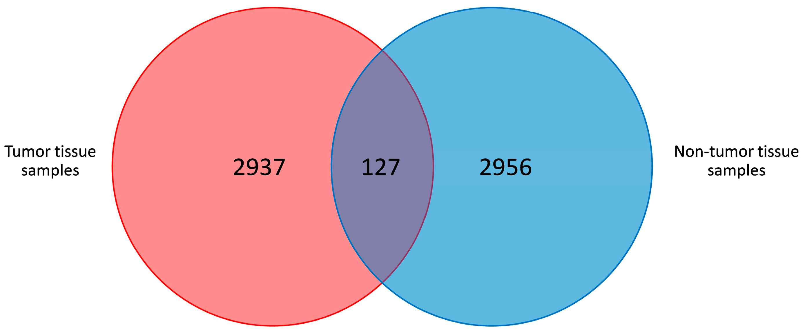
3.1. Sequence Analysis

We analyzed the microbiota composition of paired tumor and non-tumor adjacent tissue samples from 65 CRC patients. 27,305,189 raw reads were analyzed through 16S rRNA gene sequencing with a mean length of 637.37 ± 13.2 bp, and an average quality score of 10 ± 0.36. Two paired tissue samples were excluded because no reads were obtained for the non-tumor sample. A total of 25,193,582 reads were assigned to the remaining 128 samples. After quality filtering, 18,878,209 high-quality reads from the 128 samples were obtained. The average reads per sample for the tumor and non-tumor tissues were 155,274 ± 188,803 and 136,836 ± 160,153, respectively (p = 0.421) (Figure 1). Overall, 3879 different OTUs were identified at a 97% similarity threshold.

3.2. α- and β-Diversity

3.2.1. α-Diversity of Microbiota in Tumor Compared to Non-Tumor Adjacent Tissue Samples of CRC Patients

We observed higher community richness (number of OTUs, Chao1 index) and diversity and evenness (Shannon and Simpson indexes) in the non-tumoral compared to the tumoral group.
Three thousand eight hundred and seventy-nine OTUs were identified, with 127 OTUs shared among non-tumor and tumor tissue samples (Figure 1). The average number of OTUs (mean value ± standard error) in the non-tumor and tumor tissue was 503 ± 180 and 488 ± 178, respectively (p = 0.499).
A comparison between non-tumor and tumor tissue only reported statistically significant differences for Shannon Index (5.73 ± 0.69 and 5.41 ± 1.03, respectively; p = 0.014), while no statistically significant differences were reported for Chao1 (277 ± 127 and 268 ± 123, respectively; p = 0.565) or Simpson indexes (0.93 ± 0.09 and 0.95 ± 0.03, respectively; p = 0.069) (Figure 2).

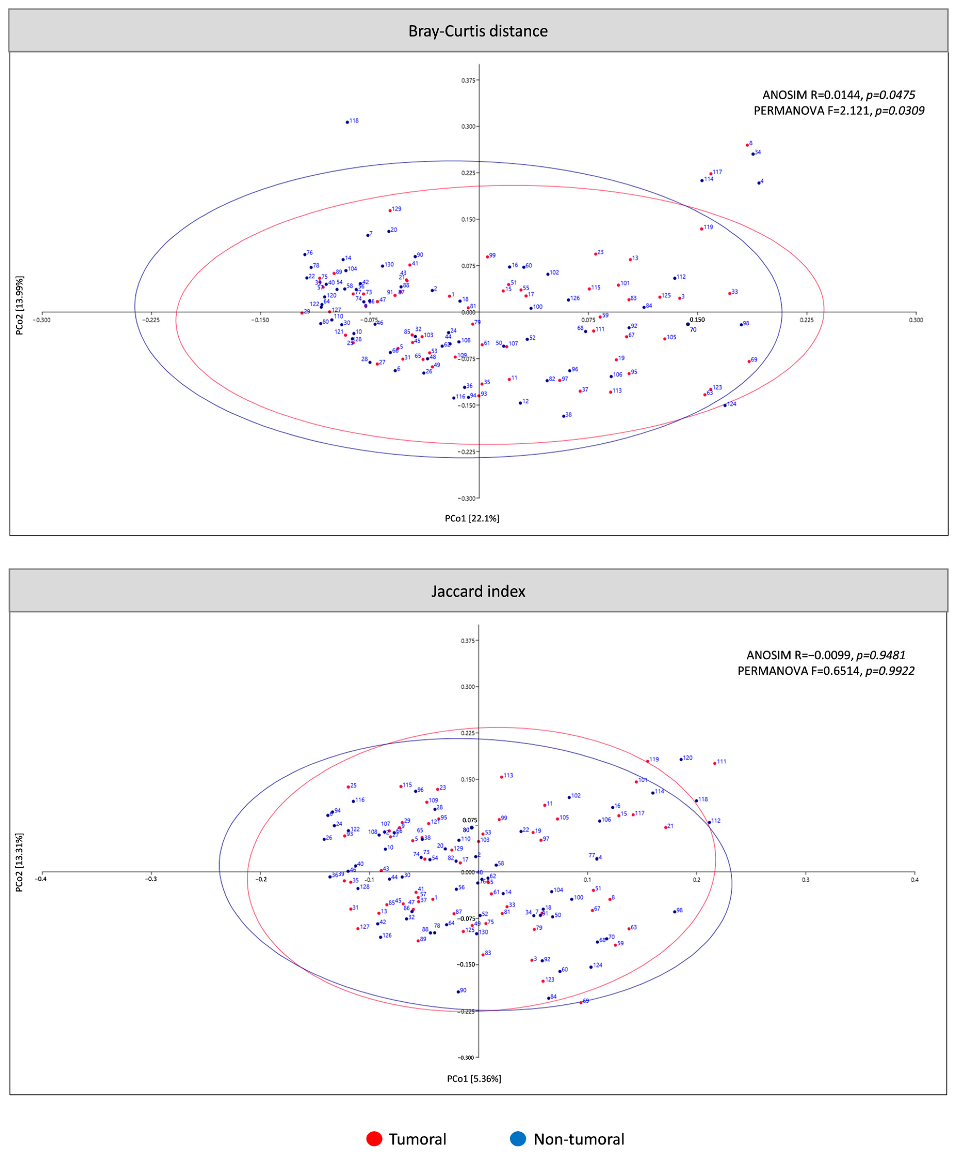
3.2.2. β-Diversity of Microbiota in Tumor Compared to Non-Tumor Adjacent Tissue Samples of CRC Patients

The present study analyzed β-diversity between tumor and non-tumor tissue using PCoA of two metrics (Bray–Curtis and Jaccard) and ANOSIM and PERMANOVA analysis (Figure 3). According to Bray–Curtis dissimilarity, the mucosal microbiota composition differed between tumoral and non-tumoral groups. No statistically significant differences were obtained for the Jaccard index. Six samples were classified as outliers by Bray–Curtis dissimilarity and consequently removed for α-diversity, taxa differential abundance and different tumor location analyses.

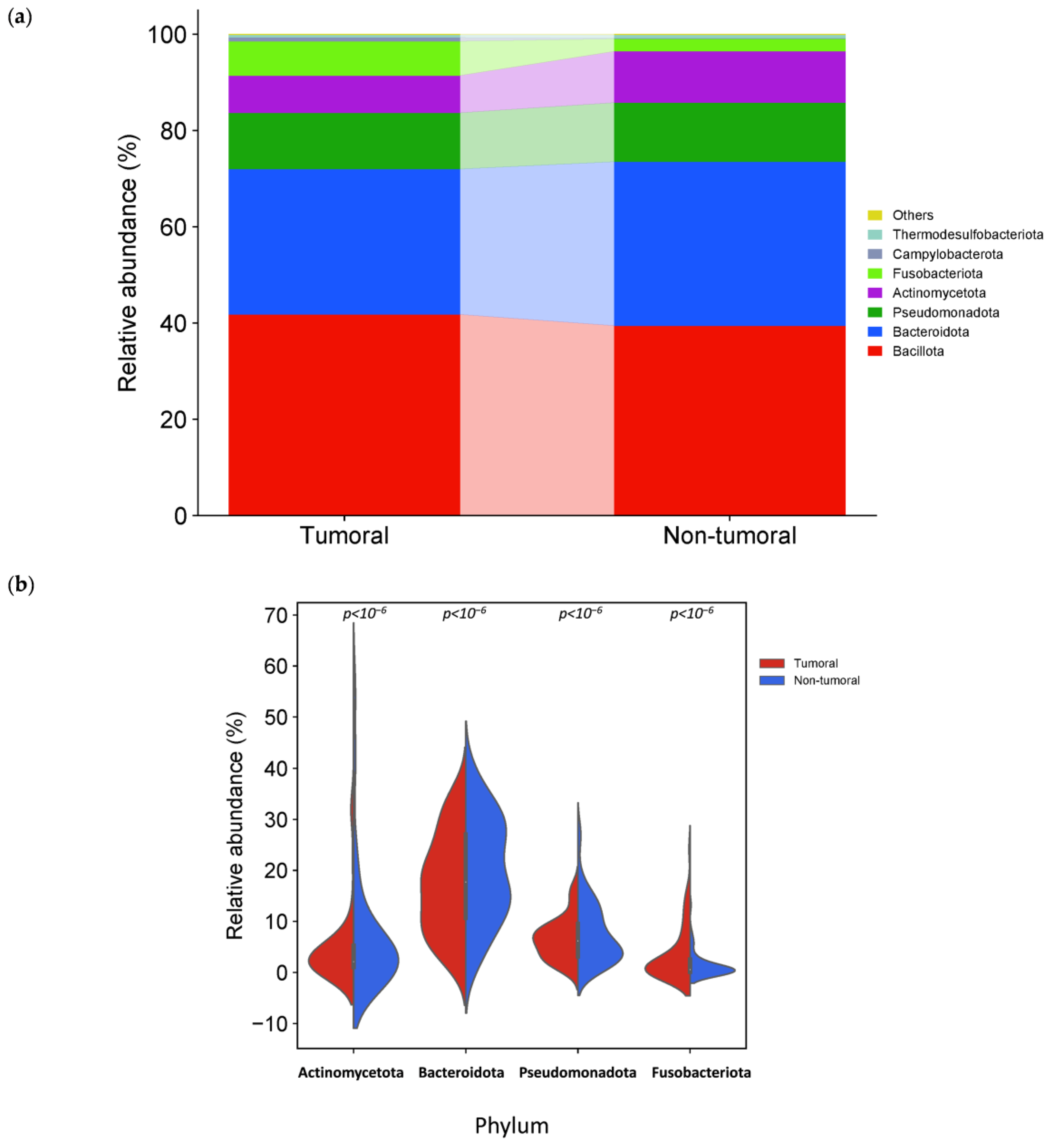
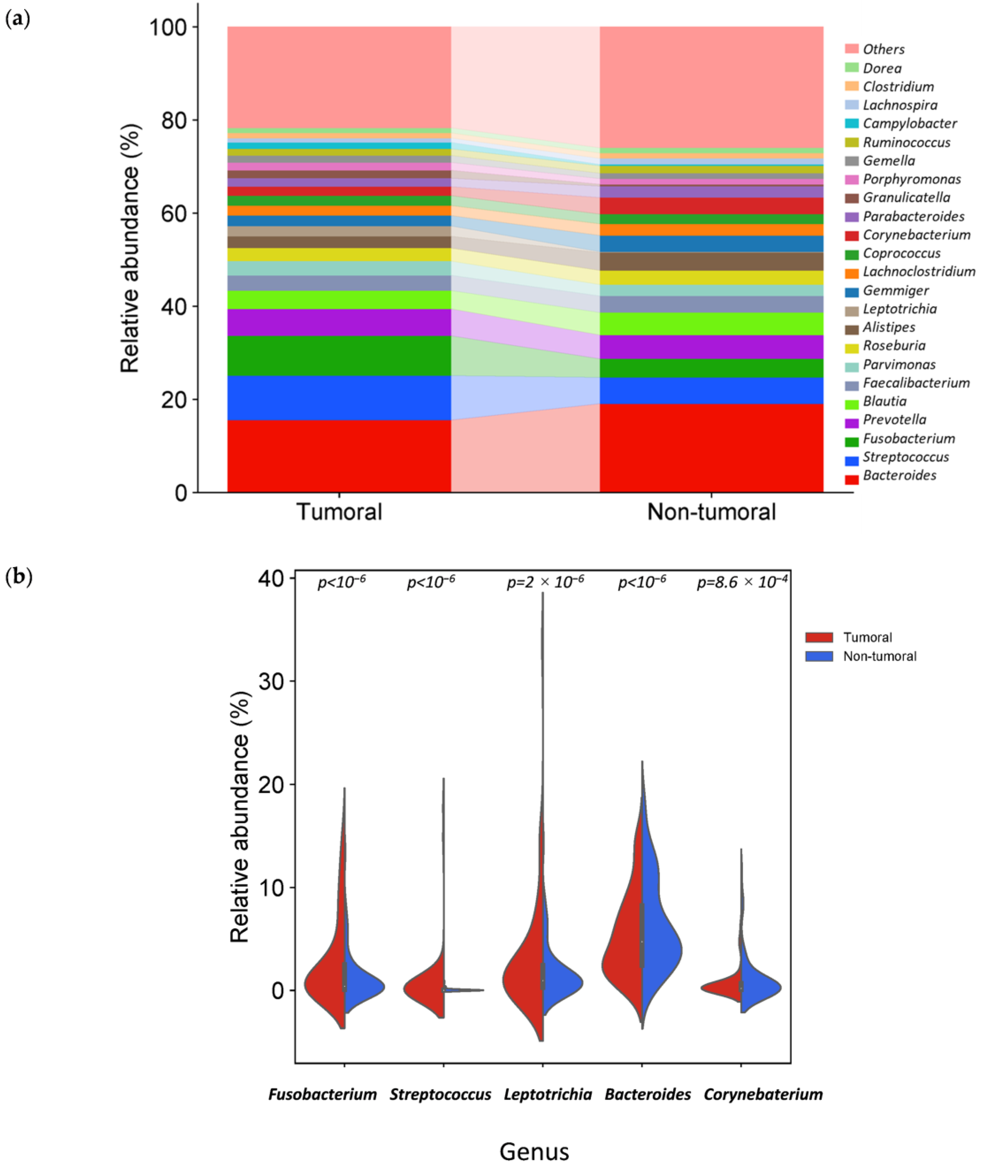
3.3. Specific Microbiota Compositional Differences Between Tumor and Non-Tumor Adjacent Tissue Samples

Significant relative abundance variations were observed in the microbiota of tumor and non-tumor tissue samples at different taxonomic bacterial levels (Table 1 and Table 2).
The phylum Bacillota, Bacteroidota, Pseudomonadota (former Proteobacteria) and Actinomycetota, common members of the human gut microbiota, formed more than 90% of bacterial phyla in tumor and non-tumor tissue samples (Figure 4a). The relative abundances at the phylum level in the tumor and non-tumor tissue samples were compared. We observed a significant difference in four detected phyla between sample groups. The relative abundance of Actinomycetota, Bacteroidota and Pseudomonadota was significantly higher in the non-tumor than in tumor samples. In contrast, Fusobacteriota was significantly higher in tumor samples (Figure 4b).
Seven taxa showed statistically significant differences between tumor and non-tumor tissue samples at the class level (Table 1 and Table 2). The relative abundance of Bacilli and Fusobacteriia in the tumor tissues was significantly higher than in the non-tumor tissues. The relative abundance was significantly lower for the tumor tissues of Actinomycetes, Alphaproteobacteria, Bacteroidia, Betaproteobacteria, and Clostridia than the non-tumor tissues.
Four taxa showed statistically significant differences between tumor and non-tumor tissue samples at the order level (Table 1 and Table 2). Fusobacteriales and Lactobacillales were significantly higher in the tumor compared to the non-tumor tissues. In contrast, Bacteroidales, Eubacteriales, and Mycobacteriales were significantly decreased in tumors compared to the non-tumor tissues.
The microbiota composition also differed at the family level, with seven significantly different families between tumor and non-tumor tissues (Table 1 and Table 2). The relative abundance of Fusobacteriaceae, Leptotrichiaceae and Streptococcaceae was significantly higher in the tumoral group than in the non-tumoral group. The relative abundance of Bacteroidaceae, Corynebacteriaceae, Lachnospiraceae and Propionibacteriaceae was significantly lower in the tumoral than in the non-tumoral group.
Bacteroides, Streptococcus, Fusobacterium, Prevotella and Blautia were the fifth most abundant genus in tumor and non-tumor tissue samples. Interestingly, among the 20 most abundant genera in tumor and non-tumor tissues, 17 were common, while Leptotrichia, Granulicatella and Campylobacter were also found in tumor tissues and Lachnospira, Clostridium and Dorea in non-tumor tissues (Figure 5a).
The relative abundance of three genera was significantly higher in tumor tissue samples (Table 1 and Table 2): Fusobacterium, Leptotrichia and Streptococcus. In contrast, two genera, Bacteroides and Corynebacterium, were significantly reduced (Figure 5b).
In tumor tissue samples, species assigned to F. nucleatum, Fusobacterium polymorphum and Streptococcus periodonticum had significantly higher relative abundances than non-tumor samples. Two strains, F. polymorphum ATCC 10953 and Fusobacterium animalis ATCC 51191, were significantly more abundant in tumor samples.

3.4. Tissue-Associated Microbiota Differences Related to Tumor Location in CRC Patients

We observed relevant differences in the composition of the microbiota according to location: colon, rectum and sigmoid colon. After removing the outliers and the only sample from the cecum, the analysis included 25 tumor samples from the colon, 24 from the rectum and 9 from the sigmoid colon. Evaluation of α-diversity between CRC tumor tissue samples from the colon, rectum and sigmoid colon revealed no significant differences (Figure 6).
The analysis of the ANOVA test for β-diversity showed significant differences between colon and rectum tumors, colon and sigmoid colon tumors, and rectum and sigmoid colon tumors for Jaccard Index but not for Bray–Curtis dissimilarity. No statistically significant differences were obtained for the PERMANOVA test (Table 3).
At the taxonomic level, there were significant differences between colon and rectum tumors, colon and sigmoid colon tumors, and rectum and sigmoid colon tumors.
Our results indicated that tumoral microbiota from the colon compared to the rectum was characterized by a preponderance of Prevotella, Roseburia, Granulicatella, Leyella stercorea, Agathobacter rectalis, Phocaeicola plebeius and Granulicatella elegans (Supplementary Table S2). In contrast, colon tumors compared to rectum showed a decrease in Alistipes, F. animalis, F. nucleatum, F. polymorphum, Fusobacterium vincentii and S. periodonticum (Supplementary Table S3).
Concerning tumors from the colon compared to the sigmoid colon, there was an increase in Roseburia, Prevotella, Granulicatella, L. stercorea and G. elegans (Supplementary Table S4). In contrast, colon tumors compared to the sigmoid colon showed a decrease in Peptoniphilus, Staphylococcus, Streptococcus and Fusobacterium. At the species level, colon tumors showed a decrease in F. nucleatum, F. polymorphum, F. vincentii, Peptoniphilus lacrimalis, Phocaeicola coprocola, Porphyromonas endodontalis, S. periodonticum and Waltera intestinalis compared to sigmoid colon tumors (Supplementary Table S5).
For tumors from the rectum compared to the sigmoid colon, there was an increase in Fusobacterium and F. animalis (Supplementary Table S6). In contrast, rectum tumors compared to sigmoid colon showed a decrease in Peptoniphilus, Staphylococcus, Streptococcus, P. lacrimalis, P. coprocola, S. periodonticum and W. intestinalis (Supplementary Table S7).

4. Discussion

The development of CRC is associated with genetic and environmental factors, among which diet stands out [5,6]. The available scientific evidence suggests that the link between diet and CRC lies in the gut microbiota [7,8]. A growing body of scientific research has recently shown that gut microbiota can directly affect colorectal tumorigenesis [30].
In the present research, we analyzed the microbiota composition of the paired tumor and non-tumor adjacent tissue samples of the large intestine of CRC patients by a large amplicon, including the V3–V4 regions of 16S rRNA gene with MinION sequencing platform and by NCBI taxonomic classification. This innovative methodology allowed for the finding of significant variations in the relative abundance of bacteria at different taxonomic levels between tumor and non-tumor tissues. These findings are largely consistent with previous studies, and simultaneously, the methodology favors revealing new key taxa even at lower taxonomic levels, such as species and strains [21,31].
The relative abundance of phylum Fusobacteriota (p < 10−6), genera Fusobacterium (p < 10−6), Leptotrichia (p = 2 × 10⁻⁶), and Streptococcus (p < 10−6) was significantly higher in tumor samples. On the other hand, the relative abundance of phyla Actinomycetota, Bacteroidota, and Pseudomonadota (p < 10−6), and genus Bacteroides (p < 10−6) and Corynebacterium (p = 8.6 × 10⁻⁴) was significantly lower in tumor samples. Moreover, although it is necessary to approach these results cautiously, this study showed a higher abundance in tumor samples of F. nucleatum (p < 10−6), F. polymorphum (p = 5.3 × 10⁻³), S. periodonticum (p < 10−6) species and F nucleatum subsp. polymorphum ATCC 10953 and F. animalis ATCC 51191 strains (p < 10−6) compared to non-tumor tissue samples. Despite the scientific interest of those results, those at species and especially at strain levels should be taken cautiously and replicated in future studies, considering the limitations inherent to the available amplicon length and the sequence databases resolution. To the best of our knowledge, the present study is the first to identify the possible increased relative abundance of S. periodonticus and decreased Corynebacterium association with CRC.
On the other hand, bacteria of the phylum Fusobacteriota were more predominant among the tumor tissue than non-tumor tissue samples, as previously reported [32,33,34]. Fusobacteriota is an understudied phylum of bacteria, including Fusobacteriaceae and Leptotrichiaceae families, both enriched in tumor compared to the non-tumor tissues, consistent with past studies [35]. Fusobacterium is a genus included in the first family and Leptotrichia in the second one, which was also increased in tumor tissue [36,37]. Notably, several studies have pointed out that the combination of Fusobacterium and Leptotrichia may contribute to the progression of CRC [38].
The molecular mechanisms through which Fusobacterium and especially genus members, such as F. nucleatum and F. animalis, influence CRC are increasingly well described, whereas the ones for Leptotrichia are still poorly known [39]. Leptotrichia invasive infections have been reported in patients receiving high-dose chemotherapy [40] and in oral disease in immunocompromised patients [41]. One of the most studied species from the genera is Leptotrichia trevisanii, an anaerotolerant, anaerobic, opportunistic gram-negative pathogen. Its ability to produce mainly lactic acid from glucose fermentation distinguishes it from closely related Fusobacterium species [41]. Leptotrichia trevisanii has been described as a causal agent of severe sepsis in immunocompetent patients, particularly in patients with hematological malignancies receiving chemotherapy [42].
According to previous studies, F. nucleatum was significantly enriched in the tumor compared to the non-tumor tissues [32,43,44]. F. nucleatum is an anaerobe gram-negative opportunistic pathogen ubiquitous in the human oral microbiota [45]. The oral cavity contributes to CRC tumor seeding [46]. Independently of clinical, pathological and molecular features, the amount of F. nucleatum in CRC tissue has been positively associated with mortality [47]. F. nucleatum has been related to genetic and epigenetic lesions in CRC tissues. F. nucleatum is an invasive and proinflammatory microorganism capable of stimulating CRC cell proliferation through different mechanisms: proliferation and metabolism promotion, immune microenvironment reprogramming, proinflammatory microenvironment creation, anticancer immune responses inhibition and metastasis and chemoresistance promotion in CRC [48]. These carcinogenic effects could be mediated by components such as lipopolysaccharides and adhesins like FadA and Fap2 [35]. Hence, F. nucleatum is considered a diagnosis biomarker, a prognostic predictor and a promising target in CRC treatment [35,36].
The F. nucleatum subspecies genetic and phenotypic heterogeneity has prompted research into differential genetic attributes among species contributing to CRC initiation and progression [49]. In this regard, currently, there is a controversy about the taxonomic classification of F.nucleatum. Previous genetic analyses have reported that it comprises five subspecies usually found in oral microbiota: F. nucleatum subsp. nucleatum, subsp. polymorphum, subsp. vincentii, subsp. fusiforme, and subsp. animalis [50]. However, the taxonomic positions of members have been under discussion. In 2010, it was proposed that F. nucleatum subsp. vincentii and subsp. fusiforme could be classified into one subspecies [51]. In 2017, it was proposed to elevate the four F. nucleatum subspecies (subps. nucleatum, subps. polymorphum, subps. vincentii, and subps. animalis) to species-level status with a wide diversity of significant strains [52].
In the present study, F. polymorphum showed higher relative abundance in tumor than non-tumor tissue samples. Recently, F. polymorphum has been isolated from CRC patients in their tumor tissue and oral cavity [53] and pre-CRC patients’ saliva [54]. Therefore, to our knowledge, F. polymorphum has not been reported to be significantly enriched in CRC tissue compared to non-tumor tissue, and hence, more studies are needed to confirm these findings. The information scarcity could be because it was only in 2017 that F. polymorphum was considered a species rather than a subspecies of F. nucleatum [52].
On the other hand, F. animalis is considered the most common and pathogenic species in CRC [55,56]; it also presented a higher relative abundance in tumor tissue than in non-tumor tissue in our study. The presence of F. animalis has been previously associated with right-sided tumor location and advanced tumor stages (stages II and III), with higher CRC-specific mortality, and with specific mutations in somatic genes in CRC. In contrast, Fusobacterium vincentii or F. nucleatum was not and may be driven by a stage shift and chemoresistance [55]. The interaction of F. animalis ATCC 51191 with human intestinal epithelial and tumor cells has been verified [57]. Recently, F. animalis has been suggested as a primary target for mechanistic and therapeutic studies in CRC [49].
We observed that Streptococcus and S. periodonticus relative abundance was higher in tumor than in non-tumor tissue samples. To the best of the author’s knowledge, this is the first time that the association of S. periodonticus in CRC has been found. S. periodonticus is a gram-positive coccus within the genus Streptococcus that has been recently isolated from human subgingival dental plaque of periodontitis lesion [58]. S. periodonticus has been associated with a pediatric case of bacterial meningitis after cranial surgery [59]. Interestingly, Streptococcus spp. internalization into epithelial cells could be facilitated by the effect of Fusobacterium, which promotes coaggregation and facilitates internalization processes in these normally non-invasive bacteria [60,61]. Interestingly, a bacterium of the genus Streptococcus, S. gallolyticus, is a proinflammatory species remarkably associated with CRC. This bacterium can express collagen-binding proteins such as Pil 1, allowing it to colonize tissues and induce the secretion of proinflammatory mediators that can promote CRC [11].
We observed that the relative abundance of phylum Actinomycetota, Bacteroidota and Pseudomonadota was significantly higher in non-tumor than in tumor tissues. Past studies have reached similar results [33,62,63,64]. However, some studies have reported enrichment of the phylum Pseudomonadota in tumor tissue [65,66]. Although a Pseudomonadota increase is generally considered an intestinal dysbiosis marker and gut commensals with pathogenic potential [67], bacteria of the same taxonomic group may exert different effects depending on functional characteristics, interactions and environment [63].
We observed that Bacteroides was enriched in non-tumor compared to tumor tissues, which is in coherence with several previous studies [68,69,70]. However, Gao et al. observed that Bacteroides was highly enriched in tumor tissues [63]. Bacteroides is thought to have both positive and negative impacts on host health via their colitogenic or probiotic effects [71].
Corynebacterium genus showed higher relative abundance in non-tumor compared to tumor tissues. To the best of our knowledge, there is no previous information related to this genus in CRC tissue. Regarding cancer, the evidence is scarce and contradictory; one study reported that oxidative tryptamine dimers from Corynebacterium durum exert anticancer properties [72], whereas another study reported its contribution to induced colitis [73].
Differences in the microbial profiles observed between tumor and non-tumor tissues could reflect the role of certain microbiota members in the initiation and development of CRC or changes associated with the TME. Two main hypotheses have been proposed to explain these interactions.
The “alpha-bug” hypothesis proposed by Sears and Pardoll suggests that detrimental members of the microbiota, such as enterotoxigenic Bacteroides fragilis (ETBF), S. gallolyticus, superoxide-producing Enterococcus faecalis and Escherichia coli, may act as cancer initiators by directly inducing alterations in colonic epithelial cells and remodeling the colonic microbial community to promote these alterations and disrupt immune responses [18].
In contrast, the bacterial driver-passenger model proposed by Tjalsma et al. suggests that certain commensal bacteria (bacterial drivers), such as Enterococcus faecalis, can cause epithelial DNA damage, leading to CRC initiation. Next, the process of tumorigenesis induces modifications in the microenvironment that benefit relatively poor colonizing bacteria (bacterial passengers) [19]. Bacterial passengers have been proposed to include opportunistic pathogens that feed in the tumor (Fusobacterium or Streptococcus spp.), intestinal commensal or probiotic bacteria (Coriobacteriaceae family), and bacteria that have a competitive advantage in the TME [19].
The microbial α-diversity refers to within-sample diversity. Although no significant differences between groups were obtained for observed OTUs, Chao1 and Simpson indices, the Shannon index was significantly reduced in tumor than in non-tumor tissue samples. From our study, we can conclude that tumor samples present a lower microbial diversity. Previous studies revealed similar results [33,65,74], no statistically significant differences [68,75,76,77], and even reported that microbial α-diversity was significantly higher in the tumor samples [78]. Therefore, there is no consensus between α-diversity metrics behavior in tumor vs. non-tumor tissues.
The microbial β-diversity refers to between-sample diversity. In this study, Bray-Curtis dissimilarity based on OTUs revealed that the microbiota composition of the tumor tissues could be differentiated from non-tumor tissues. In contrast, no statistically significant differences were observed with the Jaccard index. Some previous studies have revealed similar results [33,64,66,69], while others reported no statistically significant differences [74,76,79,80]. These discrepancies could be caused by the Bray–Curtis dissimilarity being based on the relative abundances of the OTUs, while the Jaccard index is based on their presence/absence.
On the other hand, we identified associations between gut microbiota composition and tumor location. Although there were no significant differences in α and β-diversity analysis, we identified an enrichment of L. stercorea and G. elegans in colon tumors compared to the rectum and the sigmoid colon. Likewise, the sigmoid colon showed a higher relative abundance of Peptoniphilus, Staphylococcus, Streptococcus, P. lacrimalis, P. coprocola, S. periodonticum and W. intestinalis than the colon and the rectum. The rectal tumors were enriched in F. animalis compared to the colon and the sigmoid colon. Our results align with previous evidence, supporting that characterization of the microbiota composition at different sites in the large intestine can contribute to a better description of the molecular subtypes of CRC [81,82].
As mentioned, CRC-associated microbiota research has key limitations, such as the sample type selection, the sequencing platform used and the taxonomy classification employed.
Regarding sample type, the present study analyzed the tissue CRC-associated microbiota from 130 paired tumor and non-tumor adjacent tissue samples, in contrast to traditional microbiota analysis from fecal samples [21]. Fecal samples are commonly used because they are easy to collect, non-invasive and repeatable, which makes them potential tools for CRC screening and early detection. In addition, they are less likely to be contaminated by eukaryotic DNA [83,84]. Fecal microbiota can provide valuable information, but it primarily reflects the composition of the intestinal lumen microbiota, and it does not capture the same perturbations and interactions with the colonic mucosa critical to CRC pathogenesis [85,86].
In contrast, as analyzed in the present study, mucosa-associated microbiota interacts more closely with colonocytes and host local immunity, possibly playing a greater role in CRC initiation and development than luminal microbiota. These interactions can lead to gene expression modifications and inflammation, which may influence colorectal tumorigenesis [87]. Therefore, tissue samples are ideal for investigating the pathogenesis of CRC. Moreover, tissue samples are considered more accurate for bacterial detection, show higher microbial diversity than fecal samples, and are more suitable for identifying potentially minor alterations in the microbiota of precancerous colorectal lesions [84]. Despite the potential importance of tissue samples, studies examining the differential abundance of associated microbiota in tumor and non-tumor tissue in CRC patients are scarce [21].
However, the analysis of tissue samples showed additional practical challenges. While in fecal samples, our group usually analyzed the whole 16S rRNA gene (approximately 1500 bp) from fecal samples [88], after several attempts in tissue samples, the maximum amplicon size reached was approximately 450 bp corresponding V3–V4 region of the 16SrRNA gene. Similar limitations have been observed when investigating mucosal microbiota with shotgun metagenomics [89]. We hypothesized that studying the complete 16SrRNA gene amplicon in tissue samples is unfeasible due to human DNA’s high presence and the target gene’s fragmentation. Nevertheless, the 450 bp reached in the current article comprises significantly larger amplicons than those usually obtained by sequence platforms different from MinION (200–300 bp), improving the potential taxonomic profiling [90]. The larger read size combined with NCBI taxonomic classification improves the taxonomic profiling.
On the other hand, due to operative limitations, the present study did not include a CRC-free control group because of the difficulty in obtaining colon samples from healthy people. However, using paired tumor and non-tumor samples avoids the potential confounding factors when analyzing CRC and healthy controls, such as age, sex, body mass index, diet and other characteristics [21,82].
Otherwise, tissue sampling is more invasive than fecal, limiting the number of samples collected from the tumor and non-tumor tissues in the present study. It would be advisable to analyze more samples from each patient for future research to examine the possible heterogeneity of the microbiota composition associated with CRC tumor tissue [76]. In addition, future longitudinal studies looking at changes in the microbiota over time would provide a better understanding of whether specific bacteria play a role in the initiation and progression of CRC or whether they are responding to TME. Finally, considering the pros and cons of analyzing fecal or tissue samples, combining both seems valuable for future research.
On the other hand, selecting a taxonomic classification database is essential for analyzing CRC-associated microbiota. The most used taxonomic classifications for 16S rRNA gene studies vary in size (taxa amount) and resolution capacity (classification level). The current scientific evidence shows that the taxonomic classification at the genus and species level is essential to decoding the role of microbiota composition in CRC. In the present article, we have selected the NCBI database because it contains the largest number of nodes and the highest resolution. Thus, NCBI allows a more thorough classification, going down to the species rank and below and offering several intermediate ranks [25].
In contrast, SILVA and RDP are limited to classifying the genus level as the lowest rank. Although Greengenes goes down to species, the comparative analysis carried out by Balvočiūtė & Huson showed that SILVA, RDP and Greengenes map well with NCBI but not vice versa. Hence, they recommended using the NCBI taxonomy as a common framework for 16S rRNA gene studies [25]. In addition, the NCBI taxonomy is manually curated, covering more than 150 sources, and updated daily [91].
Nevertheless, taxonomy selection up to date is generally determined by the pipeline used, and most studies that analyze tumor microbiota composition in CRC patients use SILVA, RDP or Greengenes. Therefore, it dramatically limits the resolution and amplitude of the previous results and reanalyzing them using the NCBI database could improve the available scientific evidence regarding CRC-associated microbiota.
Regarding sequence platform selection in CRC research, access to sequencing technology is an important limitation for implementing CRC-associated gut microbiota analysis in precision medicine [22]. MinION from ONT is a revolutionary sequencing platform because of its small size and portability, and it allows real-time sequencing of various samples on demand at a competitive cost [23,24]. In the present article, MinION is used for its potential to facilitate the implementation of microbiota analysis in precision medicine strategies for CRC. Despite its advantages, to the best of our knowledge, the previous evidence using the MinION sequencer for microbiota analysis of paired tumor and non-tumor tissues in CRC patients is limited to a single article [22]. In this article, the authors did not carry out 16S rRNA gene amplicon sequencing with MinION but used MinION sequencing using genomic DNA to analyze the microbiota. They conclude that long-read sequences generated using MinION allow differentiation between bacterial strains and plasmids, and as a cost-effective and rapid sequencing tool, it has the potential for use in clinical settings [22].

5. Conclusions

In this study, we detected an enrichment in genera such as Fusobacterium, Leptotrichia and Streptococcus in tumor compared to non-tumor tissue samples. In addition, species such as F. nucleatum, F. polymorphum, S. periodonticus and strains such as F. polymorphum ATCC 10953 and F. animalis ATCC 51191 were also enriched in tumor tissue. On the other hand, genera such as Bacteroides and Corynebacterium were enriched in non-tumor tissues. In addition, differences in microbiota composition were observed between tumor locations (colon, rectum and sigmoid colon).
The present study faces the main technical challenges in CRC-associated microbiota regarding sample type, sequence platform, and taxonomic database. It comprises the analysis of 130 paired tumor and non-tumor adjacent tissue samples in different locations by larger amplicon sizes, including the V3–V4 region of 16S rRNA gene analyzed by MinION suitable sequence platform, and using NCBI database, that increases the amplitude and resolution in sequence taxonomy classification. These methodological advances agree with earlier findings of microbiota composition differences between tumor and non-tumor tissues. They also reveal the possible involvement of specific taxa, such as S. periodonticus and Corynebacterium, in CRC biology for the first time.
Therefore, the results obtained in this study facilitate the implementation of individual gut microbiota analysis in personalized medicine. This approach allows the development of therapeutic strategies for CRC that consider this essential component of TME [20].

Supplementary Materials

The following supporting information can be downloaded at https://www.mdpi.com/article/10.3390/cancers16234008/s1, Supplementary Table S1. Description of tumor and non-tumor adjacent mucosal tissue samples; Supplementary Table S2. Over-represented bacterial taxa in tumoral colon tissue compared to the rectum; Supplementary Table S3. Under-represented bacterial taxa in tumor colon tissue compared to the rectum; Supplementary Table S4. Over-represented bacterial taxa in tumor colon tissue compared to sigmoid colon; Supplementary Table S5. Under-represented bacterial taxa in tumor colon tissue compared to sigmoid colon; Supplementary Table S6. Over-represented bacterial taxa in tumor rectum compared to sigmoid colon; Supplementary Table S7. Under-represented bacterial taxa in tumor rectum compared to sigmoid colon.

Author Contributions

Conceptualization, A.G., A.F. and A.O.; methodology, A.G., D.N., C.T. and A.O.; writing—original draft preparation, A.G.; writing—review and editing, D.N., A.F., J.R., C.T. and A.O.; supervision, A.O. All authors have read and agreed to the published version of the manuscript.

Funding

This research has received specific grants from the Basque Government, Department of Education (IT1547-22).

Institutional Review Board Statement

This work was approved for research ethics by the Ethics Committee of Clinica Universitaria de Navarra (CUN) (REINFORCE_0011-1411-2020-000102 of the 10 September 2020).

Informed Consent Statement

All participants provided informed consent to participate in this study.

Data Availability Statement

The original contributions presented in the study are included in the article/Supplementary Materials; further inquiries can be directed to the corresponding author.

Acknowledgments

We particularly acknowledge the patients for their participation and the Biobank of the University of Navarra for its collaboration.

Conflicts of Interest

Author David Navarro was employed by the company Danagen-Bioted S.L. The remaining authors declare that the research was conducted in the absence of any commercial or financial relationships that could be construed as a potential conflict of interest.

References

  1. Cancer (IARC), T.I.A. for R. on Global Cancer Observatory. Available online: https://gco.iarc.fr/ (accessed on 15 November 2024).
  2. Hanahan, D. Hallmarks of Cancer: New Dimensions. Cancer Discov. 2022, 12, 31–46. [Google Scholar] [CrossRef] [PubMed]
  3. Jobin, C. Colorectal Cancer: Looking for Answers in the Microbiota. Cancer Discov. 2013, 3, 384–387. [Google Scholar] [CrossRef] [PubMed]
  4. Rothschild, D.; Weissbrod, O.; Barkan, E.; Kurilshikov, A.; Korem, T.; Zeevi, D.; Costea, P.I.; Godneva, A.; Kalka, I.N.; Bar, N.; et al. Environment Dominates over Host Genetics in Shaping Human Gut Microbiota. Nature 2018, 555, 210–215. [Google Scholar] [CrossRef] [PubMed]
  5. Janout, V.; Kollárová, H. Epidemiology of Colorectal Cancer. Biomed. Pap. Med. Fac. Univ. Palacky Olomouc Czechoslov. 2001, 145, 5–10. [Google Scholar] [CrossRef]
  6. Willett, W.C. Diet and Cancer: An Evolving Picture. JAMA 2005, 293, 233–234. [Google Scholar] [CrossRef]
  7. De Filippo, C.; Cavalieri, D.; Di Paola, M.; Ramazzotti, M.; Poullet, J.B.; Massart, S.; Collini, S.; Pieraccini, G.; Lionetti, P. Impact of Diet in Shaping Gut Microbiota Revealed by a Comparative Study in Children from Europe and Rural Africa. Proc. Natl. Acad. Sci. USA 2010, 107, 14691–14696. [Google Scholar] [CrossRef]
  8. Nakatsu, G.; Andreeva, N.; MacDonald, M.H.; Garrett, W.S. Interactions between Diet and Gut Microbiota in Cancer. Nat. Microbiol. 2024, 9, 1644–1654. [Google Scholar] [CrossRef]
  9. Casasanta, M.A.; Yoo, C.C.; Udayasuryan, B.; Sanders, B.E.; Umaña, A.; Zhang, Y.; Peng, H.; Duncan, A.J.; Wang, Y.; Li, L.; et al. Fusobacterium nucleatum Host-Cell Binding and Invasion Induces IL-8 and CXCL1 Secretion That Drives Colorectal Cancer Cell Migration. Sci. Signal. 2020, 13, eaba9157. [Google Scholar] [CrossRef]
  10. Rubinstein, M.R.; Wang, X.; Liu, W.; Hao, Y.; Cai, G.; Han, Y.W. Fusobacterium nucleatum Promotes Colorectal Carcinogenesis by Modulating E-Cadherin/β-Catenin Signaling via Its FadA Adhesin. Cell Host Microbe 2013, 14, 195–206. [Google Scholar] [CrossRef]
  11. Abdulamir, A.S.; Hafidh, R.R.; Bakar, F.A. Molecular Detection, Quantification, and Isolation of Streptococcus gallolyticus Bacteria Colonizing Colorectal Tumors: Inflammation-Driven Potential of Carcinogenesis via IL-1, COX-2, and IL-8. Mol. Cancer 2010, 9, 249. [Google Scholar] [CrossRef]
  12. Mazmanian, S.K.; Liu, C.H.; Tzianabos, A.O.; Kasper, D.L. An Immunomodulatory Molecule of Symbiotic Bacteria Directs Maturation of the Host Immune System. Cell 2005, 122, 107–118. [Google Scholar] [CrossRef] [PubMed]
  13. Long, X.; Wong, C.C.; Tong, L.; Chu, E.S.H.; Ho Szeto, C.; Go, M.Y.Y.; Coker, O.O.; Chan, A.W.H.; Chan, F.K.L.; Sung, J.J.Y.; et al. Peptostreptococcus Anaerobius Promotes Colorectal Carcinogenesis and Modulates Tumour Immunity. Nat. Microbiol. 2019, 4, 2319–2330. [Google Scholar] [CrossRef] [PubMed]
  14. Drewes, J.L.; Chen, J.; Markham, N.O.; Knippel, R.J.; Domingue, J.C.; Tam, A.J.; Chan, J.L.; Kim, L.; McMann, M.; Stevens, C.; et al. Human Colon Cancer–Derived Clostridioides Difficile Strains Drive Colonic Tumorigenesis in Mice. Cancer Discov. 2022, 12, 1873–1885. [Google Scholar] [CrossRef] [PubMed]
  15. Cheng, Y.; Ling, Z.; Li, L. The Intestinal Microbiota and Colorectal Cancer. Front. Immunol. 2020, 11, 615056. [Google Scholar] [CrossRef]
  16. Wong, S.H.; Yu, J. Gut Microbiota in Colorectal Cancer: Mechanisms of Action and Clinical Applications. Nat. Rev. Gastroenterol. Hepatol. 2019, 16, 690–704. [Google Scholar] [CrossRef]
  17. González, A.; Fullaondo, A.; Odriozola, A. Microbiota-Associated Mechanisms in Colorectal Cancer. Adv. Genet. 2024, 112, 123–205. [Google Scholar] [CrossRef]
  18. Sears, C.L.; Pardoll, D.M. Perspective: Alpha-Bugs, Their Microbial Partners, and the Link to Colon Cancer. J. Infect. Dis. 2011, 203, 306–311. [Google Scholar] [CrossRef]
  19. Tjalsma, H.; Boleij, A.; Marchesi, J.R.; Dutilh, B.E. A Bacterial Driver–Passenger Model for Colorectal Cancer: Beyond the Usual Suspects. Nat. Rev. Microbiol. 2012, 10, 575–582. [Google Scholar] [CrossRef]
  20. Kiran, N.S.; Yashaswini, C.; Maheshwari, R.; Bhattacharya, S.; Prajapati, B.G. Advances in Precision Medicine Approaches for Colorectal Cancer: From Molecular Profiling to Targeted Therapies. ACS Pharmacol. Transl. Sci. 2024, 7, 967–990. [Google Scholar] [CrossRef]
  21. da Costa, C.P.; Vieira, P.; Mendes-Rocha, M.; Pereira-Marques, J.; Ferreira, R.M.; Figueiredo, C. The Tissue-Associated Microbiota in Colorectal Cancer: A Systematic Review. Cancers 2022, 14, 3385. [Google Scholar] [CrossRef]
  22. Taylor, W.S.; Pearson, J.; Miller, A.; Schmeier, S.; Frizelle, F.A.; Purcell, R.V. MinION Sequencing of Colorectal Cancer Tumour Microbiomes—A Comparison with Amplicon-Based and RNA-Sequencing. PLoS ONE 2020, 15, e0233170. [Google Scholar] [CrossRef] [PubMed]
  23. Jain, M.; Olsen, H.E.; Paten, B.; Akeson, M. The Oxford Nanopore MinION: Delivery of Nanopore Sequencing to the Genomics Community. Genome Biol. 2016, 17, 239. [Google Scholar] [CrossRef]
  24. Scholz, M.B.; Lo, C.-C.; Chain, P.S. Next Generation Sequencing and Bioinformatic Bottlenecks: The Current State of Metagenomic Data Analysis. Curr. Opin. Biotechnol. 2012, 23, 9–15. [Google Scholar] [CrossRef] [PubMed]
  25. Balvočiūtė, M.; Huson, D.H. SILVA, RDP, Greengenes, NCBI and OTT—How Do These Taxonomies Compare? BMC Genom. 2017, 18, 114. [Google Scholar] [CrossRef]
  26. Stoddard, S.F.; Smith, B.J.; Hein, R.; Roller, B.R.K.; Schmidt, T.M. rrnDB: Improved Tools for Interpreting rRNA Gene Abundance in Bacteria and Archaea and a New Foundation for Future Development. Nucleic Acids Res. 2015, 43, D593–D598. [Google Scholar] [CrossRef]
  27. Shannon, C.E. A Mathematical Theory of Communication. Bell Syst. Tech. J. 1948, 27, 379–423. [Google Scholar] [CrossRef]
  28. Simpson, E.H. Measurement of Diversity. Nature 1949, 163, 688. [Google Scholar] [CrossRef]
  29. Tang, D.; Chen, M.; Huang, X.; Zhang, G.; Zeng, L.; Zhang, G.; Wu, S.; Wang, Y. SRplot: A Free Online Platform for Data Visualization and Graphing. PLoS ONE 2023, 18, e0294236. [Google Scholar] [CrossRef]
  30. Zhen, J.; Liu, C.; Liao, F.; Zhang, J.; Xie, H.; Tan, C.; Dong, W. The Global Research of Microbiota in Colorectal Cancer Screening: A Bibliometric and Visualization Analysis. Front. Oncol. 2023, 13, 1169369. [Google Scholar] [CrossRef]
  31. Ternes, D.; Karta, J.; Tsenkova, M.; Wilmes, P.; Haan, S.; Letellier, E. Microbiome in Colorectal Cancer: How to Get from Meta-Omics to Mechanism? Trends Microbiol. 2020, 28, 401–423. [Google Scholar] [CrossRef]
  32. Allali, I.; Delgado, S.; Marron, P.I.; Astudillo, A.; Yeh, J.J.; Ghazal, H.; Amzazi, S.; Keku, T.; Azcarate-Peril, M.A. Gut Microbiome Compositional and Functional Differences between Tumor and Non-Tumor Adjacent Tissues from Cohorts from the US and Spain. Gut Microbes 2015, 6, 161–172. [Google Scholar] [CrossRef] [PubMed]
  33. Choi, S.; Chung, J.; Cho, M.-L.; Park, D.; Choi, S.S. Analysis of Changes in Microbiome Compositions Related to the Prognosis of Colorectal Cancer Patients Based on Tissue-Derived 16S rRNA Sequences. J. Transl. Med. 2021, 19, 485. [Google Scholar] [CrossRef] [PubMed]
  34. Cremonesi, E.; Governa, V.; Garzon, J.F.G.; Mele, V.; Amicarella, F.; Muraro, M.G.; Trella, E.; Galati-Fournier, V.; Oertli, D.; Däster, S.R.; et al. Gut Microbiota Modulate T Cell Trafficking into Human Colorectal Cancer. Gut 2018, 67, 1984–1994. [Google Scholar] [CrossRef] [PubMed]
  35. El Tekle, G.; Andreeva, N.; Garrett, W.S. The Role of the Microbiome in the Etiopathogenesis of Colon Cancer. Annu. Rev. Physiol. 2024, 86, 453–478. [Google Scholar] [CrossRef]
  36. Wang, N.; Fang, J.-Y. Fusobacterium nucleatum, a Key Pathogenic Factor and Microbial Biomarker for Colorectal Cancer. Trends Microbiol. 2023, 31, 159–172. [Google Scholar] [CrossRef]
  37. Xu, K.; Jiang, B. Analysis of Mucosa-Associated Microbiota in Colorectal Cancer. Med. Sci. Monit. Int. Med. J. Exp. Clin. Res. 2017, 23, 4422–4430. [Google Scholar] [CrossRef]
  38. Nakatsu, G.; Li, X.; Zhou, H.; Sheng, J.; Wong, S.H.; Wu, W.K.K.; Ng, S.C.; Tsoi, H.; Dong, Y.; Zhang, N.; et al. Gut Mucosal Microbiome across Stages of Colorectal Carcinogenesis. Nat. Commun. 2015, 6, 8727. [Google Scholar] [CrossRef]
  39. Fukuoka, H.; Tourlousse, D.M.; Ohashi, A.; Suzuki, S.; Nakagawa, K.; Ozawa, M.; Ishibe, A.; Endo, I.; Sekiguchi, Y. Elucidating Colorectal Cancer-Associated Bacteria through Profiling of Minimally Perturbed Tissue-Associated Microbiota. Front. Cell. Infect. Microbiol. 2023, 13, 1216024. [Google Scholar] [CrossRef]
  40. Couturier, M.R.; Slechta, E.S.; Goulston, C.; Fisher, M.A.; Hanson, K.E. Leptotrichia Bacteremia in Patients Receiving High-Dose Chemotherapy. J. Clin. Microbiol. 2012, 50, 1228–1232. [Google Scholar] [CrossRef]
  41. Tee, W.; Midolo, P.; Janssen, P.; Kerr, T.; Dyall-Smith, M. Bacteremia Due to Leptotrichia trevisanii sp. nov. Eur. J. Clin. Microbiol. Infect. Dis. 2001, 20, 765–769. [Google Scholar] [CrossRef]
  42. Chady, A.; Emmanuel, G.-M.; Antony, S. Leptotrichia trevisanii: Case Report and Review of the Literature on Patients with Leptotrichia trevisanii Bacteremia in Acute Myeloid Leukemia. Infect. Disord. Drug Targets 2023, 23, 73–76. [Google Scholar] [CrossRef] [PubMed]
  43. Drewes, J.L.; White, J.R.; Dejea, C.M.; Fathi, P.; Iyadorai, T.; Vadivelu, J.; Roslani, A.C.; Wick, E.C.; Mongodin, E.F.; Loke, M.F.; et al. High-Resolution Bacterial 16S rRNA Gene Profile Meta-Analysis and Biofilm Status Reveal Common Colorectal Cancer Consortia. NPJ Biofilms Microbiomes 2017, 3, 34. [Google Scholar] [CrossRef] [PubMed]
  44. Zeller, G.; Tap, J.; Voigt, A.Y.; Sunagawa, S.; Kultima, J.R.; Costea, P.I.; Amiot, A.; Böhm, J.; Brunetti, F.; Habermann, N.; et al. Potential of Fecal Microbiota for Early-Stage Detection of Colorectal Cancer. Mol. Syst. Biol. 2014, 10, 766. [Google Scholar] [CrossRef] [PubMed]
  45. Brennan, C.A.; Garrett, W.S. Fusobacterium nucleatum—Symbiont, Opportunist and Oncobacterium. Nat. Rev. Microbiol. 2019, 17, 156–166. [Google Scholar] [CrossRef] [PubMed]
  46. Abed, J.; Maalouf, N.; Manson, A.L.; Earl, A.M.; Parhi, L.; Emgård, J.E.M.; Klutstein, M.; Tayeb, S.; Almogy, G.; Atlan, K.A.; et al. Colon Cancer-Associated Fusobacterium nucleatum May Originate from the Oral Cavity and Reach Colon Tumors via the Circulatory System. Front. Cell. Infect. Microbiol. 2020, 10, 400. [Google Scholar] [CrossRef]
  47. Mima, K.; Nishihara, R.; Qian, Z.R.; Cao, Y.; Sukawa, Y.; Nowak, J.A.; Yang, J.; Dou, R.; Masugi, Y.; Song, M.; et al. Fusobacterium nucleatum in Colorectal Carcinoma Tissue and Patient Prognosis. Gut 2016, 65, 1973–1980. [Google Scholar] [CrossRef]
  48. Ganesan, K.; Guo, S.; Fayyaz, S.; Zhang, G.; Xu, B. Targeting Programmed Fusobacterium nucleatum Fap2 for Colorectal Cancer Therapy. Cancers 2019, 11, 1592. [Google Scholar] [CrossRef]
  49. Zepeda-Rivera, M.; Minot, S.S.; Bouzek, H.; Wu, H.; Blanco-Míguez, A.; Manghi, P.; Jones, D.S.; LaCourse, K.D.; Wu, Y.; McMahon, E.F.; et al. A Distinct Fusobacterium nucleatum Clade Dominates the Colorectal Cancer Niche. Nature 2024, 628, 424–432. [Google Scholar] [CrossRef]
  50. Tomida, J.; Akiyama-Miyoshi, T.; Tanaka, K.; Hayashi, M.; Kutsuna, R.; Fujiwara, N.; Kawamura, Y. Fusobacterium watanabei sp. nov. As Additional Species within the Genus Fusobacerium, Isolated from Human Clinical Specimens. Anaerobe 2021, 69, 102323. [Google Scholar] [CrossRef]
  51. Kim, H.-S.; Lee, D.-S.; Chang, Y.-H.; Kim, M.J.; Koh, S.; Kim, J.; Seong, J.-H.; Song, S.K.; Shin, H.S.; Son, J.-B.; et al. Application of rpoB and Zinc Protease Gene for Use in Molecular Discrimination of Fusobacterium nucleatum Subspecies. J. Clin. Microbiol. 2010, 48, 545–553. [Google Scholar] [CrossRef]
  52. Kook, J.-K.; Park, S.-N.; Lim, Y.K.; Cho, E.; Jo, E.; Roh, H.; Shin, Y.; Paek, J.; Kim, H.-S.; Kim, H.; et al. Genome-Based Reclassification of Fusobacterium nucleatum Subspecies at the Species Level. Curr. Microbiol. 2017, 74, 1137–1147. [Google Scholar] [CrossRef] [PubMed]
  53. Komiya, Y.; Shimomura, Y.; Higurashi, T.; Sugi, Y.; Arimoto, J.; Umezawa, S.; Uchiyama, S.; Matsumoto, M.; Nakajima, A. Patients with Colorectal Cancer Have Identical Strains of Fusobacterium nucleatum in Their Colorectal Cancer and Oral Cavity. Gut 2019, 68, 1335–1337. [Google Scholar] [CrossRef] [PubMed]
  54. Morsi, H.; Golizeh, M.; Brosseau, N.; Janati, A.I.; Emami, E.; Ndao, M.; Tran, S.D. Detection of Fusobacterium nucleatum Subspecies in the Saliva of Pre-Colorectal Cancer Patients, Using Tandem Mass Spectrometry. Arch. Oral Biol. 2022, 134, 105337. [Google Scholar] [CrossRef] [PubMed]
  55. Borozan, I.; Zaidi, S.H.; Harrison, T.A.; Phipps, A.I.; Zheng, J.; Lee, S.; Trinh, Q.M.; Steinfelder, R.S.; Adams, J.; Banbury, B.L.; et al. Molecular and Pathology Features of Colorectal Tumors and Patient Outcomes Are Associated with Fusobacterium nucleatum and Its Subspecies Animalis. Cancer Epidemiol. Biomark. Prev. 2022, 31, 210–220. [Google Scholar] [CrossRef]
  56. Ye, X.; Wang, R.; Bhattacharya, R.; Boulbes, D.R.; Fan, F.; Xia, L.; Adoni, H.; Ajami, N.J.; Wong, M.C.; Smith, D.P.; et al. Fusobacterium nucleatum Subspecies Animalis Influences Proinflammatory Cytokine Expression and Monocyte Activation in Human Colorectal Tumors. Cancer Prev. Res. Phila. Pa 2017, 10, 398–409. [Google Scholar] [CrossRef]
  57. Zhang, H.; Jin, K.; Xiong, K.; Jing, W.; Pang, Z.; Feng, M.; Cheng, X. Disease-Associated Gut Microbiome and Critical Metabolomic Alterations in Patients with Colorectal Cancer. Cancer Med. 2023, 12, 15720–15735. [Google Scholar] [CrossRef]
  58. Lim, Y.K.; Park, S.-N.; Shin, J.H.; Chang, Y.-H.; Shin, Y.; Paek, J.; Kim, H.; Kook, J.-K. Streptococcus periodonticum sp. nov., Isolated from Human Subgingival Dental Plaque of Periodontitis Lesion. Curr. Microbiol. 2019, 76, 835–841. [Google Scholar] [CrossRef]
  59. Nagashio, H.; Yoneda, T.; Hoshina, T.; Saito, R.; Kusuhara, K. A Pediatric Case of Previously Unrecognized Streptococcus periodonticum-Associated Meningitis After Cranial Surgery. Pediatr. Infect. Dis. J. 2023, 42, e99. [Google Scholar] [CrossRef]
  60. Edwards, A.M.; Grossman, T.J.; Rudney, J.D. Fusobacterium nucleatum Transports Noninvasive Streptococcus cristatus into Human Epithelial Cells. Infect. Immun. 2006, 74, 654–662. [Google Scholar] [CrossRef]
  61. Warren, R.L.; Freeman, D.J.; Pleasance, S.; Watson, P.; Moore, R.A.; Cochrane, K.; Allen-Vercoe, E.; Holt, R.A. Co-Occurrence of Anaerobic Bacteria in Colorectal Carcinomas. Microbiome 2013, 1, 16. [Google Scholar] [CrossRef]
  62. Zhang, J.; Tao, J.; Gao, R.-N.; Wei, Z.-Y.; He, Y.-S.; Ren, C.-Y.; Li, Q.-C.; Liu, Y.-S.; Wang, K.-W.; Yang, G.; et al. Cytotoxic T-Cell Trafficking Chemokine Profiles Correlate with Defined Mucosal Microbial Communities in Colorectal Cancer. Front. Immunol. 2021, 12, 715559. [Google Scholar] [CrossRef] [PubMed]
  63. Gao, Z.; Guo, B.; Gao, R.; Zhu, Q.; Qin, H. Microbiota Disbiosis Is Associated with Colorectal Cancer. Front. Microbiol. 2015, 6, 20. [Google Scholar] [CrossRef] [PubMed]
  64. Malik, S.A.; Zhu, C.; Li, J.; LaComb, J.F.; Denoya, P.I.; Kravets, I.; Miller, J.D.; Yang, J.; Kramer, M.; McCombie, W.R.; et al. Impact of Preoperative Antibiotics and Other Variables on Integrated Microbiome-Host Transcriptomic Data Generated from Colorectal Cancer Resections. World J. Gastroenterol. 2021, 27, 1465–1482. [Google Scholar] [CrossRef] [PubMed]
  65. Niccolai, E.; Russo, E.; Baldi, S.; Ricci, F.; Nannini, G.; Pedone, M.; Stingo, F.C.; Taddei, A.; Ringressi, M.N.; Bechi, P.; et al. Significant and Conflicting Correlation of IL-9 with Prevotella and Bacteroides in Human Colorectal Cancer. Front. Immunol. 2020, 11, 573158. [Google Scholar] [CrossRef] [PubMed]
  66. Wang, Q.; Ye, J.; Fang, D.; Lv, L.; Wu, W.; Shi, D.; Li, Y.; Yang, L.; Bian, X.; Wu, J.; et al. Multi-Omic Profiling Reveals Associations between the Gut Mucosal Microbiome, the Metabolome, and Host DNA Methylation Associated Gene Expression in Patients with Colorectal Cancer. BMC Microbiol. 2020, 20, 83. [Google Scholar] [CrossRef]
  67. Shin, N.-R.; Whon, T.W.; Bae, J.-W. Proteobacteria: Microbial Signature of Dysbiosis in Gut Microbiota. Trends Biotechnol. 2015, 33, 496–503. [Google Scholar] [CrossRef]
  68. de Carvalho, A.C.; de Mattos Pereira, L.; Datorre, J.G.; Dos Santos, W.; Berardinelli, G.N.; Matsushita, M.d.M.; Oliveira, M.A.; Durães, R.O.; Guimarães, D.P.; Reis, R.M. Microbiota Profile and Impact of Fusobacterium nucleatum in Colorectal Cancer Patients of Barretos Cancer Hospital. Front. Oncol. 2019, 9, 813. [Google Scholar] [CrossRef]
  69. Loke, M.F.; Chua, E.G.; Gan, H.M.; Thulasi, K.; Wanyiri, J.W.; Thevambiga, I.; Goh, K.L.; Wong, W.F.; Vadivelu, J. Metabolomics and 16S rRNA Sequencing of Human Colorectal Cancers and Adjacent Mucosa. PLoS ONE 2018, 13, e0208584. [Google Scholar] [CrossRef]
  70. Okuda, S.; Shimada, Y.; Tajima, Y.; Yuza, K.; Hirose, Y.; Ichikawa, H.; Nagahashi, M.; Sakata, J.; Ling, Y.; Miura, N.; et al. Profiling of Host Genetic Alterations and Intra-Tumor Microbiomes in Colorectal Cancer. Comput. Struct. Biotechnol. J. 2021, 19, 3330–3338. [Google Scholar] [CrossRef]
  71. Zhu, Q.; Jin, Z.; Wu, W.; Gao, R.; Guo, B.; Gao, Z.; Yang, Y.; Qin, H. Analysis of the Intestinal Lumen Microbiota in an Animal Model of Colorectal Cancer. PLoS ONE 2014, 9, e90849. [Google Scholar] [CrossRef]
  72. Kim, S.; Lee, M.; Kim, N.-Y.; Kwon, Y.-S.; Nam, G.S.; Lee, K.; Kwon, K.M.; Kim, D.K.; Hwang, I.H. Oxidative Tryptamine Dimers from Corynebacterium durum Directly Target Survivin to Induce AIF-Mediated Apoptosis in Cancer Cells. Biomed. Pharmacother. 2024, 173, 116335. [Google Scholar] [CrossRef] [PubMed]
  73. Cruse, J.P.; Lewin, M.R.; Clark, C.G. Corynebacterium Parvum Enhances Colonic Cancer in Dimethylhydrazine-Treated Rats. Br. J. Cancer 1978, 37, 639–643. [Google Scholar] [CrossRef] [PubMed]
  74. Wirth, U.; Garzetti, D.; Jochum, L.M.; Spriewald, S.; Kühn, F.; Ilmer, M.; Lee, S.M.L.; Niess, H.; Bazhin, A.V.; Andrassy, J.; et al. Microbiome Analysis from Paired Mucosal and Fecal Samples of a Colorectal Cancer Biobank. Cancers 2020, 12, 3702. [Google Scholar] [CrossRef] [PubMed]
  75. Debesa-Tur, G.; Pérez-Brocal, V.; Ruiz-Ruiz, S.; Castillejo, A.; Latorre, A.; Soto, J.L.; Moya, A. Metagenomic Analysis of Formalin-Fixed Paraffin-Embedded Tumor and Normal Mucosa Reveals Differences in the Microbiome of Colorectal Cancer Patients. Sci. Rep. 2021, 11, 391. [Google Scholar] [CrossRef] [PubMed]
  76. Liu, W.; Zhang, X.; Xu, H.; Li, S.; Lau, H.C.-H.; Chen, Q.; Zhang, B.; Zhao, L.; Chen, H.; Sung, J.J.-Y.; et al. Microbial Community Heterogeneity Within Colorectal Neoplasia and Its Correlation with Colorectal Carcinogenesis. Gastroenterology 2021, 160, 2395–2408. [Google Scholar] [CrossRef]
  77. Sheng, Q.-S.; He, K.-X.; Li, J.-J.; Zhong, Z.-F.; Wang, F.-X.; Pan, L.-L.; Lin, J.-J. Comparison of Gut Microbiome in Human Colorectal Cancer in Paired Tumor and Adjacent Normal Tissues. OncoTargets Ther. 2020, 13, 635–646. [Google Scholar] [CrossRef]
  78. Burns, M.B.; Lynch, J.; Starr, T.K.; Knights, D.; Blekhman, R. Virulence Genes Are a Signature of the Microbiome in the Colorectal Tumor Microenvironment. Genome Med. 2015, 7, 55. [Google Scholar] [CrossRef]
  79. Leung, P.H.M.; Subramanya, R.; Mou, Q.; Lee, K.T.-W.; Islam, F.; Gopalan, V.; Lu, C.-T.; Lam, A.K.-Y. Characterization of Mucosa-Associated Microbiota in Matched Cancer and Non-Neoplastic Mucosa from Patients with Colorectal Cancer. Front. Microbiol. 2019, 10, 1317. [Google Scholar] [CrossRef]
  80. Richard, M.L.; Liguori, G.; Lamas, B.; Brandi, G.; da Costa, G.; Hoffmann, T.W.; Pierluigi Di Simone, M.; Calabrese, C.; Poggioli, G.; Langella, P.; et al. Mucosa-Associated Microbiota Dysbiosis in Colitis Associated Cancer. Gut Microbes 2018, 9, 131–142. [Google Scholar] [CrossRef]
  81. Guinney, J.; Dienstmann, R.; Wang, X.; de Reyniès, A.; Schlicker, A.; Soneson, C.; Marisa, L.; Roepman, P.; Nyamundanda, G.; Angelino, P.; et al. The Consensus Molecular Subtypes of Colorectal Cancer. Nat. Med. 2015, 21, 1350–1356. [Google Scholar] [CrossRef]
  82. Tesolato, S.; Ortega-Hernández, A.; Gómez-Garre, D.; Claver, P.; Juan, C.D.; la Serna, S.D.; Paz, M.; Domínguez-Serrano, I.; Dziakova, J.; Rivera, D.; et al. Gut Microbiota Profiles in Feces and Paired Tumor and Non-Tumor Tissues from Colorectal Cancer Patients. Relationship to the Body Mass Index. PLoS ONE 2023, 18, e0292551. [Google Scholar] [CrossRef] [PubMed]
  83. Dai, Z.; Coker, O.O.; Nakatsu, G.; Wu, W.K.K.; Zhao, L.; Chen, Z.; Chan, F.K.L.; Kristiansen, K.; Sung, J.J.Y.; Wong, S.H.; et al. Multi-Cohort Analysis of Colorectal Cancer Metagenome Identified Altered Bacteria across Populations and Universal Bacterial Markers. Microbiome 2018, 6, 70. [Google Scholar] [CrossRef] [PubMed]
  84. Valciukiene, J.; Strupas, K.; Poskus, T. Tissue vs. Fecal-Derived Bacterial Dysbiosis in Precancerous Colorectal Lesions: A Systematic Review. Cancers 2023, 15, 1602. [Google Scholar] [CrossRef] [PubMed]
  85. Parthasarathy, G.; Chen, J.; Chen, X.; Chia, N.; O’Connor, H.M.; Wolf, P.G.; Gaskins, H.R.; Bharucha, A.E. Relationship Between Microbiota of the Colonic Mucosa vs. Feces and Symptoms, Colonic Transit, and Methane Production in Female Patients with Chronic Constipation. Gastroenterology 2016, 150, 367–379.e1. [Google Scholar] [CrossRef]
  86. Zoetendal, E.G.; von Wright, A.; Vilpponen-Salmela, T.; Ben-Amor, K.; Akkermans, A.D.L.; de Vos, W.M. Mucosa-Associated Bacteria in the Human Gastrointestinal Tract Are Uniformly Distributed along the Colon and Differ from the Community Recovered from Feces. Appl. Environ. Microbiol. 2002, 68, 3401–3407. [Google Scholar] [CrossRef]
  87. Chen, W.; Liu, F.; Ling, Z.; Tong, X.; Xiang, C. Human Intestinal Lumen and Mucosa-Associated Microbiota in Patients with Colorectal Cancer. PLoS ONE 2012, 7, e39743. [Google Scholar] [CrossRef]
  88. Álvarez-Herms, J.; Burtscher, M.; González-Benito, A.; Corbi, F.; Odriozola-Martínez, A. The Gut Microbiota Characterization of a World-Class Mountain Trail Runner during a Complete Competition Season: A Case Report. J. Athl. Train. 2024; ahead of print. [Google Scholar] [CrossRef]
  89. Quince, C.; Walker, A.W.; Simpson, J.T.; Loman, N.J.; Segata, N. Shotgun Metagenomics, from Sampling to Analysis. Nat. Biotechnol. 2017, 35, 833–844. [Google Scholar] [CrossRef]
  90. Osman, M.-A.; Neoh, H.; Ab Mutalib, N.-S.; Chin, S.-F.; Jamal, R. 16S rRNA Gene Sequencing for Deciphering the Colorectal Cancer Gut Microbiome: Current Protocols and Workflows. Front. Microbiol. 2018, 9, 767. [Google Scholar] [CrossRef]
  91. Federhen, S. The NCBI Taxonomy Database. Nucleic Acids Res. 2012, 40, D136–D143. [Google Scholar] [CrossRef]
Figure 1. Venn diagram of the shared OTUs among tumor and non-tumor adjacent tissue samples.
Cancers 16 04008 g001
Figure 2. Community richness (number of OTUs, Chao1 index) and diversity and evenness (Shannon and Simpson indexes) were analyzed between tumoral and non-tumoral groups, with statistically significant differences discovered in the case of the Shannon Index.
Cancers 16 04008 g002
Figure 3. β-diversity of microbiota in tumoral compared to non-tumoral groups of CRC patients. The principal coordinates analysis (PCoA) plots were based on two metrics: Bray–Curtis dissimilarity and Jaccard index. Ellipses represent the area in which the sample is expected to be with a 95% confidence level.
Cancers 16 04008 g003
Figure 4. Different bacterial distribution among tumor and non-tumor adjacent tissue samples at the phylum level. (a) Stacked bar plots of bacterial taxa distribution at the phylum level. (b) Violin plot representing the relative abundance of the phylum significantly different between tumor and non-tumor tissues: Actinomycetota, Bacteroidota, Pseudomonadota and Fusobacteriota.
Cancers 16 04008 g004
Figure 5. Different bacterial distribution among tumor and non-tumor adjacent tissue samples at the genus level. (a) Stacked bar plots of bacterial taxa distribution at the genus level. (b) Violin plot representing the relative abundance of the genus significantly different between tumor and non-tumor tissues: Fusobacterium, Leptotrichia, Streptococcus, Bacteroidetes and Corynebacterium.
Cancers 16 04008 g005
Figure 6. Community richness (number of OTUs, Chao1 index) and diversity and evenness (Shannon and Simpson indexes) were analyzed between CRC tumor tissue samples from the colon, rectum and sigmoid colon.
Cancers 16 04008 g006
Table 1. Over-represented bacterial taxa in tumor compared to non-tumor adjacent tissue samples.
Taxa NameRankMean of Tumor Relative Abundance (%)Mean of Non-Tumor Relative Abundance (%)p-ValueLog 2 Fold ChangeOccurrence in Tumor (%)Occurrence in Non-Tumor (%)
FusobacteriotaPhylum3.491.38<10−61.33100100
BacilliClass4.092.47<10−60.72100100
FusobacteriiaClass3.481.39<10−61.33100100
FusobacterialesOrder3.491.39<10−61.33100100
LactobacillalesOrder4.252.44<10−60.80100100
FusobacteriaceaeFamily2.181.01<10−61.11100100
LeptotrichiaceaeFamily0.910.08<10−63.5866.157.6
StreptococcaceaeFamily3.171.89<10−60.75100100
FusobacteriumGenus2.891.34<10−61.10100100
LeptotrichiaGenus0.750.062 × 10−63.5966.157.6
StreptococcusGenus3.231.90<10−60.77100100
Fusobacterium nucleatumSpecies2.341.03<10−61.19100100
Fusobacterium polymorphumSpecies0.620.135.3 × 10−32.2976.372.9
Streptococcus periodonticumSpecies2.250.99<10−61.19100100
Taxonomic name refers to the current name in the NCBI Taxonomy Browser (https://www.ncbi.nlm.nih.gov/Taxonomy/Browser/wwwtax.cgi; accessed on 19 September 2024).
Table 2. Under-represented bacterial taxa in tumor compared to non-tumor adjacent tissue samples.
Taxa NameRankMean of Tumor Relative Abundance (%)Mean of Non-Tumor Relative Abundance (%)p-ValueLog 2 Fold ChangeOccurrence in Tumor (%)Occurrence in Non-Tumor (%)
ActinomycetotaPhylum4.767.39<10−6−0.64100100
BacteroidotaPhylum17.1220.08<10−6−0.23100100
PseudomonadotaPhylum6.607.71<10−6−0.22100100
ActinomycetesClass2.724.39<10−6−0.6910098.3
AlphaproteobacteriaClass0.501.31<10−6−1.4081.488.1
BacteroidiaClass11.4213.35<10−6−0.23100100
BetaproteobacteriaClass1.722.66<10−6−0.6298.398.3
ClostridiaClass18.1920.25<10−6−0.15100100
BacteroidalesOrder11.7513.74<10−6−0.23100100
EubacterialesOrder14.1615.77<10−6−0.15100100
MycobacterialesOrder1.082.03<10−6−0.9296.698.3
BacteroidaceaeFamily4.365.32<10−6−0.29100100
CorynebacteriaceaeFamily0.641.198.6 × 10−4−0.8996.696.6
LachnospiraceaeFamily7.488.46<10−6−0.18100100
PropionibacteriaceaeFamily1.201.851 × 10−5−0.6288.193.2
BacteroidesGenus5.256.41<10−6−0.29100100
CorynebacteriumGenus0.641.198.6 × 10−4−0.8996.696.6
Taxonomic name refers to the current name in the NCBI Taxonomy Browser (https://www.ncbi.nlm.nih.gov/Taxonomy/Browser/wwwtax.cgi; accessed on 19 September 2024).
Table 3. ANOVA and PERMANOVA test for β-diversity of microbiota between tumor tissue samples with different locations.
β-Diversity
Colon vs. RectumColon vs. Sigmoid ColonRectum vs. Sigmoid Colon
MetricANOSIMPERMANOVAANOSIMPERMANOVAANOSIMPERMANOVA
Bray–CurtisR = 0.02825
p = 0.0978
F = 1.821
p = 0.0624
R = 0.1895
p = 0.054
F = 1.087
p = 0.335
R = 0.099
p = 0.1587
F = 0.7444
p = 0.6527
JaccardR = 0.05181
p = 0.0285
F = 1.34
p = 0.0649
R = 0.3916
p = 0.0016
F = 1.422
p = 0.0534
R = 0.2703
p = 0.0095
F = 1.131
p = 0.2193
Disclaimer/Publisher’s Note: The statements, opinions and data contained in all publications are solely those of the individual author(s) and contributor(s) and not of MDPI and/or the editor(s). MDPI and/or the editor(s) disclaim responsibility for any injury to people or property resulting from any ideas, methods, instructions or products referred to in the content.

