Review

Sorcin in Cancer Development and Chemotherapeutic Drug Resistance

1
Institute of Molecular Biology and Pathology, Italian National Research Council (IBPM-CNR), c/o Department Biochemical Sciences, Sapienza University of Rome, Ed. CU027, P.le A.Moro 5, 00185 Rome, Italy
2
Department Biochemical Sciences, Sapienza University of Rome, Ed. CU027, P.le A.Moro 5, 00185 Rome, Italy
3
Department of Clinical Sciences and Translational Medicine, University of Rome “Tor Vergata”, Via Montpellier 1, 00133 Rome, Italy
4
Child Neuropsychiatry Unit, Child Neuropsychiatry School, University Hospital of Tor Vergata, Via Montpellier 1, 00133 Rome, Italy
*
Author to whom correspondence should be addressed.
Cancers 2024, 16(16), 2810; https://doi.org/10.3390/cancers16162810
Submission received: 29 May 2024 / Revised: 31 July 2024 / Accepted: 8 August 2024 / Published: 9 August 2024
(This article belongs to the Special Issue Mechanisms of Therapy Resistance in Cancers)

Abstract

:

Simple Summary

Sorcin is a protein that helps cells handle calcium. It is often found in high amounts in cancer cells, especially those that are resistant to treatment. Sorcin plays a key role in cancer growth and spread by helping cancer cells avoid the toxic effects of chemotherapy drugs, acting on multiple cellular mechanisms. This review will explore sorcin’s structure and function, its role in cancer, and how we might be able to target it for new treatments.

Abstract

SOluble Resistance-related Calcium-binding proteIN (sorcin) earned its name due to its co-amplification with ABCB1 in multidrug-resistant cells. Initially thought to be an accidental consequence of this co-amplification, recent research indicates that sorcin plays a more active role as an oncoprotein, significantly impacting multidrug resistance (MDR). Sorcin is a highly expressed calcium-binding protein, often overproduced in human tumors and multidrug-resistant cancers, and is a promising novel MDR marker. In tumors, sorcin levels inversely correlate with both patient response to chemotherapy and overall prognosis. Multidrug-resistant cell lines consistently exhibit higher sorcin expression compared to their parental counterparts. Furthermore, sorcin overexpression via gene transfection enhances drug resistance to various chemotherapeutic drugs across numerous cancer lines. Conversely, silencing sorcin expression reverses drug resistance in many cell lines. Sorcin participates in several mechanisms of MDR, including drug efflux, drug sequestering, cell death inhibition, gene amplification, epithelial-to-mesenchymal transition, angiogenesis, and metastasis. The present review focuses on the structure and function of sorcin, on sorcin’s role in cancer and drug resistance, and on the approaches aimed at targeting sorcin.

Graphical Abstract

1. Introduction

Cancer is a complex disease arising from the uncontrolled growth of abnormal cells. This aberrant growth is the result of a series of genetic and epigenetic alterations that disrupt normal cellular processes. The transformation of a normal cell into a malignant one is a gradual process involving multiple steps. Initially, genetic mutations occur due to factors such as environmental carcinogens, radiation, or inherited genetic predispositions. These mutations can affect genes involved in cell growth, differentiation, and repair. Over time, these genetic changes accumulate, leading to the activation of oncogenes (genes that promote cell growth) and the inactivation of tumor suppressor genes (genes that inhibit cell growth).
To successfully progress from a benign tumor to a malignant one, cancer cells acquire a set of characteristic capabilities known as cancer hallmarks. These hallmarks, proposed by Hanahan and Weinberg, provide a conceptual framework for understanding the key features of cancer [1,2]. They include the following: (i) Sustaining proliferative signaling: Cancer cells develop mechanisms to stimulate continuous growth and division, often through the activation of growth factor receptors or aberrant signaling pathways; (ii) Evading growth suppressors: Cancer cells inactivate tumor suppressor genes, which normally act as brakes on cell proliferation; (iii) Resisting cell death: Cancer cells become resistant to programmed cell death (apoptosis), allowing them to survive and proliferate; (iv) Enabling replicative immortality: Cancer cells acquire the ability to divide indefinitely, overcoming the normal limits of a cell’s lifespan; (v) Inducing angiogenesis: Cancer cells stimulate the formation of new blood vessels to supply nutrients and oxygen for tumor growth; (vi) Activating invasion and metastasis: Cancer cells acquire the ability to invade surrounding tissues and spread to distant sites, forming secondary tumors; (vii) Deregulating cellular metabolism: Cancer cells reprogram their energy metabolism to support rapid growth and proliferation; (viii) Avoiding immune destruction: Cancer cells develop strategies to evade the immune system and prevent their elimination. These hallmarks are not mutually exclusive and often interact with each other to promote tumor growth and progression.
Traditional chemotherapy works by targeting these rapidly dividing cells. However, over time, tumors can develop various mechanisms to evade drugs. This is known as drug resistance (see Section 5 and Section 6 below).
Sorcin, a highly expressed calcium-binding protein found in the top 3% of expressed proteins in the human body, is a multifaceted protein important in both tumorigenesis and chemotherapeutic drug resistance.
This review will explore the following: (i) sorcin’s structure and activation, (ii) sorcin’s cellular functions, (iii) sorcin overexpression in tumors and cancer cells, and its functions as an oncoprotein, (iv) the molecular mechanisms underlying sorcin’s role in multidrug resistance (MDR), and (v) the approaches aiming at targeting sorcin.

2. Sorcin Structure and Activation

Sorcin (SOluble Resistance-related Calcium-binding proteIN) was first identified co-amplified with a drug transporter protein (ABCB1) in cells resistant to chemotherapeutic drugs [3].
The sorcin gene (SRI) is located on chromosome 7, specifically in region 7q21, and extends approximately 21.9 kilobases. Humans have at least four different sorcin isoforms. Isoform A, the most studied form, is a 22 kDa protein with 198 amino acids, encoded by a transcript with eight exons and seven introns (15 kb long). Other isoforms (B, C, and D) are shorter (19 kDa) due to missing parts of the N-terminal or C-terminal domains. Interestingly, some studies refer to 19 kDa forms of sorcin, despite isoform A being the dominant form. Notably, a pseudogene similar to sorcin (SRIL) also exists and it is found on chromosome 4 [4].
Sorcin is a relatively recent protein in evolutionary terms, found in vertebrates and, more broadly, in multicellular organisms. The sorcin protein sequence is remarkably similar across different species. For instance, human and mouse sorcin proteins differ by only eight amino acids, most of which are minor variations.
Structurally, sorcin belongs to the penta-EF-hand (PEF) protein family, which also includes calpains, grancalcin, PDCD6, and peflin [5].
EF hands are structural motifs composed of helix-loop-helix structures. A specific 12-amino acid sequence within this motif enables the high-affinity binding of calcium ions (Ca2+), forming a distinctive pentagonal bipyramidal arrangement.
Calcium binding acts as a signaling mechanism in many cellular processes. In the absence of calcium, sorcin exists as a homodimer. Each subunit consists of two domains: a flexible N-terminal domain rich in glycine residues (positions 1–32) and a C-terminal calcium-binding domain (SCBD, positions 33–198) containing five EF hands [6,7]. Typically, proteins that bind calcium have an even number of EF hands for optimal structure and function. However, in sorcin, the EF hands are connected by short two-stranded β-sheets, forming pairs (EF1-EF2 and EF3-EF4). EF5, although not paired within a single sorcin subunit, interacts with the EF5 hand from the other subunit in the sorcin homodimer, contributing to the dimer interface [8,9].
Sorcin activation relies on calcium binding to EF1, EF2 and EF3 hands. When Ca2+ binds to the EF3 hand, in particular, sorcin undergoes a significant conformational change (Figure 1). This change involves a rotation of the long D-helix connecting the EF1-EF2 subdomain to EF3. Additionally, it opens EF1 and exposes hydrophobic surfaces in the EF1-EF3 region. These structural changes allow sorcin to either aggregate in the absence of protein targets or bind and regulate other proteins in a calcium-dependent manner [6,7,8,9,10,11,12,13,14].
Peptide phage display, a technique used to identify protein–protein interactions, revealed two consensus sequences in proteins that bind to sorcin upon calcium binding. These sequences conform to a Φ/Gly/Met-Φ/Gly/Met-x-P motif (where Φ is an aromatic amino acid: tryptophan, tyrosine, or phenylalanine, and x can be any amino acid), and an acidic-Φ motif.
The Φ/Gly/Met-Φ/Gly/Met-x-P motif closely resembles the sequence of sorcin’s N-terminal peptide, which is located within a hydrophobic pocket formed by the D helix and the EF3 region. Trp105 and His108 within this pocket are likely critical for sorcin’s interaction with target proteins [7,9,12,13,14]. Sorcin interacts in a calcium-dependent manner with intrinsically disordered regions of target proteins, such as PPP1R3G, involved in glucose homeostasis [15].
Sorcin is a multifaceted player in cellular calcium homeostasis. Sorcin is a highly expressed calcium-binding protein and plays an important role in various processes, including cell division and survival.
Sorcin dynamically regulates calcium levels within the cell, particularly in the cytosol and in the endoplasmic reticulum (ER) and its associated vesicles [16].

3. Sorcin Regulates Several Physiological Processes

Sorcin acts in several ways, by interacting with and regulating several target proteins [17].
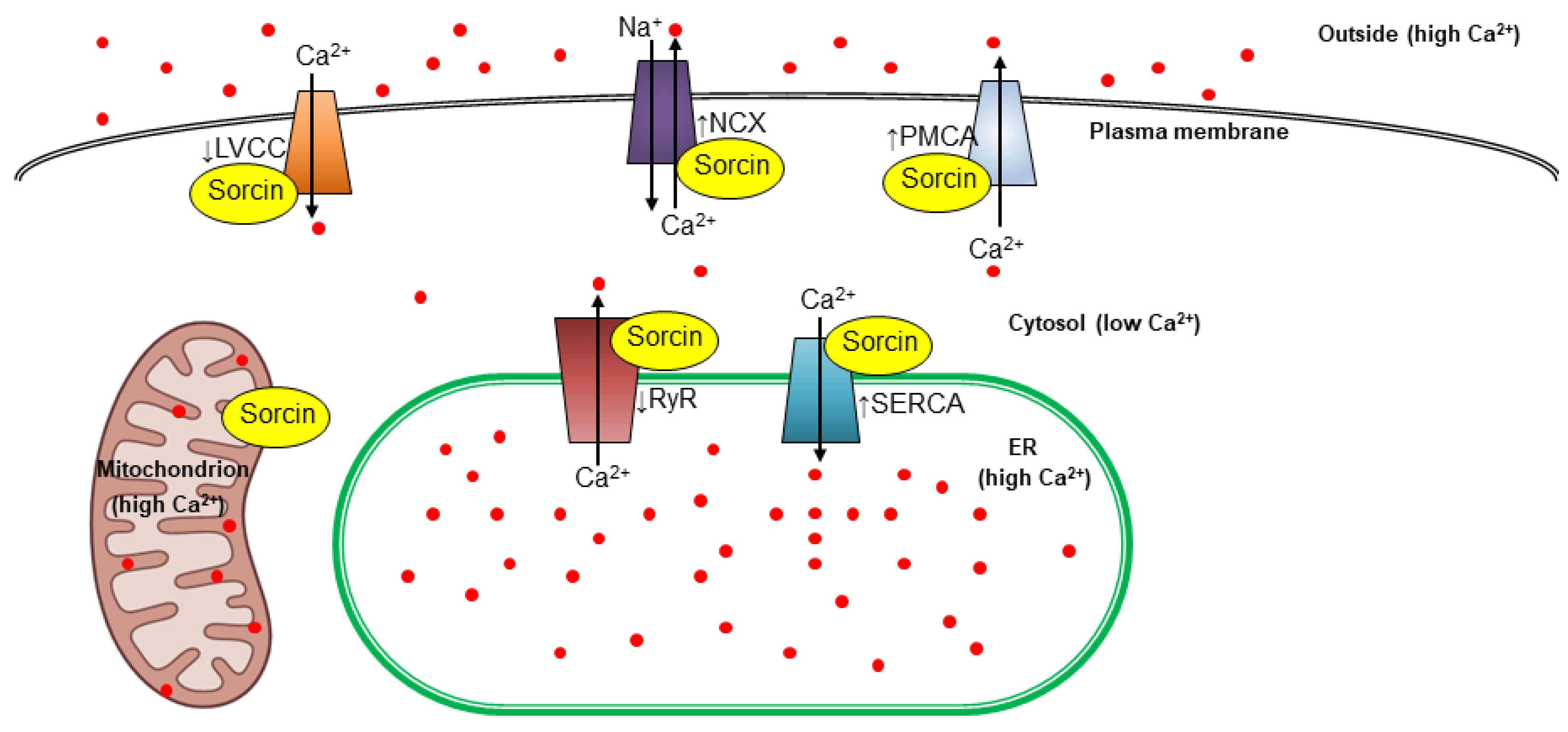
In particular, sorcin regulates calcium homeostasis by regulating channels, pumps, and exchangers (Figure 2). It inhibits calcium release by the ER, by binding to ryanodine receptors (RyRs), which are important channels that extrude calcium from the ER, acting as a brake on calcium release into the cell. In the heart, sorcin binds to cardiac RyRs with high affinity and completely inhibits channel activity. Sorcin significantly inhibits both the spontaneous activity of RyRs in quiescent cells and the inward Ca2+ current-triggered activity of RyRs that gives rise to Ca2+-induced Ca2+ release (CICR), i.e., the amplifying Ca2+ signaling mechanism that triggers cardiac contraction [18,19,20,21]. Sorcin also inhibits RyRs in endothelial and smooth muscle cells, effectively regulating Ca2+ signaling in vascular smooth muscle [22].
Furthermore, sorcin increases calcium uptake from the cytosol to the ER: sorcin increases the activity of the sarco/endoplasmic reticulum Ca2+ pump (SERCA), which actively transports calcium from the cytosol into the ER, further increasing ER calcium levels and reducing cytosolic ones [23].
Sorcin regulates other membrane calcium transporters: it activates the sarcolemmal Na+-Ca2+ exchanger (NCX), the plasma membrane Ca2+ pump (PMCA), and modulates the L-type voltage-gated calcium channel (LVCC) [14,24,25,26,27]. By regulating calcium transporters, sorcin plays a critical role in various cellular processes.
In heart muscle cells, sorcin helps regulate the contraction and relaxation cycle by controlling calcium fluxes [19,20]. Sorcin has a critical role in maintaining Ca2+ homeostasis, especially during the adrenergic response of the heart: in a sorcin knockout (KO) mouse model, sorcin deficiency can lead to ventricular arrhythmias and heart failure [28]. Sorcin KO hearts developed overexpression of LVCC and NCX, increasing spontaneous Ca2+ release events and leading to Ca2+ disturbances and arrhythmias. In turn, in vivo adenoviral transfer of sorcin reduces cardiac contractile abnormalities [29].
Additionally, sorcin also appears to modulate calcium handling of mitochondria: sorcin-overexpressing cardiac myocytes are more calcium-tolerant than wild-type cells and their mitochondria contain increased levels of calcium. Sorcin prevents cytochrome c release and caspase activation [30].
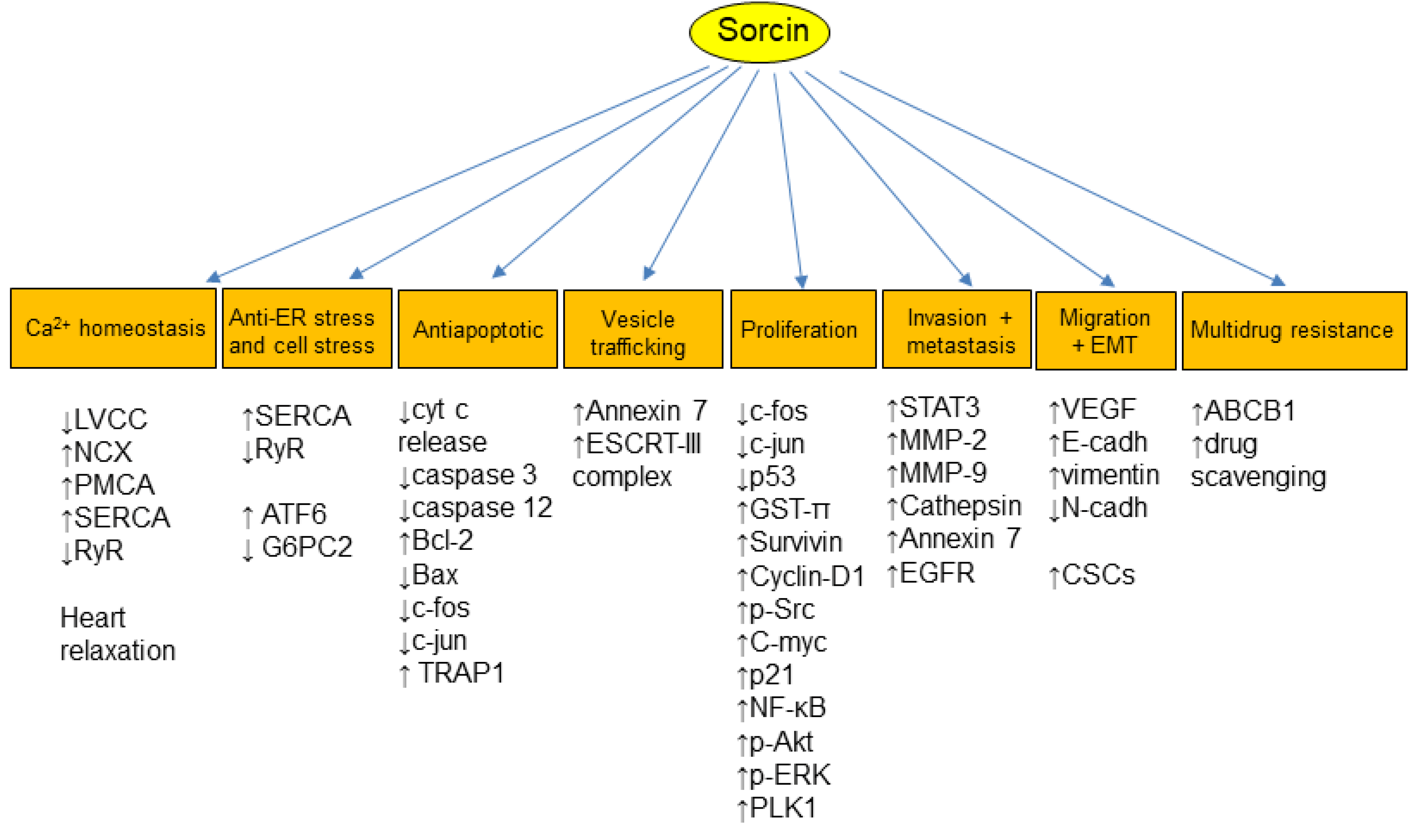
Sorcin calcium-dependently interacts with multiple proteins (Figure 3), including important kinases involved in the regulation of the cell cycle, such as Polo-like kinase 1 (PLK1), Aurora A, and Aurora B kinases. Sorcin physically interacts with PLK1, serving as a substrate for PLK1-mediated phosphorylation and stimulating PLK1 autophosphorylation, thereby regulating kinase activity. Sorcin is crucial for successful cell division, encompassing both mitosis and cytokinesis [16]. In fact, reducing sorcin levels leads to mitotic and cytokinetic abnormalities, an increase in abnormal, multinucleated cells, cell cycle arrest in the G2/M phase, and programmed cell death (apoptosis). Sorcin silencing triggers apoptosis by activating caspases 3 and 12 [16,30,31,32,33].
It is also worth mentioning that sorcin may help protect cells from ER stress, a condition caused by imbalanced calcium levels in the ER; sorcin overexpression can reduce apoptosis (programmed cell death) triggered by ER stress, by maintaining a high ER calcium concentration [16,30,32,33]. Sorcin overexpression activates ATF6, a key player in the unfolded protein response (UPR), which protects cells during ER stress, whereas sorcin KO reduces ATF6 transcriptional activity and increases ER stress. Notably, sorcin downregulation during lipotoxic stress may impair ATF6 activation and a normal UPR during the progression of obesity and insulin resistance [34].
Therefore, sorcin is emerging as a key player not only in UPR but also in cellular stress response and potentially in various pathologies. Sorcin interacts with carbohydrate-responsive element-binding protein (ChREBP), a key regulator of gene expression in pancreatic β cells and a significant contributor to glucose toxicity, sequestering it in the cytosol in a calcium-dependent fashion. Sorcin silencing inhibits ATP-induced increases in intracellular Ca2+ and glucose-stimulated insulin secretion. Sorcin may act as a Ca2+ sensor for glucose-induced nuclear translocation and the activation of ChREBP-dependent genes; a high carbohydrate diet enforces nuclear shuttling of hepatic NF-κB p65 and represses sorcin transcription, leading to de novo lipogenesis and intrahepatic lipid accumulation [35,36].
Sorcin overexpression in pancreatic beta cells increases ER Ca2+ levels and improves glucose tolerance and glucose-stimulated insulin secretion, highlighting its potential benefit in diabetes. Conversely, sorcin KO mice are glucose intolerant, with markedly impaired GSIS and increased expression of G6PC2, which contributes to lipotoxicity [33].
Another interesting aspect is the high sorcin expression in the brain, particularly in areas related to memory and emotion, which suggests its involvement in brain function [37]. Indeed, altered sorcin levels are observed in various neurodegenerative diseases, including Alzheimer’s disease (AD), Huntington’s disease, frontotemporal dementia, and Parkinson’s disease (PD). Interestingly, sorcin may offer protection against the rapid progression of these diseases. In neurons and microglia cells, sorcin colocalizes with ER proteins, such as RyRs, SERCA2, and the Sigma-1 receptor, and regulates ER calcium transients. Sorcin may represent both a novel early marker of neurodegenerative diseases and a response to cellular stress dependent on neurodegeneration [38,39,40,41,42,43,44,45,46,47,48].
Sorcin interacts with microtubule-associated tau, a protein that represents a pathological marker of AD. Binding to aberrant forms of tau impairs sorcin functions, such as calcium homeostasis and cellular resistance by ER stress, possibly resulting in a contribution to the progression of AD [49]. In addition, sorcin acts as an activator of PMCA, preventing the inhibitory effects of Aβ and tau on the pump; it also binds both Aβ and tau, counteracting their neurotoxicity [27].
Sorcin can interact with proteins like presenilin 2 (PS2) and alpha-synuclein (AS), implicated in AD and PD, respectively [50,51]. These interactions could influence both calcium signaling and disease development.
Furthermore, sorcin is involved in fertility. Sorcin is highly expressed in the endometrium, during the window of implantation, and decreased sorcin levels are observed in the endometrium of women with unexplained infertility. Sorcin is important in the regulation of Ca2+-mediated angiogenesis via the VEGF/PI3K/Akt pathway in endometrial cells and plays a crucial role in preparing the endometrium for implantation [52].
Sorcin is found in different types of vesicles within the cell, suggesting a potential role in cellular trafficking. High levels of sorcin have been identified in ER and in ER-derived vesicles positioned along microtubules. These vesicles are characterized by the presence of RyR, SERCA, calreticulin, and Rab10. Sorcin-containing vesicles are located at the mitotic spindle during mitosis, at the cleavage furrow during telophase, and finally at the midbody. Sorcin regulates the size and calcium content of ER vesicles by decreasing RYR activity and stimulating SERCA function [16]. Sorcin is found at high levels in lipid rafts and in exosomes [53,54,55,56,57,58,59,60,61,62,63,64]. Further, endosomal recruitment of sorcin was identified as an essential step in apoptotic exosome formation since it participates with annexins A2 and A7 and the HD-PTP protein in the assembly of the ESCRT-III complex [63].

4. Sorcin and Cancer

Sorcin is often overexpressed in cancer cells, in many types of tumors, such as neuroblastoma, adenocarcinoma, glioma, ovarian cancer, gastric cancer, lung cancer, oral cancer, prostate cancer, colorectal cancer, central nervous system cancer, gallbladder cancer, hepatocellular carcinoma, and leukemias [32,65,66,67,68,69,70,71,72,73,74,75,76,77,78,79,80,81,82,83,84,85,86,87,88,89,90].
A recent study investigated the expression of sorcin in normal tissues vs. cancer tissues using multiple databases, i.e., Genotype-Tissue Expression (GTEX), The Cancer Genome Atlas (TCGA), and Oncomine [91]. The analysis showed that sorcin is significantly overexpressed in 25 types of cancer (it is also downregulated in 3 cancers; no significant difference is reported for 5 cancers), and that sorcin expression dysregulation could predict worse prognosis in the patients with many types of cancer. The same study reports a correlation of sorcin expression with patient survival, clinical features, cancer stem cell properties, tumor mutation burden, microsatellite instability, and immune cell infiltration; aberrant sorcin expression and/or gene amplification was also found related to epithelial-mesenchymal-transition (EMT), tumor immune microenvironment, and drug resistance (see below) [91].
Sorcin overexpression seems to contribute to cancer cell survival by means of different features. Ca2+ signaling plays a crucial role in regulating many cancer hallmarks, such as angiogenesis, migration, and invasion; abnormal calcium fluxes due to altered channel/pump expression or activation participate in carcinogenesis and promote cancer development and resistance to chemotherapeutic drugs (for a review, see reference [92]). Sorcin contributes to the regulation of LVCC, NCX1, PMCA, SERCA, and RyRs (Figure 2), and its overexpression leads to calcium dyshomeostasis. As already mentioned, sorcin can help cancer cells evade apoptosis by modulating calcium homeostasis and, in particular, increasing calcium levels in the ER and mitochondria, thereby reducing ER stress and cellular stress [16,30,32,33,93].
Sorcin modulates cell cycle and apoptosis, also by regulating important regulation pathways (Figure 3). Leukemia and breast carcinoma cells overexpressing sorcin also showed high expression of Bcl-2, along with decreased level of Bax [75,94]; upon sorcin silencing, cells show increased Bax, c-fos and c-jun expression, while the level of Bcl-2 is decreased [94]. In nasopharyngeal carcinoma, sorcin silencing results in the expression of the multidrug-resistance genes MDR1 (ABCB1), MRP1, and ERCC1, the detoxification enzyme GST-π, the Rho GTPase RhoE, and the anti-apoptotic proteins Bcl-2 and Survivin. Conversely, PTEN expression increased, while the levels of phosphorylated Akt (p-Akt) and NF-κB decreased, leading to reduced apoptosis [95]. Similarly, in myeloma cells, sorcin silencing results in reduced cell proliferation, cell cycle arrest, and cell apoptosis, with reduced expression levels of ABCB1, MRP1, GST-π, Survivin, Livin, Bcl-2, Cyclin-D1, phospho-Src, C-myc, p21, NF-κB and phospho-AKT, while p53 expression and caspase-3 and caspase-8 activity significantly increased [96]. Sorcin silencing also activates GRP78/BiP and caspases 3 and 12, triggering apoptosis via the mitochondrial pathway [32].
Sorcin interacts with various other proteins involved in cell cycle regulation and signaling pathways, such as Polo-like kinase 1 (PLK1), Aurora A and Aurora B kinases. Sorcin silencing results in defects in mitosis and cytokinesis and in increased apoptosis [16]. Sorcin interacts with the calcium/calmodulin-dependent protein kinase II (CaMKIIdeltaC), inhibiting it in a calcium- and concentration-dependent manner [97].
TRAP1, a mitochondrial anti-apoptotic heat shock protein, involved in protection from oxidative stress and apoptosis, specifically interacts with sorcin in a Ca2+-dependent fashion; this interaction is required for the anti-apoptotic function of TRAP1 [98].
Sorcin is an important player in angiogenesis, proliferation, EMT and tumor metastasis (Figure 3). Sorcin silencing decreases the expression of the angiogenic factor VEGF and of its downstream effector molecules, i.e., PI3K, Akt, and NOS. Sorcin regulates Ca2+-mediated angiogenesis via the VEGF/PI3K/Akt pathway [52].
Sorcin overexpression increases EMT, migration, and invasion in cancer cells, while sorcin silencing reduces the pool of cancer stem cells (CSCs), decreasing the EMT transition and suppressing metastases via the reduced expression of E-cadherin and VEGF and by the PI3K/Akt axis [81,99]. Sorcin knockdown decreases the expression of matrix metalloproteinases (MMP2 and MMP9), cathepsin Z, and STAT3, leading to suppressed tumor growth and metastasis [100]. In ovarian cancer, sorcin was found to drive malignant progression via the Smad4/ZEB1/miR-142-5p axis [86]. The interaction between sorcin and Annexin 7, both overexpressed in hepatocellular carcinoma, is able to promote EMT, migration, invasion, and proliferation, contributing to tumor aggressiveness [101].
Sorcin and EGFR expression levels are significantly correlated and linked to decreased overall survival in cancer patients. Sorcin directly interacts with the EGFR protein in a calcium-dependent manner, regulating calcium balance associated with EGFR signaling induced by EGF. Sorcin influences EGFR protein stability and signaling, enhancing its phosphorylation, and subsequently promoting EGF-driven cell migration and invasion. Notably, suppressing sorcin expression enhances the effectiveness of EGFR inhibitors in controlling cell migration [102].
In ovarian cancer, sorcin overexpression is associated with an impaired TGF-β signaling pathway. TGF-β1 administration, in fact, inhibits sorcin expression [103].
Sorcin not only regulates apoptosis but also pyroptosis. High sorcin expression in hepatocarcinoma cells negatively regulates pyroptosis by interacting with the NLRP3 inflammasome to promote tumor proliferation, migration, and invasion [88].

5. Multidrug Resistance

Multidrug resistance (MDR) is the main reason why many cancers become resistant to chemotherapy drugs. This significantly reduces the effectiveness of many chemotherapy treatments. It affects patients with a wide range of blood cancers and solid tumors. Tumors are often composed of a mix of malignant cells, with some being sensitive to drugs while others are resistant. Chemotherapy eliminates the drug-sensitive cells, but this leaves behind a larger proportion of resistant cells. When the tumor starts to regrow, chemotherapy may fail because the remaining cells are now drug-resistant. Over 90% of metastatic cancers exhibit MDR, leading to treatment failure [104].
A multitude of both intrinsic and acquired mechanisms contribute to drug resistance in cancer (Figure 4) (for a review, see [104,105,106]). The efficacy of chemotherapy is hindered by factors such as poor drug solubility, toxicity to healthy tissues, and pharmacokinetic limitations on drug delivery to the tumor.
At the tumor level, diverse resistance mechanisms emerge, including impaired drug uptake, enhanced drug efflux, altered cellular composition, inhibited cell death, increased DNA repair, cell cycle abnormalities, drug sequestration, accelerated drug metabolism, drug target modifications, and epithelial-mesenchymal transition (EMT). Some of these mechanisms confer resistance to specific drugs, while others contribute to MDR, rendering tumors refractory to multiple treatments and significantly reducing cure rates.
Further, tumor heterogeneity, driven by genetic and non-genetic factors, fuels the development of drug resistance. Clonal selection during therapy favors the survival and expansion of resistant cancer cell subpopulations. Notably, cancer stem cells (CSCs) play a pivotal role in chemoresistance due to their intrinsic resistance, mediated by epigenetic factors, increased expression of anti-apoptotic proteins and drug efflux pumps, and their dormant state. Consequently, chemotherapy primarily eliminates sensitive cancer cells, allowing resistant subpopulations, including CSCs, to proliferate and cause treatment failure.

6. Sorcin in Multidrug Resistance

Sorcin was initially discovered as a marker of MDR, although it is increasingly considered a cause of MDR. Sorcin was initially discovered in vincristine-resistant hamster lung cancer cells, and it is often overexpressed in cancers exhibiting an MDR phenotype [31,32,65,66,73,75,78,80,82,83,84,86,91,95,96,103,107,108,109,110,111].
Sorcin transfection in several tumor cell lines (leukemia, lung, gastric, ovarian, and breast cancers) increases resistance to common chemotherapy drugs, e.g., paclitaxel, doxorubicin, vincristine, etoposide, 5-fluorouracil and homoharringtonine [73,75,80,84,86,94,109,110,111].
Conversely, silencing sorcin increases chemotherapeutic drug toxicity and reverses MDR in various cancers, including leukemia, cervical cancer, breast cancer, colorectal cancer, and nasopharyngeal carcinoma [31,32,73,78,82,83,84,86,94,95,103,107,108,109,112,113].
Sorcin’s influence on ABCB1 (MDR1 or P-glycoprotein) is perhaps the most well-studied and significant mechanism of MDR it contributes to. ABCB1, an ATP-dependent efflux pump, is a critical factor in resistance to a broad spectrum of chemotherapy drugs, including anthracyclines, taxanes, and Vinca alkaloids, in various cancers (solid and blood-based tumors) [114,115]. Its overexpression or amplification significantly reduces the effectiveness of these drugs, increasing their efflux from cancer cells.
Interestingly, the genes for sorcin and ABCB1 (the most significant ATP-dependent drug efflux pump) are located near each other on chromosome 7 and can be co-amplified, leading to their simultaneous overexpression in MDR cancer cells [3,65].
For a long time, sorcin overexpression was thought to be only an accidental consequence of this co-amplification. However, in the last two decades, many studies showed that sorcin is not just a marker but also a cause of MDR, since it contributes to many processes leading to resistance (Figure 4).
Sorcin plays a key role in regulating ABCB1 expression. Sorcin overexpression leads to increased ABCB1 levels, ultimately enhancing drug resistance in various cancer cell lines, including those from gastric cancer, lung tumors, nasopharyngeal carcinoma, cervical carcinoma, and leukemias, while sorcin silencing reduces ABCB1 expression [31,80,83,95,96,109,110,111,116].
Mechanistically, sorcin stimulates the phosphorylation of CREB1 by PKA (protein kinase A). This activated CREB1 then binds to the cAMP response element (CRE) within the ABCB1 gene promoter, located at −716 to −709 bp, leading to increased ABCB1 expression [80]. Conversely, sorcin silencing can suppress ABCB1 expression by inhibiting ERK and Akt signaling pathways [111].
Interestingly, the genes for sorcin (SRI) and ABCB1 reside within the same chromosomal region (7q21.12) in both humans and mice. Chromosomal amplification events at the 7q21 locus lead to marked ABCB1 overexpression, which in turn causes MDR (for a review, [115]). This shared location suggests a potential link between their co-amplification, where both genes are duplicated together. Amplification of this region is observed in many multidrug-resistant cancers, leading to the overexpression of both sorcin and ABCB1, further contributing to MDR [3,77,117,118,119,120,121,122,123,124,125,126,127,128,129,130,131].
The amplicon at the 7q21.12 region encompasses several other genes beyond sorcin and ABCB1. Many of these genes, including ABCB4 (MDR3), ADAM22, and DBF4, have been implicated in cancer development and MDR, and are considered possible therapeutic targets [132,133,134,135,136,137,138,139,140,141,142,143,144,145,146].
Sorcin’s contribution to a multidrug-resistant phenotype may also involve its ability to directly bind chemotherapeutic drugs (Figure 4). Studies using techniques like surface plasmon resonance and X-ray diffraction demonstrate sorcin’s high-affinity binding to doxorubicin, paclitaxel, vincristine, and cisplatin in vitro [110]. The crystal structure of the sorcin-doxorubicin complex revealed one of the binding sites—near the interface of two sorcin monomers. This binding pocket involves interactions between specific sorcin residues and the doxorubicin molecule [108]. Additionally, sorcin’s cellular localization changes upon doxorubicin treatment, suggesting potential drug interactions within the cell. These findings suggest sorcin could act as a “drug scavenger,” limiting the toxic effects of doxorubicin and potentially other drugs within the cell [110].

7. Targeting Sorcin: Promising Strategies for Overcoming Cancer and MDR

Sorcin’s involvement in tumorigenesis and MDR across various cancer cell lines suggests that lowering its expression could be a viable strategy to reverse both cancer onset and resistance to chemotherapeutic targets. This has led to the exploration of several promising approaches targeting sorcin (Table 1).
MicroRNAs (miRNAs) act as natural gene regulators. miR-1, for example, targets the 3′ untranslated region (3′-UTR) of the sorcin gene (SRI) and modulates calcium transients in heart muscle cells. Interestingly, miR-1 expression is downregulated in human heart failure and corresponding mouse models [147]. In MDR gastric cancer cells, miR-1 levels are significantly low with respect to non-tumor tissues. Reintroducing miR-1 promotes apoptosis (programmed cell death), inhibits migration, and increases the accumulation of chemotherapeutic drugs like doxorubicin and vincristine by acting on sorcin expression [112,148]. While sorcin overexpression can partially counteract miR-1’s effect, this finding supports the potential of miR-1 as a therapeutic strategy against sorcin-dependent MDR. However, off-target effects are a concern, as miR-1 regulates other genes like HSP60 (involved in diabetic heart damage) and the ets1 proto-oncogene (important for extracellular matrix breakdown) [149,150]. In ovarian cancer, miR-142-5p suppresses sorcin expression by binding to the 3′-UTR of the SRI gene; interestingly, this is part of a complex homeostatic loop, since the transcription factor ZEB1 inhibits the transcription of miR-142-5p by directly binding to the E-box fragment in the miR-142 promoter region, while furthermore, ZEB1 is itself negatively regulated by sorcin [86].
Many molecules have effects on sorcin levels and may represent options to control tumorigenesis, MDR, and other pathological conditions (Table 1).
Since sorcin overexpression in ovarian cancer is associated with an impaired TGF-β signaling pathway, this pathway may represent a target to regulate sorcin expression. In fact, the administration of TGF-β1 inhibits sorcin expression in ovarian cancer and non-small cell lung cancer [103].
Tetrandrine, a bis-benzylisoquinoline alkaloid, is a well-known calcium channel blocker, which inhibits voltage-gated Ca2+ current (LVCC) and Ca2+-activated K+ current. Tetrandrine decreases sorcin expression and reverses MDR in leukemia cell lines resistant to daunorubicin, in a concentration-dependent fashion [151].
The oxindole derivative PH II-7 is a powerful cytotoxic agent, able to increase intracellular drug concentration in multidrug-resistant cells by inhibiting the expressions of both ABCB1 and sorcin, and to induce the apoptosis of multidrug-resistant tumor cells [152].
Dihydromyricetin (DMY), a natural compound with anti-oxidant, anti-inflammatory, and anti-tumor properties, has shown promise in reversing MDR in leukemia and breast cancer cells resistant to adriamycin, as well as in animal models. It increases adriamycin cytotoxicity by decreasing sorcin (both mRNA and protein) and, consequently, ABCB1 levels, likely via the ERK/Akt signaling pathway [84,111]. Additionally, DMY increases intracellular-free calcium levels, reactive oxygen species (ROS), and caspase 12 expression (markers of ER stress-induced apoptosis). It also regulates markers of mitochondrial apoptosis, such as caspases 3 and 9, Bcl-2, Bax, and PARP [84].
Ondansetron (OND), an anti-emetic drug used during chemotherapy, has also been shown to reverse MDR associated with sorcin expression, particularly when combined with DMY. This combination increases cell cycle arrest and apoptosis, possibly by decreasing MDM2/MDMX, and consequently increasing p53 function. Both DMY and OND may act by binding sorcin with high affinity, although DMY’s effect seems specific and dependent on sorcin binding [111].
Haishengsu (HSS) is a protein extract, produced by Tegillarca granosa seashells. HSS administration reduces sorcin, BCR/ABL, and ABCB1 expression in mouse models and results in increased apoptosis in MDR leukemia cells. While its effects on other targets remain unclear, HSS (added to conventional chemotherapy) has shown promising results in clinical trials for acute leukemia patients, increasing treatment efficacy and improving quality of life [153,154].
Calcitriol (vitamin D) binds to the vitamin D receptor, which acts as a transcription factor regulating various cellular processes, including sorcin expression. Calcitriol treatment leads to differentiation and anti-proliferative effects and appears to increase the 19 kDa form of sorcin, suggesting potential for further investigation [155].
Palmitate, a free fatty acid, downregulates sorcin in pancreatic beta cells, potentially contributing to lipotoxicity and ER stress. In addition, palmitate treatment reduces phosphorylation of AMPK and increases the levels of markers of the proapoptotic arm of ER stress response (pEIF2α and CHOP) and apoptosis (cleaved caspase 3). Interestingly, metformin treatment normalizes the levels of pAMPK, pEIF2α, CHOP, and cleaved caspase 3 and increases the levels of sorcin; palmitate controlling may thus represent a treatment strategy aiming at decreasing insulin levels in obese children [156,157].
TNFα decreases sorcin expression in pancreatic cells, possibly by increasing phosphorylation of key proteins of insulin secretion pathway such as mTOR and NFκB; fractalkine (CX3CL1) restores sorcin levels by reducing mTOR expression and decreasing phosphorylation of mTOR and NFκB [158].
Triptolide, a diterpenoid epoxide from the thunder god vine, Tripterygium wilfordii, has proapoptotic activity and anti-tumor effects in ovarian cancer cells, reducing the expression of sorcin, MMP-2, and VEGF, which are usually highly expressed in ovarian cancer cells [83]. Since triptolide is rather toxic, strategies aimed at specifically delivering it to cancer cells have been developed. The administration of triptolide-loaded exosomes (which have high drug encapsulation efficiency and a rather specific ability to deliver their payload to cancer cells) had a high capacity of inhibiting ovarian tumor cell proliferation and tumor growth, although they had toxic effects on liver and spleen [159].
A more direct way to target sorcin is the use of CRISPR or siRNA vs. sorcin to restore calcium homeostasis, limit tumorigenesis, and overcome chemoresistance. siRNAs inhibit target gene expression by binding to specific mRNA transcripts and proved to be effective in cancer cell cultures (see Section 4 and Section 5); however, siRNA is limited in clinical application due to its nature, such as low internalization in tumor cells, degradation by enzymes in the bloodstream, and off-targeting features. Loading siRNAs into nanocarriers protects them from nuclease degradation, increases their delivery to tumors by the enhanced permeability and retention (EPR) effect, and decreases siRNA-dependent immune stimulation. Lipid-coated albumin-paclitaxel nanoparticles loaded with sorcin-siRNA (LANP-PTX-siSRI) have been recently developed for PTX and siSRI co-delivery, aiming at sorcin targeting and PTX delivery into multidrug-resistant cells. The LANP-PTX-siSRI decreased sorcin expression and increased intracellular calcium, leading to the inhibition of PTX-resistant ovarian cancer cell growth [103].

8. Conclusions

Sorcin’s co-amplification with drug efflux pumps and its role in calcium signaling make it a fascinating target for cancer therapy. Extensive research has been dedicated to unraveling sorcin’s mechanisms of action in various disease contexts. This knowledge paves the way for the development of novel and effective therapeutic strategies.
Developing effective sorcin-targeting therapies is complex due to sorcin’s widespread expression in healthy tissues. Off-target effects and potential for resistance development are major concerns, similar to many chemotherapeutic drugs. Recent advancements in targeted therapies, such as antibody-drug conjugates and gene editing techniques, offer promising avenues for overcoming these challenges.
A key future direction lies in acquiring a deeper understanding of sorcin’s protein interactions (interactome). This knowledge could help identify specific interaction sites for targeted modulation, minimizing off-target effects. Additionally, structural studies suggest that calcium binding exposes specific hydrophobic regions on the sorcin molecule. These regions could potentially serve as targets for interaction with other proteins. By targeting these specific sites, researchers may achieve focused modulation of sorcin’s activity, leading to more effective and specific therapeutic strategies.

9. Perspectives

Sorcin is an important protein, involved in many cellular processes.
It is essential for ER calcium signaling, controlling ER size, calcium content, and vesicle dynamics by inhibiting RyR and activating SERCA. Upon calcium binding, sorcin undergoes conformational changes exposing hydrophobic residues, enabling interactions with (and regulation of) calcium channels and exchangers like RyR and SERCA. This modulation increases ER calcium accumulation, mitigating ER stress and the UPR. Possibly because of this action on ER stress, sorcin is also an early marker of neurodegeneration.
In particular, sorcin influences various oncogenic proteins, including NF-κB, STAT3, AKT, ERK1/2, IP3R, VEGF, MMPs, and caspases, and drug efflux pumps such as ABCB1. Its suppression induces apoptosis, mitotic and cytokinetic defects, cell cycle arrest, and the formation of abnormal cells.
Overexpressed in multiple cancers, especially chemoresistant ones, sorcin promotes proliferation, migration, invasion, EMT, and drug resistance. Conversely, its inhibition sensitizes cells to cisplatin and adriamycin, reducing proliferation and inducing apoptosis.
Given its oncogenic properties and association with MDR, sorcin is considered an oncogene and an MDR marker.
Further, it is increasingly considered a potential therapeutic target since its regulation, by means of several categories of molecules, can affect cancer and/or resistance to chemotherapeutic drugs.

Author Contributions

Conceptualization, C.E., L.A., A.F., A.I. and G.C.; methodology, C.E., L.A., A.F., R.B., B.C., A.I. and G.C.; writing—original draft preparation, C.E., L.A., A.F., R.B., B.C., A.I. and G.C.; writing—review and editing, C.E., L.A., A.F., R.B., B.C., A.I. and G.C.; funding acquisition, A.I. and G.C. All authors have read and agreed to the published version of the manuscript.

Funding

We acknowledge: CNCCS s.c.a.r.l. 2015-2023, National Collection of Chemical Compounds and Screening Center (www.cnccs.it, accessed on 31 July 2024); PNRR M4C2—Dalla ricerca all’impresa—3.1: Fondo per la realizzazione di un sistema integrato di infrastrutture di ricerca e innovazione “Potentiating the Italian Capacity for Structural Biology Services in Instruct-ERIC (ITACA.SB)” CUP: B53C22001790006; PNRR PE8 Age-IT., co-funding from Next Generation EU [DM 1557 11.10.2022], in the context of the National Recovery and Resilience Plan, Investment PE8—Project Age-It: “Ageing Well in an Ageing Society”; Project PRIN MUR 2022HYF8KS; Project DBA.AD005.225 -NUTRAGE- FOE2021 CNR.

Conflicts of Interest

The authors declare no conflicts of interest.

References

  1. Hanahan, D.; Weinberg, R.A. The hallmarks of cancer. Cell 2000, 100, 57–70. [Google Scholar] [CrossRef] [PubMed]
  2. Hanahan, D.; Weinberg, R.A. Hallmarks of cancer: The next generation. Cell 2011, 144, 646–674. [Google Scholar] [CrossRef] [PubMed]
  3. Van der Bliek, A.M.; Meyers, M.B.; Biedler, J.L.; Hes, E.; Borst, P. A 22-kd protein (sorcin/V19) encoded by an amplified gene in multidrug-resistant cells, is homologous to the calcium-binding light chain of calpain. EMBO J. 1986, 5, 3201. [Google Scholar] [CrossRef] [PubMed]
  4. Genovese, I.; Ilari, A.; Battista, T.; Chiarini, V.; Fazi, F.; Fiorillo, A.; Colotti, G. Molecular bases of Sorcin-dependent resistance to chemotherapeutic agents. Cancer Drug Resist. 2018, 1, 164–180. [Google Scholar] [CrossRef]
  5. Maki, M.; Kitaura, Y.; Satoh, H.; Ohkouchi, S.; Shibata, H. Structures, functions and molecular evolution of the penta-EF-hand Ca 2+-binding proteins. Biochim. Biophys. Acta Proteins Proteom. 2002, 1600, 51–60. [Google Scholar] [CrossRef] [PubMed]
  6. Zamparelli, C.; Ilari, A.; Verzili, D.; Giangiacomo, L.; Colotti, G.; Pascarella, S.; Chiancone, E. Structure-function relationships in sorcin, a member of the penta EF-hand family. Interaction of sorcin fragments with the ryanodine receptor and an Escherichia coli model system. Biochemistry 2000, 39, 658–666. [Google Scholar] [CrossRef] [PubMed]
  7. Mella, M.; Colotti, G.; Zamparelli, C.; Verzili, D.; Ilari, A.; Chiancone, E. Information transfer in the penta-EF-hand protein sorcin does not operate via the canonical structural/functional pairing. A study with site-specific mutants. J. Biol. Chem. 2003, 278, 24921–24928. [Google Scholar] [CrossRef] [PubMed]
  8. Ilari, A.; Johnson, K.A.; Nastopoulos, V.; Verzili, D.; Zamparelli, C.; Colotti, G.; Tsernoglou, D.; Chiancone, E. The crystal structure of the sorcin calcium binding domain provides a model of Ca2+-dependent processes in the full-length protein. J. Mol. Biol. 2002, 317, 447–458. [Google Scholar] [CrossRef]
  9. Ilari, A.; Fiorillo, A.; Poser, E.; Lalioti, V.S.; Sundell, G.N.; Ivarsson, Y.; Genovese, I.; Colotti, G. Structural basis of Sorcin-mediated calcium-dependent signal transduction. Sci. Rep. 2015, 5, 16828. [Google Scholar] [CrossRef]
  10. Meyers, M.B.; Zamparelli, C.; Verzili, D.; Dicker, A.P.; Blanck, T.J.J.; Chiancone, E. Calcium-dependent translocation of sorcin to membranes: Functional relevance in contractile tissue. FEBS Lett. 1995, 357, 230–234. [Google Scholar] [CrossRef]
  11. Nastopoulos, V.; Ilari, A.; Colotti, G.; Zamparelli, C.; Verzili, D.; Chiancone, E.; Tsernoglou, D. Two different crystal forms of sorcin, a penta-EF-hand Ca2+-binding protein. Acta Crystallogr. D Biol. Crystallogr. 2001, 57 Pt 6, 862–864. [Google Scholar] [CrossRef] [PubMed]
  12. Colotti, G.; Zamparelli, C.; Verzili, D.; Mella, M.; Loughrey, C.M.; Smith, G.L.; Chiancone, E. The W105G and W99G sorcin mutants demonstrate the role of the D helix in the Ca2+-dependent interaction with annexin VII and the cardiac ryanodine receptor. Biochemistry 2006, 45, 12519–12529. [Google Scholar] [CrossRef] [PubMed]
  13. Franceschini, S.; Ilari, A.; Verzili, D.; Zamparelli, C.; Antaramian, A.; Rueda, A.; Valdivia, H.H.; Chiancone, E.; Colotti, G. Molecular basis for the impaired function of the natural F112L sorcin mutant: X-ray crystal structure, calcium affinity, and interaction with annexin VII and the ryanodine receptor. FASEB J. 2008, 22, 295–306. [Google Scholar] [CrossRef] [PubMed]
  14. Zamparelli, C.; Macquaide, N.; Colotti, G.; Verzili, D.; Seidler, T.; Smith, G.L.; Chiancone, E. Activation of the cardiac Na+–Ca2+ exchanger by sorcin via the interaction of the respective Ca2+-binding domains. J. Mol. Cell Cardiol. 2010, 49, 132. [Google Scholar] [CrossRef] [PubMed]
  15. Genovese, I.; Carotti, A.; Ilari, A.; Fiorillo, A.; Battista, T.; Colotti, G.; Ivarsson, Y. Profiling calcium-dependent interactions between Sorcin and intrinsically disordered regions of human proteome. Biochim. Biophys. Acta Gen. Subj. 2020, 1864, 129618. [Google Scholar] [CrossRef] [PubMed]
  16. Lalioti, V.S.; Ilari, A.; O’Connell, D.J.; Poser, E.; Sandoval, I.V.; Colotti, G. Sorcin links calcium signaling to vesicle trafficking, regulates Polo-like kinase 1 and is necessary for mitosis. PLoS ONE 2014, 9, e85438. [Google Scholar] [CrossRef] [PubMed]
  17. Battista, T.; Fiorillo, A.; Chiarini, V.; Genovese, I.; Ilari, A.; Colotti, G. Roles of Sorcin in Drug Resistance in Cancer: One Protein, Many Mechanisms, for a Novel Potential Anticancer Drug Target. Cancers 2020, 12, 887. [Google Scholar] [CrossRef] [PubMed]
  18. Meyers, M.B.; Pickel, V.M.; Sheu, S.S.; Sharma, V.K.; Scotto, K.W.; Fishman, G.I. Association of sorcin with the cardiac ryanodine receptor. J. Biol. Chem. 1995, 270, 26411–26418. [Google Scholar] [CrossRef] [PubMed]
  19. Lokuta, A.J.; Meyers, M.B.; Sander, P.R.; Fishman, G.I.; Valdivia, H.H. Modulation of cardiac ryanodine receptors by Sorcin. J. Biol. Chem. 1997, 272, 25333–25338. [Google Scholar] [CrossRef] [PubMed]
  20. Farrell, E.F.; Antaramian, A.; Rueda, A.; Gómez, A.M.; Valdivia, H.H. Sorcin Inhibits Calcium Release and Modulates Excitation-Contraction Coupling in the Heart. J. Biol. Chem. 2003, 278, 34660–34666. [Google Scholar] [CrossRef]
  21. Valdivia, H.H.; Farrell, E.F.; Antaramian, A.; Benkusky, N.; Zhu, X.; Rueda, A.; Gómez, A.M. Sorcin and ryanodine receptors in heart failure. J. Muscle Res. Cell Motil. 2004, 25, 605–607. [Google Scholar] [PubMed]
  22. Rueda, A.; Song, M.; Toro, L.; Stefani, E.; Valdivia, H.H. Sorcin modulation of Ca2+ sparks in rat vascular smooth muscle cells. J. Physiol. 2006, 576, 887–901. [Google Scholar] [CrossRef]
  23. Matsumoto, T.; Hisamatsu, Y.; Ohkusa, T.; Inoue, N.; Sato, T.; Suzuki, S.; Ikeda, Y.; Matsuzaki, M. Sorcin interacts with sarcoplasmic reticulum Ca2+-ATPase and modulates excitation-contraction coupling in the heart. Basic. Res. Cardiol. 2005, 100, 250–262. [Google Scholar] [CrossRef] [PubMed]
  24. Fowler, M.R.; Colotti, G.; Chiancone, E.; Smith, G.L.; Fearon, I.M. Sorcin modulates cardiac L-type Ca2+ current by functional interaction with the α1C subunit in rabbits. Exp. Physiol. 2008, 93, 1233–1238. [Google Scholar] [CrossRef] [PubMed]
  25. Fowler, M.R.; Colotti, G.; Chiancone, E.; Higuchi, Y.; Seidler, T.; Smith, G.L. Complex modulation of L-type Ca2+ current inactivation by sorcin in isolated rabbit cardiomyocytes. Pflug. Arch. 2009, 457, 1049–1060. [Google Scholar] [CrossRef] [PubMed]
  26. Seidler, T.; Miller, S.L.; Loughrey, C.M.; Kania, A.; Burow, A.; Kettlewell, S.; Teucher, N.; Wagner, S.; Kögler, H.; Meyers, M.B.; et al. Effects of adenovirus-mediated sorcin overexpression on excitation-contraction coupling in isolated rabbit cardiomyocytes. Circ. Res. 2003, 93, 132–139. [Google Scholar] [CrossRef] [PubMed]
  27. Berrocal, M.; Saez, L.; Mata, A.M. Sorcin activates the brain PMCA and blocks the inhibitory effects of molecular markers of alzheimer’s disease on the pump activity. Int. J. Mol. Sci. 2021, 22, 6055. [Google Scholar] [CrossRef] [PubMed]
  28. Chen, X.; Weber, C.; Farrell, E.T.; Alvarado, F.J.; Zhao, Y.T.; Gómez, A.M.; Valdivia, H.H. Sorcin ablation plus β-adrenergic stimulation generate an arrhythmogenic substrate in mouse ventricular myocytes. J. Mol. Cell Cardiol. 2018, 114, 199–210. [Google Scholar] [CrossRef] [PubMed]
  29. Suarez, J.; Belke, D.D.; Gloss, B.; Dieterle, T.; McDonough, P.M.; Kim, Y.K.; Brunton, L.L.; Dillmann, W.H. In vivo adenoviral transfer of sorcin reverses cardiac contractile abnormalities of diabetic cardiomyopathy. Am. J. Physiol. Heart Circ. Physiol. 2004, 286, H68–H75. [Google Scholar] [CrossRef]
  30. Suarez, J.; McDonough, P.M.; Scott, B.T.; Suarez-Ramirez, A.; Wang, H.; Fricovsky, E.S.; Dillmann, W.H. Sorcin modulates mitochondrial Ca2+ handling and reduces apoptosis in neonatal rat cardiac myocytes. Am. J. Physiol. Cell Physiol. 2013, 304, C248–C256. [Google Scholar] [CrossRef]
  31. Kawakami, M.; Nakamura, T.; Okamura, N.; Komoto, C.; Markova, S.; Kobayashi, H.; Hashimoto, N.; Okumura, K.; Sakaeda, T. Knock-down of sorcin induces up-regulation of MDR1 in HeLa cells. Biol. Pharm. Bull. 2007, 30, 1065–1073. [Google Scholar] [CrossRef] [PubMed]
  32. Maddalena, F.; Laudiero, G.; Piscazzi, A.; Secondo, A.; Scorziello, A.; Lombardi, V.; Matassa, D.S.; Fersini, A.; Neri, V.; Esposito, F.; et al. Sorcin induces a drug-resistant phenotype in human colorectal cancer by modulating Ca2+ homeostasis. Cancer Res. 2011, 71, 7659–7669. [Google Scholar] [CrossRef]
  33. Marmugi, A.; Parnis, J.; Chen, X.; Carmichael, L.; Hardy, J.; Mannan, N.; Marchetti, P.; Piemonti, L.; Bosco, D.; Johnson, P.; et al. Sorcin links pancreatic β-cell lipotoxicity to ER Ca2+ stores. Diabetes 2016, 65, 1009–1021. [Google Scholar] [CrossRef] [PubMed]
  34. Parks, S.Z.; Gao, T.; Jimenez Awuapura, N.; Ayathamattam, J.; Chabosseau, P.L.; Kalvakolanu, D.V.; Valdivia, H.H.; Rutter, G.A.; Leclerc, I. The Ca2+-binding protein sorcin stimulates transcriptional activity of the unfolded protein response mediator ATF6. FEBS Lett. 2021, 595, 1782–1796. [Google Scholar] [CrossRef] [PubMed]
  35. Noordeen, N.A.; Meur, G.; Rutter, G.A.; Leclerc, I. Glucose-induced nuclear shuttling of ChREBP is mediated by sorcin and Ca2+ ions in pancreatic β-cells. Diabetes 2012, 61, 574–585. [Google Scholar] [CrossRef]
  36. Daniel, P.V.; Dogra, S.; Rawat, P.; Choubey, A.; Khan, A.S.; Rajak, S.; Kamthan, M.; Mondal, P. NF-κB p65 regulates hepatic lipogenesis by promoting nuclear entry of ChREBP in response to a high carbohydrate diet. J. Biol. Chem. 2021, 296, 100714. [Google Scholar] [CrossRef]
  37. Canela-Xandri, O.; Rawlik, K.; Tenesa, A. An atlas of genetic associations in UK Biobank. Nat. Genet. 2018, 50, 1593–1599. [Google Scholar] [CrossRef]
  38. Andreev, V.P.; Petyuk, V.A.; Brewer, H.M.; Karpievitch, Y.V.; Xie, F.; Clarke, J.; Camp, D.; Smith, R.D.; Lieberman, A.P.; Albin, R.L.; et al. Label-free quantitative LC-MS proteomics of Alzheimer’s disease and normally aged human brains. J. Proteome Res. 2012, 11, 3053–3067. [Google Scholar] [CrossRef]
  39. Tsuji, T.; Shiozaki, A.; Kohno, R.; Yoshizato, K.; Shimohama, S. Proteomic profiling and neurodegeneration in Alzheimer’s disease. Neurochem. Res. 2002, 27, 1245–1253. [Google Scholar] [CrossRef]
  40. Seyfried, N.T.; Dammer, E.B.; Swarup, V.; Nandakumar, D.; Duong, D.M.; Yin, L.; Deng, Q.; Nguyen, T.; Hales, C.M.; Wingo, T.; et al. A Multi-network Approach Identifies Protein-Specific Co-expression in Asymptomatic and Symptomatic Alzheimer’s Disease. Cell Syst. 2017, 4, 60–72. [Google Scholar] [CrossRef]
  41. Drummond, E.; Nayak, S.; Faustin, A.; Pires, G.; Hickman, R.A.; Askenazi, M.; Cohen, M.; Haldiman, T.; Kim, C.; Han, X.; et al. Proteomic differences in amyloid plaques in rapidly progressive and sporadic Alzheimer’s disease. Acta Neuropathol. 2017, 133, 933–954. [Google Scholar] [CrossRef] [PubMed]
  42. Umoh, M.E.; Dammer, E.B.; Dai, J.; Duong, D.M.; Lah, J.J.; Levey, A.I.; Gearing, M.; Glass, J.D.; Seyfried, N.T. A proteomic network approach across the ALS—FTD disease spectrum resolves clinical phenotypes and genetic vulnerability in human brain. EMBO Mol. Med. 2018, 10, 48–62. [Google Scholar] [CrossRef] [PubMed]
  43. Jin, J.; Hulette, C.; Wang, Y.; Zhang, T.; Pan, C.; Wadhwa, R.; Zhang, J. Proteomic identification of a stress protein, mortalin/mthsp70/GRP75: Relevance to Parkinson disease. Mol. Cell. Proteom. 2006, 5, 1193–1204. [Google Scholar] [CrossRef] [PubMed]
  44. George, G.; Singh, S.; Lokappa, S.B.; Varkey, J. Gene co-expression network analysis for identifying genetic markers in Parkinson’s disease-a three-way comparative approach. Genomics 2019, 111, 819–830. [Google Scholar] [CrossRef] [PubMed]
  45. Kalathur, R.K.; Giner-Lamia, J.; Machado, S.; Barata, T.; Ayasolla, K.R.; Futschik, M.E. The unfolded protein response and its potential role in Huntington’s disease elucidated by a systems biology approach. F1000Res 2015, 4, 103. [Google Scholar] [CrossRef] [PubMed]
  46. Hondius, D.C.; Eigenhuis, K.N.; Morrema, T.H.J.; van der Schors, R.C.; van Nierop, P.; Bugiani, M.; Li, K.W.; Hoozemans, J.J.M.; Smit, A.B.; Rozemuller, A.J.M. Proteomics analysis identifies new markers associated with capillary cerebral amyloid angiopathy in Alzheimer’s disease. Acta Neuropathol. Commun. 2018, 6, 46. [Google Scholar] [CrossRef] [PubMed]
  47. Genovese, I.; Giamogante, F.; Barazzuol, L.; Battista, T.; Fiorillo, A.; Vicario, M.; D’Alessandro, G.; Cipriani, R.; Limatola, C.; Rossi, D.; et al. Sorcin is an early marker of neurodegeneration, Ca2+ dysregulation and endoplasmic reticulum stress associated to neurodegenerative diseases. Cell Death Dis. 2020, 11, 861. [Google Scholar] [CrossRef] [PubMed]
  48. Werner, C.J.; Heyny-von Haussen, R.; Mall, G.; Wolf, S. Proteome analysis of human substantia nigra in Parkinson’s disease. Proteome Sci. 2008, 6, 8. [Google Scholar] [CrossRef] [PubMed]
  49. Kim, S.I.; Lee, H.J.; Kim, S.S.; Kwon, Y.S.; Chun, W. Sequestration of sorcin by aberrant forms of tau results in the defective calcium homeostasis. Korean J. Physiol. Pharmacol. 2016, 20, 387–397. [Google Scholar] [CrossRef]
  50. Pack-Chung, E.; Meyers, M.B.; Pettingell, W.P.; Moir, R.D.; Brownawell, A.M.; Cheng, I.; Tanzi, R.E.; Kim, T.W. Presenilin 2 interacts with sorcin, a modulator of the ryanodine receptor. J. Biol. Chem. 2000, 275, 14440–14445. [Google Scholar] [CrossRef]
  51. Woods, W.S.; Boettcher, J.M.; Zhou, D.H.; Kloepper, K.D.; Hartman, K.L.; Ladror, D.T.; Qi, Z.; Rienstra, C.M.; George, J.M. Conformation-specific binding of alpha-synuclein to novel protein partners detected by phage display and NMR spectroscopy. J. Biol. Chem. 2007, 282, 34555–34567. [Google Scholar] [CrossRef] [PubMed]
  52. Gupta, K.; Sirohi, V.K.; Kumari, S.; Shukla, V.; Manohar, M.; Popli, P.; Dwivedi, A. Sorcin is involved during embryo implantation via activating VEGF/PI3K/Akt pathway in mice. J. Mol. Endocrinol. 2018, 60, 119–132. [Google Scholar] [CrossRef] [PubMed]
  53. Salzer, U.; Hinterdorfer, P.; Hunger, U.; Borken, C.; Prohaska, R. Ca++-dependent vesicle release from erythrocytes involves stomatin-specific lipid rafts, synexin (annexin VII), and sorcin. Blood 2002, 99, 2569–2577. [Google Scholar] [CrossRef] [PubMed]
  54. Lai, R.C.; Chen, T.S.; Lim, S.K. Mesenchymal stem cell exosome: A novel stem cell-based therapy for cardiovascular disease. Regen. Med. 2011, 6, 481–492. [Google Scholar] [CrossRef] [PubMed]
  55. Buschow, S.I.; van Balkom, B.W.; Aalberts, M.; Heck, A.J.; Wauben, M.; Stoorvogel, W. MHC class II-associated proteins in B-cell exosomes and potential functional implications for exosome biogenesis. Immunol. Cell Biol. 2010, 88, 851–856. [Google Scholar] [CrossRef] [PubMed]
  56. Demory Beckler, M.; Higginbotham, J.N.; Franklin, J.L.; Ham, A.J.; Halvey, P.J.; Imasuen, I.E.; Whitwell, C.; Li, M.; Liebler, D.C.; Coffey, R.J. Proteomic analysis of exosomes from mutant KRAS colon cancer cells identifies intercellular transfer of mutant KRAS. Mol. Cell Proteom. 2013, 12, 343–355. [Google Scholar] [CrossRef] [PubMed]
  57. Gonzales, P.A.; Pisitkun, T.; Hoffert, J.D.; Tchapyjnikov, D.; Star, R.A.; Kleta, R.; Wang, N.S.; Knepper, M.A. Large-scale proteomics and phosphoproteomics of urinary exosomes. J. Am. Soc. Nephrol. 2009, 20, 363–379. [Google Scholar] [CrossRef] [PubMed]
  58. Keerthikumar, S.; Gangoda, L.; Liem, M.; Fonseka, P.; Atukorala, I.; Ozcitti, C.; Mechler, A.; Adda, C.G.; Ang, C.S.; Mathivanan, S. Proteogenomic analysis reveals exosomes are more oncogenic than ectosomes. Oncotarget 2015, 6, 15375–15396. [Google Scholar] [CrossRef] [PubMed]
  59. Kharaziha, P.; Chioureas, D.; Rutishauser, D.; Baltatzis, G.; Lennartsson, L.; Fonseca, P.; Azimi, A.; Hultenby, K.; Zubarev, R.; Ullén, A.; et al. Molecular profiling of prostate cancer derived exosomes may reveal a predictive signature for response to docetaxel. Oncotarget 2015, 6, 21740–21754. [Google Scholar] [CrossRef]
  60. Pienimaeki-Roemer, A.; Kuhlmann, K.; Böttcher, A.; Konovalova, T.; Black, A.; Orsó, E.; Liebisch, G.; Ahrens, M.; Eisenacher, M.; Meyer, H.E.; et al. Lipidomic and proteomic characterization of platelet extracellular vesicle subfractions from senescent platelets. Transfusion 2015, 55, 507–521. [Google Scholar] [CrossRef]
  61. Pisitkun, T.; Shen, R.F.; Knepper, M.A. Identification and proteomic profiling of exosomes in human urine. Proc. Natl. Acad. Sci. USA 2004, 101, 13368–13373. [Google Scholar] [CrossRef] [PubMed]
  62. Gandhi, J.; Joseph, J. Proteomic profiling of extracellular vesicles derived from ARPE-19 cells challenged with Aspergillus flavus and Candida albicans: Application in fungal endophthalmitis. Pathog. Dis. 2022, 80, ftac042. [Google Scholar] [CrossRef] [PubMed]
  63. Hur, J.; Kim, Y.J.; Choi, D.A.; Kang, D.W.; Kim, J.; Yoo, H.S.; Shahriyar, S.A.; Mustajab, T.; Kim, J.; Han, K.R.; et al. Role of Gasdermin E in the Biogenesis of Apoptotic Cell–Derived Exosomes. J. Immunol. 2023, 210, 1974–1989. [Google Scholar] [CrossRef]
  64. de Oliveira Junior, G.P.; Welsh, J.A.; Pinckney, B.; Palu, C.C.; Lu, S.; Zimmerman, A.; Barbosa, R.H.; Sahu, P.; Noshin, M.; Gummuluru, S.; et al. Human red blood cells release microvesicles with distinct sizes and protein composition that alter neutrophil phagocytosis. J. Extracell. Biol. 2023, 2, e107. [Google Scholar] [CrossRef] [PubMed]
  65. Meyers, M.B.; Biedler, J.L. Increased synthesis of a low molecular weight protein in vincristine-resistant cells. Biochem. Biophys. Res. Commun. 1981, 99, 228–235. [Google Scholar] [CrossRef] [PubMed]
  66. Van der Bliek, A.M.; Baas, F.; Van der Velde-Koerts, T.; Biedler, J.L.; Meyers, M.B.; Ozols, R.F.; Hamilton, T.C.; Joenje, H.; Borst, P. Genes Amplified and Overexpressed in Human Multidrug-resistant Cell Lines. [Online]. Available online: http://aacrjournals.org/cancerres/article-pdf/48/21/5927/2433354/cr0480215927.pdf (accessed on 23 December 2022).
  67. Bouchelouche, P.; Friche, E.; Sehested, M.; Jensen, P.B.; Skovsgaard, T. Cytosolic free Ca2+ in daunorubicin and vincristine resistant Ehrlich ascites tumor cells. Drug accumulation is independent of intracellular Ca2+ changes. Biochem. Pharmacol. 1991, 41, 243–253. [Google Scholar] [CrossRef] [PubMed]
  68. Pomeroy, S.L.; Tamayo, P.; Gaasenbeek, M.; Sturla, L.M.; Angelo, M.; McLaughlin, M.E.; Kim, J.Y.; Goumnerova, L.C.; Black, P.M.; Lau, C.; et al. Prediction of central nervous system embryonal tumour outcome based on gene expression. Nature 2002, 415, 436–442. [Google Scholar] [CrossRef] [PubMed]
  69. Tan, Y.; Li, G.; Zhao, C.; Wang, J.; Zhao, H.; Xue, Y.; Han, M.; Yang, C. Expression of sorcin predicts poor outcome in acute myeloid leukemia. Leuk. Res. 2003, 27, 125–131. [Google Scholar] [CrossRef] [PubMed]
  70. Shai, R.; Shi, T.; Kremen, T.J.; Horvath, S.; Liau, L.M.; Cloughesy, T.F.; Mischel, P.S.; Nelson, S.F. Gene expression profiling identifies molecular subtypes of gliomas. Oncogene 2003, 22, 4918–4923. [Google Scholar] [CrossRef]
  71. Padar, S.; van Breemen, C.; Thomas, D.W.; Uchizono, J.A.; Livesey, J.C.; Rahimian, R. Differential regulation of calcium homeostasis in adenocarcinoma cell line A549 and its Taxol-resistant subclone. Br. J. Pharmacol. 2004, 142, 305–316. [Google Scholar] [CrossRef]
  72. French, P.J.; Swagemakers, S.M.; Nagel, J.H.; Kouwenhoven, M.C.; Brouwer, E.; van der Spek, P.; Luider, T.M.; Kros, J.M.; van den Bent, M.J.; Sillevis Smitt, P.A. Gene expression profiles associated with treatment response in oligodendrogliomas. Cancer Res. 2005, 24, 11335–11344. [Google Scholar] [CrossRef] [PubMed]
  73. Zhou, Y.; Xu, Y.; Tan, Y.; Qi, J.; Xiao, Y.; Yang, C.; Zhu, Z.; Xiong, D. Sorcin, an important gene associated with multidrug-resistance in human leukemia cells. Leuk. Res. 2006, 30, 469–476. [Google Scholar] [CrossRef] [PubMed]
  74. Yokota, T.; Kouno, J.; Adachi, K.; Takahashi, H.; Teramoto, A.; Matsumoto, K.; Sugisaki, Y.; Onda, M.; Tsunoda, T. Identification of histological markers for malignant glioma by genome-wide expression analysis: Dynein, alpha-PIX and sorcin. Acta Neuropathol. 2006, 111, 29–38. [Google Scholar] [CrossRef] [PubMed]
  75. Qi, J.; Liu, N.; Zhou, Y.; Tan, Y.; Cheng, Y.; Yang, C.; Zhu, Z.; Xiong, D. Overexpression of sorcin in multidrug resistant human leukemia cells and its role in regulating cell apoptosis. Biochem. Biophys. Res. Commun. 2006, 349, 303–309. [Google Scholar] [CrossRef] [PubMed]
  76. Nagpal, J.K.; Das, B.R. Identification of differentially expressed genes in tobacco chewing-mediated oral cancer by differential display–polymerase chain reaction. Eur. J. Clin. Investig. 2007, 37, 658–664. [Google Scholar] [CrossRef] [PubMed]
  77. Chen, J.; Watanabe, M.; Huang, P.; Sakaguchi, M.; Ochiai, K.; Nasu, Y.; Ouchida, M.; Huh, N.H.; Shimizu, K.; Kashiwakura, Y.; et al. REIC/Dkk-3 stable transfection reduces the malignant phenotype of mouse prostate cancer RM9 cells. Int. J. Mol. Med. 2009, 24, 789–794. [Google Scholar] [CrossRef] [PubMed]
  78. Qu, Y.; Yang, Y.; Liu, B.; Xiao, W. Comparative proteomic profiling identified sorcin being associated with gemcitabine resistance in non-small cell lung cancer. Med. Oncol. 2010, 27, 1303–1308. [Google Scholar] [CrossRef] [PubMed]
  79. Deng, L.; Su, T.; Leng, A.; Zhang, X.; Xu, M.; Yan, L.; Gu, H.; Zhang, G. Upregulation of soluble resistance-related calcium-binding protein (sorcin) in gastric cancer. Med. Oncol. 2010, 27, 1102–1108. [Google Scholar] [CrossRef] [PubMed]
  80. Yamagishi, N.; Nakao, R.; Kondo, R.; Nishitsuji, M.; Saito, Y.; Kuga, T.; Hatayama, T.; Nakayama, Y. Increased expression of sorcin is associated with multidrug resistance in leukemia cells via up-regulation of MDR1 expression through cAMP response element-binding protein. Biochem. Biophys. Res. Commun. 2014, 448, 430–436. [Google Scholar] [CrossRef]
  81. Tong, W.; Sun, D.; Wang, Q.; Suo, J. Sorcin Enhances Metastasis and Promotes Epithelial-to-Mesenchymal Transition of Colorectal Cancer. Cell Biochem. Biophys. 2015, 72, 453–459. [Google Scholar] [CrossRef]
  82. Gao, Y.; Li, W.; Liu, X.; Gao, F.; Zhao, X. Reversing effect and mechanism of soluble resistance-relatedcalcium-binding protein on multidrug resistance in human lung cancer A549/DDP cells. Mol. Med. Rep. 2015, 11, 2118–2124. [Google Scholar] [CrossRef] [PubMed]
  83. Dabaghi, M.; Rahgozar, S.; Moshtaghian, J.; Moafi, A.; Abedi, M.; Pourabutaleb, E. Overexpression of SORCIN is a Prognostic Biomarker for Multidrug-Resistant Pediatric Acute Lymphoblastic Leukemia and Correlates with Upregulated MDR1/P-gp. Genet. Test. Mol. Biomarkers 2016, 20, 516–521. [Google Scholar] [CrossRef] [PubMed]
  84. Sun, Y.; Wang, C.; Meng, Q.; Liu, Z.; Huo, X.; Sun, P.; Sun, H.; Ma, X.; Peng, J.; Liu, K. Targeting P-glycoprotein and SORCIN: Dihydromyricetin strengthens anti-proliferative efficiency of adriamycin via MAPK/ERK and Ca2+-mediated apoptosis pathways in MCF-7/ADR and K562/ADR. J. Cell Physiol. 2018, 233, 3066–3079. [Google Scholar] [CrossRef] [PubMed]
  85. Hu, H.; Zhu, S.; Tong, Y.; Huang, G.; Tan, B.; Yang, L. Antitumor activity of triptolide in SKOV3 cells and SKOV3/DDP in vivo and in vitro. Anticancer Drugs 2020, 31, 483–491. [Google Scholar] [CrossRef] [PubMed]
  86. Zhang, J.; Guan, W.; Xu, X.; Wang, F.; Li, X.; Xu, G. A novel homeostatic loop of sorcin drives paclitaxel-resistance and malignant progression via Smad4/ZEB1/miR-142-5p in human ovarian cancer. Oncogene 2021, 40, 4906–4918. [Google Scholar] [CrossRef] [PubMed]
  87. Jain, V.; Akhtar, J.; Priya, R.; Sakhuja, P.; Goyal, S.; Agarwal, A.K.; Ghose, V.; Polisetty, R.V.; Sirdeshmukh, R.; Siraj, F.; et al. Tissue proteome analysis for profiling proteins associated with lymph node metastasis in gallbladder cancer. BMC Cancer 2023, 23, 402. [Google Scholar] [CrossRef] [PubMed]
  88. Li, Z.; Yang, Z.; Zhu, Y.; Fu, C.; Li, N.; Peng, F. Sorcin regulate pyroptosis by interacting with NLRP3 inflammasomes to facilitate the progression of hepatocellular carcinoma. Cell Death Dis. 2023, 14, 678. [Google Scholar] [CrossRef] [PubMed]
  89. Zhang, H.; Hu, S.; Sanches, J.G.P.; Li, Y.; Wei, Y.; Pu, C.; Zhang, J. Sorcin promotes proliferation of hepatocellular carcinoma by regulating VEGFA/B via PI3K pathway. J. Physiol. Biochem. 2024, 80, 381–392. [Google Scholar] [CrossRef] [PubMed]
  90. Yang, Y.X.; Chen, Z.C.; Zhang, G.Y.; Yi, H.; Xiao, Z.Q. A subcelluar proteomic investigation into vincristine-resistant gastric cancer cell line. J. Cell Biochem. 2008, 104, 1010–1021. [Google Scholar] [CrossRef]
  91. Zhang, J.; Chen, J.; Shan, B.; Lin, L.; Dong, J.; Sun, Q.; Zhou, Q.; Han, X. Clinical Significance and Prognostic Value of Human Soluble Resistance-Related Calcium-Binding Protein: A Pan-Cancer Analysis. Front. Med. 2021, 8, 752619. [Google Scholar] [CrossRef]
  92. Romito, O.; Guéguinou, M.; Raoul, W.; Champion, O.; Robert, A.; Trebak, M.; Goupille, C.; Potier-Cartereau, M. Calcium signaling: A therapeutic target to overcome resistance to therapies in cancer. Cell Calcium 2022, 108, 102673. [Google Scholar] [CrossRef] [PubMed]
  93. Choi, S.S.; Lee, S.K.; Kim, J.K.; Park, H.K.; Lee, E.; Jang, J.; Lee, Y.H.; Khim, K.W.; Hyun, J.M.; Eom, H.J.; et al. Flightless-1 inhibits ER stress-induced apoptosis in colorectal cancer cells by regulating Ca2+ homeostasis. Exp. Mol. Med. 2020, 52, 940–950. [Google Scholar] [CrossRef] [PubMed]
  94. Hu, Y.; Cheng, X.; Li, S.; Zhou, Y.; Wang, J.; Cheng, T.; Yang, M.; Xiong, D. Inhibition of sorcin reverses multidrug resistance of K562/A02 cells and MCF-7/A02 cells via regulating apoptosis-related proteins. Cancer Chemother. Pharmacol. 2013, 72, 789–798. [Google Scholar] [CrossRef] [PubMed]
  95. Liu, X.; Chen, L.; Feng, B.; Liu, G. Reversing effect of sorcin in the drug resistance of human nasopharyngeal carcinoma. Anat. Rec. 2014, 297, 215–221. [Google Scholar] [CrossRef] [PubMed]
  96. Xu, P.; Jiang, Y.F.; Wang, J.H. shRNA-mediated silencing of sorcin increases drug chemosensitivity in myeloma KM3/DDP and U266/ADM cell lines. Int. J. Clin. Exp. Pathol. 2015, 8, 2300–2310. [Google Scholar] [PubMed]
  97. Anthony, D.F.; Beattie, J.; Paul, A.; Currie, S. Interaction of calcium/calmodulin-dependent protein kinase IIδC with sorcin indirectly modulates ryanodine receptor function in cardiac myocytes. J. Mol. Cell Cardiol. 2007, 43, 492–503. [Google Scholar] [CrossRef] [PubMed]
  98. Landriscina, M.; Laudiero, G.; Maddalena, F.; Amoroso, M.R.; Piscazzi, A.; Cozzolino, F.; Monti, M.; Garbi, C.; Fersini, A.; Pucci, P.; et al. Mitochondrial chaperone Trap1 and the calcium binding protein sorcin interact and protect cells against apoptosis induced by antiblastic agents. Cancer Res. 2010, 70, 6577–6586. [Google Scholar] [CrossRef]
  99. Hu, Y.; Li, S.; Yang, M.; Yan, C.; Fan, D.; Zhou, Y.; Zhang, Y.; Yagüe, E.; Xiong, D. Sorcin silencing inhibits epithelial-to-mesenchymal transition and suppresses breast cancer metastasis in vivo. Breast Cancer Res. Treat. 2014, 143, 287–299. [Google Scholar] [CrossRef]
  100. Tuo, H.; Shu, F.; She, S.; Yang, M.; Zou, X.Q.; Huang, J.; Hu, H.D.; Hu, P.; Ren, H.; Peng, S.F.; et al. Sorcin induces gastric cancer cell migration and invasion contributing to STAT3 activation. Oncotarget 2017, 8, 104258–104271. [Google Scholar] [CrossRef]
  101. Ling, F.; Zhang, H.; Sun, Y.; Meng, J.; Sanches, J.G.P.; Huang, H.; Zhang, Q.; Yu, X.; Wang, B.; Hou, L.; et al. AnnexinA7 promotes epithelial–mesenchymal transition by interacting with Sorcin and contributes to aggressiveness in hepatocellular carcinoma. Cell Death Dis. 2021, 12, 1018. [Google Scholar] [CrossRef]
  102. Tito, C.; Genovese, I.; Giamogante, F.; Benedetti, A.; Miglietta, S.; Barazzuol, L.; Cristiano, L.; Iaiza, A.; Carolini, S.; De Angelis, L.; et al. Sorcin promotes migration in cancer and regulates the EGF-dependent EGFR signaling pathways. Cell. Mol. Life Sci. 2023, 80. [Google Scholar] [CrossRef]
  103. Wang, C.; Xu, X.; Zhang, P.; Xiong, S.; Yuan, J.; Gao, X.; Guan, W.; Wang, F.; Li, X.; Dou, H.; et al. Lipid-coated albumin-paclitaxel nanoparticles loaded with sorcin-siRNA reverse cancer chemoresistance via restoring intracellular calcium ion homeostasis. J. Nanobiotechnol. 2022, 20, 319. [Google Scholar] [CrossRef]
  104. Holohan, C.; Van Schaeybroeck, S.; Longley, D.B.; Johnston, P.G. Cancer drug resistance: An evolving paradigm. Nat. Rev. Cancer 2013, 13, 714–726. [Google Scholar] [CrossRef]
  105. Gottesman, M.M. Mechanisms of cancer drug resistance. Annu. Rev. Med. 2002, 53, 615–627. [Google Scholar] [CrossRef]
  106. Housman, G.; Byler, S.; Heerboth, S.; Lapinska, K.; Longacre, M.; Snyder, N.; Sarkar, S. Drug resistance in cancer: An overview. Cancers 2014, 6, 1769–1792. [Google Scholar] [CrossRef]
  107. Demidova, N.S.; Ilyinskaya, G.V.; Shiryaeva, O.A.; Chernova, O.B.; Goncharova, S.A.; Kopnin, B.P. Decreased sensitivity of multidrug-resistant tumor cells to cisplatin is correlated with sorcin gene co-amplification. Neoplasma 1995, 42, 195–201. [Google Scholar]
  108. Parekh, H.K.; Deng, H.B.; Choudhary, K.; Houser, S.R.; Simpkins, H. Overexpression of sorcin, a calcium-binding protein, induces a low level of paclitaxel resistance in human ovarian and breast cancer cells. Biochem. Pharmacol. 2002, 63, 1149–1158. [Google Scholar] [CrossRef]
  109. He, Q.; Zhang, G.; Hou, D.; Leng, A.; Xu, M.; Peng, J.; Liu, T. Overexpression of sorcin results in multidrug resistance in gastric cancer cells with up-regulation of P-gp. Oncol. Rep. 2011, 25, 237–243. [Google Scholar] [CrossRef]
  110. Genovese, I.; Fiorillo, A.; Ilari, A.; Masciarelli, S.; Fazi, F.; Colotti, G. Binding of doxorubicin to Sorcin impairs cell death and increases drug resistance in cancer cells. Cell Death Dis. 2017, 8, e2950. [Google Scholar] [CrossRef]
  111. Sun, Y.; Liu, W.; Wang, C.; Meng, Q.; Liu, Z.; Huo, X.; Yang, X.; Sun, P.; Sun, H.; Ma, X.; et al. Combination of dihydromyricetin and ondansetron strengthens antiproliferative efficiency of adriamycin in K562/ADR through downregulation of SORCIN: A new strategy of inhibiting P-glycoprotein. J. Cell Physiol. 2019, 234, 3685–3696. [Google Scholar] [CrossRef]
  112. Deng, L.M.; Tan, T.; Zhang, T.Y.; Xiao, X.F.; Gu, H. MiR-1 reverses multidrug resistance in gastric cancer cells via downregulation of sorcin through promoting the accumulation of intracellular drugs and apoptosis of cells. Int. J. Oncol. 2019, 55, 451–461. [Google Scholar] [CrossRef]
  113. Liu, Y.; Zheng, H.; Gu, A.M.; Li, Y.; Wang, T.; Li, C.; Gu, Y.; Lin, J.; Ding, X. Identification and Validation of a Metabolism-Related Prognostic Signature Associated with M2 Macrophage Infiltration in Gastric Cancer. Int. J. Mol. Sci. 2023, 24, 10625. [Google Scholar] [CrossRef]
  114. Fletcher, J.I.; Williams, R.T.; Henderson, M.J.; Norris, M.D.; Haber, M. ABC transporters as mediators of drug resistance and contributors to cancer cell biology. Drug Resist. Updates 2016, 26, 1–9. [Google Scholar] [CrossRef]
  115. Genovese, I.; Ilari, A.; Assaraf, Y.G.; Fazi, F.; Colotti, G. Not only P-glycoprotein: Amplification of the ABCB1-containing chromosome region 7q21 confers multidrug resistance upon cancer cells by coordinated overexpression of an assortment of resistance-related proteins. Drug Resist. Updates 2017, 32, 23–46. [Google Scholar] [CrossRef]
  116. Limniatis, G.; Georges, E. Knockout of P-glycoprotein abolish the collateral sensitivity of CHORC5 multidrug resistant cells. Biochem. Biophys. Res. Commun. 2022, 608, 23–29. [Google Scholar] [CrossRef]
  117. Van der Bliek, A.M.; Van der Velde-Koerts, T.; Ling, V.; Borst, P. Overexpression and Amplification of Five Genes in a Multidrug-Resistant Chinese Hamster Ovary Cell Line. Mol. Cell Biol. 1986, 6, 1671–1678. [Google Scholar] [CrossRef]
  118. Torigoe, K.; Sato, S.; Kusaba, H.; Kohno, K.; Kuwano, M.; Okumura, K.; Green, E.D.; Tsui, L.C.; Scherer, S.W.; Schlessinger, D.; et al. A YAC-based contig of 1.5 mb spanning the human multidrug resistance gene region and delineating the amplification unit in three human multidrug-resistant cell lines. Genome Res. 1995, 5, 233–244. [Google Scholar] [CrossRef]
  119. Wang, Y.C.; Juric, D.; Francisco, B.; Yu, R.X.; Duran, G.E.; Chen, K.G.; Chen, X.; Sikic, B.I. Regional activation of chromosomal arm 7q with and without gene amplification in taxane-selected human ovarian cancer cell lines. Genes. Chromosomes Cancer 2006, 45, 365–374. [Google Scholar] [CrossRef]
  120. Flahaut, M.; Mühlethaler-Mottet, A.; Martinet, D.; Fattet, S.; Bourloud, K.B.; Auderset, K.; Meier, R.; Schmutz, N.B.; Delattre, O.; Joseph, J.M.; et al. Molecular cytogenetic characterization of doxorubicin-resistant neuroblastoma cell lines: Evidence that acquired multidrug resistance results from a unique large amplification of the 7q21 region. Genes. Chromosomes Cancer 2006, 45, 495–508. [Google Scholar] [CrossRef]
  121. Yabuki, N.; Sakata, K.; Yamasaki, T.; Terashima, H.; Mio, T.; Miyazaki, Y.; Fujii, T.; Kitada, K. Gene amplification and expression in lung cancer cells with acquired paclitaxel resistance. Cancer Genet. Cytogenet. 2007, 173, 1–9. [Google Scholar] [CrossRef]
  122. Kitada, K.; Yamasaki, T. The MDR1/ABCB1 regional amplification in large inverted repeats with asymmetric sequences and microhomologies at the junction sites. Cancer Genet. Cytogenet. 2007, 178, 120–127. [Google Scholar] [CrossRef]
  123. Finalet Ferreiro, J.; Rouhigharabaei, L.; Urbankova, H.; van der Krogt, J.A.; Michaux, L.; Shetty, S.; Krenacs, L.; Tousseyn, T.; De Paepe, P.; Uyttebroeck, A.; et al. Integrative genomic and transcriptomic analysis identified candidate genes implicated in the pathogenesis of hepatosplenic T-cell lymphoma. PLoS ONE 2014, 9, e102977. [Google Scholar] [CrossRef]
  124. Patch, A.M.; Christie, E.L.; Etemadmoghadam, D.; Garsed, D.W.; George, J.; Fereday, S.; Nones, K.; Cowin, P.; Alsop, K.; Bailey, P.J.; et al. Whole-genome characterization of chemoresistant ovarian cancer. Nature 2015, 521, 489–494. [Google Scholar] [CrossRef]
  125. Hansen, S.N.; Ehlers, N.S.; Zhu, S.; Thomsen, M.B.; Nielsen, R.L.; Liu, D.; Wang, G.; Hou, Y.; Zhang, X.; Xu, X.; et al. The stepwise evolution of the exome during acquisition of docetaxel resistance in breast cancer cells. BMC Genom. 2016, 17, 442. [Google Scholar] [CrossRef]
  126. Litviakov, N.V.; Cherdyntseva, N.V.; Tsyganov, M.M.; Slonimskaya, E.M.; Ibragimova, M.K.; Kazantseva, P.V.; Kzhyshkowska, J.; Choinzonov, E.L. Deletions of multidrug resistance gene loci in breast cancer leads to the down-regulation of its expression and predict tumor response to neoadjuvant chemotherapy. Oncotarget 2016, 7, 7829–7841. [Google Scholar] [CrossRef]
  127. Januchowski, R.; Sterzyńska, K.; Zawierucha, P.; Ruciński, M.; Świerczewska, M.; Partyka, M.; Bednarek-Rajewska, K.; Brązert, M.; Nowicki, M.; Zabel, M.; et al. Microarray-based detection and expression analysis of new genes associated with drug resistance in ovarian cancer cell lines. Oncotarget 2017, 8, 49944–49958. [Google Scholar] [CrossRef]
  128. Lee, S.; Kim, K.; Ho, J.N.; Jin, H.; Byun, S.S.; Lee, E. Analysis of resistance-associated gene expression in docetaxel-resistant prostate cancer cells. Oncol. Lett. 2017, 14, 3011–3018. [Google Scholar] [CrossRef]
  129. Ibrahim, S.M.; Karim, S.; Abusamra, H.; Pushparaj, P.N.; Khan, J.A.; Abuzenadah, A.M.; Gari, M.A.; Bakhashab, S.; Ahmed, F.; Al-Qahtani, M.H. Genomic amplification of chromosome 7 in the Doxorubicin resistant K562 cell line. Bioinformation 2018, 14, 587–593. [Google Scholar] [CrossRef]
  130. Lombard, A.P.; Lou, W.; Armstrong, C.M.; D’Abronzo, L.S.; Ning, S.; Evans, C.P.; Gao, A.C. Activation of the ABCB1 Amplicon in Docetaxel- And Cabazitaxel-Resistant Prostate Cancer Cells. Mol. Cancer Ther. 2021, 20, 2061–2070. [Google Scholar] [CrossRef]
  131. Bergonzini, C.; Gregori, A.; Hagens, T.M.S.; van der Noord, V.E.; van de Water, B.; Zweemer, A.J.M.; Coban, B.; Capula, M.; Mantini, G.; Botto, A.; et al. ABCB1 overexpression through locus amplification represents an actionable target to combat paclitaxel resistance in pancreatic cancer cells. J. Exp. Clin. Cancer Res. 2024, 43, 4. [Google Scholar] [CrossRef]
  132. Huang, J.F.; Wen, C.J.; Zhao, G.Z.; Dai, Y.; Li, Y.; Wu, L.X.; Zhou, H.H. Overexpression of ABCB4 contributes to acquired doxorubicin resistance in breast cancer cells in vitro. Cancer Chemother. Pharmacol. 2018, 82, 199–210. [Google Scholar] [CrossRef]
  133. Han, L.; Long, Q.; Li, S.; Xu, Q.; Zhang, B.; Dou, X.; Qian, M.; Jiramongkol, Y.; Guo, J.; Cao, L.; et al. Senescent stromal cells promote cancer resistance through SIRT1 loss-potentiated overproduction of small extracellular vesicles. Cancer Res. 2020, 80, 3383–3398. [Google Scholar] [CrossRef]
  134. Xu, X.; Zheng, Y.; Luo, L.; You, Z.; Chen, H.; Wang, J.; Zhang, F.; Liu, Y.; Ke, Y. Glioblastoma stem cells deliver ABCB4 transcribed by ATF3 via exosomes conferring glioblastoma resistance to temozolomide. Cell Death Dis. 2024, 15, 318. [Google Scholar] [CrossRef]
  135. Nambiar, S.; Mirmohammadsadegh, A.; Hassan, M.; Mota, R.; Marini, A.; Alaoui, A.; Tannapfel, A.; Hegemann, J.H.; Hengge, U.R. Identification and functional characterization of ASK/Dbf4, a novel cell survival gene in cutaneous melanoma with prognostic relevance. Carcinogenesis 2007, 28, 2501–2510. [Google Scholar] [CrossRef]
  136. Bonte, D.; Lindvall, C.; Liu, H.; Dykema, K.; Furge, K.; Weinreich, M. Cdc7-Dbf4 kinase overexpression in multiple cancers and tumor cell lines is correlated with p53 inactivation. Neoplasia 2008, 10, 920–931. [Google Scholar] [CrossRef]
  137. Kulkarni, A.A.; Kingsbury, S.R.; Tudzarova, S.; Hong, H.K.; Loddo, M.; Rashid, M.; Rodriguez-Acebes, S.; Prevost, A.T.; Ledermann, J.A.; Stoeber, K.; et al. Cdc7 kinase is a predictor of survival and a novel therapeutic target in epithelial ovarian carcinoma. Clin. Cancer Res. 2009, 15, 2417–2425. [Google Scholar] [CrossRef]
  138. Choschzick, M.; Lebeau, A.; Marx, A.H.; Tharun, L.; Terracciano, L.; Heilenkötter, U.; Jaenicke, F.; Bokemeyer, C.; Simon, R.; Sauter, G.; et al. Overexpression of cell division cycle 7 homolog is associated with gene amplification frequency in breast cancer. Hum. Pathol. 2010, 41, 358–365. [Google Scholar] [CrossRef]
  139. Hou, Y.; Wang, H.Q.; Ba, Y. High expression of cell division cycle 7 protein correlates with poor prognosis in patients with diffuse large B-cell lymphoma. Med. Oncol. 2012, 29, 3498–3503. [Google Scholar] [CrossRef]
  140. Cheng, A.N.; Jiang, S.S.; Fan, C.C.; Lo, Y.K.; Kuo, C.Y.; Chen, C.H.; Liu, Y.L.; Lee, C.C.; Chen, W.S.; Huang, T.S.; et al. Increased Cdc7 expression is a marker of oral squamous cell carcinoma and overexpression of Cdc7 contributes to the resistance to DNA-damaging agents. Cancer Lett. 2013, 337, 218–225. [Google Scholar] [CrossRef]
  141. Sasi, N.K.; Bhutkar, A.; Lanning, N.J.; MacKeigan, J.P.; Weinreich, M. DDK Promotes Tumor Chemoresistance and Survival via Multiple Pathways. Neoplasia 2017, 19, 439–450. [Google Scholar] [CrossRef]
  142. Wu, Z.; Zhang, L.; Li, X.; Liu, L.; Kuang, T.; Qiu, Z.; Deng, W.; Wang, W. The prognostic significance and potential mechanism of DBF4 zinc finger in hepatocellular carcinoma. Sci. Rep. 2024, 14, 10662. [Google Scholar] [CrossRef]
  143. McCartan, D.; Bolger, J.C.; Fagan, A.; Byrne, C.; Hao, Y.; Qin, L.; McIlroy, M.; Xu, J.; Hill, A.D.; Gaora, P.Ó.; et al. Global characterization of the SRC-1 transcriptome identifies ADAM22 as an ER-independent mediator of endocrine-resistant breast cancer. Cancer Res. 2012, 72, 220–229. [Google Scholar] [CrossRef]
  144. Li, J.; Lu, M.; Jin, J.; Lu, X.; Xu, T.; Jin, S. MiR-449a Suppresses Tamoxifen Resistance in Human Breast Cancer Cells by Targeting ADAM22. Cell. Physiol. Biochem. 2018, 50, 66–78. [Google Scholar] [CrossRef]
  145. Xing, B.; Lei, Z.; Wang, Z.; Wang, Q.; Jiang, Q.; Zhang, Z.; Liu, X.; Qi, Y.; Li, S.; Guo, X.; et al. A disintegrin and metalloproteinase 22 activates integrin β1 through its disintegrin domain to promote the progression of pituitary adenoma. Neuro Oncol. 2024, 26, 137–152. [Google Scholar] [CrossRef]
  146. Xu, K.; Jiang, P.; Chen, Z.; Gu, X.; Zhang, T. ADAM22 acts as a novel predictive biomarker for unfavorable prognosis and facilitates metastasis via PI3K/AKT signaling pathway in nasopharyngeal carcinoma. Pathol. Res. Pract. 2024, 256, 155264. [Google Scholar] [CrossRef]
  147. Ali, R.; Huang, Y.; Maher, S.E.; Kim, R.W.; Giordano, F.J.; Tellides, G.; Geirsson, A. MiR-1 mediated suppression of Sorcin regulates myocardial contractility through modulation of Ca2+ signaling. J. Mol. Cell Cardiol. 2012, 52, 1027–1037. [Google Scholar] [CrossRef]
  148. Lin, X.Q.; Wu, W.; Chen, X.; Chen, R.P.; Wu, F.; Chen, Z.F.; Huang, E.J.; Chen, C. MiR-1 inhibits migration of gastric cancer cells. Front. Biosci.–Landmark 2020, 25, 452–462. [Google Scholar] [CrossRef]
  149. Shan, Z.X.; Lin, Q.X.; Deng, C.Y.; Zhu, J.N.; Mai, L.P.; Liu, J.L.; Fu, Y.H.; Liu, X.Y.; Li, Y.X.; Zhang, Y.Y.; et al. MiR-1/miR-206 regulate Hsp60 expression contributing to glucose-mediated apoptosis in cardiomyocytes. FEBS Lett. 2010, 584, 3592–3600. [Google Scholar] [CrossRef]
  150. Wei, W.; Hu, Z.; Fu, H.; Tie, Y.; Zhang, H.; Wu, Y.; Zheng, X. MicroRNA-1 and microRNA-499 downregulate the expression of the ets1 proto-oncogene in HepG2 cells. Oncol. Rep. 2012, 28, 701–706. [Google Scholar] [CrossRef]
  151. Li, J.; Chen, B.A.; Zhu, M.S.; Gao, F.; Ding, J.H.; Gao, C.; Sun, Y.Y.; Cheng, J.; Wang, J.; Zhao, G.; et al. Influence of tetrandrine on SORCIN gene expression in K562/A02 cell line. Zhongguo Shi Yan Xue Ye Xue Za Zhi / Zhongguo Bing Li Sheng Li Xue Hui = J. Exp. Hematol./Chin. Assoc. Pathophysiol. 2008, 16, 65–69. [Google Scholar]
  152. Tan, Y.H.; Qi, J.; Liu, W.J.; Yang, C.Z. Preliminary studies on the mechanisms of a new anti-tumor agent PH II-7 with special preference to multidrug resistant tumor cells. Zhongguo Yi Xue Ke Xue Yuan Xue Bao 2002, 24, 134–139. [Google Scholar]
  153. Li, G.Y.; Liu, J.Z.; Zhang, B.; Yang, M.; Chen, S.G.; Hou, M.; Wang, L.X. Tegillarca granosa extract Haishengsu (HSS) suppresses expression of mdr1, BCR/ABL and sorcin in drug-resistant K562/ADM tumors in mice. Adv. Med. Sci. 2013, 58, 112–117. [Google Scholar] [CrossRef]
  154. Li, G.Y.; Zhang, L.; Liu, J.Z.; Chen, S.G.; Xiao, T.W.; Liu, G.Z.; Wang, J.X.; Wang, L.X.; Hou, M. Marine drug Haishengsu increases chemosensitivity to conventional chemotherapy and improves quality of life in patients with acute leukemia. Biomed. Pharmacother. 2016, 81, 160–165. [Google Scholar] [CrossRef]
  155. Wood, R.J.; Tchack, L.; Angelo, G.; Pratt, R.E.; Sonna, L.A. DNA microarray analysis of vitamin D-induced gene expression in a human colon carcinoma cell line. Physiol. Genom. 2004, 17, 122–129. [Google Scholar] [CrossRef]
  156. Groebe, K.; Cen, J.; Schvartz, D.; Sargsyan, E.; Chowdhury, A.; Roomp, K.; Schneider, R.; Alderborn, A.; Sanchez, J.C.; Bergsten, P. Palmitate-Induced Insulin Hypersecretion and Later Secretory Decline Associated with Changes in Protein Expression Patterns in Human Pancreatic Islets. J. Proteome Res. 2018, 17, 3824–3836. [Google Scholar] [CrossRef]
  157. Cen, J.; Sargsyan, E.; Forslund, A.; Bergsten, P. Mechanisms of beneficial effects of metformin on fatty acid-treated human islets. J. Mol. Endocrinol. 2018, 61, 91–99. [Google Scholar] [CrossRef]
  158. Rutti, S.; Arous, C.; Schvartz, D.; Timper, K.; Sanchez, J.C.; Dermitzakis, E.; Donath, M.Y.; Halban, P.A.; Bouzakri, K. Fractalkine (CX3CL1), a new factor protecting β-cells against TNFα. Mol. Metab. 2014, 3, 731–741. [Google Scholar] [CrossRef]
  159. Liu, H.; Shen, M.; Zhao, D.; Ru, D.; Duan, Y.; Ding, C.; Li, H. The Effect of Triptolide-Loaded Exosomes on the Proliferation and Apoptosis of Human Ovarian Cancer SKOV3 Cells. Biomed. Res. Int. 2019, 2019, 2595801. [Google Scholar] [CrossRef]
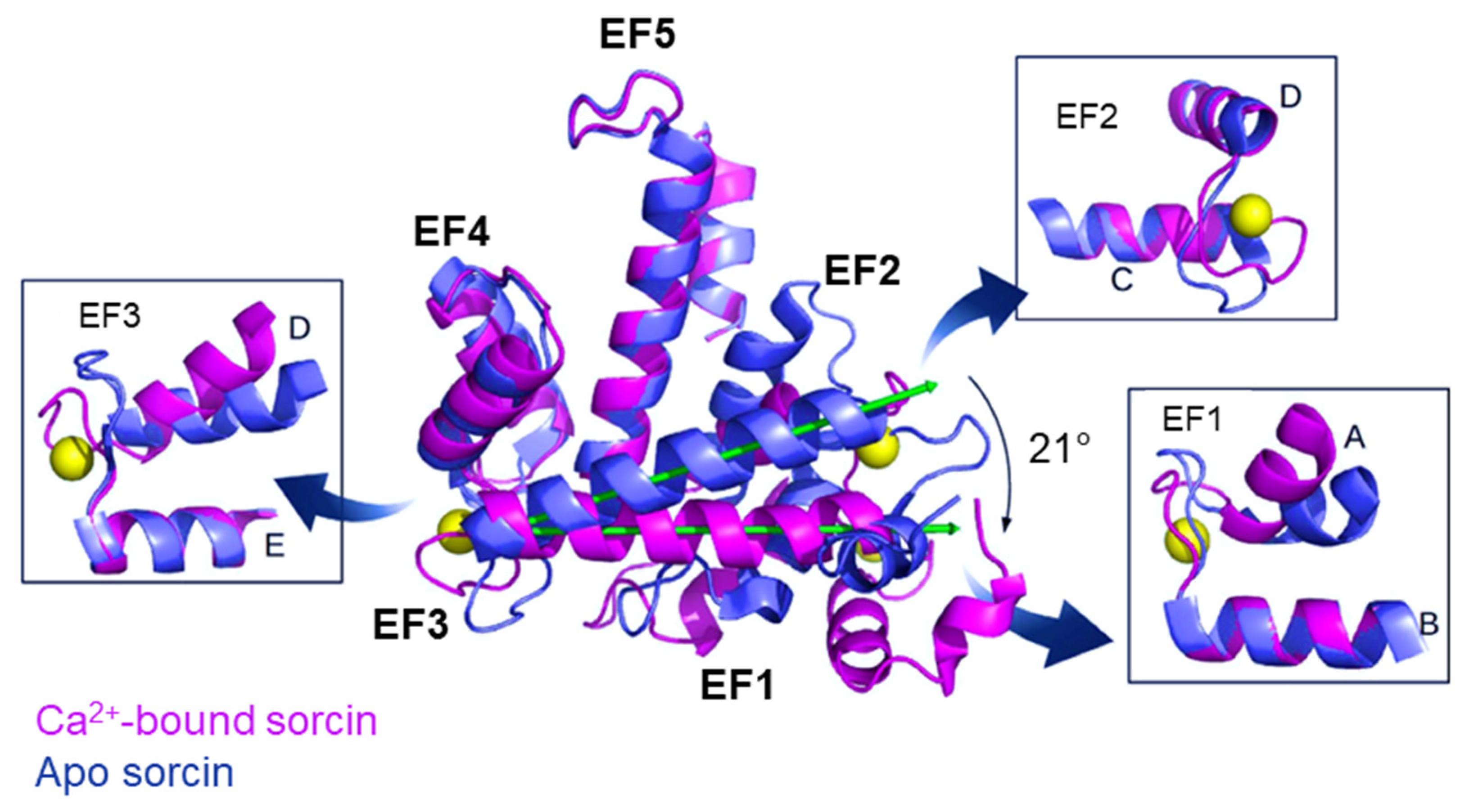
Figure 1. The X-ray crystal structure of human sorcin in the apo form (PDB: 4UPG, blue) and in the calcium-bound form (PDB: 4USL, magenta; calcium ions are represented by yellow spheres). Upon calcium binding, sorcin activation occurs, with a transition from a closed to an open structure, involving a 21° rotation of the long D-helix of 21° and the exposure of hydrophobic surfaces that leads to the interaction with targets. More details of the EF1 hand (part of the A and B helices, and the AB loop), EF2 hand (part of the C and D helices, and the CD loop), and EF3 hand (part of the D and E helices, and the DE loop) are shown in the boxes. Image drawn with the PyMol software (PyMol v.0.99 DeLano Scientific LLC, San Francisco, CA, USA).
Cancers 16 02810 g001
Figure 2. Sorcin regulates calcium homeostasis in the cells. Sorcin activates the activates the Na+-Ca2+ exchanger (NCX), the plasma membrane Ca2+ pump (PMCA) and modulates the L-type voltage-gated calcium channel (LVCC). In the endoplasmic reticulum (ER), sorcin inhibits ryanodine receptors (RyRs) and activates sarco/endoplasmic reticulum Ca2+−ATPase (SERCA), thereby increasing the Ca2+ load of the ER and decreasing ER stress (the arrows indicate Ca2+ flux). When sorcin expression is low, ER Ca2+ load (and also mitochondrial calcium load) is decreased, thereby increasing ER stress.
Cancers 16 02810 g002
Figure 3. Sorcin regulates many cellular processes, by interacting with many proteins and/or regulating their activity or expression (see text). Sorcin participates in the regulation of calcium homeostasis, ER or cellular stress, apoptosis, vesicle trafficking, proliferation, invasion, metastasis, migration, epithelial-to-mesenchymal transition, and multidrug resistance. ↑: increased activity or expression; ↓: decreased activity or expression.
Cancers 16 02810 g003
Figure 4. Upon administration of chemotherapeutic drugs, many factors determine multidrug resistance (MDR). These factors include absorption, distribution, metabolism, and elimination (ADME), drug influx, drug efflux by efflux pumps, drug activation and inactivation, drug target alteration, DNA damage repair, the inhibition of cell death, and in particular of apoptosis, gene amplification, epigenetic effects, epithelial-to-mesenchymal transition (EMT), angiogenesis, changes in tumor environment, and metastasis. Sorcin participates in several mechanisms of MDR onset (indicated in red, see text).
Cancers 16 02810 g004
Table 1. Experimental approaches targeting sorcin expression. Sorcin expression was shown to be selectively decreased by using microRNAs (miR-1, miR-142-5p), siRNAs (in nanocarriers), growth factors (TGF-β1, TNFα), small molecules (Tetrandrine, PH II-7, Dihydromyricetin, Ondansetron, Calcitriol, Palmitate, Triptolide) and protein extracts (Haishengsu). The chemokine fractalkine restores sorcin expression decreased by TNFα.
Category of MoleculesMoleculeMechanism of RegulationEffect
MicroRNAsmiR-1Binding to 3′-UTR of SRI gene↓ sorcin
miR-142-5pBinding to 3′-UTR of SRI gene↓ sorcin
siRNAssiRNA vs. sorcinin nanocarriersBinding to sorcin mRNA↓ sorcin
Growth factorsTGF-β1↑ SMAD2 phosphorylation↓ sorcin
TNFα↑ mTOR, NFκB phosphoryl.↓ sorcin
ChemokineFractalkine↓ mTOR, NFκB phosphoryl.↑ sorcin
Small moleculesTetrandrineCalcium channel blocking↓ sorcin
PH II-7?↓ sorcin
Dihydromyricetin↓ ERK-Akt phosphorylation↓ sorcin
OndansetronSorcin binding↓ sorcin
Calcitriol?↓ sorcin
Palmitate↓ AMPK- pEIF2α phosphoryl.↓ sorcin
Triptolide?↓ sorcin
Protein extractsHaishengsu?↓ sorcin
↑: increased activity or expression; ↓: decreased activity or expression; ?: unknown.
Disclaimer/Publisher’s Note: The statements, opinions and data contained in all publications are solely those of the individual author(s) and contributor(s) and not of MDPI and/or the editor(s). MDPI and/or the editor(s) disclaim responsibility for any injury to people or property resulting from any ideas, methods, instructions or products referred to in the content.