Non-Invasive 3D Breast Tumor Localization: A Viable Alternative to Invasive Tumor Marking
Abstract
Simple Summary
Abstract
1. Introduction
2. Materials and Methods
3. Technique Description
3.1. Diagnostic Procedure
3.2. Surgical Procedure
4. Results
5. Discussion
5.1. Limitations of the Presented Technique
5.2. Advantages of the Presented Technique
- -
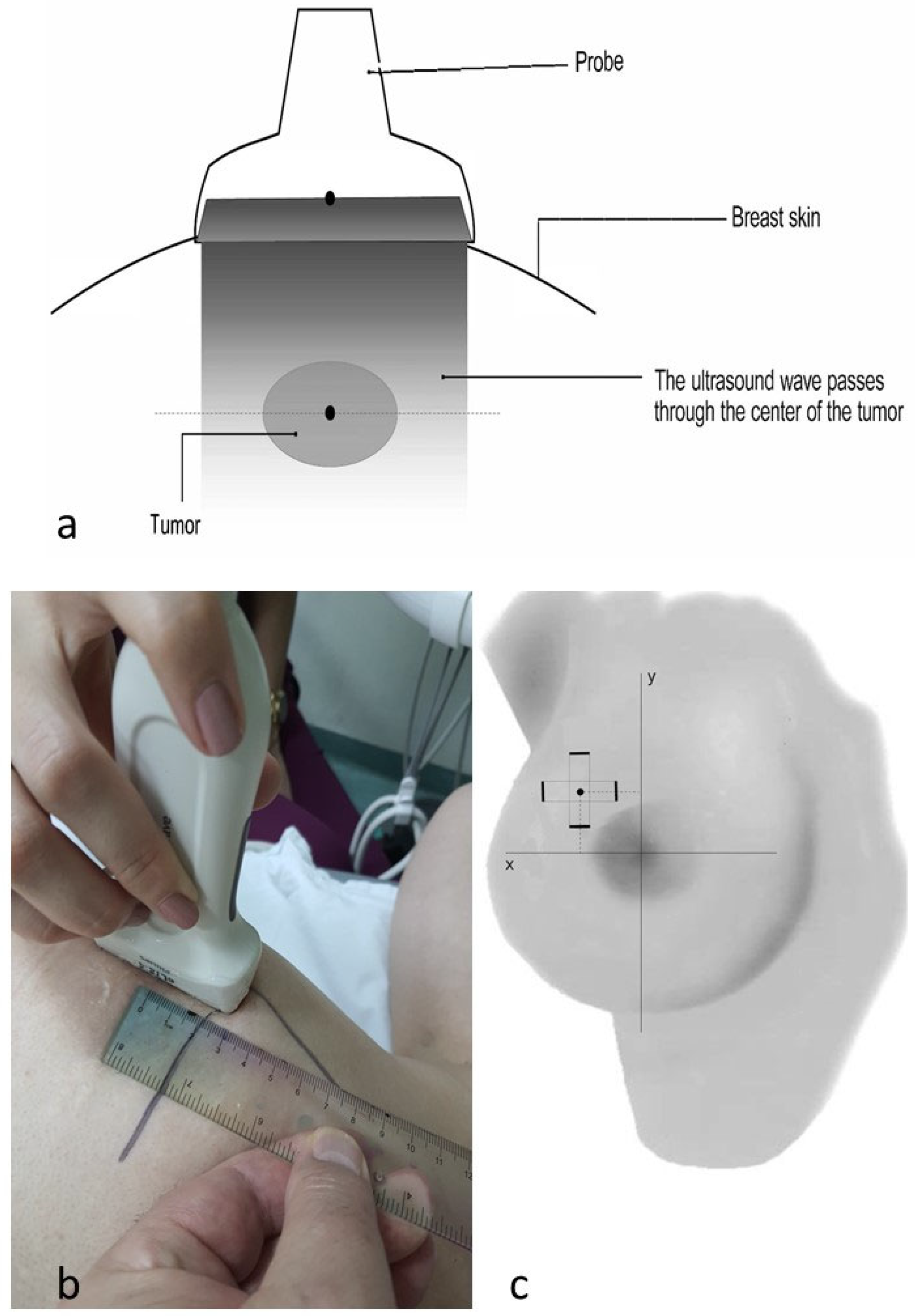
- The incorporation of the third dimension and objective multiple ultrasound measurements of tumor localization during NAST. Lannin et al. [7] performed excisions based on a one-time pre-NAST projection of the tumor on the breast skin, which potentially unnecessarily increases the volume of the excised tissue. The percentage of positive resection margins in that study was 10%, while our study featured no positive resection margins.
- -
- The avoidance of complications associated with invasive marking procedures: hematoma, infection, marker misplacement, marker migration, and poor marker visibility at the time of surgery. Every study addressing the problem of invasive tumor marking states the presence of a certain percentage of these complications, which threaten the precision of surgery [26] or require preoperative WNL [27]. Our technique does not encounter complications of this type.
- -
- Simplicity and cost–benefit ratio. Our technique can be implemented by a radiologist or surgeon who is not trained in invasive marking techniques, and it can be applied in health centers lacking specific marking equipment, avoiding additional financial outlays and inconveniences associated with invasive marking.
6. Conclusions
Author Contributions
Funding
Institutional Review Board Statement
Informed Consent Statement
Data Availability Statement
Conflicts of Interest
Abbreviations
| NAST | Neoadjuvant systemic therapy |
| pCR | Complete pathohistological response |
| CCR | Complete clinical regression |
| BCS | Breast-conservative surgery |
| ROLL | Radioguided occult lesion localization |
| WNL | Wire-needle localization |
References
- Scholl, S.M.; Asselain, B.; Palangie, T.; Dorval, T.; Jouve, M.; Giralt, E.G.; Vilcoq, J.; Durand, J.C.; Pouillart, P. Neoadjuvant chemotherapy in operable breast cancer. Eur. J. Cancer Clin. Oncol. 1991, 27, 1668–1671. [Google Scholar] [CrossRef] [PubMed]
- Fisher, B.; Bryant, J.; Wolmark, N.; Mamounas, E.; Brown, A.; Fisher, E.R.; Wickerham, D.L.; Begovic, M.; DeCillis, A.; Robidoux, A.; et al. Effect of preoperative chemotherapy on the outcome of women with operable breast cancer. J. Clin. Oncol. 1998, 16, 2672–2685. [Google Scholar] [CrossRef] [PubMed]
- von Minckwitz, G.; Huang, C.-S.; Mano, M.S.; Loibl, S.; Mamounas, E.P.; Untch, M.; Wolmark, N.; Rastogi, P.; Schneeweiss, A.; Redondo, A.; et al. Trastuzumab emtansine for residual invasive HER2-positive breast cancer. N. Engl. J. Med. 2019, 380, 617–628. [Google Scholar] [CrossRef] [PubMed]
- Masuda, N.; Lee, S.-J.; Ohtani, S.; Im, Y.-H.; Lee, E.-S.; Yokota, I.; Kuroi, K.; Im, S.A.; Park, B.W.; Kim, S.B.; et al. Adjuvant capecitabine for breast cancer after preoperative chemotherapy. N. Engl. J. Med. 2017, 376, 2147–2159. [Google Scholar] [CrossRef] [PubMed]
- Pfob, A.; Dubsky, P. The underused potential of breast conserving therapy after neoadjuvant system treatment—Causes and solutions. Breast 2023, 67, 110–115. [Google Scholar] [CrossRef] [PubMed]
- Minella, C.; Villasco, A.; D’Alonzo, M.; Cellini, L.; Accomasso, F.; Actis, S.; Biglia, N. Surgery after Neoadjuvant Chemotherapy: A Clip-Based Technique to Improve Surgical Outcomes, a Single-Center Experience. Cancers 2022, 14, 2229. [Google Scholar] [CrossRef] [PubMed]
- Lannin, D.R.; Grube, B.; Black, D.S.; Ponn, T. Breast tattoos for planning surgery following neoadjuvant chemotherapy. Am. J. Surg. 2007, 194, 518–520. [Google Scholar] [CrossRef] [PubMed]
- Espinosa-Bravo, M.; Avilés, A.S.; Esgueva, A.; Córdoba, O.; Rodriguez, J.; Cortadellas, T.; Mendoza, C.; Salvador, R.; Xercavins, J.; Rubio, I. Breast conservative surgery after neoadjuvant chemotherapy in breast cancer patients: Comparison of two tumor localization methods. Eur. J. Surg. Oncol. 2011, 37, 1038–1043. [Google Scholar] [CrossRef] [PubMed]
- Oh, J.L.; Nguyen, G.; Whitman, G.J.; Hunt, K.K.; Yu, T.K.; Woodward, W.A.; Tereffe, W.; Strom, E.A.; Perkins, G.H.; Buchholz, T.A. Placement of radiopaque clips for tumor localization in patients undergoing neoadjuvant chemotherapy and breast conservation therapy. Cancer 2007, 110, 2420–2427. [Google Scholar] [CrossRef]
- Cha, C.; Lee, J.; Kim, D.; Park, S.; Bae, S.J.; Eun, N.L.; Ahn, S.G.; Son, E.J.; Jeong, J. Comparison of resection margin status after single or double radiopaque marker insertion for tumor localization in breast cancer patients receiving neoadjuvant chemotherapy. Breast Cancer Res. Treat. 2020, 184, 797–803. [Google Scholar] [CrossRef]
- Alonso-Bartolome, P.; Ortega-Garcia, E.; Garijo-Ayensa, F.; de Juan-Ferre, A.; Vega-Bolivar, A. Utility of the tumor bed marker in patients with breast cancer receiving induction chemotherapy. Acta Radiol. 2002, 43, 29–33. [Google Scholar] [CrossRef] [PubMed]
- Ramos, M.; Díez, J.C.; Ramos, T.; Ruano, R.; Sancho, M.; González-Orús, J.M. Intraoperative ultrasound in conservative surgery for non-palpable breast cancer after neoadjuvant chemotherapy. Int. J. Surg. 2014, 12, 572–577. [Google Scholar] [CrossRef] [PubMed]
- Janssen, N.N.Y.; Nijkamp, J.; Alderliesten, T.; Loo, C.E.; Rutgers, E.J.T.; Sonke, J.; Peeters, M.T.F.D.V. Radioactive seed localization in breast cancer treatment. Br. J. Surg. 2015, 103, 70–80. [Google Scholar] [CrossRef] [PubMed]
- Van Riet, Y.E.A.; Maaskant, A.J.G.; Creemers, G.J.; Van Warmerdam, L.J.C.; Jansen, F.H.; Van de Velde, C.J.H.; Rutten, H.; Nieuwenhuijzen, G. Identification of residual breast tumour localization after neo-adjuvant chemotherapy using a radioactive 125 Iodine seed. Eur. J. Surg. Oncol. 2010, 36, 164–169. [Google Scholar] [CrossRef] [PubMed]
- Alderliesten, T.; Loo, C.E.; Pengel, K.E.; Rutgers, E.J.T.; Gilhuijs, K.G.A.; Vrancken Peeters, M.J.T.F.D. Radioactive Seed Localization of Breast Lesions: An Adequate Localization Method without Seed Migration. Breast J. 2011, 17, 594–601. [Google Scholar] [CrossRef] [PubMed]
- Gobardhan, P.D.; de Wall, L.L.; Van der Laan, L.; ten Tije, A.J.; Van der Meer, D.C.H.; Tetteroo, E.; Poortmans, P.M.P.; Luiten, E.J.T. The role of radioactive iodine-125 seed localization in breast-conserving therapy following neoadjuvant chemotherapy. Ann. Oncol. 2013, 24, 668–673. [Google Scholar] [CrossRef] [PubMed]
- Donker, M.; Drukker, C.A.; Valdés Olmos, R.A.; Rutgers, E.J.T.; Loo, C.E.; Sonke, G.S.; Wesseling, J.; Alderliesten, T.; Peeters, M.-J.T.F.D.V. Guiding Breast-Conserving Surgery in Patients After Neoadjuvant Systemic Therapy for Breast Cancer: A Comparison of Radioactive Seed Localization with the ROLL Technique. Ann. Surg. Oncol. 2013, 20, 2569–2575. [Google Scholar] [CrossRef] [PubMed]
- Donker, M.; Straver, M.E.; Rutgers, E.J.T.; Valdés Olmos, R.A.; Loo, C.E.; Sonke, G.S.; Wesseling, J.; Peeters, M.-J.V. Radioguided occult lesion localisation [ ROLL] in breast-conserving surgery after neoadjuvant chemotherapy. Eur. J. Surg. Oncol. 2012, 38, 1218–1224. [Google Scholar] [CrossRef] [PubMed]
- Aggarwal, V.; Agarwal, G.; Lal, P.; Krishnani, N.; Mishra, A.; Verma, A.K.; Mishra, S.K. Feasibility Study of Safe Breast Conservation in Large and Locally Advanced Cancers with Use of Radiopaque Markers to Mark Pre-Neoadjuvant Chemotherapy Tumor Margins. World J. Surg. 2007, 32, 2562–2569. [Google Scholar] [CrossRef]
- Hossam, A.; El-Badrawy, A.; Khater, A.; Setit, A.; Roshdy, S.; Abdelwahab, K.; Hamed, E. The Evaluation of a Cost-Effective Method for Tumour Marking Prior to Neo-Adjuvant Chemotherapy Using Silver Rods. Eur. J. Breast Health 2022, 19, 99–105. [Google Scholar] [CrossRef]
- Sever, A.R.; O’Brien, M.E.R.; Humphreys, S.; Singh, I.; Jones, S.E.; Jones, P.A. Radiopaque coil insertion into breast cancers prior to neoadjuvant chemotherapy. Breast 2005, 14, 108–117. [Google Scholar] [CrossRef] [PubMed]
- Rubio, I.T.; Esgueva-Colmenarejo, A.; Espinosa-Bravo, M.; Salazar, J.P.; Miranda, I.; Peg, V. Intraoperative Ultrasound-Guided Lumpectomy Versus Mammographic Wire Localization for Breast Cancer Patients After Neoadjuvant Treatment. Ann. Surg. Oncol. 2015, 23, 38–43. [Google Scholar] [CrossRef] [PubMed]
- Almalki, H.; Rankin, A.C.; Juette, A.; Youssef, M.M.G. Radio-frequency identification [RFID] tag localisation of non-palpable breast lesions a single centre experience. Breast 2023, 69, 417–421. [Google Scholar] [CrossRef] [PubMed]
- Tinterri, C.; Fernandes, B.; Zambelli, A.; Sagona, A.; Barbieri, E.; Di Maria Grimaldi, S.; Darwish, S.S.; Jacobs, F.; De Carlo, C.; Iuzzolino, M. The Impact of Different Patterns of Residual Disease on Long-Term Oncological Outcomes in Breast Cancer Patients Treated with Neo-Adjuvant Chemotherapy. Cancers 2024, 16, 376. [Google Scholar] [CrossRef] [PubMed]
- Pastorello, G.R.; Laws, A.; Grossmith, S.; King, C.; McGrath, M.; Mittendorf, A.E.; King, T.A.; Schnitt, S.J. Clinico-pathologic predictors of patterns of residual disease following neoadjuvant chemotherapy for breast cancer. Mod. Pathol. 2021, 34, 875–882. [Google Scholar] [CrossRef] [PubMed]
- Thomassin-Naggara, I.; Lalonde, L.; David, J.; Darai, E.; Uzan, S.; Trop, I. A plea for the biopsy marker: How, why and why not clipping after breast biopsy? Breast Cancer Res. Treat. 2011, 132, 881–893. [Google Scholar] [CrossRef] [PubMed]
- Green, R.T.; Weiser, R.; Golan, O.; Menes, T.S. In Search of the Lost Clip: Outcome of Women After Needle-Guided Lumpectomy of a Marking Clip. Ann. Surg. Oncol. 2021, 28, 4974–4980. [Google Scholar] [CrossRef]
- Jha, C.K.; Johri, G.; Singh, P.K.; Yadav, S.K.; Sinha, U. Does Tumor Marking Before Neoadjuvant Chemotherapy Helps Achieve Better Outcomes in Patients Undergoing Breast Conservative Surgery? A Systematic Review. Indian J. Surg. Oncol. 2021, 12, 624–631. [Google Scholar] [CrossRef] [PubMed]
- Fusco, N.; Rizzo, A.; Costarelli, L.; Santinelli, A.; Cerbelli, B.; Scatena, C.; Macrì, E.; Pietribiasi, F.; d’Amati, G.; Sapino, A.; et al. Pathological examination of breast cancer samples before and after neoadjuvant therapy: Recommendations from the Italian Group for the Study of Breast Pathology-Italian Society of Pathology [GIPaM-SIAPeC]. Pathologica 2022, 114, 104–110. [Google Scholar] [CrossRef]
- Bi, Z.; Qiu, P.F.; Yang, T.; Chen, P.; Song, X.R.; Zhao, T.; Zhang, Z.P.; Wang, Y.S. The modified shrinkage classification modes could help to guide breast conserving surgery after neoadjuvant therapy in breast cancer. Front. Oncol. 2022, 12, 982011. [Google Scholar] [CrossRef]
- Early Breast Cancer Trialists’ Collaborative Group (EBCTCG). Effects of radiotherapy and of differences in the extent of surgery for early breast cancer on local recurrence and 15-year survival: An overview of the randomised trials. Lancet 2005, 366, 2087–2106. [Google Scholar] [CrossRef] [PubMed]
- Early Breast Cancer Trialists’ Collaborative Group (EBCTCG). Long-term outcomes for neoadjuvant versus adjuvant chemotherapy in early breast cancer: Meta-analysis of individual patient data from ten randomised trials. Lancet Oncol. 2018, 19, 27–39. [Google Scholar] [CrossRef] [PubMed]
- Ivanovic, N.; Bjelica, D.; Loboda, B.; Bogdanovski, M.; Colakovic, N.; Petricevic, S.; Gojgic, M.; Zecic, O.; Zecic, K.; Zdravkovic, D. Changing the role of pCR in breast cancer treatment—An unjustifiable interpretation of a good prognostic factor as a “factor for a good prognosis”. Front. Oncol. 2023, 13, 1207948. [Google Scholar] [CrossRef] [PubMed]
- Tinterri, C.; Barbieri, E.; Sagona, A.; Bottini, A.; Canavese, G.; Gentile, D. De-Escalation Surgery in cT3-4 Breast Cancer Patients after Neoadjuvant Therapy: Predictors of Breast Conservation and Comparison of Long-Term Oncological Outcomes with Mastectomy. Cancers 2024, 16, 1169. [Google Scholar] [CrossRef] [PubMed]
- Hadar, T.; Koretz, M.; Nawass, M.; Allweis, M.T. Innovative Standards in Surgery of the Breast after Neoadjuvant Systemic Therapy. Breast Care 2021, 16, 590–597. [Google Scholar] [CrossRef] [PubMed]
- Lee, H.-S.; Kim, H.-J.; Chung, I.-Y.; Kim, J.; Lee, S.-B.; Lee, J.-W.; Son, B.H.; Ahn, S.H.; Kim, H.H.; Seo, J.B.; et al. Usefulness of 3D-surgical guides in breast conserving surgery after neoadjuvant treatment. Sci. Rep. 2021, 11, 3376. [Google Scholar] [CrossRef]
- Conti, M.; Morciano, F.; Bufi, E.; D’Angelo, A.; Panico, C.; Di Paola, V.; Gori, E.; Russo, G.; Cimino, G.; Palma, S.; et al. Surgical Planning after Neoadjuvant Treatment in Breast Cancer: A Multimodality Imaging-Based Approach Focused on MRI. Cancers 2023, 15, 1439. [Google Scholar] [CrossRef]


| Tumor Positioning Record | ||
|---|---|---|
| Name and Surname: | ||
| Status of Neoadjuvant therapy: | Before starting therapy | |
| After ________________ cycles of NAHT | ||
| Breast: | Right | Left ○ |
| Date of examination: | ||
| Tumor dimensions: | 1. cranio-caudal: | 22 mm |
| 2. medio-lateral: | 18 mm | |
| 3. vertical: | 28 mm | |
| Distance between superficial margin of the tumor and the skin: | 6 mm | |
| Distance between deepest margin of the tumor and pectoral fascia: | 0 mm | |
| Coordinates of tumor’s central point on the skin: | X axis | +32 mm |
| Y axis | −22 mm | |
| Description | Number of Patients (%) |
|---|---|
| Total patients included | 93 (94 tumors) (100%) |
| Patients on whom the entire procedure was completed | 79 (84%) |
| Excluded due to unsuitability for the procedure | 15 (16%) |
| Patients with CCR * who underwent surgery using our technique | 31/79 (39% of those where the procedure was completed) |
| Characteristic | Patients in the Study (N = 79) | |
|---|---|---|
| Response to NAST—no. of patients (%) | CCR achieved 31 (39) | No CCR 48 (61) |
| Age—no. of patients (%) | ||
| <50 yrs | 16 (20) | 21 (27) |
| >50 yrs | 15 (19) | 27 (34) |
| US tumor size—no. of tumors (%) | ||
| 18–30 mm | 22 (28) | 8 (10) |
| 31–47 mm | 9 (11.4) | 40 (50.6) |
| Hormone receptor status—no. of patients (%) | ||
| Positive | 16 (20) | 30 (38) |
| Negative | 15 (19) | 18 (23) |
| Her2 receptor status—no. of patients (%) | ||
| Positive | 14 (18) | 13 (16.5) |
| Negative | 17 (21.5) | 35 (44) |
| Nodal status—no. of patients (%) | ||
| N0 | 13 (16.5) | 20 (25) |
| N1 | 18 (23) | 28 (35.5) |
| Triple negative—no. of patients (%) | 5 (6) | 6 (7.6) |
| Parameter | Mastectomy | BCS (Breast-Conserving Surgery) |
|---|---|---|
| Number (%) | 2 (6.5%) | 29 (93.5%) |
| pCR (Pathological Complete Response) | 2 | 22 |
| Residual Microfoci of Carcinoma | 0 | 7 |
| Positive Margin | 0 | 0 |
| Histological Signs of Pre-existing Tumor | 2 (100%) | 29 (100%) |
| Concentric Regression Pattern | 2 | 19 |
| Non-Concentric Regression Pattern | 0 | pCR 7/22, non-pCR 3/7 |
| Ratio of Excised Specimen Volume to Pre-NAST Tumor Volume | X | 0.91 (0.01–2.38) |
Disclaimer/Publisher’s Note: The statements, opinions and data contained in all publications are solely those of the individual author(s) and contributor(s) and not of MDPI and/or the editor(s). MDPI and/or the editor(s) disclaim responsibility for any injury to people or property resulting from any ideas, methods, instructions or products referred to in the content. |
© 2024 by the authors. Licensee MDPI, Basel, Switzerland. This article is an open access article distributed under the terms and conditions of the Creative Commons Attribution (CC BY) license (https://creativecommons.org/licenses/by/4.0/).
Share and Cite
Bjelica, D.; Colakovic, N.; Opric, S.; Zdravkovic, D.; Loboda, B.; Petricevic, S.; Gojgic, M.; Zecic, O.; Skuric, Z.; Zecic, K.; et al. Non-Invasive 3D Breast Tumor Localization: A Viable Alternative to Invasive Tumor Marking. Cancers 2024, 16, 2564. https://doi.org/10.3390/cancers16142564
Bjelica D, Colakovic N, Opric S, Zdravkovic D, Loboda B, Petricevic S, Gojgic M, Zecic O, Skuric Z, Zecic K, et al. Non-Invasive 3D Breast Tumor Localization: A Viable Alternative to Invasive Tumor Marking. Cancers. 2024; 16(14):2564. https://doi.org/10.3390/cancers16142564
Chicago/Turabian StyleBjelica, Dragana, Natasa Colakovic, Svetlana Opric, Darko Zdravkovic, Barbara Loboda, Simona Petricevic, Milan Gojgic, Ognjen Zecic, Zlatko Skuric, Katarina Zecic, and et al. 2024. "Non-Invasive 3D Breast Tumor Localization: A Viable Alternative to Invasive Tumor Marking" Cancers 16, no. 14: 2564. https://doi.org/10.3390/cancers16142564
APA StyleBjelica, D., Colakovic, N., Opric, S., Zdravkovic, D., Loboda, B., Petricevic, S., Gojgic, M., Zecic, O., Skuric, Z., Zecic, K., & Ivanovic, N. (2024). Non-Invasive 3D Breast Tumor Localization: A Viable Alternative to Invasive Tumor Marking. Cancers, 16(14), 2564. https://doi.org/10.3390/cancers16142564


