Congenital Tumors—Magnetic Resonance Imaging Findings with Focus on Rare Tumors
Abstract
Simple Summary
Abstract
1. Introduction
2. Materials and Methods
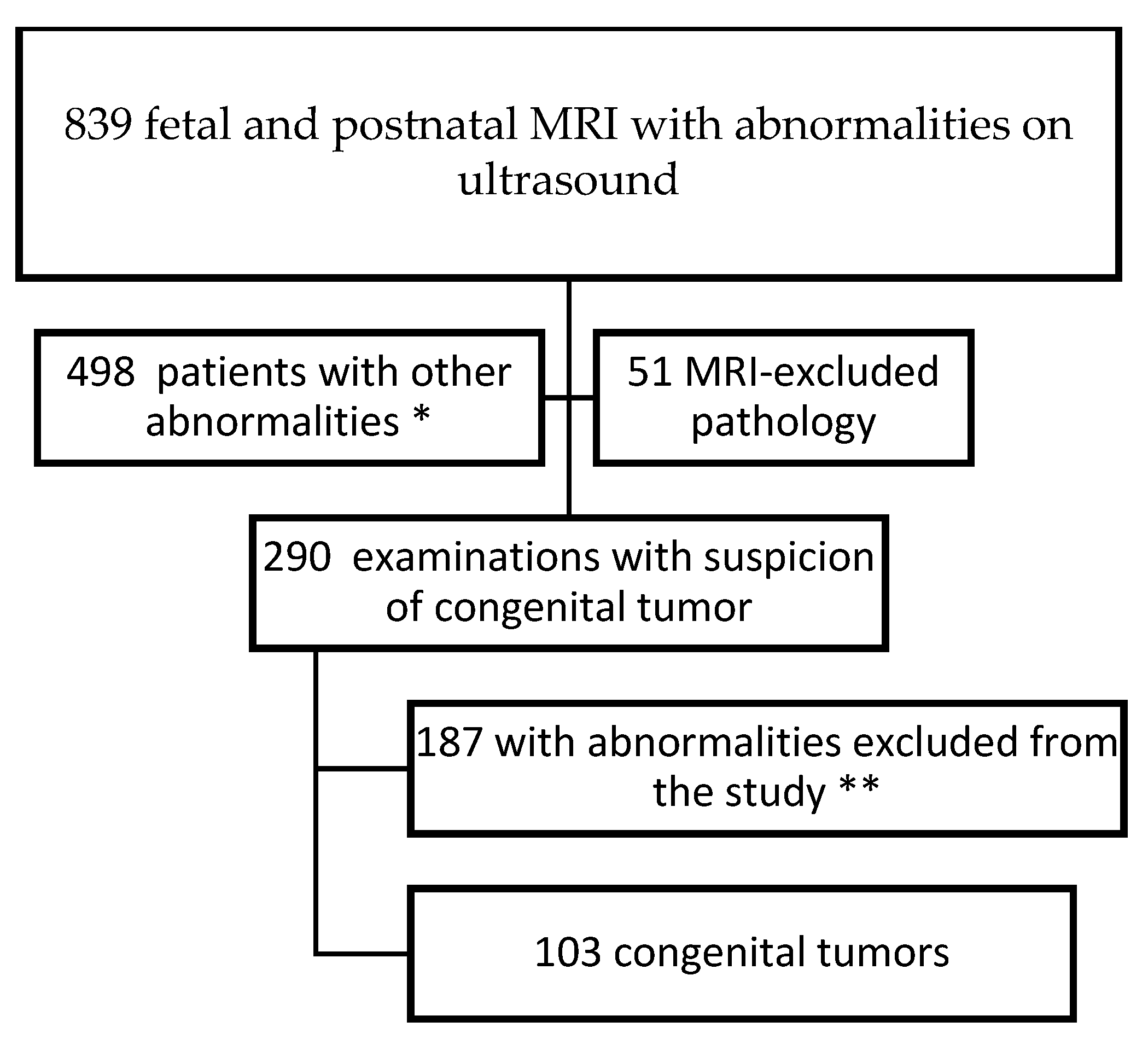
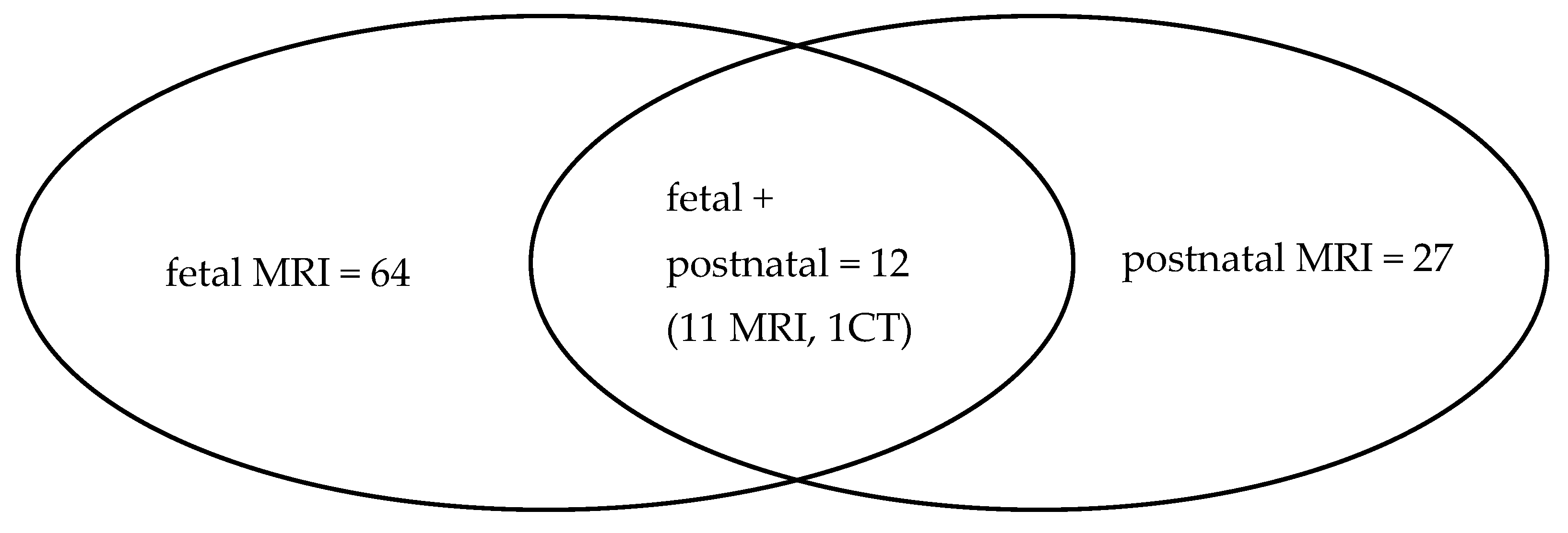
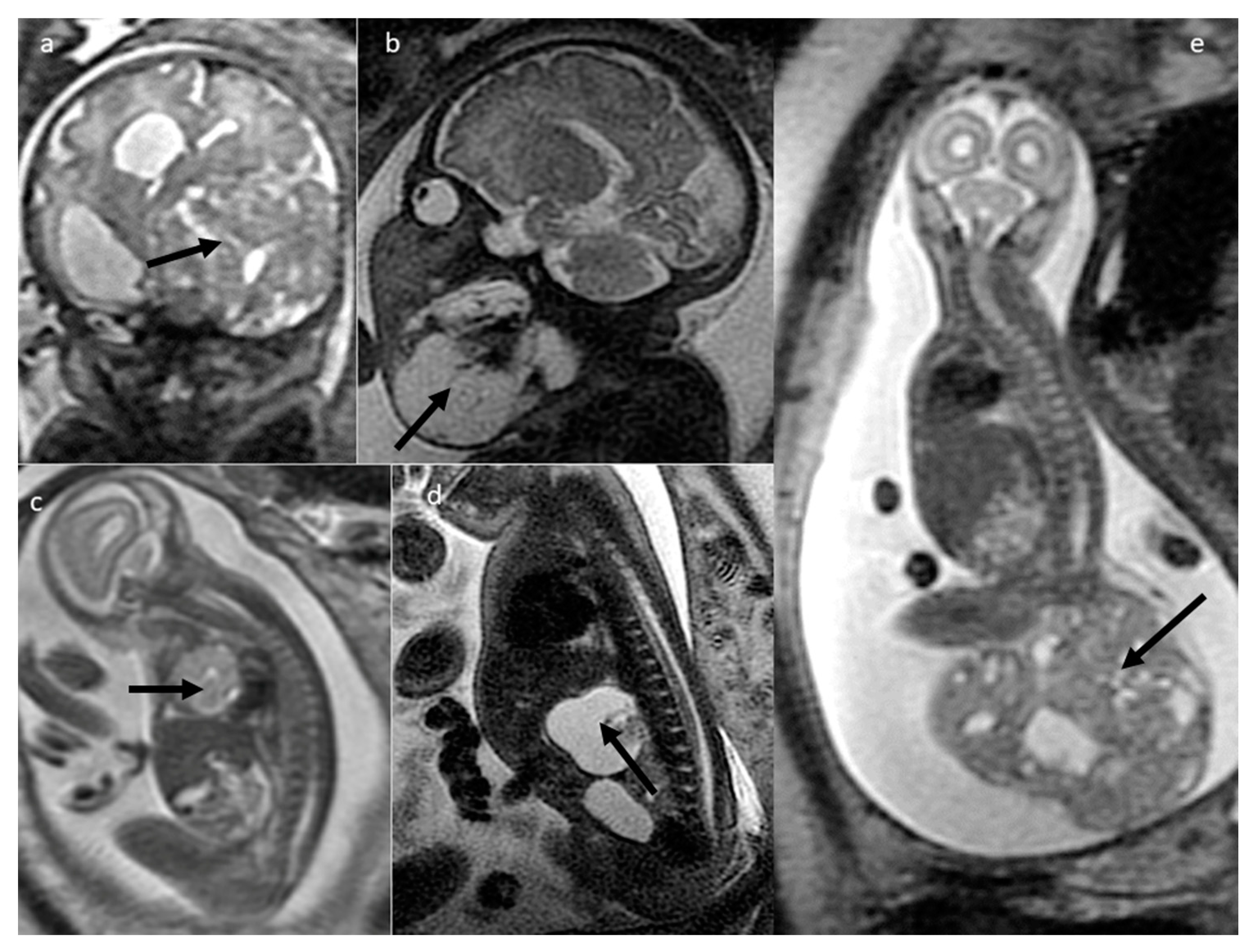
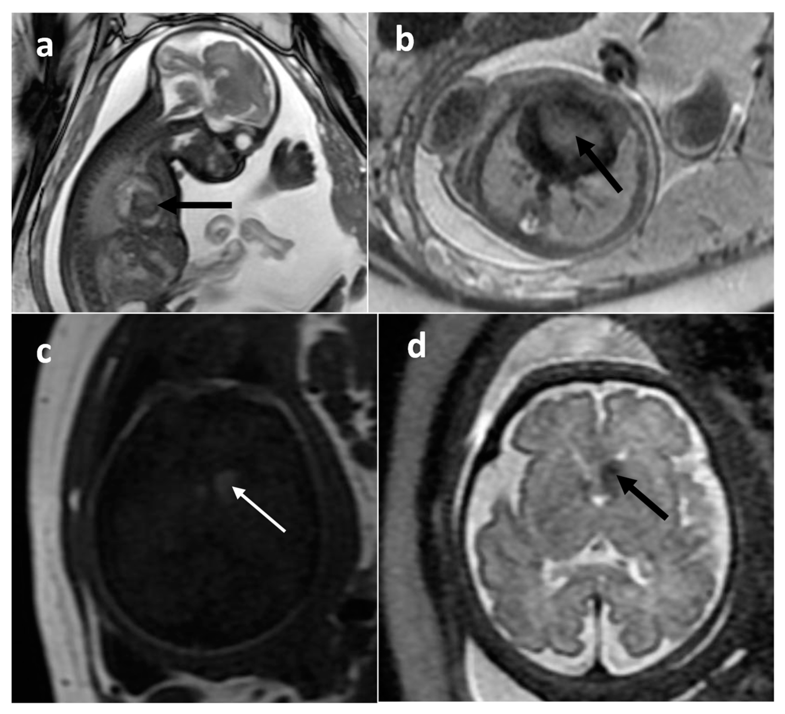
3. Results
4. Discussion
- External mass only
- Equal internal/external components (dumbbell shape)
- Primary location in abdomen or pelvis
- Entirely internal, no external components visible
5. Conclusions
Supplementary Materials
Author Contributions
Funding
Institutional Review Board Statement
Informed Consent Statement
Data Availability Statement
Acknowledgments
Conflicts of Interest
References
- Woodward, P.J.; Sohaey, R.; Kennedy, A.; Koeller, K.K. A comprehensive review of fetal tumors with pathologic correlation. Radiographics 2005, 25, 215–242. [Google Scholar] [CrossRef]
- Isaacs, H. Perinatal (congenital and neonatal) neoplasms: A report of 110 cases. Pediatr. Pathol. 1985, 3, 165–210. [Google Scholar] [CrossRef]
- Moore, S.W.; Satg’e, D.; Sasco, A.J.; Zimmermann, A.; Plaschkes, J. The epidemiology of neonatal tumours. Report of an international working group. Pediatr. Surg. Int. 2003, 19, 509–519. [Google Scholar] [CrossRef]
- Alamo, L.; Beck-Popovic, M.; Gudinchet, F.; Meuli, R. Congenital tumors: Imaging when life just begins. Insights Imaging 2011, 2, 297–308. [Google Scholar] [CrossRef]
- Bekiesinska-Figatowska, M.; Jurkiewicz, E.; Duczkowski, M.; Duczkowska, A.; Romaniuk-Doroszewska, A.; Brągoszewska, H.; Ceran, A. Congenital CNS tumors diagnosed on prenatal MRI. Neuroradiol. J. 2011, 24, 477–481. [Google Scholar] [CrossRef]
- Blaicher, W.; Bernaschek, G.; Deutinger, J.; Messerschmidt, A.; Schindler, E.; Prayer, D. Fetal and early postnatal magnetic resonance imaging—Is there a difference? J. Perinat. Med. 2004, 32, 53–57. [Google Scholar] [CrossRef]
- Miller, E.; Ben-Sira, L.; Constantini, S.; Beni-Adani, L. Impact of prenatal magnetic resonance imaging on postnatal neurosurgical treatment. J. Neurosurg. 2006, 105, 203–209. [Google Scholar] [CrossRef]
- Prayer, D.; Malinger, G.; Brugger, P.C.; Cassady, C.; De Catte, L.; De Keersmaecker, B.; Fernandes, G.L.; Glanc, P.; Goncalves, L.F.; Gruber, G.M.; et al. ISUOG Practice Guidelines: Performance of fetal magnetic resonance imaging. Ultrasound Obstet. Gynecol. 2017, 49, 671–680. [Google Scholar] [CrossRef]
- Bijma, H.H.; Van der Heide, A.; Wildschut, H.I.; Van der Maas, P.J.; Wladimiroff, J.W. Impact of decision-making in a multidisciplinary perinatal team. Prenat. Diagn. 2007, 27, 97–103. [Google Scholar] [CrossRef]
- Milani, H.J.; Araujo Júnior, E.; Cavalheiro, S.; Oliveira, P.S.; Hisaba, W.J.; Barreto, E.Q.; Barbosa, M.M.; Nardozza, L.M.; Moron, A.F. Fetal brain tumors: Prenatal diagnosis by ultrasound and magnetic resonance imaging. World J. Radiol. 2015, 7, 17–21. [Google Scholar] [CrossRef]
- Nemec, S.F.; Horcher, E.; Kasprian, G.; Brugger, P.C.; Bettelheim, D.; Amann, G.; Nemec, U.; Rotmensch, S.; Rimoin, D.L.; Graham, J.M., Jr.; et al. Tumor disease and associated congenital abnormalities on prenatal MRI. Eur. J. Radiol. 2012, 81, e115–e122. [Google Scholar] [CrossRef]
- Avni, F.E.; Massez, A.; Cassart, M. Tumours of the fetal body: A review. Pediatr. Radiol. 2009, 39, 1147–1157. [Google Scholar] [CrossRef]
- Cassart, M.; Bosson, N.; Garel, C.; Eurin, D.; Avni, F. Fetal intracranial tumors: A review of 27 cases. Eur. Radiol. 2008, 18, 2060–2066. [Google Scholar] [CrossRef]
- Available online: www.imagegently.org (accessed on 12 December 2023).
- Bekiesinska-Figatowska, M.; Sobieraj, P.; Pasieczna, M.; Szymkiewicz-Dangel, J. Early diagnosis of tuberous sclerosis complex: Prenatal diagnosis. AJNR Am. J. Neuroradiol. 2023, 44, 1070–1076. [Google Scholar] [CrossRef]
- PDQ Pediatric Treatment Editorial Board. Childhood Brain Stem Glioma Treatment (PDQ®): Health Professional Version. 2022 Feb 23. In PDQ Cancer Information Summaries [Internet]; National Cancer Institute (US): Bethesda, MD, USA, 2002. Available online: https://www.ncbi.nlm.nih.gov/books/NBK65812/ (accessed on 12 December 2023).
- Davidson, J.R.; Uus, A.; Matthew, J.; Egloff, A.M.; Deprez, M.; Yardley, I.; De Coppi, P.; David, A.; Carmichael, J.; Rutherford, M.A. Fetal body MRI and its application to fetal and neonatal treatment: An illustrative review. Lancet Child. Adolesc. Health. 2021, 5, 447–458. [Google Scholar] [CrossRef]
- Altman, R.P.; Randolph, J.G.; Lilly, J.R. Sacrococcygeal teratoma: American Academy of Pediatrics Surgical Section Survey-1973. J. Pediatr. Surg. 1974, 9, 389–398. [Google Scholar] [CrossRef]
- Gabra, H.O.; Jesudason, E.C.; McDowell, H.P.; Pizer, B.L.; Losty, P.D. Sacrococcygeal teratoma—A 25-year experience in a UK regional center. J. Pediatr. Surg. 2006, 41, 1513–1516. [Google Scholar] [CrossRef]
- Chen, J.; Wang, J.; Sun, H.; Gu, X.; Hao, X.; Fu, Y.; Zhang, Y.; Liu, X.; Zhang, H.; Han, L.; et al. Fetal cardiac tumor: Echocardiography, clinical outcome and genetic analysis in 53 cases. Ultrasound Obstet. Gynecol. 2019, 54, 103–109. [Google Scholar] [CrossRef]
- du Toit, J.; Wieselthaler, N. Let’s face it—13 unusual causes of facial masses in children. Insights Imaging 2015, 6, 519–530. [Google Scholar] [CrossRef][Green Version]
- Goh, S.; Butler, W.; Thiele, E.A. Subependymal giant cell tumors in tuberous sclerosis complex. Neurology 2004, 63, 1457–1461. [Google Scholar] [CrossRef]
- Goergen, S.K.; Fahey, M.C. Prenatal MR Imaging Phenotype of Fetuses with Tuberous Sclerosis: An Institutional Case Series and Literature Review. AJNR Am. J. Neuroradiol. 2022, 43, 633–638. [Google Scholar] [CrossRef] [PubMed]
- Nuchtern, J.G. Perinatal neuroblastoma. Semin. Pediatr. Surg. 2006, 15, 10–16. [Google Scholar] [CrossRef] [PubMed]
- Elsayes, K.M.; Mukundan, G.; Narra, V.R.; Lewis, J.S., Jr.; Shirkhoda, A.; Farooki, A.; Brown, J.J. Adrenal masses: Mr imaging features with pathologic correlation. Radiographics 2004, 24, S73–S86. [Google Scholar] [CrossRef] [PubMed]
- Canale, S.; Vanel, D.; Couanet, D.; Patte, C.; Caramella, C.; Dromain, C. Infantile fibrosarcoma: Magnetic resonance imaging findings in six cases. Eur. J. Radiol. 2009, 72, 30–37. [Google Scholar] [CrossRef] [PubMed]
- Isaacs, H., Jr. Fetal and neonatal rhabdoid tumor. J. Pediatr. Surg. 2010, 45, 619–626. [Google Scholar] [CrossRef] [PubMed]
- Yang, C.; Chen, W.; Han, P. Congenital soft tissue Ewing’s sarcoma: A case report of pre- and postnatal magnetic resonance imaging findings. Medicine 2022, 101, e28587. [Google Scholar] [CrossRef] [PubMed]
- Jin, S.G.; Jiang, X.P.; Zhong, L. Congenital Ewing’s Sarcoma/Peripheral Primitive Neuroectodermal Tumor: A Case Report and Review of the Literature. Pediatr. Neonatol. 2016, 57, 436–439. [Google Scholar] [CrossRef]
- Glick, R.D.; Hicks, M.J.; Nuchtern, J.G.; Wesson, D.E.; Olutoye, O.O.; Cass, D.L. Renal tumors in infants less than 6 months of age. J. Pediatr. Surg. 2004, 39, 522–525. [Google Scholar] [CrossRef]
- Ko, S.M.; Kim, M.J.; Im, Y.J.; Park, K.I.; Lee, M.J. Cellular mesoblastic nephroma with liver metastasis in a neonate: Prenatal and postnatal diffusion-weighted MR imaging. Korean J. Radiol. 2013, 14, 361–365. [Google Scholar] [CrossRef]
- Jurkiewicz, E.; Bekiesinska-Figatowska, M.; Duczkowski, M.; Grajkowska, W.; Roszkowski, M.; Czech-Kowalska, J.; Dobrzańska, A. Antenatal diagnosis of the congenital craniopharyngioma. Pol. J. Radiol. 2010, 75, 98–102. [Google Scholar]
- Suo-Palosaari, M.; Rantala, H.; Lehtinen, S.; Kumpulainen, T.; Salokorpi, N. Long-term survival of an infant with diffuse brainstem lesion diagnosed by prenatal MRI: A case report and review of the literature. Childs Nerv. Syst. 2016, 32, 1163–1168. [Google Scholar] [CrossRef] [PubMed]
- Schumacher, M.; Schulte-Mönting, J.; Stoeter, P.; Warmuth-Metz, M.; Solymosi, L. Magnetic resonance imaging compared with biopsy in the diagnosis of brainstem diseases of childhood: A multicenter review. J. Neurosurg. 2007, 106, 111–119. [Google Scholar] [CrossRef] [PubMed]
- Beppu, T.; Sato, Y.; Uesugi, N.; Kuzu, Y.; Ogasawara, K.; Ogawa, A. Desmoplastic infantile astrocytoma and characteristics of the accompanying cyst. Case report. J. Neurosurg. Pediatr. 2008, 1, 148–151. [Google Scholar] [CrossRef] [PubMed]
- Trehan, G.; Bruge, H.; Vinchon, M.; Khalil, C.; Ruchoux, M.M.; Dhellemmes, P.; Ares, G.S. MR imaging in the diagnosis of desmoplastic infantile tumor: Retrospective study of six cases. AJNR Am. J. Neuroradiol. 2004, 25, 1028–1033. [Google Scholar] [PubMed]
- Maran-Gonzalez, A.; Laquerrière, A.; Bigi, N.; Develay-Morice, J.E.; Rouleau, C. Posterior fossa solitary fibrous tumour: Report of a fetal case and review of the literature. J. Neurooncol. 2011, 101, 297–300. [Google Scholar] [CrossRef] [PubMed]
- Laure-Kamionowska, M.; Szymanska, K.; Bekiesinska-Figatowska, M.; Gierowska-Bogusz, B.; Michalak, E.; Klepacka, T. Congenital glioblastoma coexisting with vascular developmental anomaly. Folia Neuropathol. 2013, 51, 333–339. [Google Scholar] [CrossRef] [PubMed]
- Isaacs, H., Jr. Perinatal (fetal and neonatal) astrocytoma: A review. Childs Nerv. Syst. 2016, 32, 2085–2096. [Google Scholar] [CrossRef] [PubMed]
- Louis, D.N.; Perry, A.; Wesseling, P.; Cree, I.A.; Figarella-Branger, D.; Hawkins, C.; Ng, H.K.; Pfister, S.M.; Reifenberger, G.; Soffietti, R.; et al. The 2021 WHO Classification of Tumors of the Central Nervous System: A summary. Neuro. Oncol. 2021, 23, 1231–1251. [Google Scholar] [CrossRef]
- Sobel, G.; Halász, J.; Bogdányi, K.; Szabó, I.; Borka, K.; Molnár, P.; Schaff, Z.; Paulin, F.; Bánhidy, F. Prenatal diagnosis of a giant congenital primary cerebral hemangiopericytoma. Pathol. Oncol. Res. 2006, 12, 46–49. [Google Scholar] [CrossRef]
- Cavalheiro, S.; Sparapani, F.V.; Moron, A.F.; da Silva, M.C.; Stávale, J.N. Fetal meningeal hemangiopericytoma. Case report. J. Neurosurg. 2002, 97, 1217–1220. [Google Scholar] [CrossRef]
- Keraliya, A.R.; Tirumani, S.H.; Shinagare, A.B.; Zaheer, A.; Ramaiya, N.H. Solitary Fibrous Tumors: 2016 Imaging Update. Radiol. Clin. N. Am. 2016, 54, 565–579. [Google Scholar] [CrossRef] [PubMed]
- Chigurupati, R.; Alfatooni, A.; Myall, R.W.T.; Hawkins, D.; Oda, D. Orofacial rhabdomyosarcoma in neonates and young children: A review of literature and management of four cases. Oral. Oncol. 2002, 38, 508–515. [Google Scholar] [CrossRef] [PubMed]
- Tariq, S.; Shallwani, H.; Waqas, M.; Bari, M.E. Congenital and infantile malignant melanoma of the scalp: A systematic review. Ann. Med. Surg. 2017, 21, 93–95. [Google Scholar] [CrossRef] [PubMed]
- Makin, E.; Davenport, M. Fetal and neonatal liver tumours. Early Hum. Dev. 2010, 86, 637–642. [Google Scholar] [CrossRef] [PubMed]
- Sargar, K.M.; Sheybani, E.F.; Shenoy, A.; Aranake-Chrisinger, J.; Khanna, G. Pediatric Fibroblastic and Myofibroblastic Tuors: A Pictorial Review. Radiographics 2016, 36, 1195–1214. [Google Scholar] [CrossRef] [PubMed]
- Li, R.; Kelly, D.; Siegal, G.P. Bilateral Mesenchymal Hamartoma of the Chest Wall in an Infant Boy. Fetal Pediatr. Pathol. 2012, 31, 415–422. [Google Scholar] [CrossRef] [PubMed]
- Nakatani, T.; Morimoto, A.; Kato, R.; Tokuda, S.; Sugimoto, T.; Tokiwa, K.; Tsuchihashi, Y.; Imashuku, S. Successful treatment of congenital systemic juvenile xanthogranuloma with Langerhans cell histiocytosis-based chemotherapy. J. Pediatr. Hematol. Oncol. 2004, 26, 371–374. [Google Scholar] [CrossRef]
- He, S.; Jin, K.; Deng, X.; Zhou, Z.; McKinstry, R.C.; Wang, Y. Imaging features of neonatal systemic juvenile xanthogranuloma: A case report and review of the literature. J. Int. Med. Res. 2020, 48, 300060520956416. [Google Scholar] [CrossRef]
- Konez, O.; Burrows, P.E.; Mulliken, J.B. Cervicofacial venous malformations. MRI features and interventional strategies. Interv. Neuroradiol. 2002, 8, 227–234. [Google Scholar] [CrossRef]
- Bekiesinska-Figatowska, M.; Herman-Sucharska, I.; Romaniuk-Doroszewska, A.; Jaczynska, R.; Furmanek, M.; Bragoszewska, H. Diagnostic problems in case of twin pregnancies: US vs. MRI study. J. Perinat. Med. 2013, 41, 535–541. [Google Scholar] [CrossRef]

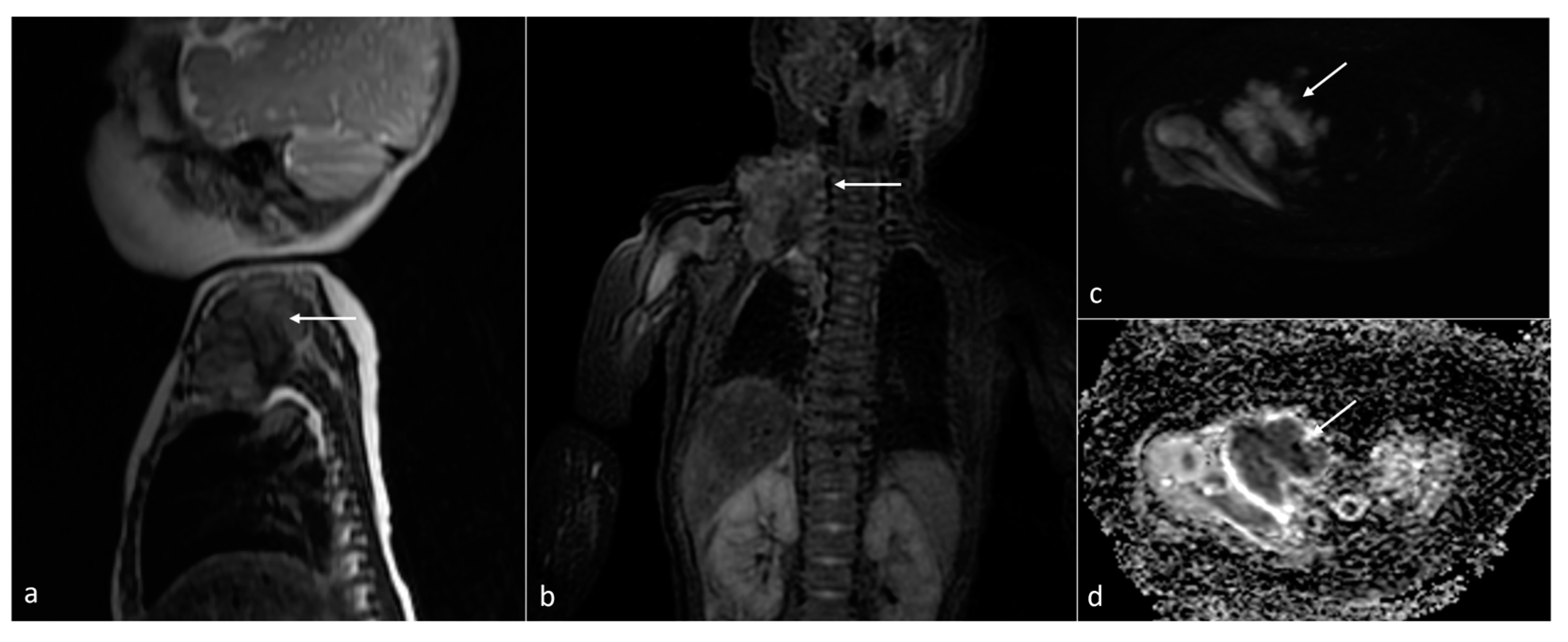
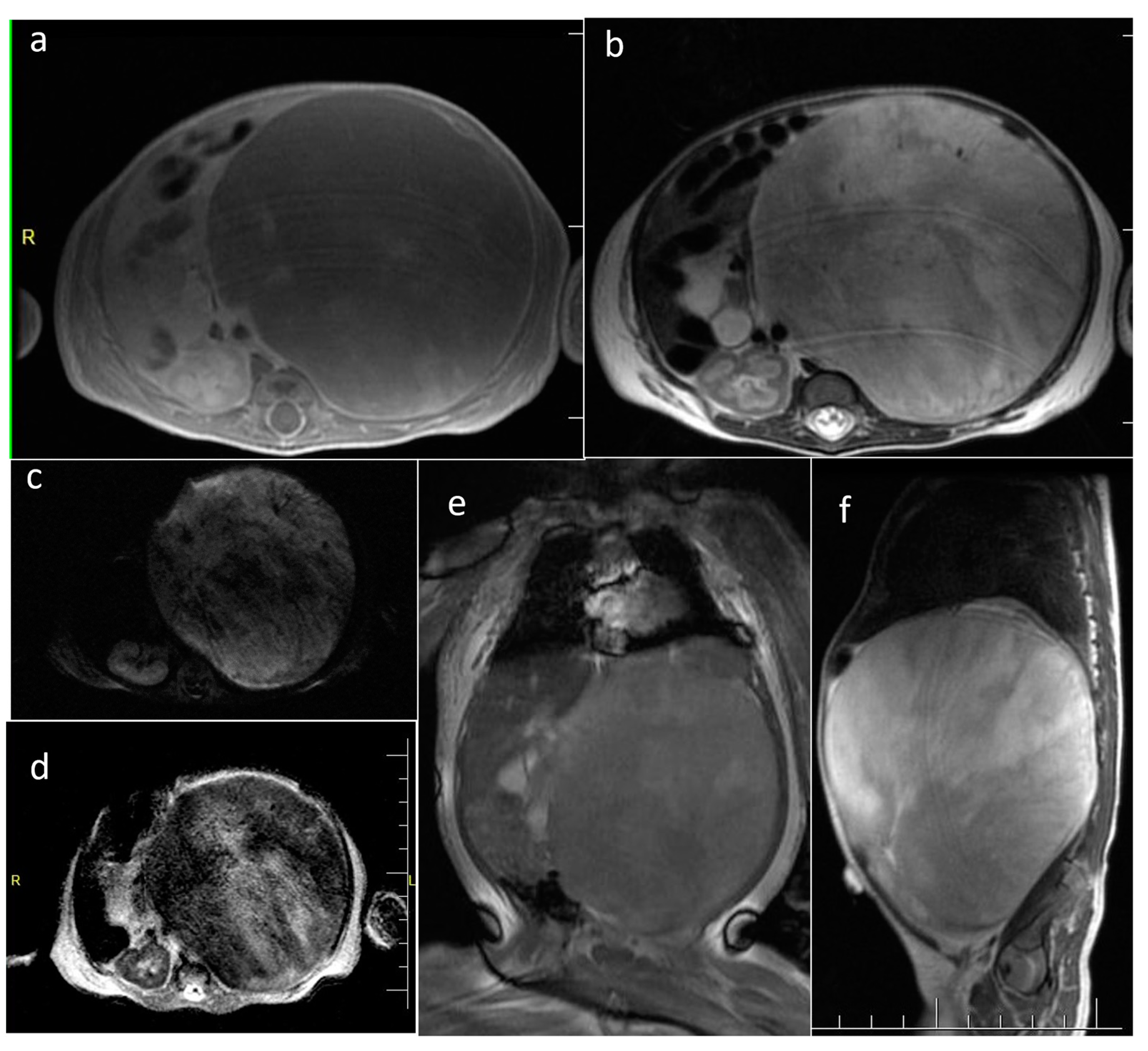
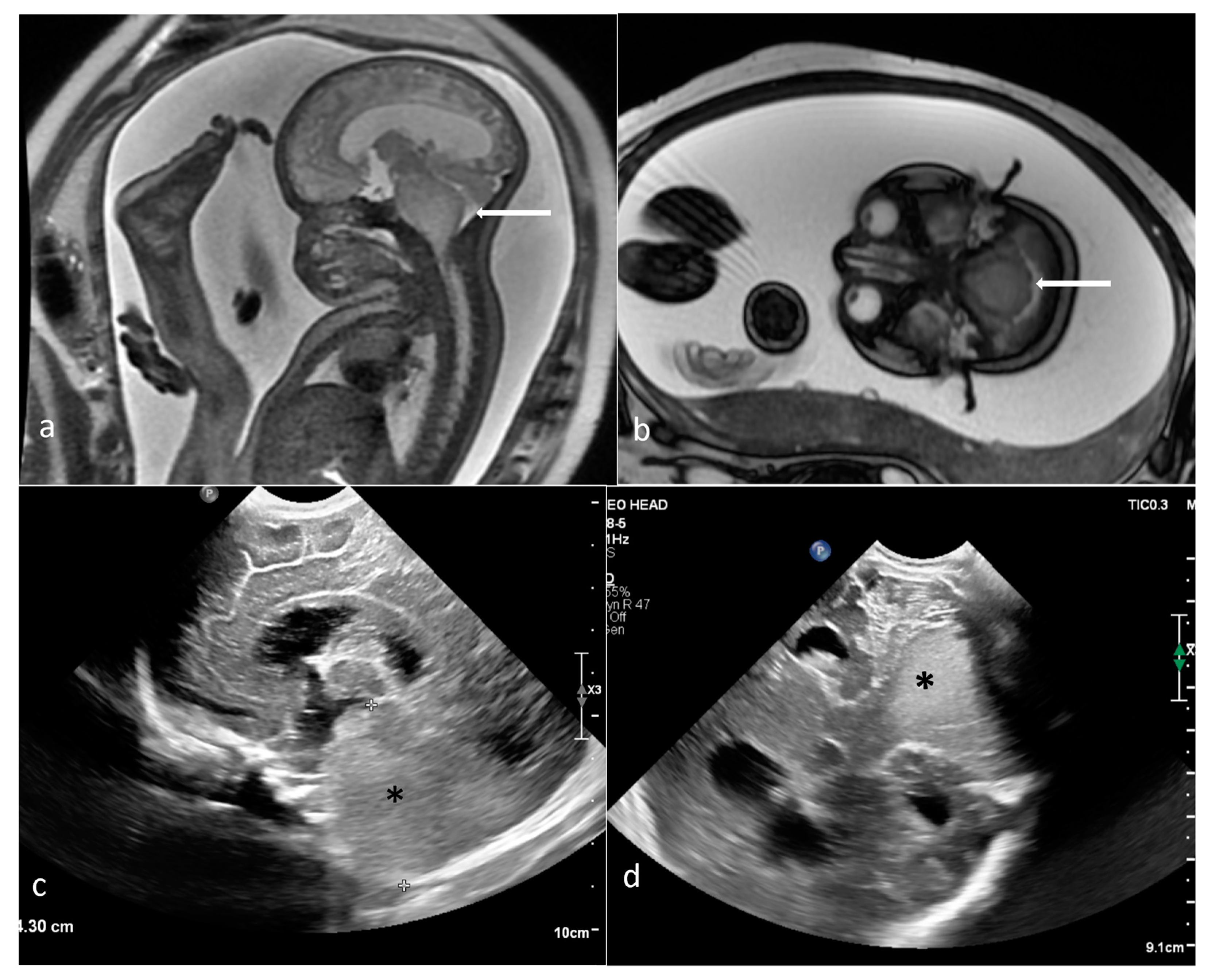
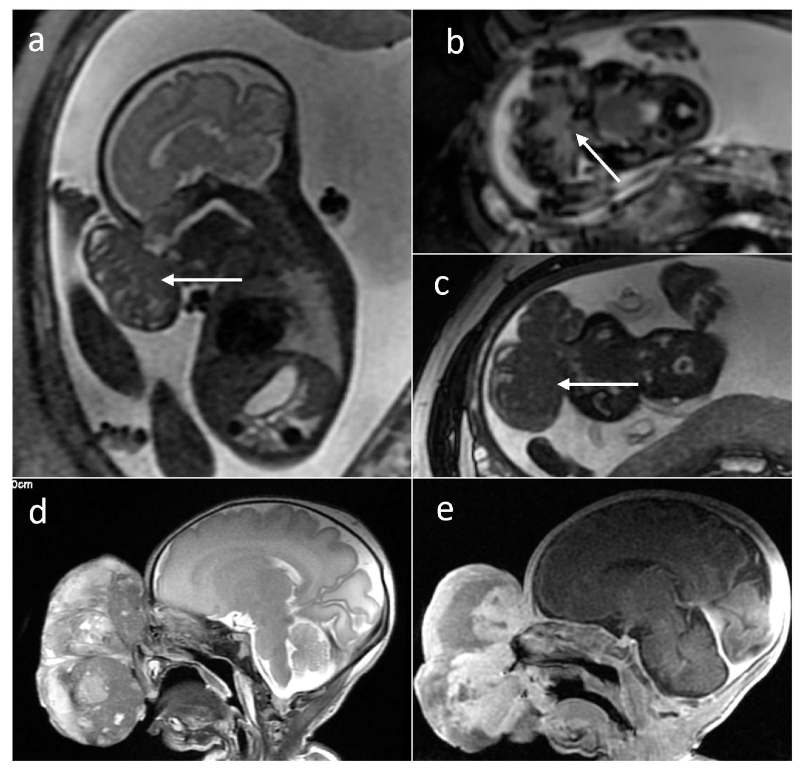

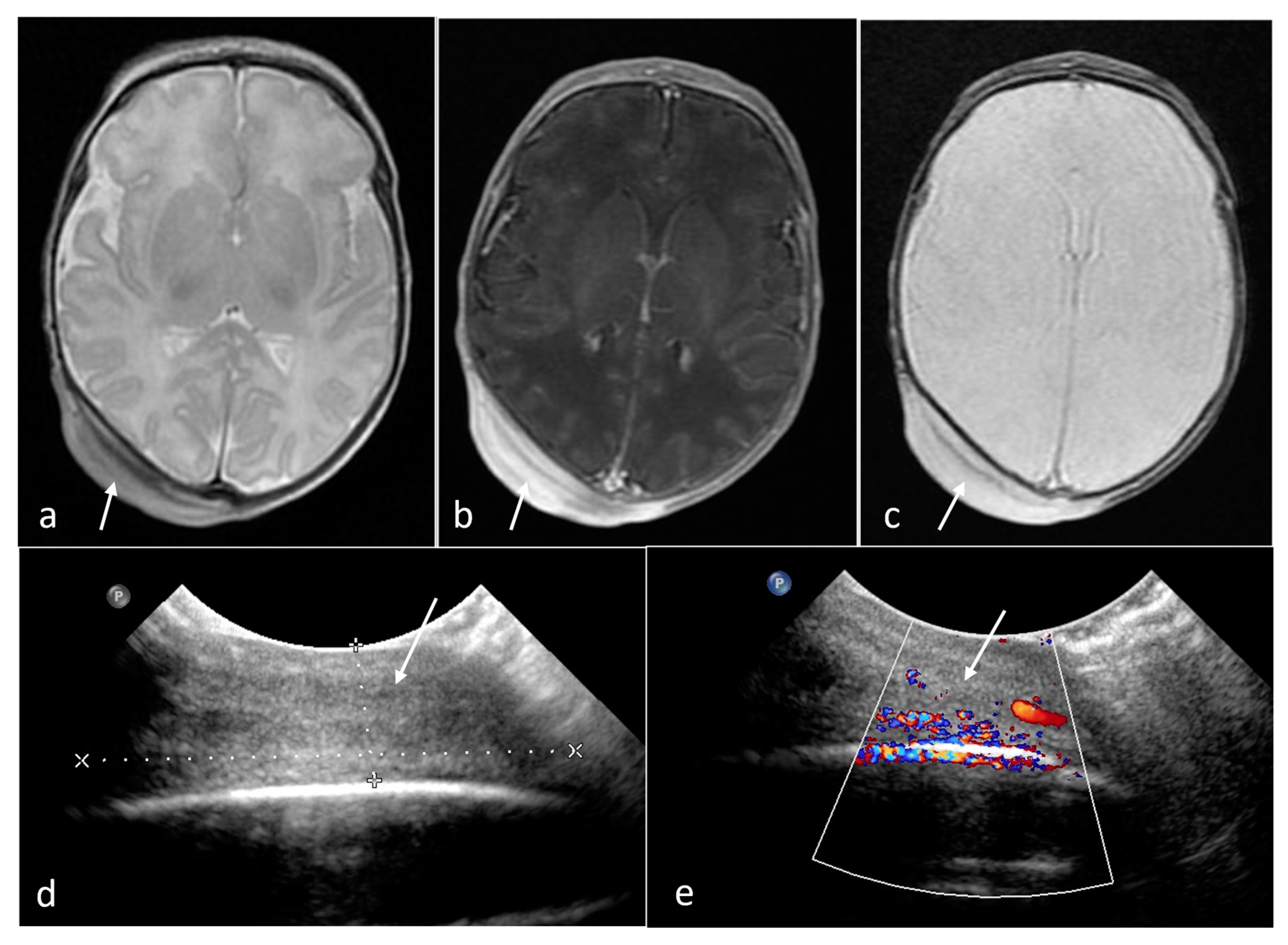
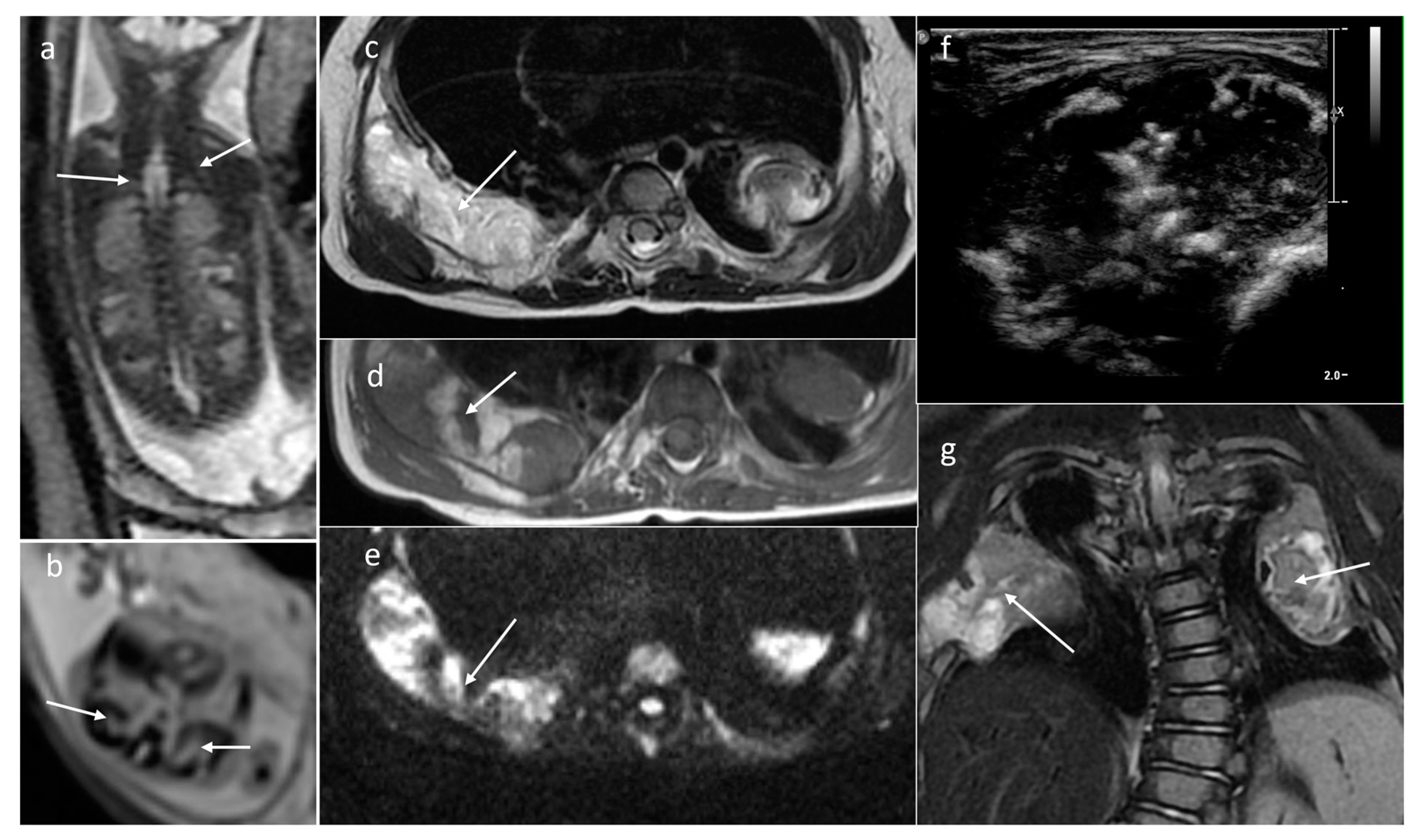
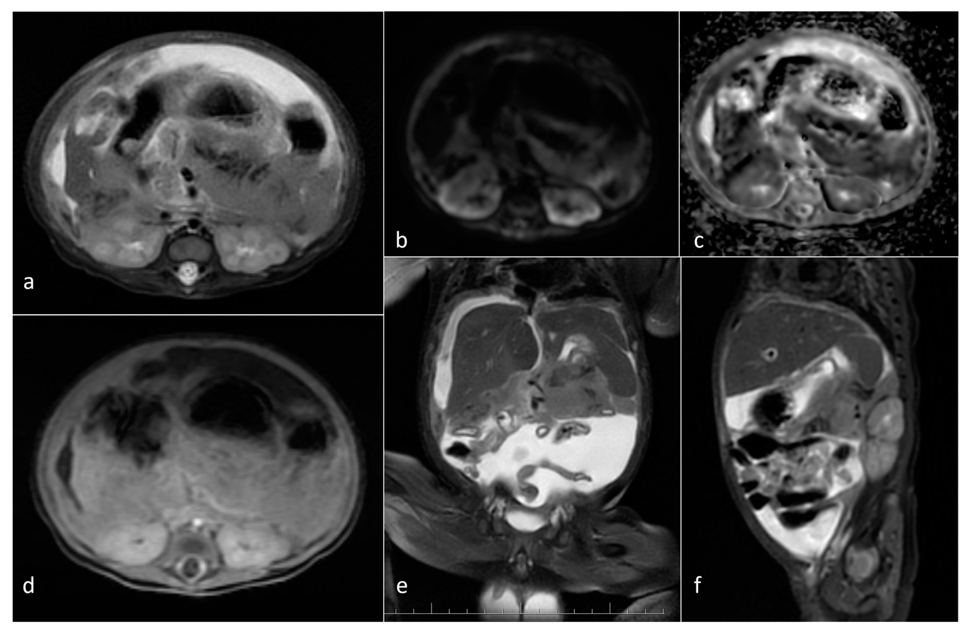
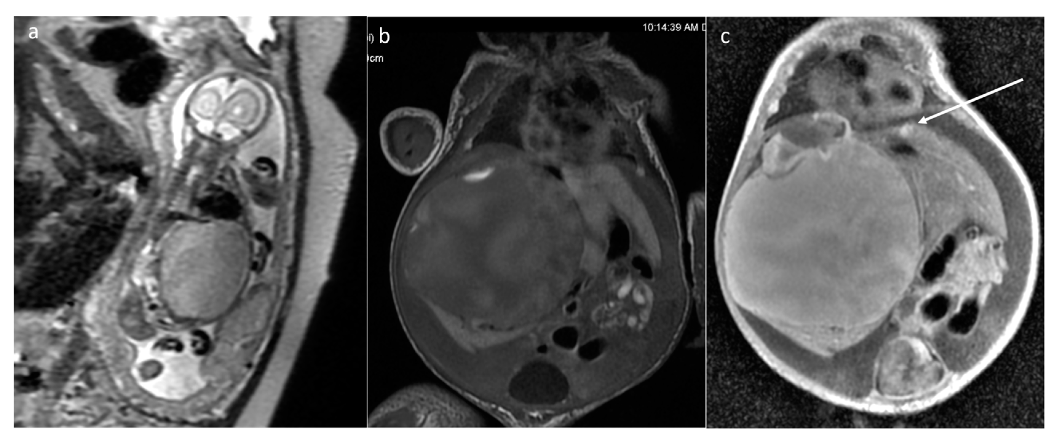
| Tumor Type | Incidence (%) | Additional Statistics |
|---|---|---|
| Extracranial teratoma | 23–29 | 45% SCT |
| Neuroblastoma | 22–30 | 90% adrenal gland |
| Soft tissue | 8–12 | Infantile fibrosarcoma most common |
| CNS | 6–10 | 50% teratoma |
| Renal | 5–7 | 66% mesoblastic nephroma |
| Hepatic | 5 | hemangioma 60% mesoblastic hamartoma 23% hepatoblastoma 16% (excluding metastases) |
| Thoracic | 3 | cardiac rhabdomyoma (TSC) 78% teratoma 18% |
| Tumor | Localization | Number of Patients | Volume (cm3) Mean [Min–Max] | Diffusion Restriction (ADC Values Min–Max × 10−6 mm2/s) | Solid/Cystic | Hemosiderin Deposits or Calcifications on T2* | Contrast Enhancement |
|---|---|---|---|---|---|---|---|
| Teratoma | sacrococcygeal | 25 | 268 [2.5–1145.165] | yes (13/17) [918–1661) | cystic (6) mixed (19) | yes 8/13 | yes (3/4) |
| head and neck | 13 | 176.1 [1.78–877.11] | yes (9/13) (796–1294) | solid (3) mixed (8) cystic (2) | yes (8/13) | yes (1) | |
| thorax and abdomen | 8 | 32 [1.49–134.98] | yes (3/6) | mixed (3) solid (3) cystic (2) | yes (4/5) | yes (1) | |
| Rhabdomyoma | heart | 22 | 5.08 [0.1–22] | no | Solid | No | N/A |
| Subependymal giant cell astrocytoma | foramen of Monro | 6 | 0.74 [0.59–0.89] | no | Solid | yes (2/2) | N/A |
| Neuroblastoma | adrenal gland/infrarenal space | 5 | 11.9 [2.6–24.2] | yes (3/5) (754–1200) | solid/mixed | yes (2/5) | yes (3/3) |
| Infantile fibrosarcoma | soft tissues of the back | 2 | 235 [47–423] | yes (2/2) (1025–1030) | solid (1) mixed (1) | yes (1/1) | yes 1/1 |
| Malignant rhabdoid tumor | soft tissues in upper right chest, soft tissues of left arm | 2 | 33.8 [20.5–47] | yes (2/2) (638–791) | Solid | no (0/1) | yes 1/1 |
| Mesoblastic nephroma | kidney | 2 | 98.4 [42.9–153.9] | yes (1100–1310) | Solid | 0/2 | yes (1/2) |
| Wilms tumor | kidney | 1 | 366.9 | yes (548) | Mixed | N/A | yes (solid part) |
| Craniopharyngioma | sella turcica/suprasellar | 2 | 34.15 [24.8–43.5] | yes (1/2) (548) | Mixed | no (0/1) | N/A |
| Brain stem glioma | brain stem | 2 | [12.5–18.41] | no (1266–1430) | microcystic/solid | 1/2 | 0/1 |
| Desmoplastic infantile astrocytoma (DIA) | right middle cranial fossa | 1 | 52.7 | N/A | Solid | N/A | N/A |
| Choroid plexus carcinoma | left cerebral hemisphere | 1 | 182 | N/A | Mixed | N/A | N/A |
| Glioblastoma | right cerebral hemisphere | 1 | 140 | yes (492) | Mixed | yes | N/A |
| Extra-pleural hemangiopericytoma/Solitary fibrous tumor (HPC/SFT) | nasal region | 1 | 159.1 | yes (1197) | Solid | yes | yes |
| Rhabdomyosarcoma | forearm | 1 | 7 | yes (885) | Solid | N/A | yes |
| Melanoma | scalp | 1 | 17.5 | yes (1230–1480) | Solid | No | yes |
| Hepatoblastoma | liver | 1 | 282 | yes (770) | mixed | No | yes (solid part) |
| Infantile myofibroma | left buttock and thigh, pelvis | 1 | 461 | yes (1056) | Solid | No | yes |
| Mesenchymal hamartoma | liver | 1 | 157 | N/A | Cystic | N/A | N/A |
| chest wall | 1 | 2.67 | yes (1450) | Solid | yes | N/A | |
| Juvenile xanthogranuloma (JXG) | abdomen | 1 | irregular shape | no | Cystic | N/A | yes |
| Blue rubber bleb nevus syndrome/Bean syndrome (BRBNS) | abdomen, tongue | 2 | 141 [124–158] | no | Solid | 1/2 | N/A |
Disclaimer/Publisher’s Note: The statements, opinions and data contained in all publications are solely those of the individual author(s) and contributor(s) and not of MDPI and/or the editor(s). MDPI and/or the editor(s) disclaim responsibility for any injury to people or property resulting from any ideas, methods, instructions or products referred to in the content. |
© 2023 by the authors. Licensee MDPI, Basel, Switzerland. This article is an open access article distributed under the terms and conditions of the Creative Commons Attribution (CC BY) license (https://creativecommons.org/licenses/by/4.0/).
Share and Cite
Kwasniewicz, P.; Wieczorek-Pastusiak, J.; Romaniuk-Doroszewska, A.; Bekiesinska-Figatowska, M. Congenital Tumors—Magnetic Resonance Imaging Findings with Focus on Rare Tumors. Cancers 2024, 16, 43. https://doi.org/10.3390/cancers16010043
Kwasniewicz P, Wieczorek-Pastusiak J, Romaniuk-Doroszewska A, Bekiesinska-Figatowska M. Congenital Tumors—Magnetic Resonance Imaging Findings with Focus on Rare Tumors. Cancers. 2024; 16(1):43. https://doi.org/10.3390/cancers16010043
Chicago/Turabian StyleKwasniewicz, Piotr, Julia Wieczorek-Pastusiak, Anna Romaniuk-Doroszewska, and Monika Bekiesinska-Figatowska. 2024. "Congenital Tumors—Magnetic Resonance Imaging Findings with Focus on Rare Tumors" Cancers 16, no. 1: 43. https://doi.org/10.3390/cancers16010043
APA StyleKwasniewicz, P., Wieczorek-Pastusiak, J., Romaniuk-Doroszewska, A., & Bekiesinska-Figatowska, M. (2024). Congenital Tumors—Magnetic Resonance Imaging Findings with Focus on Rare Tumors. Cancers, 16(1), 43. https://doi.org/10.3390/cancers16010043


