Article

Expression of Hemangioblast Proteins in von Hippel-Lindau Disease Related Tumors

by
Evelynn Vergauwen
1,2,
Ramses Forsyth
3,4,
Alexander Vortmeyer
5 and
Sven Gläsker
1,6,*
1
Department of Neurosurgery, Vrije Universiteit Brussel, Laarbeeklaan 101, 1090 Jette, Belgium
2
Department of Neurology, Universiteit Antwerpen, Universiteitsplein 1, 2610 Antwerpen, Belgium
3
Department of Pathology, Universitair Ziekenhuis Brussel, Laarbeeklaan 101, 1090 Jette, Belgium
4
Experimental Pathology Research Group, Vrije Universiteit Brussel, Laarbeeklaan 103, 1090 Jette, Belgium
5
Department of Pathology, Indiana University-Purdue University, 420 University Blvd, Indianapolis, IN 46202, USA
6
Neurosurgery Section, Gesundheitsverbund Landkreis Konstanz (GLKN), 78224 Singen am Hohentwiel, Germany
*
Author to whom correspondence should be addressed.
Cancers 2023, 15(9), 2551; https://doi.org/10.3390/cancers15092551
Submission received: 27 March 2023 / Revised: 23 April 2023 / Accepted: 27 April 2023 / Published: 29 April 2023
(This article belongs to the Section Cancer Pathophysiology)

Abstract

:

Simple Summary

Von Hippel-Lindau (VHL) disease is a rare tumor syndrome with autosomal dominant inheritance. We are a team of physicians who established the first VHL disease reference center in Belgium in 2016. Clear cell renal cell carcinomas can be a manifestation of a VHL disease. These tumors are currently a “hot topic” in VHL research, because of the research on the VHL protein pathway by Nobel Prize winner William Kaelin Jr. However, VHL patients also suffer from hemangioblastomas in the cerebellum, retina, and spinal cord. The pathogenesis of these tumors is less well studied. Based on histologic observation of VHL hemangioblastomas, we have gained insights that can be useful for understanding their pathogenesis.

Abstract

Von Hippel-Lindau (VHL) disease is a hereditary tumor syndrome that targets a highly selective subset of organs causing specific types of tumors. The biological basis for this principle of organ selectivity and tumor specificity is not well understood. VHL-associated hemangioblastomas share similar molecular and morphological features with embryonic blood and vascular precursor cells. Therefore, we suggest that VHL hemangioblastomas are derived from developmentally arrested hemangioblastic lineage keeping their potential of further differentiation. Due to these common features, it is of major interest to investigate whether VHL-associated tumors other than hemangioblastoma also share these pathways and molecular features. The expression of hemangioblast proteins has not yet been assessed in other VHL-related tumors. To gain a better understanding of VHL tumorigenesis, the expression of hemangioblastic proteins in different VHL-associated tumors was investigated. The expression of embryonic hemangioblast proteins Brachyury and TAL1 (T-cell acute lymphocytic leukemia protein 1) was assessed by immunohistochemistry staining on 75 VHL-related tumors of 51 patients: 47 hemangioblastomas, 13 clear cell renal cell carcinomas, 8 pheochromocytomas, 5 pancreatic neuroendocrine tumors, and 2 extra-adrenal paragangliomas. Brachyury and TAL1 expression was, respectively, observed in 26% and 93% of cerebellar hemangioblastomas, 55% and 95% of spinal hemangioblastomas, 23% and 92% of clear cell renal cell carcinomas, 38% and 88% of pheochromocytomas, 60% and 100% of pancreatic neuroendocrine tumors, and 50% and 100% of paragangliomas. We concluded that the expression of hemangioblast proteins in different VHL-associated tumors indicates a common embryological origin of these lesions. This may also explain the specific topographic distribution of VHL-associated tumors.

1. Introduction

Von Hippel-Lindau (VHL) disease is a rare hereditary tumor syndrome caused by germline mutations of the VHL gene on chromosome 3p25.3. Although all cells are affected, VHL disease targets a highly selective subset of organs giving rise to very specific tumors [1,2,3,4]. VHL patients develop central nervous system hemangioblastomas in a remarkable typical topographic distribution throughout the retina, cerebellum, and spinal cord. Other frequent manifestations include renal cysts and clear cell carcinomas, pancreatic cysts, and neuroendocrine tumors, as well as epididymal cysts in males and broad ligament cysts in females. A smaller group of patients is affected by pheochromocytomas, extra-adrenal paragangliomas, and endolymphatic sac or duct tumors [5,6,7,8,9,10]. The biology of organ selectivity and tumor specificity is not well understood. During our research on the pathogenesis of VHL disease, we made three important observations regarding VHL-associated hemangioblastomas.
First, hemangioblastomas express several embryonic proteins (such as Brachyury and TAL1) and embryonic protein receptors [10,11]. The proteins and receptors mirror the expression profile of an embryonic cell: the hemangioblast. This embryonic cell is a precursor of erythrocytes, vascular smooth muscle cells, and blood vessels. Brachyury and TAL1 are needed for differentiation from mesoderm into hemangioblast and subsequent blood cell precursors.
Second, we observed that hemangioblastomas share similar histomorphological features with the hemangioblast, such as blood island formation and extramedullary hematopoiesis (with VHL-deficient daughter cells) [12]. In addition, many histological similarities exist between different VHL-associated lesions, such as the clear cell aspect.
Third, we discovered preneoplastic lesions in a steady topographic distribution throughout the central nervous system of VHL patients, corresponding to the localization of hemangioblast proteins, such as TAL1, during embryogenesis [13].
Based on these three observations, we have hypothesized that VHL-associated hemangioblastomas originate from developmentally arrested embryonic hemangioblasts containing a neoplastic potential and the protracted capacity to differentiate [12,14].
The presence of embryonic hemangioblast proteins (Brachyury and TAL1) has not yet been investigated in VHL-associated tumors other than hemangioblastomas. Due to the common morphological features and the common molecular basis of the different VHL tumors, it is of major interest to investigate whether VHL-associated tumors, other than hemangioblastomas, share these aforementioned features.

2. Materials and Methods

2.1. Tissue Selection

The study protocol was approved by the local research ethics service (reference number 2016/271). To access surgically resected tumors of VHL patients, a query through the Belgian Cancer Registry (national central tumor databank) was performed. Additionally, colleague neurosurgeons throughout Belgium were contacted, and several anatomic pathological institutions were visited. Specimens were collected from the following institutions: University Hospital Brussels (Jette, Belgium), Jules Bordet Institute (Brussels, Belgium), Erasme University Hospital Brussels (Anderlecht, Belgium), University Hospital Saint-Luc (Woluwe-Saint-Lambert, Belgium), University Hospital Liège (Liège, Belgium), and University Hospital Antwerp (Edegem, Belgium). Other samples were contributed by the biomaterial database of the German VHL family alliance (Verein VHL betroffener Familien e.V.) at the Dr. Margarete Fischer-Bosch-Institut für Klinische Pharmakologie (IKP) at the Robert-Bosch-Krankenhaus (RBK) (Stuttgart, Germany).
Any tumor related to VHL disease was included in the samples cohort (central nervous system hemangioblastomas, clear cell renal cell carcinomas, pheochromocytomas, pancreatic tumors, endolymphatic sac tumors, epididymis/broad ligament cystadenomas and extra-adrenal paragangliomas). Patients of all ages and both genders, with either a genetical or clinical confirmation of VHL disease were included.
Seventy-five surgically resected tumors from 51 different von Hippel-Lindau patients were obtained (27 cerebellar hemangioblastomas, 20 spinal hemangioblastomas, 13 clear cell renal cell carcinomas, 8 pheochromocytomas, 5 pancreatic tumors, and 2 extra-adrenal paragangliomas). In total, 20 male and 31 female patients were included, their ages ranging from 13 to 70 years old. All patients previously had either genetical or clinical confirmation of VHL disease. Patients had been operated on between 1989 and 2018. Patient characteristics and tumor data are summarized in Table 1.

2.2. Tissue Preparation and Histological Classification

In-house tumors and control tissues were fixed in 10% neutral-buffered formalin and embedded in paraffin. Tumors obtained from other institutions had been processed in different devices, however, with the same formalin concentration. Tumors and control tissue blocks were cut in 4 µm sections, mounted on TOMO slides (Matsunami Glass IND Ltd., Osaka, Japan) and dried for 1 h at 50 °C. Haematoxylin and eosin (H & E) staining was performed on all sections for diagnostic confirmation of tumor type. Clear cell renal cell carcinomas, pheochromocytomas, pancreatic neuroendocrine tumors, and extra-adrenal paragangliomas were histologically graded according to the following grading systems: World Health Organization (WHO)/International Society of Urological Pathology (ISUP) grading system [15], Pheochromocytoma of the Adrenal Gland Scaled Score (PASS) [16], WHO grading system [17], and for paragangliomas again the Pheochromocytoma of the Adrenal Gland Scaled Score (PASS) [16].

2.3. Immunohistochemistry

All experiments were performed in the laboratory of anatomic pathology at the University Hospital Brussels. Tumor and control sections were stained in the same way for each run, using an automated slide staining system (BenchMark ULTRA, Ventana Medical Systems, Inc., Oro Valley, AZ, USA) with standard settings. All used products were produced by Ventana Medical Systems, Inc, Oro Valley, Arizona, United States. In brief, sections were manually preheated for 20 min at 60 °C, then heated automatically for 4 min at 72 °C and deparaffinized with EZ Prep. Next, Long Cell Conditioner 1 and CC Medium Coverslip were alternately applied at 95 °C for breaking of formalin bonds/antigen retrieval and rehydration, respectively. Sections were rinsed with a Tris based Reaction Buffer at 36 °C. ISH Peroxidase Inhibitor was applied to decrease non-specific background staining, and sections were rinsed with several cycles of Tris based Reaction Buffer wash at 36 °C. Secondary antibodies were diluted in Antibody Diluent and applied manually. HQ Universal Linker, HRP Multimer, and Amplifier H2O2 (OptiView Amplification Kit) were applied for optimal linking of primary to secondary antibody, alternated by rinsing cycles of Tris based Reaction Buffer. Incubation with the secondary antibody was performed for 8 min (anti-TAL1) and 2 h (anti-Brachyury) at 36 °C. The tertiary antibody was visualized with hydrogen peroxide substrate and three, 3′-diaminobenzidine tetrahydrochloride (DAB) chromogen (ultraView Universal DAB Detection Kit). Sections were counterstained with Mayer’s haematoxylin, postcounterstained with bluing agent, manually dehydrated with 70–100% graded alcohol series and xylene, and cover-slipped.
Secondary antibodies were validated on validated positive and negative (tissue microarray) control tissues in our institution. Data about concentration, dilution, distributor, cellular localization, and positive control tissues are summarized in Table 2. Human chordoma tumor was used as positive control tissue for Brachyury. A human T-cell acute lymphoblastic leukemia cell line was grown and used as positive control tissue for TAL1. Tissue microarray was used as a negative control, containing different human tissues (spleen, stomach, appendix, colon, liver, pancreas, kidney, adrenal gland, testis, prostate, lung, tonsil, cerebrum, cerebellum, and spinal cord). Brachyury and TAL1 sporadically stained isolated cells in the negative control tissues (presumably mast cells).

2.4. Evaluation of Immunohistochemical Staining

To date, there are no standard immunohistochemical (IHC) scoring systems for the antibodies that were used [18]. Different semi-quantitative IHC scoring systems were evaluated. It was concluded that the Remelle composite scoring system (immunoreactive score, IRS) suits our study best [19]. IHC staining was scored as intensity of staining signal multiplied by percentage of positive tumor cells. Intensity was classified as 0 (no detectable staining), 1 (weak, visible at high to intermediate magnification: ×40; nuclear: faint, membranous: only part of the membrane), 2 (moderate, visible at intermediate to low magnification: ×10–20; nuclear: moderate, membranous: large part), and 3 (strong, visible at low magnification: ×2.5–5; nuclear: strong, membranous: strong, whole membrane, and also: same intensity as control tissue). Percentage of positive tumor cells was evaluated as 0 (0%), 1 (<10%), 2 (10–50%), 3 (51–80%), or 4 (>80%). Scores for different VHL-related tumors were given as mean values +/− SD. Ultimately, tumor and control tissues were classified according to ‘no expression’ (total score 0), ‘low expression’ (total score 1 to 4) or high ‘expression’ (total score 5 to 9).

2.5. Statistical Analysis

All statistical data analyses were conducted with Real Statistics Resource Pack for Excel 2016 (Microsoft Office, Microsoft, Redmont, Washington, DC, USA). Prevalence of embryonic marker expression in different types of VHL tumors was summarized using descriptive statistics. The nonparametric Kruskal Wallis test was used to determine if there was a statistical difference in embryonic marker expression—represented by the Remelle composite scoring system (dependent variable) between VHL tumors (independent variable). The differences were reported in medians.

3. Results

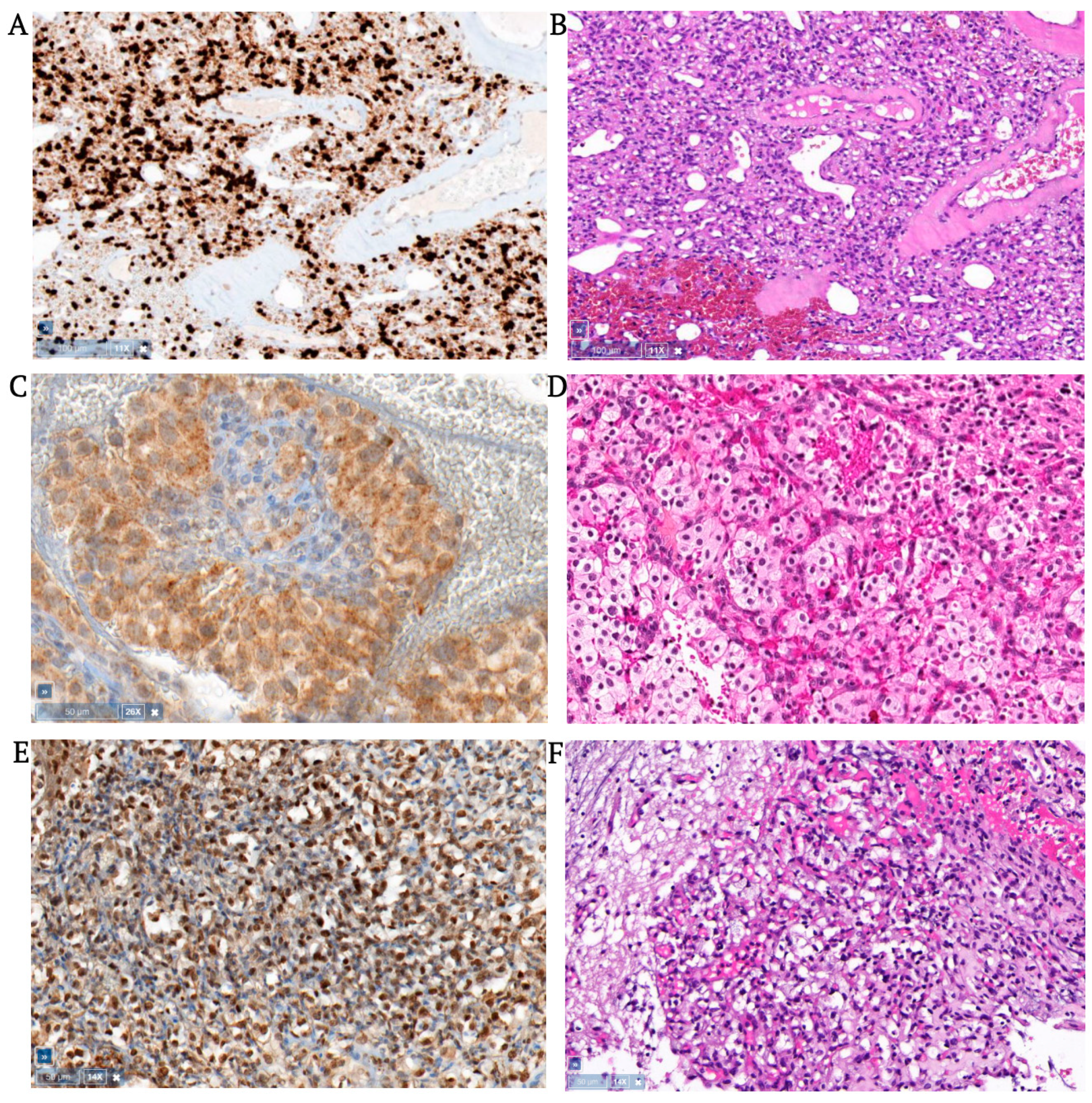
3.1. Hemangioblastomas

All 47 hemangioblastomas (27 cerebellar, 20 spinal) corresponded histologically to WHO grade I and consisted of large round, oval, or polygonal tumor cells in a capillary vascular network. In 13 of 47 hemangioblastomas (3 cerebellar, 10 spinal), moderate to strong nuclear Brachyury expression was seen; mostly in half of the tumor cell population. Two of 20 spinal hemangioblastomas showed strong nuclear Brachyury staining in almost all tumor cells (an example can be seen in Figure 1). Weak to moderately strong cytoplasmic Brachyury expression was seen in 8 of 47 hemangioblastomas (5 cerebellar, 3 spinal), covering various amounts of tumor areas, including three tumors with nuclear expression as well. Figure 1 shows an example of cytoplasmic Brachyury staining in a cerebellar hemangioblastoma.
Sometimes weak, but mostly moderately strong, nuclear TAL1 expression was seen in 43 of 47 hemangioblastomas (25 cerebellar, 18 spinal), staining approximately half of the tumor area. TAL1 cytoplasmic expression was seen in 40 of 47 hemangioblastomas (22 cerebellar, 18 spinal), covering more than half of the tumor area.
Brachyury and/or TAL1 staining was never seen in the whole tumor cell population. Tumors that expressed Brachyury always co-expressed TAL1, but not vice versa. Tumor cells surrounding cysts had the same staining pattern. Sometimes, weak to strong cytoplasmic Brachyury and TAL1 staining was seen in some, but not all, endothelial cells that were contained by positive nuclear or cytoplasmic tumor areas, and sometimes in negative areas. Data are summarized in Supplementary Tables S1 and S2.

3.2. Clear Cell Renal Cell Carcinomas

Two of 13 clear cell renal cell carcinomas showed weak to moderate nuclear Brachyury staining, more specifically in a small part of the tumor cell population. Weak Brachyury cytoplasmic staining was seen in three tumors, in less than half of the tumor area, arranged in small groups or cyst walls. Additionally, one of these cytoplasmic-staining tumors contained few tumoral and nearby endothelial cells with strong nuclear Brachyury expression (<10% of tumor cells). Twelve of 13 renal cell carcinomas showed moderate to strong nuclear TAL1 expression, varying from small to mostly very large areas (see Figure 2). Eleven renal cell carcinomas showed a weak to moderate cytoplasmic TAL1 expression in large tumor areas. Occasionally, TAL1 was weakly expressed in endothelial cytoplasm as well. Similar to hemangioblastomas, tumors with nuclear Brachyury expression always co-expressed TAL1, but not vice versa. Data are summarized in Supplementary Tables S3 and S4.

3.3. Pheochromocytomas

In three of eight pheochromocytomas, weak to moderately strong cytoplasmic Brachyury expression was found in groups of tumor cells, or the whole population in one tumor (an example can be seen in Figure 2). Nuclear Brachyury expression was not seen, except in isolated sporadic cells, in all tumors. Moderate to strong nuclear TAL1 staining was seen in three pheochromocytomas in a small part of the tumor area. Weak to moderate cytoplasmic TAL1 staining was seen in seven of eight pheochromocytomas, mostly in large areas. Some endothelial cells showed weak and incomplete cytoplasmic Brachyury staining, however, in one pheochromocytoma a vascular structure was found with moderate Brachyury expression in nuclei of plump endothelial cells. TAL1 was sometimes weakly expressed in endothelial cytoplasm as well. Data are summarized in Supplementary Tables S5 and S6.

3.4. Pancreatic Neuroendocrine Tumors

Two of five pancreatic neuroendocrine tumors showed strong nuclear Brachyury expression in a small population of tumor cells. In between tumor cell groups, endothelial cells showed weak to moderate cytoplasmic Brachyury staining. Another tumor showed weak cytoplasmic Brachyury staining in a small population of tumor cells. Normal pancreatic tissue showed negligible cytoplasmic staining with Brachyury and was negative for TAL1.
In three of five tumors, moderately strong nuclear TAL1 staining was observed in a larger population of tumor cells. Some endothelial cells showed cytoplasmic staining with TAL1 (but not in all tumors), usually in larger vessels with plump endothelial cells, and sometimes large vascular structures surrounded by tumor cells. Weak to moderate cytoplasmic TAL1 staining was seen in large areas of four tumors (see Figure 2). In comparison to other VHL tumors, Brachyury and TAL1 staining was more dissociated. Data are summarized in Supplementary Tables S7 and S8.

3.5. Extra-Adrenal Aragangliomas

In one of two paragangliomas, small tumor areas showed strong nuclear and weak cytoplasmic Brachyury expression. In addition, this paraganglioma showed strong nuclear and moderate cytoplasmic TAL1 expression in a large area (Figure 2). In the other paraganglioma, only moderately strong cytoplasmic expression was seen in almost all tumor cells. In both tumors, some vascular structures showed weak cytoplasmic Brachyury staining. Data are summarized in Supplementary Tables S9 and S10.

3.6. Statistical Analysis

Brachyury and TAL1 expression was, respectively, observed in 26% and 93% of cerebellar hemangioblastomas, 55% and 95% of spinal hemangioblastomas, 23% and 92% of clear cell renal cell carcinomas, 38% and 88% of pheochromocytomas, 60% and 100% of pancreatic neuroendocrine tumors, and 50% and 100% of paragangliomas. Median and mean Remelle scores are described in the Supplementary Tables. The difference between Remelle scores (both nuclear and cytoplasmic) was statistically significant between different VHL tumors. In general, TAL1 staining was stronger compared to Brachyury staining in all VHL tumors (according to Remelle scores). Additionally, there was a statistically significant difference in amount of staining between different tumors, which could be ranked according to decreasing nuclear Brachyury expression: spinal hemangioblastomas, extra-adrenal paragangliomas, pancreatic neuroendocrine tumors, cerebellar hemangioblastomas, clear cell renal cell carcinomas, and pheochromocytomas. Cytoplasmic Brachyury staining was mostly observed in pheochromocytomas, cerebellar hemangioblastomas, and spinal hemangioblastomas.
Ranking for amount of TAL1 staining was as follows: cerebellar hemangioblastomas, clear cell renal cell carcinomas, spinal hemangioblastomas, extra-adrenal paragangliomas, pancreatic neuroendocrine tumors, and pheochromocytomas. Cytoplasmic TAL1 staining was observed in all VHL tumors and was strongest in extra-adrenal paragangliomas, cerebellar hemangioblastomas, and spinal hemangioblastomas.

4. Discussion

VHL disease targets a highly selective subset of organs with very specific types of tumors. To date, this principle of organ selectivity and tumor specificity, as well as the origin of the “stromal tumor cell” of VHL tumors, has been an ongoing subject of debate, and its growth mechanisms remain largely unknown.
Per this study, evidence for the expression of hemangioblast proteins in different types of VHL-associated tumors is provided, including cerebellar and spinal hemangioblastomas, clear cell renal cell carcinomas, pheochromocytomas, pancreatic neuroendocrine tumors, and paragangliomas. The expression of hemangioblast proteins in different types of VHL tumors provides evidence to the pre-hemangioblastic stem cells as a key role player in the origin of the different VHL-associated tumors. This point of view could be a possible explanation for the principle of organ selectivity and tumor specificity in VHL disease. We noted with interest that the distribution of TAL1 expression in mammalian embryonic development matches the vast topographic distribution of VHL tumors [20] (Figure 3).

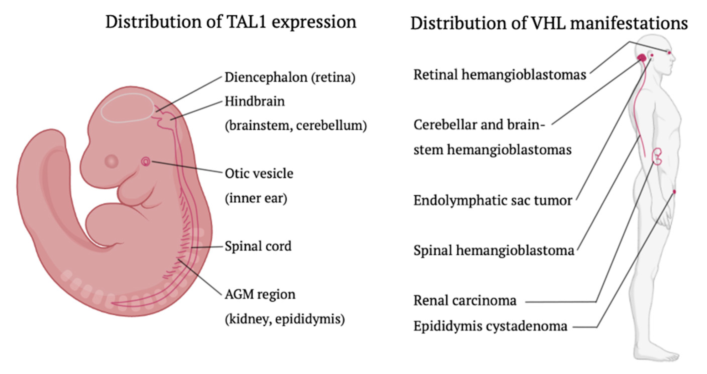
4.1. The Embryonic Hemangioblast: Fact or Fiction?

The irrefutable existence of the embryonic hemangioblast in vivo has not been confirmed to date. In 1880, Wilhelm His was the first to hypothesize a common ancestor cell for hematopoietic cells and endothelium [21], a concept which was consolidated in 1917 by Sabin Florence on living chick embryos. Sabin named the progenitor cell “the angioblast” [22]. Murray P.D.F. later renamed the ancestor cell as “the hemangioblast” [23].
Beyond question, hematopoietic and endothelial cell lineages share common typical mesodermal markers (such as Brachyury, BMP4, and VEGFR2), surface markers (such as CD34, VE cadherin, CD31), and transcription factors (such as RUNX1 and GATA2) throughout their development. Nevertheless, two different hypotheses co-exist in the current literature. Some researchers have hypothesized that endothelial cells and hematopoietic stem/progenitor cells derive from independent epiblast populations. The second hypothesis states that the mesodermally-derived bipotent hemangioblast gives rise to endothelial cells and hematopoietic stem/progenitor cells, whether or not after a transitional phase of hemogenic endothelium [24,25,26,27].

4.2. Hypothesis of Embryonic Origin

The multiplicity, histological heterogeneity, and specific topographic distribution of angiomatous tumors in “von Hippel’s disease” or “angiomatosis retinae” made Arvid Lindau (1926) originally hypothesize that “cerebellar (capillary hem) angiomas”, being part of “central nervous system angiomatosis”, originated from a vascular congenital maldevelopment. The specific region of interest was a mesoderm-derived capillary network in the inferior medullary velum, its malformation taking place in the third fetal month [28]. This vascular network was described by Karlefors [29]. Based on the observation of immature vascular elements and blood cell formation, Lindau, Cushing, and Bailey (1928), and Stein (1960), hypothesized that the original cells of “(hem) angioblastomas” were maldeveloped angioblastic cells which, later on, transformed into fat-laden “pseudoxanthoma tumor cells”: now known as the intravascular tumor stromal cell. They all described the simultaneous occurrence of mature and immature vascular elements in cerebellar hemangioblastomas [8,9,30,31].
Notwithstanding its ingenuity, the embryonic hypothesis was never proven on all DNA, RNA, and protein levels, and it does not completely cover the broad neoplastic and extra-nervous system diversity of the VHL syndrome. Since 1926, many other origin cells have been suspected and conflicting immunohistochemical expression patterns have emerged ever since, as summarized by Ma and Zu [32].
Not until recently, the embryonic hypothesis regained attention by the discovery of early embryonal maldevelopmental structures: developmentally arrested structural elements (DASEs). They have been found in supposedly normal cerebellum and dorsal nerve roots on surgical and autopsy tissue from VHL patients. Interestingly, DASEs already exhibit loss of VHL-heterozygosity (in contrast to normal surrounding tissue), and some have the ability to transform into frank VHL hemangioblastomas with mesenchymal components. Indeed, the more progressive epithelioid hemangioblastomas feature VHL-deficient vascular structures and extramedullary haematopoiesis, which may also explain tumor diversity in VHL disease [13,33,34,35,36,37,38]. Comparable embryonal precursor tissue was found in the kidney, epididymal and endolymphatic duct and sac from VHL patients [14].
One major finding of this study is that stronger evidence is provided for the embryonic hypothesis by proposing the hemangioblast as the cell of origin of a broader spectrum of VHL tumors. It was shown that the cell’s typical proteins Brachyury and TAL1 are expressed by a broader spectrum of VHL tumors rather than by hemangioblastomas only.
Our previous research showed the expression of Brachyury and TAL1 in 10 of 10 VHL-related hemangioblastomas [39]. Ma et al. and Park et al. later confirmed the expression of Brachyury and TAL1 in another small series of VHL-related hemangioblastomas, of which the exact amount of positively staining tumors was not mentioned [32,40,41]. In our current study some hemangioblastomas showed very specific nuclear and/or cytoplasmic Brachyury expression in select tumor areas, with varying staining intensities, suggesting different tumor cell development stages.

4.3. New Insights into Brachyury Distribution

Interestingly, this manuscript is the first that describes both nuclear and cytoplasmic Brachyury expression in VHL hemangioblastoma tumor cells. Thus far, two articles described exclusive cytoplasmic expression in 20 of 22 non-VHL hemangioblastomas [42,43], reserving nuclear expression for human chordomas. In some chordomas, also cytoplasmic can be observed although in lower dilutions [44,45,46,47]. Tirabosco et al. described nuclear expression in both intra- and -extra axial bony chordomas and hemangioblastomas, without information about VHL-status. They are also the first to report Brachyury expression at the protein level in normal adult tissue: the testis [47]. An article from Doyle et al. describes the absence of either nuclear and cytoplasmic Brachyury expression in 22 peripheral VHL and non-VHL hemangioblastomas [48]. These discrepancies might be due to different Brachyury isoforms of different antibodies, as previously suggested by Hamilton et al. [49].

5. Conclusions

Based on the abovementioned observations and results, it is hypothesized that VHL tumors originate from the pre-hemangioblastic lineage, a hemangioblast daughter cell (also known as “hemogenic endothelium”) or that they have at least a hemangioblast-like-phenotype. Additionally, this hypothesis (especially with regard to TAL1) may explain the vast topographic distribution of VHL tumors in a highly selective subset of organs.

Supplementary Materials

The following supporting information can be downloaded at: https://www.mdpi.com/article/10.3390/cancers15092551/s1, Table S1: nuclear and cytoplasmic Brachyury staining in VHL hemangioblastomas; Table S2: nuclear and cytoplasmic TAL1 staining in VHL hemangioblastomas; Table S3: nuclear and cytoplasmic Brachyury staining in VHL clear cell renal cell carcinomas; Table S4: nuclear and cytoplasmic TAL1 staining in VHL clear cell renal cell carcinomas; Table S5: nuclear and cytoplasmic Brachyury staining in VHL pheochromocytomas; Table S6: nuclear and cytoplasmic TAL1 staining in VHL pheochromocytomas; Table S7: nuclear and cytoplasmic Brachyury staining in VHL pancreatic neuroendocrine tumours; Table S8: nuclear and cytoplasmic TAL1 staining in VHL pancreatic neuroendocrine tumours; Table S9: nuclear and cytoplasmic Brachyury staining in VHL paragangliomas; Table S10: nuclear and cytoplasmic TAL1 staining in VHL paragangliomas.

Author Contributions

All authors contributed to the design, data analysis, writing, and final approval of the manuscript. E.V., S.G., R.F., and A.V. contributed extensively to histological interpretation. All authors have read and agreed to the published version of the manuscript.

Funding

This research was funded by the FWO (Fonds Wetenschappelijk Onderzoek Vlaanderen, FWO-V 1831017 N).

Institutional Review Board Statement

The study protocol was approved by the local research ethics service (reference number 2016/271).

Informed Consent Statement

Not applicable.

Data Availability Statement

Data is contained within the article or supplementary material.

Acknowledgments

The authors would like to thank the following institutions for their contribution in tissue material: University Hospital Brussels (Jette, Belgium), Jules Bordet Institute (Brussels, Belgium), Erasme University Hospital Brussels (Anderlecht, Belgium), University Hospital Saint-Luc (Woluwe-Saint-Lambert, Belgium), University Hospital Liège (Liège, Belgium), and University Hospital Antwerp (Edegem, Belgium). Other samples were a kind gift of the Margarete Fischer-Bosch-Institut für Klinische Pharmakologie (IKP) at the Robert-Bosch-Krankenhaus (RBK) (Stuttgart, Germany).

Conflicts of Interest

The authors declare no conflict of interest. The funders had no role in the design of the study; in the collection, analyses, or interpretation of data; in the writing of the manuscript; or in the decision to publish the results.

References

  1. Seizinger, B.R.; Rouleau, G.A.; Ozelius, L.J.; Lane, A.H.; Farmer, G.E.; Lamiell, J.M.; Haines, J.; Yuen, J.W.; Collins, D.; Majoor-Krakauer, D.; et al. Von Hippel-Lindau Disease Maps to the Region of Chromosome 3 Associated with Renal Cell Carcinoma. Nature 1988, 332, 268–269. [Google Scholar] [CrossRef] [PubMed]
  2. Latif, F.; Tory, K.; Gnarra, J.; Yao, M.; Duh, F.M.; Orcutt, M.L.; Stackhouse, T.; Kuzmin, I.; Modi, W.; Geil, L.; et al. Identification of the Von Hippel-Lindau Disease Tumor Suppressor Gene. Science 1993, 260, 1317–1320. [Google Scholar] [CrossRef] [PubMed]
  3. Cohen, A.J.; Li, F.P.; Berg, S.; Marchetto, D.J.; Tsai, S.; Jacobs, S.C.; Brown, R.S. Hereditary Renal-Cell Carcinoma Associated with a Chromosomal Translocation. N. Engl. J. Med. 1979, 301, 592–595. [Google Scholar] [CrossRef] [PubMed]
  4. Whaley, J.M.; Naglich, J.; Gelbert, L.; Hsia, Y.E.; Lamiell, J.M.; Green, J.S.; Collins, D.; Neumann, H.P.; Laidlaw, J.; Li, F.P.; et al. Germ-Line Mutations in the Von Hippel-Lindau Tumor-Suppressor Gene Are Similar to Somatic Von Hippel-Lindau Aberrations in Sporadic Renal Cell Carcinoma. Am. J. Hum. Genet. 1994, 55, 1092–1102. [Google Scholar]
  5. Gaal, J.; Van Nederveen, F.H.; Erlic, Z.; Korpershoek, E.; Oldenburg, R.; Boedeker, C.C.; Kontny, U.; Neumann, H.P.; Dinjens, W.N.; De Krijger, R.R. Parasympathetic Paragangliomas Are Part of the Von Hippel-Lindau Syndrome. J. Clin. Endocrinol. Metab. 2009, 94, 4367–4371. [Google Scholar] [CrossRef]
  6. Brandt, R. Zur Frage Der Angiomatosis Retinae. Albrecht Von Graæes Arch. Für Ophthalmol. 1921, 106, 126–136. [Google Scholar] [CrossRef]
  7. Manski, T.J.; Heffner, D.K.; Glenn, G.M.; Patronas, N.J.; Pikus, A.T.; Katz, D.; Lebovics, R.; Sledjeski, K.; Choyke, P.L.; Zbar, B.; et al. Endolymphatic Sac Tumors. A Source of Morbid Hearing Loss in Von Hippel-Lindau Disease. JAMA 1997, 277, 1461–1466. [Google Scholar] [CrossRef]
  8. Lindau, A. Studien ÜBer Kleinhirncysten: Bau, Pathogenese Und Beziehungen Zur Angiomatosis Retinae. Acta Pathol. Et Microbiol. Scandinavica Suppl. 1926, 1, 1–128. [Google Scholar]
  9. Cushing, H.; Bailey, P. Tumors Arising from Blood-Vessels of the Brain: Angiomatous Malformations and Hemangioblastomas; Charles C. Thomas: Springfield, IL, USA, 1928. [Google Scholar]
  10. Plate, K.H.; Aldape, K.D.; Vortmeyer, A.O.; Zagzag, D.; Neumann, H.P. WHO Classification of Tumours of the Central Nervous System: Haemangioblastoma, 4th ed.; Louis, D.N., Ohgaki, H., Wiestler, O.D., Cavenee, W.K., Eds.; International Agency for Research on Cancer: Lyon, France, 2016. [Google Scholar]
  11. Vortmeyer, A.O.; Frank, S.; Jeong, S.Y.; Yuan, K.; Ikejiri, B.; Lee, Y.S.; Bhowmick, D.; Lonser, R.R.; Smith, R.; Rodgers, G.; et al. Developmental Arrest of Angioblastic Lineage Initiates Tumorigenesis in Von Hippel-Lindau Disease. Cancer Res. 2003, 63, 7051–7055. [Google Scholar]
  12. Glasker, S.; Smith, J.; Raffeld, M.; Li, J.; Oldfield, E.H.; Vortmeyer, A.O. Vhl-Deficient Vasculogenesis in Hemangioblastoma. Exp. Mol. Pathol. 2014, 96, 162–167. [Google Scholar] [CrossRef]
  13. Vortmeyer, A.O.; Yuan, Q.; Lee, Y.S.; Zhuang, Z.; Oldfield, E.H. Developmental Effects of Von Hippel-Lindau Gene Deficiency. Ann. Neurol. 2004, 55, 721–728. [Google Scholar] [CrossRef]
  14. Vortmeyer, A.O.; Alomari, A.K. Pathology of the Nervous System in Von Hippel-Lindau Disease. J. Kidney Cancer VHL 2015, 2, 114–129. [Google Scholar] [CrossRef]
  15. Srigley, J.R.; Delahunt, B.; Eble, J.N.; Egevad, L.; Epstein, J.I.; Grignon, D.; Hes, O.; Moch, H.; Montironi, R.; Tickoo, S.K.; et al. The International Society of Urological Pathology (Isup) Vancouver Classification of Renal Neoplasia. Am. J. Surg. Pathol. 2013, 37, 1469–1489. [Google Scholar] [CrossRef]
  16. Thompson, L.D. Pheochromocytoma of the Adrenal Gland Scaled Score (Pass) to Separate Benign from Malignant Neoplasms: A Clinicopathologic and Immunophenotypic Study of 100 Cases. Am. J. Surg. Pathol. 2002, 26, 551–566. [Google Scholar] [CrossRef]
  17. Rindi, G.; Arnold, R.; Bosman, F.T. Nomenclature and Classification of Neuroendocrine Neoplasms of the Digestive System, 4th ed.; Bosman, F.T., Carneiro, F., Hruban, R.H., Theise, N.D., Eds.; International Agency for Reseach on Cancer: Lyon, France, 2010. [Google Scholar]
  18. Fedchenko, N.; Reifenrath, J. Different Approaches for Interpretation and Reporting of Immunohistochemistry Analysis Results in the Bone Tissue—A Review. Diagn. Pathol. 2014, 9, 221. [Google Scholar] [CrossRef]
  19. Remmele, W.; Stegner, H.E. Recommendation for Uniform Definition of an Immunoreactive Score (Irs) for Immunohistochemical Estrogen Receptor Detection (Er-Ica) in Breast Cancer Tissue. Pathologe 1987, 8, 138–140. [Google Scholar]
  20. Herberth, B.; Minko, K.; Csillag, A.; Jaffredo, T.; Madarasz, E. Scl, Gata-2 and Lmo2 Expression in Neurogenesis. Int. J. Dev. Neurosci. 2005, 23, 449–463. [Google Scholar] [CrossRef]
  21. His, W. Anatomie Menslicher Embyronen: Die Aortenbogen; FCW Vogel: Leipzig, Germany, 1882. [Google Scholar]
  22. Sabin, F. Preliminary Note on the Differentiation of Angioblasts and the Method by Which They Produce Blood-Vessels, Blood-Plasma and Red Blood-Cells as Seen in the Living Chick. Anat. Rec. 1917, 13, 199–204. [Google Scholar] [CrossRef]
  23. Murray, P. The Development in Vitro of the Blood of the Early Chick Embryo. Proc. R. Soc. B Biol. Sci. 1932, 111, 497–521. [Google Scholar]
  24. Gritz, E.; Hirschi, K.K. Specification and Function of Hemogenic Endothelium During Embryogenesis. Cell Mol. Life Sci. 2016, 73, 1547–1567. [Google Scholar] [CrossRef]
  25. Lacaud, G.; Kouskoff, V. Hemangioblast, Hemogenic Endothelium, and Primitive Versus Definitive Hematopoiesis. Exp. Hematol 2017, 49, 19–24. [Google Scholar] [CrossRef]
  26. Huber, T.L.; Kouskoff, V.; Fehling, H.J.; Palis, J.; Keller, G. Haemangioblast Commitment Is Initiated in the Primitive Streak of the Mouse Embryo. Nature 2004, 432, 625–630. [Google Scholar] [CrossRef] [PubMed]
  27. Lancrin, C.; Sroczynska, P.; Stephenson, C.; Allen, T.; Kouskoff, V.; Lacaud, G. The Haemangioblast Generates Haematopoietic Cells through a Haemogenic Endothelium Stage. Nature 2009, 457, 892–895. [Google Scholar] [CrossRef] [PubMed]
  28. Lindau, A. Zur Frage Der Angiomatosis Retinae Und Ihrer Hirnkomplikationen. Acta Ophthalmol. Scand. 1926, 4, 193–226. [Google Scholar] [CrossRef]
  29. Karlefors, J. Die Hirnhauträume Des Kleinhirns, Die Verbindungen Des 4. Ventrikels Mit Den Subarachnoidalräumen Und Der Aquaeductus Cochleae Bei Kindern Und Erwachsenen. Acta Oto-Laryngol. 1924, 6, 87–184. [Google Scholar]
  30. Paton, L. Discussion on Vascular Tumours of the Brain and Spinal Cord. Proc. R. Soc. Med. 1930, 24, 363. [Google Scholar]
  31. Stein, A.A.; Schilp, A.O.; Whitfield, R.D. The Histogenesis of Hemangioblastoma of the Brain. A Review of Twenty-One Cases. J. Neurosurg. 1960, 17, 751–761. [Google Scholar] [CrossRef]
  32. Ma, D.; Zhu, W.; Zhang, M.; Ding, X.; Xu, F.; Hua, W.; Tang, X.; Zhu, J.; Mao, Y.; Zhou, L. Identification of Tumorigenic Cells and Implication of Their Aberrant Differentiation in Human Hemangioblastomas. Cancer Biol. Ther. 2011, 12, 727–736. [Google Scholar] [CrossRef]
  33. Vortmeyer, A.O.; Tran, M.G.; Zeng, W.; Glasker, S.; Riley, C.; Tsokos, M.; Ikejiri, B.; Merrill, M.J.; Raffeld, M.; Zhuang, Z.; et al. Evolution of Vhl Tumourigenesis in Nerve Root Tissue. J. Pathol. 2006, 210, 374–382. [Google Scholar] [CrossRef]
  34. Shively, S.B.; Beltaifa, S.; Gehrs, B.; Duong, H.; Smith, J.; Edwards, N.A.; Lonser, R.; Raffeld, M.; Vortmeyer, A.O. Protracted Haemangioblastic Proliferation and Differentiation in Von Hippel-Lindau Disease. J. Pathol. 2008, 216, 514–520. [Google Scholar] [CrossRef]
  35. Glasker, S.; Lonser, R.R.; Tran, M.G.; Ikejiri, B.; Butman, J.A.; Zeng, W.; Maxwell, P.H.; Zhuang, Z.; Oldfield, E.H.; Vortmeyer, A.O. Effects of Vhl Deficiency on Endolymphatic Duct and Sac. Cancer Res. 2005, 65, 10847–10853. [Google Scholar] [CrossRef]
  36. Shively, S.B.; Falke, E.A.; Li, J.; Tran, M.G.; Thompson, E.R.; Maxwell, P.H.; Roessler, E.; Oldfield, E.H.; Lonser, R.R.; Vortmeyer, A.O. Developmentally Arrested Structures Preceding Cerebellar Tumors in Von Hippel-Lindau Disease. Mod. Pathol. 2011, 24, 1023–1030. [Google Scholar] [CrossRef]
  37. Glasker, S.; Tran, M.G.; Shively, S.B.; Ikejiri, B.; Lonser, R.R.; Maxwell, P.H.; Zhuang, Z.; Oldfield, E.H.; Vortmeyer, A.O. Epididymal Cystadenomas and Epithelial Tumourlets: Effects of Vhl Deficiency on the Human Epididymis. J. Pathol. 2006, 210, 32–41. [Google Scholar] [CrossRef]
  38. Mandriota, S.J.; Turner, K.J.; Davies, D.R.; Murray, P.G.; Morgan, N.V.; Sowter, H.M.; Wykoff, C.C.; Maher, E.R.; Harris, A.L.; Ratcliffe, P.J.; et al. Hif Activation Identifies Early Lesions in Vhl Kidneys: Evidence for Site-Specific Tumor Suppressor Function in the Nephron. Cancer Cell 2002, 1, 459–468. [Google Scholar] [CrossRef]
  39. Glasker, S.; Li, J.; Xia, J.B.; Okamoto, H.; Zeng, W.; Lonser, R.R.; Zhuang, Z.; Oldfield, E.H.; Vortmeyer, A.O. Hemangioblastomas Share Protein Expression with Embryonal Hemangioblast Progenitor Cell. Cancer Res. 2006, 66, 4167–4172. [Google Scholar] [CrossRef]
  40. Park, D.M.; Zhuang, Z.; Chen, L.; Szerlip, N.; Maric, I.; Li, J.; Sohn, T.; Kim, S.H.; Lubensky, I.A.; Vortmeyer, A.O.; et al. Von Hippel-Lindau Disease-Associated Hemangioblastomas Are Derived from Embryologic Multipotent Cells. PLoS Med. 2007, 4, e60. [Google Scholar] [CrossRef]
  41. Ma, D.; Wang, Y.; Du, G.; Yang, J.; Tang, Q.; Zhou, L. Cd41 and Cd45 Expression Marks the Angioformative Initiation of Neovascularisation in Human Haemangioblastoma. Tumour. Biol. 2016, 37, 3765–3774. [Google Scholar] [CrossRef]
  42. Barresi, V.; Vitarelli, E.; Branca, G.; Antonelli, M.; Giangaspero, F.; Barresi, G. Expression of Brachyury in Hemangioblastoma: Potential Use in Differential Diagnosis. Am. J. Surg. Pathol. 2012, 36, 1052–1057. [Google Scholar] [CrossRef]
  43. Barresi, V.; Ieni, A.; Branca, G.; Tuccari, G. Brachyury: A Diagnostic Marker for the Differential Diagnosis of Chordoma and Hemangioblastoma Versus Neoplastic Histological Mimickers. Dis. Markers 2014, 2014, 514753. [Google Scholar] [CrossRef]
  44. Jambhekar, N.A.; Rekhi, B.; Thorat, K.; Dikshit, R.; Agrawal, M.; Puri, A. Revisiting Chordoma with Brachyury, a “New Age” Marker: Analysis of a Validation Study on 51 Cases. Arch. Pathol. Lab. Med. 2010, 134, 1181–1187. [Google Scholar] [CrossRef]
  45. Vujovic, S.; Henderson, S.; Presneau, N.; Odell, E.; Jacques, T.S.; Tirabosco, R.; Boshoff, C.; Flanagan, A.M. Brachyury, a Crucial Regulator of Notochordal Development, Is a Novel Biomarker for Chordomas. J. Pathol. 2006, 209, 157–165. [Google Scholar] [CrossRef] [PubMed]
  46. Miettinen, M.; Wang, Z.; Lasota, J.; Heery, C.; Schlom, J.; Palena, C. Nuclear Brachyury Expression Is Consistent in Chordoma, Common in Germ Cell Tumors and Small Cell Carcinomas, and Rare in Other Carcinomas and Sarcomas: An Immunohistochemical Study of 5229 Cases. Am. J. Surg. Pathol. 2015, 39, 1305–1312. [Google Scholar] [CrossRef] [PubMed]
  47. Tirabosco, R.; Mangham, D.C.; Rosenberg, A.E.; Vujovic, S.; Bousdras, K.; Pizzolitto, S.; De Maglio, G.; den Bakker, M.A.; Di Francesco, L.; Kalil, R.K.; et al. Brachyury Expression in Extra-Axial Skeletal and Soft Tissue Chordomas: A Marker That Distinguishes Chordoma from Mixed Tumor/Myoepithelioma/Parachordoma in Soft Tissue. Am. J. Surg. Pathol. 2008, 32, 572–580. [Google Scholar] [CrossRef] [PubMed]
  48. Doyle, L.A.; Fletcher, C.D. Peripheral Hemangioblastoma: Clinicopathologic Characterization in a Series of 22 Cases. Am. J. Surg. Pathol. 2014, 38, 119–127. [Google Scholar] [CrossRef]
  49. Hamilton, D.H.; Fernando, R.I.; Schlom, J.; Palena, C. Aberrant Expression of the Embryonic Transcription Factor Brachyury in Human Tumors Detected with a Novel Rabbit Monoclonal Antibody. Oncotarget 2015, 6, 4853–4862. [Google Scholar] [CrossRef]
Figure 1. Expression of Brachyury and TAL1 in VHL hemangioblastomas. Panel (A) shows spinal hemangioblastoma with nuclear Brachyury staining with Remelle score 12 (i.e., strong staining in >80% of tumor cells), and (B) H & E staining. Panel (C) shows cerebellar hemangioblastoma with cytoplasmic Brachyury staining with Remelle score 8 (i.e., moderate staining in >80% of tumor cells), and (D) H&E staining. Panel (E) shows cerebellar hemangioblastoma with nuclear TAL1 staining with Remelle score 9 (i.e., strong staining in 51–80% of tumor cells), and (F) H & E staining. (Screenshot made by Pathomation Digital Pathology Software v2.1.1.1985).
Cancers 15 02551 g001
Figure 2. Expression of TAL1 and Brachyury in other VHL-related tumors. Clear cell carcinoma with (A) nuclear TAL1 staining with Remelle score 12 (i.e., strong staining in >80% of tumor cells), and (B) nuclear Brachyury staining with Remelle score 2 (i.e., weak staining in 10–50% of tumor cells). Pheochromocytoma with (C) cytoplasmic TAL1 staining with Remelle score 4 (i.e., moderate staining in 10–50% of tumor cells), and (D) cytoplasmic Brachyury staining with Remelle score 4 (i.e., moderate staining in 10–50% of tumor cells). Pancreatic neuroendocrine tumor with (E) cytoplasmic TAL1 staining with Remelle score 6 (i.e., moderate staining in 51–80% of tumor cells), and (F) cytoplasmic Brachyury staining with Remelle score 2 (i.e., weak staining in 10–50% of tumor cells). Extra-adrenal paraganglioma with (G) cytoplasmic and nuclear TAL1 staining with Remelle score 8 (i.e., moderate staining in >80% of tumor cells), and (H) cytoplasmic Brachyury staining with Remelle score 4 (i.e., moderate staining in 10–50% of tumor cells). (Screenshots made by Pathomation Digital Pathology Software v2.1.1.1985).
Cancers 15 02551 g002
Figure 3. The distribution of TAL expression during normal embryonic development closely recapitulates the distribution of VHL manifestations. Note that for every anatomic location of intraembryonic TAL1 expression (left), there is a corresponding VHL manifestation (right). Image design with Biorender and SketchBook.
Cancers 15 02551 g003
Table 1. Clinical and pathological findings.
CaseSexAge (Year)Tumor SiteGradeTumor Size (mm³)Remelle BrachyuryRemelle TAL1
NuclearCytoplasmicNuclearCytoplasmic
Cerebellar hemangioblastoma WHO
1M (18)17unknown1unknown3023
2M (6)17right hemisphere123000090
3M (6)18left hemisphere142000064
4M (6)20left hemisphere148,0000096
5M (18)25right hemisphere198800046
6F (7)26unknown134910000
7M (3)27unknown112803048
8F30left hemisphere13560196
9M (2)30left hemisphere17000046
10F (7)30unknown1unknown0066
11M (1)31right hemisphere11601 (3 tumors)0026
12F32right hemisphere12265 (2 tumors)0066
13M (1)37midline11760 (2 tumors)0066
14M (4)41left hemisphere147520066
15F (8)42left hemisphere1unknown0046
16M (1)43left + right hemisphere17560 (2 tumors)0896
17F (5)43intraventricular119200066
18F (8)47right hemisphere12880366
19M (4)50unknown19478 (2 tumors)3246
20F55left hemisphere14756 (2 tumors)0066
21M56right hemisphere140480066
22F65right hemisphere1952503126
23F67vermis117280006
24F22right hemisphere154000046
25M44left hemisphere15320046
26F (19)40right hemisphere112410066
Spinal hemangioblastoma WHO
27M (6)17C6110000042
28F24C1 + C5192 (2 tumors)0024
29F24T7–811923060
30M (15) *25medulla oblongata1unknown0002
31F (9)26medulla, obex135886066
32F (10)26T10–11125000128
33F (9)27C5–612240296
34F (10)27C5–6169026
35M (3)30T12-L11336012013
36M (14)31nerve root115006066
37F34T511080 (2 tumors)0094
38F (11)34T10110002042
39F36C3-C5163021
40M38C1-C3136003666
41F42C4-C611803346
42F (12)42lumbar nerve root 117286042
43M (13)47filum terminale116000066
44F58T121430000
45F33borderzone cerebellum12250064
46F (19)37C4–C51910034
47M70L1128560048
Clear cell renal cell carcinoma WHO/ISUP
48F (11)31left kidney223,4000100
49F (16)37right kidney143350020
50M (14)37right kidney287,5000096
51M38right kidney158320020
52F (16)38left kidney1unknown2120
53F46left kidney1389,01700120
54F (12)46left kidney160000063
55M (13)47right kidney1782000126
56F (5)52right kidney180002244
57M (17) *52right kidney221970028
58F57unknown112,16700120
59F35right kidney210,1660068
60M44left kidney2123,786 (11 tumors)0064
Pheochromocytoma PASS (Tompson)
61M (2)13right adrenal gland6192,5000001
62M20right adrenal gland022400044
63F (11)21right adrenal gland379200200
64M (6)24right adrenal gland 32100001
65M (15) *46right adrenal gland 025200034
66M (17) *47right adrenal gland214,5200008
67M51left adrenal gland021,0000224
68F54unknown761,8450408
Pancreatic neuroendocrine tumor WHO/AJCC
69F23pancreatic head210,6480026
70F29pancreatic head488000040
71F45pancreatic head3unknown3004
72F46pancreatic head + body3unknown0102
73M48pancreatic head3unknown3024
Extra-adrenal paraganglioma
74F31carotid body020,8250008
75M60carotid body1151,7403168
M = male, F = female, * = twin brothers, numbers between brackets = same patient. WHO: World Health Organization; ISUP: International Society of Urological Pathology; PASS score: Pheochromocytoma of the Adrenal gland Score; AJCC: American Joint Committee on Cancer.
Table 2. Data about antibodies.
Target ProteinType of AntibodyConcentration, Dilution DistributorCellular LocalizationPositive Control
TAL1Rabbit polyclonal IgG to Tal1 C-terminal (ab155195)1 mg/mL, 1:2000Abcam, Cambridge, UKNuclear/cytoplasmicHuman T-ALL (DSMZ)
BrachyuryRabbit monoclonal IgG [EPR18113] to Brachyury (ab209665)0.413 mg/mL, 1:50Abcam, Cambridge, UKNuclear/cytoplasmicHuman chordoma
Disclaimer/Publisher’s Note: The statements, opinions and data contained in all publications are solely those of the individual author(s) and contributor(s) and not of MDPI and/or the editor(s). MDPI and/or the editor(s) disclaim responsibility for any injury to people or property resulting from any ideas, methods, instructions or products referred to in the content.

