Article

Prognostic Significance of MRE11 Overexpression in Colorectal Cancer Patients

1
School of Medicine, Western Sydney University, Sydney, NSW 2560, Australia
2
Ingham Institute for Applied Medical Research, Liverpool, NSW 2170, Australia
3
Department of Medical Oncology, Liverpool Hospital, Liverpool, NSW 2170, Australia
4
Department of Anatomical Pathology, Liverpool Hospital, Liverpool, NSW 2170, Australia
5
Macarthur Cancer Therapy Centre, Campbelltown Hospital, Sydney, NSW 2560, Australia
6
Department of Radiation Oncology, Liverpool Hospital, Liverpool, NSW 2170, Australia
7
South Western Sydney Clinical School, University of New South Wales, Liverpool Hospital, Liverpool, NSW 2170, Australia
8
Discipline of Medical Oncology, School of Medicine, Western Sydney University, Liverpool Hospital, Liverpool, NSW 2170, Australia
9
Discipline of Pathology, School of Medicine, Western Sydney University, Sydney, NSW 2560, Australia
*
Author to whom correspondence should be addressed.
These authors contributed equally to this work.
Cancers 2023, 15(9), 2438; https://doi.org/10.3390/cancers15092438
Submission received: 16 March 2023 / Revised: 20 April 2023 / Accepted: 22 April 2023 / Published: 24 April 2023
(This article belongs to the Special Issue Contemporary Treatment of Colorectal Cancer)

Abstract

:

Simple Summary

Colorectal cancer (CRC) is the third-most frequent cancer in the world and the second in terms of mortality rate. Consequently, the identification of biomarkers that can be used to predict CRC prognosis is of considerable interest. Proteins involved in the detection and repair of DNA damage have been implicated in the development, evolution, and response to therapy for many cancers, including CRC. In this study, we investigated the prognostic value of one such factor, known as meiotic recombination 11 (MRE11)—a member of an essential DNA repair complex. We found that elevated MRE11 expression was associated with poor overall and disease-free survival, showing high prognostic value for the subgroup of patients with right-sided primary CRC. Collectively, our findings suggest that MRE11 is an independent biomarker in CRC, which can be leveraged to improve patient outcomes.

Abstract

Meiotic recombination 11 (MRE11) plays a critical role in the DNA damage response and maintenance of genome stability and is associated with the prognosis for numerous malignancies. Here, we explored the clinicopathological significance and prognostic value of MRE11 expression in colorectal cancer (CRC), a leading cause of cancer-related deaths worldwide. Samples from 408 patients who underwent surgery for colon and rectal cancer between 2006 and 2011, including a sub-cohort of 127 (31%) patients treated with adjuvant therapy, were analyzed. In Kaplan–Meier survival analyses, we found that high MRE11 expression in the tumor center (TC) was significantly associated with poor disease-free survival (DFS; p = 0.045) and overall survival (OS; p = 0.039). Intriguingly, high MRE11 expression in the TC was also significantly correlated with reduced DFS (p = 0.005) and OS (p = 0.010) in the subgroup with right-sided primary CRC. In multivariate analyses, high MRE11 expression (hazard ratio [HR] = 1.697, 95% confidence interval [CI]: 1.034–2.785; p = 0.036) and lymphovascular/perineural invasion (LVI/PNI; HR = 1.922, 95% CI 1.122–3.293; p = 0.017) showed significant association with worse OS in patients with right-sided tumors but not those with left-sided tumors. Moreover, in patients with right-sided tumors, high MRE11 was associated with worse OS for those with lymph node involvement (p = 0.006) and LVI/PNI (p = 0.049). Collectively, our results suggest that MRE11 may serve as an independent prognostic marker in those with right-sided severe CRC, with clinical value in the management of these patients.

1. Introduction

Colorectal cancer (CRC) ranks among the top three causes of cancer and cancer-related mortality worldwide, leading to approximately 10% of all cancer deaths [1]. In recent years, the implementation of screening guidelines and the development of improved treatment options have reduced CRC incidence and mortality in older individuals. However, rising rates of CRC in those under 50 and in developing countries associated with the adoption of a western diet have highlighted the need for biomarkers to better determine disease prognosis and predict patient response to chemo/radiotherapy, which can be highly variable [1,2,3]. Accordingly, numerous studies have investigated the prognostic value of various molecular markers in CRC [4,5,6,7,8]. However, only a small number have been adopted for clinical use, and predicting prognosis and response to therapy remains a considerable challenge for patients with this complex and heterogeneous disease [9].
Genomic instability is a hallmark and a driver of many cancers, and consequently, tumors often show mutations in, or aberrant expression of, key genes involved in the detection and repair of DNA damage [10,11]. Such factors can further serve as prognostic indicators for disease severity and progression, a possibility that has been extensively explored by us and others in CRC [12,13,14,15,16,17]. Moreover, given that radiotherapy and chemotherapies induce cell cycle arrest and cell death via the generation of double-strand breaks (DSBs) and other DNA lesions, alterations in damage-sensing and repair proteins can further affect individual patient responses to cancer treatments [9,18,19].
The conserved heterotrimeric complex formed by meiotic recombination 11 (MRE11), DNA repair protein Rad50 (RAD50), and Nijmegen breakage syndrome 1 (NBS1) detects DSBs and acts upstream of several DNA damage response (DDR) pathways [20]. The MRN (Mre11/Rad50/Nbs1) complex binds to and joins DNA ends together, generating single-stranded DNA through the nuclease activity of MRE11 and initiating repair [20]. MRN further recruits and activates ataxia telangiectasia mutated (ATM), a serine/threonine protein kinase in the phosphatidylinositol 3-kinase-related kinase (PIKK) family [21]. ATM, in turn, phosphorylates all three MRN subunits, further activating downstream repair pathways and expression of checkpoint proteins, leading to cell cycle arrest and repair of DNA damage [22]. MRE11 was further shown to be necessary for the processing of topoisomerase II–DNA complexes that arise as part of normal cellular metabolism, potentially protecting from complex-induced cytotoxicity [23]. Moreover, MRE11 is also present in mammalian mitochondria, where it binds to mitochondrial DNA and may protect from damage due to reactive oxygen species (ROS) [24]. It was recently shown that deficiencies in key mismatch repair (MMR) proteins can lead to deregulated mitochondrial metabolism and increased ROS sensitivity [25]. However, it is unclear whether elevated MRE11 may play a protective role in this context.
Given the crucial role of the MRN complex in the maintenance of genome integrity, its constituent proteins and interacting partners have been investigated as potential prognostic indicators for numerous malignancies, including CRC [26]. Pavelitz and colleagues found that loss of MRE11 is associated with improved overall survival (OS) and long-term disease-free survival (DFS) in a small cohort of patients with stage III colon cancer, independent of treatment [27]. Consistent with these findings, we previously performed a combined analysis of MRE11 and ATM expression in a cohort of 262 rectal cancer patients, which revealed that elevated expression of these proteins in the tumor center is a predictive marker for OS and DFS, as well as poor response to neoadjuvant radiotherapy [28]. Similarly, in a separate study, we found that overexpression of the entire MRN complex in rectal cancer correlates with a worse prognosis and poor response to neoadjuvant radiotherapy [29]. Collectively, these findings highlight the potential prognostic value of the MRN proteins in CRC.
Here, to address this possibility and determine whether MRE11 expression alone holds clinicopathological significance and can predict outcomes in CRC patients, we analyzed samples from 408 individuals with colon and rectal cancer, a subset of which were treated with adjuvant therapy.

2. Materials and Methods

2.1. Patients and Tissue Specimens

Patients were recruited from Liverpool Hospital, Sydney, Australia. Enrollment criteria were histologically confirmed stage I-IV CRC. A total of 489 patients were identified, and 408, for whom tissues were available from the Anatomical Pathology Department, were enrolled in the study. In the course of standard care, all patients were treated by surgical mesorectal excision and anterior or abdominoperineal resection and chemo/radiotherapy. Chemo/radiotherapy consisted of 25 Gy radiation administered in five sessions or treatment with 50.4 Gy administered in 28 sessions, concurrently with 5-fluorouracil (5FU). Patients were followed at 12-month intervals for 150 months. Follow-up included clinic visits, blood tests, colonoscopy, and imaging, as recommended by the treating specialist. This study was approved by the South Western Sydney Local Health District Human Research Ethics Committee (HREC Reference: HREC/14/LPOOL/186; project number 14/103), Sydney, Australia.

2.2. Sample Preparation and Tissue Microarrays

Tissue samples were available from five sites: tumor center (TC); tumor periphery at the invasive edge (TP); normal mucosa adjacent to the tumor; normal mucosa distal to the tumor; and involved lymph nodes. Hematoxylin and eosin (H&E) sections were reviewed to localize the most representative areas of tumor and normal colorectal mucosa in tissue samples. Two 1 mm diameter cores were then obtained from pre- and post-operative formalin-fixed, paraffin-embedded (FFPE) tumor samples. Cores were then transferred into pre-drilled wells of tissue microarray blocks using a Beecher Manual Tissue Microarrayer (Beecher Instruments Inc., Sun Prairie, WI, USA), and the blocks were mounted on slides for immunohistochemical analysis.

2.3. Immunohistochemistry

Immunohistochemical staining was performed as described [15]. Briefly, slides were incubated with monoclonal anti-MRE11 primary antibody (1:600; Cat. #ab214; Abcam, Cambridge, UK) for 60 min at room temperature. Slides were washed in Tris-buffered saline with Tween-20 (TBS-T) and incubated for 15 min with DAKO EnVision FLEX + Mouse LINKER (Glostrup Municipality, Glostrup, Denmark), rinsed with TBS-T, and incubated for 30 min with an anti-mouse secondary antibody (Dako EnVision FLEX/HRP DM822). Color development was elicited with a mixture of EnvisionTM FLEX DAB + Chromogen DM827 and EnvisionTM FLEX Substrate Buffer DM823 (DAKO). Finally, slides were counterstained with hematoxylin, washed with cold water, and dipped 10 times in Scott’s Bluing solution. Slides were rinsed with cold water, dehydrated, and mounted.
Slides were independently scored by at least two pathologists. MRE11 expression was calculated as the product of the percent of cells stained and the average per-cell staining intensity, as described [26]. The intensity was graded as follows: 0, negative; 1, weak; 2, moderate; or 3, strong. The percent of positive cells was graded on a 0 to 4 scale as: 0 (<5%), 1 (5–25%), 2 (26–50%), 3 (51–75%), or 4 (>75%). The intensity score and the percent positive score were multiplied to obtain a weighted score ranging from 0 to 12. Tumor samples were then categorized with respect to the median of the score range as belonging to a low expression group (score range: 0–5) or a high expression group (score range: 6–12). MMR protein expression was determined based on positive or negative staining for MutL homolog (MLH)1, MutS homolog (MSH)2, MSH6, and post-meiotic segregation (PMS2), irrespective of the proportion of cells stained using the standard protocol of the Anatomical Pathology Laboratory, Liverpool Hospital, Liverpool, Australia.

2.4. Statistical Analysis

Statistical analysis was performed using SPSS for Windows v.27.0 (IBM Corporation, Armonk, NY, USA). Survival analysis was performed on the entire cohort. Further subgroup analysis was performed using early tumor stage and low-grade tumor as covariates. MRE11 protein expression in samples from the cancer core and periphery was assessed in univariate and multivariate analyses using Kaplan–Meier curves and Cox’s proportional hazard ratio (HR) survival modeling. Sex, age, tumor-node-metastasis (TNM) stage, differentiation, lymph node (LN) involvement, metastasis stage at diagnosis, LVI/PNI, and adjuvant and neoadjuvant treatments were included as covariates. The statistical significance of results from univariate and multivariate analyses was determined using the Mann–Whitney U test. p < 0.05 was considered significant.

3. Results

3.1. Study Population

The median age of the 408 patients included in this study was 70 years (range: 23–96 years). Most subjects were male (53.7%), and most were stage III or IV (80.1%) at diagnosis. Out of 408 patients, 127 (31.1%) received adjuvant chemo/radiotherapy. Patients were followed for a median period of 69.8 months (range: 0.1–157.6 months). Patient characteristics are summarized in Table 1, based on available clinical information.

3.2. Association between MRE11 Expression and Clinicopathological Features and Prognosis

We first investigated the association between MRE11 expression levels and clinicopathological characteristics (Table 2). In Table 2, the Pearson chi-square test was used to check if the results of a cross-tabulation are statistically significant. p < 0.05 was considered significant. Given that prior studies have suggested prognostic value for MRE11 expression in combination with other DNA repair proteins, specifically within the TC [28,29], we measured MRE11 expression in both the TC and TP of the samples in our cohort. Areas with the highest mitotic activity in the central region of the cancer were designated as the TC, whereas the most mitotically active areas at the outer invasive zone of the tumor were considered the TP. Representative immunohistochemical staining of high and low MRE11 protein expression, mainly distributed in the nucleus, is shown in Figure 1. There were no significant differences in gender, age, tumor stage, lymph node involvement, metastasis, or LVI/PNI in patients with high vs. low MRE11 protein expression. In Kaplan–Meier analyses, we further found that high MRE11 expression in the TC was significantly associated with worse DFS (p = 0.045; Figure 1B) and OS (p = 0.039; Figure 1C). In contrast, no significant differences in DFS (p = 0.357; Figure 1D) or OS (p = 0.304; Figure 1E) were observed in patients with high vs. low MRE11 protein expression in the TP.
Approximately 15% of CRC tumors have deficiencies in the MMR pathway, a feature that affects long-term prognosis and sensitivity to certain chemotherapies and immune checkpoint inhibitors [30,31,32,33,34,35]. MMR deficiency (dMMR) results from the loss of normal MMR gene alleles in tumor cells, leading to an increased somatic mutation rate and accumulation of mutations in microsatellite regions, known as microsatellite instability (MSI), in tumors. Ultimately, this genomic instability can affect the function of other genes, including MRE11, which was shown to be disrupted in >60% of CRC with dMMR [36,37,38].
We, therefore, examined the status of the MMR pathway in patient samples by evaluating the association between MMR (MLH1, MSH2, MSH6, and PMS2) protein expression and MRE11 protein expression. Loss of MMR protein expression can be detected by immunohistochemistry using antibodies to the target proteins of interest. In turn, this loss of immunohistochemical staining can be used to determine the identity of the mutated genes, with the absence of nuclear staining within a tumor indicating dMMR status. In this study, MMR staining was available for 87 out of 408 patients. To assess the activity of MMR proteins, we measured protein expression in the TC or peripheral invasive front (TP), as determined by interaction with and destruction of the surrounding tissues. Interestingly, we found that MRE11 expression levels in the TP at the invasive edge were associated with loss of PMS2 expression (p = 0.031; Table 2), indicating that germline mutation in PMS2 is associated with lower levels of MRE11. However, expression of the other MMR proteins was not significantly associated with MRE11 expression in either the TC or the TP (Table 2).
Using univariate Cox regression analysis, we then found that high expression of MRE11 in the TC and several other clinicopathological features were significantly associated with reduced OS (Table 3). Moreover, in multivariate Cox analysis, MRE11 expression (HR = 1.434, 95% confidence interval [CI]: 1.026–2.002; p = 0.035), age (HR = 2.314, 95% CI: 1.576–3.396; p < 0.001), lymph node stage (HR = 3.379, 95% CI: 2.263–5.047; p < 0.001), and adjuvant therapy treatment (HR = 0.288, 95% CI: 0.184–0.451; p < 0.001) remained significantly associated with OS (Table 3), implying that those markers together are strongly prognostic for OS in patients with metastatic CRC.

3.3. Correlation between MRE11 Expression and Survival Outcomes Based on CRC Location

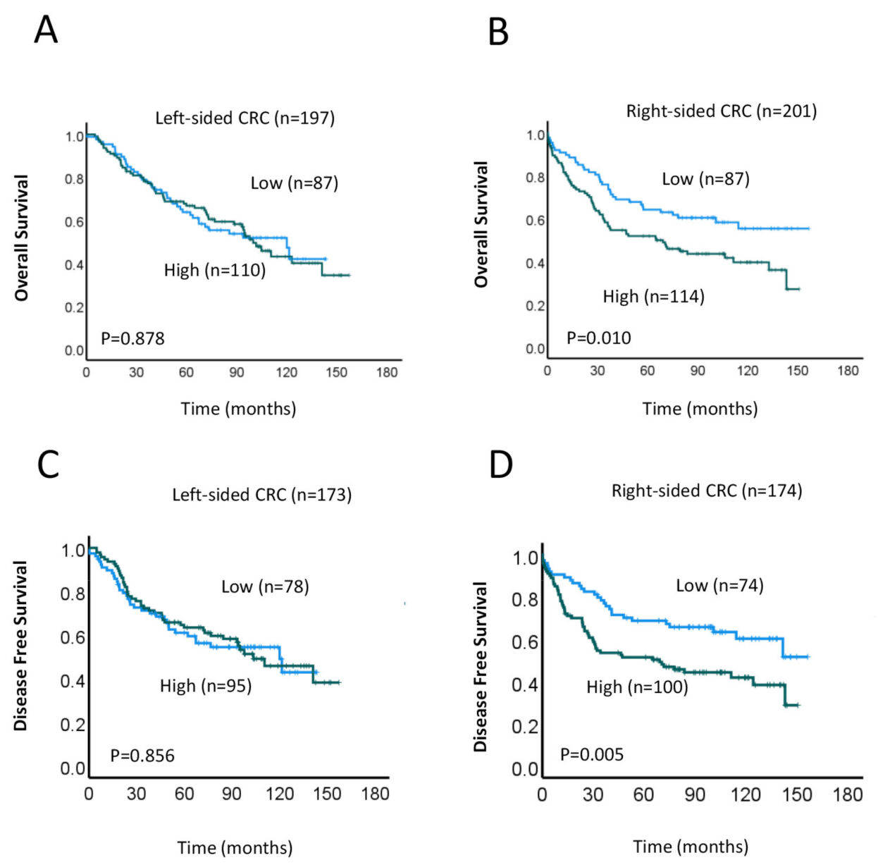
Our above findings indicate that high MRE11 expression in the TC was significantly associated with worse DFS and OS (Figure 1B,C). Given that left- and right-sided CRCs have been shown in numerous studies to be biologically and clinically distinct [39,40,41,42,43], we next measured the associations between MRE11 expression and OS and DFS in those with left- and right-sided CRC. DFS measures the time until a patient experiences disease recurrence, while OS reflects the length of time a patient survives after the start of treatment, regardless of disease status. In Kaplan–Meier analyses, we found that high expression of MRE11 within right-sided CRC was significantly correlated with worse OS (p = 0.001; Figure 2B) and DFS (p = 0.005; Figure 2D), whereas associations within left-sided CRC were not significant (OS, p = 0.878 and DFS, p = 0.856; Figure 2A,C, respectively). This finding suggests that MRE11 expression has potential prognostic value in CRC patients with right-sided tumors.
Using multivariate Cox regression analysis, we further demonstrated that high MRE11 expression (HR = 1.697, 95% CI: 1.034–2.785; p = 0.036) and LVI/PNI (HR = 1.922, 95% CI 1.122–3.293; p = 0.017) were significantly associated with worse OS in patients with right-sided tumors (Table 4), whereas no significant association was found in those with left-sided tumors. These data suggest that those markers together are strongly prognostic for OS in patients within the right-sided CRC subgroup. In contrast, LN involvement, age at/over 70 years, and adjuvant therapy treatment remained significantly associated with reduced OS for right-sided tumor subgroups (Table 4).

3.4. Prognostic Implications of MRE11 Expression in Right-Sided CRC Subgroups

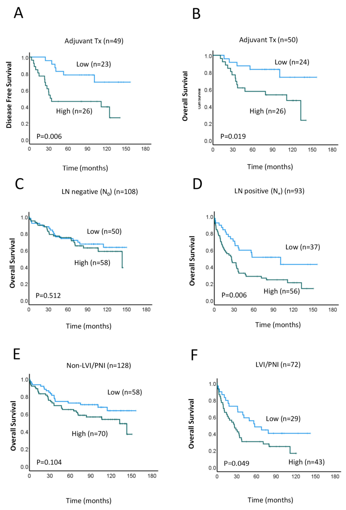
Lastly, we assessed the relationship between MRE11 expression and patient survival within the right-sided CRC subgroup. To this end, we performed Kaplan–Meier survival analyses of DFS and OS in patients with high vs. low MRE11 expression who received adjuvant treatment (combined radio- and chemotherapy treatment). Results show that patients in the high MRE11 expression group showed significantly worse OS (p = 0.019) and disease-free (p = 0.006) survival compared to those with low MRE11 expression (Figure 3A,B). We then performed similar analyses to assess OS at low vs. high MRE11 expression in patients with and without LN involvement. Interestingly, when patients were grouped in this way, high MRE11 expression was associated with worse OS and DFS only in patients with LN-positive tumors (n = 93, p = 0.006; Figure 3D and n = 70, p = 0.011; Figure S1, respectively) but not in those with LN-negative tumors (n = 108, p = 0.512; Figure 3C). Similar results were observed for LVI/PNI; that is, high MRE11 expression was associated with worse OS in patients with LVI/PNI-positive tumors (n = 72, p = 0.049; Figure 3F), but no significant association was detected for those with LVI/PNI-negative tumors (n = 128, p = 0.512; Figure 3E). These results suggest that MRE11 overexpression may contribute to tumor progression in high-risk patients with severe disease.

3.5. Clinical Significance of MRE11 in CRC

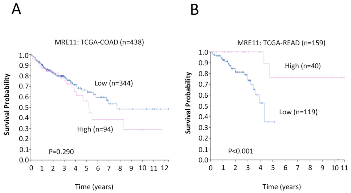
To validate our findings in this study, we also performed the experiments using RNA-seq databases via The Human Protein Atlas (HPA) (https://www.proteinatlas.org, accessed on 18 April 2023) to validate our results and we compared them with two individual cohorts from The Cancer Genome Atlas (TCGA-COAD, n = 438 and TCGA-READ, n = 159). The log-rank p-value for the Kaplan–Meier plot presented the results from the analysis of the correlation between mRNA expression level and patient survival. In colon cancer cohort (TCGA-COAD) data analysis, the Kaplan–Meier curves revealed that high MRE11 tends to correlate with worse survival probability (n = 438, p = 0.290; Figure 4A). This colon cancer cohort (TCGA-COAD) includes not only right-sided tumors (right-sided colon: ascending colon, two-thirds of the transverse colon) but also a proportion of left-sided tumors (left-sided colon: one-third of the transverse colon, sigmoid colon, and descending colon). In contrast, for the rectal cancer cohort (TCGA-READ), low MRE11 was significantly associated with poor survival probability (n = 159, p < 0.001; Figure 4B).

4. Discussion

Despite the development of improved therapies and the adoption of more stringent screening guidelines over the past decade, colon and rectal cancers remain one of the most prevalent and deadliest malignancies worldwide [1]. These cancers show a high degree of complexity regarding their molecular features and clinical behavior, underscoring the need for the identification of prognostic biomarkers to better predict patient outcomes. Here, we investigated the prognostic value of MRE11, an essential DDR protein that has been implicated in the development and evolution of numerous cancers, including CRC [26,27,28,29,36,37,44,45,46,47]. In a cohort of 408 CRC patients, we found that elevated MRE11 expression in the TC was significantly associated with poor DFS and OS, specifically for patients with right-sided CRC. Multivariate analyses further showed that both high MRE11 expression and LVI/PNI were significantly associated with worse OS in patients with right-sided tumors. Further, in this subgroup of patients, high MRE11 expression was associated with worse OS for patients with lymph node involvement and LVI/PNI, suggesting its value as a prognostic indicator for patients with right-side localized high-risk CRC.
As a member of the MRN complex, MRE11 plays critical roles as both a sensor of DNA damage and a mediator of downstream responses. Given that genomic instability is a fundamental feature of many cancers, this protein and its interacting partners have been investigated as potential prognostic indicators for CRC and other malignancies, in some cases, with conflicting results [26]. One study that analyzed a small cohort of stage III colon cancer patients reported an association between MRE11 loss and improved survival outcomes [27]. In past studies, we further found that elevated combined expression of MRE11 and ATM or of the entire MRN complex in the TC is associated with a worse prognosis and poor response to neoadjuvant radiotherapy in rectal cancer patients [28,29]. Similarly, elevated MRE11 was also associated with disease progression, poor survival outcomes, and radioresistance in oral and lung cancer patients [44,46]. Our current findings are generally consistent with these observations, suggesting that elevated MRE11 expression may contribute to worse outcomes. In contrast, a separate study by Sheridan et al. did not detect an association between MRE11 and either survival or therapeutic response in CRC [48]. Moreover, we note that in our prior study, MRE11 expression only had prognostic value in combination with ATM [28]. These discrepancies may be due, in part, to the relatively smaller sample sizes in these studies.
At present, it remains unclear how elevated MRE11 may contribute to worse survival outcomes in CRC patients. Given its essential role in DNA repair, it is possible that MRE11 helps cancer cells withstand and survive DNA-damaging assaults from chemoradiotherapy. Accordingly, we previously showed that elevated MRE11 in combination with ATM and MRN complex members was associated with poor outcomes in rectal cancer patients who received neoadjuvant therapy [28,29]. Here, only a small number of patients received neoadjuvant therapy (n = 16). However, we found that in right-side localized tumors, increased MRE11 was associated with reduced DFS and OS in patients who received adjuvant combined radio- and chemotherapy. We also note that here, as well as in our prior two studies described above [28,29], high MRE11 expression was associated with poor survival outcomes only for those with LN involvement. This may suggest that the effects of elevated MRE11 expression are more pronounced in certain contexts, such as those with advanced metastatic disease.
The right and left regions of the colorectum originate from separate sources during embryogenesis and differ in several physiological aspects, including their vasculature and lymphatic drainage systems. Accordingly, there is a growing body of evidence that right- and left-sided CRCs are also distinct in regard to their gene expression and mutational profiles, as well as outcomes, disease prognoses, and responses to various treatment regimens [41,49,50,51,52,53]. A recent study has further uncovered a possible association between tumor sidedness and MRE11, reporting that elevated MRE11 expression is associated with favorable survival outcomes for patients with left-sided CRC, as well as in the subset of left-sided CRC patients with microsatellite stability [45]. In contrast, here, we found that elevated MRE11 expression is associated with worse outcomes, specifically for CRC patients with right-side-localized CRC. Moreover, we did not detect an association between MRE11 and MMR expression. The reasons for these discrepant results are unclear, although they may result, in part, from cohort-specific or methodological differences. Intriguingly, both studies indicate that the effects of MRE11 overexpression—although opposite from one another—are dependent on tumor location, underscoring the need for future studies aimed at elucidating the precise relationship between MRE11 expression, tumor location, and clinical outcomes in CRC. Furthermore, we were able to validate our results against two individual cohorts from The Cancer Genome Atlas (TCGA). The results revealed that for both right- and left-sided cancers in the TCGA cohort, a high MRE11 expression is associated with a trend towards worse survival, but contrasting data from rectal cancers showed that low MRE11 expression confers worse outcomes. This implies that right-sided cancers by themselves have worse outcomes with high MRE11 expression, which is in keeping with our findings.
A primary strength of this study is our relatively large study population, comprising both colon and rectal cancer patients. This feature allowed us to uncover a robust and specific association between elevated MRE11 expression and worse survival outcomes in individuals with right-sided localized primary CRC. However, immunohistochemical staining for MMR proteins was only available for 87 out of the 408 subjects in our study, making it difficult to interpret our findings, which do not show an association between MRE11 expression and MMR protein expression. Prior studies have reported that dMMR can induce MRE11 mutations, leading to the formation of a truncated, nonfunction protein [36,37,38]. In turn, there is evidence that MRE11 may play a role in the MMR pathway, and loss of this protein is associated with increased MSI in dMMR CRC [54]. Increased MRN expression was also associated with lower tumor grade and improved prognosis for CRCs with functional MMR [55]. Thus, future studies are needed to better understand the prognostic relationship between the MMR pathway and MRE11 expression in the context of CRC.

5. Conclusions

In summary, immunohistochemical staining of tumor samples from 408 individuals with colon and rectal cancer followed by Cox regression analysis revealed that high MRE11 expression in the TC was significantly associated with poor DFS and OS for the overall cohort, as well as for the sub-cohort of patients with right-sided primary CRC. In multivariate analyses, both high MRE11 expression and LVI/PNI showed significant association with worse OS in patients with right-sided tumors but not those with left-sided tumors. Moreover, in patients with right-sided tumors, high MRE11 was associated with worse OS for those with lymph node involvement and LVI/PNI. Collectively, these findings suggest that MRE11 expression may represent an independent prognostic marker for patients with right-sided severe CRC, which holds potential clinical value for improving the care and management of these patients.

Supplementary Materials

The following supporting information can be downloaded at: www.mdpi.com/article/10.3390/cancers15092438/s1, Figure S1: Association between MRE11 expression and survival based on location of the primary colorectal cancer (CRC) in relation to lymph node (LN) involvement.

Author Contributions

V.H., L.C. and C.S.L. conceived and designed the research; L.C., K.W., V.L., A.A. and V.H. collected and did preliminary analysis on the data; V.H., L.C., K.W., S.H.L., W.N., M.L., T.L.R., W.C. and C.S.L. further analyzed and interpreted the data; V.H. and L.C. wrote the draft manuscript; K.W., S.H.L., V.L., W.N., M.L., T.L.R., W.C. and C.S.L. provided critical material. All authors have read and agreed to the published version of the manuscript.

Funding

This research was funded by the Ainsworth Medical Research Innovation Fund, School of Medicine, Western Sydney University, Australia, grant number 64927 and the Narellan Rotary Bowel Cancer Grant.

Institutional Review Board Statement

The study was conducted in accordance with the Declaration of Helsinki and approved the South Western Sydney Local Health District Human Research Ethics Committee (HREC Reference: HREC/14/LPOOL/186; project number 14/103; approval date 25 May 2017), Sydney, Australia.

Informed Consent Statement

Informed consent was obtained from all subjects involved in the study.

Data Availability Statement

The data presented in this study are available on request from the corresponding author. The data are not publicly available due to data size and privacy.

Acknowledgments

We acknowledge the support of the Ainsworth Medical Research Innovation Fund, School of Medicine, Western Sydney University, Australia 64927, and the Narellan Rotary Bowel Cancer Grant. We kindly thank Laura Marinelli for her editorial assistance.

Conflicts of Interest

The authors declare no conflict of interest.

References

  1. Sung, H.; Ferlay, J.; Siegel, R.L.; Laversanne, M.; Soerjomataram, I.; Jemal, A.; Bray, F. Global Cancer Statistics 2020: GLOBOCAN Estimates of Incidence and Mortality Worldwide for 36 Cancers in 185 Countries. CA A Cancer J. Clin. 2021, 71, 209–249. [Google Scholar] [CrossRef]
  2. Siegel, R.L.; Torre, L.A.; Soerjomataram, I.; Hayes, R.B.; Bray, F.; Weber, T.K.; Jemal, A. Global Patterns and Trends in Colorectal Cancer Incidence in Young Adults. Gut 2019, 68, 2179–2185. [Google Scholar] [CrossRef] [PubMed]
  3. Siegel, R.L.; Miller, K.D.; Fuchs, H.E.; Jemal, A. Cancer Statistics. CA Cancer J. Clin. 2022, 72, 7–33. [Google Scholar] [CrossRef]
  4. Dayde, D.; Tanaka, I.; Jain, R.; Tai, M.C.; Taguchi, A. Predictive and Prognostic Molecular Biomarkers for Response to Neoadjuvant Chemoradiation in Rectal Cancer. Int. J. Mol. Sci. 2017, 18, 573. [Google Scholar] [CrossRef] [PubMed]
  5. Peluso, G.; Incollingo, P.; Calogero, A.; Tammaro, V.; Rupealta, N.; Chiacchio, G.; Sandoval Sotelo, M.L.; Minieri, G.; Pisani, A.; Riccio, E.; et al. Current Tissue Molecular Markers in Colorectal Cancer: A Literature Review. Biomed. Res. Int. 2017, 2017, 2605628. [Google Scholar] [CrossRef] [PubMed]
  6. Yoo, B.C.; Yeo, S.-G. Clinical Utility of Pretreatment Prediction of Chemoradiotherapy Response in Rectal Cancer: A Review. EPMA J. 2017, 8, 61–67. [Google Scholar] [CrossRef] [PubMed]
  7. Bottarelli, L.; de’ Angelis, G.L.; Azzoni, C.; Mario, F.D.; de’ Angelis, N.; Leandro, G.; Fornaroli, F.; Gaiani, F.; Negri, F. Potential Predictive Biomarkers in Locally Advanced Rectal Cancer Treated with Preoperative Chemo-Radiotherapy. Acta Biomed. 2018, 89, 102–106. [Google Scholar] [CrossRef]
  8. Vacante, M.; Borzì, A.M.; Basile, F.; Biondi, A. Biomarkers in Colorectal Cancer: Current Clinical Utility and Future Perspectives. World J. Clin. Cases 2018, 6, 869–881. [Google Scholar] [CrossRef]
  9. Tomasini, P.P.; Guecheva, T.N.; Leguisamo, N.M.; Péricart, S.; Brunac, A.-C.; Hoffmann, J.S.; Saffi, J. Analyzing the Opportunities to Target DNA Double-Strand Breaks Repair and Replicative Stress Responses to Improve Therapeutic Index of Colorectal Cancer. Cancers 2021, 13, 3130. [Google Scholar] [CrossRef] [PubMed]
  10. Jackson, S.P.; Bartek, J. The DNA-Damage Response in Human Biology and Disease. Nature 2009, 461, 1071–1078. [Google Scholar] [CrossRef]
  11. Pećina-Šlaus, N.; Kafka, A.; Salamon, I.; Bukovac, A. Mismatch Repair Pathway, Genome Stability and Cancer. Front. Mol. Biosci. 2020, 7, 122. [Google Scholar] [CrossRef]
  12. French, A.J.; Sargent, D.J.; Burgart, L.J.; Foster, N.R.; Kabat, B.F.; Goldberg, R.; Shepherd, L.; Windschitl, H.E.; Thibodeau, S.N. Prognostic Significance of Defective Mismatch Repair and BRAF V600E in Patients with Colon Cancer. Clin. Cancer Res. 2008, 14, 3408–3415. [Google Scholar] [CrossRef]
  13. Tennstedt, P.; Fresow, R.; Simon, R.; Marx, A.; Terracciano, L.; Petersen, C.; Sauter, G.; Dikomey, E.; Borgmann, K. RAD51 Overexpression Is a Negative Prognostic Marker for Colorectal Adenocarcinoma. Int. J. Cancer 2013, 132, 2118–2126. [Google Scholar] [CrossRef]
  14. Ho, V.; Chung, L.; Singh, A.; Lea, V.; Revoltar, M.; Lim, S.H.; Tut, T.-G.; Ng, W.; Lee, M.; De Souza, P.; et al. Early Postoperative Low Expression of RAD50 in Rectal Cancer Patients Associates with Disease-Free Survival. Cancers 2017, 9, 163. [Google Scholar] [CrossRef] [PubMed]
  15. Ho, V.; Chung, L.; Singh, A.; Lea, V.; Abubakar, A.; Lim, S.H.; Chua, W.; Ng, W.; Lee, M.; Roberts, T.L.; et al. Aberrant Expression of RAD52, Its Prognostic Impact in Rectal Cancer and Association with Poor Survival of Patients. Int. J. Mol. Sci. 2020, 21, 1768. [Google Scholar] [CrossRef]
  16. Hestetun, K.E.; Aasebø, K.; Rosenlund, N.B.; Müller, Y.; Dahl, O.; Myklebust, M.P. Mismatch Repair Phenotype Determines the Implications of Tumor Grade and CDX2 Expression in Stage II–III Colon Cancer. Mod. Pathol. 2021, 34, 161–170. [Google Scholar] [CrossRef] [PubMed]
  17. Ho, V.; Chung, L.; Lim, S.H.; Ma, Y.; Wang, B.; Lea, V.; Abubakar, A.; Ng, W.; Lee, M.; Roberts, T.L.; et al. Prognostic Impact of TP53 Mutations and Tumor Mutational Load in Colorectal Cancer. Gastrointest. Disord. 2022, 4, 165–179. [Google Scholar] [CrossRef]
  18. Mladenov, E.; Magin, S.; Soni, A.; Iliakis, G. DNA Double-Strand Break Repair as Determinant of Cellular Radiosensitivity to Killing and Target in Radiation Therapy. Front. Oncol. 2013, 3, 113. [Google Scholar] [CrossRef] [PubMed]
  19. Goldstein, M.; Kastan, M.B. The DNA Damage Response: Implications for Tumor Responses to Radiation and Chemotherapy. Annu. Rev. Med. 2015, 66, 129–143. [Google Scholar] [CrossRef]
  20. Van den Bosch, M.; Bree, R.T.; Lowndes, N.F. The MRN Complex: Coordinating and Mediating the Response to Broken Chromosomes. EMBO Rep. 2003, 4, 844–849. [Google Scholar] [CrossRef]
  21. Shiloh, Y. The ATM-Mediated DNA-Damage Response: Taking Shape. Trends Biochem. Sci. 2006, 31, 402–410. [Google Scholar] [CrossRef] [PubMed]
  22. Lavin, M.F.; Kozlov, S.; Gatei, M.; Kijas, A.W. ATM-Dependent Phosphorylation of All Three Members of the MRN Complex: From Sensor to Adaptor. Biomolecules 2015, 5, 2877–2902. [Google Scholar] [CrossRef] [PubMed]
  23. Lee, K.C.; Padget, K.; Curtis, H.; Cowell, I.G.; Moiani, D.; Sondka, Z.; Morris, N.J.; Jackson, G.H.; Cockell, S.J.; Tainer, J.A.; et al. MRE11 Facilitates the Removal of Human Topoisomerase II Complexes from Genomic DNA. Biol. Open 2012, 1, 863–873. [Google Scholar] [CrossRef] [PubMed]
  24. Dmitrieva, N.I.; Malide, D.; Burg, M.B. Mre11 Is Expressed in Mammalian Mitochondria Where It Binds to Mitochondrial DNA. Am. J. Physiol. Regul. Integr. Comp. Physiol. 2011, 301, R632–R640. [Google Scholar] [CrossRef] [PubMed]
  25. Rashid, S.; Freitas, M.O.; Cucchi, D.; Bridge, G.; Yao, Z.; Gay, L.; Williams, M.; Wang, J.; Suraweera, N.; Silver, A.; et al. MLH1 Deficiency Leads to Deregulated Mitochondrial Metabolism. Cell Death Dis. 2019, 10, 795. [Google Scholar] [CrossRef] [PubMed]
  26. Situ, Y.; Chung, L.; Lee, C.S.; Ho, V. MRN (MRE11-RAD50-NBS1) Complex in Human Cancer and Prognostic Implications in Colorectal Cancer. Int. J. Mol. Sci. 2019, 20, 816. [Google Scholar] [CrossRef] [PubMed]
  27. Pavelitz, T.; Renfro, L.; Foster, N.R.; Caracol, A.; Welsch, P.; Lao, V.V.; Grady, W.B.; Niedzwiecki, D.; Saltz, L.B.; Bertagnolli, M.M.; et al. MRE11-Deficiency Associated with Improved Long-Term Disease Free Survival and Overall Survival in a Subset of Stage III Colon Cancer Patients in Randomized CALGB 89803 Trial. PLoS ONE 2014, 9, e108483. [Google Scholar] [CrossRef]
  28. Ho, V.; Chung, L.; Revoltar, M.; Lim, S.H.; Tut, T.-G.; Abubakar, A.; Henderson, C.J.; Chua, W.; Ng, W.; Lee, M.; et al. MRE11 and ATM Expression Levels Predict Rectal Cancer Survival and Their Association with Radiotherapy Response. PLoS ONE 2016, 11, e0167675. [Google Scholar] [CrossRef] [PubMed]
  29. Ho, V.; Chung, L.; Singh, A.; Lea, V.; Abubakar, A.; Lim, S.H.; Ng, W.; Lee, M.; de Souza, P.; Shin, J.-S.; et al. Overexpression of the MRE11-RAD50-NBS1 (MRN) Complex in Rectal Cancer Correlates with Poor Response to Neoadjuvant Radiotherapy and Prognosis. BMC Cancer 2018, 18, 869. [Google Scholar] [CrossRef]
  30. Ionov, Y.; Peinado, M.A.; Malkhosyan, S.; Shibata, D.; Perucho, M. Ubiquitous Somatic Mutations in Simple Repeated Sequences Reveal a New Mechanism for Colonic Carcinogenesis. Nature 1993, 363, 558–561. [Google Scholar] [CrossRef]
  31. Peltomäki, P. Deficient DNA Mismatch Repair: A Common Etiologic Factor for Colon Cancer. Hum. Mol. Genet. 2001, 10, 735–740. [Google Scholar] [CrossRef]
  32. Sinicrope, F.A.; Foster, N.R.; Thibodeau, S.N.; Marsoni, S.; Monges, G.; Labianca, R.; Kim, G.P.; Yothers, G.; Allegra, C.; Moore, M.J.; et al. DNA Mismatch Repair Status and Colon Cancer Recurrence and Survival in Clinical Trials of 5-Fluorouracil-Based Adjuvant Therapy. J. Natl. Cancer Inst. 2011, 103, 863–875. [Google Scholar] [CrossRef] [PubMed]
  33. Le, D.T.; Uram, J.N.; Wang, H.; Bartlett, B.R.; Kemberling, H.; Eyring, A.D.; Skora, A.D.; Luber, B.S.; Azad, N.S.; Laheru, D.; et al. PD-1 Blockade in Tumors with Mismatch-Repair Deficiency. N. Engl. J. Med. 2015, 372, 2509–2520. [Google Scholar] [CrossRef] [PubMed]
  34. Sinicrope, F.A.; Shi, Q.; Allegra, C.J.; Smyrk, T.C.; Thibodeau, S.N.; Goldberg, R.M.; Meyers, J.P.; Pogue-Geile, K.L.; Yothers, G.; Sargent, D.J.; et al. Association of DNA Mismatch Repair and Mutations in BRAF and KRAS With Survival After Recurrence in Stage III Colon Cancers: A Secondary Analysis of 2 Randomized Clinical Trials. JAMA Oncol. 2017, 3, 472–480. [Google Scholar] [CrossRef] [PubMed]
  35. Du, F.; Liu, Y. Predictive Molecular Markers for the Treatment with Immune CheckpoInt. Inhibitors in Colorectal Cancer. J. Clin. Lab. Anal. 2022, 36, e24141. [Google Scholar] [CrossRef] [PubMed]
  36. Giannini, G.; Ristori, E.; Cerignoli, F.; Rinaldi, C.; Zani, M.; Viel, A.; Ottini, L.; Crescenzi, M.; Martinotti, S.; Bignami, M.; et al. Human MRE11 Is Inactivated in Mismatch Repair-Deficient Cancers. EMBO Rep. 2002, 3, 248–254. [Google Scholar] [CrossRef] [PubMed]
  37. Ottini, L.; Falchetti, M.; Saieva, C.; De Marco, M.; Masala, G.; Zanna, I.; Paglierani, M.; Giannini, G.; Gulino, A.; Nesi, G.; et al. MRE11 Expression Is Impaired in Gastric Cancer with Microsatellite Instability. Carcinogenesis 2004, 25, 2337–2343. [Google Scholar] [CrossRef] [PubMed]
  38. Miquel, C.; Jacob, S.; Grandjouan, S.; Aimé, A.; Viguier, J.; Sabourin, J.-C.; Sarasin, A.; Duval, A.; Praz, F. Frequent Alteration of DNA Damage Signalling and Repair Pathways in Human Colorectal Cancers with Microsatellite Instability. Oncogene 2007, 26, 5919–5926. [Google Scholar] [CrossRef] [PubMed]
  39. Bufill, J.A. Colorectal Cancer: Evidence for Distinct Genetic Categories Based on Proximal or Distal Tumor Location. Ann. Intern. Med. 1990, 113, 779–788. [Google Scholar] [CrossRef] [PubMed]
  40. Gervaz, P.; Bucher, P.; Morel, P. Two Colons-Two Cancers: Paradigm Shift and Clinical Implications. J. Surg. Oncol. 2004, 88, 261–266. [Google Scholar] [CrossRef]
  41. Shen, H.; Yang, J.; Huang, Q.; Jiang, M.-J.; Tan, Y.-N.; Fu, J.-F.; Zhu, L.-Z.; Fang, X.-F.; Yuan, Y. Different Treatment Strategies and Molecular FeatuRes. between Right-Sided and Left-Sided Colon Cancers. World J. Gastroenterol. 2015, 21, 6470–6478. [Google Scholar] [CrossRef] [PubMed]
  42. Jung, M.-K.; Shin, U.S.; Ki, Y.-J.; Kim, Y.-B.; Moon, S.-M.; Sung, S.-J. Is the Location of the Tumor Another Prognostic Factor for Patients with Colon Cancer? Ann. Coloproctol. 2017, 33, 210–218. [Google Scholar] [CrossRef] [PubMed]
  43. Baran, B.; Mert Ozupek, N.; Yerli Tetik, N.; Acar, E.; Bekcioglu, O.; Baskin, Y. Difference Between Left-Sided and Right-Sided Colorectal Cancer: A Focused Review of Literature. Gastroenterol. Res. 2018, 11, 264–273. [Google Scholar] [CrossRef] [PubMed]
  44. Yuan, S.-S.F.; Hou, M.-F.; Hsieh, Y.-C.; Huang, C.-Y.; Lee, Y.-C.; Chen, Y.-J.; Lo, S. Role of MRE11 in Cell Proliferation, Tumor Invasion, and DNA Repair in Breast Cancer. J. Natl. Cancer Inst. 2012, 104, 1485–1502. [Google Scholar] [CrossRef]
  45. Fan, C.-W.; Kopsida, M.; Liu, Y.-B.; Zhang, H.; Gao, J.-F.; Arbman, G.; Cao, S.-Y.-W.; Li, Y.; Zhou, Z.-G.; Sun, X.-F. Prognostic Heterogeneity of MRE11 Based on the Location of Primary Colorectal Cancer Is Caused by Activation of Different Immune Signals. Front. Oncol. 2020, 9, 1465. [Google Scholar] [CrossRef] [PubMed]
  46. Wang, Y.-Y.; Chen, Y.-K.; Lo, S.; Chi, T.-C.; Chen, Y.-H.; Hu, S.C.-S.; Chen, Y.-W.; Jiang, S.S.; Tsai, F.-Y.; Liu, W.; et al. MRE11 Promotes Oral Cancer Progression through RUNX2/CXCR4/AKT/FOXA2 Signaling in a Nuclease-Independent Manner. Oncogene 2021, 40, 3510–3532. [Google Scholar] [CrossRef]
  47. Horak, J.; Dolnikova, A.; Cumaogullari, O.; Cumova, A.; Navvabi, N.; Vodickova, L.; Levy, M.; Schneiderova, M.; Liska, V.; Andera, L.; et al. MiR-140 Leads to MRE11 Downregulation and Ameliorates Oxaliplatin Treatment and Therapy Response in Colorectal Cancer Patients. Front. Oncol. 2022, 12, 959407. [Google Scholar] [CrossRef] [PubMed]
  48. Sheridan, J.; Tosetto, M.; Gorman, J.; O’Donoghue, D.; Sheahan, K.; Hyland, J.; Mulcahy, H.; Gibbons, D.; O’Sullivan, J. Effects of Radiation on Levels of DNA Damage in Normal Non-Adjacent Mucosa from Colorectal Cancer Cases. J. Gastrointest Cancer 2013, 44, 41–45. [Google Scholar] [CrossRef]
  49. Glebov, O.K.; Rodriguez, L.M.; Nakahara, K.; Jenkins, J.; Cliatt, J.; Humbyrd, C.-J.; DeNobile, J.; Soballe, P.; Simon, R.; Wright, G.; et al. Distinguishing Right from Left Colon by the Pattern of Gene Expression. Cancer Epidemiol. Biomark. Prev. 2003, 12, 755–762. [Google Scholar]
  50. Muzny, D.M.; Bainbridge, M.N.; Chang, K.; Dinh, H.H.; Drummond, J.A.; Fowler, G.; Kovar, C.L.; Lewis, L.R.; Morgan, M.B.; Newsham, I.F.; et al. Comprehensive Molecular Characterization of Human Colon and Rectal Cancer. Nature 2012, 487, 330–337. [Google Scholar] [CrossRef]
  51. Koestler, D.C.; Li, J.; Baron, J.A.; Tsongalis, G.J.; Butterly, L.F.; Goodrich, M.; Lesseur, C.; Karagas, M.R.; Marsit, C.J.; Moore, J.H.; et al. Distinct Patterns of DNA Methylation in Conventional Adenomas Involving the Right and Left Colon. Mod. Pathol. 2014, 27, 145–155. [Google Scholar] [CrossRef] [PubMed]
  52. Missiaglia, E.; Jacobs, B.; D’Ario, G.; Di Narzo, A.F.; Soneson, C.; Budinska, E.; Popovici, V.; Vecchione, L.; Gerster, S.; Yan, P.; et al. Distal and Proximal Colon Cancers Differ in Terms of Molecular, Pathological, and Clinical Features. Ann. Oncol. 2014, 25, 1995–2001. [Google Scholar] [CrossRef] [PubMed]
  53. Brulé, S.Y.; Jonker, D.J.; Karapetis, C.S.; O’Callaghan, C.J.; Moore, M.J.; Wong, R.; Tebbutt, N.C.; Underhill, C.; Yip, D.; Zalcberg, J.R.; et al. Location of Colon Cancer (Right-Sided versus Left-Sided) as a Prognostic Factor and a Predictor of Benefit from Cetuximab in NCIC CO.17. Eur. J. Cancer 2015, 51, 1405–1414. [Google Scholar] [CrossRef] [PubMed]
  54. Vo, A.T.; Zhu, F.; Wu, X.; Yuan, F.; Gao, Y.; Gu, L.; Li, G.-M.; Lee, T.-H.; Her, C. HMRE11 Deficiency Leads to Microsatellite Instability and Defective DNA Mismatch Repair. EMBO Rep. 2005, 6, 438–444. [Google Scholar] [CrossRef] [PubMed]
  55. Gao, J.; Zhang, H.; Arbman, G.; Sun, X.-F. RAD50/MRE11/NBS1 Proteins in Relation to Tumour Development and Prognosis in Patients with Microsatellite Stable Colorectal Cancer. Histol. HistoPathol. 2008, 23, 1495–1502. [Google Scholar] [CrossRef]
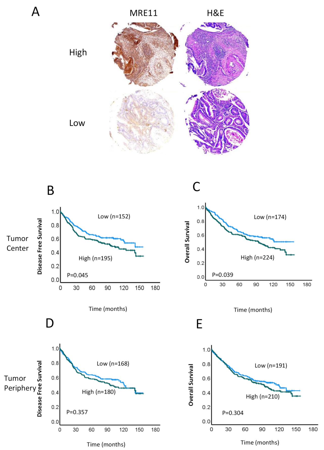
Figure 1. MRE11 expression in colorectal cancer (CRC) tissue samples. (A) Representative examples of typical nuclear staining of MRE11 in tumor cells, scored as high or low expression, and corresponding hematoxylin and eosin (H&E) staining are shown. Images were taken at 10× magnification. (40× magnification; scale bar, 50 μm). (BE) Kaplan–Meier analyses of disease-free survival (DFS) (B,D) and overall survival (OS) (C,E) for patients with high (green line) and low (blue line) MRE11 protein expression in the tumor center (B,C) and tumor periphery (D,E).
Cancers 15 02438 g001
Figure 2. Relationship between MRE11 expression in the TC and survival, based on location of the primary CRC. (AD) Kaplan–Meier analysis of OS (A,B) and DFS (C,D) in patients with high (green line) or low (blue line) MRE11 expression in left-sided (A,C) and right-sided (B,D) colorectal cancers.
Cancers 15 02438 g002
Figure 3. Association between MRE11 expression and prognosis in right-sided CRC. (A,B) Kaplan–Meier survival analyses of DFS (A) and OS (B) in patients with high (green line) or low (blue line) MRE11 expression treated with adjuvant chemo/radiotherapy. (CF) Kaplan–Meier curves of OS in patients with high (green line) or low (blue line) MRE11 expression in lymph node (LN)-negative (C), LN-positive (D) tumors, lymphovascular and perineural invasion (LVI/PNI)-negative (E) and LVI/PNI-positive (F) tumors.
Cancers 15 02438 g003
Figure 4. Kaplan–Meier survival analyses comparing survival probability in patients with low MRE11 and high MRE11. Differences between the groups were analyzed by the log-rank test, and p-values are shown. (A) Survival analyses for colon cancer cohort, and (B) for rectal cancer cohort comparing survival probabilities in patients with high (pink line) or low (blue line) MRE11. The cases were provided by The Cancer Genome Atlas (TCGA-COAD and TCGA-READ).
Cancers 15 02438 g004
Table 1. Patient characteristics for subjects included in this study.
CharacteristicsAll Patients (n = 408)%
Age median70 (23–96 years)
  <7019046.6
  ≥7021853.4
Sex
  Male21953.7
  Female18946.3
Tumor stage
  T1, T28119.9
  T3, T432780.1
Node stage
  N021352.2
  N1, N219547.8
Metastasis stage
  M035386.5
  M15513.5
Differentiation
  Well/moderate33782.5
  Poor7117.5
LVI/PNI
  Absent26565
  Present14335
Primary tumor site (Right/Left)
  Right20951.2
  Left19948.8
Treatment
Neoadjuvant therapy
  No39296.1
  Yes163.9
Adjuvant therapy
  No28168.9
  Yes12731.1
MMR deficiency
Total8721.3 (87/408)
  No6473.6
  Yes2326.4
Abbreviations: LVI/PNI, lymphovascular and perineural invasion; MMR, mismatch repair.
Table 2. Associations between MRE11 expression in the tumor center and tumor periphery and clinicohistopathological data.
Tumor CentreTumor Periphery
Low (%)High (%)p-ValueLow (%)High (%)p-Value
SexMale53.454.00.92054.551.90.618
Female46.646.0 45.548.1
Age<7044.847.30.68546.144.30.719
≥7055.252.7 53.955.7
Tumor stageT1–224.130.00.17220.921.00.998
T3–475.970.0 79.179.0
Node stageNegative55.749.60.22751.851.90.988
Positive44.350.4 48.248.1
Metastasis stageM087.487.10.92988.085.70.508
M112.612.9 12.014.3
DifferentiationWell/moderate83.382.10.75682.282.40.962
Poor16.717.9 17.817.6
LVI/PNIAbsent67.862.80.29763.266.20.562
Present32.237.2 36.833.8
Adjuvant therapyNo62.165.30.53563.365.20.714
Yes37.934.7 36.734.8
Neoadjuvant therapyNo95.496.40.60594.897.60.132
Yes4.63.6 5.22.4
MLH1 aNormal IHC97.697.80.96110097.10.209
Loss of staining2.42.2 02.9
MSH2Normal IHC95.297.70.52998.1970.732
Loss of staining4.82.3 1.93
MSH6Normal IHC71.484.10.15773.684.80.221
Loss of staining28.615.9 26.415.2
PMS2Normal IHC76.988.10.1847593.80.031
Loss of staining23.111.9 256.2
dMMR bNo64.3800.10169.879.40.322
Yes35.720 30.220.6
a Total available data for MMR protein staining (n = 87); b MMR deficiency. Abbreviations: IHC, immunohistochemistry; LVI/PNI, lymphovascular and perineural invasion; MMR, mismatch repair.
Table 3. Cox regression analyses of MRE11 expression and clinicopathological features with OS.
UnivariateMultivariate
n (%)HR95% CIp-ValueHR95% CIp-Value
MRE11 TC a
  High54.91.3521.015–1.8010.0401.4331.026–2.0020.035
  Low45.1
Sex
  Male53.71.190.905–1.5650.2140.9120.651–1.2670.584
  Female46.3
Age
  <7046.61.8281.375–2.401<0.0012.3141.576–3.396<0.001
  ≥7053.4
Tumor stage
  T1, T219.92.1031.393–3.173<0.0011.2920.807–2.0700.286
  T3, T480.1
Node stage
  Negative52.22.2861.727–3.027<0.0013.3792.263–5.047<0.001
  Positive47.8
Metastasis stage
  M086.55.8314.173–8.150<0.0012.7470.370–20.3670.323
  M113.5
Differentiation
  Well/moderate82.51.5641.119–2.1860.0091.3430.648–2.7820.427
  Poor17.5
LVI/PNI
  Absent651.9651.488–2.594<0.0011.3391.314–4.0750.112
  Present35
Adjuvant therapy
  No68.90.6030.404–0.8060.0050.2880.184–0.451<0.001
  Yes31.1
Neoadjuvant therapy
  No96.11.0490.538–2.0480.8882.2070.996–4.8880.151
  Yes3.9
a MRE11 expression in the tumor center. Abbreviations: CI, confidence interval; CRC, colorectal cancer; HR, hazard ratio; LVI/PNI, lymphovascular and perineural invasion; OS, overall survival; TC, tumor center.
Table 4. Multivariate analyses of MRE11 expression and clinicopathological features with OS, based on location of the right-sided CRC tumor.
Right-Sided CRC
HR95% CIp-Value
MRE11
High1.6971.034–2.7850.036
Low
Sex
Male0.8820.546–1.4170.598
Female
Age
<702.1141.213–3.685<0.001
≥70
Tumor stage
T1, T20.9760.487–1.9550.944
T3, T4
Node stage
Negative3.8762.237–6.716<0.001
Positive
Metastasis stage
M06.650.786–16.950.082
M1
Differentiation
Well/moderate1.4790.848–2.5810.168
Poor
LVI/PNI
Absent1.9221.122–3.2930.017
Present
Adjuvant therapy
No0.2920.154–0.565<0.001
Yes
Abbreviations: CI, confidence interval; CRC, colorectal cancer; HR, hazard ratio; LVI/PNI, lymphovascular and perineural invasion; OS, overall survival.
Disclaimer/Publisher’s Note: The statements, opinions and data contained in all publications are solely those of the individual author(s) and contributor(s) and not of MDPI and/or the editor(s). MDPI and/or the editor(s) disclaim responsibility for any injury to people or property resulting from any ideas, methods, instructions or products referred to in the content.