Article

Myeloid NGS Analyses of Paired Samples from Bone Marrow and Peripheral Blood Yield Concordant Results: A Prospective Cohort Analysis of the AGMT Study Group

by
Bettina Jansko-Gadermeir
1,2,3,4,5,6,
Michael Leisch
1,2,3,7,
Franz J. Gassner
1,2,3,4,
Nadja Zaborsky
1,2,3,4,
Thomas Dillinger
2,5,
Sonja Hutter
2,5,
Angela Risch
3,6,
Thomas Melchardt
1,2,3,7,
Alexander Egle
1,2,3,4,7,
Manuel Drost
8,
Julian Larcher-Senn
8,
Richard Greil
1,2,3,4,5,7 and
Lisa Pleyer
1,2,3,4,5,7,*
1
Salzburg Cancer Research Institute (SCRI), Center for Clinical Cancer and Immunology Trials (CCCIT), 5020 Salzburg, Austria
2
3rd Medical Department with Hematology, Medical Oncology, Hemostaseology, Rheumatology and Infectiology, Oncologic Center, Paracelsus Medical University, 5020 Salzburg, Austria
3
Cancer Cluster Salzburg (CCS), 5020 Salzburg, Austria
4
Laboratory of Immunological and Molecular Cancer Research (LIMCR), 5020 Salzburg, Austria
5
Laboratory for Molecular Cytology (MZL), 5020 Salzburg, Austria
6
Department of Biosciences and Medical Biology, Allergy-Cancer-BioNano Research Centre, University of Salzburg, 5020 Salzburg, Austria
7
Austrian Group for Medical Tumor Therapy (AGMT) Study Group, 1180 Vienna, Austria
8
Assign Data Management and Biostatistics GmbH, 6020 Innsbruck, Austria
*
Author to whom correspondence should be addressed.
Cancers 2023, 15(8), 2305; https://doi.org/10.3390/cancers15082305
Submission received: 7 March 2023 / Revised: 10 April 2023 / Accepted: 11 April 2023 / Published: 14 April 2023

Abstract

:

Simple Summary

Myelodysplastic neoplasms and acute myeloid leukemias are often caused by gene mutations. Next generation sequencing (NGS) has become indispensable for mutational assessment and is widely used for disease classification, risk stratification, prognostication, and disease monitoring. In these diseases, the bone marrow blast percentage and hence bone marrow specimen remain pre-requisite for the above. Several groups, including ours, report that bone marrow evaluations, which can be painful and time-consuming, are only performed in ~50% of patients during follow-up outside of clinical trials, indicating a clinical need for surrogate samples. We therefore aimed to compare NGS results for paired bone marrow and peripheral blood samples. Our results clearly show, in a prospective setting, that sequential molecular analyses of peripheral blood specimens can be reliably used to molecularly classify and monitor myeloid neoplasms without loss of sensitivity or specificity, even in the absence of circulating blasts or in neutropenic patients. Hence, a bone marrow evaluation for the purpose of monitoring of mutations is not necessary.

Abstract

Background: Next generation sequencing (NGS) has become indispensable for diagnosis, risk stratification, prognostication, and monitoring of response in patients with myeloid neoplasias. Guidelines require bone marrow evaluations for the above, which are often not performed outside of clinical trials, indicating a need for surrogate samples. Methods: Myeloid NGS analyses (40 genes and 29 fusion drivers) of 240 consecutive, non-selected, prospectively collected, paired bone marrow/peripheral blood samples were compared. Findings: Very strong correlation (r = 0.91, p < 0.0001), high concordance (99.6%), sensitivity (98.8%), specificity (99.9%), positive predictive value (99.8%), and negative predictive value (99.6%) between NGS analyses of paired samples was observed. A total of 9/1321 (0.68%) detected mutations were discordant, 8 of which had a variant allele frequency (VAF) ≤ 3.7%. VAFs between peripheral blood and bone marrow samples were very strongly correlated in the total cohort (r = 0.93, p = 0.0001) and in subgroups without circulating blasts (r = 0.92, p < 0.0001) or with neutropenia (r = 0.88, p < 0.0001). There was a weak correlation between the VAF of a detected mutation and the blast count in either the peripheral blood (r = 0.19) or the bone marrow (r = 0.11). Interpretation: Peripheral blood samples can be used to molecularly classify and monitor myeloid neoplasms via NGS without loss of sensitivity/specificity, even in the absence of circulating blasts or in neutropenic patients.

1. Introduction

Acute myeloid leukemia, myelodysplastic neoplasms, and chronic myelomonocytic leukemia are malignant clonal hematologic neoplasms arising within the bone marrow [1]. These diseases are caused by genetic alterations in bone marrow stem cells leading to expansion of malignant blasts and hematopoietic insufficiency resulting in peripheral blood cytopenia [1,2,3]. While acute myeloid leukemia, myelodysplastic neoplasms, and chronic myelomonocytic leukemia are different disease entities, they share similar clinical features (e.g., accumulation of blasts, cytopenia, or recurrent infections) and are often treated similarly [2,4,5,6]. Diagnosis of acute myeloid leukemia, myelodysplastic neoplasms, and chronic myelomonocytic leukemia requires morphologic examination of the peripheral blood and bone marrow to quantify the amount of blasts (and blast equivalents), other cells (i.e., monocytes), and morphologic features (i.e., cellular dysplasia, cytopenia, presence of ring sideroblasts) [1,3,7,8,9,10].
Genetic analysis of the bone marrow has been performed for several decades using conventional karyotyping [1,7,8]. With the advances and widespread availability of NGS techniques, large scale genomic testing has been introduced into daily clinical practice in the last five to ten years. The broad application of next generation sequencing (NGS) techniques in recent years has resulted in increasing knowledge about the mutational landscape of myeloid neoplasias [11,12,13,14,15,16,17,18,19,20]. This is highly reflected in the recently published 5th edition of the World Health Organization Classification of Haematolymphoid Tumours [7] as well as in the International Consensus Classification of Myeloid Neoplasms and Acute Leukemias [8]. Herein, diseases that were historically defined by morphology only are now classified mainly on a molecular basis. This allows for better inter-observer comparison and forms the basis for targeted and tailored treatment approaches. Thus, both classifications increasingly rely on defining diseases and disease subtypes based on recurrent genetic abnormalities with prognostic and in some cases therapeutic relevance.
Morphologic bone marrow examinations are mandatory for (i) accurate diagnosis and disease classification (e.g., [7,8,11]), (ii) risk stratification, which typically also includes the bone marrow blast percentage as well as conventional cytogenetics and/or fluorescence in situ hybridization from bone marrow specimen (e.g., [21,22,23]), and (iii) response assessment/disease monitoring during treatment (e.g., [22,23,24,25,26,27]). In addition, most large-scale reports on molecular data in these diseases were generated from bone marrow specimen only and hence cannot eo ipso be extrapolated to results obtained in the peripheral blood [e.g., [28,29,30,31,32]). All current response criteria for these diseases require a bone marrow evaluation for determining adequate therapeutic response [8,22,23,24,25,26]. Histologic examination has to be awaited, which can be time consuming. However, rapid identification of genetic abnormalities with prognostic and therapeutic implications is becoming increasingly important for the treatment of patients with myeloid neoplasias in general, and of acute myeloid leukemia in particular, since treatment decisions heavily rely on the genomic disease profile (i.e., integration of targeted agents to standard induction therapy). Despite that, bone marrow aspirations are not always feasible in clinical practice (i.e., dry tap or insufficient aspiration) and, after having established the initial diagnosis, are only performed in approximately 50% of treated patients with myelodysplastic neoplasms, chronic myelomonocytic leukemia, or acute myeloid leukemia during follow-up [33,34,35,36]. Hence, a real-world clinical need exists to be able to use an alternative or surrogate sample, to be able to obtain information on the mutational status of a patient’s disease. In this regard, research groups around the world have assessed whether the genetic information required for an integrated diagnosis and/or treatment monitoring may also be obtained from analysis of the peripheral blood of patients with various hematologic malignancies (Table 1 and Table S1) [20,37,38,39,40,41,42,43,44,45,46,47,48].
This monocentric, prospective analysis expands on the work by others, by evaluating the concordance of peripheral blood and bone marrow mutational analyses in a larger cohort of non-selected, consecutive adult patients treated at our institute for whom a myeloid NGS analysis was requested by the treating physicians. The commercially available AmpliSeq myeloid panel from Illumina including 40 DNA target genes and 29 RNA drivers was used with a high median (interquartile range (IQR)) read depth of 6116 (3675–9866), as well as a high minimum read depth of 500. Our main aim was to ascertain whether myeloid NGS analyses from peripheral blood could safely be used as an alternative to bone marrow samples to identify gene mutations and guide treatment decisions.

2. Materials and Methods

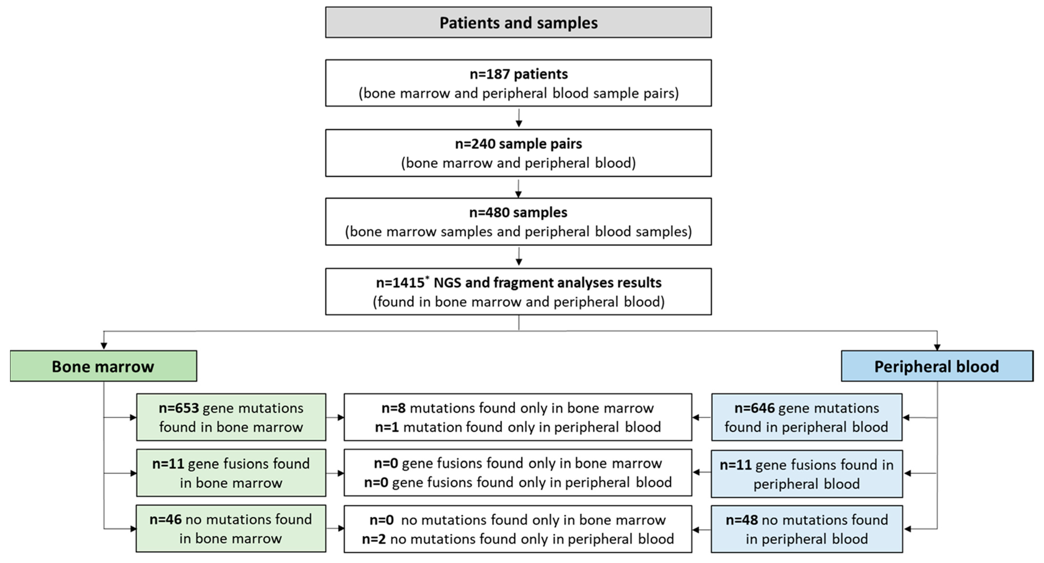
2.1. Patients and Cohort

The data collection and cleaning period was from 18 December 2019 to 11 November 2022. Database lock (last patient in) was on 13 October 2022. From 18 December 2019 to 11 November 2022 paired bone marrow and peripheral blood samples were obtained from non-selected, consecutive patients included in the Austrian Myeloid Registry (HMA cohort) and patients who were diagnosed and/or treated at the 3rd Medical Department, Laboratory for Molecular Cytology, Salzburger Landeskliniken (SALK), Paracelsus Medical University (PMU) (SALK cohort) and who had given written informed consent to this study. Patients with either a myeloid neoplasm or patients who underwent bone marrow examination during workup of a suspected myeloid disease were included. The sole inclusion criteria were the availability of a paired bone marrow and peripheral blood sample and the routine request for a myeloid NGS analysis of both materials by the treating physicians.
The Austrian Myeloid Registry of the Austrian Group of Medical tumor Therapy (AGMT) (NCT04438889; ethics committee approval 415-E/2581/Sept-2020) is a multicenter database that includes patients with acute myeloid leukemia, myelodysplastic neoplasms, and chronic myelomonocytic leukemia. This registry adheres to published quality guidelines of the U.S. Department of Health and Human Services Agency for Healthcare Research and Quality. All patients alive at the time of data entry into the electronic case report form provided written informed consent. Further details about this registry have been published previously [49].
All patients not included in the registry provided written informed consent that the NGS analyses may be performed, and that the data may be used for scientific analyses and publications.

2.2. Mutational Analyses

DNA was isolated from non-selected consecutive paired peripheral blood samples and bone marrow aspirates using Maxwell® RSC Blood DNA Kit (Promega, Madison, WI, USA). The DNA concentration was measured using the QuantiFluor® dsDNA System from Promega. The RNA concentration was measured using the QuantiFluor® RNA System from Promega. The library preparation was performed using AmpliSeq™ library plus for Illumina® using the AmpliSeq™ myeloid panel from Illumina® (San Diego, CA, USA) according to the protocol of the AmpliSeq™ for Illumina® myeloid panel reference guide. This NGS panel covers 40 DNA target genes (23 hotspot regions and 17 full genes) and 29 RNA fusion driver genes as detailed in Table S2. All NGS libraries were quantified using the QuantiFluor® dsDNA System from Promega. Buccal swab DNA NGS analysis was performed in patients harboring mutations with an allele frequency between 45 and 55%, or 95 and 100% to distinguish between germline and somatic mutation. The targeted sequencing was performed on the Illumina MiSeqDx and NextSeq550. Depending on the instrument, the starting concentration was 20 pM for the MiSeqDx and 1.6 pM for the NextSeq550.
A separate FLT3-ITD mutation analysis was performed using the LeukoStrat® FLT3 Mutation Assay 2.0 from Invivoscribe (San Diego, CA, USA). This kit is based on fragment analysis using the ABI3500 genetic analyzer and the Genemapper 6 for visualization.

2.3. Bioinformatic Analyses

The datasheets for sequencing were written using the Illumina local run manager. For read alignment and variant calling the following software tools were used: Local Run Manager DNA Amplicon Analysis Module (3.24.1.8+), Burrow-Wheeler Aligner Maximal Exact Match (BWA-MEM) [50,51] Whole-Genome Aligner (0.7.9a-isis-1.0.2), Pisces Variant Caller (5.2.9.23), Illumina Annotation Engine (2.0.11-0- g7fb24a09), binary alignment map (BAM) Metrics (v.0.0.22), and Sequence Alignment Map (SAM) tools (0.1.19-isis-1.0.3) [52]. Reads were aligned to the human genome hg19. Variant annotation and filtering were performed using Illumina VariantStudio v3.0 setting sensitivity to 1% variant allele frequency (VAF). Single nucleotide polymorphisms (SNPs) were excluded using the Exome Aggregation Consortium (ExAc) Database. Variants present in >1% of the general population, as indicated in the ExAc database, were defined as SNPs and were not included in the final data analysis. All variants were checked for sequencing artefacts using the BAM files and the Integrative Genomics Viewer (IGV) version 2.3.97 [53] for visualization. In case of discordance between bone marrow and peripheral blood samples, the discordant mutations were manually reviewed using the IGV and the corresponding BAM file. The sensitivity of the targeted NGS approach was set at 1% variant allele frequency (VAF), as it is not possible to set a lower threshold in the Local Run Manager. However, when searched manually for the mutations they could be found at lower thresholds. For mutation annotation the Catalogue of Somatic Mutations in Cancer (COSMIC) database and the database of single nucleotide polymorphism (dbSNP) were used. Variants annotated as benign or likely benign were not included in this study.

2.4. Statistical Analyses

To assess the correlation between mean VAF of the bone marrow and the mean VAF of the peripheral blood the Spearman correlation was used. Results were further illustrated by simple regression lines. If the VAF in the peripheral blood and in the bone marrow did not exactly match, the regression line deviated from the bisecting line.
Results were reported as significant when p ≤ 0.05 and a very strong correlation was reported when 0.90 < |r| < 1, a strong correlation was reported when 0.70 < |r| < 0.89, 0.40 < |r| < 0.69 indicates a moderate correlation, a weak correlation was reported when 0.10 < |r| < 0.39, and 0.00 < |r| < 0.10 indicates a negligible correlation [54]. Sensitivity analyses were performed using Kendall’s tau and by performing stratifications of paired samples (i.e. analyzing only those drawn on the same day) and of mutations (i.e., excluding found aberrations with a VAF < 5%).
As suggested by Bland et al. in the Lancet [55], a graphical representation of the agreement between the two methods of clinical measurement, i.e., mean of BMVAF and PBVAF versus (vs) the difference of BMVAF and PBVAF, was generated. Only if both values (BMVAF and PBVAF) were available, the mean and difference were calculated.
Assign Data Management and Biostatistics GmbH (Innsbruck, Austria) performed statistical analyses with SAS® 9.3. Life and Medical Sciences Institute, University of Bonn performed statistical analyses including mixed-effect linear modelling with Python 3.8.12.

3. Results

3.1. Patient Characteristics

Data from 187 non-selected, consecutive patients (108 patients from the HMA cohort and 79 from the SALK cohort) with paired bone marrow and peripheral samples were prospectively analyzed. Of these, 30 patients had more than one paired bone marrow and peripheral samples, so that the total number of paired bone marrow and peripheral samples was 240 (Figure 1). Patients with serial sample pairs had a median of 2 (IQR: 2–3, min–max: 2–5) sample pairs during the observation period. In total, 216 of 240 (90.0%) sample pairs were drawn on the same day, 5 (2.1%), 10 (4.2%), 8 (3.3%), and 1 (0.4%) sample pairs were drawn 1, >1–4, >4–8, and >8 week(s) apart, respectively.
Patient characteristics on the day of the bone marrow assessment are shown in Table 2. The median age of the entire cohort was 70 years (IQR = 58.0–77.7); 100 (53.5%) were male. Forty-six patients (24.6%) had a diagnosis of acute myeloid leukemia, 43 (23.0%) had myelodysplastic syndromes, 15 (8.0%) had a myelodysplastic/myeloproliferative overlap syndrome (MDS/MPN), 33 (17.6%) had a myeloproliferative neoplasm (MPN), and 50 (26.7%) did not have a myeloid neoplasm (termed “others” in Table 2). Ninety-one (48.7%) patients had a white blood cell count < 4000 G/L and 133 (71.1%) had no blasts in the peripheral blood count on the day of bone marrow aspiration. Treatment-related disease was present in 17 (9.1%) of 187 patients and 19 (10.2%) patients had a complex or monosomal karyotype, respectively.

3.2. Mutational Analyses—Overview

A total of 1321 variants were detected in 480 samples including 22 fusion genes, 910 single nucleotide variants (SNV), 186 insertions, 191 deletions and 12 multiple nucleotide polymorphisms (MNP). Off all detected variants, 994 had a COSMIC and/or dbSNP entry and 327 were not described in either of these two databases.
Mutations were found in 38 different genes in the bone marrow and 37 different genes in the peripheral blood, respectively. No mutations were detected in ABL1 or CSFR3 in all samples analyzed. In 41 patients and 94 samples no pathogenic mutations were detected as shown in Figure 1. The mean (SD) [min–max] number of mutations found in the bone marrow was 2.8 (2.4) [0–10] and in the peripheral blood the mean number of mutations was 2.7 (2.4) [0–10].
Table 2. Patient characteristics on the day of bone marrow assessment.
Patients with PB and BM Sample Pairs (n = 187) Total no. of PB and BM Sample Pairs (n = 240)
Mean days between PB and BM samples (SD)2.2 (10.5)2.2 (10.5)
  Median (IQR)0.0 (0.0–0.0)0.0 (0.0–0.0)
  Min–max0–1180–118
  Unknown, n (%)0 (0.0)0 (0.0)
WHO 2016 Classification: MDS, n (%)43 (23.0)63 (26.3)
  MDS/MPN15 (8.0)16 (6.7)
  AML46 (24.6)72 (30)
  MPN33 (17.6)38 (15.8)
  Others 150 (26.7)51 (21.3)
  Unknown0 (0.0)0 (0.0)
Mean age (SD), years66.1 (14.7)65.2 (14.8)
  Median (IQR)70.0 (58.0–77.7)68.5 (57.5–76.3)
  Min–max18–9018–90
  Unknown0 (0.0)0 (0.0)
Sex: Female, n (%)87 (46.5)115 (47.9)
  Male100 (53.5)125 (52.1)
  Unknown0 (0.0)0 (0.0)
Treatment-related disease: No, n (%)170 (90.9)218 (90.8)
  Yes17 (9.1)22 (9.2)
  Unknown0 (0.0)0 (0.0)
Normal karyotype: No, n (%)53 (28.3)60 (25.0)
  Yes108 (57.8)134 (55.8)
  Unknown26 (13.9)46 (19.2)
Complex karyotype: No, n (%)148 (79.1)180 (75.0)
  Yes13 (6.9)14 (5.8)
  Unknown26 (13.9)46 (19.2)
Monosomal karyotype: No, n (%)155 (82.9)188 (78.3)
  Yes6 (3.2)6 (2.5)
  Unknown26 (13.9)46 (19.2)
Peripheral blood blasts, %: Mean (SD)5.8 (16.5)5.0 (14.9)
  Median (IQR)0.0 (0.0–2.0)0.0 (0.0–1.0)
  Min–max0.0–99.00.0–99.0
  Unknown, n (%)0 (0.0)3 (1.3)
Bone marrow blasts histology, %: Mean (SD)9.0 (18.9)10.1 (20.7)
  Median (IQR)2.5 (2.5–2.5)2.5 (2.5–2.5)
  Min–max0.0–95.00.0–95.0
  Unknown, n (%)21 (11.2)41 (17.2)
Bone marrow blasts aspirate, %: Mean (SD)12.7 (25.1)14.0 (25.9)
  Median (IQR)2.0 (1.0–6.0)2.0 (1.0–8.0)
  Min–max0.0–100.00.0–100.0
  Unknown, n (%)31 (16.6)43 (17.9)
White blood cell count, G/L: Mean (SD)13.9 (34.6)11.6 (30.9)
  Median (IQR)4.8 (2.7–9.1)4.2 (2.2–8.7)
  Min–max0.6–305.60.5–305.6
  Unknown, n (%)0 (0.0)0 (0.0)
Absolute neutrophil count, G/L: Mean (SD)7.9 (23.9)6.6 (21.3)
  Median (IQR)2.7 (1.2–5.3)2.3 (0.8–4.8)
  Min–max0.0–226.10.0–226.1
  Unknown, n (%)0 (0.0)0 (0.0)
Monocytes, %: Mean (SD)9.2 (9.6)9.6 (10.1)
  Median (IQR)6.8 (3.0–12.0)7 (3.0–12.0)
  Min–max0.0–72.00.0–72.0
  Unknown, n (%)0 (0.0)1 (0.4)
Lymphocytes, %: Mean (SD)27.7 (20.1)29.6 (21.18)
  Median (IQR)23.0 (13.0–36.0)25.0 (13.8–40.0)
  Min–max0.9–95.00.9–98.0
  Unknown, n (%)0 (0.0)1 (0.4)
Hemoglobin, g/dL: Mean (SD)106 (2.5)10.5 (2.4)
  Median (IQR)10.3 (8.8–12.3)10.1 (8.8–12.2)
  Min–max5.8–17.35.6–17.3
  Unknown, n (%)0 (0,0)0 (0.0)
Mean cell volume, fl: Mean (SD)92.7 (9.0)92.6 (9.1)
  Median (IQR)91.4 (86.7–97.6)91.3 (86.3–97.6)
  Min–max62.6–120.162.6–120.1
  Unknown, n (%)1 (0.5)1 (0.4)
Mean cell hemoglobin, pg: Mean (SD)31.7 (3.53)31.6 (3.5)
  Median (IQR)31.3 (29.6–33.7)31.2 (29.5–33.6)
  Min–max18.7–44.418.7–44.4
  Unknown, n (%)1 (0.5)1 (0.4)
Platelet count, G/L: Mean (SD)198.5 (234.1)191.3 (219.1)
  Median (IQR)131.0 (58.0–223.0)132.0 (53.5–230.5)
  Min–max6–18936–1893
  Unknown, n (%)0 (0,0)0 (0,0)
Ferritin, µg/L: Mean (SD)784.3 (1003.5)1030.7 (1607.6)
  Median (IQR)412.0 (189.5–1001.5)467.5 (196.5–1382.0)
  Min–max16–721211.0–1346
  Unknown, n (%)79 (42.2)116 (48.3)
Creatinine, mg/dL: Mean (SD)1.1 (0.83)1.0 (0.75)
  Median (IQR)0.9 (0.7–1.1)0.9 (0.7–1.1)
  Min–max0.3–9.50.3–9.5
  Unknown, n (%)8 (4.3)13 (5.4)
Bilirubin, mg/dL: Mean (SD)9.8 (0.9)0.7 (0.85)
  Median (IQR)0.5 (0.4–0.8)0.5 (0.4–0.8)
  Min–max0.1–8.00.1–8.0
  Unknown, n (%)9 (4.8)15 (6.3)
BM indicates bone marrow; PB, peripheral blood; WHO, World health organization; MDS, myelodysplastic neoplasia; MDS/MPN, myelodysplastic neoplasia/myeloproliferative neoplasia; AML, acute myeloid leukemia; MPN, myeloproliferative neoplasia; SD standard deviation; IQR, interquartile range. 1 Includes evaluation for unexplained cytopenia(s) and/or cytoses due to the following diagnoses: B-cell lymphomas (n = 10), myeloma (n = 7), solid tumors (n = 7), rheumatic diseases (n = 3), pernicious anemia (n = 3), reactive cytoses (n = 3), aplastic anemia (n = 2), drug-induced bone marrow toxicity (n = 2), autoimmune hemolytic anemia/kryoglobulinemia (n = 2), liver cirrhosis (n = 2), familial Mediterranean fever (n = 1), human immunodeficiency virus infection (n = 1), hyper-eosinophilic syndrome (n = 1), cutaneous mastocytosis (n = 1), and unexplained cytopenias (n = 5).

3.3. Mutational Analyses—Concordance and Predictive Value

In total, there were 702 concordant (i.e., 656 gene mutations found in both peripheral blood and bone marrow paired samples and 46 sample pairs in which no mutations were found in either the peripheral blood or the bone marrow) and 9 discordant (of which 8 were found only in the bone marrow and 1 only in the peripheral blood) reported results (Table 3). A total of 1840 presumed concordant negative calls (i.e., the number of negative sample pairs (n = 46) multiplied by the number of genes in the panel (n = 40)) were detected, resulting in an overall concordance of 99.6% between the peripheral blood and the bone marrow, with a sensitivity of 98.8% and a specificity 99.9%. The positive predictive value (PPV) was 99.8% and the negative predictive value (NPV) was 99.6% (Table 3).

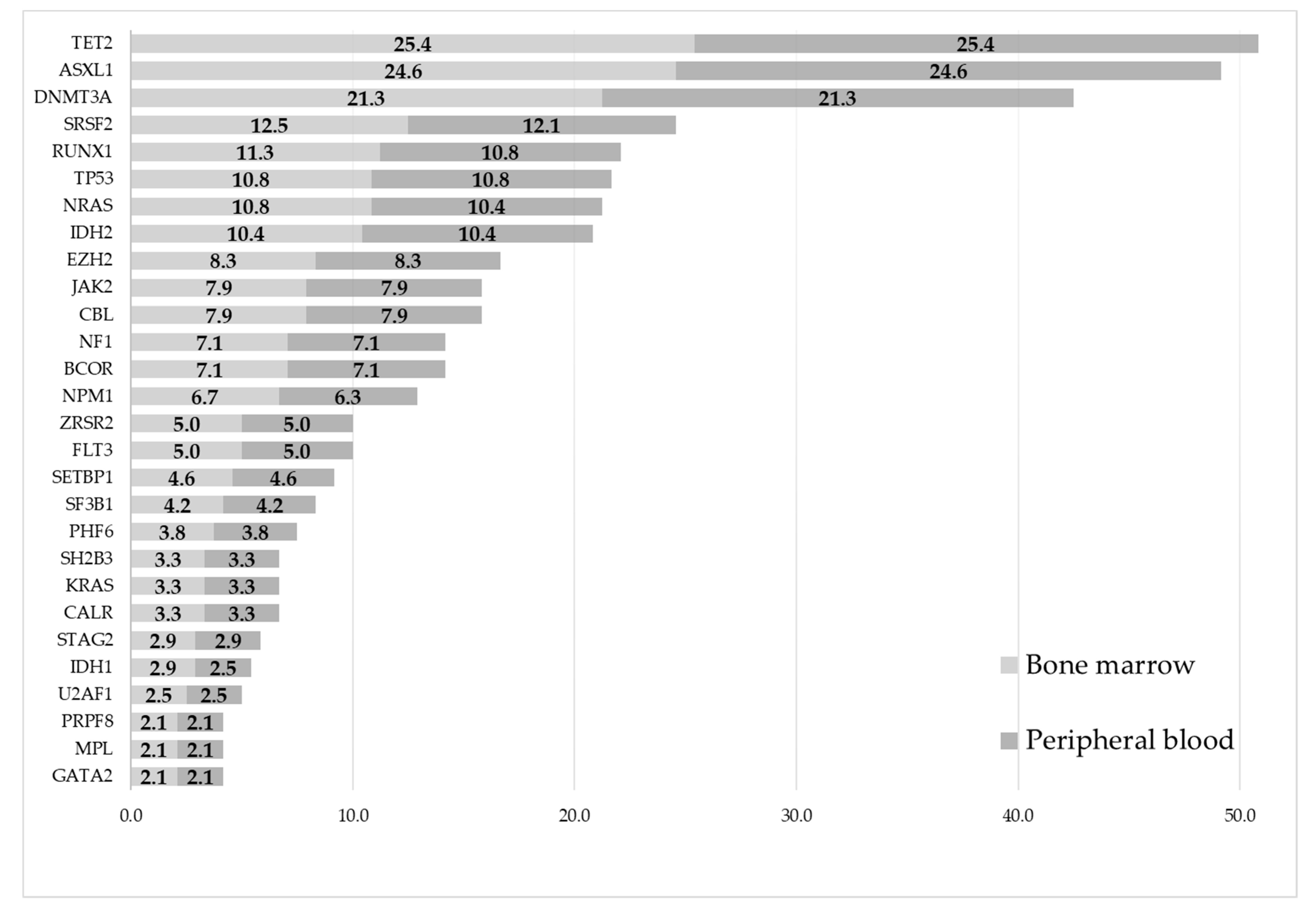
3.4. Mutational Analyses—Occurrence of Mutations

There was strong correlation between the occurrence (i.e., the detection) of mutations in peripheral blood versus bone marrow samples (r = 0.91; p < 0.0001). The occurrence of mutations by gene and sample type for all samples of the total cohort are depicted in Figure 2. In both sample types, the five most commonly mutated genes were TET2 (25.4% vs. 25.4%), ASXL1 (24.6% vs. 24.6%), DNMT3A (21.3% vs. 21.3%), SRSF2 (12.5% vs. 12.1%), and RUNX1 (11.3% vs. 10.8%) for bone marrow vs. peripheral blood, respectively (Figure 2). Two patients harbored a FLT3-ITD mutation, which was detected in the bone marrow and the peripheral blood in all sample pairs (n = 2). All detected fusion genes (n = 11) were detected in both the bone marrow and the peripheral blood in all sample pairs (n = 11).
Below, the occurrence of mutations by gene and sample type (bone marrow vs. peripheral blood) is given separately for the five most commonly mutated genes for each diagnostic subgroup (further details in Figures S1–S5):
  • Acute myeloid leukemia: DNMT3A (31.9% vs. 31.9%), NPM1 (19.4% vs. 18.1%), IDH2 (19.4% vs. 19.4%), TET2 (19.4% vs. 19.4%), and TP53 (15.3% vs. 15.3%) (Figure S1).
  • Myelodysplastic neoplasms: TET2 (37.1% vs. 37.1%), ASXL1 (33.9% vs. 33.9%), DNMT3A (21.0% vs. 21.0%), TP53 (17.7% vs. 17.7%), and RUNX1 (17.7% vs. 17.7%) (Figure S2).
  • Myelodysplastic/myeloproliferative overlap syndromes: ASXL1 (52.9% vs. 52.9%), TET2 (41.2% vs. 41.2%), SRSF2 (35.3% vs. 35.3%), NRAS (29.4% vs. 29.4%), and RUNX1 (23.5% vs. 23.5%) (Figure S3).
  • Myeloproliferative neoplasms: ASXL1 (34.2% vs. 34.2%), TET2 (31.6% vs. 31.6%), JAK2 (31.6% vs. 31.6%), SRSF (23.7% vs. 21.1%), and CALR (21.1% vs. 21.1%) (Figure S4).
  • Other (i.e., non-myeloid) diagnoses: DNMT3A (15.7% vs. 15.7%), ASXL1 (13.7% vs. 13.7%), TET2 (9.8% vs. 9.8%), MYD88 (7.8% vs. 7.8%), and CBL (7.8% vs. 7.8%) (Figure S5).
Figure 2. Occurrence of mutations by gene and by sample type for all samples (n = 150 patients, n = 191 sample pairs 1,2, n = 1299 mutations detected 3,4). The X-axis represents the percentage of samples in which the respective mutation was found. (1 Includes 53 serial sample pairs from 30 patients. 2 Total sample pairs (n = 240), excluding sample pairs without mutations (n = 46) and excluding sample pairs in which only fusion genes (and no other mutations) were found (n = 3). 3 Mutations occurring in genes in <2% of the total cohort were not included in the graph and included the following genes for bone marrow vs. peripheral blood, respectively: MYD88 1.7% vs. 1.7%; ETV6 1.7% vs. 1.7%; CEBPA 1.7% vs. 1.7%; PTPN11 1.3% vs. 1.3%; IKZF 1.3% vs. 1.3%; HRAS 1.3% vs. 1.3%; BRAF 0.8% vs. 0.8%; WT1 0.8% vs. 0.8%; KIT 0.8% vs. 0.8%; and RB1 0.4% vs. 0.0%. 4 If a sample had more than one mutation in the same gene, this was only counted one time).
Cancers 15 02305 g002

3.5. Mutational Analyses—Correlation of BMVAF vs. PBVAF

In 92 samples (i.e., in 46 sample pairs) no mutations were found in either the peripheral blood or the bone marrow. In all analyzed samples in which mutations were detected (excluding fusion genes and FLT3-ITD as the methods used do not produce VAF values, resulting in a remaining n = 382 samples, i.e., n = 191 sample pairs), the mean (SD) [min–max] of VAFs was 25.8% (22.9) [0.1–99.7%] in the bone marrow and 23.1% (21.8) [0.1–99.8%] in the peripheral blood, respectively. Figure S6 and Table S3 lists the mean BMVAF and the mean PBVAF by gene, respectively. Table S3 gives further information by diagnosis.
The strength of the correlation between the BMVAF and the PBVAF was calculated using the Spearman correlation coefficient, which was r = 0.93 (p < 0.0001), thus indicating a very strong correlation (visualized in Figure 3). Sensitivity analyses were performed using Kendall’s tau and the results remained nearly identical.
To determine whether the correlation might be different for patients with a potentially reduced tumor load in the peripheral blood, i.e., in patients with 0% peripheral blood blasts or with <1.0 G/L absolute neutrophil count, we performed the respective analyses for these patient subgroups (Figure 4). The correlation remained very strong for patients in all subgroups, i.e., in patient samples with either peripheral blood blasts = 0% (n = 165; r = 0.92, p < 0.0001) or ≥1% (n = 74, r = 0.95, p < 0.0001), and with an absolute neutrophil count of <1.0 G/L (n = 66; r = 0.88, p < 0.0001) or ≥1.0 G/L (n = 174; r = 0.95, p < 0.0001).
To determine whether the bone marrow blast percentage might correlate with the BMVAF and/or whether the peripheral blood blast percentage correlated with the PBVAF, we plotted these values against each other in Figure 5A,B. The Spearman correlation coefficients were low, with r = 0.11 (p < 0.004) in the bone marrow (Figure 5A) and r = 0.19 (p < 0.0001) in the peripheral blood (Figure 5B), indicating a weak correlation between blast counts and the detected VAFs.
Figure 4. Scatterplot of the variant allele frequency (VAF) of the mutated genes in bone marrow and peripheral blood for patient samples with the following peripheral blood parameters on the day of bone marrow sampling. (A) Peripheral blast percentage = 0% (n = 165). (B) Peripheral blast percentage ≥ 1% (n = 74). 1 (C) Absolute neutrophil count < 1.0 G/L (n = 66). (D) Absolute neutrophil count ≥ 1.0 G/L (n = 174). The black line is the bisecting line showing a perfect linear regression with slope = 1 and intercept = 0. The red dashed line is the regression line, indicating the correlation between BMVAF and PBVAF of each paired sample. (1 The peripheral blood blast percentage was missing for one patient).
Cancers 15 02305 g004

3.6. Mutational Analyses—Agreement

A graphical representation of the agreement between the two methods of clinical measurement, i.e., BMVAF versus (vs.) PBVAF, was generated according to Bland et al. [55] (Figure 6). The x-axis shows the mean of the two values, while the y-axis displays the difference between the two. The central red dashed line represents the mean of the difference ( d = 2.92), and the outer red dashed lines correspond to d ± 2 σ , where σ represents the standard deviation of the difference ( σ = 7.71). As discussed in [55], the region between these two outer lines is referred to as the “limits of agreement” and it visualizes the difference between the two methods of measurement.
The results presented in Figure 6 indicate that both methods of clinical measurement (i.e., NGS from peripheral blood vs. NGS from bone marrow) are in good agreement with each other, as the mean of the difference is close to zero and the majority of the differences between the two methods fall within the “limits of agreement” (i.e., between the two central dashed red lines). This suggests that both sample types produce similar results and provide consistent measures of the clinical parameters under study.
Data points lying outside of the “limits of agreement” are termed outliers (n = 35). These occurred in the following genes: DNMT3A (n = 8), IDH2 (n = 6), RUNX1 (n = 6), TP53 (n = 4), NPM1 (n = 3), TET2 (n = 2), PHF6 (n = 2), and BCOR, EZH2, IDH1, JAK2 and NRAS (n = 1 each).

3.7. Discordant Mutations

Of 1321 detected mutations, 9 (0.68%) were found in only one of the paired samples, i.e., were discordant (Table 4). Discordant mutations included six SNVs, two insertions and one deletion (Table 4). Of these, only one mutation (in gene ASXL1, VAF 1.0%, read depth 5014) was found exclusively in the peripheral blood but not in the bone marrow. Only eight mutations were found exclusively in the bone marrow but not in the peripheral blood, namely NPM1 (VAF 0.6%, read depth 1724), SETBP1 (VAF 1.1%, read depth 6565), NRAS (VAF 1.3%, read depth 18517), RB1 (VAF 1.9, read depth 12772), SRSF2 (VAF 2.2%, read depth 960), ASXL1 (VAF 2.5%, read depth 5816), RUNX1 (VAF 3.7%, read depth 4955), and IDH1 (VAF 9.1%, read depth 4150). The mean (SD) [min–max] VAF of all mutations exclusively found in the bone marrow was 2.7% (2.7) [0.6–3.6%]. All mutations, except the IDH1 mutation in a patient with Waldenstrom’s disease, had a VAF of <4% (Table 4). Of these patients, four had a serial sample analysis. Further details are shown in Table S4, Figure S7−S15.

3.8. Further Subanalyses

The following further subanalyses were performed: (i) limiting the analyses to those patients for whom the paired samples were drawn on the same day, (ii) exclusion of all found aberrations with a VAF < 5%, and (iii) using the filters applied in both (i) and (ii). All results were similar to those reported above (Figures S16–S27).

4. Discussion

In this prospective, monocentric analysis we report on the concordance of NGS results between paired samples obtained from the peripheral blood and the bone marrow samples of patients for whom a myeloid NGS panel was requested from our laboratory to inform their diagnostic and therapeutic decisions.
To date, bone marrow biopsies are still required to correctly classify diseases [7,8] as well as to determine (molecular) response [8,22,23,24,25,26]. As mentioned above, however, bone marrow evaluations are only performed in approximately 50% of patients with myeloid neoplasias during follow-up [33,34,35,56]. Reasons why bone marrow evaluations are often not performed outside of clinical trials include the fact that response can often be determined from clinical benefit (e.g., loss of transfusion dependence, normalization of blood counts, disappearance of peripheral blood blasts, improvement of quality of life, and performance status). Similarly, disease progression can be inferred from clinical deterioration. Other reasons include a lack of clinical consequences (e.g., due to lack of alternative treatment options), anticipated bleeding complications, and logistic reasons. The patients may also decline repeated bone marrow evaluations [35]. Thus, a real-world clinical need exists to be able to obtain information on the mutational status of a patient’s disease without the requirement for repetitive bone marrow evaluations, especially as frequent molecular monitoring is desirable for adequate risk stratification [31,57], prognostication [15,32], and for adequate clinical management [58,59,60,61].
In this regard, evidence from retrospective analyses and clinical trials is accumulating that bone marrow blast clearance [22,23,24,25,26] and even bone marrow assessments may not be mandatory to determine whether meaningful clinical response with overall survival prolongation is achieved [34,35,56]. Furthermore, high complete concordance (63–100%) of genomic, cytogenetic, and phenotypic aberrations between paired peripheral blood and bone marrow samples has been found in patients with myeloid neoplasias using methods such as fluorescence in situ hybridization, chromosome banding arrays, or flow cytometry [37,38,39,46,47,48] (Table S1). Peripheral blood samples have been identified as a viable alternative to bone marrow samples for monitoring cytogenetic data using fluorescence in situ hybridization [37,38,39,48] and flow cytometry [47] panels, as well as by conventional karyotyping [37] and SNP arrays [46].
Some research groups used targeted NGS approaches to assess whether peripheral blood may be an acceptable alternative sample to bone marrow in patients with hematologic neoplasms [40,41,42,43] (Table 1). These studies reported on 16–183 paired peripheral blood and bone marrow samples and detected a complete concordance of 69–97%. Four of these seven studies either included or were exclusively performed in patients with lymphoid neoplasms [40,42,44,45], five did not report how many days were allowed between the peripheral blood and bone marrow samples, two reported up to 334 days between the sampling, and the sensitivity of the NGS analyses and/or the mean (min–max) coverage were rarely reported. Nevertheless, these important studies underscore the relevance and the real-world clinical need to be able to diagnose and monitor treatment response without repeated bone marrow evaluations.
Our data show an exceptionally high concordance (99.6%), sensitivity (98.8%), specificity (99.9%), positive predictive value (99.8%), and negative predictive value (99.4%) between reported results obtained by NGS analyses of paired peripheral blood and bone marrow specimens.
To our best knowledge these results are the first to show a very strong correlation not only between the mutations detected in the peripheral blood and the bone marrow (r = 0.91; p < 0.0001), but also a very strong correlation between the VAFs detected in the peripheral blood and the bone marrow (r = 0.93; p < 0.0001).
We are also the first to analyze and show that the concordance of the VAFs of detected mutations between the bone marrow and the peripheral blood was similarly high in patient subgroups with no circulating blasts (r = 0.92; p < 0.0001) or with neutropenia (as defined by an absolute neutrophil count of < 1.0 G/L) (r = 0.88; p < 0.0001). These results are in line with the fact that clonal involvement has been shown for maturing myeloid cells such as neutrophils [62] as well as for all accessory cells (except T-cells), including natural killer cells [63,64,65,66,67,68], myeloid/lymphoid dendritic cells [36,67,69], monocytes [67], and B-cells [66,70,71,72,73], all of which have been shown to bear the same cytogenetic abnormalities as the malignant bone marrow myeloid progenitors and blasts in patients with myelodysplastic neoplasms or acute myeloid leukemia.
These data are also the first to show a lack of correlation between the blast percentage in the bone marrow and BMVAF of mutations, and the same accounted for the peripheral blood blast percentage and PBVAF of mutations. This is an important finding for daily clinical practice, since clinicians can safely rely on the results of peripheral blood NGS analysis, when a bone marrow evaluation is not evaluable or not feasible, even in the absence of circulating blasts or in neutropenic patients.
Of 1321 found mutations, only 9 (0.68%) were discordant between bone marrow and peripheral blood. Eight of the 9 discordant mutations were found only in the bone marrow and one was found only in the peripheral blood. Most discrepancies (eight of nine mutations that were found in only one sample) were caused by low-level sub-clonal events (VAF ≤ 3.7%). The clinical relevance of VAFs < 5% is currently thought to be low [74] and most laboratories specify a sensitivity of their NGS methods of ≥5%. A VAF of ≥2% is required for the diagnosis of clonal hematopoiesis of indeterminate potential [75] and a VAF of ≥10% is required for recent classification systems [7,22].
Our results thus confirm and expand on reports by others, in an extremely well documented patient population with prospectively collected paired peripheral blood and bone marrow samples, >90% of which were drawn on the same day. Our group used the highest read depth of those reported and also had the highest concordance, indicating that increasing the read depth (and thus sensitivity) of the method increases concordance. Hence, it will be extremely interesting and relevant to perform similar studies for minimal residual disease analyses by NGS, once such tests become commercially available.
These findings support current guidelines, which advise against a repeated NGS analysis of bone marrow samples if a mutation has already been found in the peripheral blood [20].

5. Conclusions

Taken together, our results clearly show, in a prospective setting, that sequential molecular analyses of peripheral blood specimens can be reliably used to molecularly classify and monitor myeloid neoplasms without loss of sensitivity or specificity, and that a bone marrow evaluation solely for the purpose of monitoring of mutations is not necessary in (almost) all cases. These data thus ascertain that myeloid NGS analyses from peripheral blood can safely be used as an alternative to bone marrow samples to identify and monitor gene mutations and to guide treatment decisions. This allows for less frequent follow-up of bone marrow evaluations, which may perhaps be entirely omitted in the future. This is extremely relevant information for both the treating physicians and patients, as samples of peripheral blood can be drawn easily, nearly painlessly, and at multiple time points. NGS from peripheral blood specimen is thus of particular clinical interest for its use as a minimally invasive screening tool for diagnostic and therapy monitoring, especially in special situations such as a fibrotic or hypocellular marrow.

Supplementary Materials

The following supporting information can be downloaded at: https://www.mdpi.com/article/10.3390/cancers15082305/s1. Table S1. Published articles comparing non-NGS analyses from bone marrow and peripheral blood in patients with MDS, CMML and AML. Table S2. Analyzed genes and translocation partners. Figure S1. Occurrence of mutations by gene and by sample type for all samples of patients with AML. Figure S2. Occurrence of mutations by gene and by sample type for all samples of patients with MDS. Figure S3. Occurrence of mutations by gene and by sample type for all samples of patients with MDS-MPN. Figure S4. Occurrence of mutations by gene and by sample type for all samples of patients with MPN. Figure S5. Occurrence of mutations by gene and by sample type for all samples of patients with non-myeloid diagnoses. Figure S6. Mean variant allele frequency (VAF) by gene and by sample type for all samples of all patients. Table S3. Mean variant allele frequency (VAF) by gene and by sample type and diagnosis. Table S4. Complete list of reported variants and the laboratory values in the 9 discordant patients. Figure S7. AML patient with serial analyses and a discordant NPM1 mutation in the third analysis. Figure S8. AML patient with serial analyses and a discordant ASXL1 mutation in the first analysis. Figure S9. MDS patient with a discordant SETBP1 mutation. Figure S10. AML patient with serial analyses and a discordant NRAS mutation in the second analysis. Figure S11. MDS patient with serial analyses and a discordant RB1 mutation in the second analysis. Figure S12. MPN patient with a discordant SRSF2 mutation. Figure S13. MDS patient with a discordant ASXL1 mutation. Figure S14. AML patient with a discordant RUNX1 mutation. Figure S15. Waldenstrom’s disease patient with a discordant IDH1 mutation. Figure S16. Scatterplot of the variant allele frequency (VAF) of the mutated genes in bone marrow and peripheral blood of samples drawn on the same day. Figure S17. Scatterplot of the variant allele frequency (VAF) of the mutated genes in bone marrow and peripheral blood of samples drawn on the same day. Figure S18. Scatterplots of the “variant allele frequency (VAF) of a mutation” vs. “blast percentage” of samples drawn on the same day. Figure S19. Scatterplot of the variant allele frequency (VAF): difference against the mean of samples drawn on the same day. Figure S20. Scatterplot of the variant allele frequency (VAF) of the mutated genes in bone marrow and peripheral blood of samples drawn on the same day and mutations detected with a VAF >5%. Figure S21. Scatterplot of the variant allele frequency (VAF) of the mutated genes in bone marrow and peripheral blood of samples drawn on the same day and mutations detected with a VAF >5%. Figure S22. Scatterplots of the “variant allele frequency (VAF) of a mutation” vs. “blast percentage” of samples drawn on the same day and mutations detected with a VAF >5%. Figure S23. Scatterplot of the variant allele frequency (VAF): difference against the mean of samples drawn on the same day and mutations detected with a VAF >5%. Figure S24. Scatterplot of the variant allele frequency (VAF) of the mutated genes in bone marrow and peripheral blood of samples with mutations >5% VAF. Figure S25. Scatterplot of the variant allele frequency (VAF) of the mutated genes in bone marrow and peripheral blood of samples with mutations >5% VAF. Figure S26. Scatterplots of the “variant allele frequency (VAF) of a mutation” vs. “blast percentage” of samples with mutations >5% VAF. Figure S27. Scatterplot of the variant allele frequency (VAF): difference against the mean of samples with mutations >5% VAF.

Author Contributions

Conceptualization, B.J.-G., M.L. and L.P.; Data curation, B.J.-G. and L.P.; Formal analysis, M.L.; Funding acquisition, R.G.; Investigation, B.J.-G., M.L., F.J.G., N.Z., T.D., S.H., A.R., T.M., A.E., M.D., J.L.-S., R.G. and L.P.; Methodology, B.J.-G., M.L. and L.P.; Resources, B.J.-G., M.L., F.J.G., N.Z., T.D., S.H., A.R., T.M., A.E., M.D., J.L.-S., R.G. and L.P.; Supervision, L.P.; Visualization, B.J.-G. and L.P.; Writing—original draft, B.J.-G., M.L. and L.P.; Writing—review and editing, B.J.-G., M.L., F.J.G., N.Z., T.D., S.H., A.R., T.M., A.E., M.D., J.L.-S., R.G. and L.P. All authors have read and agreed to the published version of the manuscript.

Funding

The Austrian Group for Medical Tumor Therapy (AGMT) is the sponsor for the Austrian Registry of Hypomethylating Agents and received funding from Celgene/BMS, AbbVie, and Janssen Cilag. The AGMT is a not-for-profit organization and an academic study group. The group performed administrative and legal management, as well as funding acquisition. No pharmaceutical company and no other funding source were involved in any way and had no role in the study design, data collection, data analysis, data interpretation, or writing of the report. No medical writer or editor was involved. This work was, in part, supported by the Cancer Cluster Salzburg research grant of the County of Salzburg.

Institutional Review Board Statement

Ethics committee approval was provided by the Ethikkommission für das Bundesland Salzburg (415-EP/39/Feb-2009). The Ethics Committee Approval, the study protocol, and the signed Sponsor Approval Page of the Austrian Registry of Hypomethylating Agents are available on request through the AGMT ([email protected]).

Informed Consent Statement

Informed consent was obtained from all the subjects alive at the time of data entry into the electronic case report form. The Informed Consent form of the Austrian Registry of Hypomethylating Agents is available on request through the AGMT ([email protected]). Patients not included in the registry signed a separate informed consent form allowing the use of their data.

Data Availability Statement

The datasets supporting the conclusions of this article are included within the article and the supplementary material. Data sharing of patient level data collected for the study is not planned. However, we are open to research questions asked by other researchers and we are also open to data contributions by others. Participation requests or potential joint research proposals can be made at any timepoint to the corresponding author via email ([email protected]) and are subject to approval by the AGMT and its collaborators.

Conflicts of Interest

B.J.-G.: No potential conflict of interest; M.L.: Honoraria from BMS, Celgene, Gilead, Takeda and Novartis; Travel support: Celgene and Novartis; F.J.G.: No potential conflict of interest; N.Z.: No potential conflict of interest; T.D.: No potential conflict of interest; S.H.: No potential conflict of interest; A.R.: No potential conflict of interest; T.M.: Honoraria from AbbVie and Celgene/BMS; A.E.: Honoraria, consultancy, and travel support from AbbVie and BMS/Celgene; M.D.: No potential conflict of interest; J.L.-S.: No potential conflict of interest; R.G.: Honoraria from AbbVie, Amgen, AstraZeneca, BMS/Celgene, Daiichi Sankyo, Gilead, Merck, Novartis, Roche, Takeda, BMS, MSD, Sandoz, Gilead; Research funding from Celgene, Roche, Merck, Novartis, MSD, Sandoz, and Takeda; Consulting: AbbVie, AstraZeneca, BMS/Celgene, Novartis, Roche, Takeda, Janssen, MSD, Merck, Gilead, Daiichi Sankyo; Travel support from AbbVie, Amgen, AstraZeneca, BMS/Celgene, Daiichi Sankyo, Gilead, Janssen Cilag, MSD, Novartis, and Roche; L.P.: Honoraria from AbbVie, BMS, and Novartis.

References

  1. Swerdlow, S.H.; Campo, E.; Pileri, S.A.; Harris, N.L.; Stein, H.; Siebert, R.; Advani, R.; Ghielmini, M.; Salles, G.A.; Zelenetz, A.D.; et al. The 2016 Revision of the World Health Organization Classification of Lymphoid Neoplasms. Blood 2016, 127, 2375–2390. [Google Scholar] [CrossRef] [Green Version]
  2. Pleyer, L.; Leisch, M.; Kourakli, A.; Padron, E.; Maciejewski, J.P.; Xicoy Cirici, B.; Kaivers, J.; Ungerstedt, J.; Heibl, S.; Patiou, P.; et al. Outcomes of Patients with Chronic Myelomonocytic Leukaemia Treated with Non-Curative Therapies: A Retrospective Cohort Study. Lancet Haematol. 2021, 8, e135–e148. [Google Scholar] [CrossRef] [PubMed]
  3. Pleyer, L.; Neureiter, D.; Faber, V.; Greil, R. Myelodysplastic Syndromes (MDS). In Chronic Myeloid Neoplasias and Clonal Overlap Syndromes; Greil, R., Pleyer, L., Faber, V., Neureiter, D., Eds.; Springer: Vienna, Austria, 2010; pp. 153–222. ISBN 978-3-211-79891-1. [Google Scholar]
  4. Valent, P.; Orazi, A.; Savona, M.R.; Patnaik, M.M.; Onida, F.; van de Loosdrecht, A.A.; Haase, D.; Haferlach, T.; Elena, C.; Pleyer, L.; et al. Proposed Diagnostic Criteria for Classical Chronic Myelomonocytic Leukemia (CMML), CMML Variants and Pre-CMML Conditions. Haematologica 2019, 104, 1935–1949. [Google Scholar] [CrossRef] [PubMed] [Green Version]
  5. Pleyer, L.; Döhner, H.; Dombret, H.; Seymour, J.; Schuh, A.; Beach, C.; Swern, A.; Burgstaller, S.; Stauder, R.; Girschikofsky, M.; et al. Azacitidine for Front-Line Therapy of Patients with AML: Reproducible Efficacy Established by Direct Comparison of International Phase 3 Trial Data with Registry Data from the Austrian Azacitidine Registry of the AGMT Study Group. IJMS 2017, 18, 415. [Google Scholar] [CrossRef] [PubMed] [Green Version]
  6. Pleyer, L.; Sekeres, M.A. An Early Glimpse at Azacitidine plus Venetoclax for Myelodysplastic Syndromes. Lancet Haematol. 2022, 9, e714–e716. [Google Scholar] [CrossRef]
  7. Khoury, J.D.; Solary, E.; Abla, O.; Akkari, Y.; Alaggio, R.; Apperley, J.F.; Bejar, R.; Berti, E.; Busque, L.; Chan, J.K.C.; et al. The 5th Edition of the World Health Organization Classification of Haematolymphoid Tumours: Myeloid and Histiocytic/Dendritic Neoplasms. Leukemia 2022, 36, 1703–1719. [Google Scholar] [CrossRef]
  8. Arber, D.A.; Orazi, A.; Hasserjian, R.P.; Borowitz, M.J.; Calvo, K.R.; Kvasnicka, H.-M.; Wang, S.A.; Bagg, A.; Barbui, T.; Branford, S.; et al. International Consensus Classification of Myeloid Neoplasms and Acute Leukemias: Integrating Morphologic, Clinical, and Genomic Data. Blood 2022, 140, 1200–1228. [Google Scholar] [CrossRef]
  9. Pleyer, L.; Neureiter, D.; Faber, V.; Greil, R. Chronic Myelomonocytic Leukemia (CMML). In Chronic Myeloid Neoplasias and Clonal Overlap Syndromes; Greil, R., Pleyer, L., Faber, V., Neureiter, D., Eds.; Springer: Vienna, Austria, 2010; pp. 223–233. ISBN 978-3-211-79891-1. [Google Scholar]
  10. Schuh, A.C.; Döhner, H.; Pleyer, L.; Seymour, J.F.; Fenaux, P.; Dombret, H. Azacitidine in Adult Patients with Acute Myeloid Leukemia. Crit. Rev. Oncol./Hematol. 2017, 116, 159–177. [Google Scholar] [CrossRef]
  11. Itzykson, R.; Fenaux, P.; Bowen, D.; Cross, N.C.P.; Cortes, J.; De Witte, T.; Germing, U.; Onida, F.; Padron, E.; Platzbecker, U.; et al. Diagnosis and Treatment of Chronic Myelomonocytic Leukemias in Adults: Recommendations From the European Hematology Association and the European LeukemiaNet. HemaSphere 2018, 2, e150. [Google Scholar] [CrossRef]
  12. Leisch, M.; Jansko, B.; Zaborsky, N.; Greil, R.; Pleyer, L. Next Generation Sequencing in AML—On the Way to Becoming a New Standard for Treatment Initiation and/or Modulation? Cancers 2019, 11, 252. [Google Scholar] [CrossRef] [Green Version]
  13. Gangat, N.; Mudireddy, M.; Lasho, T.L.; Finke, C.M.; Nicolosi, M.; Szuber, N.; Patnaik, M.M.; Pardanani, A.; Hanson, C.A.; Ketterling, R.P.; et al. Mutations and Prognosis in Myelodysplastic Syndromes: Karyotype-Adjusted Analysis of Targeted Sequencing in 300 Consecutive Cases and Development of a Genetic Risk Model. Am. J. Hematol. 2018, 93, 691–697. [Google Scholar] [CrossRef] [PubMed]
  14. Tefferi, A.; Lasho, T.L.; Patnaik, M.M.; Saeed, L.; Mudireddy, M.; Idossa, D.; Finke, C.; Ketterling, R.P.; Pardanani, A.; Gangat, N. Targeted Next-Generation Sequencing in Myelodysplastic Syndromes and Prognostic Interaction between Mutations and IPSS-R. Am. J. Hematol. 2017, 92, 1311–1317. [Google Scholar] [CrossRef] [Green Version]
  15. Yun, S.; Geyer, S.M.; Komrokji, R.S.; Al Ali, N.H.; Song, J.; Hussaini, M.; Sweet, K.L.; Lancet, J.E.; List, A.F.; Padron, E.; et al. Prognostic Significance of Serial Molecular Annotation in Myelodysplastic Syndromes (MDS) and Secondary Acute Myeloid Leukemia (SAML). Leukemia 2021, 35, 1145–1155. [Google Scholar] [CrossRef] [PubMed]
  16. Reinig, E.; Yang, F.; Traer, E.; Arora, R.; Brown, S.; Rattray, R.; Braziel, R.; Fan, G.; Press, R.; Dunlap, J. Targeted Next-Generation Sequencing in Myelodysplastic Syndrome and Chronic Myelomonocytic Leukemia Aids Diagnosis in Challenging Cases and Identifies Frequent Spliceosome Mutations in Transformed Acute Myeloid Leukemia. Am. J. Clin. Pathol. 2016, 145, 497–506. [Google Scholar] [CrossRef] [PubMed] [Green Version]
  17. Maggioni, G.; Della Porta, M.G. Molecular Landscape of Myelodysplastic Neoplasms in Disease Classification and Prognostication. Curr. Opin. Hematol. 2023, 30, 30–37. [Google Scholar] [CrossRef] [PubMed]
  18. Elena, C.; Gallì, A.; Such, E.; Meggendorfer, M.; Germing, U.; Rizzo, E.; Cervera, J.; Molteni, E.; Fasan, A.; Schuler, E.; et al. Integrating Clinical Features and Genetic Lesions in the Risk Assessment of Patients with Chronic Myelomonocytic Leukemia. Blood 2016, 128, 1408–1417. [Google Scholar] [CrossRef] [PubMed] [Green Version]
  19. Xie, Z.; Chen, E.C.; Stahl, M.; Zeidan, A.M. Prognostication in Myelodysplastic Syndromes (Neoplasms): Molecular Risk Stratification Finally Coming of Age. Blood Rev. 2022, 5, 101033. [Google Scholar] [CrossRef] [PubMed]
  20. Duncavage, E.J.; Bagg, A.; Hasserjian, R.P.; DiNardo, C.D.; Godley, L.A.; Iacobucci, I.; Jaiswal, S.; Malcovati, L.; Vannucchi, A.M.; Patel, K.P.; et al. Genomic Profiling for Clinical Decision Making in Myeloid Neoplasms and Acute Leukemia. Blood 2022, 140, 2228–2247. [Google Scholar] [CrossRef]
  21. Greenberg, P.L.; Tuechler, H.; Schanz, J.; Sanz, G.; Garcia-Manero, G.; Solé, F.; Bennett, J.M.; Bowen, D.; Fenaux, P.; Dreyfus, F.; et al. Revised International Prognostic Scoring System for Myelodysplastic Syndromes. Blood 2012, 120, 2454–2465. [Google Scholar] [CrossRef] [Green Version]
  22. Döhner, H.; Wei, A.H.; Appelbaum, F.R.; Craddock, C.; DiNardo, C.D.; Dombret, H.; Ebert, B.L.; Fenaux, P.; Godley, L.A.; Hasserjian, R.P.; et al. Diagnosis and Management of AML in Adults: 2022 Recommendations from an International Expert Panel on Behalf of the ELN. Blood 2022, 140, 1345–1377. [Google Scholar] [CrossRef]
  23. Döhner, H.; Estey, E.; Grimwade, D.; Amadori, S.; Appelbaum, F.R.; Büchner, T.; Dombret, H.; Ebert, B.L.; Fenaux, P.; Larson, R.A.; et al. Diagnosis and Management of AML in Adults: 2017 ELN Recommendations from an International Expert Panel. Blood 2017, 129, 424–447. [Google Scholar] [CrossRef] [PubMed] [Green Version]
  24. Cheson, B.D. Clinical Application and Proposal for Modification of the International Working Group (IWG) Response Criteria in Myelodysplasia. Blood 2006, 108, 419–425. [Google Scholar] [CrossRef] [PubMed] [Green Version]
  25. Cheson, B.D.; Bennett, J.M.; Kopecky, K.J.; Büchner, T.; Willman, C.L.; Estey, E.H.; Schiffer, C.A.; Doehner, H.; Tallman, M.S.; Lister, T.A.; et al. Revised Recommendations of the International Working Group for Diagnosis, Standardization of Response Criteria, Treatment Outcomes, and Reporting Standards for Therapeutic Trials in Acute Myeloid Leukemia. JCO 2003, 21, 4642–4649. [Google Scholar] [CrossRef]
  26. Savona, M.R.; Malcovati, L.; Komrokji, R.; Tiu, R.V.; Mughal, T.I.; Orazi, A.; Kiladjian, J.-J.; Padron, E.; Solary, E.; Tibes, R.; et al. An International Consortium Proposal of Uniform Response Criteria for Myelodysplastic/Myeloproliferative Neoplasms (MDS/MPN) in Adults. Blood 2015, 125, 1857–1865. [Google Scholar] [CrossRef] [PubMed] [Green Version]
  27. Zeidan, A.M.; Platzbecker, U.; Bewersdorf, J.P.; Stahl, M.; Adès, L.; Borate, U.; Bowen, D.T.; Buckstein, R.J.; Brunner, A.M.; Carraway, H.E.; et al. Consensus Proposal for Revised International Working Group Response Criteria for Higher Risk Myelodysplastic Syndromes. Blood, 2023; Online ahead of print. [Google Scholar] [CrossRef]
  28. Tazi, Y.; Arango-Ossa, J.E.; Zhou, Y.; Bernard, E.; Thomas, I.; Gilkes, A.; Freeman, S.; Pradat, Y.; Johnson, S.J.; Hills, R.; et al. Unified Classification and Risk-Stratification in Acute Myeloid Leukemia. Nat. Commun. 2022, 13, 4622. [Google Scholar] [CrossRef] [PubMed]
  29. Nannya, Y.; Tobiasson, M.; Sato, S.; Bernard, E.; Ohtake, S.; Takeda, J.; Creignou, M.; Zhao, L.; Kusakabe, M.; Shibata, Y.; et al. Post-Azacitidine Clone Size Predicts Outcome of Patients with Myelodysplastic Syndromes and Related Myeloid Neoplasms. Blood Adv. 2023; Online ahead of print. [Google Scholar] [CrossRef]
  30. Welch, J.S.; Petti, A.A.; Miller, C.A.; Fronick, C.C.; O’Laughlin, M.; Fulton, R.S.; Wilson, R.K.; Baty, J.D.; Duncavage, E.J.; Tandon, B.; et al. TP53 and Decitabine in Acute Myeloid Leukemia and Myelodysplastic Syndromes. N. Engl. J. Med. 2016, 375, 2023–2036. [Google Scholar] [CrossRef]
  31. Bernard, E.; Tuechler, H.; Greenberg, P.L.; Hasserjian, R.P.; Arango Ossa, J.E.; Nannya, Y.; Devlin, S.M.; Creignou, M.; Pinel, P.; Monnier, L.; et al. Molecular International Prognostic Scoring System for Myelodysplastic Syndromes. NEJM Evid. 2022, 1. [Google Scholar] [CrossRef]
  32. Papaemmanuil, E.; Gerstung, M.; Bullinger, L.; Gaidzik, V.I.; Paschka, P.; Roberts, N.D.; Potter, N.E.; Heuser, M.; Thol, F.; Bolli, N.; et al. Genomic Classification and Prognosis in Acute Myeloid Leukemia. N. Engl. J. Med. 2016, 374, 2209–2221. [Google Scholar] [CrossRef]
  33. Dinmohamed, A.G.; van Norden, Y.; Visser, O.; Posthuma, E.F.M.; Huijgens, P.C.; Sonneveld, P.; van de Loosdrecht, A.A.; Jongen-Lavrencic, M. The Use of Medical Claims to Assess Incidence, Diagnostic Procedures and Initial Treatment of Myelodysplastic Syndromes and Chronic Myelomonocytic Leukemia in the Netherlands. Leuk. Res. 2015, 39, 177–182. [Google Scholar] [CrossRef] [PubMed]
  34. Pleyer, L.; Pfeilstocker, M.; Stauder, R.; Heibl, S.; Sill, H.; Girschikofsky, M.; Stampfl-Mattesberger, M.; Tinchon, C.; Hartmann, B.; Petzer, A.; et al. Peripheral Blood Complete Remission Provides Added Value to the Classical Definition of Morphologic Complete Remission—A Prospective Cohort Study of 1441 Patients with MDS, CMML and AML Treated within the Austrian Azacitidine Registry. Blood 2021, 138, 3387. [Google Scholar] [CrossRef]
  35. Pleyer, L.; Pfeilstocker, M.; Stauder, R.; Heibl, S.; Sill, H.; Girschikofsky, M.; Stampfl-Mattersberger, M.; Tinchon, C.; Petzer, A.; Schmitt, C.A.; et al. Expanding on Current Definitions of Hematologic Improvement in MDS, CMML and AML: Landmark Analyses of 1301 Patients Treated with Azacitidine in the Austrian Registry of Hypomethylating Agents By the AGMT-Study Group. Blood 2019, 134, 3821. [Google Scholar] [CrossRef]
  36. van Lom, K.; Hagemeijer, A.; Smit, E.; Hählen, K.; Groeneveld, K.; Löwenberg, B. Cytogenetic Clonality Analysis in Myelodysplastic Syndrome: Monosomy 7 Can Be Demonstrated in the Myeloid and in the Lymphoid Lineage. Leukemia 1995, 9, 1818–1821. [Google Scholar] [PubMed]
  37. Asadi Fakhr, Z.; Mehrzad, V.; Izaditabar, A.; Salehi, M. Evaluation of the Utility of Peripheral Blood vs Bone Marrow in Karyotype and Fluorescence in Situ Hybridization for Myelodysplastic Syndrome Diagnosis. J. Clin. Lab. Anal. 2018, 32, e22586. [Google Scholar] [CrossRef] [Green Version]
  38. Braulke, F.; Jung, K.; Schanz, J.; Götze, K.; Müller-Thomas, C.; Platzbecker, U.; Germing, U.; Brümmendorf, T.H.; Bug, G.; Ottmann, O.; et al. Molecular Cytogenetic Monitoring from CD34+ Peripheral Blood Cells in Myelodysplastic Syndromes: First Results from a Prospective Multicenter German Diagnostic Study. Leuk. Res. 2013, 37, 900–906. [Google Scholar] [CrossRef]
  39. Cherry, A.M.; Slovak, M.L.; Campbell, L.J.; Chun, K.; Eclache, V.; Haase, D.; Haferlach, C.; Hildebrandt, B.; Iqbal, A.M.; Jhanwar, S.C.; et al. Will a Peripheral Blood (PB) Sample Yield the Same Diagnostic and Prognostic Cytogenetic Data as the Concomitant Bone Marrow (BM) in Myelodysplasia? Leuk. Res. 2012, 36, 832–840. [Google Scholar] [CrossRef]
  40. Lucas, F.; Michaels, P.D.; Wang, D.; Kim, A.S. Mutational Analysis of Hematologic Neoplasms in 164 Paired Peripheral Blood and Bone Marrow Samples by Next-Generation Sequencing. Blood Adv. 2020, 4, 4362–4365. [Google Scholar] [CrossRef]
  41. Ruan, M.; Liu, L.; Qi, B.; Chen, X.; Chang, L.; Zhang, A.; Liu, F.; Wang, S.; Liu, X.; Chen, X.; et al. Targeted Next-Generation Sequencing of Circulating Tumor DNA, Bone Marrow, and Peripheral Blood Mononuclear Cells in Pediatric AML. Front. Oncol. 2021, 11, 666470. [Google Scholar] [CrossRef]
  42. Jumniensuk, C.; Nobori, A.; Lee, T.; Senaratne, T.N.; Rao, D.; Pullarkat, S. Concordance of Peripheral Blood and Bone Marrow Next-Generation Sequencing in Hematologic Neoplasms. Adv. Hematol. 2022, 2022, 8091746. [Google Scholar] [CrossRef]
  43. Stasik, S.; Burkhard-Meier, C.; Kramer, M.; Middeke, J.M.; Oelschlaegel, U.; Sockel, K.; Ehninger, G.; Serve, H.; Müller-Tidow, C.; Baldus, C.D.; et al. Deep Sequencing in CD34+ Cells from Peripheral Blood Enables Sensitive Detection of Measurable Residual Disease in AML. Blood Adv. 2022, 6, 3294–3303. [Google Scholar] [CrossRef] [PubMed]
  44. Muffly, L.; Sundaram, V.; Chen, C.; Yurkiewicz, I.; Kuo, E.; Burnash, S.; Spiegel, J.Y.; Arai, S.; Frank, M.J.; Johnston, L.J.; et al. Concordance of Peripheral Blood and Bone Marrow Measurable Residual Disease in Adult Acute Lymphoblastic Leukemia. Blood Adv. 2021, 5, 3147–3151. [Google Scholar] [CrossRef] [PubMed]
  45. Fries, C.; Adlowitz, D.G.; Spence, J.M.; Spence, J.P.; Rock, P.J.; Burack, W.R. Acute Lymphoblastic Leukemia Clonal Distribution between Bone Marrow and Peripheral Blood. Pediatr. Blood Cancer 2020, 67, e28280. [Google Scholar] [CrossRef] [PubMed]
  46. Mohamedali, A.M.; Gäken, J.; Ahmed, M.; Malik, F.; Smith, A.E.; Best, S.; Mian, S.; Gaymes, T.; Ireland, R.; Kulasekararaj, A.G.; et al. High Concordance of Genomic and Cytogenetic Aberrations between Peripheral Blood and Bone Marrow in Myelodysplastic Syndrome (MDS). Leukemia 2015, 29, 1928–1938. [Google Scholar] [CrossRef] [PubMed]
  47. Godwin, C.D.; Zhou, Y.; Othus, M.; Asmuth, M.M.; Shaw, C.M.; Gardner, K.M.; Wood, B.L.; Walter, R.B.; Estey, E.H. Acute Myeloid Leukemia Measurable Residual Disease Detection by Flow Cytometry in Peripheral Blood vs Bone Marrow. Blood 2021, 137, 569–572. [Google Scholar] [CrossRef] [PubMed]
  48. Coleman, J.F.; Theil, K.S.; Tubbs, R.R.; Cook, J.R. Diagnostic Yield of Bone Marrow and Peripheral Blood FISH Panel Testing in Clinically Suspected Myelodysplastic Syndromes and/or Acute Myeloid Leukemia: A Prospective Analysis of 433 Cases. Am. J. Clin. Pathol. 2011, 135, 915–920. [Google Scholar] [CrossRef] [PubMed]
  49. Leisch, M.; Pfeilstöcker, M.; Stauder, R.; Heibl, S.; Sill, H.; Girschikofsky, M.; Stampfl-Mattersberger, M.; Tinchon, C.; Hartmann, B.; Petzer, A.; et al. Adverse Events in 1406 Patients Receiving 13,780 Cycles of Azacitidine within the Austrian Registry of Hypomethylating Agents—A Prospective Cohort Study of the AGMT Study-Group. Cancers 2022, 14, 2459. [Google Scholar] [CrossRef]
  50. Li, H.; Durbin, R. Fast and Accurate Short Read Alignment with Burrows–Wheeler Transform. Bioinformatics 2009, 25, 1754–1760. [Google Scholar] [CrossRef] [Green Version]
  51. Li, H.; Durbin, R. Fast and Accurate Long-Read Alignment with Burrows-Wheeler Transform. Bioinformatics 2010, 26, 589–595. [Google Scholar] [CrossRef] [Green Version]
  52. Danecek, P.; Bonfield, J.K.; Liddle, J.; Marshall, J.; Ohan, V.; Pollard, M.O.; Whitwham, A.; Keane, T.; McCarthy, S.A.; Davies, R.M.; et al. Twelve Years of SAMtools and BCFtools. Gigascience 2021, 10, giab008. [Google Scholar] [CrossRef]
  53. Robinson, J.T.; Thorvaldsdóttir, H.; Winckler, W.; Guttman, M.; Lander, E.S.; Getz, G.; Mesirov, J.P. Integrative Genomics Viewer. Nat. Biotechnol. 2011, 29, 24–26. [Google Scholar] [CrossRef] [PubMed] [Green Version]
  54. Schober, P.; Boer, C.; Schwarte, L.A. Correlation Coefficients: Appropriate Use and Interpretation. Anesth. Analg. 2018, 126, 1763–1768. [Google Scholar] [CrossRef]
  55. Bland, J.M.; Altman, D.G. Statistical Methods for Assessing Agreement between Two Methods of Clinical Measurement. Lancet 1986, 1, 307–310. [Google Scholar] [CrossRef]
  56. Pleyer, L.; Vaisband, M.; Pfeilstocker, M.; Stauder, R.; Heibl, S.; Sill, H.; Girschikofsky, M.; Stampf-Mattersberger, M.; Tinchon, C.; Hartmann, B.; et al. Cox Proportional Hazards Deep Neural Network Identifies Peripheral Blood Complete Remission (PB-CR) to Be at Least Equivalent to Morphologic CR in Predicting Outcomes of Patients Treated with Azacitidine—A Prospective Cohort Study by the AGMT; ASH Oral: New Orleans, LA, USA, 2022. [Google Scholar]
  57. Sargas, C.; Ayala, R.; Larráyoz, M.J.; Chillón, M.C.; Carrillo-Cruz, E.; Bilbao-Sieyro, C.; Prados de la Torre, E.; Martínez-Cuadrón, D.; Rodríguez-Veiga, R.; Boluda, B.; et al. Molecular Landscape and Validation of New Genomic Classification in 2668 Adult AML Patients: Real Life Data from the PETHEMA Registry. Cancers 2023, 15, 438. [Google Scholar] [CrossRef] [PubMed]
  58. Kayser, S.; Levis, M.J. The Clinical Impact of the Molecular Landscape of Acute Myeloid Leukemia. Haematologica 2023, 108, 308–320. [Google Scholar] [CrossRef] [PubMed]
  59. Ogawa, S. Genetics of MDS. Blood 2019, 133, 1049–1059. [Google Scholar] [CrossRef] [PubMed] [Green Version]
  60. Chiereghin, C.; Travaglino, E.; Zampini, M.; Saba, E.; Saitta, C.; Riva, E.; Bersanelli, M.; Della Porta, M.G. The Genetics of Myelodysplastic Syndromes: Clinical Relevance. Genes 2021, 12, 1144. [Google Scholar] [CrossRef]
  61. Prassek, V.V.; Rothenberg-Thurley, M.; Sauerland, M.C.; Herold, T.; Janke, H.; Ksienzyk, B.; Konstandin, N.P.; Goerlich, D.; Krug, U.; Faldum, A.; et al. Genetics of Acute Myeloid Leukemia in the Elderly: Mutation Spectrum and Clinical Impact in Intensively Treated Patients Aged 75 Years or Older. Haematologica 2018, 103, 1853–1861. [Google Scholar] [CrossRef] [Green Version]
  62. Sexauer, A.; Perl, A.; Yang, X.; Borowitz, M.; Gocke, C.; Rajkhowa, T.; Thiede, C.; Frattini, M.; Nybakken, G.E.; Pratz, K.; et al. Terminal Myeloid Differentiation in Vivo Is Induced by FLT3 Inhibition in FLT3/ITD AML. Blood 2012, 120, 4205–4214. [Google Scholar] [CrossRef] [Green Version]
  63. Kiladjian, J.-J.; Bourgeois, E.; Lobe, I.; Braun, T.; Visentin, G.; Bourhis, J.-H.; Fenaux, P.; Chouaib, S.; Caignard, A. Cytolytic Function and Survival of Natural Killer Cells Are Severely Altered in Myelodysplastic Syndromes. Leukemia 2006, 20, 463–470. [Google Scholar] [CrossRef] [Green Version]
  64. Miura, I.; Kobayashi, Y.; Takahashi, N.; Saitoh, K.; Miura, A.B. Involvement of Natural Killer Cells in Patients with Myelodysplastic Syndrome Carrying Monosomy 7 Revealed by the Application of Fluorescence in Situ Hybridization to Cells Collected by Means of Fluorescence-Activated Cell Sorting: Short Report. Br. J. Haematol. 2000, 110, 876–879. [Google Scholar] [CrossRef] [PubMed]
  65. Nakazawa, T.; Koike, K.; Agematsu, K.; Itoh, S.; Hagimoto, R.; Kitazawa, Y.; Higuchi, T.; Sawai, N.; Matsui, H.; Komiyama, A. Cytogenetic Clonality Analysis in Monosomy 7 Associated with Juvenile Myelomonocytic Leukemia: Clonality in B and NK Cells, but Not in T Cells. Leuk. Res. 1998, 22, 887–892. [Google Scholar] [CrossRef] [PubMed]
  66. Meers, S.; Vandenberghe, P.; Boogaerts, M.; Verhoef, G.; Delforge, M. The Clinical Significance of Activated Lymphocytes in Patients with Myelodysplastic Syndromes: A Single Centre Study of 131 Patients. Leuk. Res. 2008, 32, 1026–1035. [Google Scholar] [CrossRef]
  67. Ma, L.; Delforge, M.; Van Duppen, V.; Verhoef, G.; Emanuel, B.; Boogaerts, M.; Hagemeijer, A.; Vandenberghe, P. Circulating Myeloid and Lymphoid Precursor Dendritic Cells Are Clonally Involved in Myelodysplastic Syndromes. Leukemia 2004, 18, 1451–1456. [Google Scholar] [CrossRef] [PubMed] [Green Version]
  68. Mohty, M.; Jarrossay, D.; Lafage-Pochitaloff, M.; Zandotti, C.; Brière, F.; de Lamballeri, X.-N.; Isnardon, D.; Sainty, D.; Olive, D.; Gaugler, B. Circulating Blood Dendritic Cells from Myeloid Leukemia Patients Display Quantitative and Cytogenetic Abnormalities as Well as Functional Impairment. Blood 2001, 98, 3750–3756. [Google Scholar] [CrossRef] [Green Version]
  69. Matteo Rigolin, G.; Howard, J.; Buggins, A.; Sneddon, C.; Castoldi, G.; Hirst, W.J.R.; Mufti, G.J. Phenotypic and Functional Characteristics of Monocyte-Derived Dendritic Cells from Patients with Myelodysplastic Syndromes: Monocyte-Derived Dendritic Cells in Myelodysplastic Syndromes. Br. J. Haematol. 1999, 107, 844–850. [Google Scholar] [CrossRef] [PubMed]
  70. Nilsson, L.; Astrand-Grundström, I.; Arvidsson, I.; Jacobsson, B.; Hellström-Lindberg, E.; Hast, R.; Jacobsen, S.E. Isolation and Characterization of Hematopoietic Progenitor/Stem Cells in 5q-Deleted Myelodysplastic Syndromes: Evidence for Involvement at the Hematopoietic Stem Cell Level. Blood 2000, 96, 2012–2021. [Google Scholar] [CrossRef] [PubMed]
  71. Thanopoulou, E.; Cashman, J.; Kakagianne, T.; Eaves, A.; Zoumbos, N.; Eaves, C. Engraftment of NOD/SCID-Beta2 Microglobulin Null Mice with Multilineage Neoplastic Cells from Patients with Myelodysplastic Syndrome. Blood 2004, 103, 4285–4293. [Google Scholar] [CrossRef] [Green Version]
  72. Lawrence, H.J.; Broudy, V.C.; Magenis, R.E.; Olson, S.; Tomar, D.; Barton, S.; Fitchen, J.H.; Bagby, G.C. Cytogenetic Evidence for Involvement of B Lymphocytes in Acquired Idiopathic Sideroblastic Anemias. Blood 1987, 70, 1003–1005. [Google Scholar] [CrossRef] [Green Version]
  73. White, N.J.; Nacheva, E.; Asimakopoulos, F.A.; Bloxham, D.; Paul, B.; Green, A.R. Deletion of Chromosome 20q in Myelodysplasia Can Occur in a Multipotent Precursor of Both Myeloid Cells and B Cells. Blood 1994, 83, 2809–2816. [Google Scholar] [CrossRef]
  74. Sasaki, K.; Kanagal-Shamanna, R.; Montalban-Bravo, G.; Assi, R.; Jabbour, E.; Ravandi, F.; Kadia, T.; Pierce, S.; Takahashi, K.; Nogueras Gonzalez, G.; et al. Impact of the Variant Allele Frequency of ASXL1, DNMT3A, JAK2, TET2, TP53, and NPM1 on the Outcomes of Patients with Newly Diagnosed Acute Myeloid Leukemia. Cancer 2020, 126, 765–774. [Google Scholar] [CrossRef] [PubMed]
  75. Steensma, D.P.; Bejar, R.; Jaiswal, S.; Lindsley, R.C.; Sekeres, M.A.; Hasserjian, R.P.; Ebert, B.L. Clonal Hematopoiesis of Indeterminate Potential and Its Distinction from Myelodysplastic Syndromes. Blood 2015, 126, 9–16. [Google Scholar] [CrossRef] [PubMed] [Green Version]
Figure 1. Numbers of patients, samples and mutations. * Total reported molecular results (n = 1415) consist of: 1321 pathogenic variants (8 fragment analysis, 1313 NGS); 94 samples with no mutations.
Cancers 15 02305 g001
Figure 3. Scatterplot of the variant allele frequency (VAF) of the mutated genes in bone marrow and peripheral blood. The black line is the bisecting line showing a perfect linear regression with slope = 1 and intercept = 0. The red dashed line is the regression line, indicating the correlation between BMVAF and PBVAF of each paired sample.
Cancers 15 02305 g003
Figure 5. Scatterplots of the “variant allele frequency (VAF) of a mutation” vs. “blast percentage”. (A) “BMVAF” vs. “percentage of bone marrow blasts”. (B) “PBVAF” vs. “percentage of peripheral blood blasts”. The black line is the bisecting line showing a perfect linear regression with slope = 1 and intercept = 0. The red dashed line is the regression line, indicating the correlation between BMVAF and PBVAF of each paired sample.
Cancers 15 02305 g005
Figure 6. Scatterplot of the variant allele frequency (VAF): difference against the mean. 1 The X−axis shows the mean of the BMVAF and the PBVAF. The Y−axis displays the difference between the BMVAF and the PBVAF. The central red dashed line represents the mean of the difference, ( d = 2.92), and the outer red dashed lines correspond to d ± 2 σ , where σ represents the standard deviation of the difference ( σ = 7.71). As discussed in [55], the region between these two outer lines is referred to as the “limits of agreement” and it visualizes the difference between the two methods of measurement. (1 Analyzed according to Bland et al., Lancet 1986 [55]).
Cancers 15 02305 g006
Table 1. Published articles comparing NGS analyses from bone marrow and peripheral blood in patients with MDS, CMML, and AML 1.
First AuthorPts,
n
Paired Samples,
n
Disease, n/n (%)MethodCompany, kitDays between BM-PB Analyses,
Mean (min–max)
Concordance between Paired Samples
(BM and PB)
Concordance between Paired Mutations (BM and PB)Coefficient
Jansko-Gadermeir B. [current manuscript]187240AML, 46/187 (24.6%)
MDS, 43/187 (23.0%)
MDS/MPN, 15/187 (8.0%)
MPN, 33/187 (17.6%)
Others, 50/187 (26.7%)
NGSIllumina® AmpliSeq™ myeloid panel (40 genes, 29 driver fusion genes)
Leukostrat Invivoscribe 2.0 (FLT3-ITD/TKD)
2 (0–118)Complete concordance: 231/240 (96%)
Partial concordance: 7/240 (3%)
Concordance: 702/711 (99.7%)r = 0.93
p < 0.0001
Jumniensuk C. [42]163163Cytopenia, 54/163 (33%)
NHL, 31/163 (19%)
AML, 23/163 (14%)
MDS, 53/163 (13%)
MPN, 21/163 (13%)
MDS/MPN, 11/163 (7%)
Others, 2/163 (1%)
NGSIllumina® TruSight (54 genes)63 (0–334)Complete concordance: 124/163 (76%)
Partial concordance: 26/163 (16%)
Concordance: not givenκ = 0·79
p < 0·0001
Stasik S. [43]2935MDS, 2/40 (5%)
AML, 38/40 (95%)
NGS (CD34+ MRD)Life Technologies custom panel (4 genes)Not reportedComplete concordance: not given
Partial concordance: not given
Concordance: not givenr = 0·90
p <0·0001
Muffly L. [44]62126T-ALL, 8/62 (13%)
B-ALL, 54/62 (87%)
NGS (MRD)Adaptive Biotechnologies clonoSEQ assay (TCR rearrangement)Not reportedComplete concordance: 112/126 (89%)
Partial concordance: not applicable
Concordance: not givenr = 0·87
p <0·0001
Ruan M. [41]2020Pediatric AML, 20/20 (100%)NGSAcornMed Biotechnology customized Gene Panel (137 genes)Not reportedComplete concordance: 155/209 (74%)
Partial concordance: not given
Concordance: 155/239 (74%)r = 0·95
p < 0·001
Lucas F. [40]164164Myeloid neoplasias, 129/164 (79%)
Lymphoid neoplasm, 32/164 (20%)
MPAL, 3/126 (1.8%)
NGSRapid Heme Panel (95 genes)2 (0–14)Complete concordance: 130/164 (79%)
Partial concordance: not given
Concordance: 278/329 (84.5%)Not given
Fries C. [45]1616B-ALL, 16/16 (100%)NGSIGH Vh-DJh rearrangementNot reportedComplete concordance: 11/16 (69%)
Partial concordance: 4/16 (25%)
Concordance: 23/28 (82.1%) 2Not given
Mohmedali A.M. [46]183183MDS, 183/183 (100%)NGSIllumina custom panel (24 genes)Not reportedComplete concordance: 177/183 (97%)
Partial concordance: not given
Concordance: 234/240 (97.5%)Not given
1 Search terms used in PubMed from 5 June 2022 until the 7 December 2022 were: Concordance peripheral blood bone marrow, NGS peripheral blood bone marrow, Myeloid neoplasm peripheral blood bone marrow. 2 Detected clones. Pts, indicates patients; BM indicates bone marrow; PB, peripheral blood; AML, acute myeloid leukemia; MDS, myelodysplastic neoplasia; MDS/MPN, myelodysplastic neoplasia/myeloproliferative neoplasia; MPN, myeloproliferative neoplasia; NGS, next generation sequencing; ITD, internal tandem duplication; TKD, tyrosine kinase domain; NHL, non-Hodgkin lymphoma; MRD, minimal residual disease; ALL, acute lymphatic leukemia; TCR, T-cell receptor; IGH, immunoglobulin heavy chain; MPAL, mixed phenotype acute leukemia;
Table 3. NGS sensitivity and specificity.
Peripheral blood
PositiveNegativeTotal
Bone marrowPositive6568664
Negative118401841
Total65718482505
Concordance = 99.6% (i.e., 2496/2505 × 100). Positive prediction value (PPV) = 99.8% (i.e., 656/657 × 100). Negative prediction value (NPV) = 99.6% (i.e., 1840/1848 × 100). Sensitivity = 98.8% (i.e., 656/664 × 100). Specificity = 99.9% (i.e., 1840/1841 × 100).
Table 4. Discordant samples (n = 9): mutations found in only one of the paired samples (i.e., either in the peripheral blood but not in the bone marrow, or vice versa).
IDSexAge at Initial DiagnosisInitial DiagnosisBM Blasts, %PB Blasts, %WBC, G/LMutations Detected in BM, nMutations Detected in PB, nDiscordant MutationPathwayVAF in BM, %VAF in PB, %
1f55AML104.621NPM1Nucleolar multifunctional protein0.6Not found
2f84AML87112.567ASLX1DNA methylation relatedNot found1.0
3f83MDS2.505.265SETBP1DNA replication1.1Not found
4f81AML1921.754NRASRAS pathway1.3Not found
5f79MDS3not done1.521RB1Tumor suppressor1.9Not found
6f75MPN2.523.643SRSF2Splicing factor2.2Not found
7f70MDS802.510ASLX1DNA methylation related2.5Not found
8f76AML4003.198RUNX1Transcription factor3.7Not found
9f70Waldenstrom’s disease2.506.010IDH1DNA methylation related9.1Not found
ID indicates patient identification number; BM, bone marrow; PB, peripheral blood; WBC, white blood cell count; VAF, variant allele frequency; f, female; DNA, deoxyribonucleic acid; AML acute myeloid leukemia; MDS, myelodysplastic neoplasia; MPN, myeloproliferative neoplasia.
Disclaimer/Publisher’s Note: The statements, opinions and data contained in all publications are solely those of the individual author(s) and contributor(s) and not of MDPI and/or the editor(s). MDPI and/or the editor(s) disclaim responsibility for any injury to people or property resulting from any ideas, methods, instructions or products referred to in the content.

