Article

Prognostic Value of Measurable Residual Disease in Patients with AML Undergoing HSCT: A Multicenter Study

by
Teresa Caballero-Velázquez
1,*,†,
Olga Pérez-López
2,†,
Ana Yeguas Bermejo
3,
Eduardo Rodríguez Arbolí
1,
Enrique Colado Varela
4,
Amparo Sempere Talens
5,
María Belén Vidriales
3,
María Solé-Rodríguez
6,
Covadonga Quirós Caso
7,
Estefanía Pérez López
3,
Marta Reinoso Segura
1,
Concepción Prats-Martín
1,
Pau Montesinos
5 and
Jose A. Pérez-Simón
1
1
Department of Haematology, Instituto de Biomedicina de Sevilla (IBIS/CSIC), University Hospital Virgen del Rocío, Universidad de Sevilla, 41013 Seville, Spain
2
Department of Haematology, University Hospital Virgen del Macarena, 41009 Seville, Spain
3
Department of Haematology, Centro de Investigación del Cáncer (Instituto de Biología Molecular y Celular del Cáncer, CSIC-USAL), Instituto Biosanitario de Salamanca (IBSAL), Hospital Universitario de Salamanca, 37007 Salamanca, Spain
4
Laboratory Medicine Program, Department of Hematology, Hospital Universitario Central de Asturias, 33011 Asturias, Spain
5
Department of Haematology, CIBERONC, Instituto Carlos III, Hospital Universitario y Politécnico La Fe, 46026 Valencia, Spain
6
Department of Haematology, Hospital Juan Ramón Jiménez, 21005 Huelva, Spain
7
Laboratory Medicine Program, Department of Clinical Biochemistry, Hospital Universitario Central de Asturias, 33011 Asturias, Spain
*
Author to whom correspondence should be addressed.
These authors contributed equally to this work.
Cancers 2023, 15(5), 1609; https://doi.org/10.3390/cancers15051609
Submission received: 16 December 2022 / Revised: 19 February 2023 / Accepted: 3 March 2023 / Published: 5 March 2023

Abstract

:

Simple Summary

In patients diagnosed with acute myeloid leukemia (AML), relapse remains the main cause of mortality after allogeneic hematopoietic stem cell transplantation (HSCT). The detection of measurable residual disease (MRD) by multiparameter flow cytometry in AML patients undergoing HSCT is a powerful predictor of outcome. The aim of this study was performed a retrospective multicenter study to evaluate the prognostic value of MRD by second generation of MFC among patients undergoing HSCT, using recommendations from the Euroflow consortium. MRD levels prior to transplantation significantly influenced outcomes irrespective of the conditioning regimen. Positive MRD on day +100 after transplantation was associated with an extremely poor prognosis. Detection of positive MRD prior to and after transplantation performed with standardized technical conditions has prognostic value in real life.

Abstract

Allogeneic hematopoietic stem cell transplantation (HSCT) represents the best therapeutic option for many patients with acute myeloid leukemia (AML). However, relapse remains the main cause of mortality after transplantation. The detection of measurable residual disease (MRD) by multiparameter flow cytometry (MFC) in AML, before and after HSCT, has been described as a powerful predictor of outcome. Nevertheless, multicenter and standardized studies are lacking. A retrospective analysis was performed, including 295 AML patients undergoing HSCT in 4 centers that worked according to recommendations from the Euroflow consortium. Among patients in complete remission (CR), MRD levels prior to transplantation significantly influenced outcomes, with overall (OS) and leukemia free survival (LFS) at 2 years of 76.7% and 67.6% for MRD-negative patients, 68.5% and 49.7% for MRD-low patients (MRD < 0.1), and 50.5% and 36.6% for MRD-high patients (MRD ≥ 0.1) (p < 0.001), respectively. MRD level did influence the outcome, irrespective of the conditioning regimen. In our patient cohort, positive MRD on day +100 after transplantation was associated with an extremely poor prognosis, with a cumulative incidence of relapse of 93.3%. In conclusion, our multicenter study confirms the prognostic value of MRD performed in accordance with standardized recommendations.

1. Introduction

Acute myeloid leukemia (AML) is a heterogeneous disease with different molecular and prognostic characteristics [1]. Allogeneic hematopoietic stem cell transplantation (HSCT) represents the best therapeutic option for many patients with intermediate- or high-risk AML [2]. However, relapse remains the main cause of mortality after transplantation.
The prognostic value of measurable residual disease (MRD) in AML patients is well recognized. Accordingly, 2017 European LeukemiaNet (ELN2017) introduced the new response category, complete remission (CR) without minimal residual disease [1]. The techniques used for MRD monitoring must be applicable, sensitive, specific, and reproducible. In this regard, quantitative polymerase chain reaction (PCR), and more recently, next generation sequencing and digital PCR, have a high sensitivity and applicability. However, they are limited to patients with certain genetic alterations, therefore, their applicability in routine clinical practice can be reduced. Multiparametric flow cytometry (MFC) is almost universally applicable, but display a lower sensitivity; the development of new generation flow (NGF) allows to achieve a similar sensitivity compared to molecular techniques [3]. Nevertheless, NGF is only validated in multiple myeloma and B acute lymphoblastic leukemia [4,5]. An elevated level of expertise is needed to perform MFC-MRD, and the harmonization of the technical issues for the measurement of MRD is necessary. According to ELN consensus, MRD by MFC must integrate the “leukemia-associated phenotype (LAP)” assessment, plus the “different from normal (DfN)” approach [1,6]. Other recommendations include considering the cut-off point of 0.1% to define MRD as positive, the use of prospectively validated panels, such as the Euroflow consortium [7], and the use of standardized flow cytometers [8]. However, there are no studies reporting routine clinical practice based on these standards.
In 2022, the update of the ELN classification emphasized the relevance of the early MRD evaluation that can modify the individual risk classification [9]. In addition, ELN 2017 and 2022 imply a broad genetic characterization, which is not considered in this study, since it includes patients transplanted from 2012 to 2020, who are therefore classified according to ELN 2011.
The detection of MRD by MFC in AML patients undergoing HSCT is a powerful predictor of outcome, and might allow individualized therapeutic strategies, as shown in different studies [10,11,12,13,14,15,16,17]. However, multicenter and standardized studies are lacking.
We performed a retrospective multicenter study to evaluate the prognostic value of MRD by second generation of MFC among patients undergoing HSCT, using recommendations from the Euroflow consortium.

2. Methods

2.1. Patients

A retrospective analysis was performed, including 295 AML patients treated according to PETHEMA protocols, undergoing HSCT from 2012 to 2020 in 4 transplant centers. CR was defined, based on ELN2011, as less than 5% of blasts in bone marrow (BM), and no evidence of extramedullary leukemia [17]. All patients who had flow cytometric evaluation prior to transplantation were included in the analysis. All patients provided written informed consent in accordance with the Declaration of Helsinki. This study has been approved by a formally constituted review board: C.P. S2200072, C.I. 0466-N-22, CEI de los Hospitales Universitarios Virgen Macarena y Virgen del Rocío (Protocolo V.1-21 February 2022).

2.2. Detection of Measurable Residual Disease by Second Generation Flow Cytometry

BM aspirate was obtained before starting the conditioning regimen, and at day one hundred after HSCT. The MRD was carried out using 8-color panels based on Euroflow protocols [7]. The Euroflow consortium periodically organizes an Internal Quality Assurance (QA) program to verify that the laboratories work in a standardized way, and according to the published recommendations. Two centers included in the study participate in the QA program organized since 2015, and another one from 2016. Supplementary Table S1 shows the information from the QA program. All the centers carry out calibration and stability controls of the cytometers daily, with CS&T (Becton Dickinson, San Jose, CA, USA) and SPHEROTM Rainbow Calibration Particles, EuroFlowTM. If it fails, and failure is not due to a fluid problem, the technical service is notified. Normal cells were used for internal control, case by case. Fifty µL of BM per tube were stained with monoclonal antibodies. After 30 min of incubation at room temperature in the dark, erythrocytes were lysed, and the sample was washed. The samples were acquired in 8-color digital cytometers FACSCanto II (Becton Dickinson, San Jose, CA, USA). Cytometers were calibrated and compensated according to Euroflow protocols using the Diva software (Becton Dickinson) [8]. More than five hundred thousand viable cells per tube were acquired. For the design of the MRD, the different laboratories used panels from the myelodysplastic syndrome (MDS) panel of Euroflow (T1, T2, and T3 in more than 80% ± T4 when lymphoid markers were expressed, or when the phenotype at diagnosis was not available), along with additional tubes, depending on the patient’s specific “leukemia-associated phenotype” (LAP). The tubes that include the LAP had to include the 4 backbones, HLADR, CD45, CD34, and CD117, together with another 4 antibodies at the discretion of each laboratory, in accordance with the recommendations by ELN for MRD [6]. In 240 cases, tubes were added to the maturation tubes of the Euroflow MDS diagnostic panel. The combinations for LAP analysis are shown in Supplementary Table S2. When the phenotype of the blasts at diagnosis corresponded to monocytes, more than 1 tube associated with LAP was designated.
Analysis was performed with Infinicyt software 1.6 to 2.0 (Cytognos). Both LAP assessment plus the “different from normal (DfN)” approach were used to analyze MRD according to the criteria of each laboratory. Any measurable MRD level was considered as positive. A representative MRD image is shown in Supplementary Figure S1. The abnormal population was quantified as a percentage of the total viable cells (including the erythroblast population). There was no reference laboratory, but each laboratory independently carried out the analysis of its samples. All centers carried out the same initial strategy of population selection analysis based on the 4 common markers. The 4 centers confirmed the MRD in more than 1 tube. MRD was considered assessable when the peripheral blood cell counts were recovered. Maturation patterns were analyzed. MRD assays were performed by 2 experts, and most included the lead analyst. A comparative study of 6 MRD samples was carried out between the 4 centers (Supplementary Table S3). MRD included in this study achieved a sensitivity of 0.1%, and more than 90% was 0.01%. The level of sensitivity was considered based on the following conditions: number of acquired events, viable cells, patient’s LAP if available, and bone marrow status (representation of bone marrow cells: mast cells, plasma cells, B precursors...). We considered the marrow evaluable for analysis if mast cells, red series, and less than 80% mature neutrophils were found. A cluster of 20 cells with phenotypic abnormalities was needed for the detection of MRD.

2.3. Statistical Analysis

The objective of our study was to evaluate the impact of MRD on the outcome of patients with AML undergoing HSCT. The following endpoints were analyzed: cumulative incidence of relapse (CIR), acute and chronic graft versus host disease (GvHD), non-relapse mortality (NRM), overall survival (OS), and leukemia free survival (LFS). Probabilities of OS and LFS were calculated using the Kaplan-Meier method. Cumulative incidence functions were used to estimate CIR, NRM, aGvHD, and cGvHD rates in the setting of competing risks. OS was calculated from the time of transplantation to death from any cause, and those who survived were censored at last follow-up. LFS was calculated from the time of transplantation to relapse or death, and those patients who did not obtain a CR were considered events. Neutrophil recovery was considered as more than 0.5 × 10⁹/L for at least 2 consecutive days, and platelet engraftment was more than 20 × 109/L platelets for at least 2 consecutive days. Comparisons were performed using the log-rank test for LFS and OS, and Gray’s test for RI and NRM. The impact of age, type of conditioning, GvHD prophylaxis, ELN classification, and MRD (according to ELN criteria) was evaluated both in univariate and multivariate analysis. Cox proportional hazards regression models were constructed for OS and LFS. GvHD was diagnosed according to NIH criteria. The occurrence of chronic GvHD was treated as a time-dependent covariate. Patients who lived more than 100 days after transplantation were evaluated for chronic GvHD. Cumulative incidences were calculated with the cmprsk package for R version 2.14.0, and other analyses were performed using SPSS 20.0 and Stata 14.2.

3. Results

3.1. Patient Characteristics

A total of 295 patients were included into the analysis; Table 1 shows the characteristics of the patients. Of them, 285 (96.7%) were in CR at the time of HSCT, 207 had negative MRD (MRD neg), in 21 patients MRD was positive but below 0.1% (namely MRD-low), and in 57 patients MRD was positive and greater than or equal to 0.1% (MRD-high or MRD ≥ 0.1). Ten patients had active disease. A total of 47.1% received HSCT from a matched sibling donor, while 39.7% had unrelated donors. A total of 59.7% of patients received myeloablative conditioning. Considering only patients who were in CR at the time of transplant, no significant differences were observed between patients with MRD positive (MRD pos) or negative, except for age, the prior being slightly older (54.5 vs. 51 years, p = 0.02), and GvHD prophylaxis, with MRD pos patients receiving more T-cell depletion-based approaches (Table 1).

3.2. Transplant Toxicities and GvHD

Regarding the toxicity of the procedure, no patient in the MRD pos, and three in the MRD neg group, had neutrophil graft failure. Regarding neutrophil engraftment, no differences were observed between MRD neg and MRD pos patients: median of 16 days (range 8–385) among MRD pos patients, versus 16 days (range 9–181) for MRD neg patients. Regarding platelet engraftment, two patients in both MRD pos and MRD neg groups did not engraft. There were no differences in the speed of platelet engraftment between MRD pos (15, range 5–171) versus MRD neg patients (12, range 3–1096) (Table 2).
One hundred and eighty-four (62.4%) patients developed acute GvHD: 56 presented grade one, 100 grade two, 15 grade three, and 12 grade four. Chronic GvHD was observed in 121 patients (41%); in 60 (20%) of them it was mild, and in 61 (20.7%) it was moderate/severe. There were no differences in the incidence of acute or chronic GvHD between MRD pos and MRD neg groups (Table 2).
On the other hand, 76 MRD positive patients with were evaluable for acute GvHD, and 32 of them relapsed. Of the patients, 63.5% (28 of 44) who did not relapse developed acute GvHD, versus 40.6% (13 of 32) of patients who relapsed (p = 0.063). In other words, we observed a trend towards a higher risk of aGvHD in MRD positive patients who did not relapse, as compared to those who did. No significant differences in chronic GvHD were observed: 61.3% (27 of 44) of patients who did not relapse developed chronic GvHD, versus 71.9% (23 of 32) of patients who did (p = 0.5).

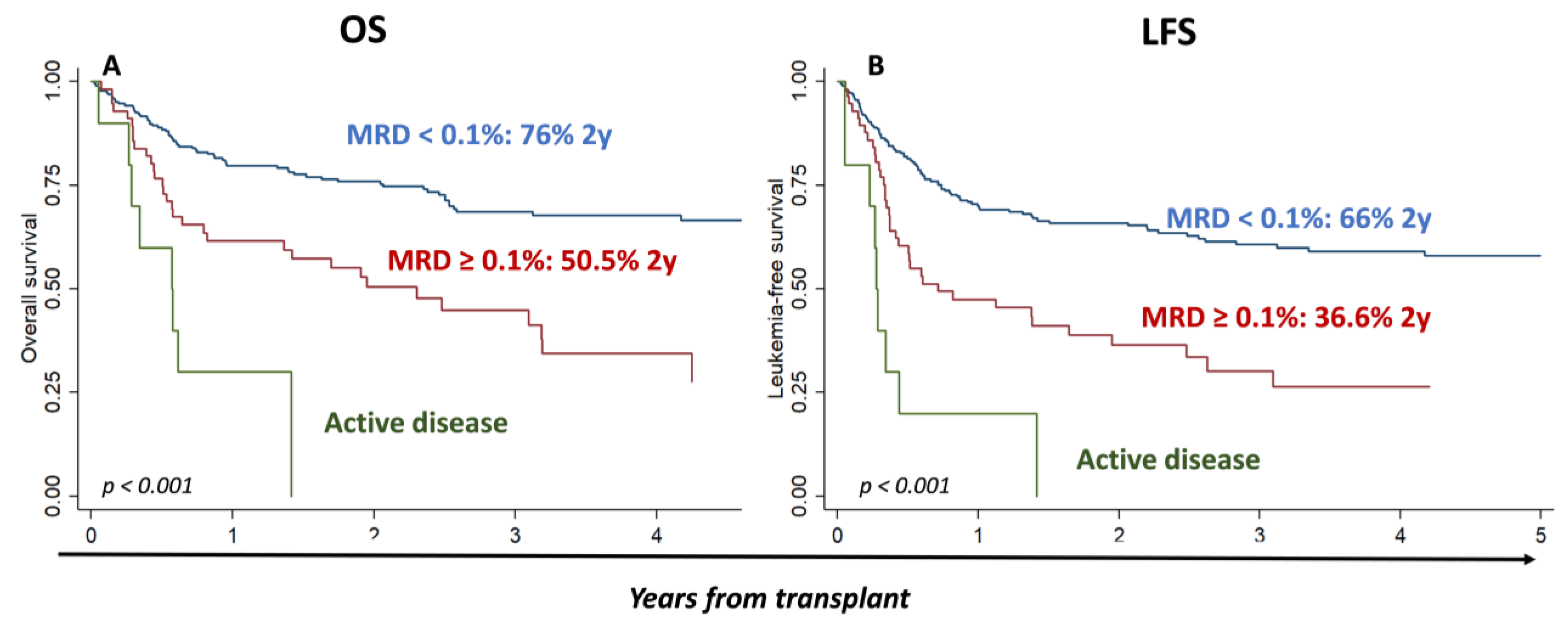
3.3. Impact of MRD Prior to Transplantation on Outcomes after HSCT

Overall survival at 2 and 5 years was 69% [95% CI 63.18–74.18] and 58.4% [95% CI 52.4–63.9], respectively, and the LFS at 2 and 5 years was 57.8% [95% CI 50.8−64.7%] and 50.4% [95% CI 44–57], respectively. Among patients in CR, MRD levels significantly influenced outcomes, with OS and LFS at 2 years of 76.7% [95% CI 70.1–82] and 67.6% [95% CI 60.6–73.6] for MRD neg patients, 68.5% [95% CI 42.1–84.7] and 49.7% [95% CI 26.4–69.3] for MRD-low patients, and 50.5% [95% CI 36–63.2] and 36.6% [95% CI 23.7–49.5] for MRD-high patients, respectively (MRD ≥ 0.1) (p < 0.001) (Supplementary Figure S2). Next, we attempted to confirm the prognostic value of the ELN17 proposed cut-off (0.1%) to identify patients at a higher risk of relapse and, therefore, we combined the MRD neg and MRD-low groups into MRD < 0.1: at 2 and 5 years, the respective values for these MRD < 0.1 patients were 76% [95% CI 69.7–81.1] and 66.7% [95% CI 59.1–73.1] for OS (p < 0.001 as compared to those with MRD ≥ 0.1), and 66% [95% CI 59.3–71.8] and 58.1% [95% CI 50.6–64.8] for LFS (p < 0.001 as compared to MRD ≥ 0.1), respectively. In Figure 1, the OS and LFS of the three groups MRD < 1, MRD ≥ 0.1, and active disease are shown (p < 0.001).
In univariate analysis, age (HR = 1.03 [95% CI 1.01–1.04], p = 0.002 and HR = 1.02 [95% CI 1.01–1.03], p = 0.008), conditioning (HR = 1.73 [95% CI 1.18–2.53], p = 0.005 and HR = 1.43 [95% CI 1.01–2.01], p = 0.041), adverse risk of ELN2011 (HR = 3.26 [95% CI 1.62–6.55], p = 0.001 and HR = 3.75 [95% CI 1.98–7.09], p < 0.001), and MRD ≥ 0.1 (HR = 2.51 [95% CI 1.64–3.83], p < 0.001 and HR = 2.29 [95% CI 1.55–3.38], p < 0.001) significantly influenced OS and LFS, respectively (Supplementary Table S4).
In multivariate analysis, adverse risk of ELN2011 (HR = 2.42 [95% CI 1.18–5.00], p = 0.016 for OS and HR = 3.16 [95% CI 1.64–6.07], p = 0.001 for LFS) and MRD ≥ 0.1 (HR = 2.07 [95% CI 1.26–3.39], p = 0.004 for OS and HR 2.1 [95% CI 1.36–3.29], p = 0.001 for LFS) significantly influenced OS and LFS (Table 3).
In multivariate time-dependent analysis (time-dependent variable GvHD), the adverse risk group, according to ELN2011 (for OS HR= 2.8 95% [CI 1.33–5.89], p = 0.006 and for LFS HR = 3.43 [95% CI 1.76–6.7], p < 0.001), and MRD ≥ 0.1 before transplant (for OS HR = 1.98 [95% CI 1.26–3.11], p < 0.001 and for LFS HR = 1.89 [95% CI 1.25–2.86], p < 0.001) significantly influenced the outcome (Supplementary Table S5).
Next, we attempted to identify whether these differences, in terms of OS and LFS between MRD ≥ 0.1 and MRD < 0.1, were due to a higher RI and/or NRM.
Considering only patients in CR, CIR at 2 and 5 years were significantly lower among MRD < 0.1 patients: 22% [95% CI 17–28.1%] and 27% [95% CI 21−33.5%], versus 46.5% [95% CI 32.4−59.5%] and 50% [95% CI 34.8−63.2%] among MRD ≥ 0.1 patients, respectively, p = 0.0005.
By contrast, no differences were observed in terms of NRM (11.7% [95% CI 7.9−16.3%] and 14.8% [95% CI 10.1–20.4%] for MRD < 0.1, and 16.9% [95% CI 8.2−28.3%] and 23.7% [95% CI 11.9−37.8%] for MRD ≥ 0.1 p = 0.2), (Figure 2).
In univariate analysis, age (HR = 1.02 [95% CI 1–1.04], p = 0.037), adverse risk of ELN2011 (HR = 4.88 [95% CI 1.99–11.99], p = 0.01), and MRD ≥ 0.1 (HR = 2.29 [95% CI 1.43–3.65], p = 0.001) significantly influenced CIR. As far as NRM is concerned, only the type of donor had a statistically significant influence in univariate analysis: unrelated donor (HR = 2.04 [95% CI 1.06–3.95], p = 0.033) and haplo-identical donor (HR = 2.73 [95% CI 1.16–6.42], p = 0.021) (Supplementary Table S4).
Finally, in multivariate analysis, haplo-identical donor (HR = −0.27 [95% CI 0.11–0.67], p = 0.005), T-cell depletion (HR 1.78 [95% CI 1.01–3.14], p = 0.045), adverse risk ELN2011 (HR = 4.37 [95% CI 1.67–11.4], p = 0.003), and MRD ≥ 0.1 (HR = 2.47 [95% CI 1.4–4.33], p = 0.002) significantly influenced CIR. On the other hand, the use of haplo-identical donor (HR = −2.63 [95% CI 1.04–6.65], p = 0.042) significantly influenced NRM (Table 3).
In Supplementary Table S6, we describe 43 patients who had MRD pos and did not relapse. Of these 43 patients, 30 (70%) developed acute GvHD (70%): 15 grade two, 4 grade four, and the rest grade one. Nineteen developed chronic GvHD (44.2%). Fourteen patients had MRD < 0.1, and three of them died because of an infection within one year after transplantation. The remaining 30 had MRD ≥ 0.1 and 11 (37%) of them have died, 8 before one year post-transplant because of infections (2 bilateral pneumonia), 2 due to GVHD, 1 due to veno-occlusive disease, 1 due to ureteral carcinoma, and another of unknown cause. Four of these thirty patients are still alive with a follow-up of less than 1 year.

3.4. Impact of MRD before Transplantation on OS and LFS among the Different ELN2011 Subgroups and Conditioning Regimens

MRD before transplantation also identified patients with different outcomes within the ELN2011 subgroups: OS and LFS at 2 years among high-risk ELN2011 patients were 58% and 41.1% in MRD < 0.1 vs. 39.3% and 20% for MRD ≥ 0.1 patients, p = 0.16 and p = 0.216, respectively; for intermediate risk: 77.6% and 70.2% for MRD < 0.1 vs. 60.6% and 43.5% among MRD ≥ 0.1 patients, p = 0.0.018 and p = 0.0037, respectively; and for favorable ELN2011 risk: 88.6% and 82.9% among MRD < 0.1 vs. 48.5% and 49.1% for patients with MRD ≥ 0.1, p= 0.0098 and p= 0.0315, respectively (Figure 3).
Finally, MRD level did influence the outcome, irrespective of the conditioning regimen: patients with MRD < 0.1 before transplantation had a better OS and LFS at 2 years (82% and 71.4% among those who received myeloablative conditioning, and 65% and 57.6% among those who received reduced intensity conditioning, respectively) than those who had MRD ≥ 0.1 prior to transplantation (56% and 44.4% for myeloablative, and 43% and 25.5% for those who received reduced intensity conditioning) (p < 0.001) (Figure 4).

3.5. Impact of MRD on 100 Days after HSCT

Next, we analyzed the prognostic value of MRD at 100 days post transplantation; at 2 years, OS and LFS were significantly higher in patients with MRD < 0.1 on day +100 (83% and 76% in MRD < 0.1 vs. 76% and 7% in MRD ≥ 0.1, respectively, p < 0.001) (Figure 5). Similarly, CIR at 2 years was significantly lower in patients with MRD < 0.1 (12.8% [95% CI 8.4–18%] as compared to 93.3% [95% CI 41.4%−99.5%]) among those with MRD ≥ 0.1, p < 0.001. By contrast, no differences were observed in terms of NRM (10.6% [95% CI 6.7–15.6] in MRD < 0.1 vs. 0% in in MRD ≥ 0.1, p = 0.19).
However, the main limitation for the analysis of the MRD at 100 days after transplantation is that only 16 patients with positive MRD remained in CR at this time-point.

4. Discussion

Numerous studies have described the prognostic value of measurable residual disease after induction and/or consolidation [18,19,20,21,22,23,24,25,26,27] and, based on these results, it is currently used to tailor the intensification strategy in patients with AML [21]. Terwijn et al. showed the independent prognostic value of MRD ≥ 0.1% by MCF after induction and consolidation therapy, identifying a subgroup of patients with higher risk of relapse [22]. Similarly, a second multicenter prospective study identified that patients with positive MRD had a poor outcome [23]. On the other hand, Balsat et al. showed the prognostic value of the detection of mutated NPM1 in peripheral blood after induction, thus identifying a subgroup of patients who would be candidates for transplantation [24]. Likewise, RUNX1-RUNX1T1 MRD monitoring also identifies a subgroup of patients at high risk of relapse [25]. Moreover, Jongen-Lavrencic et al. showed that the combination of detection of MRD by next-generation sequencing and flow cytometry has additional prognostic value in terms of relapse and overall survival [26]. Based on these findings, Cornelissen et al. proposed an algorithm, considering not only the risk subgroup at diagnosis and the performance status of the patients, but also the levels of MRD [27].
Unfortunately, although HSCT is considered the best option for patients with persistent MRD after first line treatment, tumor load, even at the MRD level, is one of the variables with the highest impact on outcome, after allogeneic stem cell transplantation, as described in different studies using either molecular or flow cytometry techniques. Regarding the latter, available studies have not used international standardized protocols; different cut-off values have been used, and the number of combinations of the fluorochrome-conjugated antibodies used, or the analysis strategy, have also greatly varied. Likewise, the need to maintain calibration and compensation protocols is not considered. Araki et al. showed that patients in CR with positive MRD at the time of transplantation had a similar outcome compared to patients with active disease with OS at 3 years, of 26% and 23%, respectively [14]. In this study, 1 million events were acquired, and a panel of 10 colors with three combinations of antibodies is carried out, considering any level of detection to determine positive MRD. However, it is a single center study conducted by the same group of hematopathologists. Additionally, a different from normal approach is used, considering a population showing a deviation from antigen expression patterns on reactive or regeneration hematopoietic cells. However, this approach might not display a uniform sensitivity in all cases. Moreover, it is not considered an international standardization of cytometers, allowing an adequate reproducibility within different laboratories. Likewise, in the same group in the context of myeloablative transplantation, the presence of MRD before or after transplantation identified patients with poor outcome [13] As in our study, they observed that 16 patients with MRD pos after transplantation had shorter OS and RFS. However, the MRD measurement is performed at an earlier post-transplant period, between days +28 and +35. Other studies that show the prognostic value of positive MRD use numbers of fluorescence less than six [17,28,29], or combine the detection of disease with low-sensitivity techniques, such as karyotype or fluorescence in situ hybridization (FISH) [28]. In a recent retrospective study, patients with monosomal karyotype had a higher likelihood of MRD positivity by MFC prior to transplant, with worse OS and higher relapse risk than those without it [12]. However, in multivariate analysis, only MRD positivity was associated with a shorter survival.
The current study has been conducted in routine clinical practice, in real life, outside of a specific trial, in four centers that had to meet certain requirements. Three laboratories participated in the quality control organized within the Euroflow QA program. Two different approaches were used to determinate MRD: “leukemia-associated phenotype (LAP)” and “different from normal (DfN)”. The latter allowed us to identify MRD even if the diagnostic phenotype was not available, or in case of changes in the phenotype during the disease [30,31,32]. Both strategies have been used, as recommended by ELN [1,6]. All laboratories work with the same quality criteria, and each one carried out its own analyses by experts in clinical cytometry.
In the current study, we confirmed the prognostic value of MRD monitoring, both pre- and post-allogeneic stem cell transplantation. In fact, cumulative incidence of relapse was significantly higher, both if we consider patients with MRD pos as compared to MRD neg, and by using the cut-off value suggested by ELN, and this difference in relapse incidence significantly influenced OS and LFS.
Considering ELN2011 subgroups, MRD also allowed the identification of subgroups of patients with different prognoses among favorable and intermediate ELN2011, while in the adverse risk group, no clear difference was observed. The number of patients included in each subgroup, when the analysis is separately performed, might explain the lack of statistically significant differences in this subgroup, in terms of overall survival. Alternatively, the poor outcome of these patients might not be influenced by the MRD levels. Further studies would be required to elucidate this point.
An area of current interest, in the context of allogeneic transplantation, is whether the therapeutic strategy should be modified in those patients with positive MRD, before transplantation. In fact, several studies have suggested that patients with positive MRD prior to transplant might benefit from receiving myeloablative conditionings [17,33,34]. However, in a recent retrospective study of 746 patients, Morsink et al. observed that in all patients, regardless of the type of conditioning, the risk of relapse, relapse free survival, and OS were higher in patients with positive, as compared to those with negative, MRD [11]. Similarly, no effect of the conditioning regimen intensity on OS was observed in NPM1 mutated AML patients who remained MRD positive [35]. In the same way, in the current analysis, regardless of the type of conditioning, the presence of MRD by flow cytometry implied a poor prognosis. Recently, Paras et al. conducted a retrospective study of 810 patients, showing that the conditioning regimen can influence the ability to eliminate MRD; however, the elimination of MRD in non-myeloablative regimens had a greater impact on the outcome [36]. With these contradictory results, it remains unclear whether MRD should determine the choice of conditioning regimen. An alternative approach would be to identify these patients for alternative therapy after HSCT, such as withdraw of immunosuppression, hypomethylating agents [37], infusion of donor lymphocytes [38], or venetoclax [39], among other approaches. For example, the RELAZA2 prospective study demonstrated that treatment with azacytidine [37] in patients with MRD pos could avoid or delay hematology relapse of AML or high-risk myelodysplastic syndromes (MDS). In this regard, MRD monitoring after transplantation would further allow the identification of patients at a high risk of relapse, and establish therapeutic maneuvers.
In our patient’s cohort, positive MRD on day +100 after transplantation was associates with an extremely poor prognosis, with a CIR of 93.3%. It is possible that the identification of MRD should be conducted in an earlier post-transplant period, to assess any preventive strategy to avoid relapse. Moreover, in our study, those patients who reach day +100 post-transplant and persist, or have a new positive MRD, have an unfavorable short-term prognosis, regardless of the status of the pre-transplant MRD. In this way, a recent study of Paras et al. showed that the dynamics of MRD before, and early after transplantation (+20 or 40 days after transplantation), improve the accuracy of risk assessment; in that study, patients with MRDpos/MRDpos and MRDneg/MRDpos had high relapse rates and poor survival estimates [36].
In relation to the type of donor, is relevant to point out that Yu et al. have shown that a haplo-identical donor might increase the graft versus leukemia effect [40]. Therefore, the positivity of MRD before transplant could modify the selection of the donor.
Other authors have identified the CD34+/CD38– leukemic stem cell (LSC) phenotype, and have shown its prognostic value, both at diagnosis and during follow-up [41,42,43,44]. Furthermore, this phenotype might be used in combination with the MRD assessment in a single tube combining six markers [43]. Therefore, future studies might allow the further improvement of MRD analysis, for example, by incorporating the LSC phenotype into the standardized MRD strategy currently used.
The main limitation of this study is its retrospective nature over a long period of time, in which changes have been incorporated to the treatment according to the molecular results. Likewise, GvHD conditioning and prophylaxis vary during this period. On the other hand, each center carried out its analysis independently. However, this further reinforces the fact that a positive MRD performed with standardized technical conditions has prognostic value in real life.

5. Conclusions

Our multicenter study confirms the prognostic value of MRD performed in accordance with standardized recommendations. Regardless of the conditioning regimen, positive MRD maintains its poor prognostic value, and might allow the identification of patients who are candidates to receive early post-transplant therapeutic procedures.

Supplementary Materials

The following supporting information can be downloaded at: https://www.mdpi.com/article/10.3390/cancers15051609/s1, Figure S1: Representative image of MRD analysis by flow cytometry. Figure S2: Impact of MRD levels in OS and LFS. Table S1. The information per fluorchrome from QA program, Table S2. Designed panels to LAIP analysis, Table S3. Comparative analysis of MRD samples in the 4 laboratories: The table represents the MRD value determined by each laboratory, Table S4. Univariate analysis of CIR, NRM, OS and LFS, Table S5. OS and LFS according to multivariate time-dependent analysis (time-dependent variable GvHD, Table S6. Evolution of patients who did not relapse with positive MRD prior to transplantation.

Author Contributions

T.C.-V. and J.A.P.-S. conceived the idea and designed the study. T.C.-V., O.P.-L., A.Y.B., E.C.V., A.S.T., M.B.V., M.R.S., C.Q.C., E.P.L., M.S.-R., C.P.-M., P.M. and J.A.P.-S. supplied study material or patients. T.C.-V., O.P.-L., A.Y.B., E.C.V., A.S.T., M.B.V., C.Q.C. and C.P.-M. analyzed flow cy-tometry data. T.C.-V. and O.P.-L. performed statistical analysis. T.C.-V., O.P.-L., E.R.A. and J.A.P.-S. analyzed and interpreted data. T.C.-V. and J.A.P.-S. wrote the manuscript. All authors have read and agreed to the published version of the manuscript.

Funding

This work was supported by Instituto de Salud Carlos III/Subdirección General de Investigación Sanitaria Fondo de Investigación en Salud (proyect PI17/02283), Red de terapia celular (TERCEL RD16/0011/0035), and RICORS (RD21/0017/0016).

Institutional Review Board Statement

C.P. S2200072, C.I. 0466-N-22, CEI de los Hospitales Universitarios Virgen Macarena y Virgen del Rocío (Protocolo V.1-21 February 2022).

Informed Consent Statement

All patients provided written informed consent in accordance with the Declaration of Helsinki. This study has been approved by a formally constituted review board: C.P. S2200072, C.I. 0466-N-22, CEI de los Hospitales Universitarios Virgen Macarena y Virgen del Rocío (Protocolo V.1-21 February 2022).

Data Availability Statement

The original data will be available by request to the corresponding author.

Acknowledgments

This work was supported by Instituto de Salud Carlos III/Subdirección General de Investigación Sanitaria Fondo de Investigación en Salud (proyect PI17/02283), Red de terapia celular (TERCEL RD16/0011/0035), and RICORS (RD21/0017/0016). The authors TCV, OPL, AYB, ECV, AST, MBV, and CPM belong to Grupo Español de Citometría de Flujo Hematológica. The authors declare that they have no conflict of interest.

Conflicts of Interest

The authors declare no conflict of interest.

References

  1. Döhner, H.; Estey, E.; Grimwade, D.; Amadori, S.; Appelbaum, F.R.; Büchner, T.; Dombret, H.; Ebert, B.L.; Fenaux, P.; Larson, R.A.; et al. Diagnosis and management of AML in adults: 2017 ELN recommendations from an international expert panel. Blood 2017, 129, 424–447. [Google Scholar] [CrossRef] [Green Version]
  2. Grimm, J.; Jentzsch, M.; Bill, M.; Goldmann, K.; Schulz, J.; Niederwieser, D.; Platzbecker, U.; Schwind, S. Prognostic impact of the ELN2017 risk classification in patients with AML receiving allogeneic transplantation. Blood Adv. 2020, 4, 3864–3874. [Google Scholar] [CrossRef] [PubMed]
  3. Riva, G.; Nasillo, V.; Ottomano, A.M.; Bergonzini, G.; Paolini, A.; Forghieri, F.; Lusenti, B.; Barozzi, P.; Lagreca, I.; Fiorcari, S.; et al. Multiparametric Flow Cytometry for MRD Monitoring in Hematologic Malignancies: Clinical Applications and New Challenges. Cancers 2021, 13, 4582. [Google Scholar] [CrossRef] [PubMed]
  4. Flores-Montero, J.; Sanoja-Flores, L.; Paiva, B.; Puig, N.; García-Sánchez, O.; Böttcher, S.; van der Velden, V.H.J.; Pérez-Morán, J.J.; Vidriales, M.B.; García-Sanz, R.; et al. Next Generation Flow for highly sensitive and standardized detection of minimal residual disease in multiple myeloma. Leukemia 2017, 31, 2094–2103. [Google Scholar] [CrossRef] [Green Version]
  5. Theunissen, P.; Mejstrikova, E.; Sedek, L.; van der Sluijs-Gelling, A.J.; Gaipa, G.; Bartels, M.; Sobral da Costa, E.; Kotrová, M.; Novakova, M.; Sonneveld, E.; et al. EuroFlow Consortium. Standardized flow cytometry for highly sensitive MRD measurements in B-cell acute lymphoblastic leukemia. Blood 2017, 129, 347–357. [Google Scholar] [CrossRef]
  6. Heuser, M.; Freeman, S.D.; Ossenkoppele, G.J.; Buccisano, F.; Hourigan, C.S.; Ngai, L.L.; Tettero, J.M.; Bachas, C.; Baer, C.; Béné, M.C.; et al. 2021 Update on MRD in acute myeloid leukemia: A consensus document from the European LeukemiaNet MRD Working Party. Blood 2021, 138, 2753–2767. [Google Scholar] [CrossRef]
  7. Kalina, T.; Flores-Montero, J.; van der Velden, V.H.; Martin-Ayuso, M.; Böttcher, S.; Ritgen, M.; Almeida, J.; Lhermitte, L.; Asnafi, V.; Mendonça, A.; et al. EuroFlow Consortium (EU-FP6, LSHB-CT-2006-018708). EuroFlow standardization of flow cytometer instrument settings and immunophenotyping protocols. Leukemia 2012, 26, 1986–2010. [Google Scholar] [CrossRef] [PubMed] [Green Version]
  8. van Dongen, J.J.; Lhermitte, L.; Böttcher, S.; Almeida, J.; van der Velden, V.H.; Flores-Montero, J.; Rawstron, A.; Asnafi, V.; Lécrevisse, Q.; Lucio, P.; et al. EuroFlow Consortium (EU-FP6, LSHB-CT-2006-018708). EuroFlow antibody panels for standardized n-dimensional flow cytometric immunophenotyping of normal, reactive and malignant leukocytes. Leukemia 2012, 26, 1908–1975. [Google Scholar] [CrossRef] [Green Version]
  9. Döhner, H.; Wei, A.H.; Appelbaum, F.R.; Craddock, C.; DiNardo, C.D.; Dombret, H.; Ebert, B.L.; Fenaux, P.; Godley, L.A.; Hasserjian, R.P.; et al. Diagnosis and management of AML in adults: 2022 recommendations from an international expert panel on behalf of the ELN. Blood 2022, 140, 1345–1377. [Google Scholar] [CrossRef] [PubMed]
  10. Walter, R.B.; Gooley, T.A.; Wood, B.L.; Milano, F.; Fang, M.; Sorror, M.L.; Estey, E.H.; Salter, A.I.; Lansverk, E.; Chien, J.W.; et al. Impact of pretransplantation minimal residual disease, as detected by multiparametric flow cytometry, on outcome of myeloablative hematopoietic cell transplantation for acute myeloid leukemia. J. Clin. Oncol. 2011, 29, 1190–1197. [Google Scholar] [CrossRef] [PubMed] [Green Version]
  11. Morsink, L.M.; Sandmaier, B.M.; Othus, M.; Palmieri, R.; Granot, N.; Bezerra, E.D.; Wood, B.L.; Mielcarek, M.; Schoch, G.; Davis, C.; et al. Conditioning Intensity, Pre-Transplant Flow Cytometric Measurable Residual Disease, and Outcome in Adults with Acute Myeloid Leukemia Undergoing Allogeneic Hematopoietic Cell Transplantation. Cancers 2020, 12, 2339. [Google Scholar] [CrossRef] [PubMed]
  12. Morsink, L.M.; Othus, M.; Bezerra, E.D.; Wood, B.L.; Fang, M.; Sandmaier, B.M.; Mielcarek, M.; Schoch, G.; Storb, R.; Deeg, H.J.; et al. Impact of pretransplant measurable residual disease on the outcome of allogeneic hematopoietic cell transplantation in adult monosomal karyotype AML. Leukemia 2020, 34, 1577–1587. [Google Scholar] [CrossRef] [PubMed]
  13. Zhou, Y.; Othus, M.; Araki, D.; Wood, B.L.; Radich, J.P.; Halpern, A.B.; Mielcarek, M.; Estey, E.H.; Appelbaum, F.R.; Walter, R.B. Pre- and post-transplant quantification of measurable (‘minimal’) residual disease via multiparameter flow cytometry in adult acute myeloid leukemia. Leukemia 2016, 30, 1456–1464. [Google Scholar] [CrossRef]
  14. Araki, D.; Wood, B.L.; Othus, M.; Radich, J.P.; Halpern, A.B.; Zhou, Y.; Mielcarek, M.; Estey, E.H.; Appelbaum, F.R.; Walter, R.B. Allogeneic Hematopoietic Cell Transplantation for Acute Myeloid Leukemia: Time to Move Toward a Minimal Residual Disease-Based Definition of Complete Remission? J. Clin. Oncol. 2016, 34, 329–336. [Google Scholar] [CrossRef] [Green Version]
  15. Walter, R.B.; Gyurkocza, B.; Storer, B.E.; Godwin, C.D.; Pagel, J.M.; Buckley, S.A.; Sorror, M.L.; Wood, B.L.; Storb, R.; Appelbaum, F.R.; et al. Comparison of minimal residual disease as outcome predictor for AML patients in first complete remission undergoing myeloablative or nonmyeloablative allogeneic hematopoietic cell transplantation. Leukemia 2015, 29, 137–144. [Google Scholar] [CrossRef] [Green Version]
  16. Walter, R.B.; Buckley, S.A.; Pagel, J.M.; Wood, B.L.; Storer, B.E.; Sandmaier, B.M.; Fang, M.; Gyurkocza, B.; Delaney, C.; Radich, J.P.; et al. Significance of minimal residual disease before myeloablative allogeneic hematopoietic cell transplantation for AML in first and second complete remission. Blood 2013, 122, 1813–1821. [Google Scholar] [CrossRef]
  17. Ustun, C.; Courville, E.L.; DeFor, T.; Dolan, M.; Randall, N.; Yohe, S.; Bejanyan, N.; Warlick, E.; Brunstein, C.; Weisdorf, D.J.; et al. Myeloablative, but not Reduced-Intensity, Conditioning Overcomes the Negative Effect of Flow-Cytometric Evidence of Leukemia in Acute Myeloid Leukemia. Biol. Blood Marrow Transplant. 2016, 22, 669–675. [Google Scholar] [CrossRef] [Green Version]
  18. Döhner, H.; Estey, E.H.; Amadori, S.; Appelbaum, F.R.; Büchner, T.; Burnett, A.K.; Dombret, H.; Fenaux, P.; Grimwade, D.; Larson, R.A.; et al. European LeukemiaNet. Diagnosis and management of acute myeloid leukemia in adults: Recommendations from an international expert panel, on behalf of the European LeukemiaNet. Blood 2010, 115, 453–474. [Google Scholar] [CrossRef] [Green Version]
  19. San Miguel, J.F.; Vidriales, M.B.; López-Berges, C.; Díaz-Mediavilla, J.; Gutiérrez, N.; Cañizo, C.; Ramos, F.; Calmuntia, M.J.; Pérez, J.J.; González, M.; et al. Early immunophenotypical evaluation of minimal residual disease in acute myeloid leukemia identifies different patient risk groups and may contribute to postinduction treatment stratification. Blood 2001, 98, 1746–1751. [Google Scholar] [CrossRef] [PubMed] [Green Version]
  20. Vidriales, M.B.; Pérez-López, E.; Pegenaute, C.; Castellanos, M.; Pérez, J.J.; Chandía, M.; Díaz-Mediavilla, J.; Rayón, C.; de Las Heras, N.; Fernández-Abellán, P.; et al. PETHEMA Programa para el Estudio de la Terapéutica en Hemopatías Malignas Cooperative Study Group. Minimal residual disease evaluation by flow cytometry is a complementary tool to cytogenetics for treatment decisions in acute myeloid leukaemia. Leuk. Res. 2016, 40, 1–9. [Google Scholar] [CrossRef]
  21. Venditti, A.; Piciocchi, A.; Candoni, A.; Melillo, L.; Calafiore, V.; Cairoli, R.; de Fabritiis, P.; Storti, G.; Salutari, P.; Lanza, F.; et al. GIMEMA AML1310 trial of risk-adapted, MRD-directed therapy for young adults with newly diagnosed acute myeloid leukemia. Blood 2019, 134, 935–945. [Google Scholar] [CrossRef] [PubMed]
  22. Terwijn, M.; van Putten, W.L.; Kelder, A.; van der Velden, V.H.; Brooimans, R.A.; Pabst, T.; Maertens, J.; Boeckx, N.; de Greef, G.E.; Valk, P.J.; et al. High prognostic impact of flow cytometric minimal residual disease detection in acute myeloid leukemia: Data from the HOVON/SAKK AML 42A study. J. Clin. Oncol. 2013, 31, 3889–3897. [Google Scholar] [CrossRef] [PubMed]
  23. Freeman, S.D.; Virgo, P.; Couzens, S.; Grimwade, D.; Russell, N.; Hills, R.K.; Burnett, A.K. Prognostic relevance of treatment response measured by flow cytometric residual disease detection in older patients with acute myeloid leukemia. J. Clin. Oncol. 2013, 31, 4123–4131. [Google Scholar] [CrossRef]
  24. Balsat, M.; Renneville, A.; Thomas, X.; de Botton, S.; Caillot, D.; Marceau, A.; Lemasle, E.; Marolleau, J.P.; Nibourel, O.; Berthon, C.; et al. Postinduction Minimal Residual Disease Predicts Outcome and Benefit From Allogeneic Stem Cell Transplantation in Acute Myeloid Leukemia With NPM1 Mutation: A Study by the Acute Leukemia French Association Group. J. Clin. Oncol. 2017, 35, 185–193. [Google Scholar] [CrossRef] [PubMed]
  25. Rücker, F.G.; Agrawal, M.; Corbacioglu, A.; Weber, D.; Kapp-Schwoerer, S.; Gaidzik, V.I.; Jahn, N.; Schroeder, T.; Wattad, M.; Lübbert, M.; et al. Measurable residual disease monitoring in acute myeloid leukemia with t(8;21)(q22;q22.1): Results from the AML Study Group. Blood 2019, 134, 1608–1618. [Google Scholar] [CrossRef] [PubMed]
  26. Jongen-Lavrencic, M.; Grob, T.; Hanekamp, D.; Kavelaars, F.G.; Al Hinai, A.; Zeilemaker, A.; Erpelinck-Verschueren, C.A.J.; Gradowska, P.L.; Meijer, R.; Cloos, J.; et al. Molecular Minimal Residual Disease in Acute Myeloid Leukemia. N. Engl. J. Med. 2018, 378, 1189–1199. [Google Scholar] [CrossRef]
  27. Cornelissen, J.J.; Gratwohl, A.; Schlenk, R.F.; Sierra, J.; Bornhäuser, M.; Juliusson, G.; Råcil, Z.; Rowe, J.M.; Russell, N.; Mohty, M.; et al. The European LeukemiaNet AML Working Party consensus statement on allogeneic HSCT for patients with AML in remission: An integrated-risk adapted approach. Nat. Rev. Clin. Oncol. 2012, 9, 579–590. [Google Scholar] [CrossRef] [Green Version]
  28. Norkin, M.; Katragadda, L.; Zou, F.; Xiong, S.; Chang, M.; Dai, Y.; Hsu, J.W.; Moreb, J.S.; Leather, H.; Murthy, H.S.; et al. Minimal residual disease by either flow cytometry or cytogenetics prior to an allogeneic hematopoietic stem cell transplant is associated with poor outcome in acute myeloid leukemia. Blood Cancer J. 2017, 7, 634. [Google Scholar] [CrossRef] [Green Version]
  29. Maurillo, L.; Buccisano, F.; Del Principe, M.I.; Del Poeta, G.; Spagnoli, A.; Panetta, P.; Ammatuna, E.; Neri, B.; Ottaviani, L.; Sarlo, C.; et al. Toward optimization of postremission therapy for residual disease-positive patients with acute myeloid leukemia. J. Clin. Oncol. 2008, 26, 4944–4951. [Google Scholar] [CrossRef] [Green Version]
  30. Baer, M.R.; Stewart, C.C.; Dodge, R.K.; Leget, G.; Sulé, N.; Mrózek, K.; Schiffer, C.A.; Powell, B.L.; Kolitz, J.E.; Moore, J.O.; et al. High frequency of immunophenotype changes in acute myeloid leukemia at relapse: Implications for residual disease detection (Cancer and Leukemia Group B Study 8361). Blood 2001, 97, 3574–3580. [Google Scholar] [CrossRef] [Green Version]
  31. Langebrake, C.; Brinkmann, I.; Teigler-Schlegel, A.; Creutzig, U.; Griesinger, F.; Puhlmann, U.; Reinhardt, D. Immunophenotypic differences between diagnosis and relapse in childhood AML: Implications for MRD monitoring. Cytom. B Clin. Cytom. 2005, 63, 1–9. [Google Scholar] [CrossRef]
  32. Al-Mawali, A.; Gillis, D.; Hissaria, P.; Lewis, I. Incidence, sensitivity, and specificity of leukemia-associated phenotypes in acute myeloid leukemia using specific five-color multiparameter flow cytometry. Am. J. Clin. Pathol. 2008, 129, 934–945. [Google Scholar] [CrossRef]
  33. Hourigan, C.S.; Dillon, L.W.; Gui, G.; Logan, B.R.; Fei, M.; Ghannam, J.; Li, Y.; Licon, A.; Alyea, E.P.; Bashey, A.; et al. Impact of Conditioning Intensity of Allogeneic Transplantation for Acute Myeloid Leukemia With Genomic Evidence of Residual Disease. J. Clin. Oncol. 2020, 38, 1273–1283. [Google Scholar] [CrossRef] [PubMed]
  34. Gilleece, M.H.; Labopin, M.; Yakoub-Agha, I.; Volin, L.; Socié, G.; Ljungman, P.; Huynh, A.; Deconinck, E.; Wu, D.; Bourhis, J.H.; et al. Measurable residual disease, conditioning regimen intensity, and age predict outcome of allogeneic hematopoietic cell transplantation for acute myeloid leukemia in first remission: A registry analysis of 2292 patients by the Acute Leukemia Working Party European Society of Blood and Marrow Transplantation. Am. J. Hematol. 2018, 93, 1142–1152. [Google Scholar] [PubMed] [Green Version]
  35. Dillon, R.; Hills, R.; Freeman, S.; Potter, N.; Jovanovic, J.; Ivey, A.; Kanda, A.S.; Runglall, M.; Foot, N.; Valganon, M.; et al. Molecular MRD status and outcome after transplantation in NPM1-mutated AML. Blood 2020, 135, 680–688. [Google Scholar] [CrossRef] [PubMed] [Green Version]
  36. Paras, G.; Morsink, L.M.; Othus, M.; Milano, F.; Sandmaier, B.M.; Zarling, L.C.; Palmieri, R.; Schoch, G.; Davis, C.; Bleakley, M.; et al. Conditioning intensity and peritransplant flow cytometric MRD dynamics in adult AML. Blood 2022, 139, 1694–1706. [Google Scholar] [CrossRef]
  37. Platzbecker, U.; Middeke, J.M.; Sockel, K.; Herbst, R.; Wolf, D.; Baldus, C.D.; Oelschlägel, U.; Mütherig, A.; Fransecky, L.; Noppeney, R.; et al. Measurable residual disease-guided treatment with azacitidine to prevent haematological relapse in patients with myelodysplastic syndrome and acute myeloid leukaemia (RELAZA2): An open-label, multicentre, phase 2 trial. Lancet Oncol. 2018, 19, 1668–1679. [Google Scholar] [CrossRef]
  38. Schmid, C.; Labopin, M.; Nagler, A.; Bornhäuser, M.; Finke, J.; Fassas, A.; Volin, L.; Gürman, G.; Maertens, J.; Bordigoni, P.; et al. EBMT Acute Leukemia Working Party. Donor lymphocyte infusion in the treatment of first hematological relapse after allogeneic stem-cell transplantation in adults with acute myeloid leukemia: A retrospective risk factors analysis and comparison with other strategies by the EBMT Acute Leukemia Working Party. J. Clin. Oncol. 2007, 25, 4938–4945. [Google Scholar] [PubMed]
  39. Todisco, E.; Gigli, F.; Sammassimo, S.; Camisaschi, C.; Mancuso, P.; Ronchini, C.; Ramadan, S.; Bertolini, F.; Pastano, R.; Tarella, C. Efficacy of venetoclax based salvage chemotherapy followed by “Minimal Residual Disease driven”-venetoclax maintenance therapy post-allotransplant in a young patient with high-risk primary refractory acute myeloid leukemia. Leuk. Lymphoma 2020, 61, 2277–2279. [Google Scholar] [CrossRef]
  40. Yu, S.; Huang, F.; Wang, Y.; Xu, Y.; Yang, T.; Fan, Z.; Lin, R.; Xu, N.; Xuan, L.; Ye, J.; et al. Haploidentical transplantation might have superior graft-versus-leukemia effect than HLA-matched sibling transplantation for high-risk acute myeloid leukemia in first complete remission: A prospective multicentre cohort study. Leukemia 2020, 34, 1433–1443. [Google Scholar] [CrossRef]
  41. Zeijlemaker, W.; Grob, T.; Meijer, R.; Hanekamp, D.; Kelder, A.; Carbaat-Ham, J.C.; Oussoren-Brockhoff, Y.J.M.; Snel, A.N.; Veldhuizen, D.; Scholten, W.J.; et al. CD34+CD38- leukemic stem cell frequency to predict outcome in acute myeloid leukemia. Leukemia 2019, 33, 1102–1112. [Google Scholar] [CrossRef]
  42. Plesa, A.; Dumontet, C.; Mattei, E.; Tagoug, I.; Hayette, S.; Sujobert, P.; Tigaud, I.; Pages, M.P.; Chelghoum, Y.; Baracco, F.; et al. High frequency of CD34+CD38-/low immature leukemia cells is correlated with unfavorable prognosis in acute myeloid leukemia. World J. Stem Cells 2017, 9, 227–234. [Google Scholar] [CrossRef]
  43. Kandeel, E.Z.; El Sharkawy, N.; Hanafi, M.; Samra, M.; Kamel, A. Tracing Leukemia Stem Cells and Their Influence on Clinical Course of Adult Acute Myeloid Leukemia. Clin. Lymphoma Myeloma Leuk. 2020, 20, 383–393. [Google Scholar] [CrossRef]
  44. Zeijlemaker, W.; Kelder, A.; Oussoren-Brockhoff, Y.J.; Scholten, W.J.; Snel, A.N.; Veldhuizen, D.; Cloos, J.; Ossenkoppele, G.J.; Schuurhuis, G.J. A simple one-tube assay for immunophenotypical quantification of leukemic stem cells in acute myeloid leukemia. Leukemia 2016, 30, 439–446. [Google Scholar] [CrossRef]
Figure 1. Impact of MRD levels in OS and LFS, considering MRD < 0.1%, MRD ≥ 0.1%, and group of active disease.
Cancers 15 01609 g001
Figure 2. Impact of MRD in CIR and NRM: (A) CIR at 2 and 5 years were significantly lower among those with MRD ≥ 0.1. (B) However, no differences were observed in terms of non-relapse mortality.
Cancers 15 01609 g002
Figure 3. Impact of MRD in OS and LFS according to risk group ELN2011: survival was significantly worse in patients with MRD ≥ 0.1 in favorable (A,D) and intermediate groups (B,E). However, no differences were observed in the adverse risk group (C,F).
Cancers 15 01609 g003
Figure 4. Impact of MRD in OS and LFS, according to conditioning regimen. Patients with MRD < 0.1 before transplantation had a better OS (A) and LFS (B) at 2 years, with both types of conditioning.
Cancers 15 01609 g004
Figure 5. Impact of MRD on day +100 after transplantation. At 2 years, OS and LFS were significantly higher in patients with MRD ≥ 0.1 on day +100.
Cancers 15 01609 g005
Table 1. Characteristics of the patients according to MRD before transplantation.
All Group
(n = 295)
MRD Negative
(n = 207)
MRD Positive
(n = 78)
p-Value
AGE mean (range)51 (2–71)51 (2–71)54.5 (18–69) 0.02
Recipient Gender, n (%)
Male
 
148(50.2)
 
102(49.3)
 
40(51.3)
 
0.63
ELN2011, n (%)
favorable
intermediate I
intermediate II
adverse
 
47 (15.9)
57 (19.3)
111 (37.6)
70 (23.7)
 
31 (15)
48 (23.2)
79 (38.2)
44 (21.3)
 
15 (19.2)
8 (10.3)
30 (38.5)
21 (28.2)
 
 
0.217
Disease Status at transplant, n (%)
1st CR
2nd CR
Others CR
Active disease
Aplasia
 
235 (79.7)
28 (9.5)
19 (6.4)
10 (3.4)
3 (1)
 
176 (85)
17 (8.2)
13 (6.3)
1 (0.5)
0
 
59 (77.6)
11 (14.5)
6 (7.9)
0
2
 
0.271
Donor, n (%)
Matched sibling
Unrelated donor
Haplo-identical donor
 
139 (47.1)
117 (39.7)
38 (12.9)
 
103 (49.8)
82 (39.6)
22 (10.6)
 
31 (39.7)
30 (38.5)
16 (20.5)
 
 
0.092
Conditioning, n (%)
Myeloablative
No myeloablative
 
176 (59.7)
117 (40.4)
 
119 (57.5)
88 (42.5)
 
50 (64.1)
28 (35.9)
 
0.781
Conditioning Therapy, n (%)
BUCy
BUCy + Thiothepa
FLUBU
FLUBU + Cy
FLUBU + THIOTHEPA
FLUBU + THIOTHEPA + ATG
FLUBU + ATG
Cy TBI ± ATG
FLUMEL ± Thiothepa
Others
 
56 (19)
3 (1)
162 (54.9%)
8 (2.7%)
42 (14.2)
2 (0.7)
2 (0.7)
5 (1.3)
7 (2.3)
8 (2.7)
 
44 (21.3)
1 (0.5)
113 (54.6)
5 (2.4)
33 (15.9)
2 (1)
1 (0.5)
2 (1)
2 (1)
4 (1.9)
 
10 (12.8)
0 (0)
44 (56.4)
3 (3.8)
9 (11.5)
0 (0)
1 (1.3)
2 (2.6)
5 (6.4)
4 (5.2)


0.385
Donor gender, n (%)
Male
 
192 (65.1)
 
135 (65.2)
 
51 (65.4)
 
0.945
Donor/Recipient gender, n (%)
Female/male
 
52 (17.6)
 
38 (18.4)
 
12 (15.4)
 
0.807
GvHD Prophylaxis, n (%)
Tacrolimus/CsA + MTX
Tacrolimus + Sirolimus ± MMF
Tacrolimus/CsA + MTX + ATG
Tacrolimus/CsA + MMF
Tacrolimus/CsA + MMF + Cy
Sirolimus + MMF + Cy
CsA + Pred
 
121 (41)
82 (27.8)
28 (9.5)
25 (8.5)
24 (8.1)
10 (3.4)
2 (0.7)
 
87 (42)
64 (30.9)
14 (6.7)
18 (8.7)
13 (6.2)
8 (3.9)
2 (1)
 
28 (35.9)
16 (21)
13 (16.6)
7 (9)
11 (14.1)
2 (2.6)
0 (0)
0.001
ELN: European LeukemiaNet; CR: complete remission; BUCy: busulfan + cyclophosphamide; FLUBU: fludarabine + busulfan; Cy: cyclophosphamide; ATG: thymoglobulin; TBI: total body irradiation; MTX: methotrexate; MMF: mycophenolate mofetil; CsA: cyclosporine A; GvHD: graft versus host disease.
Table 2. Toxicities: engraftment and GvHD.
All Group
(n = 295)
MRD Negative before Transplantation
(n = 207)
MRD Positive before Transplantation
(n = 78)
p-Value
Engraftment (YES/patients)
Neutrophil
Platelets
 
286/289
287/293
 
204/207
205/207
 
78/7
876/78
Engraftment day mean (range)
Neutrophil
Platelets
 
16 (8–385)
13 (3–1096)
 
16 (9–181)
12 (3–1096)
 
16 (8–385)
15 (5–171)
 
0.889
0.317
Acute GvHD, n(%)
Grade 1
Grade 2
Grade 3
Grade 4
184 (62.4)
56 (19)
100 (33.9)
15 (5.1)
12 (4.1)
137 (55.6)
45 (21.7)
71 (34.3)
12 (5.8)
8 (3.9)
43 (55.1)
11 (14.1)
28 (36.8)
0 (0)
3 (3.9)
0.137
Chronic GvHD, n (%)
Mild
Moderate/severe
121 (59)
60 (20)
61 (20.7)
92 (44.4)
47 (22.7)
41 (19.8)
28 (35.9)
13 (16.7)
14 (17.9)
0.356
MRD: measurable residual disease; GvHD: graft versus host disease.
Table 3. Multivariate analysis considering MRD before transplantation.
VariableCIR
HR (95% CI)
NRM
HR (95% CI)
OS
HR (95% CI)
LFS
HR (95% CI)
Sex
male
 
0.93 (0.59–1.48),
p = 0.785
 
1.0 (0.53–1.92),
p = 0.980
 
1.05 (0.68–1.60),
p = 0.837
 
1.01 (0.7–1.47), p = 0.852
Age1.01 (0.98–1.04),
p = 0.456
1.02 (0.99–1.05),
p = 0.231
1.02 (1.00–1.04),
p = 0.077
1.02 (0.99–1.04),
p = 0.065
Conditioning
RIC
 
1.17 (0.65–2.10),
p = 0.595
 
1.32 (0.62–2.84),
p = 0.467
 
1.65 (0.95–2.88),
p = 0.077
 
1.32 (0.82–2.12), p = 0.248
Donor
Unrelated
 
Haploidentical
 
1.04 (0.64–1.69),
p = 0.886
0.27 (0.11–0.67),
p = 0.005
 
2.05 (0.94–4.50),
p = 0.073
2.63 (1.04–6.65),
p = 0.042
 
1.25 (0.79–1.99),
p = 0.333
0.86 (0.43–1.69),
p = 0.655
 
1.29 (0.87–1.94), p = 0.206
0.69 (0.37–1.29), p = 0.248
Depletion T
Yes
 
1.78 (1.01–3.14),
p = 0.045
 
0.56 (0.26–1.21),
p = 0.138
 
0.94 (0.56–1.57),
p = 0.812
 
1.13(0.73–1.76),
p = 0.583
ELN_2011
Intermediate
 
Adverse
 
2.51 (0.99–6.34), p = 0.052
4.37 (1.67–11.4), p = 0.003
 
1.09 (0.43–2.82),
p = 0.851
1.45 (0.53–4.00),
p = 0.470
 
1.51 (0.75–3.04),
p = 0.243
2.42 (1.18–5.00),
p = 0.016
 
1.88 (1.00–3.54), p = 0.05
3.16 (1.64–6.07),
p = 0.001
MRD ≥ 0.1 before transplantation 2.47 (1.4–4.33),
p = 0.002
1.10 (0.47–2.59),
p = 0.821
2.07 (1.26–3.39),
p = 0.004
2.1 (1.36–3.29),
p = 0.001
CIR: cumulative incidence of relapse; NRM: non-relapse mortality; OS: overall survival; LFS: leukemia free survival; HR: hazard ratio; CI: confidence interval; Mielo: myeloablative; RIC: reduced intensity conditioning; ELN: European LeukemiaNet; MRD: measurable residual disease.
Disclaimer/Publisher’s Note: The statements, opinions and data contained in all publications are solely those of the individual author(s) and contributor(s) and not of MDPI and/or the editor(s). MDPI and/or the editor(s) disclaim responsibility for any injury to people or property resulting from any ideas, methods, instructions or products referred to in the content.

