Article

Prognostic Impact of Metastatic Site in Patients Receiving First-Line Sorafenib Therapy for Advanced Hepatocellular Carcinoma

1
Division of Internal Medicine, Hepatobiliary and Immunoallergic Diseases, IRCCS Azienda Ospedaliero-Universitaria di Bologna, 40138 Bologna, Italy
2
Department of Medical and Surgical Sciences, University of Bologna, 40138 Bologna, Italy
3
Liver Unit, Department of Transplantation, Cardarelli Hospital, 80131 Naples, Italy
4
Department of Gastroenterology and Transplant Hepatology, Papa Giovanni XXIII Hospital, 24127 Bergamo, Italy
5
Gastroenterology Unit, Azienda Ospedaliero-Universitaria Pisana, 56126 Pisa, Italy
6
Gastroenterology and Digestive Endoscopy Unit, Foggia University Hospital, 71122 Foggia, Italy
7
Humanitas Cancer Center, IRCCS Humanitas Research Hospital, Rozzano, 20089 Milan, Italy
8
Semeiotica Medica, IRCCS Azienda Ospedaliero-Universitaria di Bologna, 40138 Bologna, Italy
9
Medical Oncology Unit, University Hospital of Parma, 43126 Parma, Italy
*
Author to whom correspondence should be addressed.
Cancers 2023, 15(5), 1523; https://doi.org/10.3390/cancers15051523
Submission received: 12 January 2023 / Revised: 14 February 2023 / Accepted: 27 February 2023 / Published: 28 February 2023
(This article belongs to the Section Cancer Metastasis)

Abstract

:

Simple Summary

Several retrospective studies tried to assess the prognostic role of different sites of metastases in patients with advanced HCC, but results are often contradictory. These studies also presented results based on population samples with several confounding factors. Although the therapeutic scenario is moving towards immunotherapy, a better knowledge of a different metastatic site response rate to sorafenib is needed, also considering the potential future advent of combination therapies with immune checkpoint and tyrosine-kinase inhibitors. We tried to perform a large-scale multicentric study by enrolling metastatic HCC patients treated with sorafenib as front-line therapy. A low rate of concomitant locoregional treatments during sorafenib in our population study allowed us to focus on the actual response of different sites of metastases to systemic treatment with sorafenib, showing that lymph nodes and lung metastases have worse prognosis.

Abstract

Extrahepatic spread is a well-known negative prognostic factor in patients with advanced hepatocellular carcinoma (HCC). The prognostic role of different metastatic sites and their response rate to systemic treatment is still being debated. We considered 237 metastatic HCC patients treated with sorafenib as first-line therapy in five different Italian centers from 2010 to 2020. The most common metastatic sites were lymph nodes, lungs, bone and adrenal glands. In survival analysis, the presence of dissemination to lymph nodes (OS 7.1 vs. 10.2 months; p = 0.007) and lungs (OS 5.9 vs. 10.2 months; p < 0.001) were significantly related to worse survival rates compared with all other sites. In the subgroup analysis of patients with only a single metastatic site, this prognostic effect remained statistically significant. Palliative radiation therapy on bone metastases significantly prolonged survival in this cohort of patients (OS 19.4 vs. 6.5 months; p < 0.001). Furthermore, patients with lymph node and lung metastases had worse disease control rates (39.4% and 30.5%, respectively) and shorter radiological progression-free survival (3.4 and 3.1 months, respectively). In conclusion, some sites of an extrahepatic spread of HCC have a prognostic impact on survival in patients treated with sorafenib; in particular, lymph nodes and lung metastases have worse prognosis and treatment response rate.

1. Introduction

Primary liver tumors are the sixth most common cancer and the third cancer-related cause of death worldwide with more than 900,000 new cases/year and 830,000 deaths/year. Among liver tumors, hepatocellular carcinoma (HCC) represents by far the most frequent histological type [1].
Despite the improvement in therapeutic strategies, the overall survival for advanced HCC patients remains poor. According to Barcelona Clinic Liver Cancer, advanced HCC (BCLC-C) is defined by the presence of macrovascular invasion (MVI), extrahepatic spread (EHS) and/or a cancer-related deterioration of general condition based on the Eastern Cooperative Oncology Group Performance Status (ECOG-PS > 0) [2].
International guidelines all agree on starting systemic therapy for advanced HCC or intermediate stage (BCLC-B) not amenable or refractory to locoregional therapies [3,4].
Since 2008, tyrosine-kinase inhibitors (TKIs) have played a central role in the pharmacologic scenario of advanced HCC. Sorafenib was the first approved drug as first-line therapy, and it represented the only therapeutic choice for these patients for about a decade [5]. In the last five years, several new TKIs have been approved for the treatment of HCC patients: lenvatinib as an alternative first-line therapy [6], regorafenib as a second-line therapy after progression to sorafenib [7], and cabozantinib as both second- and third-line therapy [8].
The presence of EHS represents an indirect sign of tumor biological aggressiveness, and its negative prognostic impact on patients’ survival has been widely demonstrated [9]. The most common metastatic sites of HCC are lymph nodes, lungs, bone and adrenal glands [10]. The prognostic role played by the different metastatic sites is less known, and their response rate to TKIs have not been yet defined in literature.
The aim of the present study is to verify whether the different metastatic sites have clinical relevance in a large multicentric population of patients treated with sorafenib for metastatic HCC.

2. Materials and Methods

2.1. Design of the Study

This study was performed using medical records from the ARPES database, a prospective multicenter registry of all consecutive HCC patients treated with sorafenib as first-line therapy. This registry was created in 2010, and it includes patients from five different Italian Centers (IRCCS Azienda Ospedaliero-Universitaria di Bologna, Bologna; Cardarelli Hospital, Naples; Papa Giovanni XXIII Hospital, Bergamo; Azienda Ospedaliero-Universitaria Pisana, Pisa; Humanitas Clinical and Research Center, Milan, Italy). Data were entered every 3–6 months by co-investigators from each center and checked by the coordinating center for internal consistency. For this study, we considered patients with radiological detection of extrahepatic HCC localizations who started sorafenib from January 2012 to December 2020. The closing follow-up date was 31 October 2022 in order to allow an adequate follow-up period.

2.2. Baseline and Re-Evaluation

Baseline characteristics including sex, age, laboratory findings (bilirubin, albumin, alpha-fetoprotein), liver disease characteristics (etiology, Child-Pugh score, intrahepatic tumor burden) and tumor features (MVI, EHS, ECOG-PS) were present for all patients. In all patients with EHS, the organ(s) affected by metastases was known and recorded.
In all patients, a baseline contrast-enhanced CT scan of thorax and abdomen was performed within 30 days before start of sorafenib and then every 12 ± 2 week for tumor response assessment. Treatment response from target lesions was recorded according to Response Evaluation Criteria In Solid Tumours (RECIST) v1.1 [11].

2.3. Management of Sorafenib

Sorafenib was started at the usual dosage of 400 mg bid. Dose modifications (reduction or temporary discontinuation) were performed in case of occurrence of intolerable adverse events. Sorafenib was permanently discontinued in case of (i) clinical and radiological progression of disease (for patients eligible for a second-line licensed drug or a clinical trial, radiological progression was considered sufficient for sorafenib interruption), (ii) unacceptable severe toxicity or intolerance and (iii) significant liver function deterioration.
According to daily clinical practice and following a multidisciplinary team discussion, concomitant or sequential locoregional treatments could have been potentially performed for better tumor burden control; such patients were also included in the database considering the real-life observational nature of the registry.

2.4. Statistical Analysis

Categorical variables were expressed as absolute and relative frequencies; continuous variables were expressed as mean and standard deviation. Comparisons between groups were performed using the chi-squared test for categorical variables and with Student’s t test for continuous variables.
Overall survival (OS) was measured from sorafenib starting date until date of patient death; last follow-up visit if no additional information could be retrieved or end of follow-up period. Radiological progression-free survival (rPFS) was measured from sorafenib starting date until date of radiological progression of disease, death or end of follow-up period. Survival curves were estimated using the Kaplan–Meier method.
Stratification of predicting factors was analyzed with Log-rank test. In order to define the independent correlation between predictive variables and survival, we performed a time-dependent covariate survival approach, including the statistically significant (p < 0.05) variables from the univariate Cox analysis.
Statistical analysis was performed using SPSS Statistic for MacOSX (version 24.0; IBM, Armonk, NY, USA).

3. Results

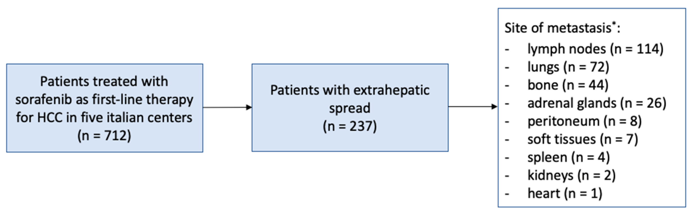
3.1. Study Population

From the entire ARPES database of 712 patients, we considered for this study only 237 patients (33.2%) with radiological detection of EHS. Most patients were male (85.2%), cirrhotic (94.1%) and with viral etiologies (69.6%). Almost all patients (95.4%) presented preserved liver function defined as Child-Pugh A; remaining patients were all in Child-Pugh score B7 liver functional class. Baseline characteristics of study population compared with other patients in ARPES registry are summarized in Table 1.
The most frequent sites of metastases were lymph nodes (48.1%), lungs (30.4%), bone (18.6%) and adrenal glands (11.0%); remaining metastatic sites presented a frequency < 5%. Multiple concomitant sites of metastases were recorded in 48 patients (20.2%) (Figure 1).
Baseline characteristics of patients with lymph node, lung, bone and adrenal gland metastases compared with other patients of the study population are reported in Table 2.

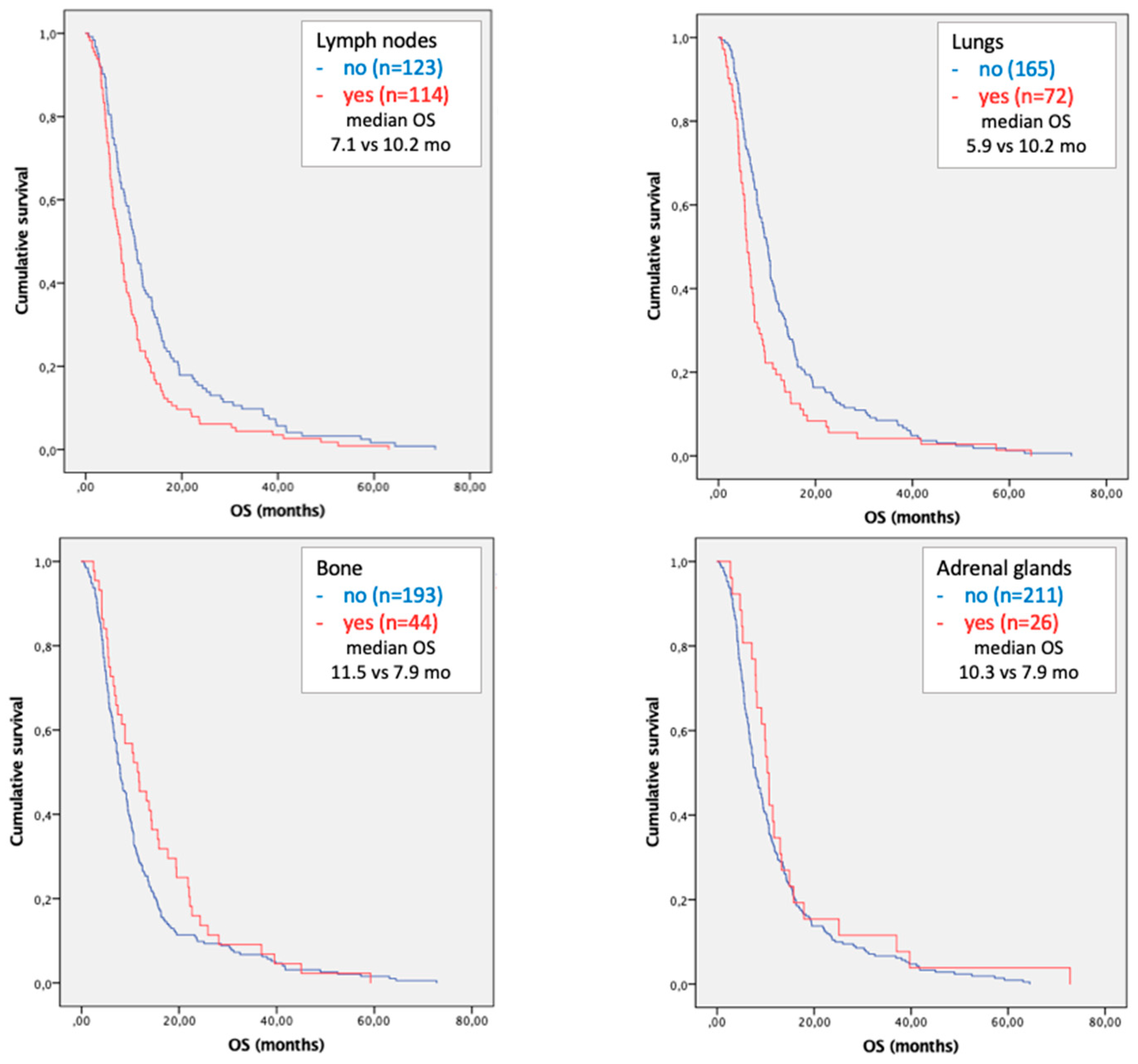
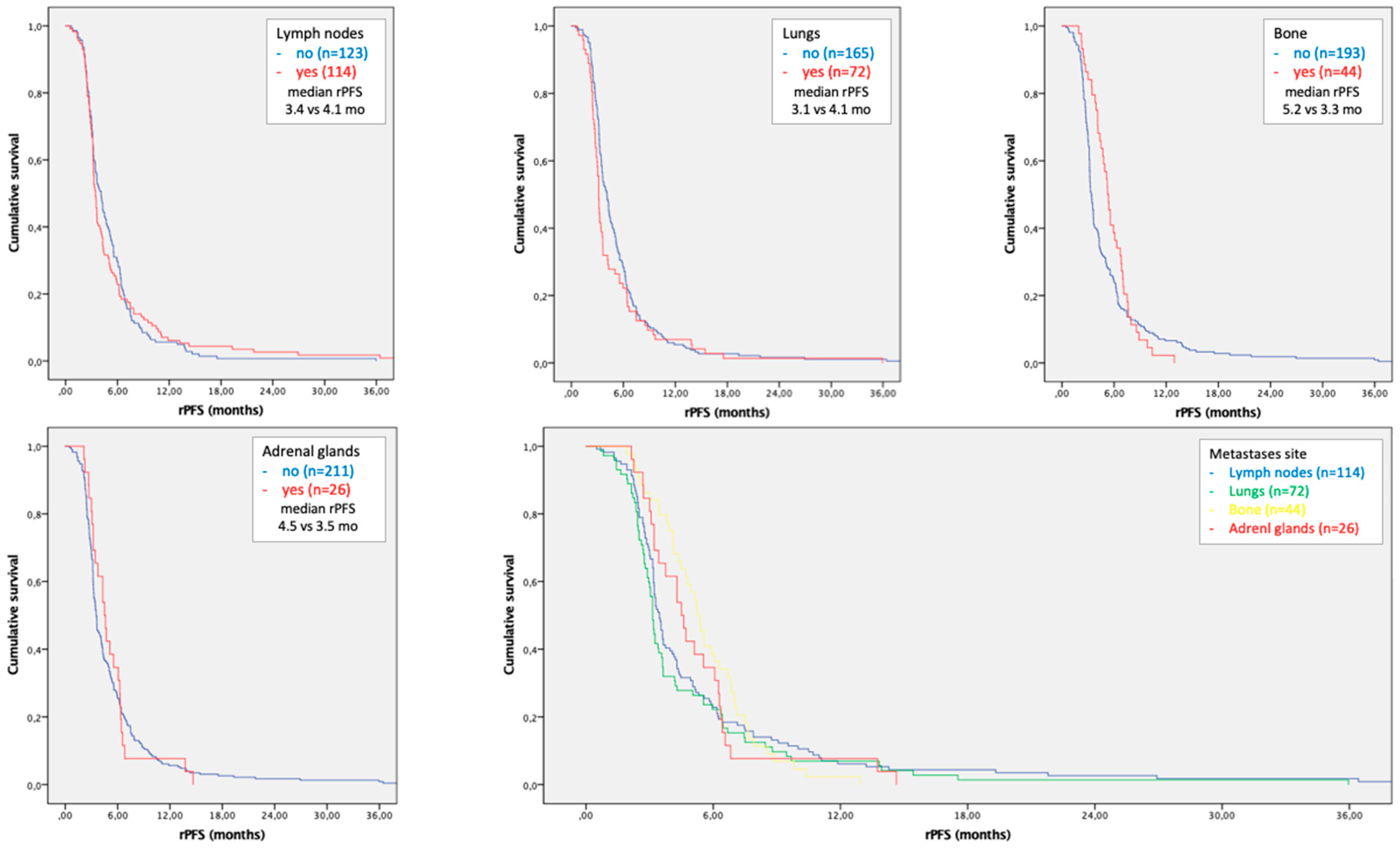
3.2. Survival Analysis

The univariate analysis of OS showed that compromised liver function (Child-Pugh B), high albumin-bilirubin grade (ALBI > 1), elevated serum alpha-fetoprotein values (AFP > 400 ng/mL), intrahepatic tumor burden > 50% (ITB > 50%), cancer-related deterioration of general conditions (ECOG-PS > 0), presence of microvascular invasion (MVI) and occurrence of dermatological adverse events during treatment (DAEs) were associated with patients’ prognosis.
A statistically significant independent correlation with survival was confirmed in the multivariate analysis for Child–Pugh B (HR 3.105, p = 0.002), AFP > 400 ng/mL (HR 1.396, p = 0.030), ITB > 50% (HR 1.543, p = 0.006), MVI (HR 1.770, p < 0.001) and DAEs (HR 0.755, p = 0.045).
Regarding the sites of metastases, the presence of lymph node, lung and multiple site localizations of disease were associated with worse prognosis. A statistically independent correlation with survival was confirmed in the multivariate analysis for lymph node (HR 1.545; OS 7.1 vs. 10.2 mo; p = 0.007) and lung metastases (HR 2.110; OS 5.9 vs. 10.2 mo; p < 0.001) compared with patients with metastatic HCC without lymph node or lung localizations, respectively (Table 3 and Figure 2).
After stratification of patients with lymph node and lung metastases, patients with the co-presence of these two metastatic sites had worse survival rates compared to those who presented only lymph node (HR 2.014; OS 5.0 vs. 7.4 mo; p = 0.006) or only lung metastases (HR 1.762; OS 5.0 vs. 6.5 mo; p = 0.040).

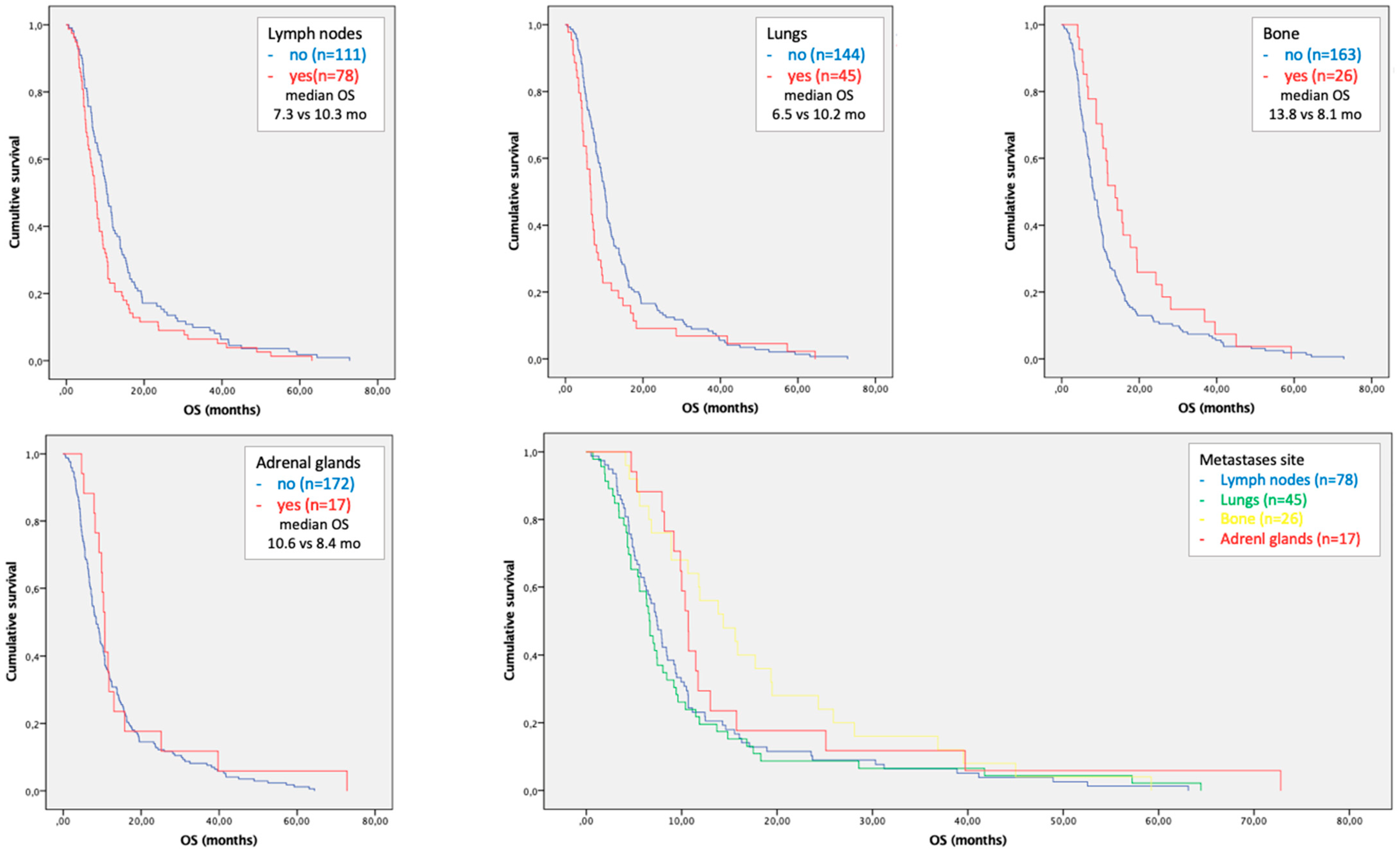
3.3. Subgroup Analysis in Patients with Single Site of Metastases

In order to more accurately define the prognostic role of different site of metastases, we performed a subgroup survival analysis in patients with single metastatic site (n = 189). In this cohort of patients, Child–Pugh B (HR 5.661, p < 0.001), AFP > 400 ng/mL (1.464, p = 0.026), ITB > 50% (HR 1.614, p = 0.009) and presence of MVI (HR 1.770, p = 0.001) were significantly related to worse survivals.
Regarding the sites of metastases, the presence of lymph nodes, lung and bone localizations of disease were associated with patient prognosis. In the multivariate analysis a statistically independent correlation with survival was confirmed only for lymph node (HR 1.645; OS 7.3 vs. 10.3 mo; p = 0.021) and lung metastases (HR 2.182; OS 6.5 vs. 10.2 mo; p = 0.002) compared with patients with metastatic HCC without lymph node or lung localizations, respectively (Table 4 and Figure 3).

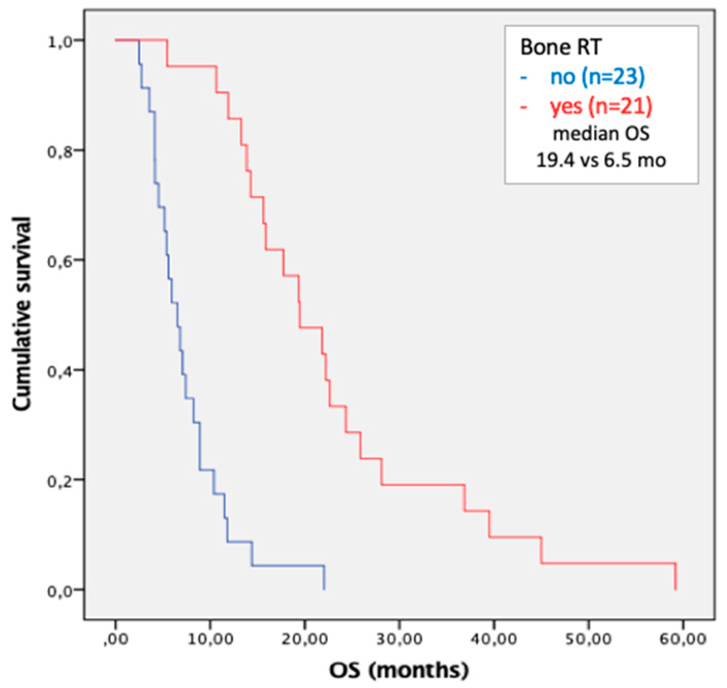
3.4. Metastases Focused Combination Treatments

In order to control extrahepatic tumor burden, locoregional combination treatments were performed in 28 patients. Among these, 3 patients had lymph node metastases (all received stereotaxic radiotherapy), 2 patients had pulmonary metastases (a patient received stereotaxic radiotherapy, the other received radiofrequency ablation) and 21 patients had bone metastases (all received radiotherapy with antalgic purpose). No surgical treatments (i.e., metastasectomy) have been performed in any patients of the study population.
Due to the high number of treatments focused on bone metastases, we performed survival analysis in this cohort of patients. Radiation therapy was associated with a significant survival benefit in patients with bone metastases (HR 0.137; OS 19.4 vs. 6.5 mo; p < 0.001) (Figure 4).

3.5. Time to Progression According to Site of Metastases

A re-evaluation of all CT images was also performed in order to assess the radiological progression-free survival (rPFS) of each site of metastases. A dimensional increase of at least 20% from baseline of a target metastatic lesion or the occurrence of a new lesion in a specific site of metastases was considered as progression of disease in that metastatic site.
The rPFS of lymph node, lung, bone and adrenal gland metastases was 3.4 (95% CI 3.1–3.7; p = 0.930), 3.1 (95% CI 2.9–3.3; p = 0.107), 5.2 (95% CI 4.6–5.8; p = 0.126) and 4.5 (95% CI 3.5–5.4; p = 0.719) months, respectively. No statistically significant differences in rPFS were observed among different metastatic sites (Figure 5). The disease control rates (DCRs) of the four abovementioned sites of metastases were 39.5%, 30.5%, 75.0% and 61.5%, respectively.

4. Discussion

Sorafenib represented the only available therapeutic option for patients with advanced HCC for several years. Since its approval, real-life studies have shown a progressive improvement in the survival of patients treated with sorafenib thanks to a better management of drug-related adverse events [12,13].
In order to better understand the wide response variability among patients, several authors investigated predictors of survival. In the last few years, several studies showed how clinical [14,15,16,17], laboratory [18,19,20,21] and pharmacologic features [22,23] could influence the prognosis of patients with advanced HCC; for instance, the presence of EHS is a well-known predictor of negative prognosis.
Regarding the actual impact of different sites of metastases on prognosis, evidence is scarce, and results are often contradictory. Two recently published works on this topic are the study of Zhan H et al., based on the American epidemiologic registry SEER (Surveillance, Epidemiology and End Results) that gives OS results [24], and the study of Huang SF et al., based on the national registry of Taiwan that gives PFS results [25]. In the first study, lymph nodes were not considered as a metastatic site, and less than a half of patients were on systemic treatment; in the second study all patients with advanced HCC were analyzed, including those without EHS.
In the study of Zhan H et al., the OS was negatively related to multiple metastatic sites, pulmonary, bone (only after propensity score matching) and lymph nodes metastases (considered as a potential influencing factor in survival analysis). In the study of Huang SF et al., the PFS was negatively related to lung and bone metastases and positively related to lymph node metastases (only in the cohort of patients with single metastatic site).
In both studies, high rates of surgical and radiation treatments were reported, but the target metastatic site was not specified.
In the current study we reported that the presence of lung or lymph node metastases are negative independent prognostic factors with a median OS of 5.9 and 7.1 months, respectively; the co-presence of these two metastatic sites reduced the median OS at 5.0 months. In the subgroup analysis of patients with a single metastatic site, a statistically significant correlation between lung and lymph node metastases and prognosis was confirmed.
Lung and lymph node metastases did not reach statistical significance in rPFS analysis even though a negative trend is represented in survival curves.
Concerning adrenal gland metastases, the fourth most common metastatic site, they did not reach statistical significance in any survival analysis, but the small sample size (26 cases) could be a determinant. Adrenal gland metastases have not been evaluated in any above-mentioned study. There are only case reports and case series on this topic in the literature, so our results on OS and rPFS (10.3 and 4.5 months, respectively) provide original, relevant information.
Combination treatments, both surgical and radiation ones, are widely used to control the tumor burden and to improve patients’ outcome [26,27]. In our study population, palliative radiation therapy on bone metastases was the most common treatment performed, so it was possible to conduct subgroup analysis in this cohort of patients. Although there was no oncologic radical purpose, antalgic radiation therapy contributed to a significant improvement in survival (median OS 19.4 vs. 6.5 months). This result could explain the positive trend observed in bone metastases OS curves. Recently, it has been demonstrated how curative radiation therapy increases survival in patients with oligometastatic (up to 5 bone lesions) solid tumors and HCC [28,29]. Nowadays, no conclusive results on the prognostic impact of palliative radiation therapy have been published [30].
In the last few years, a rapid and progressive change in the management of advanced HCC patients has occurred, leading to improved prognosis. This was due to the approval of drugs for post-sorafenib treatment [31,32] and the advent of immune checkpoint inhibitors (ICIs). Atezolizumab, a PD-L1 inhibitor, in association with bevacizumab, is the current front-line therapy for advanced HCC [33,34], and several clinical trial with ICIs, some of these in combination with TKIs, are now ongoing.
With these preconditions, TKI monotherapy seems to be outdated, but it still assumes a central role in the therapeutic scenario for patients with advanced HCC and contraindications to ICIs [35] or solid organ recipients [36]; furthermore, recent studies seem to highlight that the etiology of underling liver disease could influence the response to different first-line therapies [37].
In the next few years, the ICIs actual efficacy on the different target lesions needs to be assessed due to the close relationship between these new drugs and tumor microenvironment. The final possible aim is to tailor individual therapeutic strategies for each patient with advanced HCC.
Despite the large population of this study, the small sample size of less common sites of metastases did not allow us to perform a survival analysis in these subgroups of patients. In the current study, the enrollment of only metastatic patients in therapy with sorafenib and the low rate of concomitant treatments, with the exception of radiation therapy on bone metastases, significantly reduced possible confounders for survival analysis compared with previous studies.

5. Conclusions

The presence of lymph node and lung metastases are independent negative prognostic factors in patients with metastatic HCC. Radiation therapy of bone metastases, even with a palliative antalgic purpose, seems to give a survival benefit, and it should be therefore considered for the management of these patients.

Author Contributions

Conceptualization, L.I. and I.G.; methodology, L.I., F.T. (Francesco Tovoli) and A.G.; formal analysis, L.I. and F.T. (Francesco Tovoli); data curation: L.I., M.T., B.S., R.T., G.M., F.T. (Franco Trevisani), R.S. and T.P.; writing—original draft preparation, L.I.; writing—review and editing, A.G., F.T. (Francesco Tovoli) and F.P. All authors have read and agreed to the published version of the manuscript.

Funding

This research received no external funding.

Institutional Review Board Statement

The study was conducted according to the guidelines of the Declaration of Helsinki and approved by the Ethics Committee of the Area Vasta Emilia Centro (protocol code 2014/Oss/147).

Informed Consent Statement

Informed consent was obtained from all subjects involved in the study.

Data Availability Statement

The data presented in this study are available on request from the corresponding author. The data are not publicly available due to privacy restrictions.

Conflicts of Interest

F.T. (Francesco Tovoli) has served as a consultant for Bayer, Ipsen, and Eisai and an advisory board member for Laforce. F.T. (Franco Trevisani) is an advisor and a consultant for Bayer and an advisory board member for Sirtex, Alfasigma, and Bristol–Myers Squibb. F.P. has been lecturer or consultant for Bayer, Bracco, Eisai, Esaote, Exact Sciences, Ipsen, Samsung, AstraZeneca, MSD, Roche. All remaining authors have declared no conflict of interest.

References

  1. Sung, H.; Ferlay, J.; Siegel, R.L.; Laversanne, M.; Soerjomataram, I.; Jemal, A.; Bray, F. Global Cancer Statistics 2020: GLOBOCAN Estimates of Incidence and Mortality Worldwide for 36 Cancers in 185 Countries. CA Cancer J. Clin. 2021, 71, 209–249. [Google Scholar] [CrossRef] [PubMed]
  2. Reig, M.; Forner, A.; Rimola, J.; Ferrer-Fàbrega, J.; Burrel, M.; Garcia-Criado, Á.; Kelley, R.K.; Galle, P.R.; Mazzaferro, V.; Salem, R.; et al. BCLC strategy for prognosis prediction and treatment recommendation Barcelona Clinic Liver Cancer (BCLC) staging system: The 2022 update. J. Hepatol. 2021, 76, 681–693. [Google Scholar] [CrossRef] [PubMed]
  3. Heimbach, J.K.; Kulik, L.M.; Finn, R.S.; Sirlin, C.B.; Abecassis, M.M.; Roberts, L.R.; Zhu, A.X.; Murad, M.H.; Marrero, J.A. AASLD guidelines for the treatment of hepatocellular carcinoma. Hepatology 2018, 67, 358–380. [Google Scholar] [CrossRef] [Green Version]
  4. European Association for the Study of the Liver. EASL Clinical Practice Guidelines: Management of hepatocellular carcinoma. J. Hepatol. 2018, 69, 182–236. [Google Scholar] [CrossRef] [Green Version]
  5. Llovet, J.M.; Ricci, S.; Mazzaferro, V.; Hilgard, P.; Gane, E.; Blanc, J.F.; Oliveira, A.C.; Santoro, A.; Raoul, J.L.; Forner, A.; et al. Sorafenib in advanced hepatocellular carcinoma. N. Engl. J. Med. 2008, 359, 378–390. [Google Scholar] [CrossRef] [Green Version]
  6. Kudo, M.; Finn, R.S.; Qin, S.; Han, K.-H.; Ikeda, K.; Piscaglia, F.; Baron, A.; Park, J.-W.; Han, G.; Jassem, J.; et al. Lenvatinib versus sorafenib in first-line treatment of patients with unresectable hepatocellular carcinoma: A randomised phase 3 non-inferiority trial. Lancet 2018, 391, 1163–1173. [Google Scholar] [CrossRef] [Green Version]
  7. Bruix, J.; Qin, S.; Merle, P.; Granito, A.; Huang, Y.-H.; Bodoky, G.; Pracht, M.; Yokosuka, O.; Rosmorduc, O.; Breder, V.; et al. Regorafenib for patients with hepatocellular carcinoma who progressed on sorafenib treatment (RESORCE): A randomised, double-blind, placebo-controlled, phase 3 trial. Lancet 2017, 389, 56–66. [Google Scholar] [CrossRef] [Green Version]
  8. Abou-Alfa, G.K.; Meyer, T.; Cheng, A.-L.; El-Khoueiry, A.B.; Rimassa, L.; Ryoo, B.-Y.; Cicin, I.; Merle, P.; Chen, Y.; Park, J.-W.; et al. Cabozantinib in Patients with Advanced and Progressing Hepatocellular Carcinoma. N. Engl. J. Med. 2018, 379, 54–63. [Google Scholar] [CrossRef]
  9. Uka, K. Clinical features and prognosis of patients with extrahepatic metastases from hepatocellular carcinoma. World J. Gastroenterol. 2007, 13, 414. [Google Scholar] [CrossRef] [PubMed]
  10. Schlageter, M.; Quagliata, L.; Matter, M.; Perrina, V.; Tornillo, L.; Terracciano, L. Clinicopathological Features and Metastatic Pattern of Hepatocellular Carcinoma: An Autopsy Study of 398 Patients. Pathobiology 2016, 83, 301–307. [Google Scholar] [CrossRef] [PubMed]
  11. Eisenhauer, E.A.; Therasse, P.; Bogaerts, J.; Schwartz, L.H.; Sargent, D.; Ford, R.; Dancey, J.; Arbuck, S.; Gwyther, S.; Mooney, M.; et al. New response evaluation criteria in solid tumours: Revised RECIST guideline (version 1.1). Eur. J. Cancer 2009, 45, 228–247. [Google Scholar] [CrossRef] [PubMed]
  12. Iavarone, M.; Cabibbo, G.; Piscaglia, F.; Zavaglia, C.; Grieco, A.; Villa, E.; Cammà, C.; Colombo, M. Field-practice study of sorafenib therapy for hepatocellular carcinoma: A prospective multicenter study in Italy. Hepatology 2011, 54, 2055–2063. [Google Scholar] [CrossRef] [PubMed]
  13. Tovoli, F.; Ielasi, L.; Gardini, A.C.; Granito, A.; Foschi, F.G.; Rovesti, G.; Negrini, G.; Orsi, G.; Renzulli, M.; Piscaglia, F. Management of adverse events with tailored sorafenib dosing prolongs survival of hepatocellular carcinoma patients. J. Hepatol. 2019, 71, 1175–1183. [Google Scholar] [CrossRef] [PubMed] [Green Version]
  14. Bruix, J.; Cheng, A.L.; Meinhardt, G.; Nakajima, K.; De Sanctis, Y.; Llovet, J. Prognostic factors and predictors of sorafenib benefit in patients with hepatocellular carcinoma: Analysis of two phase III studies. J. Hepatol. 2017, 67, 999–1008. [Google Scholar] [CrossRef] [Green Version]
  15. Reig, M.; Torres, F.; Rodriguez-Lope, C.; Forner, A.; Llarch, N.; Rimola, J.; Darnell, A.; Ríos, J.; Ayuso, C.; Bruix, J. Early dermatologic adverse events predict better outcome in HCC patients treated with sorafenib. J. Hepatol. 2014, 61, 318–324. [Google Scholar] [CrossRef]
  16. Sansone, V.; Tovoli, F.; Casadei-Gardini, A.; Di Costanzo, G.G.; Magini, G.; Sacco, R.; Pressiani, T.; Trevisani, F.; Rimini, M.; Tortora, R.; et al. Comparison of Prognostic Scores in Patients with Hepatocellular Carcinoma Treated with Sorafenib. Clin. Transl. Gastroenterol. 2021, 12, e00286. [Google Scholar] [CrossRef]
  17. Nakano, M.; Tanaka, M.; Kuromatsu, R.; Nagamatsu, H.; Tajiri, N.; Satani, M.; Niizeki, T.; Aino, H.; Okamura, S.; Iwamoto, H.; et al. Sorafenib for the treatment of advanced hepatocellular carcinoma with extrahepatic metastasis: A prospective multicenter cohort study. Cancer Med. 2015, 4, 1836–1843. [Google Scholar] [CrossRef]
  18. Johnson, P.J.; Berhane, S.; Kagebayashi, C.; Satomura, S.; Teng, M.; Reeves, H.L.; O’Beirne, J.; Fox, R.; Skowronska, A.; Palmer, D.; et al. Assessment of Liver Function in Patients with Hepatocellular Carcinoma: A New Evidence-Based Approach—The ALBI Grade. J. Clin. Oncol. 2015, 33, 550–558. [Google Scholar] [CrossRef]
  19. Orsi, G.; Tovoli, F.; Dadduzio, V.; Vivaldi, C.; Brunetti, O.; Ielasi, L.; Conti, F.; Rovesti, G.; Gramantieri, L.; Rizzato, M.D.; et al. Prognostic Role of Blood Eosinophil Count in Patients with Sorafenib-Treated Hepatocellular Carcinoma. Target. Oncol. 2020, 15, 773–785. [Google Scholar] [CrossRef]
  20. Lué, A.; Serrano, M.T.; Bustamante, F.J.; Iñarrairaegui, M.; Arenas, J.I.; Testillano, M.; Lorente, S.; Gil, C.; de la Torre, M.; Gomez, A.; et al. Neutrophil-to-lymphocyte ratio predicts survival in European patients with hepatocellular carcinoma administered sorafenib. Oncotarget 2017, 8, 103077–103086. [Google Scholar] [CrossRef] [Green Version]
  21. Lin, W.; Zhong, M.; Zhang, Y.; Wang, H.; Zhao, H.; Cheng, B.B.; Ling, C. Prognostic Role of Platelet-to-Lymphocyte Ratio in Hepatocellular Carcinoma with Different BCLC Stages: A Systematic Review and Meta-Analysis. Gastroenterol Res. Pract. 2018, 2018, 5670949. [Google Scholar] [CrossRef] [PubMed]
  22. Casadei Gardini, A.; Faloppi, L.; De Matteis, S.; Foschi, F.G.; Silvestris, N.; Tovoli, F.; Palmieri, V.; Marisi, G.; Brunetti, O.; Umberto, V.-G.; et al. Metformin and insulin impact on clinical outcome in patients with advanced hepatocellular carcinoma receiving sorafenib: Validation study and biological rationale. Eur. J. Cancer 2017, 86, 106–114. [Google Scholar] [CrossRef] [PubMed] [Green Version]
  23. Ielasi, L.; Tovoli, F.; Tonnini, M.; Tortora, R.; Magini, G.; Sacco, R.; Pressiani, T.; Trevisani, F.; Sansone, V.; Marasco, G.; et al. Beneficial Prognostic Effects of Aspirin in Patients Receiving Sorafenib for Hepatocellular Carcinoma: A Tale of Multiple Confounders. Cancers 2021, 13, 6376. [Google Scholar] [CrossRef]
  24. Zhan, H.; Zhao, X.; Lu, Z.; Yao, Y.; Zhang, X. Correlation and Survival Analysis of Distant Metastasis Site and Prognosis in Patients with Hepatocellular Carcinoma. Front. Oncol. 2021, 11, 652768. [Google Scholar] [CrossRef] [PubMed]
  25. Huang, S.F.; Chong, S.W.; Huang, C.W.; Hsu, H.Y.; Pan, K.T.; Hung, C.F.; Wu, T.H.; Lee, C.W.; Hsieh, C.H.; Wang, C.T.; et al. Differential Response to Sorafenib Administration for Advanced Hepatocellular Carcinoma. Biomedicines 2022, 10, 2277. [Google Scholar] [CrossRef] [PubMed]
  26. Chang, W.I.; Kim, B.H.; Kim, Y.J.; Yoon, J.; Jung, Y.J.; Chie, E.K. Role of radiotherapy in Barcelona Clinic Liver Cancer stage C hepatocellular carcinoma treated with sorafenib. J. Gastroenterol. Hepatol. 2021, 37, 387–394. [Google Scholar] [CrossRef] [PubMed]
  27. Goio, E.; Ielasi, L.; Benevento, F.; Renzulli, M.; Tovoli, F. Long-lasting remission in a metastatic hepatocellular carcinoma patient after combined regorafenib therapy and surgery. Hepatic Oncol. 2020, 7, HEP24. [Google Scholar] [CrossRef] [PubMed]
  28. Palma, D.A.; Olson, R.; Harrow, S.; Gaede, S.; Louie, A.V.; Haasbeek, C.; Mulroy, L.; Lock, M.; Rodrigues, G.B.; Yaremko, B.P.; et al. Stereotactic Ablative Radiotherapy for the Comprehensive Treatment of Oligometastatic Cancers: Long-Term Results of the SABR-COMET Phase II Randomized Trial. J. Clin. Oncol. 2020, 38, 2830–2838. [Google Scholar] [CrossRef]
  29. Kim, T.H.; Park, S.; Rim, C.H.; Choi, C.; Seong, J. Improved oncologic outcomes by ablative radiotherapy in patients with bone metastasis from hepatocellular carcinoma. J. Cancer Res. Clin. Oncol. 2021, 147, 2693–2700. [Google Scholar] [CrossRef]
  30. Ielasi, L.; Stefanini, B.; Piscaglia, F.; Granito, A.; Tovoli, F. A Case of Advanced Hepatocellular Carcinoma with Bone Metastases Managed with Tyrosine Kinase Inhibitors and Aggressive Palliative Radiation Therapy: Role of Combination Therapy for Extending Survival. Gastroenterol. Insights 2023, 14, 38–44. [Google Scholar] [CrossRef]
  31. Granito, A.; Forgione, A.; Marinelli, S.; Renzulli, M.; Ielasi, L.; Sansone, V.; Benevento, F.; Piscaglia, F.; Tovoli, F. Experience with regorafenib in the treatment of hepatocellular carcinoma. Ther. Adv. Gastroenterol. 2021, 14, 17562848211016959. [Google Scholar] [CrossRef] [PubMed]
  32. Ielasi, L.; Sansone, V.; Granito, A.; Benevento, F.; De Lorenzo, S.; Tovoli, F. An update of treatments of hepatocellular carcinoma in patients refractory to sorafenib. Drugs Today 2018, 54, 615–627. [Google Scholar] [CrossRef] [PubMed]
  33. Finn, R.S.; Qin, S.; Ikeda, M.; Galle, P.R.; Ducreux, M.; Kim, T.Y.; Kudo, M.; Breder, V.; Merle, P.; Kaseb, A.O.; et al. Atezolizumab plus Bevacizumab in Unresectable Hepatocellular Carcinoma. N. Engl. J. Med. 2020, 382, 1894–1905. [Google Scholar] [CrossRef] [PubMed]
  34. Ielasi, L.; Sansone, V.; Forgione, A.; Granito, A.; Benevento, F.; Tovoli, F. An update on atezolizumab for hepatocellular carcinoma. Drugs Today 2021, 57, 365–375. [Google Scholar] [CrossRef] [PubMed]
  35. Stefanini, B.; Bucci, L.; Santi, V.; Reggidori, N.; Rampoldi, D.; Lani, L.; Granito, A.; Sangiovanni, A.; Cabibbo, G.; Farinati, F.; et al. Potential feasibility of atezolizumab-bevacizumab therapy in patients with hepatocellular carcinoma treated with tyrosine-kinase inhibitors. Dig. Liver Dis. 2022, 54, 1563–1572. [Google Scholar] [CrossRef] [PubMed]
  36. Tovoli, F.; Pallotta, D.P.; Sansone, V.; Iavarone, M.; De Giorgio, M.; Ielasi, L.; Di Costanzo, G.G.; Giuffrida, P.; Sacco, R.; Pressiani, T.; et al. Outcomes of Sorafenib for Recurrent Hepatocellular Carcinoma After Liver Transplantation in the Era of Combined and Sequential Treatments. Transplantation 2023, 107, 156–161. [Google Scholar] [CrossRef] [PubMed]
  37. Rimini, M.; Rimassa, L.; Ueshima, K.; Burgio, V.; Shigeo, S.; Tada, T.; Suda, G.; Yoo, C.; Cheon, J.; Pinato, D.; et al. Atezolizumab plus bevacizumab versus lenvatinib or sorafenib in non-viral unresectable hepatocellular carcinoma: An international propensity score matching analysis. ESMO Open 2022, 7, 100591. [Google Scholar] [CrossRef]
Figure 1. Study population and metastases localization distribution. * Multiple concomitant sites of metastases were recorded in 48 patients.
Cancers 15 01523 g001
Figure 2. Kaplan–Meier curves of overall survival in patients with lymph node, lung, bone and adrenal gland metastases.
Cancers 15 01523 g002
Figure 3. Kaplan–Meier curves of overall survival in patients with lymph node, lung, bone and adrenal gland metastases in the cohort of patients with single site of metastases. The last figure compares the survival curves of four overmentioned most frequent sites of metastases.
Cancers 15 01523 g003
Figure 4. Kaplan–Meier curves of overall survival in patients with bone metastases treated with combination radiation therapy.
Cancers 15 01523 g004
Figure 5. Kaplan–Meier curves of radiological progression-free survival for each site of metastases. The last figure compares the survival curves of lymph node, lung, bone and adrenal gland site of metastases.
Cancers 15 01523 g005
Table 1. Baseline characteristics of patients with extrahepatic spread (EHS) compared with all remaining patients without extrahepatic spread (all w/o EHS). The latter is reported and also divided into two groups, which were themselves split in advanced HCC without metastases (BCLC-C w/o EHS) or intermediate HCC (BCLC-B).
VariableEHS
(n = 237)
All w/o EHS
(n = 475)
pBCLC-C w/o EHS
(n = 311)
pBCLC-B
(n = 164)
p
Male sex202 (85.2%)398 (83.8%)0.637264 (84.9%)0.951134 (85.2%)0.355
Age67.6 ± 11.567.8 ± 9.80.82566.7 ± 10.40.37469.7 ± 11.50.056
Viral etiology165 (69.6%)349 (73.5%)0.281231 (74.3%)0.224118 (69.6%)0.630
Child-Pugh B11 (4.6%)47 (9.9%)0.01440 (12.9%)0.0017 (4.6%)0.792
ALBI > 1154 (65.0%)381 (80.2%)<0.001251 (80.7%)<0.001130 (65.0%)0.003
AFP > 400 ng/mL76 (32.1%)156 (32.8%)0.841116 (37.3%)0.20940 (32.1%)0.115
ITB > 50%69 (29.1%)149 (31.4%)0.545106 (34.1%)0.24543 (29.1%)0.628
ECOG-PS > 060 (25.3%)126 (26.5%)0.743126 (26.5%)0.743N.A.-
MVI78 (32.9%)232 (48.8%)<0.001232 (48.8%)<0.001N.A.-
AFP: alfa-fetoprotein; ECOG-PS: Eastern Cooperative Oncology Group—Performance Status; ITB > 50%: intrahepatic tumor burden > 50%; MVI: macrovascular invasion. N.A. not applicable.
Table 2. Baseline characteristics of patients with lymph node, lung, bone and adrenal gland metastases compared with the rest of study population.
VariableYes (n = 114)Lymph Nodes
No (n = 123)
pYes (n = 72)Lungs
No (n = 165)
p
Male sex93 (81.5%)109 (88.6%)0.10861 (84.7%)141 (85.4%)0.845
Age67.7 ± 12.567.6 ± 10.60.95566.7 ± 9.868.0 ± 12.20.436
Viral etiology81 (71.0%)84 (68.2%)0.67346 (63.8%)119 (72.1%)0.062
Child-Pugh B5 (4.3%)6 (4.8%)1.0005 (6.9%)6 (3.6%)0.316
ALBI > 181 (71.0%)73 (59.3%)0.07044 (61.1%)110 (66.6%)0.449
AFP > 400 ng/mL36 (31.5%)40 (32.5%)0.89024 (33.3%)52 (31.5%)0.880
ITB > 50%33 (28.9%)36 (29.2)0.98420 (27.7%)49 (29.6%)0.874
ECOG-PS > 023 (20.1%)37 (30.0%)0.10020 (27.7%)40 (24.2%)0.627
MVI44 (38.5%)34 (27.6%)0.09025 (34.7%)53 (32.1%)0.764
VariableYes (n = 44)Bone
No (n = 193)
pYes (n = 26)Adrenal Glands
No (n = 211)
p
Male sex41 (93.1%)161 (83.4%)0.15525 (96.1%)177 (83.8%)0.141
Age68.4 ± 9.667.4 ± 11.90.61867.3 ± 10.667.7 ± 11.70.867
Viral etiology34 (77.2%)131 (67.8%)0.27720 (76.9%)145 (68.7%)0.134
Child-Pugh B3 (6.8%)8 (4.1%)0.4331 (3.8%)10 (4.7%)1.000
ALBI > 126 (59.0%)128 (66.3%)0.29315 (57.6%)139 (65.8%)0.375
AFP > 400 ng/mL16 (36.3%)60 (31.0%)0.5927 (26.9%)69 (32.7%)0.659
ITB > 50%10 (22.7%)59 (30.5)0.3416 (23.0%)63 (29.8%)0.813
ECOG-PS > 015 (34.0%)45 (23.3%)0.2045 (19.2%)55 (26.0%)0.633
MVI9 (20.4%)69 (35.7%)0.0537 (26.9%)71 (33.6%)0.659
AFP: alfa-fetoprotein; ECOG-PS: Eastern Cooperative Oncology Group—Performance Status; ITB > 50%: intrahepatic tumor burden > 50%; MVI: macrovascular invasion.
Table 3. Univariate and multivariate Cox regression analysis of overall survival.
VariableUnivariate
HR (95% CI)
pMultivariate
HR (95% CI)
p
Male sex1.089 (0.758–1.564)0.644
Viral etiology0.918 (0.695–1.212)0.546
Child-Pugh B3.615 (1.952–6.696)<0.0013.105 (1.538–6.268)0.002
ALBI > 11.367 (1.038–1.801)0.0261.216 (0.904–1.637)0.196
AFP > 400 ng/mL1.478 (1.120–1.950)0.0061.396 (1.033–1.887)0.030
ITB > 50%1.550 (1.550–2.080)0.0031.543 (1.130–2.109)0.006
ECOG-PS > 01.552 (1.152–2.092)0.0041.288 (0.926–1.791)0.133
MVI2.014 (1.522–2.080)<0.0011.770 (1.301–2.409)<0.001
DAEs0.759 (0.579–0.971)0.0290.755 (0.573–0.994)0.045
Lymph nodes1.487 (1.149–1.925)0.0031.545 (1.127–2.117)0.007
Lungs1.569 (1.186–2.075)0.0022.110 (1.484–2.999)<0.001
Bone0.759 (0.546–1.055)0.100
Adrenal glands0.768 (0.506–1.164)0.213
Multiple sites1.505 (1.0.91–2.075)0.0131.041 (0.702–1.544)0.842
AFP: alfa-fetoprotein; DAEs: dermatologic adverse events; ECOG-PS: Eastern Cooperative Oncology Group—Performance Status; ITB > 50%: intrahepatic tumor burden > 50%; MVI: macrovascular invasion. N.A. not applicable.
Table 4. Univariate and multivariate Cox regression analysis of overall survival in the cohort of patients with a single site of metastases.
VariableUnivariate
HR (95% CI)
pMultivariate
HR (95% CI)
p
Male sex1.079 (0.723–1.609)0.710
Viral etiology0.921 (0.672–1.262)0.610
Child-Pugh B5.356 (2.577–11.131)<0.0016.326 (2.696–14.843)<0.001
ALBI > 11.423 (1.045–1.939)0.0251.291 (0.913–1.825)0.148
AFP > 400 ng/mL1.500 (1.098–2.048)0.0111.464 (1.046–2.049)0.026
ITB > 50%1.580 (1.140–2.191)0.0061.614 (1.130–2.307)0.009
ECOG-PS > 01.587 (1.133–2.222)0.0071.311 (0.905–1.898)0.152
MVI1.963 (1.434–2.686)<0.0011.770 (1.251–2.503)0.001
DAEs0.780 (0.584–1.042)0.0930.755 (0.573–0.994)0.045
Lymph nodes1.411 (1.054–1.890)0.0211.645 (1.078–2.509)0.021
Lungs1.431 (1.018–2.013)0.0392.182 (1.331–3.578)0.002
Bone0.663 (0.440–0.998)0.0491.018 (0.586–1.766)0.950
Adrenal glands0.727 (0.434–1.218)0.226
AFP: alfa-fetoprotein; DAEs: dermatologic adverse events; ECOG-PS: Eastern Cooperative Oncology Group—Performance Status; ITB > 50%: intrahepatic tumor burden > 50%; MVI: macrovascular invasion. N.A. not applicable.
Disclaimer/Publisher’s Note: The statements, opinions and data contained in all publications are solely those of the individual author(s) and contributor(s) and not of MDPI and/or the editor(s). MDPI and/or the editor(s) disclaim responsibility for any injury to people or property resulting from any ideas, methods, instructions or products referred to in the content.

