Article

Management and Oncologic Outcomes of Close and Positive Margins after Transoral CO2 Laser Microsurgery for Early Glottic Carcinoma

1
Unit of Otorhinolaryngology, Department of Surgery, Azienda Ospedaliero-Universitaria di Cagliari, University of Cagliari, 09100 Cagliari, Italy
2
Unit of Pathology, Department of Medical Sciences and Public Health, Azienda Ospedaliero-Universitaria di Cagliari, University of Cagliari, 09100 Cagliari, Italy
3
ENT Department, King’s College Hospital London-Dubai, Dubai P.O. Box 340901, United Arab Emirates
*
Author to whom correspondence should be addressed.
These authors contributed equally to this work.
Cancers 2023, 15(5), 1490; https://doi.org/10.3390/cancers15051490
Submission received: 7 February 2023 / Revised: 22 February 2023 / Accepted: 24 February 2023 / Published: 27 February 2023

Abstract

:

Simple Summary

The management of close and/or positive margins after transoral CO2 laser microsurgery (CO2 TOLMS) is still an ongoing matter of discussion. Different options have been suggested on the basis of the number of the involved margins (single vs. multiple) and site (deep vs. superficial): strict follow-up, revision surgery or radiotherapy. However, precise indications about additional treatment and its effective impact on local control and survival rates are still lacking. The authors reviewed 351 patients with early glottic cancer treated using CO2 TOLMS with the aim of analyzing the impact of margin status on local control and survival, and discussing the therapeutic options in cases of close and positive margins.

Abstract

Background: The present study analyzed the impact of margin status on local control and survival, and the management of close/positive margins after transoral CO2 laser microsurgery for early glottic carcinoma. Methods: 351 patients (328 males, 23 females, mean age 65.6 years) underwent surgery. We identified the following margin statuses: negative, close superficial (CS), close deep (CD), positive single superficial (SS), positive multiple superficial (MS), and positive deep (DEEP). Results: A total of 286 patients (81.5%) had negative margins, 23 (6.5%) had close margins (8 CS, 15 CD) and 42 (12%) had positive margins (16 SS, 9 MS, 17 DEEP). Among the 65 patients with close/positive margins, 44 patients underwent enlargement, 6 radiotherapy and 15 follow-up. Twenty-two patients (6.3%) recurred. Patients with DEEP or CD margins showed a higher risk of recurrence (hazard ratios of 2.863 and 2.537, respectively), compared to patients with negative margins. Local control with laser alone, overall laryngeal preservation and disease-specific survival decreased significantly in patients with DEEP margins (57.5%, 86.9% and 92.9%, p < 0.05). Conclusions: Patients with CS or SS margins could be safely submitted to follow-up. In the case of CD and MS margins, any additional treatment should be discussed with the patient. In the case of DEEP margin, additional treatment is always recommended.

1. Introduction

Transoral CO2 laser microsurgery (CO2 TOLMS) [1] represents a well-standardized and minimally invasive approach allowing good oncological and functional results in patients with early glottic squamous cell carcinoma (SCC) [2,3]. This ultraconservative approach can be burdened by an increased incidence of close and positive margins of resection, which are unanimously associated with a higher risk of local relapse [4,5,6,7,8].
In the case of inadequate margins, different options have been suggested on the basis of the number of the involved margins (single vs. multiple) and site (deep vs. superficial): strict follow-up, revision surgery or radiotherapy (RT) [9]. However, precise indications for additional treatment and its effective impact on local control and survival rates are still debated.
There is a common consensus that further treatments are required in patients with positive deep margin of resection at definitive histology, while the management of close and/or positive superficial margins is still an ongoing matter of discussion [9] since additional resection can potentially hinder the functional results.
The high rate of false positive margins after enlargement reported in the literature (up to 80%) [6,9,10,11,12,13] has driven several authors to reduce second-look procedures in recent years, implementing a watchful waiting policy [6,9].
We reviewed a large homogeneous cohort of patients affected by Tis-T1-T2 glottic SCC treated using CO2 TOLMS with the aim of analyzing the impact of margin status on local control, survival and organ preservation. The decision-making process and effective indication of additional therapeutic options in cases of close and positive margins have been discussed.

2. Materials and Methods
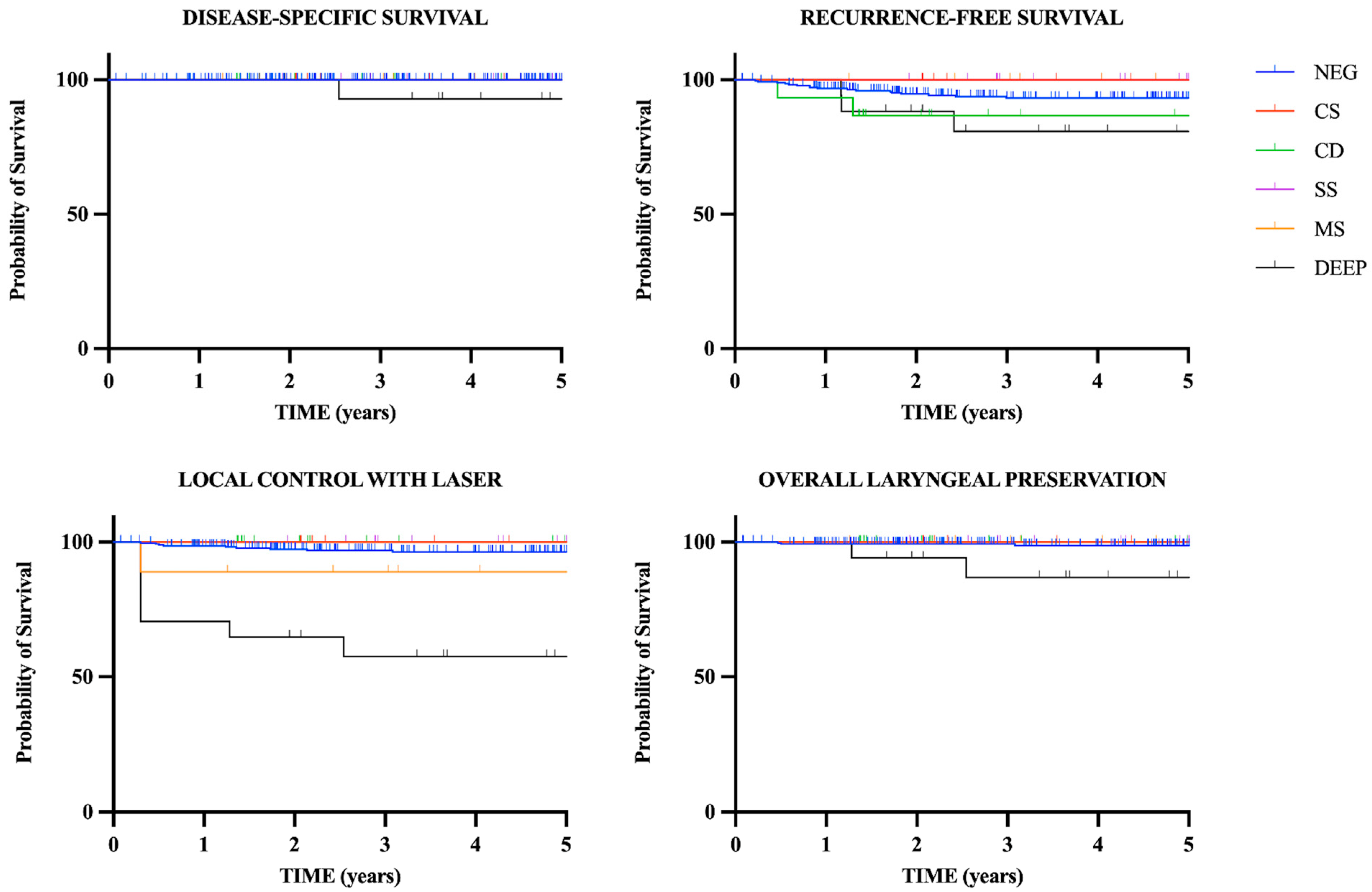
The authors performed a retrospective analysis of 351 patients with early glottic SCC (Tis-T1-T2) treated by the senior author with CO2 TOLMS from October 1993 to November 2005 and from December 2010 to December 2020 at the Department of Otorhinolaryngology of an Italian institution (ethics committee protocol number 895/2018). All the patients included in the study had no clinical involvement of the lymph node at the time of surgery.
During the preoperative work up, all patients underwent fiberlaryngoscopy, while computed tomography (CT) or magnetic resonance imaging (MRI) of the neck with contrast medium were considered necessary in selected patients to rule out invasion of the paraglottic space (PGS), of the preepiglottic space and of the cartilage. Intraoperative work up was always performed using rigid 0° and 70° scopes. From the end of 2013, preoperative and intraoperative endoscopic work up was coupled with narrow-band imaging (NBI) (Olympus Medical Systems Corporation, Tokyo, Japan) and the IMAGE1 S SystemTM (Storz, Tuttlingen, Germany) plus enhanced contact endoscopy (ECE) [14].
On the day of surgery, all patients received ceftriaxone (1000 mg IV) or, as a substitute if allergic, ciprofloxacin (400 mg IV), according to the antibiotic prophylaxis protocol of our institution.
All patients underwent CO2 TOLMS under general anesthesia with orotracheal intubation (Mallinckrodt laser safe tube, I.D. 5.0–7.0 mm; Athlone, Ireland). Sharplan 1030 and Acupulse CO2 lasers with an Acuspot, Acublade 712 micromanipulator and Digital AcuBladeTM (Lumenis®, Yokneam, Israel) set on the superpulsed mode (10 W, continuous, acublade 1–3 mm) were used in most of the cases. The UltraPulse/Surgitouch CO2 laser (Lumenis®, Yokneam, Israel) was used from 2020.
Adequate laryngeal exposure in microlaryngoscopy was obtained using the Kleinsasser laser laryngoscopes modified by Rudert with the Riecker–Kleinsasser suspension system (Karl Storz, Tüttlingen, Germany).
Endoscopic cordectomies were classified according to the European Laryngological Society (ELS) [15,16].
Resections were always performed using an en bloc procedure when the volume of the tumor allowed it. Larger tumors were removed using a piecemeal technique. When indicated, the anterior commissure was resected through a subperichondrial dissection. Resections were performed in macroscopic free margins. Specimens were sent for histology opportunely oriented by the surgeon by staining the superior edge with ink to obtain the precise mapping of the lesions, also after piecemeal removal. Frozen sections were not routinely performed because they are unrepresentative of the whole mucosal margins, and the time of execution can become excessive for organizational reasons and hospital logistics.
After definitive histology, all lesions were staged and restaged according to the eighth edition of the Union for International Cancer Control–American Joint Committee on Cancer (UICC–AJCC) TNM staging system [17].
According to Fiz et al. [5], the margin status was classified as follows: negative, close (tumor–margin distance < 1 mm) superficial (CS), close deep (CD), positive (presence of at least carcinoma in situ at the surgical margin) single superficial (SS), positive multiple superficial (MS) and positive deep (DEEP).
In the case of close or positive margins, intraoperative recording was reviewed and discussed in a multidisciplinary team. The policy after histology was as follows: CO2 transoral enlargement or postoperative RT was always performed in the case of DEEP or MS margins; SS and CD margins were almost constantly enlarged with a laser, except selected cases who underwent a close wait-and-see policy; CS margins were managed with close follow-up.
Patients scheduled for a second look with CO2 TOLMS were treated at 30 to 40 days after the first cordectomy, because by that point the scar tissue is completely healed and the glottic aspect can be better evaluated.
Voice rehabilitation and regular follow-up were scheduled according to the NCCN guidelines in all cases [18]: patients underwent fiberlaryngoscopy every month during the first year, every 2 months during the second year and every 3–4 months until the fifth year after surgery, in the absence of any recurrence and/or secondary disease. From 2013, fiberlaryngoscopy was coupled with NBI. Patients included in the present study were followed up from the date of surgery until December 2022, when possible.
Statistical analysis was performed on the basis of the data reported as supplementary material (Table S1), using GraphPad Prism software (GraphPad, San Diego, CA, USA). Survival probabilities over time were estimated using the Kaplan–Meier method, considering the six different types of margins (negative, CS, CD, SS, MS, DEEP). The entry point was the date of laser cordectomy. The first studied outcome was disease-specific survival (DSS), with the end point being patient’s death due to laryngeal cancer or last follow-up. The second outcome was recurrence-free survival (RFS), with the end point set at the date of recurrence or at the last available visit. The third outcome was local control with laser alone (LCL), with the end point set at the date of RT or open procedure for recurrences. Organ laryngeal preservation (OLP) was the fourth measured outcome, with the end point set at the date of total laryngectomy or at last follow-up. The log-rank (Mantel–Cox) test was applied to compare recurrence rates between patients with negative margins versus patients with close/positive margins. A p value < 0.05 was considered to be statistically significant.
The influence of the routine intraoperative use of NBI, IMAGE1 S and ECE in the incidence of positive superficial margins was evaluated.

3. Results

Three hundred and fifty-one patients (328 males, 23 females, mean age 65.6 years, age range 29–90 years) with early glottic SCC treated with CO2 TOLMS were included in the study. Of the 351 patients treated with CO2 TOLMS, 34 (9.7%) underwent type I cordectomy, 94 (26.8%) type II cordectomy, 77 (21.9%) type III cordectomy, 21 (6%) type IV cordectomy, 122 (34.8%) type V cordectomy and 3 (0.8%) type VI cordectomy. Patient and tumor characteristics, and the numbers and types of surgical cordectomies are detailed in Table 1.
The mean hospitalization time was 2.9 days. Three patients experienced a postoperative bleeding that required an endoscopic cautery under general anesthesia. Thirty-four patients (9.7%) developed an anterior glottic web. Among them, the 12 patients with moderate to severe symptoms were managed endoscopically with the laser incision of the web and the harvesting of a mucosal microflap, while the 22 patients with mild symptoms were referred to voice therapists, as suggested in the literature [19].
Two hundred and eighty-six patients (81.5%) had negative margins after primary surgery, while 65 patients (18.5%) had close or positive margins. Twenty-three patients (6.5%) had close margins, among whom 8 had CS and 15 had CD margins. Forty-two patients (12%) had positive margins: 16 had SS margin, 9 MS margins and 17 DEEP margin (Table 1).
Seventeen out of the 229 patients (7.4%) who underwent CO2 TOLMS before the systematic intraoperative use of enhancement systems (NBI, IMAGE1 S and ECE) had positive superficial margins. After the implementation of bioendoscopy, only 8 out of 122 patients (6.6%) experienced positive superficial margins. Therefore, the use of enhancement tools reduced the incidence of positive superficial margins, although the decrease was not statically significant (p = 0.76).
Forty-four (67.7%) of the 65 patients with close/positive margins underwent CO2 laser enlargement, obtaining negative margins in all cases. Among these patients, 12 had DEEP margin, 8 had MS margins, 14 had SS margin and 10 had CD margin. Definitive histology showed the presence of residual carcinoma in only 8 (18.2%) of these 44 cases: 3 patients with initially DEEP margin, one with MS margins, and 4 with SS margin. Definitively, 330 patients (94%) showed negative margins after primary surgery and subsequent enlargements.
A total of 7 patients underwent postoperative RT: six (9.2%) of the 65 patients had close/positive margins (5 with DEEP margin and one with MS margins), and one patient had negative margins but lymphovascular invasion at definitive histology.
Fifteen (23.1%) of the 65 patients with close/positive margins underwent a close wait-and-see policy, among whom 8 had CS margin, 5 had CD margin and 2 had SS margin.
The mean follow-up was 4.97 years.
Twenty-two of the 351 patients (6.3%) experienced recurrence: laryngeal recurrence in 21 cases and nodal recurrence in 1 case. No patients developed distant metastasis. Recurrence occurred in 17 patients with negative margins, in 3 patients with DEEP margin who underwent surgical enlargement without evidence of residual tumor at histology, and in 2 patients with CD margin who underwent follow-up. Twenty-one patients underwent salvage treatment; one patient refused additional treatment and died of disease. Salvage therapy of the 21 patients with recurrences included: CO2 TOLMS alone in 11 cases, CO2 TOLMS and RT in 1 case, RT alone in 1 case, type II OPHL in 1 case, type III OPHL in 1 case, type II OPHL and RT in 1 case, total laryngectomy (TL) in 3 cases, TL and RT in 1 case and radical neck dissection and RT in 1 case (Table 2).
The five-year DSS, RFS, LCL and OLP of the whole series were 99.6%, 92.9%, 94.6% and 98.2%, respectively. Survival rates and Kaplan–Meier survival curves relative to the different subtypes of margins are reported in Table 3 and Figure 1.
In the univariate analysis, patients with DEEP margin showed a 2.863 (p = 0.08) times increased risk for recurrence compared to patients with negative margins (Table 4). Furthermore, in the case of DEEP margin, both LCL, OLP and DSS decreased in a statistically significant way: 57.5%, 86.9% and 92.9%, respectively, in patients with DEEP margin versus 96.4%, 98.7% and 100%, respectively, in patients with negative margins, p < 0.05. Patients with CD margin experienced a 2.537 (p = 0.2) times increased risk of recurrence (RFS of 86.7% versus 93.2% in patients with negative margins) (Table 4).

4. Discussion

The surgical margins required in early glottic SCC are narrower than those considered necessary in other head and neck cancers because of the scarce glottic submucosal lymphatic network. In the literature, margins ≥ 1 mm of healthy tissue are generally considered adequate in patients treated with CO2 TOLMS [4,20,21]. The high magnification available with the operative microscope and modern biologic endoscopic techniques makes the surgical approach with such narrow margins easier. In our series, we found negative margins in 286 cases (81.5%) and close margins in 23 cases (6.5%), whereas positive margins were present in 42 cases (12%). Our rate of positive margins is, encouragingly, in the range reported in the literature (9.3–45.4%) [4,5,6,7,10,22,23,24,25,26].
Different prognostic factors, such as stage or anterior commissure involvement, may be associated with local recurrence, but various studies have demonstrated that positive margins represent an independent risk factor for local failure [9,10,21,25,27]. In the literature, it is reported that a local recurrence rate ranging from 3.1% to 22.8% is observed in cases of negative surgical margins, while the recurrence rate rises from 8% to 51% in cases of positive margins [10]. This great variability of incidences of local relapse could be related to the experience of the surgeon and to the different interpretation of the margin status performed by the pathologist. According to Fiz et al. [5], we analyzed our oncological outcomes, classifying the margins as negative, CS, CD, SS, MS and DEEP.
The intraoperative use of rigid endoscopy with bio endoscopic tools such as NBI and IMAGE1 S has been suggested as a useful tool in achieving optimal superficial margins outlines, and has been shown to potentially decrease the rate of positive superficial margins [5,14,28,29,30]. According to the literature, in our series, the systematic use of NBI, IMAGE1 S and ECE decreased the number of positive superficial margins (6.6% vs. 7.4%), although the difference was not statistically significant (p = 0.76).
In the present series, the CS and SS margins did not negatively impact RFS. Close superficial margins were always managed with close follow-up. Regarding SS margin, in the early years of the present series, our policy was for systematic enlargement, but since the majority of revision surgeries resulted in negative specimens (71.4%), in the second part of the present series, we adopted a strict endoscopic follow-up, performing a second CO2 TOLMS only when the surgeon expressed doubts concerning the resection, as suggested in the literature [6,21,31,32,33]. Avoiding unnecessary surgical enlargements has the advantage of sparing the voice quality, because a second TOLMS clearly results in a further loss of tissue, increasing the scarring of the residual vocal cords. The low incidence of cases with residual disease at histology after the second look could be explained by the loss of the narrow healthy tissue due to the laser effect (thermal damage), and/or because of the shrinkage of the specimen [6,9,12]. Human tissues, especially the mucosa, have an intrinsic propensity to undergo shrinkage after surgical resection because of the presence of contractile proteins in the connective tissue and their release from the surrounding structures. In the literature, a mean shrinkage of mucosal specimens after CO2 TOLMS, from intralaryngeal measurement to postresection, of 3.8 ± 0.3 mm in the anteroposterior length of the glottic plane is reported [34]. Such important shrinkage could explain the high number of unnecessary enlargements to obtain wider or free margins. Therefore, careful harvesting and orientation of the surgical specimen is mandatory for the correct evaluation of the tumor extent. Several protocols have been described in the literature to improve margin assessment. Michel et al. [35] and Aluffi Valletti et al. [6] suggested the use of two different colored inks to tag superficial and deep mucosal sides before formalin fixation. In the present series, all the specimens were systematically three-dimensionally oriented by staining the superior margin with ink, and were analyzed by a dedicated pathologist.
Some authors reported an increased risk of recurrence in the presence of MS margins [5,36]. In our series, none of the nine cases with MS margins experienced recurrences, and postoperative RT was deemed necessary in one patient with multifocal carcinoma. In patients with multifocal SCC, it is difficult to assess the true superficial extension of the lesion and obtain a radical excision; thus, multiple CO2 TOLMS are needed, and, in some instances, RT represents an additional tool. In the eight patients with MS margins who underwent CO2 laser enlargement, the resection was carried out starting from a wider macroscopically free margin including the scar of the previous surgery. Definitive histology showed the presence of residual carcinoma in only one (12.5%) of these eight patients. These data suggest that the majority of patients with MS margins could be overtreated with revision surgery.
Nowadays, the use of HD flexible endoscopy with bioendoscopy performed in the office setting during the follow-up improves the accuracy of the early detection of persistent or recurrent disease after CO2 TOLMS. This could be a reason to shift to a less aggressive attitude concerning second-look procedures in cases of doubtful or positive superficial margins, even multiple, opting for a close follow-up with NBI endoscopy [29].
Fiz et al. [5] reported that close deep margins were related to an increased number of relapses, with an RFS of 77.1%. This was also confirmed by our findings, since patients with CD margin experienced a lower RFS (86.7% versus 93.2% in patients with negative margins) with a 2.537 times increased risk of recurrence; however, this result was not statistically significant (p = 0.2). Two of the five patients with CD margin who underwent follow-up experienced recurrence and were managed with additional CO2 TOLMS. Among the 10 patients with CD margin who underwent the second look, none showed residual carcinoma at histology. Ultimately, LCL, OLP and DSS were not negatively impacted by the CD margin status. As a consequence, we believe that the CD margin does not represent an absolute indication for additional treatment, although a strict follow-up with imaging is mandatory to detect early recurrence.
According to the literature, DEEP margins are generally associated with the highest recurrence rate [5,6,7,8]. In our experience, patients with DEEP margin showed an increased risk of recurrence compared to patients with negative margins, with a hazard ratio of 2.863 (p = 0.08). Five patients required RT and one patient underwent total laryngectomy after recurrence, whereas one patient refused treatment after recurrence and died of disease. Consequently, both LCL, OLP and DSS were negatively affected in patients with DEEP margin (respectively 57.5%, 86.9% and 92.9% versus 96.4%, 98.7% and 100% in patients with negative margins, p < 0.05).
In our series, none of the five patients who underwent postoperative RT after DEEP margin at histology experienced recurrence. In the case of positive margins, the role of postoperative RT is still debated. Some authors found a benefit in RFS after postoperative RT, whereas others could not demonstrate any significant difference in patients submitted for adjuvant treatment when compared with those followed up with a compulsory protocol of surveillance [6,9,36]. Furthermore, postoperative RT results in a multimodal therapeutic approach for early tumors that could have been managed by RT alone from the beginning, with additional biological and economic costs [20]. Moreover, the patient would lose the possibility of being treated with RT in the event of a laryngeal second primary or recurrence [20].
A recent review [37] pointed out that the application of postoperative RT in patients with positive margins following the first resection depends on the confidence of the surgeon with wider resection, and can range from 10% [36,38] to 44% [4,39,40]. However, if a second CO2 TOLMS is judged unlikely to result in “true negative margins”, then open partial surgery or RT are the most appropriate choices [37].
Among the 12 patients with DEEP margin who underwent additional CO2 TOLMS, 3 patients recurred, despite surgical enlargement with no residual tumor at the histology. These “false” negative margins are difficult to interpret: although missing at histology, the residual disease cannot be excluded. In two of these cases, the patients were initially treated with extended type V cordectomies for cT2 tumors, with clinical involvement of the posterior third of the vocal cord. In these cases, histology confirmed the presence of the cancer immediately lateral to the vocal process and close to the PGS. A lower local control can be observed in the case of understaging of T2/T3 tumors. The misdiagnosis of cT2 can be associated with the complexity of the assessment of the PGS. The correct identification of the involvement of the posterior PGS is essential to choose the appropriate therapeutic strategy, since patients with posterior glottic tumors have poor local control when treated with CO2 TOLMS [41]. The limit that separates the anterior from the posterior laryngeal compartments is a virtual plane described as tangential to the vocal process and perpendicular to the ipsilateral thyroid lamina. We believe that, in the case of posterior DEEP margins after extended cordectomies, the surgeon should consider the possibility of micro infiltration of the posterior PGS and, consequently, decide on postoperative RT or open surgery.
Evidently, our analysis was limited by the reduced number of each subgroup of patients, which did not allow us to perform a multivariate analysis. Therefore, to draw any definite conclusions, large controlled multi-center retrospective and prospective trials are needed.

5. Conclusions

The present study confirms that, although CO2 TOLMS in early glottic cancer offers optimal oncological outcomes, the margin status impacts on local control, and it is mandatory to stratify the different types of margins for their distinctive prognostic significance.
In the present series, the CS and SS margins behaved similarly, and could be followed-up after adequate counseling with the patients.
We observed that the CD and MS margins do not have a statistically significant negative impact on DSS, RFS, LCL or OLP, and any additional treatment should be thoroughly discussed with the patient so as to avoid unnecessary overtreatment.
Patients with DEEP margin should always undergo additional treatment, and if a second CO2 TOLMS is judged unlikely to result in “true negative margins”, open surgery or RT must be considered.

Supplementary Materials

The following supporting information can be downloaded at: https://www.mdpi.com/article/10.3390/cancers15051490/s1, supplementary material are reported in the Methods as Table S1.

Author Contributions

Conceptualization, C.M., F.C. and R.P.; data curation, C.M., M.B., V.M., M.T., V.P. and C.G.; formal analysis, C.M. and F.C.; investigation, C.M. and F.C.; methodology, C.M. and F.C.; resources, C.G. and R.P.; supervision, R.P.; validation, C.M., F.C. and R.P.; writing—original draft, C.M. and F.C.; writing—review and editing, C.M., F.C. and R.P. All authors have read and agreed to the published version of the manuscript.

Funding

This research received no external funding.

Institutional Review Board Statement

This research did not involve any animal models; this research involved human participants in accordance with the ethical standards of the institutional and/or national research committees and with the 1964 Helsinki Declaration and its later amendments or comparable ethical standards. The research was approved by the Ethics Committee of the University Hospital of Cagliari (protocol number 895/2018).

Informed Consent Statement

Written informed consent has been obtained from the patients included in the study.

Data Availability Statement

The data that support the findings of this study are available as electronic supplementary material.

Conflicts of Interest

The authors declare that this manuscript was conceived and written by the cited authors, they do not have any financial interest to disclose, and they confirm no conflict of interest concerning this manuscript.

References

  1. Remacle, M.; Arens, C.; Eldin, M.B.; Campos, G.; Estomba, C.C.; Dulguerov, P.; Fiz, I.; Hantzakos, A.; Keghian, J.; Mora, F.; et al. Laser-assisted surgery of the upper aero-digestive tract: A clarification of nomenclature. A consensus statement of the European Laryngological Society. Eur. Arch. Otorhinolaryngol. 2018, 275, 657–658, Erratum for: Eur. Arch. Otorhinolaryngol. 2017, 274, 3723–3727. [Google Scholar] [CrossRef] [Green Version]
  2. Carta, F.; Bandino, F.; Olla, A.M.; Chuchueva, N.; Gerosa, C.; Puxeddu, R. Prognostic value of age, subglottic, and anterior commissure involvement for early glottic carcinoma treated with CO2 laser transoral microsurgery: A retrospective, single-center cohort study of 261 patients. Eur. Arch. Otorhinolaryngol. 2018, 275, 1199–1210. [Google Scholar] [CrossRef] [PubMed]
  3. Sjögren, E.V. Transoral laser microsurgery in early glottic lesions. Curr. Otorhinolaryngol. Rep. 2017, 5, 56–68. [Google Scholar] [CrossRef] [PubMed] [Green Version]
  4. Ansarin, M.; Santoro, L.; Cattaneo, A.; Massaro, M.A.; Calabrese, L.; Giugliano, G.; Maffini, F.; Ostuni, A.; Chiesa, F. Laser surgery of early glottic cancer; impact of margin status on local control and organ preservation. Arch. Otolaryngol. Head Neck Surg. 2009, 135, 385–390. [Google Scholar] [CrossRef] [PubMed]
  5. Fiz, I.; Mazzola, F.; Fiz, F.; Marchi, F.; Filauro, M.; Paderno, A.; Parrinello, G.; Piazza, C.; Peretti, G. Impact of close and positive margins in transoral laser microsurgery for Tis-T2 glottic cancer. Front. Oncol. 2017, 7, 245. [Google Scholar] [CrossRef] [PubMed]
  6. Aluffi Valletti, P.; Taranto, F.; Chiesa, A.; Pia, F.; Valente, G. Impact of resection margin status on oncological outcomes after CO2 laser cordectomy. Acta Otorhinolaryngol. Ital. 2018, 38, 24–30. [Google Scholar] [CrossRef] [PubMed]
  7. Galli, A.; Giordano, L.; Sarandria, D.; Di Santo, D.; Bussi, M. Oncological and complication assessment of CO2 laser-assisted endoscopic surgery for T1-T2 glottic tumours: Clinical experience. Acta Otorhinolaryngol. Ital. 2016, 36, 167–173. [Google Scholar] [CrossRef]
  8. Bertolin, A.; Lionello, M.; Salis, G.; Rizzotto, G.; Lucioni, M. Two-stage CO2 -laser-assisted bilateral cordectomy for cT1b glottic carcinoma. Am. J. Otolaryngol. 2017, 38, 183–187. [Google Scholar] [CrossRef]
  9. Fiz, I.; Koelmel, J.C.; Sittel, C. Nature and role of surgical margins in transoral laser microsurgery for early and intermediate glottic cancer. Curr. Opin. Otolaryngol. Head Neck Surg. 2018, 26, 78–83. [Google Scholar] [CrossRef]
  10. Gallet, P.; Rumeau, C.; Nguyen, D.T.; Teixeira, P.A.; Baumann, C.; Toussaint, B. “Watchful observation” follow-up scheme after endoscopic CO2 laser treatment for small glottic carcinomas: A retrospective study of 93 cases. Clin. Otolaryngol. 2017, 42, 1193–1199. [Google Scholar] [CrossRef]
  11. Canis, M.; Ihler, F.; Martin, A.; Matthias, C.; Steiner, W. Transoral laser microsurgery for T1a glottic cancer: Review of 404 cases. Head Neck. 2015, 37, 889–895. [Google Scholar] [CrossRef] [PubMed]
  12. Jäckel, M.C.; Ambrosch, P.; Martin, A.; Steiner, W. Impact of re-resection for inadequate margins on the prognosis of upper aerodigestive tract cancer treated by laser microsurgery. Laryngoscope 2007, 117, 350–356. [Google Scholar] [CrossRef]
  13. Peretti, G.; Piazza, C.; Bolzoni, A.; Mensi, M.C.; Rossini, M.; Parrinello, G.; Shapshay, S.M.; Antonelli, A.R. Analysis of recurrences in 322 Tis, T1, or T2 glottic carcinomas treated by carbon dioxide laser. Ann. Otol. Rhinol. Laryngol. 2004, 113, 853–858. [Google Scholar] [CrossRef] [PubMed]
  14. Puxeddu, R.; Sionis, S.; Gerosa, C.; Carta, F. Enhanced contact endoscopy for the detection of neoangiogenesis in tumors of the larynx and hypopharynx. Laryngoscope 2015, 125, 1600–1606. [Google Scholar] [CrossRef]
  15. Remacle, M.; Eckel, H.E.; Antonelli, A.; Brasnu, D.; Chevalier, D.; Friedrich, G.; Olofsson, J.; Rudert, H.H.; Thumfart, W.; de Vincentiis, M.; et al. Endoscopic cordectomy. A proposal for a classification by the Working Committee, European Laryngological Society. Eur. Arch. Otorhinolaryngol. 2000, 257, 227–231. [Google Scholar] [CrossRef] [PubMed]
  16. Remacle, M.; van Haverbeke, C.; Eckel, H.; Bradley, P.; Chevalier, D.; Djukic, V.; de Vicentiis, M.; Friedrich, G.; Olofsson, J.; Peretti, G.; et al. Proposal for revision of the European Laryngological Society classification of endoscopic cordectomies. Eur. Arch. Otorhinolaryngol. 2007, 264, 499–504, Erratum in: Eur. Arch. Otorhinolaryngol. 2007, 264, 709. [Google Scholar] [CrossRef]
  17. Lydiatt, W.M.; Patel, S.G.; O’Sullivan, B.; Brandwein, M.S.; Ridge, J.A.; Migliacci, J.C.; Loomis, A.M.; Shah, J.P. Head and neck cancers-major changes in the American joint committee on cancer eighth edition cancer staging manual. CA Cancer J. Clin. 2017, 67, 122–137. [Google Scholar] [CrossRef] [PubMed]
  18. Roman, B.R.; Goldenberg, D.; Givi, B. AHNS series-do you know your guidelines? Guideline recommended follow-up and surveillance of head and neck cancer survivors. Head Neck 2016, 38, 168–174. [Google Scholar] [CrossRef]
  19. Carta, F.; Mariani, C.; Quartu, D.; Gioia, E.; Chuchueva, N.; Puxeddu, R. Single and double mucosal microflap CO2 laser “sliding” technique in the management of iatrogenic glottic web: Anatomical and functional results. Eur. Arch. Otorhinolaryngol. 2019, 276, 3405–3412. [Google Scholar] [CrossRef]
  20. Piazza, C.; Paderno, A.; Grazioli, P.; Del Bon, F.; Montalto, N.; Perotti, P.; Morello, R.; Filauro, M.; Nicolai, P.; Peretti, G. Laryngeal exposure and margin status in glottic cancer treated by transoral laser microsurgery. Laryngoscope 2017, 128, 1146–1151. [Google Scholar] [CrossRef] [PubMed]
  21. Bertino, G.; Degiorgi, G.; Tinelli, C.; Cacciola, S.; Occhini, A.; Benazzo, M. CO2 laser cordectomy for T1-T2 glottic cancer: Oncological and functional long-term results. Eur. Arch. Otorhinolaryngol. 2015, 272, 2389–2395. [Google Scholar] [CrossRef] [PubMed]
  22. Charbonnier, Q.; Thisse, A.S.; Sleghem, L.; Mouawad, F.; Chevalier, D.; Page, C.; Mortuaire, G. Oncologic outcomes of patients with positive margins after laser cordectomy for T1 and T2 glottic squamous cell carcinoma. Head Neck 2016, 38, 1804–1809. [Google Scholar] [CrossRef] [PubMed]
  23. Lee, H.S.; Chun, B.G.; Kim, S.W.; Kim, S.T.; Oh, J.H.; Hong, J.C.; Lee, K.D. Transoral laser microsurgery for early glottic cancer as one-stage single-modality therapy. Laryngoscope 2013, 123, 2670–2674. [Google Scholar] [CrossRef]
  24. Hartl, D.M.; de Monès, E.; Hans, S.; Janot, F.; Brasnu, D. Treatment of early-stage glottic cancer by transoral laser resection. Ann. Otol. Rhinol. Laryngol. 2007, 116, 832–836. [Google Scholar] [CrossRef] [PubMed]
  25. Karatzanis, A.D.; Waldfahrer, F.; Psychogios, G.; Hornung, J.; Zenk, J.; Velegrakis, G.A.; Iro, H. Resection margins and other prognostic factors regarding surgically treated glottic carcinomas. J. Surg. Oncol. 2010, 101, 131–136. [Google Scholar] [CrossRef]
  26. Jumaily, M.; Faraji, F.; Osazuwa-Peters, N.; Walker, R.J.; Ward, G.M. Prognostic significance of surgical margins after transoral laser microsurgery for early-stage glottic squamous cell carcinoma. Oral Oncol. 2019, 97, 105–111. [Google Scholar] [CrossRef]
  27. Zhang, S.Y.; Lu, Z.M.; Luo, X.N.; Chen, L.S.; Ge, P.J.; Song, X.H.; Chen, S.H.; Wu, Y.L. Retrospective analysis of prognostic factors in 205 patients with laryngeal squamous cell carcinoma who underwent surgical treatment. PLoS ONE 2013, 8, e60157. [Google Scholar] [CrossRef] [Green Version]
  28. Garofolo, S.; Piazza, C.; Del Bon, F.; Mangili, S.; Guastini, L.; Mora, F.; Nicolai, P.; Peretti, G. Intraoperative narrow band imaging better delineates superficial resection margins during transoral laser microsurgery for early glottic cancer. Ann. Otol. Rhinol. Laryngol. 2015, 124, 294–298. [Google Scholar] [CrossRef]
  29. Meulemans, J.; Hauben, E.; Peeperkorn, S.; Nuyts, S.; Delaere, P.; Poorten, V.V. Transoral laser microsurgery (TLM) for glottic cancer: Prospective assessment of a new pathology workup protocol. Front. Surg. 2020, 7, 56. [Google Scholar] [CrossRef]
  30. Campo, F.; D’Aguanno, V.; Greco, A.; Ralli, M.; De Vincentiis, M. The prognostic value of adding narrow-band imaging in transoral laser microsurgery for early glottic cancer: A review. Lasers Surg. Med. 2020, 52, 301–306. [Google Scholar] [CrossRef]
  31. Blanch, J.L.; Vilaseca, I.; Bernal-Sprekelsen, M.; Grau, J.J.; Moragas, M.; Traserra-Coderch, J.; Caballero, M.; Sabater, F.; Guilemany, J.M.; Alos, L. Prognostic significance of surgical margins in transoral CO2 laser microsurgery for T1–T4 pharyngo- laryngeal cancers. Eur. Arch. Otorhinolaryngol. 2007, 264, 1045–1051. [Google Scholar] [CrossRef] [PubMed]
  32. Hendriksma, M.; Montagne, M.W.; Langeveld, T.P.M.; Veselic, M.; van Benthem, P.P.G.; Sjögren, E.V. Evaluation of surgical margin status in patients with early glottic cancer (Tis-T2) treated with transoral CO2 laser microsurgery, on local control. Eur. Arch. Otorhinolaryngol. 2018, 275, 2333–2340. [Google Scholar] [CrossRef] [PubMed] [Green Version]
  33. Verro, B.; Greco, G.; Chianetta, E.; Saraniti, C. Management of Early Glottic Cancer Treated by CO2 Laser According to Surgical-Margin Status: A Systematic Review of the Literature. Int. Arch. Otorhinolaryngol. 2021, 25, e301–e308. [Google Scholar] [CrossRef]
  34. Mariani, C.; Carta, F.; Tatti, M.; Marrosu, V.; Gerosa, C.; Puxeddu, R. Shrinkage of specimens after CO2 laser cordectomy: An objective intraoperative evaluation. Eur. Arch. Otorhinolaryngol. 2021, 278, 1515–1521. [Google Scholar] [CrossRef] [PubMed]
  35. Michel, J.; Fakhry, N.; Duflo, S.; Lagier, A.; Mancini, J.; Dessi, P.; Giovanni, A. Prognostic value of the status of resection margins after endoscopic laser cordectomy for T1a glottic carcinoma. Eur. Ann. Otorhinolaryngol. Head Neck Dis. 2011, 128, 297–300. [Google Scholar] [CrossRef] [Green Version]
  36. Ansarin, M.; Cattaneo, A.; de Benedetto, L.; Zorzi, S.; Lombardi, F.; Alterio, D.; Rocca, M.C.; Scelsi, D.; Preda, L.; Chiesa, F.; et al. Retrospective analysis of factors influencing oncologic outcome in 590 patients with early-intermediate glottic cancer treated by transoral laser microsurgery. Head Neck 2017, 39, 71–81. [Google Scholar] [CrossRef]
  37. Poorten, V.V.; Meulemans, J.; Van Lierde, C.; Nuyts, S.; Delaere, P. Current indications for adjuvant treatment following transoral laser microsurgery of early and intermediate laryngeal cancer. Curr. Opin. Otolaryngol. Head Neck Surg. 2021, 29, 79–85. [Google Scholar] [CrossRef]
  38. Meulemans, J.; Bijnens, J.; Delaere, P.; Poorten, V.V. Up-front and salvage transoral laser microsurgery for early glottic squamous cell carcinoma: A single centre retrospective case series. Front. Oncol. 2018, 8, 186. [Google Scholar] [CrossRef]
  39. Djukic, V.; Milovanović, J.; Jotić, A.D.; Vukasinovic, M.; Folic, M.M.; Ivanov, S.Y.; Satueva, D.B. Laser transoral microsurgery in treatment of early laryngeal carcinoma. Eur. Arch. Otorhinolaryngol. 2019, 276, 1747–1755. [Google Scholar] [CrossRef]
  40. Grant, D.G.; Bradley, P.T.; Parmar, A.; Toll, E.C.; Baldwin, D.L.; Porter, G.C.; Tierney, P.A. Implications of positive margins or incomplete excision in laryngeal cancer treated by transoral laser microsurgery: How we do it. Clin. Otolaryngol. 2009, 34, 485–489. [Google Scholar] [CrossRef]
  41. Peretti, G.; Piazza, C.; Mensi, M.C.; Magnoni, L.; Bolzoni, A. Endoscopic treatment of cT2 glottic carcinoma: Prognostic impact of different pT subcategories. Ann. Otol. Rhinol. Laryngol. 2005, 114, 579–586. [Google Scholar] [CrossRef] [PubMed]
Figure 1. Kaplan–Meier curves for the entire cohort of patients showing disease-specific survival, recurrence-free survival, local control with laser alone and overall laryngeal preservation in relation to margin status.
Cancers 15 01490 g001
Table 1. Patients who underwent CO2 TOLMS for early glottic cancer.
VariablesNo. of Patients
All351
Age65.6 (range 29–90 years)
Male/female328/23
Type of surgical cordectomies34 type I cordectomy
94 type II cordectomy
77 type III cordectomy
21 type IV cordectomy
122 type V cordectomy
  (61 a, 7 ab, 4 abc, 11 abcd, 2 abd, 10 ac,
  8 acd, 7 ad, 7 b, 3 bc, 2 c)
3 type VI cordectomy
Clinical N classification351 cN0
Pathological T classification34 pTis
193 pT1a
61 pT1b
63 pT2
Margin status286 NEG
8 CS
15 CD
16 SS
9 MS
17 DEEP
NEG = negative. CS = close superficial. CD = close deep. SS = positive single superficial. MS = positive multiple superficial. DEEP = positive deep. a = extended cordectomy encompassing the contralateral vocal fold. b = extended cordectomy encompassing the arytenoid. c = extended cordectomy encompassing the ventricular fold. d = extended cordectomy encompassing the subglottis.
Table 2. Recurrences after CO2 TOLMS for early glottic cancer.
Patient/Sex/Age (Years)Type of CordectomypTMargin Status after Primary TreatmentCO2 Laser EnlargementSite of Relapse/Time of Relapse (Years)Salvage TreatmentOutcome/Time of Last Follow-Up (Years)
DG/M/76.3Va1bNEG-Larynx/1.7CO2 TOLMS and radiotherapyDOC/2.5
FE/M/68.5IV1bNEG-Larynx/0.3CO2 TOLMSNED/7
MG/M/70.1II1aNEG-Larynx/0.6Type II horizontal laryngectomy and radiotherapyNED/5
VA/M/61.9II1aNEG-Larynx/2.4CO2 TOLMSDOC/11.1
RB/M/82.7Vabc2DEEPYes
(no residual tumor at histology)
Larynx/2.4Refused treatmentDOD/2.5
DE/M/72.8II1aNEG-Larynx/0.7CO2 TOLMSDOC/4.9
LS/M/76.6Va2NEG-Larynx/1.7CO2 TOLMSDOC/4.3
FG/M/64.1Vacd2NEG-Larynx/1.3Type II horizontal laryngectomyNED/11
MS/M/53.5IV1aNEG-Larynx/3Total laryngectomyNED/7.8
SC/M/61.4III1aNEG-Larynx/0.8CO2 TOLMSNED/6.6
CS/M/60.9II1aNEG-Larynx/1.8CO2 TOLMSNED/2.1
ZF/M/71.5Vabcd2NEG-Larynx/0.2Total laryngectomyDOC/0.5
PB/M/64.1Vac2DEEPYes
(no residual tumor at histology)
Larynx/1.2Total laryngectomyNED/5.2
RM/M/73.8I1aNEG-Larynx/1.2RadiotherapyNED/5.7
DS/F/62.8Vabcd2NEG-Larynx/0.5Total laryngectomy and radiotherapyNED/5.1
BM/M/51.7Vb1aNEG-Larynx/2.1Type III horizontal laryngectomyNED/3.2
SG/M/68.3Vac1bNEG-Neck node/0.9Neck dissection and radiotherapyNED/1.5
MI/M/48.1II1aDEEPYes
(no residual tumor at histology)
Larynx/1.2CO2 TOLMSNED/4.8
LM/M/68.7II1bCDNoLarynx/0.5CO2 TOLMSNED/5
PA/M/74.1Va1bNEG-Larynx/0.8CO2 TOLMSNED/5
CA/M/70.3III1bNEG-Larynx/0.6CO2 TOLMSDOC/2.2
MV/M/65.7II1bCDNoLarynx/1.3CO2 TOLMSNED/1.6
CO2 TOLMS = transoral CO2 laser microsurgery. NEG = negative. CD = close deep. DEEP = positive deep. NED = no evidence of disease. DOD = died of disease. DOC = died of other causes.
Table 3. Survival rates of the cohort of patients who underwent CO2 TOLMS for early glottic cancer.
DSS
5 Years
RFS
5 Years
LCL
5 Years
OLP
5 Years
All patients
(n = 351)
99.6%92.9%94.6%98.2%
NEG margins
(n = 286)
100%93.2%96.4%98.7%
CS margin
(n = 8)
100%100%100%100%
CD margin
(n = 15)
100%86.7%100%100%
SS margin
(n = 16)
100%100%100%100%
MS margins
(n = 9)
100%100%88.9%100%
DEEP margin
(n = 17)
92.9%80.9%57.5%86.9%
DSS = disease-specific survival. RFS = recurrence-free survival. LCL = local control with laser alone. OLP = overall laryngeal preservation. NEG = negative. CS = close superficial. CD = close deep. SS = positive single superficial. MS = positive multiple superficial. DEEP = positive deep.
Table 4. Univariate analysis of different types of margins for recurrence-free survival.
Recurrence5-Year RFSHazard Ratio (95% CI)p
NEG margins
(n = 286)
17 (5.9%)93.2%1 (Reference)NA
CD margin
(n = 15)
2 (13.3%)86.7%2.537 (0.2854–22.55)=0.2
DEEP margin
(n = 17)
3 (17.6%)80.9%2.863 (0.4395–18.66)=0.08
NA = not applicable. CI = confidence interval. NEG = negative. CD = close deep. DEEP = positive deep.
Disclaimer/Publisher’s Note: The statements, opinions and data contained in all publications are solely those of the individual author(s) and contributor(s) and not of MDPI and/or the editor(s). MDPI and/or the editor(s) disclaim responsibility for any injury to people or property resulting from any ideas, methods, instructions or products referred to in the content.

