Systematic Review

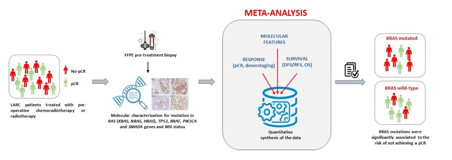
Predictive and Prognostic Value of Oncogene Mutations and Microsatellite Instability in Locally-Advanced Rectal Cancer Treated with Neoadjuvant Radiation-Based Therapy: A Systematic Review and Meta-Analysis

1
Experimental and Clinical Pharmacology, Centro di Riferimento Oncologico di Aviano (CRO) IRCCS, Via Franco Gallini n. 2, 33081 Aviano, Italy
2
Unit of Cancer Epidemiology, Centro di Riferimento Oncologico di Aviano (CRO) IRCCS, Via Franco Gallini n. 2, 33081 Aviano, Italy
3
Radiation Oncology, Centro di Riferimento Oncologico di Aviano (CRO) IRCCS, Via Franco Gallini n. 2, 33081 Aviano, Italy
4
Surgical Oncology, Centro di Riferimento Oncologico di Aviano (CRO) IRCCS, Via Franco Gallini n. 2, 33081 Aviano, Italy
*
Author to whom correspondence should be addressed.
Cancers 2023, 15(5), 1469; https://doi.org/10.3390/cancers15051469
Submission received: 6 December 2022 / Revised: 17 February 2023 / Accepted: 22 February 2023 / Published: 25 February 2023
(This article belongs to the Special Issue Advances in Radiotherapy and Prognosis of Rectal Cancer)

Abstract

:

Simple Summary

Identification of novel molecular markers of pathological complete response (pCR) to preoperative radiation-based therapy in locally advanced rectal cancer (LARC) is strongly needed. Given the established predictive and/or prognostic role of somatic mutations in key oncogenes (RAS, TP53, BRAF, PIK3CA, SMAD4) and microsatellite instability (MSI) status in colorectal cancer, we aimed to investigate the clinical value of the same markers in LARC patients by systematically reviewing the published literature and performing a quantitative analysis of the data. We found that KRAS mutations were significantly associated with the risk of not achieving pCR after preoperative treatment. This association was even more significant in patients who did not receive cetuximab than in patients who did. No other markers were associated with pCR. Based on our results, the implementation of KRAS mutation testing into clinical practice could improve the management of LARC patients.

Abstract

Markers of pathological complete response (pCR) to preoperative radiation-based therapy in locally advanced rectal cancer (LARC) are strongly needed. This meta-analysis aimed at elucidating the predictive/prognostic role of tumor markers in LARC. We systematically reviewed the impact of RAS, TP53, BRAF, PIK3CA, and SMAD4 mutations and MSI status on response (pCR, downstaging) and prognosis (risk of recurrence, survival) in LARC according to PRISMA guidelines and the PICO model. PubMed, Cochrane Library, and Web of Science Core Collection were systematically searched to identify relevant studies published before October 2022. KRAS mutations were significantly associated with the risk of not achieving pCR after preoperative treatment (summary OR = 1.80, 95% CI: 1.23–2.64). This association was even more significant in patients not receiving cetuximab (summary OR = 2.17, 95% CI: 1.41–3.33) than in patients receiving cetuximab (summary OR = 0.89, 95% CI: 0.39–20.05). MSI status was not associated with pCR (summary OR = 0.80, 95% CI: 0.41–1.57). No effect of KRAS mutation or MSI status on downstaging was detected. Meta-analysis of survival outcomes was not possible due to the large heterogeneity among studies in endpoint assessment. The minimum number of eligible studies to assess the predictive/prognostic role of TP53, BRAF, PIK3CA, and SMAD4 mutations was not reached. KRAS mutation, but not MSI status, proved to be a detrimental marker for response to preoperative radiation-based therapy in LARC. Translating this finding into the clinic could improve the management of LARC patients. More data are needed to clarify the clinical impact of TP53, BRAF, PIK3CA, and SMAD4 mutations.

Graphical Abstract

1. Introduction

Colorectal cancer (CRC) is one of the most commonly diagnosed cancers and one of the leading causes of cancer-related death worldwide [1]. Rectal cancers account for approximately 30–35% of all colorectal cancers, and about half of them are diagnosed at a locally advanced stage (i.e., locally advanced rectal cancer, LARC) [2]. A combined modality approach involving the use of fluoropyrimidine-based neoadjuvant chemoradiotherapy (nCRT), followed by total mesorectal surgical excision, is the standard of care for LARC patients [3,4]. At the time of surgery, a variable proportion of patients (8–30%) achieve pathologic complete response (pCR) [5,6], a condition associated with favorable long-term outcome [7], and with the possibility of opting for an organ-preserving approach (i.e., local excision or watch-and-wait strategy) [8]. On the other hand, early identification of patients with a poor response could be helpful in selecting patients for intensified pre-operative chemotherapy (e.g., total neoadjuvant therapy, TNT) [3,9].
Currently, the baseline assessment of patients for treatment planning is mainly based on clinical-pathological criteria [10] and has only recently been integrated by specific radiomic features [11].
Nowadays, testing for somatic tumor mutations in RAS and BRAF is mandatory to select the most appropriate treatment for CRC patients, and characterization of microsatellite instability (MSI) status of CRC tumor is assessed to evaluate the use of immune checkpoint inhibitors [12,13]. Although there is increasing evidence of the potential role of these predictive/prognostic molecular markers in LARC, they are still not included in risk algorithms used in clinics.
The mechanism of oncogenesis and the spectrum of molecular changes in tumor tissue have been described as significantly different between colon and rectal tumors [14,15,16]. It has been reported that 82% of non-metastatic rectal cancers have mutations in cancer-driving genes belonging to the PI3K and MAPK pathways, including KRAS, PIK3CA, and TP53 [17], similar to colon cancer, but the distribution of these mutations was different between rectal and colon samples [14,16]. For example, TP53 pathway mutations were more common in rectal tumors, whereas colon carcinomas had more RAS and PI3K pathway alterations [14,16]. The pharmacogenomic role of KRAS and TP53 mutations has been extensively studied in LARC patients treated with nCRT [18], but their predictive and prognostic value in this setting remains uncertain due to the large heterogeneity of published data. Other genes that have been studied in rectal cancer with contrasting results include BRAF, PIK3CA, SMAD4, and tumor MSI [18,19].
The aim of the present study was to perform a systematic review and meta-analysis of the impact of somatic mutations in the RAS (i.e., KRAS, NRAS, HRAS), TP53, BRAF, PIK3CA, and SMAD4 genes, and MSI status on response to neoadjuvant radiation-based therapy in patients with LARC. The primary endpoints were pCR and tumor downstaging and the secondary endpoints were disease-free survival (DFS) or relapse-free survival (RFS) and overall survival (OS). Determining the true predictive/prognostic value of somatic alterations in LARC could support their application in the clinic to improve selection of the most appropriate therapeutic option.

2. Materials and Methods

2.1. Literature Search

The systematic review was conducted according to the PRISMA (Preferred Reporting Items for Systematic Reviews and Meta-Analyses) guidelines and the PICO (Patients, Interventions, Comparisons, Outcomes) model (see Supplementary Methods for details on the PICO framework). This review was not registered on PROSPERO.
The literature search was performed for all studies published related to candidate gene mutation/MSI status and its impact on response to neoadjuvant chemotherapy or radiotherapy in LARC patients. Three databases, MEDLINE (PubMed), Cochrane Library, and Web of Science Core Collection (Clarivate), were used to search for relevant articles published in English in a peer-reviewed journal with the last search update on 1 October 2022. Because MEDLINE included all articles found in the other two databases, only MEDLINE was used. Search algorithms included all keywords to indicate ‘rectal cancer’, ‘chemoradiotherapy’ or ‘radiotherapy’, and ‘candidate gene’ or ‘MSI’, combined using Boolean operators (OR/AND) (see Supplementary Methods for the exact literature search algorithm). Additional studies were identified by hand-searching the references of relevant articles. Retrieved articles were screened and selected by two independent authors (EDM and SM) on the basis of inclusion and exclusion criteria; disagreements were resolved by a third researcher (EC). When studies overlapped, data from the publication with the largest number of patients were considered.

2.2. Inclusion and Exclusion Criteria

Inclusion criteria were as follows: (1) studies of patients diagnosed with primary adenocarcinoma of the rectum by pathological confirmation and eligible for preoperative radiation-based treatment. Patients with disease at all stages (I–IV) were included for analysis of response (i.e., pCR and downstaging), whereas only patients with stage II–III disease were considered for analysis of DFS/RFS and OS; (2) studies of patients treated with neoadjuvant chemoradiotherapy or radiotherapy; (3) studies that assessed the impact of mutational status in at least one candidate gene (i.e., RAS, TP53, BRAF, PIK3CA, and SMAD4) or MSI profile on clinical outcome (i.e., pathologic response as assessed by tumor regression grade, downstaging, recurrence, and survival) after neoadjuvant chemoradiotherapy or radiotherapy; (4) studies that obtained molecular data from pre-treatment tumor tissue sample; and (5) studies reporting pCR by mutation/MSI status or odds ratio (OR) with 95% confidence interval (CI). Exclusion criteria included: (1) reviews, meta-analyses, case reports, and conference abstracts; (2) preclinical studies; (3) studies without clinicopathologic endpoints; (4) studies not related to locally advanced rectal cancer; (5) studies with unclear, inadequate, insufficient, or incomplete data; and (6) studies that obtained molecular data from post-treatment biological samples to exclude the effects of chemoradiotherapy on tumor biology (e.g., induced change in the mutational pattern or complete disappearance of tumor cells in samples from patients with a pCR); and (7) case selection or treatment bias.

2.3. Data Extraction and Outcomes

Based on inclusion/exclusion criteria, relevant data were extracted in duplicate from all eligible studies by two independent investigators (EDM and SM) and recorded in a dedicated form designed at the beginning of the study. Particularly, the following items were collected for descriptive purposes: first author name, year of publication, country of origin, total number of patients, mean/median age of patients, gender, study type (retrospective/prospective), enrollment interval, disease stage, median follow-up, neoadjuvant treatment, type of chemotherapy, radiotherapy dose, interval time to surgery, type of surgery, and adjuvant treatment information, clinical outcome evaluated, tumor regression grade (TRG) classification system for evaluating pathologic response (i.e., American Joint Commission on Cancer—AJCC [20], Japanese Society for Cancer of the Colon and Rectum —JSCCR [21], Gavioli et al. [22], Dworak et al. [23], Wheeler et al. [24], Ryan et al. [25]), molecular markers studied and the related analytical method, source of biological sample. Discrepancies were resolved by discussion. The number of pCR by mutational/MSI status and the OR with corresponding CI—when available—were retrieved for the statistical analysis.
In this study, the outcomes collected include pCR, downstaging, DFS/RFS, and OS.

2.4. Quality Assessment

The methodological quality of each article included in the present meta-analysis was independently assessed by two authors using the Newcastle–Ottawa Scale (NOS) [26] (The Newcastle–Ottawa Scale, 2022), with scores ranging from 0 to 9. A NOS score of ≥7 was considered high quality.

2.5. Statistical Analysis

Summary estimates of the proportion of pCR or OR with the corresponding 95% CI were calculated when three or more studies were available. ORs were retrieved from the original publications, when available, or derived from the absolute number of patients with pCR by mutational/MSI status. The random-effects models by DerSimonian and Laird [27] were applied to incorporate both within- and between-study variability, as a weighted average, giving each study a weight proportional to its precision using the logit transformation. Statistical heterogeneity between studies was assessed using the I2 and τ2 statistics [27]. Influence analysis was performed when the summary estimate was estimated from five or more studies: the summary estimate was calculated by omitting one study at a time. Publication bias was assessed using a funnel plot [28]. Considering the existing gene/drug interaction between KRAS and cetuximab, analyses for KRAS status were stratified by cetuximab neo-adjuvant treatment. Results of the meta-analysis were presented graphically using forest plots, plotting the estimates from individual papers, summary estimate, proportion, and 95% CI. Statistical significance was claimed for p < 0.05. Analyses were performed using R’ software.

3. Results

3.1. Eligible Studies
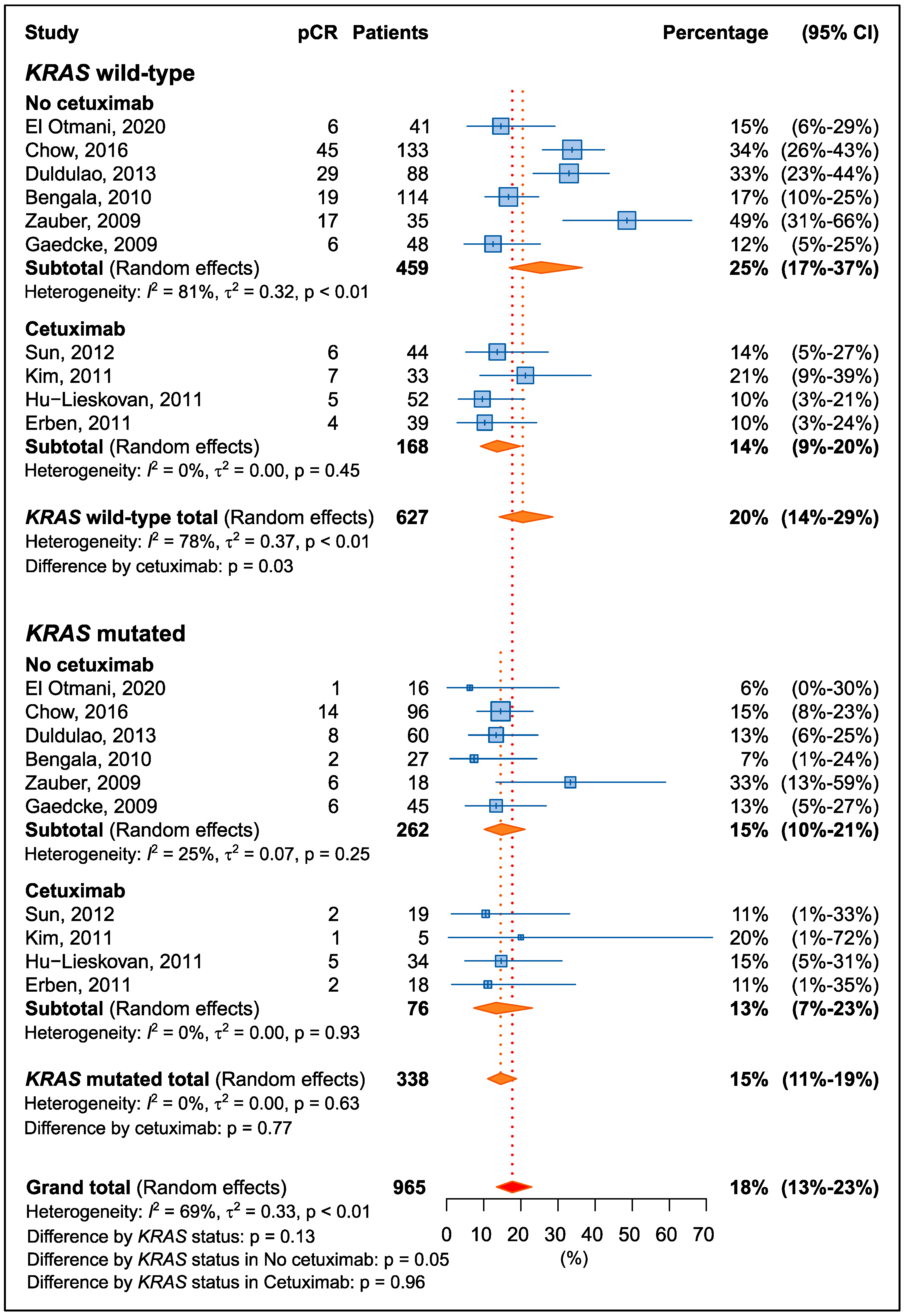
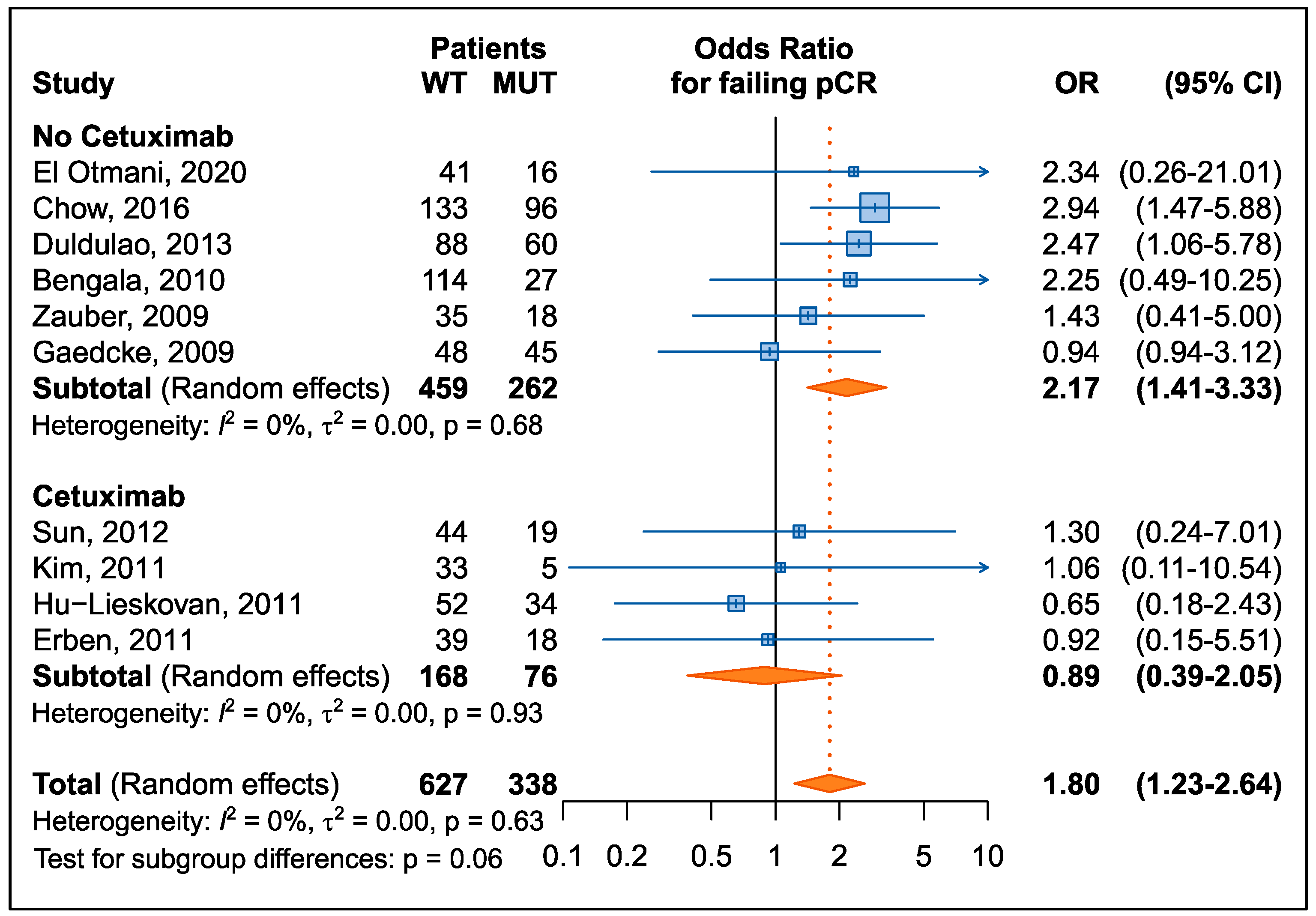
Figure 1 summarizes the results of the literature search.
By applying the inclusion/exclusion criteria, the minimum number of three eligible articles required for quantitative synthesis of data was not reached for the TP53, BRAF, PIK3CA, and SMAD4 genes for both response (i.e., pCR and tumor downstaging) and prognosis (i.e., DFS/RFS, OS) endpoints.
By applying the inclusion/exclusion criteria, sufficient articles were found for RAS genes and MSI status to perform a quantitative synthesis of the data for the pCR endpoint. For RAS genes, tumor downstaging was also analyzed. For DFS/RFS and OS, quantitative analysis was not possible due to large heterogeneity in endpoint assessment and data presentation (e.g., assessment of local or distant recurrence, different follow-up, different statistical methods); therefore, only descriptive synthesis was reported. Regarding the RAS genes, only KRAS was considered in the present meta-analysis, as no eligible records were identified for the other members of the RAS family, with the exception of two articles that investigated NRAS mutations [29,30].

3.2. KRAS

3.2.1. Pathological Complete Response

Ten eligible studies [29,31,32,33,34,35,36,37,38,39] were considered for quantitative synthesis (Figure 1). Among others, a large study on LARC cases extracted from the national oncologic outcome database was excluded due to the large heterogeneity of the study population, a lack of information on all inclusion criteria, and the risk of replicated data [40]. The characteristics of the included studies and details on the method of molecular analysis and response assessment are provided in Table S1. A summary of the main features of the studies is presented in Table 1. The total number of patients included in the analysis was 965, with an incidence of KRAS gene mutation of 35.0% (338/965) and a percentage of pCR of 19.8% (191/965). Seven of the ten eligible studies were conducted in European or North American populations. Most of the studies utilized long course radiotherapy delivered over 5 weeks. Preoperative treatment regimens varied, and the interval to surgery ranged from 0 to 61.4 weeks. The mutational status of KRAS was determined mainly by sequencing-based methods (8 of the 10 studies), and exon 2 (i.e., codons 12 and 13) was the most frequently studied. The quality of the included studies was high, with a NOS score equal to 7 for all articles (Table S2).
Overall, patients with a KRAS mutation have a non-significant lower rate of pCR compared to patients with wild-type KRAS (15% and 20%, respectively; p = 0.13) (Figure 2). However, treatment with cetuximab had a significant impact on pCR, particularly in patients with wild-type KRAS (p = 0.03). When analyses were restricted to patients not treated with cetuximab, patients with the KRAS mutation had worse pCR (15%; 95% CI: 10–21%) than wild-type patients (25%; 95% CI: 17–37%; p = 0.05). There was no difference in patients treated with cetuximab by KRAS status (p = 0.96).
To account for heterogeneity between studies, the effect of KRAS mutational status on the risk of not achieving pCR was expressed as OR for each study (Figure 3). The results showed that the presence of a KRAS mutation was significantly associated with an increased risk of not achieving pCR (summary OR = 1.80, 95% CI: 1.23–2.64); no heterogeneity was observed (p = 0.63). An analysis stratified by cetuximab use confirmed previous findings: the risk of not achieving pCR was 2.17 (95% CI: 1.41–3.33) in patients not treated with cetuximab compared to 0.89 (95% CI: 0.39–2.05) in patients treated with cetuximab; however, this difference was only marginally significant (p = 0.06).

3.2.2. Downstaging

A total of five eligible studies [31,34,35,38,41] that reported data on KRAS mutations and their effects on downstaging were considered for data extraction (Figure 1). Four articles [31,35,38,41] analyzed tumor downstaging, three [34,35,41] analyzed T-downstaging, and one [41] analyzed N-downstaging (Table 2). No significant association was found with KRAS mutation.

3.2.3. Recurrence Risk

Nine studies [29,30,34,35,37,38,41,42,43] were identified that investigated the impact of KRAS mutations on recurrence risk (i.e., DFS, RFS) (Figure 1). However, quantitative analysis of the data could not be performed due to the large heterogeneity of the studies in terms of the method of endpoint assessment (e.g., evaluation of local or distant recurrence, different follow-up, different statistical methods). In summary, eight of nine eligible studies [29,34,35,37,38,41,42,43] showed no association between tumor KRAS mutation status and DFS or RFS. Only one study [30] indicated that patients with KRAS mutations had a lower 3-year DFS (68% vs. 88.3%, p = 0.016) than patients without KRAS mutations. Of note, El Otmani et al. [29], in a subgroup analysis according to the specific codon mutations of KRAS, showed a significant association between mutations detected at codon 146 (i.e., A146T and A146V) and the presence of both recurrence and distant metastases (p = 0.019).

3.2.4. Overall Survival
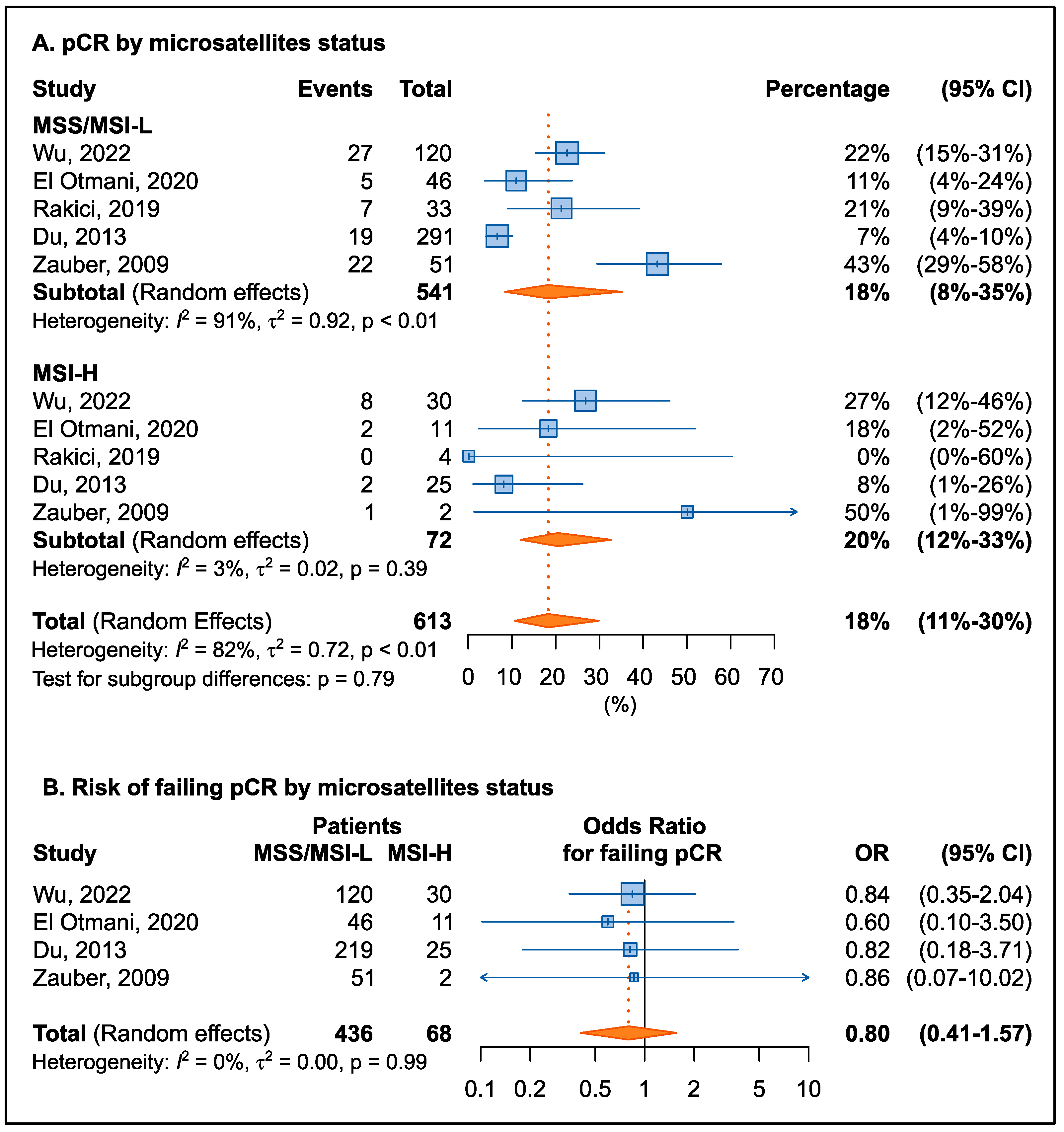
Eight articles [29,30,35,38,40,41,42,44] examining the effects of KRAS mutations on OS were eligible (Figure 1). However, quantitative analysis of the data could not be performed because of the large heterogeneity in the clinical assessment of the endpoint (e.g., different follow-up, different statistical methods). In brief, five studies [29,35,38,41,42] found no significant association between KRAS mutations and OS. On the contrary, three studies [30,40,44] reported that patients with KRAS mutations had a worse prognosis and an increased risk of death.

3.3. MSI Status

3.3.1. Pathological Complete Response

Five eligible studies [29,39,45,46,47] were included in this analysis (Figure 1). Mismatch repair deficiency (dMMR) was classified as high-frequency MSI (MSI-H), whereas proficient mismatch repair (pMMR) or low-frequency MSI (MSI-L) was considered microsatellite stable (MSS) [48]. Among others, two large studies that used a national oncologic outcome database to select LARC cases were excluded due to the large heterogeneity of the study population, lack of information on all inclusion criteria, and risk of replicated data [40,49]. A recent article was also excluded due to treatment bias, as patients received the immune checkpoint inhibitor nivolumab, whose efficacy is known to be related to MSI status [50]. The characteristics of the studies included in the meta-analysis, as well as details on the method of molecular analysis and response assessment, are provided in Table S3. A summary of the main features of the studies can be found in Table 3. The total number of patients included in the analysis was 613, the incidence of MSI-H was 11.7% (72/613), and the percentage of pCR was 15.2% (93/613). The studies were performed in different countries. Preoperative treatment regimens varied, and the interval time to surgery ranged from 3 to 16 weeks. MSI status was determined by immunohistochemistry in three articles and by an allelic size analysis in two articles. The quality of the included studies was high, with a NOS score equal to 7 for all but one article, which had a score of 9 (Table S2).
Microsatellite status (MSI-H or MSI-L/MMS) was not associated with pCR rate (20% vs. 18% for MSI-H and MSI-L/MSS, respectively) (Figure 4A). To control heterogeneity between studies, OR was calculated for each study (Figure 4B). The results confirmed that there was no association between microsatellite status and risk of not responding to therapy (summary OR = 0.80, 95% CI: 0.41–1.57). It should be noted, however, that all studies consistently reported a nonsignificant trend toward a lower risk of non-response to therapy for carriers of tumor MSI-H status.

3.3.2. Downstaging

No eligible article was found that reported data on the association between MSI status and tumor downstaging (Figure 1).

3.3.3. Recurrence Risk

Three eligible studies [29,45,46] were identified that examined the role of MSI status on recurrence risk (i.e., DFS, RFS) (Figure 1). In all studies, no significant association was found between the molecular marker and clinical outcome. Notably, Du et al. [45], in a subgroup analysis performed by tumor stage, showed that in the ypN0 group, patients with MSI-H had significantly better DFS than those with MSI-L or MSS status (100% vs. 79.8%, p < 0.05), whereas in the ypN + group no DFS improvement was observed for patients with MSI-H.

3.3.4. Overall Survival

Only one eligible article [29] (Figure 1) examined the impact of MSI status on OS and showed no significant association.

3.4. TP53, BRAF, PIK3CA, and SMAD4

3.4.1. Pathological Complete Response

According to the inclusion/exclusion criteria for study selection applied here, no eligible study was identified for the BRAF and SMAD4 genes evaluating the role of mutations on pCR (Figure 1).
For the TP53 gene, two eligible articles were retrieved [32,51]. The prospective study by Lopez-Crapez et al. [51], which included 70 LARC patients treated with preoperative radiotherapy or chemoradiotherapy, failed to demonstrate an association between TP53 mutations and pCR. Similarly, the retrospective analysis by Chow et al. [32] of 229 tumor biopsies from LARC patients who received nCRT confirmed that the presence of TP53 mutations did not affect pCR. For PIK3CA gene, two eligible retrospective studies were identified [52,53]. The study by Abdul-Jalil et al. [52] on 201 LARC biopsy specimens from patients treated with nCRT showed that mutations in the PIK3CA gene tended to be associated with a lack of pCR (OR: 3.33; p = 0.094). In contrast, the work of Russo et al. [53] in 47 LARC patients who had received nCRT showed no association between PIK3CA mutations and pCR.

3.4.2. Downstaging

For the BRAF, PIK3CA, and SMAD4 genes, no eligible studies were found that investigated the role of genetic tumor mutations on tumor downstaging (Figure 1).
For the TP53 gene, two eligible articles were found [51,54]. In the retrospective study by Kandioler et al. [54], which included 64 LARC patients treated with preoperative short-term radiotherapy, it was reported that TP53 mutations were significantly associated with no response to radiotherapy (p < 0.005) in terms of T-downstaging. However, this finding was not confirmed by the prospective analysis of Lopez-Crapez et al. [51], which was performed on 70 LARC patients treated with preoperative radiotherapy or chemoradiotherapy and found no association between TP53 mutational status and T-downstaging.

3.4.3. Recurrence Risk

For the TP53, BRAF, and SMAD4 genes, no eligible articles evaluating the role of mutations on recurrence risk were found (Figure 1).
For the PIK3CA gene, only one eligible study was identified [30]. This retrospective work by Peng et al., involving 70 LARC patients treated with preoperative chemoradiotherapy, found no association between PIK3CA mutations and 3-years DFS (3-year rate, 68.6% vs. 82.8% for mutated and wild-type patients, respectively, p = 0.632).

3.4.4. Overall Survival

For the TP53, BRAF, and SMAD4 genes, no eligible papers were identified examining the association between mutations and survival (Figure 1).
For the PIK3CA gene, only one eligible study was found [30]. This retrospective work by Peng et al., including 70 LARC patients treated with nCRT, found no association between PIK3CA mutations and 3-years OS (3-year rate, 77.8% versus 94.9% for mutated and wild-type patients, respectively, p = 0.870).

3.5. Sensitivity Analysis and Publication Bias

Influence analysis was conducted by performing meta-analysis, excluding one study at a time. The results were stable for the estimation of the pCR proportion according to KRAS status (Figure S1A,B). Conversely, substantial variability was observed when analyzed by microsatellite stability: for MSS/MSI-L (Figure S1C), the pCR proportion ranged from 14% excluding the study by Zauber et al. [39] to 24%, excluding the study by Du et al. [45]. Similarly, for MSI-H, the pCR proportion ranged from 14% (excluding the study by Wu et al. [46]) to 25% (excluding the study by Du et al. [45]) (Figure S1D). Sensitivity analysis for the risk of not achieving pCR showed no significant variability for both KRAS and microsatellite status (Figure S2).
To evaluate a possible confounding due to cancer stage, analyses for pCR by KRAS status were further restricted to studies performed in patients at stage II–III LARC [32,33,34,35,37,38]. The summary percentage of pCR was 21% (95% CI: 14–31%) for KRAS wild-type and 14% (95% CI: 10–19%; p = 0.11) for KRAS mutated. Among patients receiving cetuximab, pCR was achieved in 27% (95% CI: 17–40%) of KRAS wild-type patients and in 14% (95% CI: 8–23%; p = 0.02) of KRAS mutated patients, confirming the results of the main analysis.
No publication bias was detected in any analyses (Figure S3).

4. Discussion

The possibility to identify good and poor responders in advance for neoadjuvant treatment is a crucial issue in the management of LARC patients, as it could help clinicians select the most appropriate personalized strategy, including intensified pre-operative therapy (e.g., TNT) and organ-preserving approaches [3,9].
Somatic mutations in specific oncogenes (i.e., RAS, TP53, BRAF, PIK3CA, SMAD4) and MSI status have been widely studied as predictive markers of response (i.e., pCR) to neoadjuvant chemotherapy or radiotherapy, but the results are difficult to interpret due to the large heterogeneity of the studies performed [18,19]. With this meta-analysis, we attempted to overcome this issue by setting strict inclusion criteria to limit the analysis to a homogeneous study population and avoid potential bias.
This meta-analysis highlighted a significant detrimental role of the KRAS mutation, which was found to be predictive of poor response to neo-adjuvant treatment in LARC patients. KRAS is a key molecule in the MAPK and PI3K/AKT signaling pathways, which play important roles in cellular differentiation and apoptosis [18]. In colon cancer, somatic KRAS mutations, located mainly in codon 12 and 13 of exon 2, have been reported to lead to a more aggressive and invasive tumor and have been associated with resistance to anti-epidermal growth factor receptor (EGFR) monoclonal antibodies such as cetuximab and panitumumab [18,55]. Although the clinical significance of KRAS mutations in colon cancer is well established [56], the role of the same mutations in rectal cancer has not been fully elucidated. Pre-clinical investigations have shown that KRAS mutations could cause not only a more aggressive tumor phenotype, but also resistance to radiotherapy in rectal cancer [57,58,59]. However, clinical studies attempting to replicate this observation in patients receiving neoadjuvant treatment have been heterogeneous, and no consensus has been reached. Indeed, some analyses have highlighted a possible role of the KRAS mutation in predicting a lower pCR rate [32,33], but these results have not been confirmed by other studies [31,35,37]. Our meta-analysis showed that in a group of ten studies that met the inclusion criteria [29,31,32,33,34,35,36,37,38,39], KRAS mutations conferred an increased risk of not responding to neoadjuvant treatment (i.e., no-pCR), which is consistent with pre-clinical observations in rectal cancer and the well-established detrimental impact of KRAS mutations on the behavior of other tumors. This finding is in contrast to the results of two previously published meta-analyses [60,61], which found no significant association between KRAS status and pCR rate. The less stringent inclusion criteria adopted by the two previous meta-analyses could likely account for the discrepancy in results. In addition, one of the two papers [60] was published ten years ago and did not include the most recent data obtained with next-generation genotyping techniques.
During the years 2011–2013, the inclusion of cetuximab in preoperative treatment regimes in LARC patients was investigated. In this context, cetuximab was administered to all patients, regardless of KRAS mutational status. Therefore, a subgroup analysis on the predictive role of KRAS according to cetuximab administration was possible. The KRAS mutation was significantly predictive of pCR only in the group of patients who did not receive cetuximab, whereas it was not associated with pCR in patients who received cetuximab. EGFR has been shown to be a key molecule in the pathogenesis of rectal cancer, and its expression in the tumor of LARC patients undergoing neoadjuvant therapy was associated with significantly lower DFS [36,38]. Moreover, a radiosensitizing effect of anti-EGFR agents was noticed [34,36,37,38]. It could be hypothesized that an interaction between EGFR signaling pathway and radio/chemotherapy is the basis for the observed effect in patients not receiving an anti-EGFR agent such as cetuximab. On the other hand, the specific architecture of the rectal cancer molecular background may have disrupted the interaction between the use of cetuximab and the KRAS mutation observed in colon cancer, resulting in no effect of the mutation on the response to the specific anti-EGFR drug [34,36,37,38].
The effect of the KRAS mutation on T or N downstaging has also been investigated, but without significant results. This could probably be due to the small number of eligible studies (range 4 to 1), the heterogeneity in preoperative treatment (e.g., studies including cetuximab), and the different endpoints.
In the present meta-analysis, MSI status was not found to be predictive of pCR after neoadjuvant chemoradiation or radiation therapy in LARC patients. This finding confirms the results of two previous meta-analyses, in which a pooled analysis showed no significant effect of MSI status on pCR rates [19,62]. Accumulating data point out a resistance to 5-fluorouracil-based chemotherapy in colorectal cancer patients with MSI tumors [19,62,63]. However, although the MSI status has been reported as a predictive factor for the benefit of adjuvant fluoropyrimidine-based chemotherapy and overall prognosis in colon cancer [64], this finding has not been confirmed in rectal cancer, where MSI leads to different molecular and clinicopathological characteristics than colon MSI tumors [65]. Moreover, an interaction between chemoradiotherapy and MSI may also support our findings. Indeed, chemotherapy and radiotherapy have been reported to reprogram the tumor microenvironment and induce immunostimulatory effects, possibly by promoting a tumor antigen-specific immune response [66,67]. Similarly, MSI status has been shown to alter the radiosensitivity [68,69,70] and influence immunological status during nCRT for rectal cancer [71,72]. Therefore, both chemoradiotherapy and MSI status could affect the immune response, leading to unpredictable outcomes [72]. Our meta-analysis showed a non-significant trend for a lower risk of non-response to therapy in MSI-H tumors, suggesting a positive interaction between the immunomodulatory effect of radiotherapy and MSI status. However, the limited number of studies included in our meta-analysis and the relatively low incidence of MSI in rectal cancer may have compromised the power of the analysis, and further well-designed studies with large samples are needed to definitively clarify whether MSI status can be used to select patients for neoadjuvant treatment in rectal cancer.
Quantitative analysis of the role of KRAS mutation and MSI status on recurrence risk and survival was not possible. The literature search revealed that KRAS or MSI status did not appear to have a significant impact on the risk of local recurrence or distant metastasis. Subgroup analyses, e.g., by clinicopathological features (e.g., tumor stage [45]) or molecular features (e.g., specific KRAS codon mutations [29]), could potentially reveal an overlooked association. Moreover, a polygenic risk score that takes into account the interplay of multiple signaling pathways (e.g., the mutated KRAS gene and concomitant high expression of vascular endothelial growth factor) could likely better capture the tumor recurrence phenotype [42].
Meta-analysis of the predictive/prognostic value of the somatic mutation in the other oncogenes studied (i.e., TP53, BRAF, PIK3CA, and SMAD4) was not possible because the minimum number of three eligible articles required for quantitative synthesis of the data was not reached for both response and survival assessment. The gene most frequently studied in this context was TP53, with available studies reporting nonsignificant results of association with outcome in LARC patients, both in terms of pCR rate [32,51] and tumor downstaging [51,54]. No eligible studies on the impact of TP53 mutational status on recurrence risk or survival were found in the literature. Another gene examined in the present meta-analysis is PIK3CA; its mutational status was associated with a lack of pCR in one eligible study [52], but this result was not confirmed in another study [53]. The effect of PIK3CA mutation on DFS [30] and OS [30] was analyzed by one eligible study without finding a significant association. For the BRAF and SMAD4 genes, no eligible studies were detected for both pCR and prognosis, and the clinical value of their mutational status remains to be investigated.
This study has some limitations. First, there is high heterogeneity among the available studies. A thorough cleaning was performed to select a homogeneous group of studies eligible for meta-analysis. To this end, only studies with molecular data obtained from pre-treatment samples were included in the meta-analysis [70,73]. Two large population-based studies [40,49] were excluded because they collected data from national oncologic databases, and this register-based exploration did not allow the collection of all the information necessary to verify that all eligible criteria, including the timing of molecular analysis, were met. The work of Hasan et al. [49] has also been the subject of criticism of the methodology [62,70]. Nevertheless, a high degree of heterogeneity, including differences in study design (retrospective versus prospective), preoperative treatment (e.g., chemoradiotherapy, intensified chemoradiotherapy, or radiotherapy alone; administration of other drugs in addition to fluoropyrimidines; radiation dose), techniques used for molecular testing, and TRG classification system used to classify response, may still have influenced our work. Second, there are notable differences in the frequency of KRAS mutation (range 48–13%) or MSI-H status (range 4–20%) and pCR rate (range 7–43%) between studies, which may reflect the inhomogeneity of molecular techniques across laboratories and over time, as well as different clinical procedures for tumor response assessment or different treatment protocols. However, these differences were accounted for in the statistical procedure chosen. Third, despite some preliminary evidence for a role of the specific KRAS mutation (i.e., codon 12, 13, or 146) in tumor phenotype, the limited number of studies on this topic did not allow us to examine the association between specific mutation sites of the KRAS gene and treatment response.

5. Conclusions

In conclusion, the present meta-analysis provides evidence for the predictive role of tumor KRAS mutation in predicting the likelihood of achieving pCR in LARC patients, thus promoting the introduction of a pre-treatment molecular testing to improve the definition of individual risk of treatment failure and guide therapeutic planning. This could translate into personalized treatment, allowing a selective, safe, and organ-sparing approach (e.g., watch-and-wait strategy) in patients with a high probability of pCR and alternative treatment strategies (e.g., total neoadjuvant therapy) in patients with unresponsive tumors. This could lead to fewer surgery-related complications, better quality of life, and fewer unnecessary treatments.
On the contrary, the role of MSI status in predicting response to preoperative treatment remains unclear, and future studies are needed to definitively define its clinical value in rectal cancer. Further research efforts are also required to clarify the predictive significance of somatic mutations in other key oncogenes, such as TP53, BRAF, PIK3CA, and SMAD4.

Supplementary Materials

The following supporting information can be downloaded at: https://www.mdpi.com/article/10.3390/cancers15051469/s1, Supplementary methods: 1. PICO framework; 2. Search algorithms. Table S1: (A) Characteristics of included studies and (B) details on molecular analysis and response assessment for KRAS gene. Table S2: Quality assessment of included studies for (A) KRAS gene and (B) MSI status. Table S3: (A) Characteristics of included studies and (B) details on molecular analysis and response assessment for microsatellite instability (MSI) status. Figure S1: Influence analyses for percentage of pathological complete response (pCR) according to KRAS mutation and microsatellite status. Figure S2: Influence analyses for the risk of not achieving a pathological complete response (pCR) according to KRAS mutation and microsatellite status. Figure S3: Funnel plots for publication bias.

Author Contributions

E.D.M.: Conceptualization, Data curation, Writing—original draft; J.P.: Data curation, Formal analysis, Visualization; S.M.: Data curation, Validation; E.P.: Writing—review & editing; S.P.: Writing—review & editing; G.T.: Funding acquisition; E.C.: Conceptualization, Supervision, Writing—review & editing. All authors have read and agreed to the published version of the manuscript.

Funding

This research was funded by the Italian Ministry of Health (Ricerca Corrente), “Ricerca Sanitaria”.

Conflicts of Interest

The authors declare no conflict of interest.

References

  1. Sung, H.; Ferlay, J.; Siegel, R.L.; Laversanne, M.; Soerjomataram, I.; Jemal, A.; Bray, F. Global Cancer Statistics 2020: GLOBOCAN Estimates of Incidence and Mortality Worldwide for 36 Cancers in 185 Countries. CA Cancer J. Clin. 2021, 71, 209–249. [Google Scholar] [CrossRef] [PubMed]
  2. Ng, S.P.; Ngan, S.Y.; Leong, T. Current State of Neoadjuvant Radiotherapy for Rectal Cancer. Clin. Color. Cancer 2022, 21, 63–70. [Google Scholar] [CrossRef]
  3. Roeder, F.; Meldolesi, E.; Gerum, S.; Valentini, V.; Rodel, C. Recent advances in (chemo-)radiation therapy for rectal cancer: A comprehensive review. Radiat. Oncol. 2020, 15, 262. [Google Scholar] [CrossRef] [PubMed]
  4. Yoo, R.N.; Kim, H.J. Total neoadjuvant therapy in locally advanced rectal cancer: Role of systemic chemotherapy. Ann. Gastroenterol. Surg. 2019, 3, 356–367. [Google Scholar] [CrossRef] [PubMed] [Green Version]
  5. Belluco, C.; De Paoli, A.; Canzonieri, V.; Sigon, R.; Fornasarig, M.; Buonadonna, A.; Boz, G.; Innocente, R.; Perin, T.; Cossaro, M.; et al. Long-term outcome of patients with complete pathologic response after neoadjuvant chemoradiation for cT3 rectal cancer: Implications for local excision surgical strategies. Ann. Surg. Oncol. 2011, 18, 3686–3693. [Google Scholar] [CrossRef] [Green Version]
  6. Valentini, V.; van Stiphout, R.G.; Lammering, G.; Gambacorta, M.A.; Barba, M.C.; Bebenek, M.; Bonnetain, F.; Bosset, J.F.; Bujko, K.; Cionini, L.; et al. Nomograms for predicting local recurrence, distant metastases, and overall survival for patients with locally advanced rectal cancer on the basis of European randomized clinical trials. J. Clin. Oncol. 2011, 29, 3163–3172. [Google Scholar] [CrossRef]
  7. Barina, A.; De Paoli, A.; Delrio, P.; Guerrieri, M.; Muratore, A.; Bianco, F.; Vespa, D.; Asteria, C.; Morpurgo, E.; Restivo, A.; et al. Rectal sparing approach after preoperative radio- and/or chemotherapy (RESARCH) in patients with rectal cancer: A multicentre observational study. Technol. Coloproctology 2017, 21, 633–640. [Google Scholar] [CrossRef]
  8. Capelli, G.; De Simone, I.; Spolverato, G.; Cinquini, M.; Moschetti, I.; Lonardi, S.; Masi, G.; Carlomagno, C.; Corsi, D.; Luppi, G.; et al. Non-Operative Management Versus Total Mesorectal Excision for Locally Advanced Rectal Cancer with Clinical Complete Response After Neoadjuvant Chemoradiotherapy: A GRADE Approach by the Rectal Cancer Guidelines Writing Group of the Italian Association of Medical Oncology (AIOM). J. Gastrointest. Surg. 2020, 24, 2150–2159. [Google Scholar] [CrossRef] [PubMed]
  9. Liu, S.; Jiang, T.; Xiao, L.; Yang, S.; Liu, Q.; Gao, Y.; Chen, G.; Xiao, W. Total Neoadjuvant Therapy (TNT) versus Standard Neoadjuvant Chemoradiotherapy for Locally Advanced Rectal Cancer: A Systematic Review and Meta-Analysis. Oncologist 2021, 26, e1555–e1566. [Google Scholar] [CrossRef]
  10. Bitterman, D.S.; Resende Salgado, L.; Moore, H.G.; Sanfilippo, N.J.; Gu, P.; Hatzaras, I.; Du, K.L. Predictors of Complete Response and Disease Recurrence Following Chemoradiation for Rectal Cancer. Front. Oncol. 2015, 5, 286. [Google Scholar] [CrossRef] [PubMed] [Green Version]
  11. Zhang, S.; Yu, M.; Chen, D.; Li, P.; Tang, B.; Li, J. Role of MRIbased radiomics in locally advanced rectal cancer (Review). Oncol. Rep. 2022, 47, 34. [Google Scholar] [CrossRef] [PubMed]
  12. Boukouris, A.E.; Theochari, M.; Stefanou, D.; Papalambros, A.; Felekouras, E.; Gogas, H.; Ziogas, D.C. Latest evidence on immune checkpoint inhibitors in metastatic colorectal cancer: A 2022 update. Crit. Rev. Oncol. Hematol. 2022, 173, 103663. [Google Scholar] [CrossRef]
  13. Di Nicolantonio, F.; Vitiello, P.P.; Marsoni, S.; Siena, S.; Tabernero, J.; Trusolino, L.; Bernards, R.; Bardelli, A. Precision oncology in metastatic colorectal cancer—From biology to medicine. Nat. Rev. Clin. Oncol. 2021, 18, 506–525. [Google Scholar] [CrossRef]
  14. Chatila, W.K.; Kim, J.K.; Walch, H.; Marco, M.R.; Chen, C.T.; Wu, F.; Omer, D.M.; Khalil, D.N.; Ganesh, K.; Qu, X.; et al. Genomic and transcriptomic determinants of response to neoadjuvant therapy in rectal cancer. Nat. Med. 2022, 28, 1646–1655. [Google Scholar] [CrossRef]
  15. Frattini, M.; Balestra, D.; Suardi, S.; Oggionni, M.; Alberici, P.; Radice, P.; Costa, A.; Daidone, M.G.; Leo, E.; Pilotti, S.; et al. Different genetic features associated with colon and rectal carcinogenesis. Clin. Cancer Res. 2004, 10, 4015–4021. [Google Scholar] [CrossRef] [PubMed] [Green Version]
  16. Kapiteijn, E.; Liefers, G.J.; Los, L.C.; Kranenbarg, E.K.; Hermans, J.; Tollenaar, R.A.; Moriya, Y.; van de Velde, C.J.; van Krieken, J.H. Mechanisms of oncogenesis in colon versus rectal cancer. J. Pathol. 2001, 195, 171–178. [Google Scholar] [CrossRef] [PubMed]
  17. Iseas, S.; Sendoya, J.M.; Robbio, J.; Coraglio, M.; Kujaruk, M.; Mikolaitis, V.; Rizzolo, M.; Cabanne, A.; Ruiz, G.; Salanova, R.; et al. Prognostic Impact of An Integrative Landscape of Clinical, Immune, and Molecular Features in Non-Metastatic Rectal Cancer. Front. Oncol. 2021, 11, 801880. [Google Scholar] [CrossRef] [PubMed]
  18. De Mattia, E.; Roncato, R.; Palazzari, E.; Toffoli, G.; Cecchin, E. Germline and Somatic Pharmacogenomics to Refine Rectal Cancer Patients Selection for Neo-Adjuvant Chemoradiotherapy. Front. Pharmacol. 2020, 11, 897. [Google Scholar] [CrossRef]
  19. O’Connell, E.; Reynolds, I.S.; McNamara, D.A.; Prehn, J.H.M.; Burke, J.P. Microsatellite instability and response to neoadjuvant chemoradiotherapy in rectal cancer: A systematic review and meta-analysis. Surg. Oncol. 2020, 34, 57–62. [Google Scholar] [CrossRef]
  20. Mace, A.G.; Pai, R.K.; Stocchi, L.; Kalady, M.F. American Joint Committee on Cancer and College of American Pathologists regression grade: A new prognostic factor in rectal cancer. Dis. Colon Rectum 2015, 58, 32–44. [Google Scholar] [CrossRef] [PubMed] [Green Version]
  21. Yasutomi, M.; Baba, S.; Hojo, K.; Kato, Y.; Kodaira, S.; Koyama, Y. Japanese Classification of Colorectal Carcinoma, 1st ed.; Kanehara & Co., Ltd.: Tokyo, Japan, 1997. [Google Scholar]
  22. Gavioli, M.; Bagni, A.; Piccagli, I.; Fundaro, S.; Natalini, G. Usefulness of endorectal ultrasound after preoperative radiotherapy in rectal cancer: Comparison between sonographic and histopathologic changes. Dis. Colon Rectum 2000, 43, 1075–1083. [Google Scholar] [CrossRef]
  23. Dworak, O.; Keilholz, L.; Hoffmann, A. Pathological features of rectal cancer after preoperative radiochemotherapy. Int. J. Color. Dis. 1997, 12, 19–23. [Google Scholar] [CrossRef] [PubMed]
  24. Wheeler, J.M.; Dodds, E.; Warren, B.F.; Cunningham, C.; George, B.D.; Jones, A.C.; Mortensen, N.J. Preoperative chemoradiotherapy and total mesorectal excision surgery for locally advanced rectal cancer: Correlation with rectal cancer regression grade. Dis. Colon Rectum 2004, 47, 2025–2031. [Google Scholar] [CrossRef]
  25. Ryan, R.; Gibbons, D.; Hyland, J.M.; Treanor, D.; White, A.; Mulcahy, H.E.; O’Donoghue, D.P.; Moriarty, M.; Fennelly, D.; Sheahan, K. Pathological response following long-course neoadjuvant chemoradiotherapy for locally advanced rectal cancer. Histopathology 2005, 47, 141–146. [Google Scholar] [CrossRef]
  26. The Newcastle-Ottawa Scale (NOS). Available online: https://www.ohri.ca/programs/clinical_epidemiology/oxford.asp (accessed on 1 October 2022).
  27. Normand, S.L. Meta-analysis: Formulating, evaluating, combining, and reporting. Stat. Med. 1999, 18, 321–359. [Google Scholar] [CrossRef]
  28. Sterne, J.A.; Egger, M. Funnel plots for detecting bias in meta-analysis: Guidelines on choice of axis. J. Clin. Epidemiol. 2001, 54, 1046–1055. [Google Scholar] [CrossRef]
  29. El Otmani, I.; El Agy, F.; El Baradai, S.; Bouguenouch, L.; Lahmidani, N.; El Abkari, M.; Benajah, D.A.; Toughrai, I.; El Bouhaddouti, H.; Mouaqit, O.; et al. Analysis of Molecular Pretreated Tumor Profiles as Predictive Biomarkers of Therapeutic Response and Survival Outcomes after Neoadjuvant Therapy for Rectal Cancer in Moroccan Population. Dis. Markers 2020, 2020, 8459303. [Google Scholar] [CrossRef] [Green Version]
  30. Peng, J.; Lin, J.; Qiu, M.; Zhao, Y.; Deng, Y.; Shao, J.; Ding, P.; Zhang, H.; Wan, D.; Lu, Z.; et al. Oncogene mutation profile predicts tumor regression and survival in locally advanced rectal cancer patients treated with preoperative chemoradiotherapy and radical surgery. Tumor Biol. 2017, 39, 1010428317709638. [Google Scholar] [CrossRef] [Green Version]
  31. Bengala, C.; Bettelli, S.; Bertolini, F.; Sartori, G.; Fontana, A.; Malavasi, N.; Depenni, R.; Zironi, S.; Del Giovane, C.; Luppi, G.; et al. Prognostic role of EGFR gene copy number and KRAS mutation in patients with locally advanced rectal cancer treated with preoperative chemoradiotherapy. Br. J. Cancer 2010, 103, 1019–1024. [Google Scholar] [CrossRef]
  32. Chow, O.S.; Kuk, D.; Keskin, M.; Smith, J.J.; Camacho, N.; Pelossof, R.; Chen, C.T.; Chen, Z.; Avila, K.; Weiser, M.R.; et al. KRAS and Combined KRAS/TP53 Mutations in Locally Advanced Rectal Cancer are Independently Associated with Decreased Response to Neoadjuvant Therapy. Ann. Surg. Oncol. 2016, 23, 2548–2555. [Google Scholar] [CrossRef] [PubMed] [Green Version]
  33. Duldulao, M.P.; Lee, W.; Nelson, R.A.; Li, W.; Chen, Z.; Kim, J.; Garcia-Aguilar, J. Mutations in specific codons of the KRAS oncogene are associated with variable resistance to neoadjuvant chemoradiation therapy in patients with rectal adenocarcinoma. Ann. Surg. Oncol. 2013, 20, 2166–2171. [Google Scholar] [CrossRef]
  34. Erben, P.; Strobel, P.; Horisberger, K.; Popa, J.; Bohn, B.; Hanfstein, B.; Kahler, G.; Kienle, P.; Post, S.; Wenz, F.; et al. KRAS and BRAF mutations and PTEN expression do not predict efficacy of cetuximab-based chemoradiotherapy in locally advanced rectal cancer. Int. J. Radiat. Oncol. Biol. Phys. 2011, 81, 1032–1038. [Google Scholar] [CrossRef]
  35. Gaedcke, J.; Grade, M.; Jung, K.; Schirmer, M.; Jo, P.; Obermeyer, C.; Wolff, H.A.; Herrmann, M.K.; Beissbarth, T.; Becker, H.; et al. KRAS and BRAF mutations in patients with rectal cancer treated with preoperative chemoradiotherapy. Radiother. Oncol. 2010, 94, 76–81. [Google Scholar] [CrossRef]
  36. Hu-Lieskovan, S.; Vallbohmer, D.; Zhang, W.; Yang, D.; Pohl, A.; Labonte, M.J.; Grimminger, P.P.; Holscher, A.H.; Semrau, R.; Arnold, D.; et al. EGF61 polymorphism predicts complete pathologic response to cetuximab-based chemoradiation independent of KRAS status in locally advanced rectal cancer patients. Clin. Cancer Res. 2011, 17, 5161–5169. [Google Scholar] [CrossRef] [Green Version]
  37. Kim, S.Y.; Hong, Y.S.; Kim, D.Y.; Kim, T.W.; Kim, J.H.; Im, S.A.; Lee, K.S.; Yun, T.; Jeong, S.Y.; Choi, H.S.; et al. Preoperative chemoradiation with cetuximab, irinotecan, and capecitabine in patients with locally advanced resectable rectal cancer: A multicenter Phase II study. Int. J. Radiat. Oncol. Biol. Phys. 2011, 81, 677–683. [Google Scholar] [CrossRef] [PubMed]
  38. Sun, P.L.; Li, B.; Ye, Q.F. Effect of neoadjuvant cetuximab, capecitabine, and radiotherapy for locally advanced rectal cancer: Results of a phase II study. Int. J. Color. Dis. 2012, 27, 1325–1332. [Google Scholar] [CrossRef] [PubMed]
  39. Zauber, N.P.; Marotta, S.P.; Berman, E.; Grann, A.; Rao, M.; Komati, N.; Ribiero, K.; Bishop, D.T. Molecular genetic changes associated with colorectal carcinogenesis are not prognostic for tumor regression following preoperative chemoradiation of rectal carcinoma. Int. J. Radiat. Oncol. Biol. Phys. 2009, 74, 472–476. [Google Scholar] [CrossRef]
  40. Zhou, P.; Goffredo, P.; Ginader, T.; Thompson, D.; Hrabe, J.; Gribovskaja-Rupp, I.; Kapadia, M.; Hassan, I. Impact of KRAS status on tumor response and survival after neoadjuvant treatment of locally advanced rectal cancer. J. Surg. Oncol. 2021, 123, 278–285. [Google Scholar] [CrossRef] [PubMed]
  41. Lee, J.W.; Lee, J.H.; Shim, B.Y.; Kim, S.H.; Chung, M.J.; Kye, B.H.; Kim, H.J.; Cho, H.M.; Jang, H.S. KRAS Mutation Status Is Not a Predictor for Tumor Response and Survival in Rectal Cancer Patients Who Received Preoperative Radiotherapy With 5-Fluoropyrimidine Followed by Curative Surgery. Medicine 2015, 94, e1284. [Google Scholar] [CrossRef]
  42. Krajnovic, M.; Markovic, B.; Knezevic-Usaj, S.; Nikolic, I.; Stanojevic, M.; Nikolic, V.; Siljic, M.; Jovanovic Cupic, S.; Dimitrijevic, B. Locally advanced rectal cancers with simultaneous occurrence of KRAS mutation and high VEGF expression show invasive characteristics. Pathol. Res. Pract. 2016, 212, 598–603. [Google Scholar] [CrossRef]
  43. Velenik, V.; Ocvirk, J.; Oblak, I.; Anderluh, F. Cetuximab in preoperative treatment of rectal cancer—Term outcome of the XERT trial. Radiol. Oncol. 2012, 46, 252–257. [Google Scholar] [CrossRef] [PubMed]
  44. Asawa, P.; Bakalov, V.; Kancharla, P.; Abel, S.; Chahine, Z.; Monga, D.K.; Kirichenko, A.V.; Wegner, R.E. The prognostic value of KRAS mutation in locally advanced rectal cancer. Int. J. Color. Dis. 2022, 37, 1199–1207. [Google Scholar] [CrossRef]
  45. Du, C.; Zhao, J.; Xue, W.; Dou, F.; Gu, J. Prognostic value of microsatellite instability in sporadic locally advanced rectal cancer following neoadjuvant radiotherapy. Histopathology 2013, 62, 723–730. [Google Scholar] [CrossRef] [PubMed]
  46. Wu, Z.; Hu, H.; Wang, C.; Zhang, J.; Cai, Y.; Xie, X.; Huang, Y.; Deng, Y. The prognostic and predictive value of mismatch repair status in patients with locally advanced rectal cancer following neoadjuvant therapy. Ann. Transl. Med. 2022, 10, 491. [Google Scholar] [CrossRef]
  47. Yilmaz Rakici, S.; Bedir, R.; Hatipoglu, C. Are there predictors that can determine neoadjuvant treatment responses in rectal cancer? Turk. J. Gastroenterol. 2019, 30, 220–227. [Google Scholar] [CrossRef] [PubMed]
  48. Bai, Z.; Zhou, Y.; Ye, Z.; Xiong, J.; Lan, H.; Wang, F. Tumor-Infiltrating Lymphocytes in Colorectal Cancer: The Fundamental Indication and Application on Immunotherapy. Front. Immunol. 2021, 12, 808964. [Google Scholar] [CrossRef] [PubMed]
  49. Hasan, S.; Renz, P.; Wegner, R.E.; Finley, G.; Raj, M.; Monga, D.; McCormick, J.; Kirichenko, A. Microsatellite Instability (MSI) as an Independent Predictor of Pathologic Complete Response (PCR) in Locally Advanced Rectal Cancer: A National Cancer Database (NCDB) Analysis. Ann. Surg. 2020, 271, 716–723. [Google Scholar] [CrossRef]
  50. Bando, H.; Tsukada, Y.; Inamori, K.; Togashi, Y.; Koyama, S.; Kotani, D.; Fukuoka, S.; Yuki, S.; Komatsu, Y.; Homma, S.; et al. Preoperative Chemoradiotherapy plus Nivolumab before Surgery in Patients with Microsatellite Stable and Microsatellite Instability-High Locally Advanced Rectal Cancer. Clin. Cancer Res. 2022, 28, 1136–1146. [Google Scholar] [CrossRef]
  51. Lopez-Crapez, E.; Bibeau, F.; Thezenas, S.; Ychou, M.; Simony-Lafontaine, J.; Thirion, A.; Azria, D.; Grenier, J.; Senesse, P. p53 status and response to radiotherapy in rectal cancer: A prospective multilevel analysis. Br. J. Cancer 2005, 92, 2114–2121. [Google Scholar] [CrossRef] [Green Version]
  52. Abdul-Jalil, K.I.; Sheehan, K.M.; Toomey, S.; Schmid, J.; Prehn, J.; O’Grady, A.; Cummins, R.; O’Neill, B.; McNamara, D.A.; Deasy, J.; et al. The frequencies and clinical implications of mutations in 33 kinase-related genes in locally advanced rectal cancer: A pilot study. Ann. Surg. Oncol. 2014, 21, 2642–2649. [Google Scholar] [CrossRef]
  53. Russo, A.L.; Ryan, D.P.; Borger, D.R.; Wo, J.Y.; Szymonifka, J.; Liang, W.Y.; Kwak, E.L.; Blaszkowsky, L.S.; Clark, J.W.; Allen, J.N.; et al. Mutational and clinical predictors of pathologic complete response in the treatment of locally advanced rectal cancer. J. Gastrointest. Cancer 2014, 45, 34–39. [Google Scholar] [CrossRef] [Green Version]
  54. Kandioler, D.; Zwrtek, R.; Ludwig, C.; Janschek, E.; Ploner, M.; Hofbauer, F.; Kuhrer, I.; Kappel, S.; Wrba, F.; Horvath, M.; et al. TP53 genotype but not p53 immunohistochemical result predicts response to preoperative short-term radiotherapy in rectal cancer. Ann. Surg. 2002, 235, 493–498. [Google Scholar] [CrossRef] [PubMed]
  55. Bignucolo, A.; Scarabel, L.; Toffoli, G.; Cecchin, E.; De Mattia, E. Predicting drug response and toxicity in metastatic colorectal cancer: The role of germline markers. Expert Rev. Clin. Pharmacol. 2022, 15, 689–713. [Google Scholar] [CrossRef]
  56. Bignucolo, A.; De Mattia, E.; Cecchin, E.; Roncato, R.; Toffoli, G. Pharmacogenomics of Targeted Agents for Personalization of Colorectal Cancer Treatment. Int. J. Mol. Sci. 2017, 18, 1522. [Google Scholar] [CrossRef] [Green Version]
  57. Bernhard, E.J.; Stanbridge, E.J.; Gupta, S.; Gupta, A.K.; Soto, D.; Bakanauskas, V.J.; Cerniglia, G.J.; Muschel, R.J.; McKenna, W.G. Direct evidence for the contribution of activated N-ras and K-ras oncogenes to increased intrinsic radiation resistance in human tumor cell lines. Cancer Res. 2000, 60, 6597–6600. [Google Scholar]
  58. Jones, H.A.; Hahn, S.M.; Bernhard, E.; McKenna, W.G. Ras inhibitors and radiation therapy. Semin. Radiat. Oncol. 2001, 11, 328–337. [Google Scholar] [CrossRef]
  59. McKenna, W.G.; Weiss, M.C.; Bakanauskas, V.J.; Sandler, H.; Kelsten, M.L.; Biaglow, J.; Tuttle, S.W.; Endlich, B.; Ling, C.C.; Muschel, R.J. The role of the H-ras oncogene in radiation resistance and metastasis. Int. J. Radiat. Oncol. Biol. Phys. 1990, 18, 849–859. [Google Scholar] [CrossRef] [PubMed]
  60. Clancy, C.; Burke, J.P.; Coffey, J.C. KRAS mutation does not predict the efficacy of neo-adjuvant chemoradiotherapy in rectal cancer: A systematic review and meta-analysis. Surg. Oncol. 2013, 22, 105–111. [Google Scholar] [CrossRef]
  61. Peng, J.; Lv, J.; Peng, J. KRAS mutation is predictive for poor prognosis in rectal cancer patients with neoadjuvant chemoradiotherapy: A systemic review and meta-analysis. Int. J. Color. Dis. 2021, 36, 1781–1790. [Google Scholar] [CrossRef] [PubMed]
  62. Swets, M.; Graham Martinez, C.; van Vliet, S.; van Tilburg, A.; Gelderblom, H.; Marijnen, C.A.M.; van de Velde, C.J.H.; Nagtegaal, I.D. Microsatellite instability in rectal cancer: What does it mean? A study of two randomized trials and a systematic review of the literature. Histopathology 2022, 81, 352–362. [Google Scholar] [CrossRef]
  63. Cercek, A.; Dos Santos Fernandes, G.; Roxburgh, C.S.; Ganesh, K.; Ng, S.; Sanchez-Vega, F.; Yaeger, R.; Segal, N.H.; Reidy-Lagunes, D.L.; Varghese, A.M.; et al. Mismatch Repair-Deficient Rectal Cancer and Resistance to Neoadjuvant Chemotherapy. Clin. Cancer Res. 2020, 26, 3271–3279. [Google Scholar] [CrossRef] [Green Version]
  64. Cao, Y.; Wang, X. Effects of molecular markers on the treatment decision and prognosis of colorectal cancer: A narrative review. J. Gastrointest. Oncol. 2021, 12, 1191–1196. [Google Scholar] [CrossRef] [PubMed]
  65. Hong, S.P.; Min, B.S.; Kim, T.I.; Cheon, J.H.; Kim, N.K.; Kim, H.; Kim, W.H. The differential impact of microsatellite instability as a marker of prognosis and tumour response between colon cancer and rectal cancer. Eur. J. Cancer 2012, 48, 1235–1243. [Google Scholar] [CrossRef] [PubMed]
  66. De Mattia, E.; Canzonieri, V.; Polesel, J.; Mezzalira, S.; Dalle Fratte, C.; Dreussi, E.; Roncato, R.; Bignucolo, A.; Innocente, R.; Belluco, C.; et al. SMAD3 Host and Tumor Profiling to Identify Locally Advanced Rectal Cancer Patients at High Risk of Poor Response to Neoadjuvant Chemoradiotherapy. Front. Pharmacol. 2021, 12, 778781. [Google Scholar] [CrossRef] [PubMed]
  67. De Mattia, E.; Polesel, J.; Roncato, R.; Labriet, A.; Bignucolo, A.; Gagno, S.; Buonadonna, A.; D’Andrea, M.; Levesque, E.; Jonker, D.; et al. IL15RA and SMAD3 Genetic Variants Predict Overall Survival in Metastatic Colorectal Cancer Patients Treated with FOLFIRI Therapy: A New Paradigm. Cancers 2021, 13, 1705. [Google Scholar] [CrossRef] [PubMed]
  68. Davis, T.W.; Wilson-Van Patten, C.; Meyers, M.; Kunugi, K.A.; Cuthill, S.; Reznikoff, C.; Garces, C.; Boland, C.R.; Kinsella, T.J.; Fishel, R.; et al. Defective expression of the DNA mismatch repair protein, MLH1, alters G2-M cell cycle checkpoint arrest following ionizing radiation. Cancer Res. 1998, 58, 767–778. [Google Scholar]
  69. Franchitto, A.; Pichierri, P.; Piergentili, R.; Crescenzi, M.; Bignami, M.; Palitti, F. The mammalian mismatch repair protein MSH2 is required for correct MRE11 and RAD51 relocalization and for efficient cell cycle arrest induced by ionizing radiation in G2 phase. Oncogene 2003, 22, 2110–2120. [Google Scholar] [CrossRef] [Green Version]
  70. Meillan, N.; Vernerey, D.; Lefevre, J.H.; Manceau, G.; Svrcek, M.; Augustin, J.; Flejou, J.F.; Lascols, O.; Simon, J.M.; Cohen, R.; et al. Mismatch Repair System Deficiency Is Associated With Response to Neoadjuvant Chemoradiation in Locally Advanced Rectal Cancer. Int. J. Radiat. Oncol. Biol. Phys. 2019, 105, 824–833. [Google Scholar] [CrossRef]
  71. Lee, J.H.; Kang, B.H.; Song, C.; Kang, S.B.; Lee, H.S.; Lee, K.W.; Chie, E.K.; Kim, J.S. Microsatellite Instability Correlated Inflammatory Markers and their Prognostic Value in the Rectal Cancer Following Neoadjuvant Chemoradiotherapy: A Hypothesis-generating Study. In Vivo 2020, 34, 2119–2126. [Google Scholar] [CrossRef]
  72. Ni, K.; Zhan, Y.; Liu, Z.; Zhao, X.Z.; Wang, W.; Wang, G.; Zhang, Z.; Li, G.; Zhang, X.; Zhang, C. Mismatch repair system deficiency is associated with chemoradiotherapy resistance in locally advanced rectal adenocarcinoma patients. J. Surg. Oncol. 2022, 125, 692–702. [Google Scholar] [CrossRef]
  73. Sclafani, F.; Wilson, S.H.; Cunningham, D.; Gonzalez De Castro, D.; Kalaitzaki, E.; Begum, R.; Wotherspoon, A.; Capdevila, J.; Glimelius, B.; Rosello, S.; et al. Analysis of KRAS, NRAS, BRAF, PIK3CA and TP53 mutations in a large prospective series of locally advanced rectal cancer patients. Int. J. Cancer 2020, 146, 94–102. [Google Scholar] [CrossRef] [PubMed]
Figure 1. PRISMA flow diagram. Abbreviations: DFS, disease-free survival; OS, overall survival; pCR, pathologic complete response.
Cancers 15 01469 g001
Figure 2. Forrest plot for percentage of pathological complete response (pCR) by KRAS mutation and cetuximab treatment [29,31,32,33,34,35,36,37,38,39].
Cancers 15 01469 g002
Figure 3. Forrest plot for the risk of not achieving a pathological complete response (pCR) in patients with a KRAS mutation according by cetuximab treatment [29,31,32,33,34,35,36,37,38,39].
Cancers 15 01469 g003
Figure 4. Forrest plot for percentage of pathological complete response (pCR, (A)) and for the risk of not achieving a pCR (B) by microsatellite status. dMMR was considered as MSI-H, while pMMR or MSI-L are considered as microsatellite stable MSS [29,39,45,46,47].
Cancers 15 01469 g004
Table 1. Main characteristics of included studies for KRAS gene.
First Author, YearCountryNTherapy StrategyFLsOther DrugKRAS Mut (%)pCR (%)NOS Score
El Otmani, 2020 [29]Morocco57CRT/RT + surgery5-FU-28%12%7
Chow, 2016 [32]USA229CRT/intensified CRT + surgery5-FUOXA 42%26%7
Duldulao, 2013 [33]USA 148CRT/intensified CRT + surgery5-FUOXA41%25%7
Sun, 2012 [38]China63CRT + surgeryCAPECTX30%13%7
Kim, 2011 [37]Korea38CRT + surgeryCAPECTX, IRI 13%21%7
Hu-Lieskovan, 2011 [36]Europe86CRT + surgery 5-FU, CAPE CTX, OXA40%12%7
Erben, 2011 [34]Europe 57intensified CRT + surgeryCAPECTX, IRI32%11%7
Bengala, 2010 [31]Europe 141CRT + surgery5-FU, CAPEOXA19%15%7
Zauber, 2009 [39]Europe 53CRT/RT + surgery5-FU--34%43%7
Gaedcke, 2010 [35]Europe 93CRT + surgery5-FUOXA48%13%7
Abbreviation: 5-FU, 5-fluorouracil; CAPE, capecitabine; CRT, chemoradiotherapy; CTX, cetuximab; FLs, Fluoropyrimidines; IRI, irinotecan; NOS, Newcastle–Ottawa Scale; OXA, oxaliplatin; pCR, pathological complete response; RT, radiotherapy.
Table 2. Pooled percentage of downstaging according to KRAS mutational status.
DownstagingT DownstagingN Downstaging
Rate (95% CI)PHetRate (95% CI)PHetRate (95% CI)PHet
Studies (n)4 3 1
KRAS
Wild-type0.52 (0.27–0.77)p < 0.010.54 (0.46–0.61)p = 0.480.61 (0.49–0.72)-
Mutated0.55 (0.37–0.71)p = 0.020.44 (0.34–0.54)p = 0.620.62 (0.41–0.80)-
p = 0.87 p = 0.14 p = 1.00
Table 3. Main characteristics of included studies for microsatellite instability (MSI) status.
First Author, YearCountryNTherapy StrategyFLsOther DrugMSI-H (%)pCR (%)NOS Score
Wu, 2022 [46]China150CRT + surgery 5-FUOXA 20%23%7
El Otmani, 2020 [29]Morocco57CRT/RT + surgery5-FU--19%12%7
Yilmaz Rakici, 2019 [47] Turkey37CRT/RT + surgery5-FU, CAPE--11%19%9
Du, 2013 [45]China316RT + surgery----8%7%7
Zauber, 2009 [39]USA53CRT/RT + surgery5-FU--4%43%7
Abbreviation: 5-FU, 5-fluorouracil; CAPE, capecitabine; CRT, chemoradiotherapy; FLs, Fluoropyrimidines; MSI-H, high-frequency; NOS, Newcastle–Ottawa Scale; OXA, oxaliplatin; pCR, pathological complete response; RT, radiotherapy.
Disclaimer/Publisher’s Note: The statements, opinions and data contained in all publications are solely those of the individual author(s) and contributor(s) and not of MDPI and/or the editor(s). MDPI and/or the editor(s) disclaim responsibility for any injury to people or property resulting from any ideas, methods, instructions or products referred to in the content.

