Article

Correlations between Molecular Alterations, Histopathological Characteristics, and Poor Prognosis in Esophageal Adenocarcinoma

by
Arianna Orsini
1,†,
Luca Mastracci
2,3,†,
Isotta Bozzarelli
1,‡,
Anna Ferrari
4,‡,
Federica Isidori
1,
Roberto Fiocca
2,3,
Marialuisa Lugaresi
1,4,5,
Antonietta D’Errico
6,7,
Deborah Malvi
7,
Erica Cataldi-Stagetti
1,
Paola Spaggiari
8,
Anna Tomezzoli
9,
Luca Albarello
10,
Ari Ristimäki
11,12,
Luca Bottiglieri
13,
Kausilia K. Krishnadath
14,
Riccardo Rosati
15,
Uberto Fumagalli Romario
16,
Giovanni De Manzoni
17,
Jari Räsänen
18,
Giovanni Martinelli
4,
Sandro Mattioli
5,
Elena Bonora
1,* and
on behalf of the EACSGE Consortium
§
add Show full author list remove Hide full author list
1
Department of Medical and Surgical Sciences (DIMEC), Alma Mater Studiorum, University of Bologna, 40126 Bologna, Italy
2
Unit of Anatomic Pathology, Ospedale Policlinico San Martino IRCCS, 16125 Genova, Italy
3
Department of Surgical Sciences and Integrated Diagnostics (DISC), University of Genova, 16125 Genova, Italy
4
IRCCS Istituto Romagnolo per lo Studio dei Tumori (IRST) “Dino Amadori”, 47014 Meldola, Italy
5
Division of Thoracic Surgery, Maria Cecilia Hospital, GVM Care & Research Group, Cotignola, 48022 Ravenna, Italy
6
Pathology Unit, IRCCS Azienda Ospedaliero-Universitaria di Bologna, 40126 Bologna, Italy
7
Department of Experimental, Diagnostic and Specialty Medicine (DIMES), Alma Mater Studiorum, University of Bologna, 40126 Bologna, Italy
8
Unit of Anatomic Pathology, Humanitas University, 20089 Milan, Italy
9
Unit of Anatomic Pathology, Azienda Ospedaliera di Verona, 37122 Verona, Italy
10
Pathology Unit, San Raffaele Scientific Institute, 20135 Milan, Italy
11
Department of Pathology, HUSLAB and HUS Diagnostic Center, University of Helsinki, 00170 Helsinki, Finland
12
Helsinki University Hospital, 00170 Helsinki, Finland
13
Unit of Anatomic Pathology, Istituto Europeo di Oncologia, 20122 Milan, Italy
14
Laboratory of Experimental Medicine and Pediatrics (LEMP), Department of Gastroenterology and Hepatology, University Hospital Antwerp, 2650 Antwerp, Belgium
15
Department of Gastrointestinal Surgery, San Raffaele Hospital, Vita-Salute San Raffaele University, 20135 Milan, Italy
16
Digestive Surgery, European Institute of Oncology, IRCCS, 20122 Milan, Italy
17
Department of Surgery, General and Upper G.I. Surgery Division, University of Verona, 37126 Verona, Italy
18
Department of General Thoracic and Esophageal Surgery, Helsinki University Hospital, 00170 Helsinki, Finland
*
Author to whom correspondence should be addressed.
These authors contributed equally to this work.
§
Membership of the EACSGE Consortium is provided in the Acknowledgments.
Cancers 2023, 15(5), 1408; https://doi.org/10.3390/cancers15051408
Submission received: 14 January 2023 / Revised: 15 February 2023 / Accepted: 20 February 2023 / Published: 23 February 2023
(This article belongs to the Special Issue Oesophageal Adenocarcinoma)

Abstract

:

Simple Summary

The molecular heterogeneity of esophageal adenocarcinoma (EAC), a severe malignancy with increasing incidence and low survival rates, misperceives the underlying biology of tumor onset and development. However, advances in high-throughput next-generation sequencing (NGS) technologies have highlighted the potential role of somatic DNA sequence markers for new diagnostic techniques or constitute novel therapeutic targets. Thus, in order to identify a molecular and prognostic signature in EAC patients, we decided to integrate the sequencing of specimens from naïve patients (not treated with chemo-radiotherapy) with histological classification, with the aim of identification of potential biomarkers, and patient stratification. Combining different approaches paves the way for early identification and the selection of better therapy.

Abstract

Esophageal adenocarcinoma (EAC) is a severe malignancy with increasing incidence, poorly understood pathogenesis, and low survival rates. We sequenced 164 EAC samples of naïve patients (without chemo-radiotherapy) with high coverage using next-generation sequencing technologies. A total of 337 variants were identified across the whole cohort, with TP53 as the most frequently altered gene (67.27%). Missense mutations in TP53 correlated with worse cancer-specific survival (log-rank p = 0.001). In seven cases, we found disruptive mutations in HNF1alpha associated with other gene alterations. Moreover, we detected gene fusions through massive parallel sequencing of RNA, indicating that it is not a rare event in EAC. In conclusion, we report that a specific type of TP53 mutation (missense changes) negatively affected cancer-specific survival in EAC. HNF1alpha was identified as a new EAC-mutated gene.

1. Introduction

Esophageal adenocarcinoma (EAC) is a severe malignancy with increasing incidence in Western countries over the past few decades and a relatively high mortality since overall prognosis remains bleak and the 5-year survival rate is just 35–45% [1].
EAC develops from the cells that release mucus and other fluids and may arise according to the widely accepted sequence gastroesophageal reflux disease (GERD) /intestinal metaplasia/dysplasia/ adenocarcinoma [2].
In clinical practice, complete endoscopic evaluation of GERD symptoms includes evaluation of erosive esophagitis and its complication and inspection for BE with multiple biopsies when present [3]. BE is relatively common in the general population, with a 1–2% prevalence (up to 10% in those with reflux symptoms) [4], but only about 1% of patients progress to cancer each year [5]. The ability to treat pre-invasive, dysplastic lesions with endoscopic resection and/or ablation instead of more extensive and invasive procedures such as chemotherapy with or without radiotherapy and esophagectomy, which have associated co-morbidities and poor 5-year survival rates, makes early diagnosis highly clinically relevant [6].
In a recent study on a large series of EAC cases submitted to surgery (without neoadjuvant treatment), a diagnostic algorithm which separated adenocarcinomas with glandular architecture from other rare histotypes, and further graded the former and subtyped the latter, was adopted [7]. This morphologic distinction has proven to have a significant prognostic impact on its own or dichotomized into lower and higher risk carcinomas, especially when coupled with stage. Indeed, the stage plus histotype combination showed a high discriminating power for 5-year cancer-specific survival, ranging from 87.6% in the stage II lower risk group to 14% in the stage IVA higher risk group [7]. Given the histological differences observed in EAC, it is of great interest to investigate the underlying biology of the tumor and to understand the molecular alterations correlated with those distinctive patterns which provide strong prognostic factors. Indeed, several genomic studies have included EAC in a group of tumors with one of the most frequent rates of copy number alterations (CNAs), somatic structural rearrangements, and elevated mutation frequency, with different mutational signatures and epigenetic mechanisms giving rise to significant inter and intra-tumor heterogeneity [8]. Large-scale sequencing studies have revealed distinct mutational signatures in EAC, and the presence of multiple CNAs was possibly correlated to a worse outcome; however, an exhaustive correlation with clinical outcomes and specific histotypes has not yet been provided. In a large genomic study, Secrier et al. identified three distinct molecular subtypes of molecular signatures: (i) an enrichment for BRCA signature with prevalent defects in the homologous recombination pathway; (ii) a dominant T > G mutational pattern associated with a high mutational load and neoantigen burden; and (iii) a C > A/T mutational pattern with evidence of an aging imprint. However, the clinical characteristics of the three subgroups did not differ significantly [9].
In EAC, a number of potential driver mutations have been described, with many of the mutations occurring in tumor suppressor genes (e.g., TP53, SMAD4, and ARID1A). The overall picture is of genomic instability and significant heterogeneity between patients. The accumulation of structural variants appears to be a gradual process throughout the disease’s natural history [6,10]. According to the Cancer Genome Atlas [8], EACs contain molecular changes similar to the chromosomal instability (CIN) subtype of stomach cancer. This suggests that EAC treatment may be improved by grouping them with CIN stomach cancers. Genomic alterations may also represent effective targets for therapy, i.e., frequent alterations of genes that regulate the cell cycle may be treated with existing drugs. Moreover, one-third of the EACs studied harbor an alteration to the ERBB2 gene, which encodes the HER2 protein and may be targeted with HER2 inhibitors. However, the overall genomic heterogeneity and the rearrangements occurring during tumor progression make it difficult to define valuable prognostic molecular signatures underlying biology that drive tumor onset and development [11].
We coupled a deep sequencing analysis of the most commonly mutated genes in EAC with the recent novel classification based on histological subtypes [7] in order to stratify patients based on clinical and molecular features for better personalized care. We evaluated the genetic alterations of cancer-related genes that we found recurrently mutated in a previous study on a small group of EAC cases [12]. Herein we analyzed specimens of 164 naïve patients (without chemo-radiotherapy and not subjected to neoadjuvant treatments) using next-generation sequencing approaches. Moreover, we characterized the presence of novel gene fusion transcripts, as markers of genomic alterations, in a number of these cases for which we had RNA available from tumor specimens. Molecular data were correlated with the histological subtypes and the clinical outcomes.

2. Materials and Methods

2.1. Sample Recruitment

The DNA samples were extracted from the surgical samples obtained at the following centers: Istituto Europeo di Oncologia (IEO), Milano, and IRCCS Ospedale San Raffaele, Milano, Ospedale di Verona, Verona, for a total of 164 samples. The inclusion criteria consisted of the presence of adenocarcinoma of the esophagogastric junction; no neoadjuvant treatment (chemo-radiotherapy-naïve EACs); and full clinical medical history and follow-up up to 60 months after surgery. All of the surgical resections were formalin-fixed paraffin-embedded (FFPE), re-evaluated by gastrointestinal pathologists, and classified according to the EACSGE histological classification [7]. The cases analyzed for mutation analysis of TP53 only have been previously described [12] and they were re-classified according to the EACSGE classification.

2.2. Custom EAC Panel: Library Preparation, Hybridization, Sequencing, and Bioinformatic Analysis

DNA was extracted from two 40 μm-thick, FFPE sections using a QIAMP DNA FFPE Tissue Kit (Cat. 56404; Qiagen, Hilden, Germany) according to the manufacturer’s protocol. Dual-index paired-end libraries were prepared using a Lotus DNA library prep kit (Cat. 10001074; Integrated DNA Technologies IDT Inc., Coralville, IA, USA) according to the manufacturer’s instructions. The protocol followed three major steps: an enzymatic preparation (with fragmentation to obtain 300–350 bp DNA fragments) where end-repair and dA-tailing were performed, the ligation of stubby adapters was performed, and PCR amplification for 11 cycles with indexing primers was performed (to incorporate sample-unique indexing sequences and P5 and P7 sequences to attach to the flow-cell). After purification, the single DNA libraries were run on 3% agarose gel to confirm the appropriate size and quantified using a Qubit dsDNA BR Assay Kit (Cat. Q33265; ThermoFisher Scientific, Vilnus, Lituania). Five hundred ng of each library preparation was pooled into groups of 16 samples to perform hybridization and enrichment for selected gene regions. This step was performed using an xGen Lockdown probe pool and an xGen hybridization capture of DNA libraries kit (IDT), according to the protocols. Each pool of 16 samples was hybridized to the capture probes for 16 h at 65 °C. xGen Lockdown Probes were individually synthesized, including 5′ biotinylated oligos, and were assembled in a custom panel of 26 genes for target capture. The genes selected for this study are listed in Figure 1. The hybridized regions were then captured with streptavidin magnetic beads and, after non-bound products’ removal, a post-capture PCR of 11 cycles was performed. The enriched library pools were checked for quantity and size with a Qubit dsDNA HS Assay kit (Cat. Q33230; Thermo Fisher Scientific, Waltham, MA, USA) and 2100 Bioanalyzer High Sensitivity DNA (Agilent Technologies, Santa Clara, CA, USA), respectively. Each pool was normalized to 1.3 pM and then sequenced on an Illumina NextSeq 500 platform (Illumina San Diego, CA, USA) at 150 bp paired ends. Data analysis was performed with an in-house pipeline [12]. In particular, Fastq files containing raw reads were checked using FastQC (https://www.bioinformatics.babraham.ac.uk/projects/fastqc/ v.0.11.8) and aligned using BWA (bio-bwa.sourceforge.net v.0.7.17-r1188) to the human reference (hg19). PCR-duplicated reads were marked and removed using Picard. Putative somatic variants, including SNPs and small insertions/deletions (indels), were identified using GATK software (software.broadinstitute.org/gatk/ v.4.0.10). The raw mutation calls were filtered to exclude false calls based on base quality, allele frequency of mismatched bases, and possible occurrences of strand bias. The identified mutations were further annotated and prioritized with Ensembl VEP (www.ensembl.org/Tools/VEP v.94).

2.3. RNA Analysis

RNA was extracted from the FFPE samples (five for each EACSGE subgroup). The samples were then selected among the ones with good quality RIN scores and DV200 > 40%. Starting from 100 ng of 27 RNA, libraries were prepared using a TruSight RNA Pan-Cancer Panel Kit (Illumina, San Diego, CA, USA; 1385 cancer-associated genes), following the manufacturer’s protocol. On twenty-two of the libraries that passed the protocol quality checks, paired-end RNA sequencing was performed (Reagent Kit v3-150 cycles, MiSeq, Illumina, San Diego, CA, USA) and raw sequencing data were converted to FASTQ file format and analyzed by combining FusionCatcher (FC(1)), STAR-Fusion (SF), and two Basespace applications [RNA-Seq Alignment v.1.1.0 (RSA) and TopHat Alignment v.1.0.0 (THA); Illumina, San Diego, CA, USA]. The reference Homo sapiens UCSC hg19 (RefSeq and Gencode gene annotations) was used for all the aligners.
We retained the fusions detected by at least three tools and we introduced further criteria to retain or reject fusions detected by two tools or one tool [see PCT application No. PCT/EP2021/065692 (10 June 2021): Method to identify linked genetic fusions]. The gene fusions were confirmed with Sanger sequencing, as previously described [12].

2.4. Immunohistochemistry Analysis

Immunohistochemistry (IHC) analysis was performed automatically with a Benchmark XT® immunostainer (Ventana Medical Systems) HNF1alpha antigen. The immunohistochemical analysis was validated through positive controls (as an external positive control put on the slide according to Bragoni et al. [13]) and negative controls (by omitting the primary antibody). Cases carrying predicted damaging variants in HNF1alpha and cases with no variants in the gene were evaluated by IHC from FFPE surgical specimens. IHC was performed for HNF1alpha and scoring was carried out by two independent expert pathologists, blindly with respect to the mutation status.
IHC analysis for SMAD4 and p53 was carried out as described previously [12]. Evaluation of SMAD4 immunostains was performed by two expert pathologists. For each case, the percentage of neoplastic cells with SMAD4 preserved or lost immunosignal was collected.

2.5. Statistical Analysis

The χ2 test or Fisher’s test (an expected number less than five) and the Mann–Whitney test were used to analyze categorical and continuous variables, respectively. The correlations were analyzed with Spearman’s rho coefficient. Survival analysis was performed using the Kaplan–Meier method and the log-rank test. p-values < 0.05 were considered significant. Data were analyzed using SPSS (version 15.0) (SPSS Inc., Chicago, IL, USA) and Prism (GraphPad Software Inc., San Diego, CA, USA). Univariate and multivariate (forward stepwise conditional method) Cox regression analyses were performed to estimate the effects of clinical, genetic, and pathological parameters on CSS. In the stepwise procedure, significance levels of 0.05 for entering and 0.10 for removing the respective explanatory variables were used to determine the independent risk factors. For the power calculations, we used G*Power version 3.1.9.6 [14].

3. Results

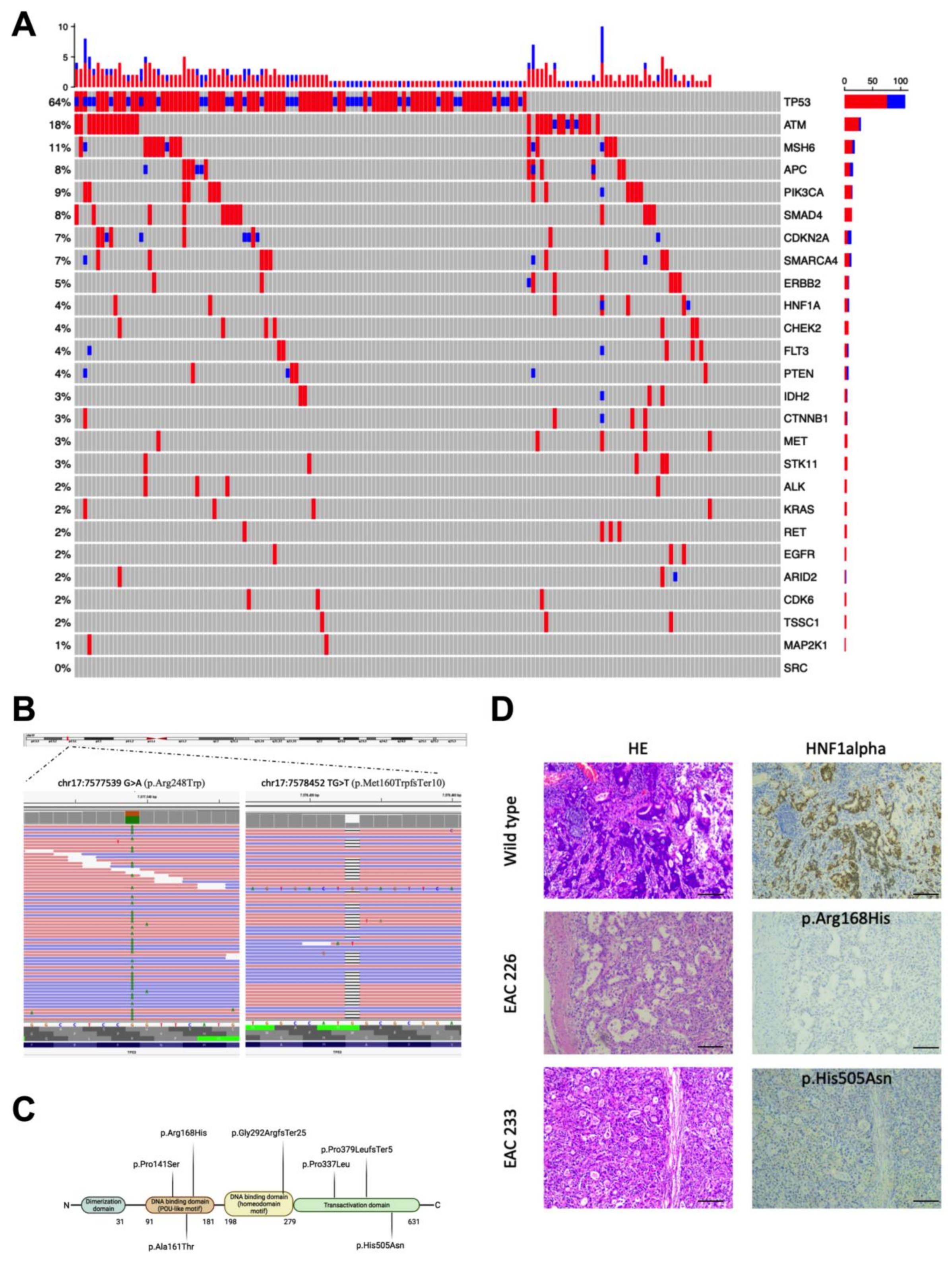
3.1. Genetic Alterations Identified in the EAC Samples

A total of 337 variants were identified across the whole cohort of 164 EAC cases (Figure 1A). All of the FFPE samples achieved good sequence representation with average coverage among samples of 700×. Examples of identified mutations are reported in Figure 1B.
Point mutations were the most frequent variations (82.21%), followed by insertions and deletions (17.78%). TP53 was the most frequently altered gene, with 110/164 cases carrying at least one mutation in this gene (67.1%). Out of the 110 variants in the TP53 gene, there was a prevalence in missense (77) vs. loss of function (LOF, including premature stop codons, splice site alterations, and frameshift variants) (33) variants. Five cases carried two variants in TP53. Most of the mutations have already been reported in the “TP53 database” (https://tp53.isb-cgc.org/ accessed on 10 October 2022) and were functionally analyzed: 76 out of 77 missense changes (98.7%) in TP53 found in our EAC cohort were functionally damaging (Supplementary Table S1).
Alterations in other genes occurred at a lower frequency, with ATM (18%), MSH6 (11%), PI3KCA (9%), APC and SMAD4 (8%), and CDKN2A and SMARCA4 (7%) being the most frequently hit genes. The majority of the samples carried concurrent variants in different genes (Figure 1A).

3.2. HNF1alpha Mutations in EAC

We found seven variants in the HNF1alpha gene, encoding for a tumor suppressor protein; in seven cases (Figure 1C), the gene that we previously found mutated in a small number of EAC cases [12]. The mutations mapped to the DNA binding domain (three single nucleotide substitutions and one frameshift indel variant) and to the transactivation domain (two single nucleotide substitutions and one frameshift indel variant) of HNF1alpha. The missense variants were predicted to be damaging according to the prediction program MCAP (MCAP score > 0.7; http://bejerano.stanford.edu/mcap/ accessed on 15 November 2022) [15]. In six out of sevem cases, HNF1alpha variants occurred in association with other gene alterations (Figure 1A).
We evaluated via IHC analysis the protein expression profiles of the available samples carrying the different HNF1alpha variants vs. a case without mutations (the control). Compared to the control sample, decreased staining was observed in the patients with HNF1alpha damaging variants. The decrease in staining correlated to an increased frequency of the variant alleles in the tumor, as detected by NGS data analysis (Figure 1D and Supplementary Table S2).

3.3. Correlation of Variants in Different Genes

As already reported in many studies, we found a variety of genetic alterations in the 164 samples analyzed via NGS, with TP53 being the most mutated gene. To evaluate whether mutations co-occurred significantly in specific genes, we performed a correlation analysis between the damaging variants identified in the different oncology-related genes, using Spearman’s rho coefficient. We identified several statistically significant positive correlations between the presence of mutations in specific genes, as well as several negative correlations, as reported in Supplementary Table S3. TP53 mutations, as an example, correlated positively with CDKN2A (0.162, p = 0.035) but correlated negatively with ATM (−0.147, p = 0.047), HNF1alpha (−0.166, p = 0.031), and MET (−0.195, p = 0.011). HNF1alpha on the other hand, correlated positively with PIK3CA (0.248, p < 0.001), CTNNB1 (0.281, p < 0.001), and RET (0.161, p = 0.034), suggesting that specific genes were concurrently mutated in these tumors. We also assessed in the COSMIC project COSU535, containing data on 409 EAC cases, and the presence of co-occurring variants between genes and found mutations of TP53 and APC (21/409, 5%), TP53 and CDKN2A in 25/409 (6%), and TP53 and SMAD4 in 30/409 (7%) in the same samples, reinforcing the concept of the genetic heterogeneity of EAC.

3.4. Evaluating Associations between Genetic Variants and Histopathological and Clinical Phenotypes

In order to connect the presence of genetic variants with clinical and/or morpho-functional characteristics, we performed further analyses taking into account cancer-specific survival (CSS), recurrence, and the EACSGE classification, introduced by Fiocca et al. [7]. This classification was based on morphological features of esophageal/esophagogastric junction adenocarcinoma, which divided the cases into two main categories with a different prognose: lower risk, including glandular well differentiated (GL WD), mucinous muconodular carcinoma (MMC), and diffuse desmoplastic (DDC) subgroups; and higher risk, including glandular poorly differentiated (GL PD), diffuse anaplastic (DAC), invasive mucinous carcinomas (IMC), and mixed (MIX) subgroups.
This analysis provided significant data only for TP53, since the number of mutations in this gene was high enough to allow a statistical association to different parameters.
We estimated the clinical outcomes in relation to the presence of TP53 variants and to the specific types of TP53 variants, i.e., missense variants or LOF variants. Poor survival and recurrence were significantly associated with TP53 mutations (p = 0.039; Supplementary Table S4A; p = 0.031, Supplementary Table S4B, respectively). Considering the EACSGE classification in the lower risk and higher risk groups, the presence of TP53 mutations and the higher risk group were significantly associated (p = 0.022; Supplementary Table S4C).
These data prompted us to extend the analysis to include additional EAC cases for which we had genetic material and the TP53 mutation status, the clinical parameters, and the morphological classification according to EACSGE for a total of 202 individuals. The overall results are presented in Figure 2A,B and Supplementary Table S5A,B. We could show that cancer-specific survival was negatively affected by the presence of missense mutations in the higher risk cases (p = 0.001, Supplementary Table S5A), as also shown by Kaplan–Meier curve analysis (Figure 2A). Considering the different subclasses of the higher and lower risk groups, we could observe how the statistical association was mainly driven by the GD-PD classes in the presence of missense variants in the TP53 gene (p = 0.001; Supplementary Table S5B and Figure 2B).
TP53 mutations and age also showed a significant association, as reported in Supplementary Table S5C (p = 0.029, Kruskal–Wallis test).
We investigated whether the presence of the different types of TP53 variants (missense and LOF) could be detected by immunohistochemical analysis of the tumor specimens. We evaluated the presence of different types of variants and the staining pattern observed for p53 in terms of overexpression or loss of staining (Supplementary Table S6), according to our previously reported methods [12], in which missense variants were associated with p53 staining. We found a significant association between the type of mutations and patterns of p53 staining also for LOF variants vs. cases without TP53 variants (Spearman correlation coefficient = 0.782; p < 0.01, Supplementary Table S7, Figure 2C–E).

3.5. SMAD4 Expression Loss and EAC Survival

We recently reported that SMAD4 loss of immunoreactivity was not an infrequent event in EAC, even in absence of gene mutations [12]. Therefore, we extended the analysis to a larger sample of EAC tissues from the EACSGE consortium and correlated it with genetic, histopathological, and clinical data [7].
First, for the group of cases where we had the TP53 status and p53 immunostaining, we evaluated whether any correlation with the SMAD4 immunostaining pattern was significant. Indeed, we found that the presence of LOF variants in TP53 correlated with SMAD4 loss of staining in the corresponding tumor tissue (as defined in the Materials and Method section, p = 0.008, Supplementary Table S8).
Therefore, we investigated whether any loss of SMAD4 immunostaining was relevant or whether a more informative SMAD4 loss cut-off value could be identified. Through ROC curve analysis (Supplementary Table S9), loss of immunostaining in at least 35% of neoplastic cells resulted to be the best discriminator with the greatest sensitivity and specificity. Applying this cut-off value in our case series, 85 cases were defined as SMAD4 loss cancers out of the 245 EAC cases from the entire EACSGE consortium (35%, Figure 3A). When considering the EACSGE histopathological classification, the SMAD4 pattern of immunostaining was significantly correlated with CCS and disease-free survival. SMAD4 loss was correlated with poor CSS (p = 0.007; Figure 3B) and disease-free survival (p = 0.002, Figure 3C) in EACSGE higher risk cases but not in lower risk cases (p = ns).
Post-hoc analysis revealed that the study had 0.772 power to detect a 0.25 effect size [16].

3.6. Univariate and Multivariate Cox Regression Analysis

Univariate and multivariate Cox regression analyses were performed to estimate the effects of clinical, genetic (TP53), SMAD4 loss (cut-off > 35), and pathological parameters on CSS. As reported in Supplementary Table S9A, the univariate Cox regression analysis showed a statistical association for age, stage, lymph node status, and EACSGE risk (p = 0.028, p = 0.001, p < 0.001, and p = 0.003, respectively), whereas in the multivariate analysis, only age, lymph node ratio, and EACSGE risk retained significance (p = 0.005, p < 0.001, and p = 0.023, Supplementary Table S9B).

3.7. Gene Fusion Analysis from RNA Sequencing

Twenty-two samples, with RNA quality compatible with massive parallel sequencing were sequenced at high coverage for 1385 oncology-relevant genes. Data analysis was performed with an in-house pipeline which is based on the combination of data obtained using four independent tools for fusion detection, e.g., FusionCatcher, STAR-Fusion, RNA-Seq Alignment v.1.1.0, and TopHat Alignment v.1.0.0. From the 64 candidate fusions identified by combining the results of the four tools, the pipeline retained eight after filtering analysis. The selected gene fusions were selected by two to four tools (Supplementary Table S10). The eight gene fusions were identified in six different EAC cases, but we could confirm with an independent method (Sanger sequencing) six gene fusions in four cases (4/22, 18.2%, Supplementary Table S10). Interestingly, in the two cases carrying two different gene fusions, one of the rearrangements was the same, the CYP2C19-CYP2C18 fusion on chromosome 10 (Figure 4A). The other gene fusions involved the GIPC1-DNAJB1 rearrangement in one case (Figure 4B) and PI4KA-MAPK1 in the other case. The GAIP-interacting protein C-terminus (GIPC1) is a regulator of autophagy and cellular trafficking and its overexpression is associated with poor survival in several cancers [17,18]. DNAJB1 encodes for a molecular chaperone involved in protein folding and autophagic mechanisms. The DNAJB1-PRKACA fusion transcript has been identified in many cases of fibrolamellar hepatocellular carcinoma [19].
The gene encoding for the phosphatidylinositol 4-kinase alpha (PI4KA) was detected in two different gene fusion EAC samples (Figure 4C). PI4KA plays a critical role in regulating tumorigenesis by activating tumor-promoting signals such as the RAS pathway [20].
The gene fusion IQCE-DGKB (Figure 4D) involved an IQ motif containing E gene, which is important in limb morphogenesis and also acts as regulator of Hedgehog signaling [21] and the gene for diacylglycerol kinase beta (DGKB). The diacylglycerol kinases are key regulators of the intracellular concentration of the second messenger diacylglycerol (DAG) and play a key role in cellular processes. This gene has been found in other fusions with different genes in prostate cancer [22].
All of the detected gene fusions involved oncology-related genes, but they have not been reported in esophageal adenocarcinoma.

4. Discussion

Esophageal adenocarcinoma represents a substantial health concern in Western countries due to its increasing incidence and poor prognosis. Rapid advances in high-throughput NGS have highlighted high EAC inter- and intratumor heterogeneity, with many structural genomic rearrangements and mutations arising even clonally [8,11,23,24], and epigenetic dysregulation of specific genes giving rise to tumor entities that may behave very differently in terms of progression and resistance [8,25,26]. Therefore, there is an increasing interest in defining molecular biomarkers for patient stratification and prognosis [27].
In our study, we focused our attention on a panel of cancer-related genes previously found recurrently mutated in a small cohort of EAC cases. In the 164 novel cases assessed, the EAC samples presented mutations in different genes, including HNF1alpha. HNF1alpha is a transcription factor, regulates epithelial to mesenchymal transition, and is considered a tumor suppressor [28]. In concordance with its role as a tumor suppressor, the majority of the variants were LOF, with a remarkable decrease in protein expression in the tumor specimens compared to cases with no mutations in this gene. HNF1alpha mutations were associated with other mutations in most of our cases, as we observed this for several genes, concurrently mutated in the same samples. However, we did not address the overall mutational signatures, in terms of specific nucleotide changes as detected with programs such as Signature Mutational Analysis (Sigma) [29] or Mix [30], but only whether specific genes showed concurrent or mutually exclusive mutations.
Regarding the identified gene mutations, TP53 was the most frequently mutated gene, as reported in previous studies [8]. Indeed, TP53 is the most mutated gene in the group of chromosomally unstable carcinomas of the esophagus and of the esophageal junction [8] and tumors that are histologically predominantly intestinal. Thus, it is not surprising that in our classification, they are mainly GD-PD with TP53 mutations, but it is remarkable that in GD-PD, this finding has prognostic significance. Moreover, we evaluated the effect of specific types of mutations (missense changes vs. loss of function) in correlation with histological and clinical data and could observe that in the cases classified as “higher risk” according to the EACSGE classification, the presence of damaging missense variants in TP53 negatively affected cancer-specific survival,
It is however worth noting that TP53 mutations are early events in the progression from esophageal dysplasia to cancer [31]; therefore, the early identification of specific types of variants, i.e., damaging amino acid substitutions, might be of critical importance, especially from the perspective of selecting the most efficient approach for targeted therapies. It is important to note that missense changes in p53 can alter protein folding, structure, and therefore DNA-binding and transcriptional activity. Targeting mutant p53 with missense changes is indeed an active research field in order to induce p53 activity similar to the wild-type protein [32].
Mutations that led to a loss of p53 proteins (such as nonsense and frameshift variants, LOF) were frequently associated with the loss of SMAD4 expression. The SMAD4 tumor-suppressor gene is pivotal for the downstream signaling of bone morphogenetic proteins (BMPs) [33]. Notably, SMAD4 has been reported as frequently lost in gastrointestinal cancer [11,34]. SMAD4 loss is associated with non-canonical BMP signaling leading to a more metastatic phenotype, poor prognosis, and poor response to treatment [35]. A previous study showed that SMAD4 mutations or homozygous deletions were associated with significantly poorer prognosis in EAC [11]. A recent study in preclinical models of EAC development showed that SMAD4 inactivation was sufficient per se to initiate tumorigenesis in a high-grade dysplastic esophagus in vivo [36]. Moreover, inhibition of the overactive non-canonical BMP signaling in SMAD4-negative tumors decreased malignancy and improved survival [37]. Notably, SMAD4 expression status resulted in being a prognostic factor in our cohort of cases when connected to the EACSGE classification, allowing us to discriminate patients with a worse prognosis into the higher risk group.
Nevertheless, when using multivariate Cox regression analyses for CSS and different variables, these associations with TP53 or SMAD4 status were not so well-defined, whereas age, lymph node status, and EACSGE risk still retained a significant correlation. Therefore, further studies in independent and large samples are warranted in order to evaluate the clinical–pathological correlation with specific types of TP53 mutations and SMAD4 expression.
A limit of our study is that on the tumor DNA extracted from paraffin-embedded tissue biopsies, we performed a targeted analysis of a discrete number of oncology-related genes and did not perform a whole exome or whole genome analysis. Thus, we were not able to also evaluate the presence of copy number alterations (CNAs), because our target gene panel was designed to test for single nucleotide or small insertion/deletion variants. Nevertheless, we observed a number of gene fusion transcripts (18.2%) using a high throughput RNA sequencing approach involving oncology-related genes. Gene fusion products can become ideal therapeutic targets, as observed in other cancers [34,38]. The identified gene fusions have not been reported previously in EAC and in three cases out of the four carrying the identified gene fusions, TP53 was mutated. Although the number of cases for which RNA was available was too small to drive statistically significant conclusions, we suggest that this analysis would add an additional step toward understanding the molecular complexity of EAC, in accordance with other studies investigating the structural rearrangements in EAC [39].
In this view, we suggest that the investigation of molecular markers together with a histopathological analysis can provide relevant clinical information for patient stratification, treatment, and prognosis. The development of therapies targeting specific pathways, based on the status of molecular biomarkers, will improve EAC clinical management.

5. Conclusions

In our study on EAC, we were able to correlate EAC histological classification, clinical outcomes, and molecular phenotypes. First, we identified loss-of-function mutations in HNF1alpha, a tumor suppressor gene with a likely role in tumor progression. Next, we showed that TP53 missense mutations are associated with higher risk cases, and in this sub-group, they could contribute to a poorer outcome (CSS). TP53 truncating mutations were associated with SMAD4 loss, and SMAD4 loss itself was a frequent event in EAC, correlated with lower CSS and disease-free survival in higher risk cases. Therefore, we showed that combining molecular and histological analyses could be a successful strategy to better stratify patients. This could contribute to identifying those patients with a worse prognosis and to selecting a tailored therapy based on molecular markers.

Supplementary Materials

The following supporting information can be downloaded at: https://www.mdpi.com/article/10.3390/cancers15051408/s1, Table S1: TP53 variant classification according to TP53 database. Table S2: Degree of staining for HNF1alpha, % of variant alleles in tumors, tumor staging and lymph node status for the samples mutated in HNF1alpha. Table S3: Correlation analyses for the different gene mutations found in 164 EAC cases. Table S4: Cancer-specific survival and recurrence for the different types of TP53 mutations. Cross tabulation analysis using Chi-square test with Pearson’s correction, degrees of freedom = 3). Table S5. Cancer-specific survival, TP53 mutations and EACSGE classification. Table S6: Correlations between TP53 mutations and p53 immunostaining. Table S7: Spearman’s correlation coefficient for TP53 mutation types and p53 immunostaining patterns (IHC).TP53 mutations: 0 = wild-type, 1 = missense, 2 = LOF, p53 immunostaining: 0 = normal expression, 1 = overexpression, 2 = null expression). Table S8: Correlation between TP53 LOF mutations and loss of SMAD4 immunostaining. (Mann-Whitney’s test; grouping variable: type of TP53 mutations). Table S9: Cox regression analyses for CSS and clinical, genetic, pathological variables. Table S10: Gene fusions detected in EAC samples.

Author Contributions

Conceptualization, E.B., A.O., R.F. and S.M.; methodology, A.O., I.B., L.M., D.M., A.D., A.F., F.I. and E.C.-S., software, A.O., F.I. and M.L.; validation, L.M. and M.L.; formal analysis, M.L.; investigation, A.O., I.B., L.M., D.M., A.D., A.F., F.I., E.C.-S. and M.L.; resources, P.S., A.T., L.A., A.R., L.B., R.R., U.F.R., G.D.M., J.R., S.M. and K.K.K.; data curation, A.D., M.L., R.F., A.O. and E.B.; writing—original draft preparation, A.O., I.B., L.M. and E.B.; writing—review and editing, K.K.K., S.M., G.M. and R.F.; visualization, D.M., L.M., R.F. and A.D.; supervision, S.M. and the EACSGE Consortium; funding acquisition, E.B. and S.M. All authors have read and agreed to the published version of the manuscript.

Funding

This research was funded by a GVM grant Q7_UNP to S.M. and was partially funded by the University of Bologna, grant RFO2019 to E.B.

Institutional Review Board Statement

This study received approval (# L3P1223) from the Ethical Committee “Comitato Etico IRST IRCCS AVR (CEIIAV)”—Italy (Reg. Sper. 109/2016 Protocol 7353/51/2016) on 5 December 2016.

Informed Consent Statement

Written informed consent was obtained from all of the patients before inclusion in the study.

Data Availability Statement

All data are available from the authors upon request.

Acknowledgments

The present study was developed and performed in the framework of the EACSGE group (Esophageal Adenocarcinoma Study Group Europe) research program. Esophageal Adenocarcinoma Study Group Europe (EACSGE)—Coordinator: Sandro Mattioli University of Bologna, Bologna, Italy: Sandro Mattioli, Marialuisa Lugaresi, Antonietta D’Errico, Deborah Malvi, Elena Bonora, Federica Isidori, Isotta Bozzarelli, Arianna Orsini, Erica Cataldi-Stagetti; Antwerp University Belgium,: Kausilia K. Krishnadath; University of Genova, Genova, Italy: Roberto Fiocca, Luca Mastracci, Federica Grillo; University of Helsinki, Helsinki, Finland: Jari Rasanen, Ari Ristimaki, Henna Sodestrom; IRST-IRCCS Meldola Oncology Institute, Meldola, Italy: Giovanni Martinelli, Anna Ferrari; European Institute of Oncology, Milan, Italy: Uberto Fumagalli Romario, Stefano De Pascale, Luca Bottiglieri; Humanitas Clinical and Research Center-IRCCS, Rozzano, Italy: Paola Spaggiari; University of Verona, Verona, Italy: Giovanni De Manzoni, Simone Giacopuzzi, Anna Tomezzoli; University Vita-Salute San Raffaele Milan, Italy: Riccardo Rosati, Paolo Parise, Luca Albarello.

Conflicts of Interest

The authors declare no conflict of interest. The funders had no role in the design of the study; in the collection, analysis, or interpretation of data; in the writing of the manuscript; or in the decision to publish the results.

References

  1. Coleman, H.G.; Xie, S.H.; Lagergren, J. The Epidemiology of Esophageal Adenocarcinoma. Gastroenterology 2018, 154, 390–405. [Google Scholar] [CrossRef] [PubMed]
  2. Curtius, K.; Rubenstein, J.H.; Chak, A.; Inadomi, J.M. Computational Modelling Suggests That Barrett’s Oesophagus May Be the Precursor of All Oesophageal Adenocarcinomas. Gut 2020, 70, 1435–1440. [Google Scholar] [CrossRef] [PubMed]
  3. Yadlapati, R.; Gyawali, C.P.; Pandolfino, J.E.; Chang, K.; Kahrilas, P.J.; Katz, P.O.; Katzka, D.; Komaduri, S.; Lipham, J.; Menard-Katcher, P.; et al. AGA Clinical Practice Update on the Personalized Approach to the Evaluation and Management of GERD: Expert Review. Clin. Gastroenterol. Hepatol. 2022, 20, 984–994.e1. [Google Scholar] [CrossRef] [PubMed]
  4. Ronkainen, J.; Aro, P.; Storskrubb, T.; Johansson, S.E.; Lind, T.; Bolling-Sternevald, E.; Vieth, M.; Stolte, M.; Talley, N.J.; Agréus, L. Prevalence of Barrett’s Esophagus in the General Population: An Endoscopic Study. Gastroenterology 2005, 129, 1825–1831. [Google Scholar] [CrossRef] [PubMed]
  5. Bhat, S.; Coleman, H.G.; Yousef, F.; Johnston, B.T.; McManus, D.T.; Gavin, A.T.; Murray, L.J. Risk of Malignant Progression in Barrett’s Esophagus Patients: Results from a Large Population-Based Study. J. Natl. Cancer Inst. 2011, 103, 1049–1057. [Google Scholar] [CrossRef] [Green Version]
  6. Katz-Summercorn, A.C.; Jammula, S.; Frangou, A.; Peneva, I.; O’Donovan, M.; Tripathi, M.; Malhotra, S.; di Pietro, M.; Abbas, S.; Devonshire, G.; et al. Multi-Omic Cross-Sectional Cohort Study of Pre-Malignant Barrett’s Esophagus Reveals Early Structural Variation and Retrotransposon Activity. Nat. Commun. 2022, 13, 1407. [Google Scholar] [CrossRef]
  7. Fiocca, R.; Mastracci, L.; Lugaresi, M.; Grillo, F.; D’errico, A.; Malvi, D.; Spaggiari, P.; Tomezzoli, A.; Albarello, L.; Ristimäki, A.; et al. The Prognostic Impact of Histology in Esophageal and Esophago-Gastric Junction Adenocarcinoma. Cancers 2021, 13, 5211. [Google Scholar] [CrossRef]
  8. Kim, J.; Bowlby, R.; Mungall, A.J.; Robertson, A.G.; Odze, R.D.; Cherniack, A.D.; Shih, J.; Pedamallu, C.S.; Cibulskis, C.; Dunford, A.; et al. Integrated Genomic Characterization of Oesophageal Carcinoma. Nature 2017, 541, 169–174. [Google Scholar] [CrossRef] [Green Version]
  9. Secrier, M.; Li, X.; de Silva, N.; Eldridge, M.D.; Contino, G.; Bornschein, J.; Macrae, S.; Grehan, N.; O’Donovan, M.; Miremadi, A.; et al. Mutational Signatures in Esophageal Adenocarcinoma Define Etiologically Distinct Subgroups with Therapeutic Relevance. Nat. Genet. 2016, 48, 1131–1141. [Google Scholar] [CrossRef] [Green Version]
  10. Isidori, F.; Malvi, D.; Fittipaldi, S.; Forcato, C.; Bozzarelli, I.; Sala, C.; Raulli, G.; D’Errico, A.; Fiorentino, M.; Seri, M.; et al. Genomic Profiles of Primary and Metastatic Esophageal Adenocarcinoma Identified via Digital Sorting of Pure Cell Populations: Results from a Case Report. BMC Cancer 2018, 18, 889. [Google Scholar] [CrossRef]
  11. Frankell, A.M.; Jammula, S.G.; Li, X.; Contino, G.; Killcoyne, S.; Abbas, S.; Perner, J.; Bower, L.; Devonshire, G.; Ococks, E.; et al. The Landscape of Selection in 551 Esophageal Adenocarcinomas Defines Genomic Biomarkers for the Clinic. Nat. Genet. 2019, 51, 506–516. [Google Scholar] [CrossRef] [PubMed]
  12. Isidori, F.; Bozzarelli, I.; Mastracci, L.; Malvi, D.; Lugaresi, M.; Molinari, C.; Söderström, H.; Räsänen, J.; D’Errico, A.; Fiocca, R.; et al. Targeted Sequencing of Sorted Esophageal Adenocarcinoma Cells Unveils Known and Novel Mutations in the Separated Subpopulations. Clin. Transl. Gastroenterol. 2020, 11, e00202. [Google Scholar] [CrossRef] [PubMed]
  13. Bragoni, A.; Gambella, A.; Pigozzi, S.; Grigolini, M.; Fiocca, R.; Mastracci, L.; Grillo, F. Quality control in diagnostic immunohistochemistry: Integrated on-slide positive controls. Histochem. Cell. Biol. 2017, 148, 569–573. [Google Scholar] [CrossRef] [PubMed]
  14. Faul, F.; Erdfelder, E.; Lang, A.G.; Buchner, A. G*Power 3: A Flexible Statistical Power Analysis Program for the Social, Behavioral, and Biomedical Sciences. Behav. Res. Methods 2007, 39, 175–191. [Google Scholar] [CrossRef] [PubMed]
  15. Jagadeesh, K.A.; Wenger, A.M.; Berger, M.J.; Guturu, H.; Stenson, P.D.; Cooper, D.N.; Bernstein, J.A.; Bejerano, G. M-CAP Eliminates a Majority of Variants of Uncertain Significance in Clinical Exomes at High Sensitivity. Nat. Genet. 2016, 48, 1581–1586. [Google Scholar] [CrossRef]
  16. Cohen, J. A Power Primer. Psychol. Bull. 1992, 112, 155–159. [Google Scholar] [CrossRef]
  17. Chittenden, T.W.; Pak, J.; Rubio, R.; Cheng, H.; Holton, K.; Prendergast, N.; Glinskii, V.; Cai, Y.; Culhane, A.; Bentink, S.; et al. Therapeutic implications of GIPC1 silencing in cancer. PLoS ONE 2010, 5, e15581. [Google Scholar] [CrossRef] [Green Version]
  18. Katoh, M. Functional proteomics, human genetics and cancer biology of GIPC family members. Exp. Mol. Med. 2013, 45, e26. [Google Scholar] [CrossRef] [Green Version]
  19. Abdelhamed, W.; El-Kassas, M. Fibrolamellar Hepatocellular Carcinoma: A Rare but Unpleasant Event. World J. Gastrointest. Oncol. 2022, 14, 1103–1114. [Google Scholar] [CrossRef]
  20. Jiang, X.; Huang, X.; Zheng, G.; Jia, G.; Li, Z.; Ding, X.; Lei, L.; Yuan, L.; Xu, S.; Gao, N. Targeting PI4KA Sensitizes Refractory Leukemia to Chemotherapy by Modulating the ERK/AMPK/OXPHOS Axis. Theranostics 2022, 12, 6972–6988. [Google Scholar] [CrossRef]
  21. Umair, M.; Shah, K.; Alhaddad, B.; Haack, T.B.; Graf, E.; Strom, T.M.; Meitinger, T.; Ahmad, W. Exome Sequencing Revealed a Splice Site Variant in the IQCE Gene Underlying Post-Axial Polydactyly Type A Restricted to Lower Limb. Eur. J. Hum. Genet. 2017, 25, 960–965. [Google Scholar] [CrossRef] [PubMed]
  22. Yang, J.; Chen, Y.; Lu, J.; Wang, X.; Wang, L.; Liang, J.; Sun, Z.S. Identification and Characterization of Novel Fusion Genes in Prostate Cancer by Targeted RNA Capture and Next-Generation Sequencing. Acta Biochim. Biophys. Sin. 2018, 50, 1166–1172. [Google Scholar] [CrossRef] [PubMed] [Green Version]
  23. Nones, K.; Waddell, N.; Wayte, N.; Patch, A.M.; Bailey, P.; Newell, F.; Holmes, O.; Fink, J.L.; Quinn, M.C.J.; Tang, Y.H.; et al. Genomic Catastrophes Frequently Arise in Esophageal Adenocarcinoma and Drive Tumorigenesis. Nat. Commun. 2014, 5, 5224. [Google Scholar] [CrossRef] [Green Version]
  24. Dulak, A.M.; Stojanov, P.; Peng, S.; Lawrence, M.S.; Fox, C.; Stewart, C.; Bandla, S.; Imamura, Y.; Schumacher, S.E.; Shefler, E.; et al. Exome and Whole-Genome Sequencing of Esophageal Adenocarcinoma Identifies Recurrent Driver Events and Mutational Complexity. Nat. Genet. 2013, 45, 478–486. [Google Scholar] [CrossRef] [PubMed] [Green Version]
  25. Xu, E.; Gu, J.; Hawk, E.T.; Wang, K.K.; Lai, M.; Huang, M.; Ajani, J.; Wu, X. Genome-Wide Methylation Analysis Shows Similar Patterns in Barrett’s Esophagus and Esophageal Adenocarcinoma. Carcinogenesis 2013, 34, 2750–2756. [Google Scholar] [CrossRef] [PubMed] [Green Version]
  26. Alvarez, H.; Opalinska, J.; Zhou, L.; Sohal, D.; Fazzari, M.J.; Yu, Y.; Montagna, C.; Montgomery, E.A.; Canto, M.; Dunbar, K.B.; et al. Widespread Hypomethylation Occurs Early and Synergizes with Gene Amplification during Esophageal Carcinogenesis. PLoS Genet. 2011, 7, e1001356. [Google Scholar] [CrossRef]
  27. Hoefnagel, S.J.M.; Boonstra, J.J.; Russchen, M.J.A.M.; Krishnadath, K.K. Towards Personalized Treatment Strategies for Esophageal Adenocarcinoma; A Review on the Molecular Characterization of Esophageal Adenocarcinoma and Current Research Efforts on Individualized Curative Treatment Regimens. Cancers 2021, 13, 4811. [Google Scholar] [CrossRef]
  28. Luo, Z.; Li, Y.; Wang, H.; Fleming, J.; Li, M.; Kang, Y.; Zhang, R.; Li, D. Hepatocyte Nuclear Factor 1A (HNF1A) as a Possible Tumor Suppressor in Pancreatic Cancer. PLoS ONE 2015, 10, e0121082. [Google Scholar] [CrossRef] [Green Version]
  29. Gulhan, D.C.; Lee, J.J.K.; Melloni, G.E.M.; Cortés-Ciriano, I.; Park, P.J. Detecting the Mutational Signature of Homologous Recombination Deficiency in Clinical Samples. Nat. Genet. 2019, 51, 912–919. [Google Scholar] [CrossRef]
  30. Sason, I.; Chen, Y.; Leiserson, M.D.M.; Sharan, R. A Mixture Model for Signature Discovery from Sparse Mutation Data. Genome Med. 2021, 13, 173. [Google Scholar] [CrossRef]
  31. Contino, G.; Vaughan, T.L.; Whiteman, D.; Fitzgerald, R.C. The Evolving Genomic Landscape of Barrett’s Esophagus and Esophageal Adenocarcinoma. Gastroenterology 2017, 153, 657–673.e1. [Google Scholar] [CrossRef]
  32. Lambert, J.M.R.; Moshfegh, A.; Hainaut, P.; Wiman, K.G.; Bykov, V.J.N. Mutant P53 Reactivation by PRIMA-1MET Induces Multiple Signaling Pathways Converging on Apoptosis. Oncogene 2009, 29, 1329–1338. [Google Scholar] [CrossRef] [PubMed] [Green Version]
  33. Milano, F.; van Baal, J.W.P.M.; Buttar, N.S.; Rygiel, A.M.; de Kort, F.; DeMars, C.J.; Rosmolen, W.D.; Bergman, J.J.G.H.M.; van Marle, J.; Wang, K.K.; et al. Bone Morphogenetic Protein 4 Expressed in Esophagitis Induces a Columnar Phenotype in Esophageal Squamous Cells. Gastroenterology 2007, 132, 2412–2421. [Google Scholar] [CrossRef] [PubMed]
  34. Singhi, A.D.; Foxwell, T.J.; Nason, K.; Cressman, K.L.; McGrath, K.M.; Sun, W.; Bahary, N.; Zeh, H.J.; Levy, R.M.; Luketich, J.D.; et al. Smad4 Loss in Esophageal Adenocarcinoma Is Associated with an Increased Propensity for Disease Recurrence and Poor Survival. Am. J. Surg. Pathol. 2015, 39, 487. [Google Scholar] [CrossRef] [PubMed] [Green Version]
  35. Chi, L.H.; Burrows, A.D.; Anderson, R.L. Bone Morphogenetic Protein Signaling in Breast Cancer Progression. Growth Factors 2019, 37, 12–28. [Google Scholar] [CrossRef] [PubMed]
  36. Gotovac, J.R.; Kader, T.; Milne, J.v.; Fujihara, K.M.; Lara-Gonzalez, L.E.; Gorringe, K.L.; Kalimuthu, S.N.; Jayawardana, M.W.; Duong, C.P.; Phillips, W.A.; et al. Loss of SMAD4 Is Sufficient to Promote Tumorigenesis in a Model of Dysplastic Barrett’s Esophagus. Cell Mol. Gastroenterol. Hepatol. 2021, 12, 689–713. [Google Scholar] [CrossRef] [PubMed]
  37. Li, S.; Hoefnagel, S.J.M.; Read, M.; Meijer, S.; van Berge Henegouwen, M.I.; Gisbertz, S.S.; Bonora, E.; Liu, D.S.H.; Phillips, W.A.; Calpe, S.; et al. Selective Targeting BMP2 and 4 in SMAD4 Negative Esophageal Adenocarcinoma Inhibits Tumor Growth and Aggressiveness in Preclinical Models. Cell Oncol. 2022, 45, 639–658. [Google Scholar] [CrossRef]
  38. Sorokin, M.; Rabushko, E.; Rozenberg, J.M.; Mohammad, T.; Seryakov, A.; Sekacheva, M.; Buzdin, A. Clinically relevant fusion oncogenes: Detection and practical implications. Adv. Med. Oncol. 2022, 14, 17588359221144108. [Google Scholar] [CrossRef]
  39. Ng, A.W.T.; Contino, G.; Killcoyne, S.; Devonshire, G.; Hsu, R.; Abbas, S.; Su, J.; Redmond, A.M.; Weaver, J.M.J.; Eldridge, M.D.; et al. Rearrangement Processes and Structural Variations Show Evidence of Selection in Oesophageal Adenocarcinomas. Commun. Biol. 2022, 5, 335. [Google Scholar] [CrossRef]
Figure 1. Gene variants identified in 164 EAC cases. (A) Variants identified in the EAC cohort; in red “missense”; in blue “LOF (frameshift and stop codon)”. (B) Representation of two TP53 mutations (visualized using Integrative Genomic Viewer, IGV): a missense (p.Arg248Trp) and a frameshift (p.Met160TrpfsTer10). The display shows individual forward (F) sequence reads in red and reverse (R) reads in blue. Selected positions are covered by a high number of aligned sequenced reads and both mutations can be seen in approximately half of the F and R reads. (C) Mutations in the HNF1alpha gene were identified in the EAC samples and mapped to the different protein domains. (D) Examples of the immunohistochemical patterns observed in a control sample (i.e., no HNF1alpha mutations) vs. cases carrying different alterations in the HNF1alpha gene. In particular, we showed the expression pattern in two cases with the HNF1alpha p.Arg168His variant or the p.His505Asn; the decreased staining suggested that the misfolded proteins might be degraded. Scale bar 500 μm.
Cancers 15 01408 g001
Figure 2. Cancer-specific survival of EAC cases with TP53 missense variants and p53 expression profiles. (A) Data are shown according to the higher and lower risk groups. (B) Data are shown for the EACSGE morphological subgroups. (CE) Immunohistochemical patterns for p53 immunostaining in the EAC cases with different types of variants of TP53: (C) control case with no variant in TP53 vs. loss of function (LOF) (D,E). Scale bar 100 μm.
Cancers 15 01408 g002
Figure 3. SMAD4 expression patterns and correlation with survival. (A) Immunohistochemical patterns observed for SMAD4 expression. Intense preserved SMAD4 immunostain in a sample of differentiated glandular adenocarcinoma (i) and in a sample of mucinous muconodular adenocarcinoma (ii). A sample of glandular adenocarcinoma with preserved (right side) and complete loss of SMAD4 expression in the same neoplastic gland (iii). Complete loss of SMAD4 in glandular adenocarcinoma with retained expression in the squamous epithelium overlying cancer (iv). Scale bar, 100 μm. (B) Cancer-specific survival and (C) disease-free survival of EAC cases with loss of SMAD4 staining (in >35% of the neoplastic area) in EACSGE higher risk cases.
Cancers 15 01408 g003
Figure 4. Reconstruction of the different gene fusions identified and validated in the EAC samples. The breakpoints in the gene transcripts and corresponding protein regions and domains are indicated for the genes involved in each gene fusion (AD).
Cancers 15 01408 g004
Disclaimer/Publisher’s Note: The statements, opinions and data contained in all publications are solely those of the individual author(s) and contributor(s) and not of MDPI and/or the editor(s). MDPI and/or the editor(s) disclaim responsibility for any injury to people or property resulting from any ideas, methods, instructions or products referred to in the content.

