Review

Exosomal LncRNAs in Gastrointestinal Cancer: Biological Functions and Emerging Clinical Applications

1
Jiangsu Key Laboratory of Medical Science and Laboratory Medicine, Department of Laboratory Medicine, School of Medicine, Jiangsu University, Zhenjiang 212013, China
2
Wujin Institute of Molecular Diagnostics and Precision Cancer Medicine of Jiangsu University, Wujin Hospital Affiliated with Jiangsu University, Changzhou 213017, China
*
Authors to whom correspondence should be addressed.
These authors contributed equally to this work.
Cancers 2023, 15(3), 959; https://doi.org/10.3390/cancers15030959
Submission received: 10 January 2023 / Revised: 28 January 2023 / Accepted: 29 January 2023 / Published: 2 February 2023
(This article belongs to the Topic Extracellular Vesicles in Cancer Diagnosis and Treatment)

Abstract

:

Simple Summary

Exosomes are nanosized vesicles secreted by living cells and exist in biological fluids. Exosomes contain various bioactive molecules, including proteins, nucleic acids, and lipids, which can reflect the pathophysiological state of donor cells. In recent years, long non-coding RNAs (lncRNAs) in exosomes have attracted wide attention in cancer research. Exosomal lncRNAs play important roles in gastrointestinal cancer progression. Blood samples-derived exosomal lncRNAs also exhibit great potential to serve as important indicators of liquid biopsy, thus providing a novel diagnosis strategy for gastrointestinal cancer. In addition, exosomes-mediated several lncRNAs delivery can exert anti-tumor effects. The aim of this review article is to summarize the recent work conducted in this field and evaluate the opportunities and challenges in utilizing exosomal lncRNAs for the diagnostic and therapeutic purposes of gastrointestinal cancer in the clinical setting.

Abstract

Due to the lack of specific and effective biomarkers and therapeutic targets, the early diagnosis and treatment of gastrointestinal cancer remain unsatisfactory. As a type of nanosized vesicles derived from living cells, exosomes mediate cell-to-cell communication by transporting bioactive molecules, thus participating in the regulation of many pathophysiological processes. Recent evidence has revealed that several long non-coding RNAs (lncRNAs) are enriched in exosomes. Exosomes-mediated lncRNAs delivery is critically involved in various aspects of gastrointestinal cancer progression, such as tumor proliferation, metastasis, angiogenesis, stemness, immune microenvironment, and drug resistance. Exosomal lncRNAs represent promising candidates to act as the diagnosis biomarkers and anti-tumor targets. This review introduces the major characteristics of exosomes and lncRNAs and describes the biological functions of exosomal lncRNAs in gastrointestinal cancer development. The preclinical studies on using exosomal lncRNAs to monitor and treat gastrointestinal cancer are also discussed, and the opportunities and challenges for translating them into clinical practice are evaluated.

1. Introduction

Gastrointestinal cancer is one of the most common malignant tumors worldwide, accounting for 26% of the global cancer incidence and 35% of all cancer-associated deaths [1]. Currently, the diagnosis of gastrointestinal cancer mainly depends on digestive endoscopy and tissue biopsy [2]. However, these invasive methods may cause surgical trauma and infection and are not feasible for early screening and dynamic monitoring [3]. Several serum tumor markers, such as carcinoembryonic antigen (CEA), cancer antigen 19-9 (CA19-9), and alpha-fetoprotein (AFP), are also used to predict the risk of gastrointestinal cancer, whereas their sensitivity and specificity are still unsatisfactory [4]. Moreover, although many attempts have been made to improve the curative effect of cancer therapy, the treatment of many patients with advanced gastrointestinal cancer remains an important challenge. The major available treatments including surgery, chemotherapy, and radiotherapy may induce strong side effects and exhibit high recurrence rates [5]. Most patients are gradually resistant to drug therapy, resulting in the poor prognosis [6]. Therefore, it is of great significance to develop more effective strategies for the diagnosis and treatment of gastrointestinal cancer.
With the length of more than 200 nucleotides, a long non-coding RNA (lncRNA) is a type of ncRNAs that does not encode a protein [7]. Increasing studies have shown that lncRNAs display regulatory roles in various diseases using multiple mechanisms [8,9]. By inducing translational inhibition or functioning as competitive endogenous RNAs, lncRNAs are involved in the occurrence and development of gastrointestinal cancer [10,11,12]. Recent evidence has revealed that lncRNAs are the major component of exosomes, which are a subtype of extracellular vesicles (EVs) derived from living cells [13]. As a new manner of intercellular communication, exosomes-mediated lncRNAs delivery plays critical roles in many biological processes associated with gastrointestinal cancer, including tumor proliferation, metastasis, angiogenesis, drug resistance, and immune microenvironment. Several exosomal lncRNAs are also reported to be promising diagnostic and prognostic biomarkers and therapeutic targets for gastrointestinal cancer.
In this review, we overview the major characteristics and functions of exosomes and lncRNAs and summarize the biological roles and underlying molecular mechanisms of exosomal lncRNAs in gastrointestinal cancer progression. We also highlight the recent advances of exosomal lncRNAs as novel diagnostic biomarkers and therapeutic targets of gastrointestinal cancer and evaluate the future opportunities and challenges of this field.

2. Exosomes and lncRNAs

2.1. Biogenesis, Characteristics, and Functions of Exosomes

Because the specific markers of EV subtypes still need further investigations, the International Society for Extracellular Vesicles (ISEV) recommends researchers use operational terms for defining EVs based on physical characteristics (size and density), biochemical composition, or descriptions of conditions or cell of origin [14]. For example, EVs can be classified into small EVs (<100 nm or <200 nm) and medium/large EVs (>200 nm). In previous studies, EVs are usually divided into exosomes, microvesicles, and apoptotic bodies. However, strictly speaking, they should only be used when data showing the formation process are provided. In this review, in order to be consistent with the original article, we use the names of EVs that appear in the original article. Exosomes are the smallest subpopulation of EVs with a size ranging from 30–200 nm [15]. The production of exosomes involves several complex procedures, including endocytosis, endosome maturation, multivesicular bodies (MVBs) formation, and exosomes release [16,17]. Specifically, lipid raft domains of the plasma membrane induce the formation of early endosomes through the endocytosis pathway [18]. With the help of Golgi complex, these early endosomes gradually mature into late endosomes. Both endosomal sorting complex required for transport (ESCRT)-dependent and independent mechanisms are involved in MVBs formation [19]. After the fusion of MVBs with the plasma membrane, exosomes are finally released into the extracellular space. The internalization of exosomes by recipient cells includes three main ways: membrane fusion, receptor-ligand interaction, and endocytosis (Figure 1) [20].
Exosomes widely exist in human body fluids, such as blood, urine, semen, and saliva, and carry various bioactive molecules, including proteins, nucleic acids, and lipids, which can reflect the pathophysiological state of donor cells [21,22]. The lipid bilayer membrane structure endows exosomes with the natural property to protect cargos from degradation [23]. Although the exact mechanisms underlying the cargo sorting into exosomes remain unclear, several possibilities have been reported. The ESCRT pathway is recognized to mediate the introduction of proteins into MVBs in a ubiquitin-dependent manner [24]. The ESCRT system consists of four ESCRT subcomplexes and the ATPase vacuolar protein sorting-associated protein 4 (VPS4). Each component exerts unique functions sequentially, including cargo clustering (ESCRT-0), cargo binding (ESCRT-I and ESCRT-II), vesicle maturation and constriction (ESCRT-III), and membrane scission (VPS4) [25]. Moreover, several ESCRT-independent mechanisms also contribute to the secretion of exosomes. It is reported that ceramide, ADP ribosylation factor 6 (ARF6), and phospholipase D2 (PLD2) are associated with the cargo loading into exosomes [26]. The specific introduction of RNAs involves several important factors, including RNA binding proteins, adenylation, and urylation at the 3′ end of miRNAs, argonaute 2, and human antigen R, which can recognize RNAs with specific sequence and structure and then load them into exosomes [27].
Initially, exosomes were considered carriers to transport cell waste. Recent studies have shown that exosomes play critical roles in pathophysiological regulation. With high biocompatibility and stability, exosomes can be rapidly absorbed by recipient cells to transfer bioactive molecules [28]. By mediating intercellular communication, exosomes serve as homeostatic integrators in disease progression. Increasing evidence has revealed that exosomes derived from different cells display significant differences in regulating the behavior of recipient cells. For instance, stem cells-derived exosomes inherit the therapeutic potential of stem cells and exhibit repairing value in many diseases, such as diabetes, renal fibrosis, and spinal cord injury [29,30,31]. In contrast, exosomes from tumor cells are usually considered key players that promote tumor progression. Luo et al. demonstrate that intrahepatic cholangiocarcinoma cells release exosomes to upregulate programmed death-ligand 1 (PD-L1)-expressing macrophages, thus resulting in immune suppression and disease progression [32]. Exosomes have gradually become an important indicator of liquid biopsy because many components in exosomes can be remarkably changed with the development of disease.

2.2. Properties and Functions of lncRNAs

LncRNAs, a class of ncRNAs with the length of more than 200 nucleotides, are generated from the RNA polymerase II-mediated transcription and do not encode proteins [33]. According to the genomic origin, lncRNAs can be divided into five types: sense, antisense, bidirectional, intronic, and intergenic [34]. The mechanisms responsible for the synthesis of lncRNAs are not fully understood. In recent years, researchers have revealed that several key factors play important roles in the biogenesis of lncRNAs, including ribonuclease P-induced cleavage to produce mature ends, small nucleolar RNA-protein (snoRNP) complexes generation at their ends, and circular structures formation [35]. Emerging studies have shown that lncRNAs exist with high abundance in all organisms from prokaryotes to mammals [36]. There is an estimation of 5400 to more than 10,000 lncRNAs transcripts in humans [37]. The expression of lncRNAs in different tissues and cells exhibits high specificity [38]. With the development of diseases, the levels of lncRNAs are also changed significantly. The results of Alkhathami et al. indicate that patients with advanced breast cancer stage have elevated levels of lncRNA UCA-1 and HIT-1 compared to those with early-stage disease [39]. In addition, most lncRNAs are relatively conserved during the evolutionary progress [40]. These properties endow lncRNAs with great potential to serve as clinical biomarkers.
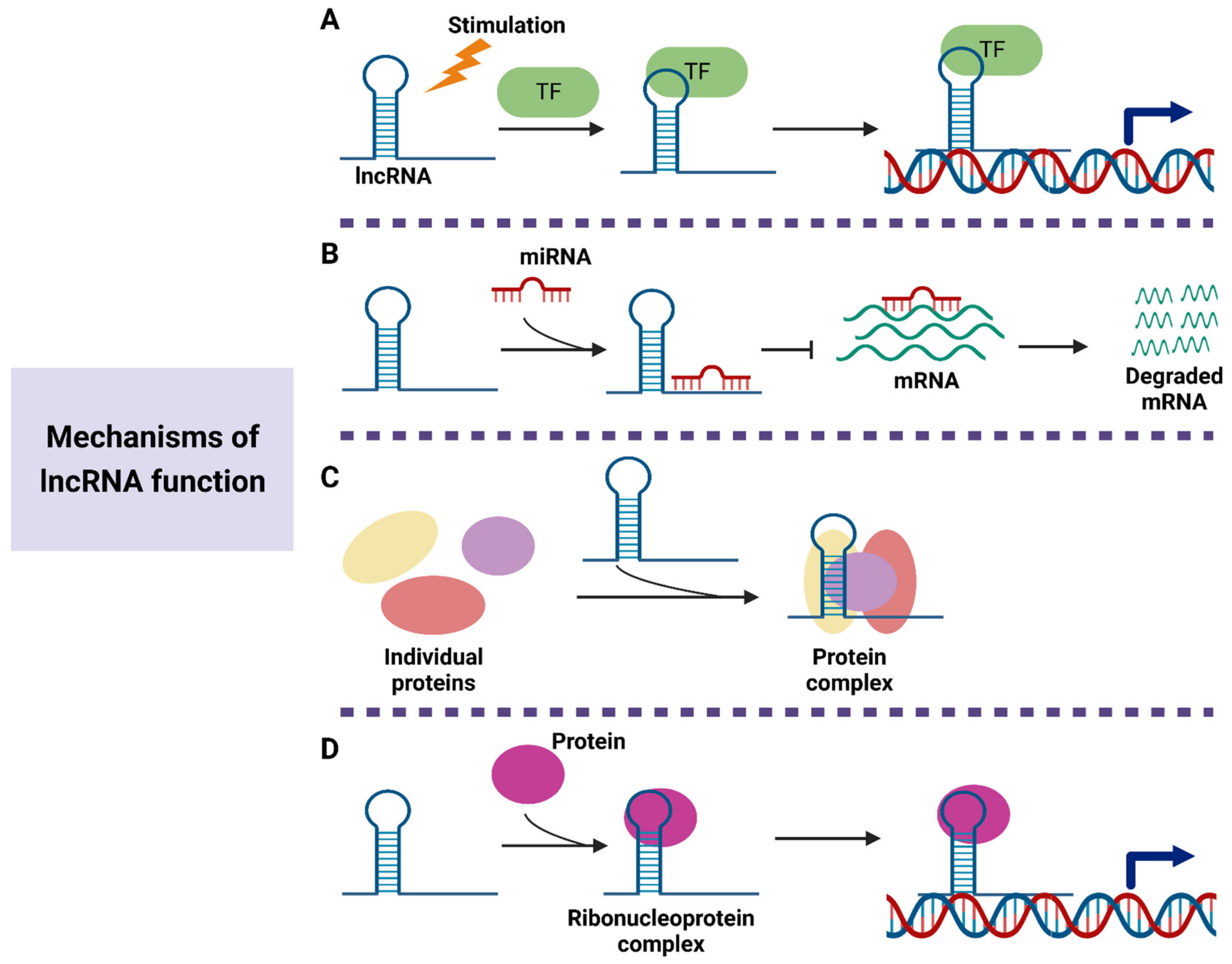
LncRNAs have been described as critical regulators in pathological and physiological processes. Through interacting with DNA, RNA, and proteins, lncRNAs are involved in the regulation of gene expression (Figure 2). The first role of lncRNAs is to regulate downstream gene transcription [41]. Under several stimulation conditions, lncRNAs can be specifically transcribed and participate in the transmission of signaling pathways. Chen et al. reveal that lncRNA HISLA inhibits the interaction of prolyl hydroxylase domain protein 2 (PHD2) and hypoxia-inducible factor-1alpha (HIF-1α) to stabilize HIF-1α expression, thereby promoting cancer aerobic glycolysis [42]. Increasing evidence has suggested that various lncRNAs can act as the sponge for miRNAs to attenuate their inhibition on downstream target genes [43]. For instance, lncRNA NEAT1 promotes the expression of myo-inositol oxygenase (MIOX) by competitively binding to miR-362-3p, thus increasing the sensitivity of hepatocellular carcinoma cells to ferroptosis [44]. LncRNA ITGB8-AS1 is reported to accelerate cell proliferation, colony formation, and tumor growth in colorectal cancer (CRC) by sponging miR-33b-5p and let-7c-5p/let-7d-5p to upregulate the expression of integrin alpha3 (ITGA3) and integrin beta3 (ITGB3) [45]. Moreover, lncRNAs are also considered as scaffolds, which enable several proteins to form complexes [46]. He et al. demonstrate that lncRNA MDHDH exhibits the ability to serve as a molecular scaffold for malate dehydrogenase 2 (MDH2) and 20S proteasomal core subunit alpha-type 1 (PSMA1), leading to the degradation of MDH2 [47]. The downregulation of MDH2 prevents the glycolysis progress in glioma cells. Zhou et al. suggest that lncRNA IFITM4P enhances PD-L1 expression to activate the immunosuppressive program in oral leukoplakia cells by facilitating the binding of SAM and SH3 domain containing protein 1 (SASH1) to phosphorylate transforming growth factor-beta-activated kinase 1 (TAK1) [48]. Another important function of lncRNAs is to combine with proteins to form complexes and then guide them to the specific DNA sequence, leading to the regulation of gene expression at the transcription level [49]. The results of Xiu et al. show that the binding of LINC02273 to heterogeneous nuclear ribonucleoprotein L (hnRNPL) activates anterior gradient homolog 2 (AGR2) expression by enhancing histone H3 lysine 4 trimethylation (H3K4me3) and histone H3 lysine 27 acetylation (H3K27ac) levels in the AGR2 promoter region [50].

3. Biological Functions of Exosomal lncRNAs in Gastrointestinal Cancer

Accumulating evidence indicates that lncRNAs are one of the major components of exosomes [51]. Several lncRNAs are enriched in exosomes. Through RNA sequencing, Wu et al. identify that 938 lncRNAs are highly expressed in ovarian endometriomas-derived exosomes [52]. Interestingly, lncRNAs in exosomes from different donor cells exhibit significant heterogeneity, implying that lncRNAs may be selectively sorted into exosomes [53]. Recently, exosomes-derived lncRNAs are reported to regulate the biological functions of recipient cells. The uptake of exosomes by recipient cells to deliver lncRNAs is regulated by many factors. The property of recipient cells may determine their interaction with exosomes [54]. The selective accumulation of exosomes in specific cells can affect the bioactive functions of exosomal lncRNAs [55]. As shown in Figure 1, exosomes transfer lncRNAs to receptor cells via three main processes: membrane fusion, endocytosis, and receptor-ligand interaction. The fusion of exosomes with the membrane of recipient cells is found to be activated with the decreased pH in the microenvironment [56]. Based on the specific cell type, exosomes can be absorbed by clathrin- or caveolae-mediated endocytosis, phagocytosis, or micropinocytosis [57]. Moreover, exosomes can also bind to the homologous receptors on the recipient cell membrane to trigger signal transduction reactions, thus transporting lncRNAs [58]. The natural plasma membrane structure of exosomes further enhances the stability of lncRNAs. Exosomal lncRNAs can be applied as diagnostic and prognostic biomarkers of various diseases [59]. Moreover, several disorders are considered to be associated with the dysregulation of lncRNAs [60]. Exosomes-mediated lncRNAs delivery represents a promising therapeutic strategy [61]. Therefore, lncRNAs have attracted widespread attention in exosome research.
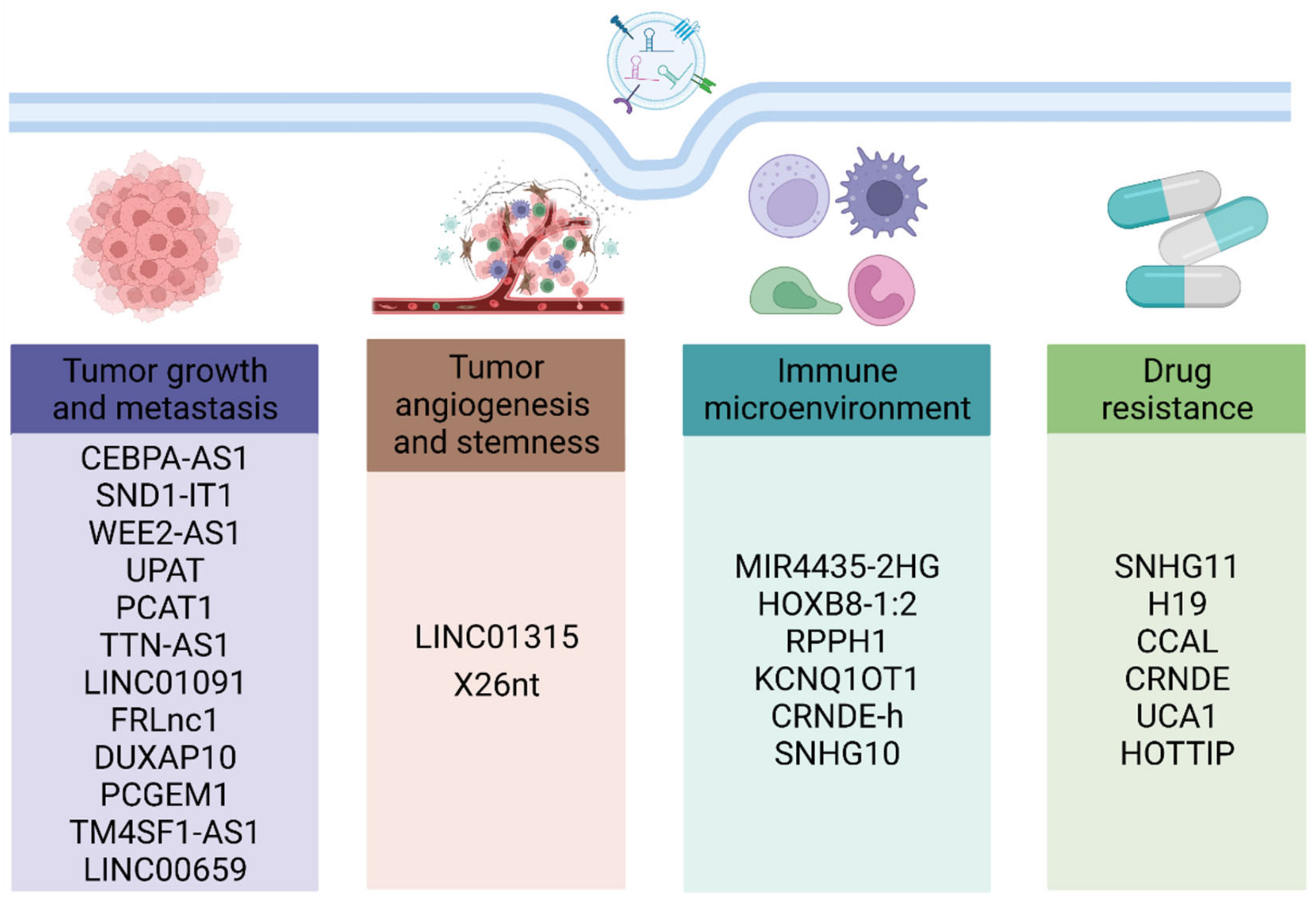
Increasing studies have demonstrated that the occurrence and progress of gastrointestinal cancer are closely associated with the release of various exosomal lncRNAs. After their absorption by recipient cells, exosomes-mediated lncRNAs delivery regulates tumor proliferation, metastasis, angiogenesis, stemness, immune microenvironment, and drug resistance through multiple mechanisms (Figure 3 and Table 1).

3.1. Roles of Exosomal lncRNAs in Tumor Proliferation, Metastasis, Angiogenesis, and Stemness

During the occurrence and development of gastrointestinal cancer, exosomes serve as key mediators of intercellular communications. Emerging studies have suggested that exosomal lncRNAs are involved in all stages of tumor progression. Piao et al. demonstrate that lncRNA CEBPA-AS1 in gastric cancer (GC) cells-derived exosomes enhances the proliferation ability of tumor cells to facilitate GC carcinogenesis [62]. The results of Jin et al. reveal that lncRNA SND1-IT1 is transmitted to gastric mucosa cells by exosomes, leading to the malignant transformation of these cells by upregulating snail family transcriptional repressor 1 (SNAI1) expression [63]. In CRC, exosomes from cancer-associated fibroblasts (CAFs) transport lncRNA WEE2-AS1 to induce MOB kinase activator 1A (MOB1A) degradation and inhibit Hippo signaling pathway, thereby accelerating the growth of CRC cells [64]. Taniue et al. indicate that lncRNA UPAT promotes the survival and tumorigenicity of CRC cells by stabilizing ubiquitin-like with PHD and ring finger domains 1 (UHRF1) [65]. Moreover, increasing evidence has revealed that various cells-released exosomes promote the metastasis of gastrointestinal cancer by the delivery of lncRNAs. For instance, Fang et al. show that lncRNA PCAT1 derived from CRC exosomes promotes the tumor circulating cells-mediated CRC liver metastasis by regulating miR-329-3p/Netrin-1-CD146 axis [66]. LncRNA TTN-AS1, LINC01091, and FRLnc1 are enriched in GC cells-derived exosomes and exhibit the property to elevate the migration and invasion potential of GC cells [67,68,69]. The findings of Lian et al. demonstrate that the upregulation of lncRNA DUXAP10 is positively associated with advanced pathological stages, larger tumor sizes, and lymph node metastasis [70]. Epithelial to mesenchymal transition (EMT) is recognized as an important mechanism underlying tumorigenesis. Several exosomal lncRNAs are reported to exert critical roles in the EMT process of gastrointestinal cancer. GC cells exosomes-delivered lncRNA PCGEM1 can maintain the stability of SNAI1 to prompt the EMT of GC [71]. LncRNA TM4SF1-AS1 is able to promote the proliferation, invasion, and EMT of GC cell by activating PI3K/AKT signalling pathway [72]. LINC00659 is highly expressed in exosomes from CAFs and exosomal LINC00659 targets miR-342-3p to enhance the annexin A2 expression, thus activating EMT progress of CRC cells [73]. In addition, the angiogenesis and stemness of tumor cells are also found to be associated with many exosomal lncRNAs. Li et al. demonstrate that LINC01315 in CRC stem cells-derived exosomes enhances the stemness of CRC cells [74]. GC-secreted exosomal lncRNA X26nt inhibits VE-cadherin level to enhance vascular permeability and promote angiogenesis in a mouse subcutaneous tumor model [75].

3.2. Roles of Exosomal lncRNAs in Tumor Immune Microenvironment

Immune cells are the important components of tumor microenvironment and critically determine tumor progression. Zhi et al. report that BRAFV600E mutant CRC cells-derived exosomes can induce the formation of local immunosuppressive microenvironment by transmitting many lncRNAs, resulting in the peritoneal and distant lymph node metastasis [89]. These findings imply that exosomes from tumor cells may affect immune microenvironment. Recent studies have shown that exosomes-mediated lncRNAs delivery is involved in the modulation of various immune cells, such as macrophages, T cells, and natural killer (NK) cells. For instance, exosomes from MKN45 cells promote gastric carcinogenesis by lncRNA MIR4435-2HG-induced macrophage M2 polarization [76]. Neuroendocrine differentiated colon cancer cells-secreted exosomal lnc-HOXB8-1:2 enhances the migration and invasion capacities of tumor-associated macrophages through miR-6825-5p/CXC chemokine receptor 3 (CXCR3) axis [77]. Macrophages treated with CRC cells-derived exosomes exhibit increased M2 polarization to accelerate CRC metastasis, which is associated with the enrichment of lncRNA RPPH1 in exosomes [78]. Moreover, Xian et al. reveal that tumor cells-derived exosomes contribute to the immune escape of CRC cells by transporting lncRNA KCNQ1OT1 to suppress CD8+ T-cell response and induce PD-L1 ubiquitination [79]. Serum exosomes from CRC patients contain elevated lncRNA CRNDE-h level to activate Th17 cell differentiation by inhibiting the Itch-induced RORγt ubiquitination [80]. NK cells have a potent cytotoxic activity and exert important roles in host immune responses against tumor development [90]. The results of Huang et al. demonstrate that exosomal lncRNA SNHG10 from CRC cells inhibits the cytotoxicity of NK cells by enhancing inhibin subunit beta C (INHBC) expression, thus further facilitating CRC progression [81]. These findings indicate that exosomal lncRNAs represent novel messengers of tumor cells to regulate immune cell functions.

3.3. Roles of Exosomal lncRNAs in Drug Resistance

Although several anti-tumor drugs provide opportunities for gastrointestinal cancer therapy, drug resistance-induced unsatisfactory treatment effects remain a major challenge. Decreased drug uptake and increased drug efflux caused by many factors are responsible for the reduced drug effectiveness. A recent study reveals that exosomal lncRNA SNHG11 is highly expressed in bevacizumab-resistant CRC cells [82]. The knockdown of SNHG11 alleviates bevacizumab resistance and inhibits cell proliferation and migration, indicating that exosomal lncRNA can regulate the drug sensitivity in tumor cells. Moreover, exosomes-mediated crosstalk between cells is reported to endow tumor cells with drug resistance by transporting lncRNAs in the tumor microenvironment. Carcinoma-associated fibroblasts release exosomal lncRNA H19 and CCAL to activate the β-catenin pathway in CRC cells, thus accelerating tumor development and chemoresistance [83,84]. The enrichment of lncRNA CRNDE in M2-polarized macrophage-derived exosomes promotes the tumor growth in cisplatin-treated nude mice by downregulating phosphatase and tensin homolog (PTEN) expression in GC cells [85]. Increasing studies have suggested that various exosomal lncRNAs are involved in the dissemination of drug resistance from resistant cells to sensitive cells. For instance, CRC cells treated with exosomal lncRNA UCA1 derived from cetuximab-resistant cells exhibit increased cetuximab resistance [86]. Wang et al. demonstrate that lncRNA HOTTIP is upregulated in exosomes from cisplatin-resistant GC cells. HOTTIP promotes the cisplatin resistance in sensitive GC cells through the regulation on high mobility group A1 (HMGA1)/miR-218 axis [87]. Mitomycin-resistant CRC cells-derived exosomes-mediated transfer of lncRNA HOTTIP also contributes to the mitomycin resistance by modulating miR-214/karyopherin subunit alpha 3 (KPNA3) pathway [88]. These findings highlight the vital role of exosomal lncRNAs in drug resistance. The combined application of a specific lncRNA inhibitor and anti-tumor drug may improve the therapeutic effects on gastrointestinal cancer.

4. Exosomal lncRNAs as Potential Biomarkers of Gastrointestinal Cancer

Due to the lack of early symptoms, most patients with gastrointestinal cancer are diagnosed in the intermediate or terminal stage [91]. Researchers need to develop novel methods for the early diagnosis of gastrointestinal cancer to reduce the mortality. In recent years, exosomes are emerging as a major content of liquid biopsy and exist in biologic fluids, such as blood, urine, semen, and saliva [92]. During the occurrence and development of many diseases, exosomes contain specific bioactive molecules to reflect the pathological state of source cells [93]. The bilayer phospholipid structure also prevents the cargos from degradation. Although exosomes are a relatively smaller component of EVs, most lncRNAs display elevated levels in exosomes than that in apoptotic bodies and microvesicles [94]. The high abundance, stability, and specificity endow various exosomal lncRNAs with the potential to serve as biomarkers of gastrointestinal cancer.
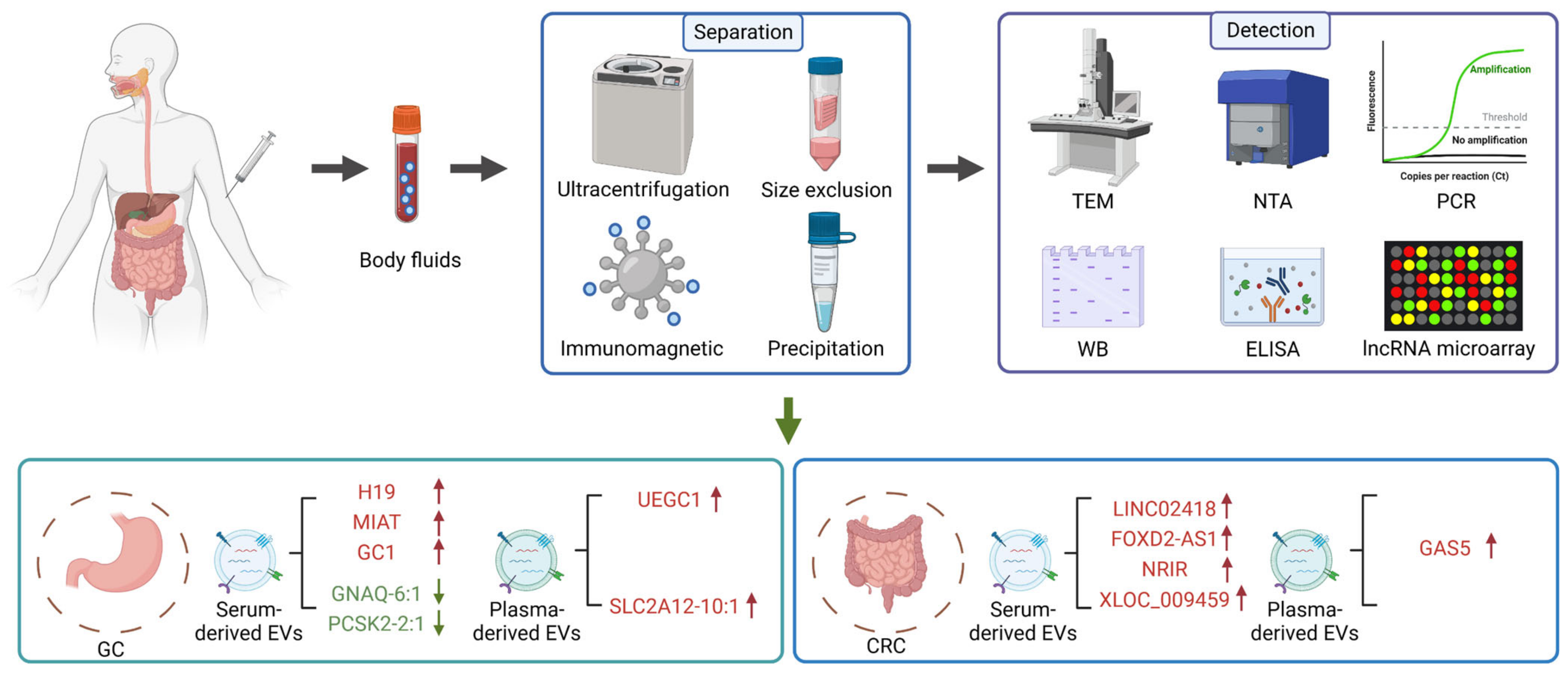
Accumulating evidence has suggested that the circulating levels of exosomes-derived lncRNAs can help to predict the risk of tumor progression. Zhou et al. demonstrate that patients with GC have increased level of serum exosomal lncRNA H19 compared with healthy controls [95]. LncRNA NR038975, which is upregulated in serum exosomes from GC patients, promotes tumor cell proliferation, migration, and invasion by interacting with the NF90/NF45 complex [96]. The elevated lncRNA MIAT expression in serum-derived exosomes is closely associated with worse clinical variables and shorter survival, indicating that serum exosomal MIAT serves as an independent prognostic factor of GC [97]. Many attempts have been made to evaluate the clinical value of exosomal lncRNAs as diagnostic and prognostic biomarkers. Serum exosomal lncRNA GNAQ-6:1 from GC patients is expressed in low level and represents a new diagnostic biomarker with an area under ROC curve (AUC) value of 0.732 [98]. Exosomal lncUEGC1 is able to facilitate the detection of early GC patients from healthy individuals and those with premalignant chronic atrophic gastritis with great sensitivity and specificity [99]. The AUC values are 0.8760 and 0.8406, respectively. Circulating exosomal lncRNA-GC1 and HOTTIP present better diagnostic performance than CEA, CA72-4, and CA19-9 for distinguishing GC patients and healthy individuals [100]. Recent studies focus on the investigation of the relationship between exosomal lncRNAs and the tumor clinicopathological parameters. Pan et al. reveal that GC patients have elevated serum exosomal lncRNA ZFAS1 levels, which may contribute to the identification of lymphatic metastasis and TNM stage [101]. The results of Zheng et al. show that the increased expression of lncRNA SLC2A12-10:1 in plasma exosomes from GC patients is remarkably correlated with tumor size, TNM stage, lymph node metastasis, and differentiation degree [102]. Moreover, serum exosomal lncRNA PCSK2-2:1 is also considered as a potential diagnostic biomarker for GC due to the fact that the downregulation of PCSK2-2:1 can predict the tumor size, tumor stage, and venous invasion [103].
In CRC, several circulating exosomal lncRNAs are found to be differentially expressed. Liu et al. demonstrate that lncRNA GAS5 is upregulated in the tissues, plasma, and exosomes from patients with CRC [104]. Wang et al. reveal that serum exosomal lncRNA CCAT2 exhibits the ability to distinguish the CRC patients from the healthy controls [105]. Recently, many exosomal lncRNAs with great diagnostic potential have also been determined. The elevated level of serum exosomal lncRNA LINC02418 serves as a promising biomarker for the early diagnosis of CRC with an AUC of 0.8978 [106]. Yu et al. detect the serum exosomal lncRNA profiles of CRC patients and healthy individuals and reveal that lncRNA FOXD2-AS1, NRIR, and XLOC_009459 are remarkably upregulated in serum exosomes from CRC patients [107]. Notably, the combination of these three lncRNAs can achieve increased diagnostic value. Moreover, increasing studies have evaluated the correlation between exosomal lncRNAs and clinical data. CRC patients are reported to carry decreased exosomal lncRNA HOTTIP, which is significantly associated with poor overall survival [108]. Elevated levels of lncRNA 91H in serum exosomes from CRC patients show a high risk in tumor recurrence and metastasis [109]. In addition, exosomal lncRNA PVT1 contributes to the CRC metastasis by inhibiting miR-152-3p [110]. Overall, these findings indicate that exosomal lncRNAs are promising biomarkers of gastrointestinal cancer (Figure 4 and Table 2).

5. Exosomal lncRNAs as Therapeutic Targets of Gastrointestinal Cancer

Current treatment strategies for gastrointestinal cancer mainly rely on the chemotherapy-based cancer cell apoptosis, whereas the side effects and drug resistance are noteworthy [111]. With the revelation of their regulatory roles in tumor progression, exosomal lncRNAs may be developed as promising therapeutic targets. Li et al. report that the level of SPINT1-AS1 is increased in CRC tissues compared with adjacent normal tissues [112]. After surgical resection, CRC patients-derived serum exosomes exhibit reduced lncRNA SPINT1-AS1 level, implying that SPINT1-AS1 may be a molecular therapy target of CRC. Several approaches based on the function of exosomal lncRNAs for gastrointestinal cancer therapy have been reported. First, the exosomes-mediated delivery of lncRNAs with antitumor effects is considered a useful strategy. For instance, lncRNA PGM5-AS1 exhibits the ability to alleviate the proliferation, migration, and oxaliplatin tolerance of CRC cells. Treatment with PGM5-AS1 and oxaliplatin-loaded exosomes derived from 293T cells enhances the drug sensitiveness to improve the effectiveness of CRC therapy [113]. Li et al. also demonstrate that the overexpression of lncRNA ADAMTS9-AS1 inhibits cell proliferation and migration by downregulating Wnt/β-catenin signalling pathway, suggesting that engineered exosomes encapsulated with ADAMTS9-AS1 serve as a novel therapeutic agent of CRC [114]. In addition, various exosomal lncRNAs are found to promote the malignant behaviors of cancer cells. The inhibition of these exosomal lncRNAs may contribute to the suppression of gastrointestinal cancer. Yin et al. reveal that the expression of lncRNA NNT-AS1 is upregulated in serum exosomes from CRC patients [115]. The knockdown of NNT-AS1 significantly reduces the proliferation, migration, and invasion of CRC cells. Overall, the exosomal lncRNAs-mediated gastrointestinal cancer therapy is still in infancy; the safety and specific mechanism associated with this strategy need further investigations.

6. Challenges and Perspectives

Exosomal lncRNAs have attracted wide attention in cancer liquid biopsy and therapy due to their unique properties and important cellular functions. Increasing studies have revealed that exosomal lncRNAs are critically involved in the progression of gastrointestinal cancer by modulating tumor proliferation, metastasis, angiogenesis, stemness, immune microenvironment, and drug resistance. Although substantial breakthroughs have been made in the field of exosomes-mediated lncRNA delivery for tumor regulation, there still exist many challenges that may hinder the clinical application of exosomal lncRNAs: (1) the secretion mechanism of exosomal lncRNAs requires further explorations. Considering the heterogeneity of lncRNAs in exosomes derived from different source cells, future studies should focus on whether exosomes can package specifical lncRNAs during the formation process to participate in the gastrointestinal cancer progression. (2) Although several isolation methods of exosomes based on their physical and chemical characteristics have been established, the separation efficiency and the quality of exosomes are still unsatisfactory. Currently, most researchers adopt commercial exosome precipitation kits to purify exosomes from blood, whereas the purity of exosomes cannot meet the requirement of Minimal information for studies of extracellular vesicles 2018 [14]. (3) The rapid isolation and detection of exosomal lncRNAs with diagnostic value are still challenges. QRT-PCR is commonly used for the content analysis of exosomal lncRNAs. However, the sensitivity and accuracy of this method need further improvement. The analysis platforms and detection techniques for exosomal lncRNAs, such as digital PCR and loop-mediated isothermal amplification, may be promising candidates in clinical applications [116,117]. (4) Accumulating evidence has suggested that various serum/plasma exosomes-derived lncRNAs exhibit diagnostic potential in gastrointestinal cancer. The sensitivity, specificity, and AUC value of several exosomal lncRNAs are better than conventional tumor markers, such as CEA, CA19-9, and AFP. However, it is necessary to further expand the number of clinical samples and analyze the correlation with clinical data to validate the diagnostic effects of these exosomal lncRNAs. Notably, the combination of exosomal lncRNAs and tumor markers may achieve elevated diagnostic efficacy. (5) Exosomes-mediated therapeutic lncRNAs delivery has shown great value in the treatment of gastrointestinal cancer. There are still many practical problems to be solved before the clinical transformation of exosomal lncRNAs, including the route of administration, biodistribution, safety, biological functions, and effective dose. Moreover, the selection of the suitable source of exosomes should also be addressed.

7. Conclusions

In conclusion, exosomal lncRNAs exert critical roles in gastrointestinal cancer progression and exhibit great potential as novel diagnostic biomarkers and therapeutic targets. With the development of advanced technologies for the isolation and detection of exosomal lncRNAs, as well as the increasing studies on exosomal lncRNAs and gastrointestinal cancer, the resolution of these key issues will contribute to their clinical translation. We believe that exosomal lncRNAs-based diagnostic and therapeutic strategies can be effectively applied to clinical practice in the future.

Author Contributions

Conceptualization, Y.S., F.S. and H.Q.; writing—original draft preparation, Y.S. and F.S.; writing—review and editing, J.J., W.X. and H.Q.; Funding acquisition, W.X. and H.Q. All authors have read and agreed to the published version of the manuscript.

Funding

This work was supported by the Advanced Science and Technology Foundation of the Jiangsu Province (BE2020680), Technology Development Project of Jiangsu University (20220516), Zhenjiang Key Laboratory of High Technology Research on Exosomes Foundation and Transformation Application (ss2018003), and the Priority Academic Program Development of Jiangsu Higher Education Institutions (Phase III).

Conflicts of Interest

The authors declare no conflict of interest.

References

  1. Arnold, M.; Abnet, C.C.; Neale, R.E.; Vignat, J.; Giovannucci, E.L.; McGlynn, K.A.; Bray, F. Global burden of 5 major types of gastrointestinal cancer. Gastroenterology 2020, 159, 335–349.e315. [Google Scholar] [CrossRef] [PubMed]
  2. Kashyap, S.; Pal, S.; Chandan, G.; Saini, V.; Chakrabarti, S.; Saini, N.K.; Mittal, A.; Thakur, V.K.; Saini, A.K.; Saini, R.V. Understanding the cross-talk between human microbiota and gastrointestinal cancer for developing potential diagnostic and prognostic biomarkers. Semin. Cancer Biol. 2022, 86, 643–651. [Google Scholar] [CrossRef] [PubMed]
  3. Yang, Z.; Wang, D.; Zhang, C.; Liu, H.; Hao, M.; Kan, S.; Liu, D.; Liu, W. The applications of gold nanoparticles in the diagnosis and treatment of gastrointestinal cancer. Front. Oncol. 2021, 11, 5855. [Google Scholar] [CrossRef] [PubMed]
  4. Raza, A.; Khan, A.Q.; Inchakalody, V.P.; Mestiri, S.; Yoosuf, Z.; Bedhiafi, T.; El-Ella, D.M.A.; Taib, N.; Hydrose, S.; Akbar, S.; et al. Dynamic liquid biopsy components as predictive and prognostic biomarkers in colorectal cancer. J. Exp. Clin. Cancer Res. 2022, 41, 99. [Google Scholar] [CrossRef] [PubMed]
  5. Yano, T.; Wang, K.K. Photodynamic therapy for gastrointestinal cancer. Photochem. Photobiol. 2020, 96, 517–523. [Google Scholar] [CrossRef]
  6. Yang, Q.; Xu, J.; Gu, J.; Shi, H.; Zhang, J.; Zhang, J.; Chen, Z.S.; Fang, X.; Zhu, T.; Zhang, X. Extracellular vesicles in cancer drug resistance: Roles, mechanisms, and implications. Adv. Sci. 2022, 9, 2201609. [Google Scholar] [CrossRef]
  7. Liu, X.; Li, Y.; Jiang, X.; Deng, Y.; Ma, C.; Yu, Q.; Gao, D. Long non-coding RNA: Multiple effects on the differentiation, maturity and cell function of dendritic cells. Clin. Immunol. 2022, 245, 109167. [Google Scholar] [CrossRef]
  8. Ruffo, P.; De Amicis, F.; Giardina, E.; Conforti, F.L. Long-noncoding RNAs as epigenetic regulators in neurodegenerative diseases. Neural Regen. Res. 2023, 18, 1243–1248. [Google Scholar] [CrossRef]
  9. Liu, J.; Ji, Q.; Cheng, F.; Chen, D.; Geng, T.; Huang, Y.; Zhang, J.; He, Y.; Song, T. The lncRNAs involved in regulating the RIG-I signaling pathway. Front. Cell. Infect. Microbiol. 2022, 12, 1664. [Google Scholar] [CrossRef]
  10. Wei, L.; Sun, J.; Zhang, N.; Zheng, Y.; Wang, X.; Lv, L.; Liu, J.; Xu, Y.; Shen, Y.; Yang, M. Noncoding RNAs in gastric cancer: Implications for drug resistance. Mol. Cancer 2020, 19, 62. [Google Scholar] [CrossRef] [Green Version]
  11. Chen, S.; Shen, X. Long noncoding RNAs: Functions and mechanisms in colon cancer. Mol. Cancer 2020, 19, 167. [Google Scholar] [CrossRef]
  12. Raziq, K.; Cai, M.; Dong, K.; Wang, P.; Afrifa, J.; Fu, S. Competitive endogenous network of lncRNA, miRNA, and mRNA in the chemoresistance of gastrointestinal tract adenocarcinomas. Biomed. Pharmacother. 2020, 130, 110570. [Google Scholar] [CrossRef]
  13. Xu, Z.; Chen, Y.; Ma, L.; Chen, Y.; Liu, J.; Guo, Y.; Yu, T.; Zhang, L.; Zhu, L.; Shu, Y. Role of exosomal non-coding RNAs from tumor cells and tumor-associated macrophages in the tumor microenvironment. Mol. Ther. 2022, 30, 3133–3154. [Google Scholar] [CrossRef]
  14. Théry, C.; Witwer, K.W.; Aikawa, E.; Alcaraz, M.J.; Anderson, J.D.; Andriantsitohaina, R.; Antoniou, A.; Arab, T.; Archer, F.; Atkin-Smith, G.K.; et al. Minimal information for studies of extracellular vesicles 2018 (MISEV2018): A position statement of the International Society for Extracellular Vesicles and update of the MISEV2014 guidelines. J. Extracell. Vesicles 2018, 7, 1535750. [Google Scholar] [CrossRef]
  15. Xiong, H.; Huang, Z.; Yang, Z.; Lin, Q.; Yang, B.; Fang, X.; Liu, B.; Chen, H.; Kong, J. Recent progress in detection and profiling of cancer cell-derived exosomes. Small 2021, 17, 2007971. [Google Scholar] [CrossRef]
  16. Tenchov, R.; Sasso, J.M.; Wang, X.; Liaw, W.S.; Chen, C.A.; Zhou, Q.A. Exosomes-nature’s lipid nanoparticles, a rising star in drug delivery and diagnostics. ACS Nano 2022, 16, 17802–17846. [Google Scholar] [CrossRef]
  17. Kalluri, R.; LeBleu, V.S. The biology, function, and biomedical applications of exosomes. Science 2020, 367, eaau6977. [Google Scholar] [CrossRef]
  18. Sun, F.; Sun, Y.; Wu, F.; Xu, W.; Qian, H. Mesenchymal stem cell-derived extracellular vesicles: A potential therapy for diabetes mellitus and diabetic complications. Pharmaceutics 2022, 14, 2208. [Google Scholar] [CrossRef]
  19. Liu, J.; Ren, L.; Li, S.; Li, W.; Zheng, X.; Yang, Y.; Fu, W.; Yi, J.; Wang, J.; Du, G. The biology, function, and applications of exosomes in cancer. Acta Pharm. Sin. B 2021, 11, 2783–2797. [Google Scholar] [CrossRef]
  20. Wei, H.; Chen, Q.; Lin, L.; Sha, C.; Li, T.; Liu, Y.; Yin, X.; Xu, Y.; Chen, L.; Gao, W.; et al. Regulation of exosome production and cargo sorting. Int. J. Biol. Sci. 2021, 17, 163–177. [Google Scholar] [CrossRef]
  21. Zhao, J.; An, Q.; Zhu, X.; Yang, B.; Gao, X.; Niu, Y.; Zhang, L.; Xu, K.; Ma, D. Research status and future prospects of extracellular vesicles in primary Sjögren’s syndrome. Stem Cell Res. Ther. 2022, 13, 230. [Google Scholar] [CrossRef] [PubMed]
  22. Noonin, C.; Thongboonkerd, V. Exosome-inflammasome crosstalk and their roles in inflammatory responses. Theranostics 2021, 11, 4436–4451. [Google Scholar] [CrossRef] [PubMed]
  23. Alptekin, A.; Parvin, M.; Chowdhury, H.I.; Rashid, M.H.; Arbab, A.S. Engineered exosomes for studies in tumor immunology. Immunol. Rev. 2022, 312, 76–102. [Google Scholar] [CrossRef] [PubMed]
  24. Zhang, X.; Zhang, H.; Gu, J.; Zhang, J.; Shi, H.; Qian, H.; Wang, D.; Xu, W.; Pan, J.; Santos, H.A. Engineered extracellular vesicles for cancer therapy. Adv. Mater. 2021, 33, 2005709. [Google Scholar] [CrossRef] [PubMed]
  25. Tallon, C.; Hollinger, K.R.; Pal, A.; Bell, B.J.; Rais, R.; Tsukamoto, T.; Witwer, K.W.; Haughey, N.J.; Slusher, B.S. Nipping disease in the bud: nSMase2 inhibitors as therapeutics in extracellular vesicle-mediated diseases. Drug Discov. Today 2021, 26, 1656–1668. [Google Scholar] [CrossRef]
  26. Wu, P.; Zhang, B.; Ocansey, D.K.W.; Xu, W.; Qian, H. Extracellular vesicles: A bright star of nanomedicine. Biomaterials 2021, 269, 120467. [Google Scholar] [CrossRef]
  27. Wei, F.; Li, Y. The emerging roles of exosome-derived noncoding RNAs in the tumor immune microenvironment and their future applications. Biomed. Pharmacother. 2022, 156, 113863. [Google Scholar] [CrossRef]
  28. Isaac, R.; Reis, F.C.G.; Ying, W.; Olefsky, J.M. Exosomes as mediators of intercellular crosstalk in metabolism. Cell Metab. 2021, 33, 1744–1762. [Google Scholar] [CrossRef]
  29. Sun, Y.; Shi, H.; Yin, S.; Ji, C.; Zhang, X.; Zhang, B.; Wu, P.; Shi, Y.; Mao, F.; Yan, Y.; et al. Human mesenchymal stem cell derived exosomes alleviate type 2 diabetes mellitus by reversing peripheral insulin resistance and relieving β-cell destruction. ACS Nano 2018, 12, 7613–7628. [Google Scholar] [CrossRef]
  30. Ji, C.; Zhang, J.; Zhu, Y.; Shi, H.; Yin, S.; Sun, F.; Wang, Q.; Zhang, L.; Yan, Y.; Zhang, X.; et al. Exosomes derived from hucMSC attenuate renal fibrosis through CK1δ/β-TRCP-mediated YAP degradation. Cell Death Dis. 2020, 11, 327. [Google Scholar] [CrossRef]
  31. Sung, S.E.; Seo, M.S.; Kim, Y.I.; Kang, K.K.; Choi, J.H.; Lee, S.; Sung, M.; Yim, S.G.; Lim, J.H.; Seok, H.G.; et al. Human epidural AD-MSC exosomes improve function recovery after spinal cord injury in rats. Biomedicines 2022, 10, 678. [Google Scholar] [CrossRef]
  32. Luo, C.; Xin, H.; Zhou, Z.; Hu, Z.; Sun, R.; Yao, N.; Sun, Q.; Borjigin, U.; Wu, X.; Fan, J.; et al. Tumor-derived exosomes induce immunosuppressive macrophages to foster intrahepatic cholangiocarcinoma progression. Hepatology 2022, 76, 982–999. [Google Scholar] [CrossRef]
  33. Herman, A.B.; Tsitsipatis, D.; Gorospe, M. Integrated lncRNA function upon genomic and epigenomic regulation. Mol. Cell 2022, 82, 2252–2266. [Google Scholar] [CrossRef]
  34. Liu, Y.; Ding, W.; Yu, W.; Zhang, Y.; Ao, X.; Wang, J. Long non-coding RNAs: Biogenesis, functions, and clinical significance in gastric cancer. Mol. Ther.-Oncolytics 2021, 23, 458–476. [Google Scholar] [CrossRef]
  35. Reggiardo, R.E.; Maroli, S.V.; Kim, D.H. LncRNA biomarkers of inflammation and cancer. Adv. Exp. Med. Biol. 2022, 1363, 121–145. [Google Scholar] [CrossRef]
  36. Marcia, M. The multiple molecular dimensions of long noncoding RNAs that regulate gene expression and tumorigenesis. Curr. Opin. Oncol. 2022, 34, 141–147. [Google Scholar] [CrossRef]
  37. Bermúdez, M.; Aguilar-Medina, M.; Lizárraga-Verdugo, E.; Avendaño-Félix, M.; Silva-Benítez, E.; López-Camarillo, C.; Ramos-Payán, R. LncRNAs as regulators of autophagy and drug resistance in colorectal cancer. Front. Oncol. 2019, 9, 1008. [Google Scholar] [CrossRef]
  38. Huang, W.; Li, H.; Yu, Q.; Xiao, W.; Wang, D.O. LncRNA-mediated DNA methylation: An emerging mechanism in cancer and beyond. J. Exp. Clin. Cancer Res. 2022, 41, 100. [Google Scholar] [CrossRef]
  39. Alkhathami, A.G.; Hadi, A.; Alfaifi, M.; Alshahrani, M.Y.; Verma, A.K.; Beg, M.M.A. Serum-based lncRNA ANRIL, TUG1, UCA1, and HIT expressions in breast cancer patients. Dis. Markers 2022, 2022, 9997212. [Google Scholar] [CrossRef]
  40. Adnane, S.; Marino, A.; Leucci, E. LncRNAs in human cancers: Signal from noise. Trends Cell Biol. 2022, 32, 565–573. [Google Scholar] [CrossRef]
  41. Shuman, S. Transcriptional interference at tandem lncRNA and protein-coding genes: An emerging theme in regulation of cellular nutrient homeostasis. Nucleic Acids Res. 2020, 48, 8243–8254. [Google Scholar] [CrossRef] [PubMed]
  42. Chen, F.; Chen, J.; Yang, L.; Liu, J.; Zhang, X.; Zhang, Y.; Tu, Q.; Yin, D.; Lin, D.; Wong, P.P.; et al. Extracellular vesicle-packaged HIF-1α-stabilizing lncRNA from tumour-associated macrophages regulates aerobic glycolysis of breast cancer cells. Nat. Cell Biol. 2019, 21, 498–510. [Google Scholar] [CrossRef] [PubMed]
  43. Venkatesh, J.; Wasson, M.D.; Brown, J.M.; Fernando, W.; Marcato, P. LncRNA-miRNA axes in breast cancer: Novel points of interaction for strategic attack. Cancer Lett. 2021, 509, 81–88. [Google Scholar] [CrossRef] [PubMed]
  44. Zhang, Y.; Luo, M.; Cui, X.; O’Connell, D.; Yang, Y. Long noncoding RNA NEAT1 promotes ferroptosis by modulating the miR-362-3p/MIOX axis as a ceRNA. Cell Death Differ. 2022, 29, 1850–1863. [Google Scholar] [CrossRef] [PubMed]
  45. Lin, X.; Zhuang, S.; Chen, X.; Du, J.; Zhong, L.; Ding, J.; Wang, L.; Yi, J.; Hu, G.; Tang, G.; et al. lncRNA ITGB8-AS1 functions as a ceRNA to promote colorectal cancer growth and migration through integrin-mediated focal adhesion signaling. Mol. Ther. 2022, 30, 688–702. [Google Scholar] [CrossRef]
  46. Han, Y.; Zhao, G.; Shi, X.; Wang, Y.; Wen, X.; Zhang, L.; Guo, X. The emerging role of long non-coding RNAs in esophageal cancer: Functions in tumorigenesis and clinical implications. Front. Pharmacol. 2022, 13, 885075. [Google Scholar] [CrossRef]
  47. He, D.; Xin, T.; Pang, B.; Sun, J.; Liu, Z.H.; Qin, Z.; Ji, X.S.; Yang, F.; Wei, Y.B.; Wang, Z.X.; et al. A novel lncRNA MDHDH suppresses glioblastoma multiforme by acting as a scaffold for MDH2 and PSMA1 to regulate NAD+ metabolism and autophagy. J. Exp. Clin. Cancer Res. 2022, 41, 349. [Google Scholar] [CrossRef]
  48. Shi, L.; Yang, Y.; Li, M.; Li, C.; Zhou, Z.; Tang, G.; Wu, L.; Yao, Y.; Shen, X.; Hou, Z.; et al. LncRNA IFITM4P promotes immune escape by up-regulating PD-L1 via dual mechanism in oral carcinogenesis. Mol. Ther. 2022, 30, 1564–1577. [Google Scholar] [CrossRef]
  49. Zhang, P.; Wu, S.; He, Y.; Li, X.; Zhu, Y.; Lin, X.; Chen, L.; Zhao, Y.; Niu, L.; Zhang, S.; et al. LncRNA-mediated adipogenesis in different adipocytes. Int. J. Mol. Sci. 2022, 23, 7488. [Google Scholar] [CrossRef]
  50. Xiu, B.; Chi, Y.; Liu, L.; Chi, W.; Zhang, Q.; Chen, J.; Guo, R.; Si, J.; Li, L.; Xue, J.; et al. LINC02273 drives breast cancer metastasis by epigenetically increasing AGR2 transcription. Mol. Cancer 2019, 18, 187. [Google Scholar] [CrossRef] [Green Version]
  51. Lakshmi, S.; Hughes, T.A.; Priya, S. Exosomes and exosomal RNAs in breast cancer: A status update. Eur. J. Cancer 2021, 144, 252–268. [Google Scholar] [CrossRef]
  52. Wu, J.; Huang, H.; Huang, W.; Wang, L.; Xia, X.; Fang, X. Analysis of exosomal lncRNA, miRNA and mRNA expression profiles and ceRNA network construction in endometriosis. Epigenomics 2020, 12, 1193–1213. [Google Scholar] [CrossRef]
  53. Chang, W.; Wang, J. Exosomes and their noncoding RNA cargo are emerging as new modulators for diabetes mellitus. Cells 2019, 8, 853. [Google Scholar] [CrossRef]
  54. Fan, Q.; Yang, L.; Zhang, X.; Peng, X.; Wei, S.; Su, D.; Zhai, Z.; Hua, X.; Li, H. The emerging role of exosome-derived non-coding RNAs in cancer biology. Cancer Lett. 2018, 414, 107–115. [Google Scholar] [CrossRef]
  55. Li, L.; Bi, Y.; Diao, S.; Li, X.; Yuan, T.; Xu, T.; Huang, C.; Li, J. Exosomal lncRNAs and hepatocellular carcinoma: From basic research to clinical practice. Biochem. Pharmacol. 2022, 200, 115032. [Google Scholar] [CrossRef]
  56. Record, M.; Carayon, K.; Poirot, M.; Silvente-Poirot, S. Exosomes as new vesicular lipid transporters involved in cell-cell communication and various pathophysiologies. Biochim. Biophys. Acta 2014, 1841, 108–120. [Google Scholar] [CrossRef]
  57. Liu, Q.W.; He, Y.; Xu, W.W. Molecular functions and therapeutic applications of exosomal noncoding RNAs in cancer. Exp. Mol. Med. 2022, 54, 216–225. [Google Scholar] [CrossRef]
  58. Dang, X.; Zeng, X. Targeted therapeutic delivery using engineered exosomes and its applications in cardiovascular diseases. Gene 2016, 575 Pt 2, 377–384. [Google Scholar] [CrossRef]
  59. Kok, V.C.; Yu, C.C. Cancer-derived exosomes: Their role in cancer biology and biomarker development. Int. J. Nanomed. 2020, 15, 8019–8036. [Google Scholar] [CrossRef]
  60. Honarmand Tamizkar, K.; Gorji, P.; Gholipour, M.; Hussen, B.M.; Mazdeh, M.; Eslami, S.; Taheri, M.; Ghafouri-Fard, S. Parkinson’s disease is associated with dysregulation of circulatory levels of lncRNAs. Front. Immunol. 2021, 12, 4706. [Google Scholar] [CrossRef]
  61. Thakur, A.; Parra, D.C.; Motallebnejad, P.; Brocchi, M.; Chen, H.J. Exosomes: Small vesicles with big roles in cancer, vaccine development, and therapeutics. Bioact. Mater. 2022, 10, 281–294. [Google Scholar] [CrossRef] [PubMed]
  62. Piao, H.Y.; Guo, S.; Wang, Y.; Zhang, J. Exosomal long non-coding RNA CEBPA-AS1 inhibits tumor apoptosis and functions as a non-invasive biomarker for diagnosis of gastric cancer. OncoTargets Ther. 2020, 13, 1365–1374. [Google Scholar] [CrossRef] [PubMed]
  63. Jin, G.; Zhang, J.; Cao, T.; Chen, B.; Tian, Y.; Shi, Y. Exosome-mediated lncRNA SND1-IT1 from gastric cancer cells enhances malignant transformation of gastric mucosa cells via up-regulating SNAIL1. J. Transl. Med. 2022, 20, 284. [Google Scholar] [CrossRef] [PubMed]
  64. Yang, P.; Zhang, D.; Wang, T.; Ji, J.; Jin, C.; Peng, C.; Tan, Y.; Zhou, J.; Wang, L.; Feng, Y.; et al. CAF-derived exosomal WEE2-AS1 facilitates colorectal cancer progression via promoting degradation of MOB1A to inhibit the Hippo pathway. Cell Death Dis. 2022, 13, 796. [Google Scholar] [CrossRef] [PubMed]
  65. Taniue, K.; Kurimoto, A.; Sugimasa, H.; Nasu, E.; Takeda, Y.; Iwasaki, K.; Nagashima, T.; Okada-Hatakeyama, M.; Oyama, M.; Kozuka-Hata, H.; et al. Long noncoding RNA UPAT promotes colon tumorigenesis by inhibiting degradation of UHRF1. Proc. Natl. Acad. Sci. USA 2016, 113, 1273–1278. [Google Scholar] [CrossRef]
  66. Fang, X.; Xu, Y.; Li, K.; Liu, P.; Zhang, H.; Jiang, Y.; Tang, J.; Li, Y. Exosomal lncRNA PCAT1 promotes tumor circulating cell-mediated colorectal cancer liver metastasis by regulating the activity of the miR-329-3p/Netrin-1-CD146 complex. J. Immunol. Res. 2022, 2022, 9916228. [Google Scholar] [CrossRef]
  67. Wang, Y.; Jiang, R.; Zhao, H.; Li, F.; Li, Y.; Zhu, M. TTN-AS1 delivered by gastric cancer cell-derived exosome induces gastric cancer progression through in vivo and in vitro studies. Cell Biol. Toxicol. 2022, 38, 1–15. [Google Scholar] [CrossRef]
  68. Wang, Q.; Zhang, C.; Cao, S.; Zhao, H.; Jiang, R.; Li, Y. Tumor-derived exosomes orchestrate the microRNA-128-3p/ELF4/CDX2 axis to facilitate the growth and metastasis of gastric cancer via delivery of LINC01091. Cell Biol. Toxicol. 2022, 38, 1–18. [Google Scholar] [CrossRef]
  69. Zhang, Y.; Chen, L.; Ye, X.; Wu, Z.; Zhang, Z.; Sun, B.; Fu, H.; Fu, C.; Liang, X.; Jiang, H. Expression and mechanism of exosome-mediated A FOXM1 related long noncoding RNA in gastric cancer. J. Nanobiotechnology 2021, 19, 133. [Google Scholar] [CrossRef]
  70. Lian, Y.; Xu, Y.; Xiao, C.; Xia, R.; Gong, H.; Yang, P.; Chen, T.; Wu, D.; Cai, Z.; Zhang, J.; et al. The pseudogene derived from long non-coding RNA DUXAP10 promotes colorectal cancer cell growth through epigenetically silencing of p21 and PTEN. Sci. Rep. 2017, 7, 7312. [Google Scholar] [CrossRef] [Green Version]
  71. Piao, H.Y.; Guo, S.; Wang, Y.; Zhang, J. Exosome-transmitted lncRNA PCGEM1 promotes invasive and metastasis in gastric cancer by maintaining the stability of SNAI1. Clin. Transl. Oncol. 2021, 23, 246–256. [Google Scholar] [CrossRef]
  72. He, C.; Qi, W.; Wang, Z. Effect and mechanism of downregulating the long-chain noncoding RNA TM4SF1-AS1 on the proliferation, apoptosis and invasion of gastric cancer cells. World J. Surg. Oncol. 2021, 19, 226. [Google Scholar] [CrossRef]
  73. Zhou, L.; Li, J.; Tang, Y.; Yang, M. Exosomal LncRNA LINC00659 transferred from cancer-associated fibroblasts promotes colorectal cancer cell progression via miR-342-3p/ANXA2 axis. J. Transl. Med. 2021, 19, 8. [Google Scholar] [CrossRef]
  74. Li, Y.; Wu, M.; Xu, S.; Huang, H.; Yan, L.; Gu, Y. Colorectal cancer stem cell-derived exosomal long intergenic noncoding RNA 01315 (LINC01315) promotes proliferation, migration, and stemness of colorectal cancer cells. Bioengineered 2022, 13, 10827–10842. [Google Scholar] [CrossRef]
  75. Chen, X.; Zhang, S.; Du, K.; Zheng, N.; Liu, Y.; Chen, H.; Xie, G.; Ma, Y.; Zhou, Y.; Zheng, Y.; et al. Gastric cancer-secreted exosomal X26nt increases angiogenesis and vascular permeability by targeting VE-cadherin. Cancer Sci. 2021, 112, 1839–1852. [Google Scholar] [CrossRef]
  76. Li, C.; Chen, Z.; Gao, J.; Tang, T.; Zhou, L.; Zhang, G.; Zhang, D.; Shen, C.; Guo, L.; Fu, T. MIR4435-2HG in exosomes promotes gastric carcinogenesis by inducing M2 polarization in macrophages. Front. Oncol. 2022, 12, 1017745. [Google Scholar] [CrossRef]
  77. Li, X.; Lan, Q.; Lai, W.; Wu, H.; Xu, H.; Fang, K.; Chu, Z.; Zeng, Y. Exosome-derived lnc-HOXB8-1:2 induces tumor-associated macrophage infiltration to promote neuroendocrine differentiated colorectal cancer progression by sponging hsa-miR-6825-5p. BMC Cancer 2022, 22, 928. [Google Scholar] [CrossRef]
  78. Liang, Z.X.; Liu, H.S.; Wang, F.W.; Xiong, L.; Zhou, C.; Hu, T.; He, X.W.; Wu, X.J.; Xie, D.; Wu, X.R.; et al. LncRNA RPPH1 promotes colorectal cancer metastasis by interacting with TUBB3 and by promoting exosomes-mediated macrophage M2 polarization. Cell Death Dis. 2019, 10, 829. [Google Scholar] [CrossRef]
  79. Xian, D.; Niu, L.; Zeng, J.; Wang, L. LncRNA KCNQ1OT1 secreted by tumor cell-derived exosomes mediates immune escape in colorectal cancer by regulating PD-L1 ubiquitination via miR-30a-5p/USP22. Front. Cell Dev. Biol. 2021, 9, 653808. [Google Scholar] [CrossRef]
  80. Sun, J.; Jia, H.; Bao, X.; Wu, Y.; Zhu, T.; Li, R.; Zhao, H. Tumor exosome promotes Th17 cell differentiation by transmitting the lncRNA CRNDE-h in colorectal cancer. Cell Death Dis. 2021, 12, 123. [Google Scholar] [CrossRef]
  81. Huang, Y.; Luo, Y.; Ou, W.; Wang, Y.; Dong, D.; Peng, X.; Luo, Y. Exosomal lncRNA SNHG10 derived from colorectal cancer cells suppresses natural killer cell cytotoxicity by upregulating INHBC. Cancer Cell Int. 2021, 21, 528. [Google Scholar] [CrossRef] [PubMed]
  82. Huang, W.; Zhang, H.; Tian, Y.; Li, Y.; Li, J.; Zhong, X.; Yuan, X. LncRNA SNHG11 enhances bevacizumab resistance in colorectal cancer by mediating miR-1207-5p/ABCC1 axis. Anti-Cancer Drugs 2022, 33, 575–586. [Google Scholar] [CrossRef] [PubMed]
  83. Ren, J.; Ding, L.; Zhang, D.; Shi, G.; Xu, Q.; Shen, S.; Wang, Y.; Wang, T.; Hou, Y. Carcinoma-associated fibroblasts promote the stemness and chemoresistance of colorectal cancer by transferring exosomal lncRNA H19. Theranostics 2018, 8, 3932–3948. [Google Scholar] [CrossRef] [PubMed]
  84. Deng, X.; Ruan, H.; Zhang, X.; Xu, X.; Zhu, Y.; Peng, H.; Zhang, X.; Kong, F.; Guan, M. Long noncoding RNA CCAL transferred from fibroblasts by exosomes promotes chemoresistance of colorectal cancer cells. Int. J. Cancer 2020, 146, 1700–1716. [Google Scholar] [CrossRef] [PubMed]
  85. Xin, L.; Zhou, L.Q.; Liu, C.; Zeng, F.; Yuan, Y.W.; Zhou, Q.; Li, S.H.; Wu, Y.; Wang, J.L.; Wu, D.Z.; et al. Transfer of lncRNA CRNDE in TAM-derived exosomes is linked with cisplatin resistance in gastric cancer. EMBO Rep. 2021, 22, e52124. [Google Scholar] [CrossRef]
  86. Yang, Y.N.; Zhang, R.; Du, J.W.; Yuan, H.H.; Li, Y.J.; Wei, X.L.; Du, X.X.; Jiang, S.L.; Han, Y. Predictive role of UCA1-containing exosomes in cetuximab-resistant colorectal cancer. Cancer Cell Int. 2018, 18, 164. [Google Scholar] [CrossRef]
  87. Wang, J.; Lv, B.; Su, Y.; Wang, X.; Bu, J.; Yao, L. Exosome-mediated transfer of lncRNA HOTTIP promotes cisplatin resistance in gastric cancer cells by regulating HMGA1/miR-218 axis. OncoTargets Ther. 2019, 12, 11325–11338. [Google Scholar] [CrossRef]
  88. Chen, X.; Liu, Y.; Zhang, Q.; Liu, B.; Cheng, Y.; Zhang, Y.; Sun, Y.; Liu, J.; Gen, H. Exosomal long non-coding RNA HOTTIP increases resistance of colorectal cancer cells to mitomycin via impairing miR-214-mediated degradation of KPNA3. Front. Cell Dev. Biol. 2020, 8, 582723. [Google Scholar] [CrossRef]
  89. Zhi, J.; Jia, X.J.; Yan, J.; Wang, H.C.; Feng, B.; Xing, H.Y.; Jia, Y.T. BRAF(V600E) mutant colorectal cancer cells mediate local immunosuppressive microenvironment through exosomal long noncoding RNAs. World J. Gastrointest. Oncol. 2021, 13, 2129–2148. [Google Scholar] [CrossRef]
  90. Poznanski, S.M.; Singh, K.; Ritchie, T.M.; Aguiar, J.A.; Fan, I.Y.; Portillo, A.L.; Rojas, E.A.; Vahedi, F.; El-Sayes, A.; Xing, S.; et al. Metabolic flexibility determines human NK cell functional fate in the tumor microenvironment. Cell Metab. 2021, 33, 1205–1220.e5. [Google Scholar] [CrossRef]
  91. Kobayashi, H.; Enomoto, A.; Woods, S.L.; Burt, A.D.; Takahashi, M.; Worthley, D.L. Cancer-associated fibroblasts in gastrointestinal cancer. Nat. Rev. Gastroenterol. Hepatol. 2019, 16, 282–295. [Google Scholar] [CrossRef]
  92. Yu, D.; Li, Y.; Wang, M.; Gu, J.; Xu, W.; Cai, H.; Fang, X.; Zhang, X. Exosomes as a new frontier of cancer liquid biopsy. Mol. Cancer 2022, 21, 56. [Google Scholar] [CrossRef]
  93. Zhou, B.; Xu, K.; Zheng, X.; Chen, T.; Wang, J.; Song, Y.; Shao, Y.; Zheng, S. Application of exosomes as liquid biopsy in clinical diagnosis. Signal Transduct. Target. Ther. 2020, 5, 144. [Google Scholar] [CrossRef]
  94. Dong, L.; Lin, W.; Qi, P.; Xu, M.D.; Wu, X.; Ni, S.; Huang, D.; Weng, W.W.; Tan, C.; Sheng, W.; et al. Circulating long RNAs in serum extracellular vesicles: Their characterization and potential application as biomarkers for diagnosis of colorectal cancer. Cancer Epidemiol. Biomark. Prev. 2016, 25, 1158–1166. [Google Scholar] [CrossRef]
  95. Zhou, H.; Shen, W.; Zou, H.; Lv, Q.; Shao, P. Circulating exosomal long non-coding RNA H19 as a potential novel diagnostic and prognostic biomarker for gastric cancer. J. Int. Med. Res. 2020, 48, 300060520934297. [Google Scholar] [CrossRef]
  96. Wei, S.; Dai, S.; Zhang, C.; Zhao, R.; Zhao, Z.; Song, Y.; Shan, B.; Zhao, L. LncRNA NR038975, a serum-based biomarker, promotes gastric tumorigenesis by interacting with NF90/NF45 complex. Front. Oncol. 2021, 11, 721604. [Google Scholar] [CrossRef]
  97. Xu, H.; Zhou, J.; Tang, J.; Min, X.; Yi, T.; Zhao, J.; Ren, Y. Identification of serum exosomal lncRNA MIAT as a novel diagnostic and prognostic biomarker for gastric cancer. J. Clin. Lab. Anal. 2020, 34, e23323. [Google Scholar] [CrossRef]
  98. Li, S.; Zhang, M.; Zhang, H.; Hu, K.; Cai, C.; Wang, J.; Shi, L.; Ma, P.; Xu, Y.; Zheng, P. Exosomal long noncoding RNA lnc-GNAQ-6:1 may serve as a diagnostic marker for gastric cancer. Clin. Chim. Acta 2020, 501, 252–257. [Google Scholar] [CrossRef]
  99. Lin, L.Y.; Yang, L.; Zeng, Q.; Wang, L.; Chen, M.L.; Zhao, Z.H.; Ye, G.D.; Luo, Q.C.; Lv, P.Y.; Guo, Q.W.; et al. Tumor-originated exosomal lncUEGC1 as a circulating biomarker for early-stage gastric cancer. Mol. Cancer 2018, 17, 84. [Google Scholar] [CrossRef]
  100. Guo, X.; Lv, X.; Ru, Y.; Zhou, F.; Wang, N.; Xi, H.; Zhang, K.; Li, J.; Chang, R.; Xie, T.; et al. Circulating exosomal gastric cancer-associated long noncoding RNA1 as a biomarker for early detection and monitoring progression of gastric cancer: A multiphase study. JAMA Surg. 2020, 155, 572–579. [Google Scholar] [CrossRef]
  101. Pan, L.; Liang, W.; Fu, M.; Huang, Z.H.; Li, X.; Zhang, W.; Zhang, P.; Qian, H.; Jiang, P.C.; Xu, W.R.; et al. Exosomes-mediated transfer of long noncoding RNA ZFAS1 promotes gastric cancer progression. J. Cancer Res. Clin. Oncol. 2017, 143, 991–1004. [Google Scholar] [CrossRef] [PubMed]
  102. Zheng, P.; Zhang, H.; Gao, H.; Sun, J.; Li, J.; Zhang, X.; Gao, L.; Ma, P.; Li, S. Plasma exosomal long noncoding RNA lnc-SLC2A12-10:1 as a novel diagnostic biomarker for gastric cancer. OncoTargets Ther. 2020, 13, 4009–4018. [Google Scholar] [CrossRef]
  103. Cai, C.; Zhang, H.; Zhu, Y.; Zheng, P.; Xu, Y.; Sun, J.; Zhang, M.; Lan, T.; Gu, B.; Li, S.; et al. Serum exosomal long noncoding RNA pcsk2-2:1 as a potential novel diagnostic biomarker for gastric cancer. OncoTargets Ther. 2019, 12, 10035–10041. [Google Scholar] [CrossRef] [PubMed]
  104. Liu, L.; Meng, T.; Yang, X.H.; Sayim, P.; Lei, C.; Jin, B.; Ge, L.; Wang, H.J. Prognostic and predictive value of long non-coding RNA GAS5 and mircoRNA-221 in colorectal cancer and their effects on colorectal cancer cell proliferation, migration and invasion. Cancer Biomark. 2018, 22, 283–299. [Google Scholar] [CrossRef] [PubMed]
  105. Wang, L.; Duan, W.; Yan, S.; Xie, Y.; Wang, C. Circulating long non-coding RNA colon cancer-associated transcript 2 protected by exosome as a potential biomarker for colorectal cancer. Biomed. Pharmacother. 2019, 113, 108758. [Google Scholar] [CrossRef] [PubMed]
  106. Zhao, Y.; Du, T.; Du, L.; Li, P.; Li, J.; Duan, W.; Wang, Y.; Wang, C. Long noncoding RNA LINC02418 regulates MELK expression by acting as a ceRNA and may serve as a diagnostic marker for colorectal cancer. Cell Death Dis. 2019, 10, 568. [Google Scholar] [CrossRef]
  107. Yu, M.; Song, X.G.; Zhao, Y.J.; Dong, X.H.; Niu, L.M.; Zhang, Z.J.; Shang, X.L.; Tang, Y.Y.; Song, X.R.; Xie, L. Circulating serum exosomal long non-coding RNAs FOXD2-AS1, NRIR, and XLOC_009459 as diagnostic biomarkers for colorectal cancer. Front. Oncol. 2021, 11, 618967. [Google Scholar] [CrossRef]
  108. Oehme, F.; Krahl, S.; Gyorffy, B.; Muessle, B.; Rao, V.; Greif, H.; Ziegler, N.; Lin, K.; Thepkaysone, M.L.; Polster, H.; et al. Low level of exosomal long non-coding RNA HOTTIP is a prognostic biomarker in colorectal cancer. RNA Biol. 2019, 16, 1339–1345. [Google Scholar] [CrossRef]
  109. Gao, T.; Liu, X.; He, B.; Nie, Z.; Zhu, C.; Zhang, P.; Wang, S. Exosomal lncRNA 91H is associated with poor development in colorectal cancer by modifying HNRNPK expression. Cancer Cell Int. 2018, 18, 11. [Google Scholar] [CrossRef]
  110. Lai, S.W.; Chen, M.Y.; Bamodu, O.A.; Hsieh, M.S.; Huang, T.Y.; Yeh, C.T.; Lee, W.H.; Cherng, Y.G. Exosomal lncRNA PVT1/VEGFA axis promotes colon cancer metastasis and stemness by downregulation of tumor suppressor miR-152-3p. Oxidative Med. Cell. Longev. 2021, 2021, 9959807. [Google Scholar] [CrossRef]
  111. Sun, Y.; Li, B.; Cao, Q.; Liu, T.; Li, J. Targeting cancer stem cells with polymer nanoparticles for gastrointestinal cancer treatment. Stem Cell Res. Ther. 2022, 13, 489. [Google Scholar] [CrossRef]
  112. Li, C.; Li, W.; Zhang, Y.; Zhang, X.; Liu, T.; Zhang, Y.; Yang, Y.; Wang, L.; Pan, H.; Ji, J.; et al. Increased expression of antisense lncRNA SPINT1-AS1 predicts a poor prognosis in colorectal cancer and is negatively correlated with its sense transcript. OncoTargets Ther. 2018, 11, 3969–3978. [Google Scholar] [CrossRef]
  113. Hui, B.; Lu, C.; Wang, J.; Xu, Y.; Yang, Y.; Ji, H.; Li, X.; Xu, L.; Wang, J.; Tang, W.; et al. Engineered exosomes for co-delivery of PGM5-AS1 and oxaliplatin to reverse drug resistance in colon cancer. J. Cell. Physiol. 2022, 237, 911–933. [Google Scholar] [CrossRef]
  114. Li, N.; Li, J.; Mi, Q.; Xie, Y.; Li, P.; Wang, L.; Binang, H.; Wang, Q.; Wang, Y.; Chen, Y.; et al. Long non-coding RNA ADAMTS9-AS1 suppresses colorectal cancer by inhibiting the Wnt/β-catenin signalling pathway and is a potential diagnostic biomarker. J. Cell. Mol. Med. 2020, 24, 11318–11329. [Google Scholar] [CrossRef]
  115. Yin, H.; Hu, J.; Ye, Z.; Chen, S.; Chen, Y. Serum long non-coding RNA NNT-AS1 protected by exosome is a potential biomarker and functions as an oncogene via the miR-496/RAP2C axis in colorectal cancer. Mol. Med. Rep. 2021, 24, 585. [Google Scholar] [CrossRef]
  116. Liu, D.; Yin, H.; Wang, Y.; Cao, Y.; Yin, J.; Zhang, J.; Yin, H.; Zhao, X. Development of a highly sensitive digital PCR assay to quantify long non-coding RNA MYU in urine samples which exhibited great potential as an alternative diagnostic biomarker for prostate cancer. Transl. Androl. Urol. 2021, 10, 3815–3825. [Google Scholar] [CrossRef]
  117. Lin, Q.; Huang, Z.; Ye, X.; Yang, B.; Fang, X.; Liu, B.; Chen, H.; Kong, J. Lab in a tube: Isolation, extraction, and isothermal amplification detection of exosomal long noncoding RNA of gastric cancer. Talanta 2021, 225, 122090. [Google Scholar] [CrossRef]
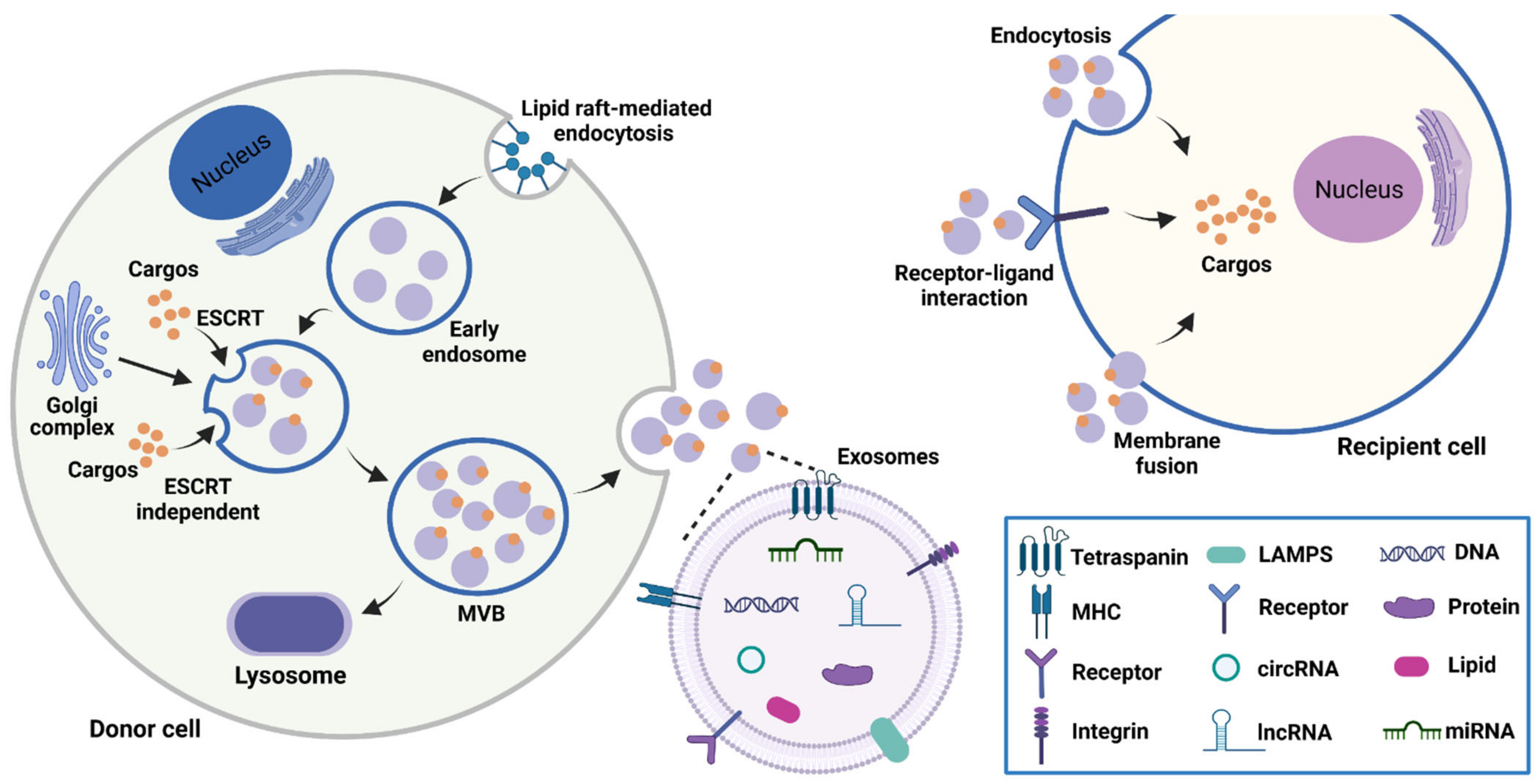
Figure 1. The biogenesis, contents, and uptake of exosomes. Exosomes are derived from the fusion of MVBs with plasma membranes. Exosomes transport cargos including nucleic acids, proteins, and lipids to recipient cells mainly through membrane fusion, receptor-ligand interaction, and endocytosis.
Cancers 15 00959 g001
Figure 2. Functional mechanisms of lncRNAs. (A) Under several stimulation conditions, lncRNAs can combine with transcription factors to regulate signaling pathway. (B) LncRNAs prevent mRNA degradation by functioning as miRNA sponge. (C) LncRNAs can act as scaffolds to form protein complexes, thus regulating the transcription of the target genes. (D) LncRNAs modulate gene expression by guiding the ribonucleoprotein complex to the DNA sequence.
Cancers 15 00959 g002
Figure 3. The biological role of exosomal lncRNAs in gastrointestinal cancer. Exosomal lncRNAs are involved in the various processes of gastrointestinal cancer including tumor growth, metastasis, angiogenesis, stemness, immune microenvironment, and drug resistance.
Cancers 15 00959 g003
Figure 4. LncRNAs in EVs as new biomarkers for the liquid biopsy of gastrointestinal cancer. Several technologies have been established to isolate EVs from body fluids and detect their cargoes. Many lncRNAs in serum/plasma-derived EVs provide a new strategy for the diagnosis of gastrointestinal cancer. Note: ↑: Upregulation; ↓: Downregulation.
Cancers 15 00959 g004
Table 1. The function and mechanism of exosomal lncRNAs in gastrointestinal cancer.
CancerlncRNAFunctionMechanismReference
GCCEBPA-AS1Promoting proliferation and
inhibiting apoptosis
N/A[62]
GCSND1-IT1Inducing malignant transformation of GES-1 cellsMiRNA sponge for miR-1245b-5b to enhance USP3 expression[63]
CRCWEE2-AS1Promoting proliferationInducing MOB1A degradation to inhibit Hippo pathway[64]
CRCUPATPromoting survival and tumorigenicity of CRC cellsstabilizing UHRF1 expression[65]
CRCPCAT1Promoting colorectal cancer liver metastasisMiRNA sponge for miR-329-3p to enhance Netrin-1 and CD146 expression[66]
GCTTN-AS1Promoting growth and metastasisMiRNA sponge for miR-499a-5p to enhance ZEB1 and CDX2 expression[67]
GCLINC01091Promoting growth and metastasisMiRNA sponge for miR-128-3p to enhance ELF4 and CDX2 expression[68]
GCFRLnc1Promoting growth and metastasisN/A[69]
CRCDUXAP10Promoting growth and lymph node metastasisBinding to LSD1 to inhibit the expression of p21 and PTEN[70]
GCPCGEM1Promoting EMT processMaintaining the stability of SNAI1[71]
GCTM4SF1-AS1Promoting the proliferation, invasion and EMTActivating PI3K/AKT signalling pathway[72]
CRCLINC00659Promoting proliferation, migration, invasion and EMT progressionMiRNA sponge for miR-342-3p to enhance annexin A2 expression[73]
CRCLINC01315Promoting proliferation, migration, and
stemness
N/A[74]
GCX26ntIncreasing angiogenesis and vascular
permeability
Reducing VE-cadherin expression[75]
GCMIR4435-2HGInducing macrophage M2 polarization to
promote tumor growth
Activating the Jagged1/Notch and JAK1/STAT3 pathways[76]
CRCHOXB8-1:2Inducing macrophage infiltration and M2
polarization to promote CRC progression
MiRNA sponge for miR-6825-5p to enhance CXCR3 expression[77]
CRCRPPH1Inducing macrophage M2 polarization to
promote the metastasis and proliferation of CRC cells
Inhibiting TUBB3 ubiquitination[78]
CRCKCNQ1OT1Inhibiting CD8+ T-cell response to promote CRC cell immune escapeMiRNA sponge for miR-30a-5p to enhance USP22 expression[79]
CRCCRNDE-hActivating Th17 cell differentiation to
promote tumor growth
Inhibiting the Itch-mediated ubiquitination and degradation of RORγt[80]
CRCSNHG10Suppressing the function of NK cells to
promote CRC cell immune escape
Upregulating INHBC expression[81]
CRCSNHG11Enhancing bevacizumab resistance in CRC cellsMiRNA sponge for miR-1207-5p to enhance ABCC1 expression[82]
CRCH19Promoting the stemness and chemoresistance
of CRC cells
MiRNA sponge for miR-141 to activate the
β-catenin pathway
[83]
CRCCCALPromoting chemoresistance of CRC cellsInteracting with HuR to enhance β-catenin
expression
[84]
GCCRNDEInducing cisplatin resistance in GC cellsPromoting NEDD4-1-mediated PTEN
ubiquitination
[85]
CRCUCA1Promoting cetuximab resistance in CRC cellsN/A[86]
GCHOTTIPPromoting cisplatin resistance in GC cellsMiRNA sponge for miR-218 to enhance HMGA1 expression[87]
CRCHOTTIPIncreasing resistance of CRC cells to mitomycinMiRNA sponge for miR-214 to enhance KPNA3 expression[88]
Abbreviations: ABCC1: ATP-binding cassette subfamily C member 1; CDX2: caudal-type homeobox-2; CRC: colorectal cancer; CXCR3: CXC chemokine receptor 3; ELF4: E74-like factor 4; EMT: epithelial–mesenchymal transition; HMGA1: high mobility group A1; INHBC: inhibin subunit beta C; JAK1: janus kinase-1; KPNA3: karyopherin subunit alpha 3; LSD1: lysine-specific demethylase 1; GC: gastric cancer; GES-1: gastric epithelial cells; MOB1A: MOB kinase activator 1A; N/A: not applicable; NEDD4-1: neuronal precursor cell-expressed developmentally downregulated 4-1; NK: natural killer; PTEN: phosphatase and tensin homolog; SNAI1: snail family transcriptional repressor 1; STAT3: signal transducer and activator of transcription 3; TUBB3: beta-III tubulin; UHRF1: ubiquitin-like with PHD and ring finger domains 1; USP22: ubiquitin specific protease 22; USP3: ubiquitin specific protease 3; ZEB1: zinc finger E-box binding homeobox 1.
Table 2. Diagnostic significance of exosomal lncRNAs in gastrointestinal cancer.
CancerlncRNAExpressionSourceCase NumberAUCReference
GCH19UpregulationSerum-derived EVs810.849[95]
GCMIATUpregulationSerum-derived EVs109N/A[97]
GCGNAQ-6:1DownregulationSerum-derived EVs430.732[98]
GCUEGC1UpregulationPlasma-derived EVs100.876 (distinguish GC patients from healthy individuals)
0.8406 (distinguish GC patients from chronic atrophic gastritis patients)
[99]
GCGC1UpregulationSerum-derived EVs8260.9033[100]
GCSLC2A12-10:1UpregulationPlasma-derived EVs1200.776[102]
GCPCSK2-2:1DownregulationSerum-derived EVs630.896[103]
CRCGAS5UpregulationPlasma-derived EVs1580.964[104]
CRCLINC02418UpregulationSerum-derived EVs1550.8978[106]
CRCFOXD2-AS1UpregulationSerum-derived EVs2030.728[107]
CRCNRIRUpregulationSerum-derived EVs2030.660[107]
CRCXLOC_009459UpregulationSerum-derived EVs2030.682[107]
Abbreviations: AUC: area under ROC curve; CRC: colorectal cancer; GC: gastric cancer; N/A: not applicable.
Disclaimer/Publisher’s Note: The statements, opinions and data contained in all publications are solely those of the individual author(s) and contributor(s) and not of MDPI and/or the editor(s). MDPI and/or the editor(s) disclaim responsibility for any injury to people or property resulting from any ideas, methods, instructions or products referred to in the content.