
Circulating Tumor DNA Methylation Biomarkers for Characterization and Determination of the Cancer Origin in Malignant Liver Tumors


Abstract
Simple Summary
Abstract
1. Introduction
2. Methylation in Cancer
3. Circulating Tumor DNA
4. Role of Methylation in the Tissue-of-Origin Determination
5. Methylation Signature of cfDNA in Malignant Liver Tumors
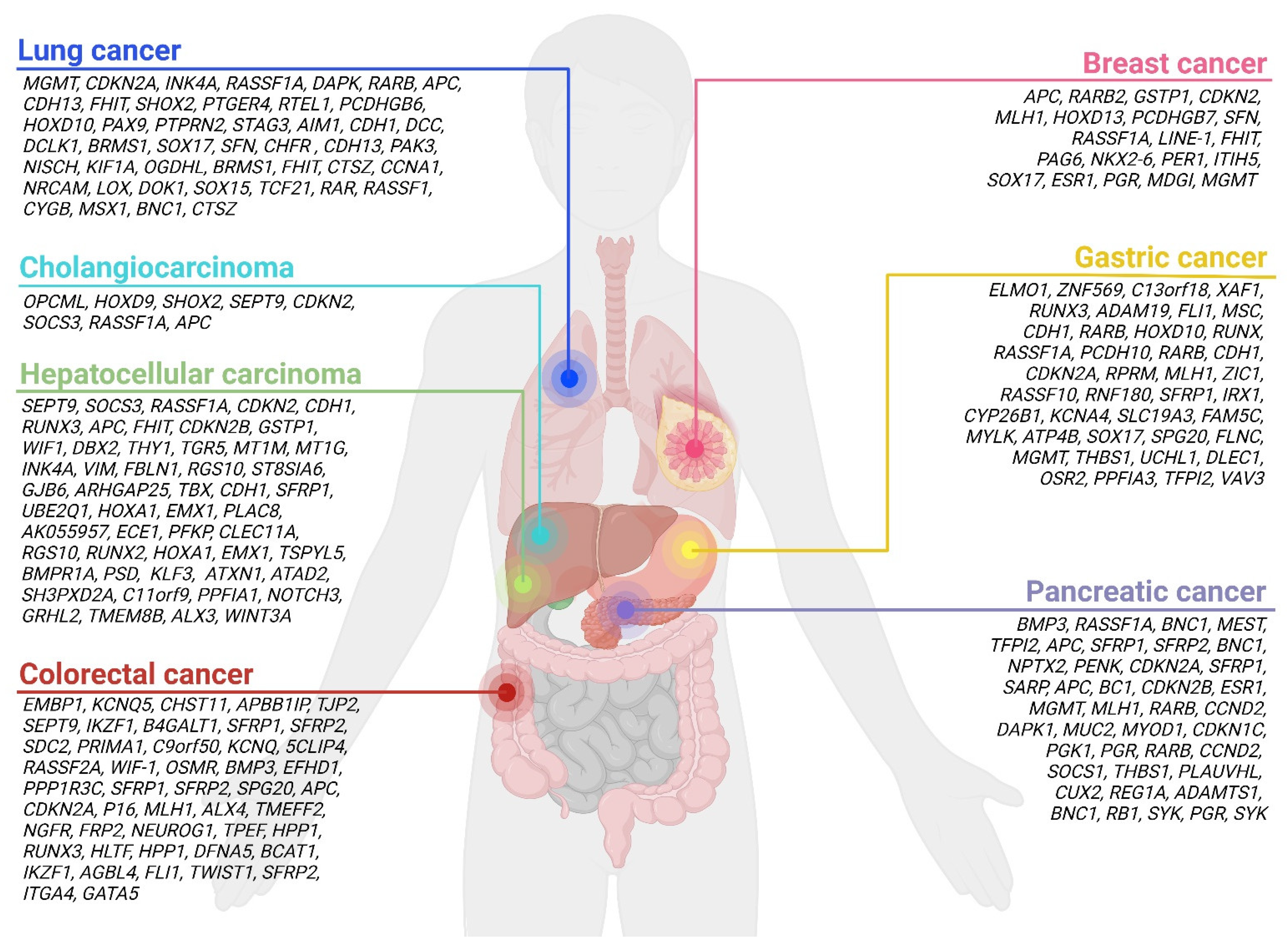
5.1. Most Common Primary Malignant Liver Tumors
5.1.1. Hepatocellular Carcinoma
5.1.2. Cholangiocarcinoma
5.2. Most Common Liver Metastases
5.2.1. Colorectal Cancer
5.2.2. Pancreatic Cancer

| Method | Sample Type | Number of Samples | Type of Methylation | Genes and/or Genetic Location | References |
|---|---|---|---|---|---|
| Clinical validation (MSP) | Plasma cfDNA | PDAC (n = 95), chronic pancreatitis (n = 97), acute pancreatitis (n = 59), patients negative for PAAD (n = 27) | Hyper | Diagnostic panel: BMP3, RASSF1A, BNC1, MEST, TFPI2, APC, SFRP1, SFRP2 Other: BNC1, NPTX2, PENK, CDKN2A, RASSF1A, SFRP1, SARP, APC, BC1, CDKN2B, ESR1, MGMT, MLH1, RARB | [94] |
| Clinical validation (MethDet56) | Plasma cfDNA | 30 chronic pancreatitis, 30 patients with pancreatic cancer, and 30 healthy controls | Hyper and hypo | CCND2, DAPK1, MLH1, MGMT, MUC2, MYOD1, CDKN2B, CDKN1C, PGK1, PGR, RARB, RB1, SYK | [95] |
| Clinical validation (multiplexed array-mediated analysis) | Plasma cfDNA | 30 PDAC patients and healthy controls | Hypo | CCND2, SOCS1, THBS1, PLAU, VHL | [96] |
| Clinical validation (real-time PCR) | Serum cfDNA | 40 PDAC patients, 60 with chronic pancreatitis, and 5 with benign biliary stone diseases | Hyper | NPTX2 | [97] |
| Review of the literature | cfDNA | / | Hyper and hypo | CUX2, REG1A, ADAMTS1, BNC1, MLH1, PGR, SYK, CCND2, CDKN1C | [98] |
5.2.3. Lung Cancer
5.2.4. Gastric Cancer
| Method | Sample Type | Number of Samples | Type of Methylation | Genes and/or Genetic Location | References |
|---|---|---|---|---|---|
| Clinical validation (MSP) | Tissue and plasma | Tissues: 14 GCs and 42 controls; plasma: 36 GCs and 38 controls | Hyper | Panel: ELMO1, ZNF569, C13orf18 | [119] |
| Clinical validation (MSP) | Tissue and serum | Tissues and plasma of 202 GC patients and their corresponding para-cancerous histological normal tissues; 88 serum healthy controls | Hyper | XAF1 | [120] |
| Clinical validation (CORD) | Serum cfDNA | 50 patients with early gastric cancer and 61 control individuals | Hyper | RUNX3 | [121] |
| Clinical validation (genome-wide methylation analysis) | Tissue and plasma cfDNA | 141 tissue and 106 plasma samples | Hyper | ADAM19, FLI1, MSC | [118] |
| Review of the literature | Serum or plasma | / | Hyper and hypo | CDKN2A, CDKN2A/CDH1, CDH1, CDKN2A/CDH1/RARB, RUNX3, ZIC1, HOXD10, RUNX, RASSF1A, PCDH10, RPRM, MLH1, RASSF10, RNF180, SFRP1, IRX1, CYP26B1/KCNA4, SLC19A3, FAM5C/MYLK, ATP4B, XAF1, SOX17, SPG20, FLNC/THBS1/UCHL1/DLEC1OSR2/VAV3/PPFIA3, TFPI2 | [115] |
| Review of the literature | Plasma and/or serum cfDNA | / | Hyper and hypo | PCDKN2A, CDH1, MGMT, RARB, RNF180 | [122] |
5.2.5. Breast Cancer
6. Conclusions and Future Directions
Author Contributions
Funding
Conflicts of Interest
References
- European Association for the Study of the Liver. EASL Clinical Practice Guidelines: Management of hepatocellular carcinoma. J. Hepatol. 2018, 69, 182–236. [Google Scholar] [CrossRef] [PubMed]
- Ananthakrishnan, A.; Gogineni, V.; Saeian, K. Epidemiology of primary and secondary liver cancers. Semin. Interv. Radiol. 2006, 23, 47–63. [Google Scholar] [CrossRef] [PubMed]
- Tsilimigras, D.I.; Brodt, P.; Clavien, P.-A.; Muschel, R.J.; D’Angelica, M.I.; Endo, I.; Parks, R.W.; Doyle, M.; de Santibañes, E.; Pawlik, T.M. Liver metastases. Nat. Rev. Dis. Prim. 2021, 7, 27. [Google Scholar] [CrossRef] [PubMed]
- Centeno, B.A. Pathology of liver metastases. Cancer Control 2006, 13, 13–26. [Google Scholar] [CrossRef]
- Abbruzzese, J.L.; Abbruzzese, M.C.; Lenzi, R.; Hess, K.R.; Raber, M.N. Analysis of a diagnostic strategy for patients with suspected tumors of unknown origin. J. Clin. Oncol. 1995, 13, 2094–2103. [Google Scholar] [CrossRef]
- Horn, S.R.; Stoltzfus, K.C.; Lehrer, E.J.; Dawson, L.A.; Tchelebi, L.; Gusani, N.J.; Sharma, N.K.; Chen, H.; Trifiletti, D.M.; Zaorsky, N.G. Epidemiology of liver metastases. Cancer Epidemiol. 2020, 67, 101760. [Google Scholar] [CrossRef]
- Fidler, I.J. The pathogenesis of cancer metastasis: The ‘seed and soil’ hypothesis revisited. Nat. Rev. Cancer 2003, 3, 453–458. [Google Scholar] [CrossRef]
- Weiss, L. Comments on hematogenous metastatic patterns in humans as revealed by autopsy. Clin. Exp. Metastasis 1992, 10, 191–199. [Google Scholar] [CrossRef]
- De Ridder, J.; de Wilt, J.H.W.; Simmer, F.; Overbeek, L.; Lemmens, V.; Nagtegaal, I. Incidence and origin of histologically confirmed liver metastases: An explorative case-study of 23,154 patients. Oncotarget 2016, 7, 55368–55376. [Google Scholar] [CrossRef]
- Van de Wouw, A.J.; Janssen-Heijnen, M.L.; Coebergh, J.W.; Hillen, H.F. Epidemiology of unknown primary tumours; incidence and population-based survival of 1285 patients in Southeast Netherlands, 1984–1992. Eur. J. Cancer 2002, 38, 409–413. [Google Scholar] [CrossRef]
- Ayoub, J.P.; Hess, K.R.; Abbruzzese, M.C.; Lenzi, R.; Raber, M.N.; Abbruzzese, J.L. Unknown primary tumors metastatic to liver. J. Clin. Oncol. 1998, 16, 2105–2112. [Google Scholar] [CrossRef]
- Pavlidis, N.; Pentheroudakis, G. Cancer of unknown primary site. Lancet 2012, 379, 1428–1435. [Google Scholar] [CrossRef] [PubMed]
- Wang, L.; Vuolo, M.; Suhrland, M.J.; Schlesinger, K. HepPar1, MOC-31, pCEA, mCEA and CD10 for distinguishing hepatocellular carcinoma vs. metastatic adenocarcinoma in liver fine needle aspirates. Acta Cytol. 2006, 50, 257–262. [Google Scholar] [CrossRef] [PubMed]
- Varadhachary, G.R.; Talantov, D.; Raber, M.N.; Meng, C.; Hess, K.R.; Jatkoe, T.; Lenzi, R.; Spigel, D.R.; Wang, Y.; Greco, F.A.; et al. Molecular profiling of carcinoma of unknown primary and correlation with clinical evaluation. J. Clin. Oncol. 2008, 26, 4442–4448. [Google Scholar] [CrossRef]
- Oweira, H.; Petrausch, U.; Helbling, D.; Schmidt, J.; Mannhart, M.; Mehrabi, A.; Schöb, O.; Giryes, A.; Decker, M.; Abdel-Rahman, O. Prognostic value of site-specific metastases in pancreatic adenocarcinoma: A surveillance epidemiology and end results database analysis. World J. Gastroenterol. 2017, 23, 1872–1880. [Google Scholar] [CrossRef] [PubMed]
- Li, J.; Zhu, H.; Sun, L.; Xu, W.; Wang, X. Prognostic value of site-specific metastases in lung cancer: A population based study. J. Cancer 2019, 10, 3079–3086. [Google Scholar] [CrossRef]
- Wang, J.; Li, S.; Liu, Y.; Zhang, C.; Li, H.; Lai, B. Metastatic patterns and survival outcomes in patients with stage IV colon cancer: A population-based analysis. Cancer Med. 2020, 9, 361–373. [Google Scholar] [CrossRef]
- Patel, S.A.; Vanharanta, S. Epigenetic determinants of metastasis. Mol. Oncol. 2017, 11, 79–96. [Google Scholar] [CrossRef]
- Kurkjian, C.; Kummar, S.; Murgo, A.J. DNA methylation: Its role in cancer development and therapy. Curr. Probl. Cancer 2008, 32, 187–235. [Google Scholar] [CrossRef]
- Bird, A. DNA methylation patterns and epigenetic memory. Genes Dev. 2002, 16, 6–21. [Google Scholar] [CrossRef]
- Fernández-Barrena, M.G.; Arechederra, M.; Colyn, L.; Berasain, C.; Avila, M.A. Epigenetics in hepatocellular carcinoma development and therapy: The tip of the iceberg. JHEP Rep. 2020, 2, 100167. [Google Scholar] [CrossRef]
- Dor, Y.; Cedar, H. Principles of DNA methylation and their implications for biology and medicine. Lancet 2018, 392, 777–786. [Google Scholar] [CrossRef]
- Aran, D.; Hellman, A. DNA methylation of transcriptional enhancers and cancer predisposition. Cell 2013, 154, 11–13. [Google Scholar] [CrossRef]
- Board, R.E.; Knight, L.; Greystoke, A.; Blackhall, F.H.; Hughes, A.; Dive, C.; Ranson, M. DNA methylation in circulating tumour DNA as a biomarker for cancer. Biomark. Insights 2008, 2, 307–319. [Google Scholar] [CrossRef]
- Cheishvili, D.; Wong, C.; Karim, M.M.; Kibria, M.G.; Jahan, N.; Das, P.C.; Yousuf, M.A.K.; Islam, M.A.; Das, D.C.; Noor-E-Alam, S.M. epiLiver a novel tumor specific, high throughput and cost-effective blood test for specific detection of liver cancer (HCC). medRxiv 2021. [Google Scholar] [CrossRef]
- Ehrlich, M. DNA hypomethylation in cancer cells. Epigenomics 2009, 1, 239–259. [Google Scholar] [CrossRef]
- Liu, X.; Ren, J.; Luo, N.; Guo, H.; Zheng, Y.; Li, J.; Tang, F.; Wen, L.; Peng, J. Comprehensive DNA methylation analysis of tissue of origin of plasma cell-free DNA by methylated CpG tandem amplification and sequencing (MCTA-Seq). Clin. Epigenetics 2019, 11, 93. [Google Scholar] [CrossRef]
- Moss, J.; Magenheim, J.; Neiman, D.; Zemmour, H.; Loyfer, N.; Korach, A.; Samet, Y.; Maoz, M.; Druid, H.; Arner, P.; et al. Comprehensive human cell-type methylation atlas reveals origins of circulating cell-free DNA in health and disease. Nat. Commun. 2018, 9, 5068. [Google Scholar] [CrossRef]
- Moran, S.; Martinez-Cardús, A.; Boussios, S.; Esteller, M. Precision medicine based on epigenomics: The paradigm of carcinoma of unknown primary. Nat. Rev. Clin. Oncol. 2017, 14, 682–694. [Google Scholar] [CrossRef]
- Liu, M.C.; Oxnard, G.R.; Klein, E.A.; Swanton, C.; Seiden, M.V.; Liu, M.C.; Oxnard, G.R.; Klein, E.A.; Smith, D.; Richards, D.; et al. Sensitive and specific multi-cancer detection and localization using methylation signatures in cell-free DNA. Ann. Oncol. 2020, 31, 745–759. [Google Scholar] [CrossRef]
- Shen, S.Y.; Singhania, R.; Fehringer, G.; Chakravarthy, A.; Roehrl, M.H.A.; Chadwick, D.; Zuzarte, P.C.; Borgida, A.; Wang, T.T.; Li, T.; et al. Sensitive tumour detection and classification using plasma cell-free DNA methylomes. Nature 2018, 563, 579–583. [Google Scholar] [CrossRef] [PubMed]
- Heyn, H.; Esteller, M. DNA methylation profiling in the clinic: Applications and challenges. Nat. Rev. Genet. 2012, 13, 679–692. [Google Scholar] [CrossRef] [PubMed]
- Song, C.X.; Yin, S.; Ma, L.; Wheeler, A.; Chen, Y.; Zhang, Y.; Liu, B.; Xiong, J.; Zhang, W.; Hu, J.; et al. 5-Hydroxymethylcytosine signatures in cell-free DNA provide information about tumor types and stages. Cell Res. 2017, 27, 1231–1242. [Google Scholar] [CrossRef] [PubMed]
- Bronkhorst, A.J.; Ungerer, V.; Holdenrieder, S. The emerging role of cell-free DNA as a molecular marker for cancer management. Biomol. Detect. Quantif. 2019, 17, 100087. [Google Scholar] [CrossRef] [PubMed]
- Cross, D.; Burmester, J.K. The promise of molecular profiling for cancer identification and treatment. Clin. Med. Res. 2004, 2, 147–150. [Google Scholar] [CrossRef]
- Karachaliou, N.; Mayo-de-Las-Casas, C.; Molina-Vila, M.A.; Rosell, R. Real-time liquid biopsies become a reality in cancer treatment. Ann. Transl. Med. 2015, 3, 36. [Google Scholar] [CrossRef]
- Wan, J.C.M.; Massie, C.; Garcia-Corbacho, J.; Mouliere, F.; Brenton, J.D.; Caldas, C.; Pacey, S.; Baird, R.; Rosenfeld, N. Liquid biopsies come of age: Towards implementation of circulating tumour DNA. Nat. Rev. Cancer 2017, 17, 223–238. [Google Scholar] [CrossRef]
- Jahr, S.; Hentze, H.; Englisch, S.; Hardt, D.; Fackelmayer, F.O.; Hesch, R.D.; Knippers, R. DNA fragments in the blood plasma of cancer patients: Quantitations and evidence for their origin from apoptotic and necrotic cells. Cancer Res. 2001, 61, 1659–1665. [Google Scholar]
- Bettegowda, C.; Sausen, M.; Leary, R.J.; Kinde, I.; Wang, Y.; Agrawal, N.; Bartlett, B.R.; Wang, H.; Luber, B.; Alani, R.M.; et al. Detection of circulating tumor DNA in early- and late-stage human malignancies. Sci. Transl. Med. 2014, 6, 224ra224. [Google Scholar] [CrossRef]
- Xu, R.-h.; Wei, W.; Krawczyk, M.; Wang, W.; Luo, H.; Flagg, K.; Yi, S.; Shi, W.; Quan, Q.; Li, K.; et al. Circulating tumour DNA methylation markers for diagnosis and prognosis of hepatocellular carcinoma. Nat. Mater. 2017, 16, 1155–1161. [Google Scholar] [CrossRef]
- Moran, S.; Martínez-Cardús, A.; Sayols, S.; Musulén, E.; Balañá, C.; Estival-Gonzalez, A.; Moutinho, C.; Heyn, H.; Diaz-Lagares, A.; de Moura, M.C.; et al. Epigenetic profiling to classify cancer of unknown primary: A multicentre, retrospective analysis. Lancet Oncol. 2016, 17, 1386–1395. [Google Scholar] [CrossRef] [PubMed]
- Oxnard, G.R.; Klein, E.A.; Seiden, M.V.; Hubbell, E.; Venn, O.; Jamshidi, A.; Zhang, N.; Beausang, J.F.; Gross, S.; Kurtzman, K.N.; et al. Simultaneous multi-cancer detection and tissue of origin (TOO) localization using targeted bisulfite sequencing of plasma cell-free DNA (cfDNA). Ann. Oncol. 2019, 30, v912. [Google Scholar] [CrossRef]
- Hlady, R.A.; Zhao, X.; Pan, X.; Yang, J.D.; Ahmed, F.; Antwi, S.O.; Giama, N.H.; Patel, T.; Roberts, L.R.; Liu, C.; et al. Genome-wide discovery and validation of diagnostic DNA methylation-based biomarkers for hepatocellular cancer detection in circulating cell free DNA. Theranostics 2019, 9, 7239–7250. [Google Scholar] [CrossRef] [PubMed]
- Kisiel, J.B.; Dukek, B.A.; Kanipakam, R.V.S.R.; Ghoz, H.M.; Yab, T.C.; Berger, C.K.; Taylor, W.R.; Foote, P.H.; Giama, N.H.; Onyirioha, K.; et al. Hepatocellular Carcinoma Detection by Plasma Methylated DNA: Discovery, Phase I Pilot, and Phase II Clinical Validation. Hepatology 2019, 69, 1180–1192. [Google Scholar] [CrossRef] [PubMed]
- Zhang, C.; Li, J.; Huang, T.; Duan, S.; Dai, D.; Jiang, D.; Sui, X.; Li, D.; Chen, Y.; Ding, F.; et al. Meta-analysis of DNA methylation biomarkers in hepatocellular carcinoma. Oncotarget 2016, 7, 81255–81267. [Google Scholar] [CrossRef]
- Iyer, P.; Zekri, A.R.; Hung, C.W.; Schiefelbein, E.; Ismail, K.; Hablas, A.; Seifeldin, I.A.; Soliman, A.S. Concordance of DNA methylation pattern in plasma and tumor DNA of Egyptian hepatocellular carcinoma patients. Exp. Mol. Pathol. 2010, 88, 107–1112. [Google Scholar] [CrossRef]
- Conway, A.M.; Mitchell, C.; Kilgour, E.; Brady, G.; Dive, C.; Cook, N. Molecular characterisation and liquid biomarkers in Carcinoma of Unknown Primary (CUP): Taking the ‘U’ out of ‘CUP’. Br. J. Cancer 2019, 120, 141–153. [Google Scholar] [CrossRef]
- Liu, M.C.; Klein, E.; Hubbell, E.; Maddala, T.; Aravanis, A.M.; Beausang, J.F.; Filippova, D.; Gross, S.; Jamshidi, A.; Kurtzman, K.; et al. Plasma cell-free DNA (cfDNA) assays for early multi-cancer detection: The circulating cell-free genome atlas (CCGA) study. Ann. Oncol. 2018, 29, viii14. [Google Scholar] [CrossRef]
- Liu, L.; Toung, J.M.; Jassowicz, A.F.; Vijayaraghavan, R.; Kang, H.; Zhang, R.; Kruglyak, K.M.; Huang, H.J.; Hinoue, T.; Shen, H.; et al. Targeted methylation sequencing of plasma cell-free DNA for cancer detection and classification. Ann. Oncol. 2018, 29, 1445–1453. [Google Scholar] [CrossRef]
- Lehmann-Werman, R.; Magenheim, J.; Moss, J.; Neiman, D.; Abraham, O.; Piyanzin, S.; Zemmour, H.; Fox, I.; Dor, T.; Grompe, M.; et al. Monitoring liver damage using hepatocyte-specific methylation markers in cell-free circulating DNA. JCI Insight 2018, 3, e120687. [Google Scholar] [CrossRef]
- Leon, S.A.; Shapiro, B.; Sklaroff, D.M.; Yaros, M.J. Free DNA in the serum of cancer patients and the effect of therapy. Cancer Res. 1977, 37, 646–650. [Google Scholar]
- Maebo, A. Plasma DNA level as a tumor marker in primary lung cancer. Nihon Kyobu Shikkan Gakkai Zasshi 1990, 28, 1085–1091. [Google Scholar]
- Cree, I.A.; Uttley, L.; Woods, H.B.; Kikuchi, H.; Reiman, A.; Harnan, S.; Whiteman, B.L.; Philips, S.T.; Messenger, M.; Cox, A.; et al. The evidence base for circulating tumour DNA blood-based biomarkers for the early detection of cancer: A systematic mapping review. BMC Cancer 2017, 17, 697. [Google Scholar] [CrossRef]
- Villanueva, A. Hepatocellular Carcinoma. N. Engl. J. Med. 2019, 380, 1450–1462. [Google Scholar] [CrossRef]
- McGlynn, K.A.; Petrick, J.L.; El-Serag, H.B. Epidemiology of Hepatocellular Carcinoma. Hepatology 2021, 73, 4–13. [Google Scholar] [CrossRef]
- Hamilton, S.R.; Aaltonen, L.A.; World Health Organization Classification of Tumours. Pathology and Genetics of Tumours of the Digestive System; IARCPress: Lyon, France, 2000; pp. 199–202. [Google Scholar]
- Wu, H.-C.; Yang, H.-I.; Wang, Q.; Chen, C.-J.; Santella, R.M. Plasma DNA methylation marker and hepatocellular carcinoma risk prediction model for the general population. Carcinogenesis 2017, 38, 1021–1028. [Google Scholar] [CrossRef]
- Chalasani, N.P.; Ramasubramanian, T.S.; Bhattacharya, A.; Olson, M.C.; Edwards, D.K.; Roberts, L.R.; Kisiel, J.B.; Reddy, K.R.; Lidgard, G.P.; Johnson, S.C.; et al. A novel blood-based panel of methylated DNA and protein markers for detection of early-stage hepatocellular carcinoma. Clin. Gastroenterol. Hepatol. 2021, 19, 2597–2605. [Google Scholar] [CrossRef]
- Wen, L.; Li, J.; Guo, H.; Liu, X.; Zheng, S.; Zhang, D.; Zhu, W.; Qu, J.; Guo, L.; Du, D.; et al. Genome-scale detection of hypermethylated CpG islands in circulating cell-free DNA of hepatocellular carcinoma patients. Cell Res. 2015, 25, 1250–1264. [Google Scholar] [CrossRef]
- Oussalah, A.; Rischer, S.; Bensenane, M.; Conroy, G.; Filhine-Tresarrieu, P.; Debard, R.; Forest-Tramoy, D.; Josse, T.; Reinicke, D.; Garcia, M.; et al. Plasma mSEPT9: A novel circulating cell-free DNA-based epigenetic biomarker to diagnose hepatocellular carcinoma. EBioMedicine 2018, 30, 138–147. [Google Scholar] [CrossRef]
- Wei, L.; Huang, Y.; Zhao, R.; Zhang, J.; Liu, Q.; Liang, W.; Ding, X.; Gao, B.; Li, B.; Sun, C.; et al. Detection of promoter methylation status of suppressor of cytokine signaling 3 (SOCS3) in tissue and plasma from Chinese patients with different hepatic diseases. Clin. Exp. Med. 2018, 18, 79–87. [Google Scholar] [CrossRef]
- Hu, N.; Fan, X.P.; Fan, Y.C.; Chen, L.Y.; Qiao, C.Y.; Han, L.Y.; Wang, K. Hypomethylated Ubiquitin-conjugating enzyme2 Q1 (UBE2Q1) gene promoter in the serum is a promising biomarker for Hepatitis B Virus-associated hepatocellular carcinoma. Tohoku J. Exp. Med. 2017, 242, 93–100. [Google Scholar] [CrossRef] [PubMed]
- Huang, Z.H.; Hu, Y.; Hua, D.; Wu, Y.Y.; Song, M.X.; Cheng, Z.H. Quantitative analysis of multiple methylated genes in plasma for the diagnosis and prognosis of hepatocellular carcinoma. Exp. Mol. Pathol. 2011, 91, 702–707. [Google Scholar] [CrossRef] [PubMed]
- Wong, I.H.; Lo, Y.M.; Yeo, W.; Lau, W.Y.; Johnson, P.J. Frequent p15 promoter methylation in tumor and peripheral blood from hepatocellular carcinoma patients. Clin. Cancer Res. 2000, 6, 3516–3521. [Google Scholar] [PubMed]
- Holmila, R.; Sklias, A.; Muller, D.C.; Degli Esposti, D.; Guilloreau, P.; McKay, J.; Sangrajrang, S.; Srivatanakul, P.; Hainaut, P.; Merle, P.; et al. Targeted deep sequencing of plasma circulating cell-free DNA reveals Vimentin and Fibulin 1 as potential epigenetic biomarkers for hepatocellular carcinoma. PLoS ONE 2017, 12, e0174265. [Google Scholar] [CrossRef] [PubMed]
- Zhang, Z.; Chen, P.; Xie, H.; Cao, P. Using circulating tumor DNA as a novel biomarker to screen and diagnose hepatocellular carcinoma: A systematic review and meta-analysis. Cancer Med. 2020, 9, 1349–1364. [Google Scholar] [CrossRef]
- Wu, X.; Li, J.; Gassa, A.; Buchner, D.; Alakus, H.; Dong, Q.; Ren, N.; Liu, M.; Odenthal, M.; Stippel, D.; et al. Circulating tumor DNA as an emerging liquid biopsy biomarker for early diagnosis and therapeutic monitoring in hepatocellular carcinoma. Int. J. Biol. Sci. 2020, 16, 1551–1562. [Google Scholar] [CrossRef]
- Li, W.; Zhang, X.; Lu, X.; You, L.; Song, Y.; Luo, Z.; Zhang, J.; Nie, J.; Zheng, W.; Xu, D.; et al. 5-Hydroxymethylcytosine signatures in circulating cell-free DNA as diagnostic biomarkers for human cancers. Cell Res. 2017, 27, 1243–12571. [Google Scholar] [CrossRef]
- Cai, J.; Chen, L.; Zhang, Z.; Zhang, X.; Lu, X.; Liu, W.; Shi, G.; Ge, Y.; Gao, P.; Yang, Y.; et al. Genome-wide mapping of 5-hydroxymethylcytosines in circulating cell-free DNA as a non-invasive approach for early detection of hepatocellular carcinoma. Gut 2019, 68, 2195–22052. [Google Scholar] [CrossRef]
- Rizvi, S.; Gores, G.J. Pathogenesis, diagnosis, and management of cholangiocarcinoma. Gastroenterology 2013, 145, 1215–1229. [Google Scholar] [CrossRef]
- Andresen, K.; Boberg, K.M.; Vedeld, H.M.; Honne, H.; Jebsen, P.; Hektoen, M.; Wadsworth, C.A.; Clausen, O.P.; Lundin, K.E.; Paulsen, V.; et al. Four DNA methylation biomarkers in biliary brush samples accurately identify the presence of cholangiocarcinoma. Hepatology 2015, 61, 1651–1659. [Google Scholar] [CrossRef]
- Isomoto, H.; Mott, J.L.; Kobayashi, S.; Werneburg, N.W.; Bronk, S.F.; Haan, S.; Gores, G.J. Sustained IL-6/STAT-3 signaling in cholangiocarcinoma cells due to SOCS-3 epigenetic silencing. Gastroenterology 2007, 132, 384–396. [Google Scholar] [CrossRef]
- Branchi, V.; Schaefer, P.; Semaan, A.; Kania, A.; Lingohr, P.; Kalff, J.C.; Schäfer, N.; Kristiansen, G.; Dietrich, D.; Matthaei, H. Promoter hypermethylation of SHOX2 and SEPT9 is a potential biomarker for minimally invasive diagnosis in adenocarcinomas of the biliary tract. Clin. Epigenetics 2016, 8, 133. [Google Scholar] [CrossRef]
- Wasenang, W.; Chaiyarit, P.; Proungvitaya, S.; Limpaiboon, T. Serum cell-free DNA methylation of OPCML and HOXD9 as a biomarker that may aid in differential diagnosis between cholangiocarcinoma and other biliary diseases. Clin. Epigenetics 2019, 11, 39. [Google Scholar] [CrossRef]
- Vedeld, H.M.; Folseraas, T.; Lind, G.E. Detecting cholangiocarcinoma in patients with primary sclerosing cholangitis—The promise of DNA methylation and molecular biomarkers. JHEP Rep. 2020, 2, 100143. [Google Scholar] [CrossRef]
- Rizvi, S.; Eaton, J.; Yang, J.D.; Chandrasekhara, V.; Gores, G.J. Emerging Technologies for the Diagnosis of Perihilar Cholangiocarcinoma. Semin. Liver Dis. 2018, 38, 160–169. [Google Scholar] [CrossRef]
- Brannon, A.R.; Vakiani, E.; Sylvester, B.E.; Scott, S.N.; McDermott, G.; Shah, R.H.; Kania, K.; Viale, A.; Oschwald, D.M.; Vacic, V.; et al. Comparative sequencing analysis reveals high genomic concordance between matched primary and metastatic colorectal cancer lesions. Genome Biol. 2014, 15, 454. [Google Scholar] [CrossRef]
- Orjuela, S.; Menigatti, M.; Schraml, P.; Kambakamba, P.; Robinson, M.D.; Marra, G. The DNA hypermethylation phenotype of colorectal cancer liver metastases resembles that of the primary colorectal cancers. BMC Cancer 2020, 20, 290. [Google Scholar] [CrossRef]
- Vignot, S.; Lefebvre, C.; Frampton, G.M.; Meurice, G.; Yelensky, R.; Palmer, G.; Capron, F.; Lazar, V.; Hannoun, L.; Miller, V.A.; et al. Comparative analysis of primary tumour and matched metastases in colorectal cancer patients: Evaluation of concordance between genomic and transcriptional profiles. Eur. J. Cancer 2015, 51, 791–799. [Google Scholar] [CrossRef]
- Ju, H.-X.; An, B.; Okamoto, Y.; Shinjo, K.; Kanemitsu, Y.; Komori, K.; Hirai, T.; Shimizu, Y.; Sano, T.; Sawaki, A.; et al. Distinct profiles of epigenetic evolution between colorectal cancers with and without metastasis. Am. J. Pathol. 2011, 178, 1835–1846. [Google Scholar] [CrossRef]
- Gai, W.; Ji, L.; Lam, W.K.J.; Sun, K.; Jiang, P.; Chan, A.W.H.; Wong, J.; Lai, P.B.S.; Ng, S.S.M.; Ma, B.B.Y.; et al. Liver- and colon-specific DNA methylation markers in plasma for investigation of colorectal cancers with or without liver metastases. Clin. Chem. 2018, 64, 1239–1249. [Google Scholar] [CrossRef]
- Li, J.; Zhou, X.; Liu, X.; Ren, J.; Wang, J.; Wang, W.; Zheng, Y.; Shi, X.; Sun, T.; Li, Z.; et al. Detection of colorectal cancer in circulating cell-free DNA by methylated CpG tandem amplification and sequencing. Clin. Chem. 2019, 65, 916–926. [Google Scholar] [CrossRef] [PubMed]
- Picardo, F.; Romanelli, A.; Muinelo-Romay, L.; Mazza, T.; Fusilli, C.; Parrella, P.; Barbazán, J.; Lopez-López, R.; Barbano, R.; De Robertis, M.; et al. Diagnostic and prognostic value of B4GALT1 hypermethylation and its clinical significance as a novel circulating cell-free DNA biomarker in colorectal cancer. Cancers 2019, 11, 1598. [Google Scholar] [CrossRef] [PubMed]
- Barták, B.K.; Kalmár, A.; Péterfia, B.; Patai, Á.V.; Galamb, O.; Valcz, G.; Spisák, S.; Wichmann, B.; Nagy, Z.B.; Tóth, K.; et al. Colorectal adenoma and cancer detection based on altered methylation pattern of SFRP1, SFRP2, SDC2, and PRIMA1 in plasma samples. Epigenetics 2017, 12, 751–763. [Google Scholar] [CrossRef] [PubMed]
- Jensen, S.; Øgaard, N.; Ørntoft, M.W.; Rasmussen, M.H.; Bramsen, J.B.; Kristensen, H.; Mouritzen, P.; Madsen, M.R.; Madsen, A.H.; Sunesen, K.G.; et al. Novel DNA methylation biomarkers show high sensitivity and specificity for blood-based detection of colorectal cancer-a clinical biomarker discovery and validation study. Clin. Epigenetics 2019, 11, 158. [Google Scholar] [CrossRef] [PubMed]
- Laugsand, E.A.; Brenne, S.S.; Skorpen, F. DNA methylation markers detected in blood, stool, urine, and tissue in colorectal cancer: A systematic review of paired samples. Int. J. Colorectal Dis. 2021, 36, 239–251. [Google Scholar] [CrossRef]
- Hauptman, N.; Glavač, D. Colorectal Cancer Blood-Based Biomarkers. Gastroenterol. Res. Pract. 2017, 2017, 2195361. [Google Scholar] [CrossRef]
- Danese, E.; Montagnana, M.; Lippi, G. Circulating molecular biomarkers for screening or early diagnosis of colorectal cancer: Which is ready for prime time? Ann. Transl. Med. 2019, 7, 610. [Google Scholar] [CrossRef]
- Mukund, K.; Syulyukina, N.; Ramamoorthy, S.; Subramaniam, S. Right and left-sided colon cancers—Specificity of molecular mechanisms in tumorigenesis and progression. BMC Cancer 2020, 20, 317. [Google Scholar] [CrossRef]
- Hess, K.R.; Varadhachary, G.R.; Taylor, S.H.; Wei, W.; Raber, M.N.; Lenzi, R.; Abbruzzese, J.L. Metastatic patterns in adenocarcinoma. Cancer 2006, 106, 1624–1633. [Google Scholar] [CrossRef]
- Lehmann-Werman, R.; Neiman, D.; Zemmour, H.; Moss, J.; Magenheim, J.; Vaknin-Dembinsky, A.; Rubertsson, S.; Nellgård, B.; Blennow, K.; Zetterberg, H.; et al. Identification of tissue-specific cell death using methylation patterns of circulating DNA. Proc. Natl. Acad. Sci. USA 2016, 113, E1826–E1834. [Google Scholar] [CrossRef]
- Lapin, M.; Oltedal, S.; Tjensvoll, K.; Buhl, T.; Smaaland, R.; Garresori, H.; Javle, M.; Glenjen, N.I.; Abelseth, B.K.; Gilje, B.; et al. Fragment size and level of cell-free DNA provide prognostic information in patients with advanced pancreatic cancer. J. Transl. Med. 2018, 16, 300. [Google Scholar] [CrossRef]
- Henriksen, S.D.; Madsen, P.H.; Krarup, H.; Thorlacius-Ussing, O. DNA Hypermethylation as a blood-based marker for pancreatic cancer: A literature review. Pancreas 2015, 44, 1036–1045. [Google Scholar] [CrossRef]
- Henriksen, S.D.; Madsen, P.H.; Larsen, A.C.; Johansen, M.B.; Drewes, A.M.; Pedersen, I.S.; Krarup, H.; Thorlacius-Ussing, O. Cell-free DNA promoter hypermethylation in plasma as a diagnostic marker for pancreatic adenocarcinoma. Clin. Epigenetics 2016, 8, 117. [Google Scholar] [CrossRef]
- Liggett, T.; Melnikov, A.; Yi, Q.L.; Replogle, C.; Brand, R.; Kaul, K.; Talamonti, M.; Abrams, R.A.; Levenson, V. Differential methylation of cell-free circulating DNA among patients with pancreatic cancer versus chronic pancreatitis. Cancer 2010, 116, 1674–1680. [Google Scholar] [CrossRef]
- Melnikov, A.A.; Scholtens, D.; Talamonti, M.S.; Bentrem, D.J.; Levenson, V.V. Methylation profile of circulating plasma DNA in patients with pancreatic cancer. J. Surg. Oncol. 2009, 99, 119–122. [Google Scholar] [CrossRef]
- Park, J.K.; Ryu, J.K.; Yoon, W.J.; Lee, S.H.; Lee, G.Y.; Jeong, K.-S.; Kim, Y.-T.; Yoon, Y.B. The role of quantitative NPTX2 hypermethylation as a novel serum diagnostic marker in pancreatic cancer. Pancreas 2012, 41, 95–101. [Google Scholar] [CrossRef]
- Brancaccio, M.; Natale, F.; Falco, G.; Angrisano, T. Cell-free DNA methylation: The new frontiers of pancreatic cancer biomarkers’ discovery. Genes 2019, 11, 14. [Google Scholar] [CrossRef]
- Yu, Y.; Wang, Z.; Mo, D.H.; Wang, Z.; Li, G. Transcriptome profiling reveals liver metastasis-associated genes in pancreatic ductal adenocarcinoma. Math. Biosci. Eng. 2021, 18, 1708–1721. [Google Scholar] [CrossRef]
- Makohon-Moore, A.P.; Zhang, M.; Reiter, J.G.; Bozic, I.; Allen, B.; Kundu, D.; Chatterjee, K.; Wong, F.; Jiao, Y.; Kohutek, Z.A.; et al. Limited heterogeneity of known driver gene mutations among the metastases of individual patients with pancreatic cancer. Nat. Genet. 2017, 49, 358–366. [Google Scholar] [CrossRef]
- McDonald, O.G.; Li, X.; Saunders, T.; Tryggvadottir, R.; Mentch, S.J.; Warmoes, M.O.; Word, A.E.; Carrer, A.; Salz, T.H.; Natsume, S.; et al. Epigenomic reprogramming during pancreatic cancer progression links anabolic glucose metabolism to distant metastasis. Nat. Genet. 2017, 49, 367–376. [Google Scholar] [CrossRef]
- Riihimäki, M.; Hemminki, A.; Fallah, M.; Thomsen, H.; Sundquist, K.; Sundquist, J.; Hemminki, K. Metastatic sites and survival in lung cancer. Lung Cancer 2014, 86, 78–84. [Google Scholar] [CrossRef] [PubMed]
- Tamura, T.; Kurishima, K.; Nakazawa, K.; Kagohashi, K.; Ishikawa, H.; Satoh, H.; Hizawa, N. Specific organ metastases and survival in metastatic non-small-cell lung cancer. Mol. Clin. Oncol. 2015, 3, 217–221. [Google Scholar] [CrossRef] [PubMed]
- Ansari, J.; Shackelford, R.E.; El-Osta, H. Epigenetics in non-small cell lung cancer: From basics to therapeutics. Transl. Lung Cancer Res. 2016, 5, 155–171. [Google Scholar] [CrossRef] [PubMed]
- Lin, R.K.; Hsieh, Y.S.; Lin, P.; Hsu, H.S.; Chen, C.Y.; Tang, Y.A.; Lee, C.F.; Wang, Y.C. The tobacco-specific carcinogen NNK induces DNA methyltransferase 1 accumulation and tumor suppressor gene hypermethylation in mice and lung cancer patients. J. Clin. Investig. 2010, 120, 521–532. [Google Scholar] [CrossRef] [PubMed]
- O’Hagan, H.M.; Wang, W.; Sen, S.; Destefano Shields, C.; Lee, S.S.; Zhang, Y.W.; Clements, E.G.; Cai, Y.; Van Neste, L.; Easwaran, H.; et al. Oxidative damage targets complexes containing DNA methyltransferases, SIRT1, and polycomb members to promoter CpG Islands. Cancer Cell 2011, 20, 606–619. [Google Scholar] [CrossRef] [PubMed]
- Fujiwara, K.; Fujimoto, N.; Tabata, M.; Nishii, K.; Matsuo, K.; Hotta, K.; Kozuki, T.; Aoe, M.; Kiura, K.; Ueoka, H.; et al. Identification of epigenetic aberrant promoter methylation in serum DNA is useful for early detection of lung cancer. Clin. Cancer Res. 2005, 11, 1219–1225. [Google Scholar] [CrossRef]
- Heller, G.; Zielinski, C.C.; Zöchbauer-Müller, S. Lung cancer: From single-gene methylation to methylome profiling. Cancer Metastasis Rev. 2010, 29, 95–107. [Google Scholar] [CrossRef]
- Lu, Y.; Li, S.S.L.; Zhu, S.S.G.; Gong, Y.Y.B.; Shi, J.J.; Xu, L.L. Methylated DNA/RNA in body fluids as biomarkers for lung cancer. Biol. Proced. Online 2017, 19, 2. [Google Scholar] [CrossRef]
- Newman, A.M.; Bratman, S.V.; To, J.; Wynne, J.F.; Eclov, N.C.W.; Modlin, L.A.; Liu, C.L.; Neal, J.W.; Wakelee, H.A.; Merritt, R.E.; et al. An ultrasensitive method for quantitating circulating tumor DNA with broad patient coverage. Nat. Med. 2014, 20, 548–554. [Google Scholar] [CrossRef]
- Jemal, A.; Center, M.M.; DeSantis, C.; Ward, E.M. Global patterns of cancer incidence and mortality rates and trends. Cancer Epidemiol. Biomark. Prev. 2010, 19, 1893–1907. [Google Scholar] [CrossRef]
- Bernards, N.; Creemers, G.J.; Nieuwenhuijzen, G.A.; Bosscha, K.; Pruijt, J.F.; Lemmens, V.E. No improvement in median survival for patients with metastatic gastric cancer despite increased use of chemotherapy. Ann. Oncol. 2013, 24, 3056–3060. [Google Scholar] [CrossRef]
- Dicken, B.J.; Bigam, D.L.; Cass, C.; Mackey, J.R.; Joy, A.A.; Hamilton, S.M. Gastric adenocarcinoma: Review and considerations for future directions. Ann. Surg. 2005, 241, 27–39. [Google Scholar] [CrossRef] [PubMed]
- Riihimäki, M.; Hemminki, A.; Sundquist, K.; Sundquist, J.; Hemminki, K. Metastatic spread in patients with gastric cancer. Oncotarget 2016, 7, 52307–52316. [Google Scholar] [CrossRef]
- Huang, Z.-B.; Zhang, H.-T.; Yu, B.; Yu, D.-H. Cell-free DNA as a liquid biopsy for early detection of gastric cancer. Oncol. Lett. 2021, 21, 3. [Google Scholar] [CrossRef]
- Hu, W.; Zheng, W.; Liu, Q.; Chu, H.; Chen, S.; Kim, J.J.; Wu, J.; Si, J. Diagnostic accuracy of DNA methylation in detection of gastric cancer: A meta-analysis. Oncotarget 2017, 8, 113142–113152. [Google Scholar] [CrossRef]
- Liu, Z.; Cheng, X.; Zhang, L.; Zhou, J.; Deng, D.; Ji, J. A panel of DNA methylated markers predicts metastasis of pN0M0 gastric carcinoma: A prospective cohort study. Br. J. Cancer 2019, 121, 529–536. [Google Scholar] [CrossRef]
- Fang, W.-L.; Chen, M.-H.; Huang, K.-H.; Chang, S.-C.; Lin, C.-H.; Chao, Y.; Lo, S.-S.; Li, A.F.-Y.; Wu, C.-W.; Shyr, Y.-M. Analysis of the clinical significance of DNA methylation in gastric cancer based on a genome-wide high-resolution array. Clin. Epigenetics 2019, 11, 154. [Google Scholar] [CrossRef]
- Anderson, B.W.; Suh, Y.S.; Choi, B.; Lee, H.J.; Yab, T.C.; Taylor, W.R.; Dukek, B.A.; Berger, C.K.; Cao, X.; Foote, P.H.; et al. Detection of gastric cancer with novel methylated DNA markers: Discovery, tissue validation, and pilot testing in plasma. Clin. Cancer Res. 2018, 24, 5724–5734. [Google Scholar] [CrossRef]
- Ling, Z.Q.; Lv, P.; Lu, X.X.; Yu, J.L.; Han, J.; Ying, L.S.; Zhu, X.; Zhu, W.Y.; Fang, X.H.; Wang, S.; et al. Circulating methylated XAF1 DNA indicates poor prognosis for gastric cancer. PLoS ONE 2013, 8, e67195. [Google Scholar] [CrossRef]
- Hideura, E.; Suehiro, Y.; Nishikawa, J.; Shuto, T.; Fujimura, H.; Ito, S.; Goto, A.; Hamabe, K.; Saeki, I.; Okamoto, T.; et al. Blood free-circulating DNA testing of methylated RUNX3 is useful for diagnosing early gastric cancer. Cancers 2020, 12, 784. [Google Scholar] [CrossRef]
- Necula, L.; Matei, L.; Dragu, D.; Neagu, A.I.; Mambet, C.; Nedeianu, S.; Bleotu, C.; Diaconu, C.C.; Chivu-Economescu, M. Recent advances in gastric cancer early diagnosis. World J. Gastroenterol. 2019, 25, 2029–2044. [Google Scholar] [CrossRef]
- Al Hannan, F.; Keogh, M.B.; Taha, S.; Al Buainain, L. Characterization of BRCA1 and BRCA2 genetic variants in a cohort of Bahraini breast cancer patients using next-generation sequencing. Mol. Genet. Genomic Med. 2019, 7, e007711. [Google Scholar] [CrossRef] [PubMed]
- Adam, R.; Aloia, T.; Krissat, J.; Bralet, M.-P.; Paule, B.; Giacchetti, S.; Delvart, V.; Azoulay, D.; Bismuth, H.; Castaing, D. Is liver resection justified for patients with hepatic metastases from breast cancer? Ann. Surg. 2006, 244, 897–908. [Google Scholar] [CrossRef] [PubMed]
- Liu, L.; Sun, L.; Li, C.; Li, X.; Zhang, Y.; Yu, Y.; Xia, W. Quantitative detection of methylation of FHIT and BRCA1 promoters in the serum of ductal breast cancer patients. Biomed. Mater. Eng. 2015, 26 (Suppl. S1), S2217–S2222. [Google Scholar] [CrossRef]
- Swellam, M.; Abdelmaksoud, M.D.; Sayed Mahmoud, M.; Ramadan, A.; Abdel-Moneem, W.; Hefny, M.M. Aberrant methylation of APC and RARβ2 genes in breast cancer patients. IUBMB Life 2015, 67, 61–68. [Google Scholar] [CrossRef]
- Hoque, M.O.; Feng, Q.; Toure, P.; Dem, A.; Critchlow, C.W.; Hawes, S.E.; Wood, T.; Jeronimo, C.; Rosenbaum, E.; Stern, J.; et al. Detection of aberrant methylation of four genes in plasma DNA for the detection of breast cancer. J. Clin. Oncol. 2006, 24, 4262–42696. [Google Scholar] [CrossRef]
- Shan, M.; Yin, H.; Li, J.; Li, X.; Wang, D.; Su, Y.; Niu, M.; Zhong, Z.; Wang, J.; Zhang, X.; et al. Detection of aberrant methylation of a six-gene panel in serum DNA for diagnosis of breast cancer. Oncotarget 2016, 7, 18485–18494. [Google Scholar] [CrossRef]
- Lee, K.H.; Shin, T.J.; Kim, W.H.; Cho, J.Y. Methylation of LINE-1 in cell-free DNA serves as a liquid biopsy biomarker for human breast cancers and dog mammary tumors. Sci. Rep. 2019, 9, 175. [Google Scholar] [CrossRef]
- Mijnes, J.; Tiedemann, J.; Eschenbruch, J.; Gasthaus, J.; Bringezu, S.; Bauerschlag, D.; Maass, N.; Arnold, N.; Weimer, J.; Anzeneder, T.; et al. SNiPER: A novel hypermethylation biomarker panel for liquid biopsy based early breast cancer detection. Oncotarget 2019, 10, 6494–6508. [Google Scholar] [CrossRef]
- Chimonidou, M.; Strati, A.; Malamos, N.; Kouneli, S.; Georgoulias, V.; Lianidou, E. Direct comparison study of DNA methylation markers in EpCAM-positive circulating tumour cells, corresponding circulating tumour DNA, and paired primary tumours in breast cancer. Oncotarget 2017, 8, 72054–72068. [Google Scholar] [CrossRef]
- Liggett, T.E.; Melnikov, A.A.; Marks, J.R.; Levenson, V.V. Methylation patterns in cell-free plasma DNA reflect removal of the primary tumor and drug treatment of breast cancer patients. Int. J. Cancer 2011, 128, 492–499. [Google Scholar] [CrossRef] [PubMed]
- Ye, M.; Huang, T.; Ying, Y.; Li, J.; Yang, P.; Ni, C.; Zhou, C.; Chen, S. Detection of 14-3-3 sigma (σ) promoter methylation as a noninvasive biomarker using blood samples for breast cancer diagnosis. Oncotarget 2017, 8, 9230–9242. [Google Scholar] [CrossRef]

| Method | Sample Type | Number of Samples | Type of Methylation | Genes and/or Genetic Location | References |
|---|---|---|---|---|---|
| Bioinformatics analysis and clinical validation | Plasma ctDNA | 1098 HCC patients and 835 normal controls | Hypo and hyper | Diagnostic panel: BMPR1A, PSD, ARHGAP25, KLF3, PLAC8, ATXN1, chr6:170, chr6:3, ATAD2, chr8:20 Prognostic prediction panel: SH3PXD2A, C11orf9, PPFIA1, chr17:78, SERPINB5, NOTCH3, GRHL2, TMEM8B | [39] |
| Clinical validation (HM450k) | Tissue and cfDNA | 127 non-tumor and 415 HCC tissue samples; 37 non-tumor and 37 HCC cfDNA samples | Hypo and hyper | Diagnostic panel: chr19:51 (intragenic region), ALX3, WINT3A, chr1:42 (intragenic region), GJB6 | [43] |
| Clinical validation (pyrosequencing) | Plasma cfDNA | 237 HCC cases and 257 controls | Hyper | TBX | [57] |
| Clinical validation (multiplex PCR) | cfDNA | 135 HCC and 302 control samples | Hyper | Detection panel: HOXA1, EMX1, TSPYL5 | [58] |
| Clinical validation (MCTA-Seq) | Tissue and plasma cfDNA | 151 tissue and plasma samples | Hyper | RGS10, ST8SIA6, RUNX2, VIM | [59] |
| Clinical validation (TELQA and MCTA-Seq) | Tissue and plasma cfDNA | 74 HCC and 29 control tissue samples; 116 HCC, 81 cirrhotic controls and 98 healthy control plasma samples | Hyper | Detection panel: HOXA1, EMX1, AK055957, ECE1, PFKP, CLEC11A (normalized by B3GALT6 level yielded) | [44] |
| Clinical validation (PCR) | Plasma cfDNA | 289 patients | Hyper | SEPT9 | [60] |
| Clinical validation | Tissue and plasma cfDNA | 116 tissues and 326 plasma samples | Hyper | SOCS3 | [61] |
| Clinical validation (MSP) | Serum cfDNA | 80 HCC, 40 liver cirrhosis, 40 chronic hepatitis B, and 20 healthy controls | Hypo | UBE2Q1 | [62] |
| Clinical validation (MSRE-qPCR) | Plasma cfDNA | 72 patients with HCC, 37 benign live diseases, and 41 normal controls | Hyper | APC, GSTP1, RASSF1A, SFRP1 | [63] |
| Clinical validation (MSP) | Tissue and plasma cfDNA | 25 tissue and 130 plasma samples | Hyper | CDKN2B, PCDKN2A | [64] |
| Clinical validation (targeted massively parallel semiconductor sequencing) | Plasma cfDNA | 84 HCC, 26 chronic liver patients, and 84 controls | Hyper | FBLN1, VIM | [65] |
| Clinical validation (MSP) | Tissue and plasma cfDNA | 24 tissue and plasma samples | Hyper | APC, FHIT, CDKN2B, PCDKN2A, CDH1 | [46] |
| Meta-analysis | 144 eligible studies included | 2044 HCC and 1371 normal serums samples | Hypo and hyper | RASSF1A, PCDKN2A, CDH1, RUNX3, GSTP1, WIF1 | [45] |
| Meta-analysis | 33 eligible studies included | 4113 subjects | Hyper | RASSF1A | [66] |
| Review of the literature | Plasma or serum ctDNA | 3442 HCC and 2696 controls | Hyper | DBX2, THY1, TGR5, MT1M, MT1G, INK4A, VIM, FBLN1, RGS10, ST8SIA6, RUNX, SEPT9 | [67] |
| Method | Sample Type | Number of Samples | Type of Methylation | Genes and/or Genetic Location | References |
|---|---|---|---|---|---|
| Clinical validation (MS-HRM) | Serum cfDNA | 40 CCA and 40 controls | Hyper | OPCML, HOXD9 | [74] |
| Clinical validation (Real-time PCR) | Tissue and plasma | Tissue: 71 tumors with pared normal samples Plasma: 20 CCA patients and 100 control patients | Hyper | SHOX2, SEPT9 | [73] |
| Meta-analysis | Tissue and serum/plasma | / | Hyper | SHOX2, SEPT9, OPCML, HOXD9 | [75] |
| Review of the literature | cfDNA | / | Hyper | CDKN2, SOCS3, RASSF1A, APC | [76] |
| Method | Sample Type | Number of Samples | Type of Methylation | Genes and/or Genetic Location | References |
|---|---|---|---|---|---|
| Clinical validation (MCTA-Seq) | Tissue and plasma cfDNA | Tissue: 66 samples Plasma: CRC (n = 147) and controls (n = 136) | Hyper | EMBP1, KCNQ5, CHST11, APBB1IP, TJP2, SEPT9, IKZF1 Additional panel with 80 markers | [82] |
| Clinical validation (Quantitative MSP) | Plasma cfDNA | 27 plasma samples | Hyper | B4GALT1 | [83] |
| Clinical validation (MethyLight PCR) | Tissue and plasma cfDNA | 21 plasma and 32 tissue biopsy samples | Hyper | SFRP1, SFRP2, SDC2, PRIMA1 | [84] |
| Clinical validation (MethyLight PCR) | Plasma cfDNA | 113 CRC patients and 87 controls | Hyper | C9orf50, KCNQ5, CLIP4 | [85] |
| Systematic review | Blood, stool, urine, and tissue | 51 studies included | Hyper and Hypo | Diagnostic panel: APC, MGMT, RASSF2A, WIF-1 Other genes: SEPT9, OSMR, BMP3, EFHD1, PPP1R3C, SFRP1, SFRP2, SPG20 | [86] |
| Review of the literature | Blood | / | Hyper and Hypo | SEPT9, APC, CDKN2A/CDKN2A, MLH1, ALX4, TMEFF2, NGFR, FRP2, NEUROG1, TPEF/HPP1, RUNX3, HLTF, HPP1, DFNA5 | [87] |
| Review of the literature | Blood | / | Hyper | Diagnostic panel: APC, MGMT, RASSF2A, WIF-1 Other genes: SEPT9, BCAT1/IKZF1, AGBL4, FLI1, TWIST1, SFRP2, ITGA4, GATA5 | [88] |
| Cancer Type | Method | Sample Type | Number of Samples | Type of Methylation | Genes and/or Genetic Location | References |
|---|---|---|---|---|---|---|
| Lung cancer, SCLC, NSCLC | Clinical validation (MS-PCR) | Serum cfDNA | 91 lung cancer patients, 9 with other malignant diseases, and 100 nonmalignant pulmonary diseases | Hyper | MGMT, PCDKN2A, INK4A, RASSF1A, DAPK, RARB | [107] |
| NSCLC | Review of the literature | Plasma or serum | / | Hyper and Hypo | MGMT, PCDKN2A, DAPK, APC, CDH13, FHIT, RARB, RASSF1A | [108] |
| Lung cancer, SCLC, NSCLC | Review of the literature | Plasma or serum | / | Hyper and Hypo | Diagnostic marker combinations: RASSF1A/RARB, SHOX2/PTGER4, RTEL1/PCDHGB6, HOXD10/PAX9/PTPRN2/STAG3, APC/AIM1/CDH1/DCC/MGMT/RASSF1A Diagnostic and prognostic markers: SHOX2, RARB2/RASSF1A, RARB, RASSF1A/APC, DCLK1, BRMS1, SOX17, SFN, CHFR, APC/RASSF1A/CDH13/CDKN2A | [109] |
| NSCLC | Review of the literature | Plasma or serum | / | Hyper | CDKN2A, PAK3, NISCH, KIF1A, OGDHL, BRMS1, FHIT, CTSZ, CCNA1, NRCAM, LOX, MGMT, DOK1, SOX15, TCF21, DAPK, RAR, RASSF1, CYGB, MSX1, BNC1, CTSZ, CDKN2A | [104] |
| Method | Sample Type | Number of Samples | Type of Methylation | Genes and/or Genetic Location | References |
|---|---|---|---|---|---|
| Clinical validation (MSP) | Serum cfDNA | 121 women breast cancer patients, 79 patients with benign breast diseases, and 66 healthy volunteers | Hyper | APC, RARβ2 | [126] |
| Clinical validation (Quantitative MSP) | Plasma cfDNA | Women with breast cancer (n = 93) compared with control women (n = 76) | Hyper | APC, GSTP1, RASSF1A, RARB2 | [127] |
| Clinical validation (MethyLight) | cfDNA in serum | 49 cases including breast cancer patients, patients with benign breast diseases, and healthy women | Hyper | Panel: SFN, CDKN2A, MLH1, HOXD13, PCDHGB7, RASSF1A | [128] |
| Clinical validation (MSRED followed by Real-time PCR) | cfDNA in plasma | 26 human BCs and 10 healthy controls | Hypo | LINE-1 | [129] |
| Clinical validation (BSP and HRM) | Serum cfDNA | 36 patients with invasive breast ductal carcinoma (BDC group), 30 patients with breast fibroadenoma (BFA group), and 30 healthy individuals | Hyper | FHIT | [125] |
| Clinical validation (Pyrosequencing) | Plasma and serum cfDNA | Serum test cohort (n = 103), a serum validation cohort (n = 368), and a plasma cohort (n = 125) | Hyper | Panel: SPAG6, NKX2-6, PER1, ITIH5 | [130] |
| Clinical validation (MSP) | FFPE, whole blood (CTC), and plasma cfDNA | 153 patients and 49 healthy controls | Hyper | SOX17 | [131] |
| Clinical validation (MethDet-56) | Plasma cfDNA | 20 patients and 20 healthy controls | Hyper | RARB, ESR1, PGR, MDGI, MGMT | [132] |
| Meta-Analysis | Tissue and blood | / | Hyper | SNF | [133] |
| Clinical validation (MSP) | Serum cfDNA | 121 women breast cancer patients, 79 patients with benign breast diseases, and 66 healthy volunteers | Hyper | APC, RARβ2 | [126] |
| Clinical validation (Quantitative MSP) | Plasma cfDNA | Women with breast cancer (n = 93) compared with control women (n = 76) | Hyper | APC, GSTP1, RASSF1A, RARB2 | [127] |
Disclaimer/Publisher’s Note: The statements, opinions and data contained in all publications are solely those of the individual author(s) and contributor(s) and not of MDPI and/or the editor(s). MDPI and/or the editor(s) disclaim responsibility for any injury to people or property resulting from any ideas, methods, instructions or products referred to in the content. |
© 2023 by the authors. Licensee MDPI, Basel, Switzerland. This article is an open access article distributed under the terms and conditions of the Creative Commons Attribution (CC BY) license (https://creativecommons.org/licenses/by/4.0/).
Share and Cite
Draškovič, T.; Zidar, N.; Hauptman, N. Circulating Tumor DNA Methylation Biomarkers for Characterization and Determination of the Cancer Origin in Malignant Liver Tumors. Cancers 2023, 15, 859. https://doi.org/10.3390/cancers15030859
Draškovič T, Zidar N, Hauptman N. Circulating Tumor DNA Methylation Biomarkers for Characterization and Determination of the Cancer Origin in Malignant Liver Tumors. Cancers. 2023; 15(3):859. https://doi.org/10.3390/cancers15030859
Chicago/Turabian StyleDraškovič, Tina, Nina Zidar, and Nina Hauptman. 2023. "Circulating Tumor DNA Methylation Biomarkers for Characterization and Determination of the Cancer Origin in Malignant Liver Tumors" Cancers 15, no. 3: 859. https://doi.org/10.3390/cancers15030859
APA StyleDraškovič, T., Zidar, N., & Hauptman, N. (2023). Circulating Tumor DNA Methylation Biomarkers for Characterization and Determination of the Cancer Origin in Malignant Liver Tumors. Cancers, 15(3), 859. https://doi.org/10.3390/cancers15030859