
Diagnostic and Therapeutic Pathway in Diffuse Malignant Peritoneal Mesothelioma

 ,
,  , , , and
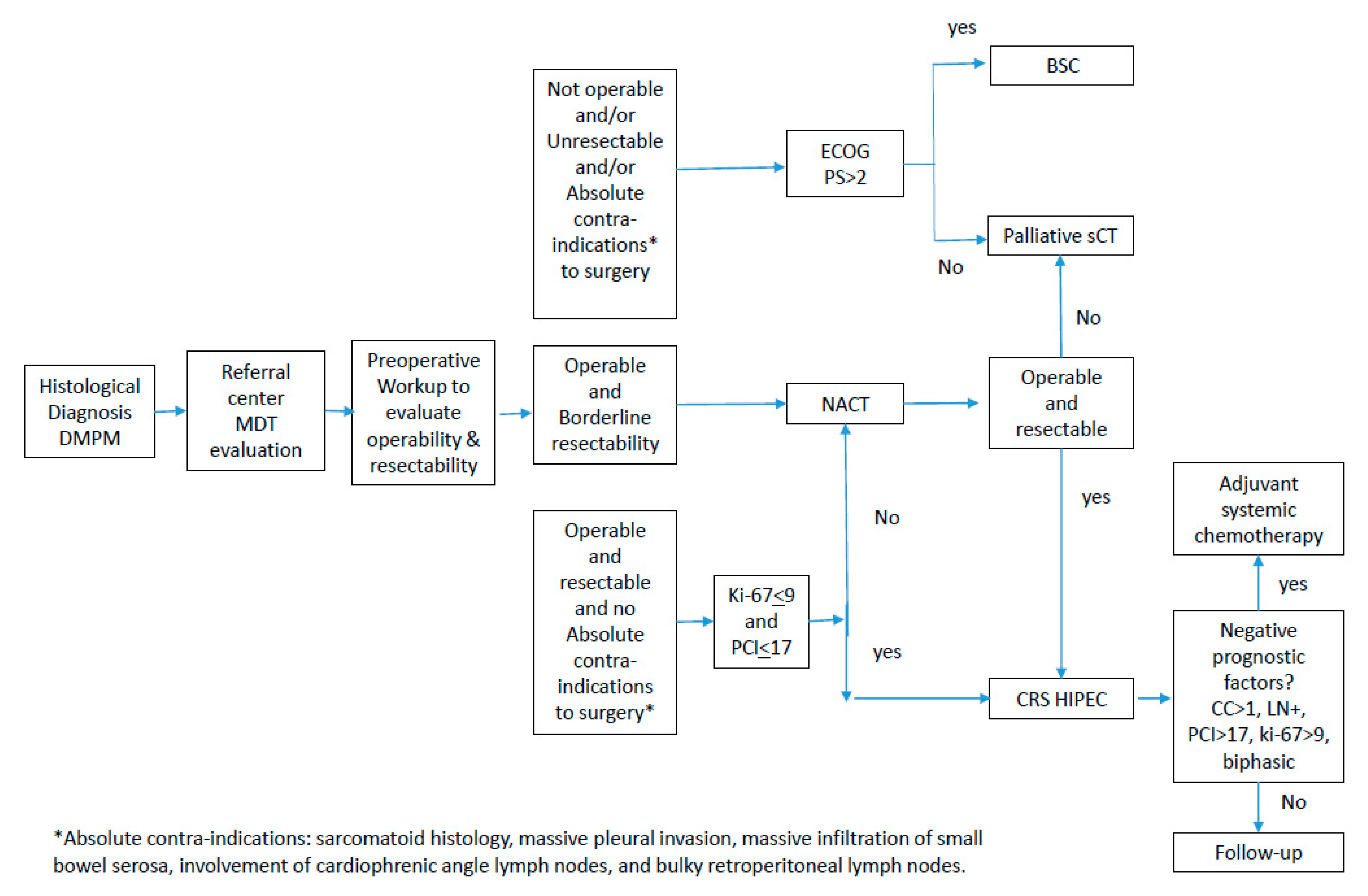
Abstract
Simple Summary
Abstract
1. Introduction
2. Diagnosis and Pathology
3. Preoperative Workup
4. Treatment
4.1. Systemic Chemotherapy and Immunotherapy
4.2. Local-Regional Treatment
4.2.1. Eligibility for Cytoreductive Surgery (CRS) and HIPEC
Evaluation of Operability and Disease Resectability
Prognostic Factors and Risk Prediction Models
Biphasic/Sarcomatoid Histologies
4.2.2. Technical Aspects of Cytoreductive Surgery
4.2.3. The Role of HIPEC and HIPEC Drug Schedules
4.2.4. Normothermic Intraperitoneal Chemotherapies
5. Follow-Up of DMPM
6. Borderline Histological Subtypes
7. Conclusions
Author Contributions
Funding
Conflicts of Interest
References
- Grogg, J.B.; Fronzaroli, J.N.; Oliveira, P.; Bode, P.-K.; Lorch, A.; Issa, A.; Beyer, J.; Eberli, D.; Sangar, V.; Hermanns, T.; et al. Clinicopathological Characteristics and Outcomes in Men with Mesothelioma of the Tunica Vaginalis Testis: Analysis of Published Case-Series Data. J. Cancer Res. Clin. Oncol. 2021, 147, 2671–2679. [Google Scholar] [CrossRef] [PubMed]
- Carteni, G.; Manegold, C.; Garcia, G.M.; Siena, S.; Zielinski, C.C.; Amadori, D.; Liu, Y.; Blatter, J.; Visseren-Grul, C.; Stahel, R. Malignant Peritoneal Mesothelioma-Results from the International Expanded Access Program Using Pemetrexed Alone or in Combination with a Platinum Agent. Lung Cancer 2009, 64, 211–218. [Google Scholar] [CrossRef] [PubMed]
- Bianchi, C.; Bianchi, T. Global Mesothelioma Epidemic: Trend and Features. Indian J. Occup. Environ. Med. 2014, 18, 82–88. [Google Scholar] [CrossRef] [PubMed]
- Bridda, A.; Padoan, I.; Mencarelli, R.; Frego, M. Peritoneal Mesothelioma: A Review. MedGenMed 2007, 9, 32. [Google Scholar] [PubMed]
- Mirarabshahii, P.; Pillai, K.; Chua, T.C.; Pourgholami, M.H.; Morris, D.L. Diffuse Malignant Peritoneal Mesothelioma—An Update on Treatment. Cancer Treat. Rev. 2012, 38, 605–612. [Google Scholar] [CrossRef] [PubMed]
- Chekol, S.S.; Sun, C.-C. Malignant Mesothelioma of the Tunica Vaginalis Testis: Diagnostic Studies and Differential Diagnosis. Arch. Pathol. Lab. Med. 2012, 136, 113–117. [Google Scholar] [CrossRef] [PubMed]
- Ullah, A.; Waheed, A.; Khan, J.; Mishra, A.; Tareen, B.; Nama, N.; Karki, N.R.; Panezai, M.S.; Zarate, L.V.; White, J.; et al. Incidence, Survival Analysis and Future Perspective of Primary Peritoneal Mesothelioma (PPM): A Population-Based Study from SEER Database. Cancers 2022, 14, 942. [Google Scholar] [CrossRef] [PubMed]
- Bijelic, L.; Darcy, K.; Stodghill, J.; Tian, C.; Cannon, T. Predictors and Outcomes of Surgery in Peritoneal Mesothelioma: An Analysis of 2000 Patients from the National Cancer Database. Ann. Surg. Oncol. 2020, 27, 2974–2982. [Google Scholar] [CrossRef]
- Acs, M.; Gerken, M.; Gajic, I.; Mayr, M.; Zustin, J.; Piso, P. Ten-Year Single-Center Experience with Treatment of Primary Diffuse Malignant Peritoneal Mesothelioma (DMPM) by Cytoreductive Surgery (CRS) and Hyperthermic Intraperitoneal Chemotherapy (HIPEC). Langenbecks Arch. Surg. 2022, 407, 3057–3067. [Google Scholar] [CrossRef]
- Chapel, D.B.; Schulte, J.J.; Absenger, G.; Attanoos, R.; Brcic, L.; Butnor, K.J.; Chirieac, L.; Churg, A.; Galateau-Sallé, F.; Hiroshima, K.; et al. Malignant Peritoneal Mesothelioma: Prognostic Significance of Clinical and Pathologic Parameters and Validation of a Nuclear-Grading System in a Multi-Institutional Series of 225 Cases. Mod. Pathol. 2021, 34, 380–395. [Google Scholar] [CrossRef]
- Helm, J.H.; Miura, J.T.; Glenn, J.A.; Marcus, R.K.; Larrieux, G.; Jayakrishnan, T.T.; Donahue, A.E.; Gamblin, T.C.; Turaga, K.K.; Johnston, F.M. Cytoreductive Surgery and Hyperthermic Intraperitoneal Chemotherapy for Malignant Peritoneal Mesothelioma: A Systematic Review and Meta-Analysis. Ann. Surg. Oncol. 2015, 22, 1686–1693. [Google Scholar] [CrossRef] [PubMed]
- Yan, T.D.; Deraco, M.; Baratti, D.; Kusamura, S.; Elias, D.; Glehen, O.; Gilly, F.N.; Levine, E.A.; Shen, P.; Mohamed, F.; et al. Cytoreductive Surgery and Hyperthermic Intraperitoneal Chemotherapy for Malignant Peritoneal Mesothelioma: Multi-Institutional Experience. J. Clin. Oncol. 2009, 27, 6237–6242. [Google Scholar] [CrossRef] [PubMed]
- Husain, A.N.; Colby, T.V.; Ordóñez, N.G.; Allen, T.C.; Attanoos, R.L.; Beasley, M.B.; Butnor, K.J.; Chirieac, L.R.; Churg, A.M.; Dacic, S.; et al. Guidelines for Pathologic Diagnosis of Malignant Mesothelioma 2017 Update of the Consensus Statement from the International Mesothelioma Interest Group. Arch. Pathol. Lab. Med. 2018, 142, 89–108. [Google Scholar] [CrossRef] [PubMed]
- Moolgavkar, S.H.; Meza, R.; Turim, J. Pleural and Peritoneal Mesotheliomas in SEER: Age Effects and Temporal Trends, 1973–2005. Cancer Causes Control 2009, 20, 935–944. [Google Scholar] [CrossRef] [PubMed]
- Teta, M.J.; Mink, P.J.; Lau, E.; Sceurman, B.K.; Foster, E.D. US Mesothelioma Patterns 1973–2002: Indicators of Change and Insights into Background Rates. Eur. J. Cancer Prev. 2008, 17, 525–534. [Google Scholar] [CrossRef]
- Boffetta, P. Epidemiology of Peritoneal Mesothelioma: A Review. Ann. Oncol. 2007, 18, 985–990. [Google Scholar] [CrossRef]
- Villa, R.; Daidone, M.G.; Motta, R.; Venturini, L.; De Marco, C.; Vannelli, A.; Kusamura, S.; Baratti, D.; Deraco, M.; Costa, A.; et al. Multiple Mechanisms of Telomere Maintenance Exist and Differentially Affect Clinical Outcome in Diffuse Malignant Peritoneal Mesothelioma. Clin. Cancer Res. 2008, 14, 4134–4140. [Google Scholar] [CrossRef]
- Zaffaroni, N.; Costa, A.; Pennati, M.; De Marco, C.; Affini, E.; Madeo, M.; Erdas, R.; Cabras, A.; Kusamura, S.; Baratti, D.; et al. Survivin Is Highly Expressed and Promotes Cell Survival in Malignant Peritoneal Mesothelioma. Cell. Oncol. 2007, 29, 453–466. [Google Scholar] [CrossRef]
- Perrone, F.; Jocollè, G.; Pennati, M.; Deraco, M.; Baratti, D.; Brich, S.; Orsenigo, M.; Tarantino, E.; De Marco, C.; Bertan, C.; et al. Receptor Tyrosine Kinase and Downstream Signalling Analysis in Diffuse Malignant Peritoneal Mesothelioma. Eur. J. Cancer 2010, 46, 2837–2848. [Google Scholar] [CrossRef]
- El Bezawy, R.; Percio, S.; Ciniselli, C.M.; De Cesare, M.; Colella, G.; Dugo, M.; Veneroni, S.; Doldi, V.; Martini, S.; Baratti, D.; et al. MiR-550a-3p Is a Prognostic Biomarker and Exerts Tumor-Suppressive Functions by Targeting HSP90AA1 in Diffuse Malignant Peritoneal Mesothelioma. Cancer Gene Ther. 2022, 29, 1394–1404. [Google Scholar] [CrossRef]
- El Bezawy, R.; De Cesare, M.; Pennati, M.; Deraco, M.; Gandellini, P.; Zuco, V.; Zaffaroni, N. Antitumor Activity of MiR-34a in Peritoneal Mesothelioma Relies on c-MET and AXL Inhibition: Persistent Activation of ERK and AKT Signaling as a Possible Cytoprotective Mechanism. J. Hematol. Oncol. 2017, 10, 19. [Google Scholar] [CrossRef] [PubMed]
- Cimino-Reale, G.; Gandellini, P.; Santambrogio, F.; Recagni, M.; Zaffaroni, N.; Folini, M. MiR-380-5p-Mediated Repression of TEP1 and TSPYL5 Interferes with Telomerase Activity and Favours the Emergence of an “ALT-like” Phenotype in Diffuse Malignant Peritoneal Mesothelioma Cells. J. Hematol. Oncol. 2017, 10, 140. [Google Scholar] [CrossRef] [PubMed]
- Bozzi, F.; Brich, S.; Dagrada, G.P.; Negri, T.; Conca, E.; Cortelazzi, B.; Belfiore, A.; Perrone, F.; Gualeni, A.V.; Gloghini, A.; et al. Epithelioid Peritoneal Mesothelioma: A Hybrid Phenotype within a Mesenchymal-Epithelial/Epithelial-Mesenchymal Transition Framework. Oncotarget 2016, 7, 75503–75517. [Google Scholar] [CrossRef] [PubMed]
- Baumann, F.; Flores, E.; Napolitano, A.; Kanodia, S.; Taioli, E.; Pass, H.; Yang, H.; Carbone, M. Mesothelioma Patients with Germline BAP1 Mutations Have 7-Fold Improved Long-Term Survival. Carcinogenesis 2015, 36, 76–81. [Google Scholar] [CrossRef]
- García-Fadrique, A.; Mehta, A.; Mohamed, F.; Dayal, S.; Cecil, T.; Moran, B.J. Clinical Presentation, Diagnosis, Classification and Management of Peritoneal Mesothelioma: A Review. J. Gastrointest. Oncol. 2017, 8, 915–924. [Google Scholar] [CrossRef]
- Kusamura, S.; Kepenekian, V.; Villeneuve, L.; Lurvink, R.J.; Govaerts, K.; De Hingh, I.H.J.T.; Moran, B.J.; Van der Speeten, K.; Deraco, M.; Glehen, O.; et al. Peritoneal Mesothelioma: PSOGI/EURACAN Clinical Practice Guidelines for Diagnosis, Treatment and Follow-Up. Eur. J. Surg. Oncol. 2021, 47, 36–59. [Google Scholar] [CrossRef]
- Kim, J.; Bhagwandin, S.; Labow, D.M. Malignant Peritoneal Mesothelioma: A Review. Ann. Transl. Med. 2017, 5, 236. [Google Scholar] [CrossRef]
- Manzini, V.D.P.; Recchia, L.; Cafferata, M.; Porta, C.; Siena, S.; Giannetta, L.; Morelli, F.; Oniga, F.; Bearz, A.; Torri, V.; et al. Malignant Peritoneal Mesothelioma: A Multicenter Study on 81 Cases. Ann. Oncol. 2010, 21, 348–353. [Google Scholar] [CrossRef]
- Henderson, D.W.; Reid, G.; Kao, S.C.; van Zandwijk, N.; Klebe, S. Challenges and Controversies in the Diagnosis of Mesothelioma: Part 1. Cytology-Only Diagnosis, Biopsies, Immunohistochemistry, Discrimination between Mesothelioma and Reactive Mesothelial Hyperplasia, and Biomarkers. J. Clin. Pathol. 2013, 66, 847–853. [Google Scholar] [CrossRef]
- Kusamura, S.; Torres Mesa, P.A.; Cabras, A.; Baratti, D.; Deraco, M. The Role of Ki-67 and Pre-Cytoreduction Parameters in Selecting Diffuse Malignant Peritoneal Mesothelioma (DMPM) Patients for Cytoreductive Surgery (CRS) and Hyperthermic Intraperitoneal Chemotherapy (HIPEC). Ann. Surg. Oncol. 2016, 23, 1468–1473. [Google Scholar] [CrossRef]
- Carbone, M.; Adusumilli, P.S.; Alexander, H.R.; Baas, P.; Bardelli, F.; Bononi, A.; Bueno, R.; Felley-Bosco, E.; Galateau-Salle, F.; Jablons, D.; et al. Mesothelioma: Scientific Clues for Prevention, Diagnosis, and Therapy. CA Cancer J. Clin. 2019, 69, 402–429. [Google Scholar] [CrossRef] [PubMed]
- Kittaneh, M.; Berkelhammer, C. Detecting Germline BAP1 Mutations in Patients with Peritoneal Mesothelioma: Benefits to Patient and Family Members. J. Transl. Med. 2018, 16, 194. [Google Scholar] [CrossRef] [PubMed]
- Singhi, A.D.; Krasinskas, A.M.; Choudry, H.A.; Bartlett, D.L.; Pingpank, J.F.; Zeh, H.J.; Luvison, A.; Fuhrer, K.; Bahary, N.; Seethala, R.R.; et al. The Prognostic Significance of BAP1, NF2, and CDKN2A in Malignant Peritoneal Mesothelioma. Mod. Pathol. 2016, 29, 14–24. [Google Scholar] [CrossRef] [PubMed]
- Leblay, N.; Leprêtre, F.; Le Stang, N.; Gautier-Stein, A.; Villeneuve, L.; Isaac, S.; Maillet, D.; Galateau-Sallé, F.; Villenet, C.; Sebda, S.; et al. BAP1 Is Altered by Copy Number Loss, Mutation, and/or Loss of Protein Expression in More Than 70% of Malignant Peritoneal Mesotheliomas. J. Thorac. Oncol. 2017, 12, 724–733. [Google Scholar] [CrossRef] [PubMed]
- Offin, M.; Yang, S.-R.; Egger, J.; Jayakumaran, G.; Spencer, R.S.; Lopardo, J.; Nash, G.M.; Cercek, A.; Travis, W.D.; Kris, M.G.; et al. Molecular Characterization of Peritoneal Mesotheliomas. J. Thorac. Oncol. 2022, 17, 455–460. [Google Scholar] [CrossRef] [PubMed]
- Dubreuil, J.; Giammarile, F.; Rousset, P.; Rubello, D.; Bakrin, N.; Passot, G.; Isaac, S.; Glehen, O.; Skanjeti, A. The Role of 18F-FDG-PET/CeCT in Peritoneal Mesothelioma. Nucl. Med. Commun. 2017, 38, 312–318. [Google Scholar] [CrossRef]
- Baratti, D.; Kusamura, S.; Deraco, M. Circulating CA125 and Diffuse Malignant Peritoneal Mesothelioma. Eur. J. Surg. Oncol. 2009, 35, 1198–1199. [Google Scholar] [CrossRef]
- Yan, T.D.; Haveric, N.; Carmignani, C.P.; Chang, D.; Sugarbaker, P.H. Abdominal Computed Tomography Scans in the Selection of Patients with Malignant Peritoneal Mesothelioma for Comprehensive Treatment with Cytoreductive Surgery and Perioperative Intraperitoneal Chemotherapy. Cancer 2005, 103, 839–849. [Google Scholar] [CrossRef]
- Laghi, A.; Bellini, D.; Rengo, M.; Accarpio, F.; Caruso, D.; Biacchi, D.; Di Giorgio, A.; Sammartino, P. Diagnostic Performance of Computed Tomography and Magnetic Resonance Imaging for Detecting Peritoneal Metastases: Systematic Review and Meta-Analysis. Radiol. Med. 2017, 122, 1–15. [Google Scholar] [CrossRef]
- Schaub, N.P.; Alimchandani, M.; Quezado, M.; Kalina, P.; Eberhardt, J.S.; Hughes, M.S.; Beresnev, T.; Hassan, R.; Bartlett, D.L.; Libutti, S.K.; et al. A Novel Nomogram for Peritoneal Mesothelioma Predicts Survival. Ann. Surg. Oncol. 2013, 20, 555–561. [Google Scholar] [CrossRef]
- Bruno, F.; Baratti, D.; Martinetti, A.; Morelli, D.; Sottotetti, E.; Bonini, C.; Guaglio, M.; Kusamura, S.; Deraco, M. Mesothelin and Osteopontin as Circulating Markers of Diffuse Malignant Peritoneal Mesothelioma: A Preliminary Study. Eur. J. Surg. Oncol. 2018, 44, 792–798. [Google Scholar] [CrossRef] [PubMed]
- Hassan, R.; Kindler, H.L.; Jahan, T.; Bazhenova, L.; Reck, M.; Thomas, A.; Pastan, I.; Parno, J.; O’Shannessy, D.J.; Fatato, P.; et al. Phase II Clinical Trial of Amatuximab, a Chimeric Antimesothelin Antibody with Pemetrexed and Cisplatin in Advanced Unresectable Pleural Mesothelioma. Clin. Cancer Res. 2014, 20, 5927–5936. [Google Scholar] [CrossRef] [PubMed]
- Randomized, M.A. Double-Blind, Placebo-Controlled Study of the Safety and Efficacy of Amatuximab in Combination with Pemetrexed and Cisplatin in Subjects with Unresectable Malignant Pleural Mesothelioma. Available online: clinicaltrials.gov (accessed on 25 November 2022).
- Iversen, L.H.; Rasmussen, P.C.; Laurberg, S. Value of Laparoscopy before Cytoreductive Surgery and Hyperthermic Intraperitoneal Chemotherapy for Peritoneal Carcinomatosis. Br. J. Surg. 2013, 100, 285–292. [Google Scholar] [CrossRef] [PubMed]
- Denzer, U.; Hoffmann, S.; Helmreich-Becker, I.; Kauczor, H.U.; Thelen, M.; Kanzler, S.; Galle, P.R.; Lohse, A.W. Minilaparoscopy in the Diagnosis of Peritoneal Tumor Spread: Prospective Controlled Comparison with Computed Tomography. Surg. Endosc. 2004, 18, 1067–1070. [Google Scholar] [CrossRef]
- Seshadri, R.A.; Hemanth Raj, E. Null Diagnostic Laparoscopy in the Preoperative Assessment of Patients Undergoing Cytoreductive Surgery and HIPEC for Peritoneal Surface Malignancies. Indian J. Surg. Oncol. 2016, 7, 230–235. [Google Scholar] [CrossRef] [PubMed]
- Tan, G.H.C.; Shamji, T.; Mehta, A.; Chandrakumaran, K.; Dayal, S.; Mohamed, F.; Carr, N.J.; Rowaiye, B.; Cecil, T.; Moran, B.J. Diagnostic and Therapeutic Laparoscopy in Assessment and Management of Patients with Appendiceal Neoplasms. Int. J. Hyperth. 2018, 34, 336–340. [Google Scholar] [CrossRef] [PubMed]
- Walsh, J.; Harrison, J.D.; Young, J.M.; Butow, P.N.; Solomon, M.J.; Masya, L. What Are the Current Barriers to Effective Cancer Care Coordination? A Qualitative Study. BMC Health Serv. Res. 2010, 10, 132. [Google Scholar] [CrossRef]
- European Partnership Action Against Cancer Consensus Group; Borras, J.M.; Albreht, T.; Audisio, R.; Briers, E.; Casali, P.; Esperou, H.; Grube, B.; Hamoir, M.; Henning, G.; et al. Policy Statement on Multidisciplinary Cancer Care. Eur. J. Cancer 2014, 50, 475–480. [Google Scholar] [CrossRef]
- Ryan, C.W.; Herndon, J.; Vogelzang, N.J. A Review of Chemotherapy Trials for Malignant Mesothelioma. Chest 1998, 113, 66S–73S. [Google Scholar] [CrossRef]
- Jänne, P.A.; Wozniak, A.J.; Belani, C.P.; Keohan, M.-L.; Ross, H.J.; Polikoff, J.A.; Mintzer, D.M.; Taylor, L.; Ashland, J.; Ye, Z.; et al. Open-Label Study of Pemetrexed Alone or in Combination with Cisplatin for the Treatment of Patients with Peritoneal Mesothelioma: Outcomes of an Expanded Access Program. Clin. Lung Cancer 2005, 7, 40–46. [Google Scholar] [CrossRef]
- Simon, G.R.; Verschraegen, C.F.; Jänne, P.A.; Langer, C.J.; Dowlati, A.; Gadgeel, S.M.; Kelly, K.; Kalemkerian, G.P.; Traynor, A.M.; Peng, G.; et al. Pemetrexed plus Gemcitabine as First-Line Chemotherapy for Patients with Peritoneal Mesothelioma: Final Report of a Phase II Trial. J. Clin. Oncol. 2008, 26, 3567–3572. [Google Scholar] [CrossRef] [PubMed]
- Deraco, M.; Baratti, D.; Hutanu, I.; Bertuli, R.; Kusamura, S. The Role of Perioperative Systemic Chemotherapy in Diffuse Malignant Peritoneal Mesothelioma Patients Treated with Cytoreductive Surgery and Hyperthermic Intraperitoneal Chemotherapy. Ann. Surg. Oncol. 2013, 20, 1093–1100. [Google Scholar] [CrossRef] [PubMed]
- Kepenekian, V.; Elias, D.; Passot, G.; Mery, E.; Goere, D.; Delroeux, D.; Quenet, F.; Ferron, G.; Pezet, D.; Guilloit, J.M.; et al. Diffuse Malignant Peritoneal Mesothelioma: Evaluation of Systemic Chemotherapy with Comprehensive Treatment through the RENAPE Database: Multi-Institutional Retrospective Study. Eur. J. Cancer 2016, 65, 69–79. [Google Scholar] [CrossRef]
- Naffouje, S.A.; Tulla, K.A.; Salti, G.I. The Impact of Chemotherapy and Its Timing on Survival in Malignant Peritoneal Mesothelioma Treated with Complete Debulking. Med. Oncol. 2018, 35, 69. [Google Scholar] [CrossRef]
- Chapel, D.B.; Stewart, R.; Furtado, L.V.; Husain, A.N.; Krausz, T.; Deftereos, G. Tumor PD-L1 Expression in Malignant Pleural and Peritoneal Mesothelioma by Dako PD-L1 22C3 PharmDx and Dako PD-L1 28-8 PharmDx Assays. Hum. Pathol. 2019, 87, 11–17. [Google Scholar] [CrossRef] [PubMed]
- Dagogo-Jack, I.; Madison, R.W.; Lennerz, J.K.; Chen, K.-T.; Hopkins, J.F.; Schrock, A.B.; Ritterhouse, L.L.; Lester, A.; Wharton, K.A.; Mino-Kenudson, M.; et al. Molecular Characterization of Mesothelioma: Impact of Histologic Type and Site of Origin on Molecular Landscape. JCO Precis. Oncol. 2022, 6, e2100422. [Google Scholar] [CrossRef] [PubMed]
- Raghav, K.; Liu, S.; Overman, M.J.; Willett, A.F.; Knafl, M.; Fu, S.-C.; Malpica, A.; Prasad, S.; Royal, R.E.; Scally, C.P.; et al. Efficacy, Safety, and Biomarker Analysis of Combined PD-L1 (Atezolizumab) and VEGF (Bevacizumab) Blockade in Advanced Malignant Peritoneal Mesothelioma. Cancer Discov. 2021, 11, 2738–2747. [Google Scholar] [CrossRef]
- Raghav, K.; Liu, S.; Overman, M.; Morani, A.; Willette, A.; Fournier, K.; Varadhachary, G. Clinical Efficacy of Immune Checkpoint Inhibitors in Patients with Advanced Malignant Peritoneal Mesothelioma. JAMA Netw. Open 2021, 4, e2119934. [Google Scholar] [CrossRef]
- Alexander, H.R.; Bartlett, D.L.; Pingpank, J.F.; Libutti, S.K.; Royal, R.; Hughes, M.S.; Holtzman, M.; Hanna, N.; Turner, K.; Beresneva, T.; et al. Treatment Factors Associated with Long-Term Survival after Cytoreductive Surgery and Regional Chemotherapy for Patients with Malignant Peritoneal Mesothelioma. Surgery 2013, 153, 779–786. [Google Scholar] [CrossRef]
- Kaya, H.; Sezgı, C.; Tanrıkulu, A.C.; Taylan, M.; Abakay, O.; Sen, H.S.; Abakay, A.; Kucukoner, M.; Kapan, M. Prognostic Factors Influencing Survival in 35 Patients with Malignant Peritoneal Mesothelioma. Neoplasma 2014, 61, 433–438. [Google Scholar] [CrossRef]
- Baratti, D.; Kusamura, S.; Cabras, A.D.; Bertulli, R.; Hutanu, I.; Deraco, M. Diffuse Malignant Peritoneal Mesothelioma: Long-Term Survival with Complete Cytoreductive Surgery Followed by Hyperthermic Intraperitoneal Chemotherapy (HIPEC). Eur. J. Cancer 2013, 49, 3140–3148. [Google Scholar] [CrossRef] [PubMed]
- Verwaal, V.J.; Kusamura, S.; Baratti, D.; Deraco, M. The Eligibility for Local-Regional Treatment of Peritoneal Surface Malignancy. J. Surg. Oncol. 2008, 98, 220–223. [Google Scholar] [CrossRef] [PubMed]
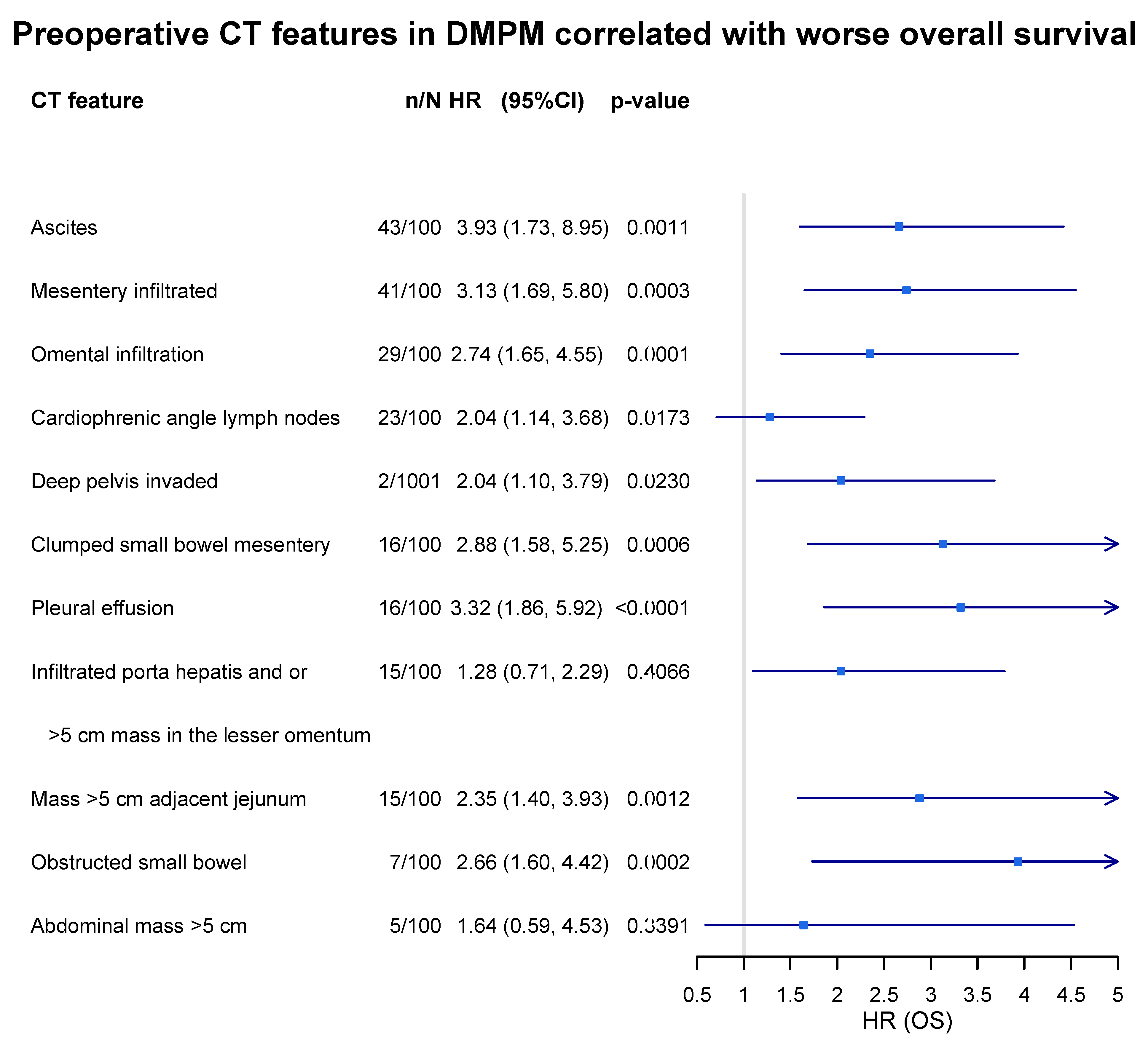
- Sugarbaker, P.H.; Chang, D.; Jelinek, J.S. Concerning CT Features Predict Outcome of Treatment in Patients with Malignant Peritoneal Mesothelioma. Eur. J. Surg. Oncol. 2021, 47, 2212–2219. [Google Scholar] [CrossRef] [PubMed]
- Laterza, B.; Kusamura, S.; Baratti, D.; Oliva, G.D.; Deraco, M. Role of Explorative Laparoscopy to Evaluate Optimal Candidates for Cytoreductive Surgery and Hyperthermic Intraperitoneal Chemotherapy (HIPEC) in Patients with Peritoneal Mesothelioma. In Vivo 2009, 23, 187–190. [Google Scholar] [PubMed]
- Magge, D.; Zenati, M.S.; Austin, F.; Mavanur, A.; Sathaiah, M.; Ramalingam, L.; Jones, H.; Zureikat, A.H.; Holtzman, M.; Ahrendt, S.; et al. Malignant Peritoneal Mesothelioma: Prognostic Factors and Oncologic Outcome Analysis. Ann. Surg. Oncol. 2014, 21, 1159–1165. [Google Scholar] [CrossRef] [PubMed]
- Verma, V.; Sleightholm, R.L.; Rusthoven, C.G.; Koshy, M.; Sher, D.J.; Grover, S.; Simone, C.B. Malignant Peritoneal Mesothelioma: National Practice Patterns, Outcomes, and Predictors of Survival. Ann. Surg. Oncol. 2018, 25, 2018–2026. [Google Scholar] [CrossRef] [PubMed]
- Valmary-Degano, S.; Colpart, P.; Villeneuve, L.; Monnien, F.; M’Hamdi, L.; Lang Averous, G.; Capovilla, M.; Bibeau, F.; Laverriere, M.-H.; Verriele-Beurrier, V.; et al. Immunohistochemical Evaluation of Two Antibodies against PD-L1 and Prognostic Significance of PD-L1 Expression in Epithelioid Peritoneal Malignant Mesothelioma: A RENAPE Study. Eur. J. Surg. Oncol. 2017, 43, 1915–1923. [Google Scholar] [CrossRef] [PubMed]
- Yan, T.D.; Deraco, M.; Elias, D.; Glehen, O.; Levine, E.A.; Moran, B.J.; Morris, D.L.; Chua, T.C.; Piso, P.; Sugarbaker, P.H.; et al. A Novel Tumor-Node-Metastasis (TNM) Staging System of Diffuse Malignant Peritoneal Mesothelioma Using Outcome Analysis of a Multi-Institutional Database*. Cancer 2011, 117, 1855–1863. [Google Scholar] [CrossRef] [PubMed]
- Votanopoulos, K.I.; Sugarbaker, P.; Deraco, M.; Morris, D.; Glehen, O.; Elias, D.; De Simone, M.; Robella, M.; Heyd, B.; Kusamura, S.; et al. Is Cytoreductive Surgery with Hyperthermic Intraperitoneal Chemotherapy Justified for Biphasic Variants of Peritoneal Mesothelioma? Outcomes from the Peritoneal Surface Oncology Group International Registry. Ann. Surg. Oncol. 2018, 25, 667–673. [Google Scholar] [CrossRef] [PubMed]
- Baratti, D.; Kusamura, S.; Cabras, A.D.; Deraco, M. Cytoreductive Surgery with Selective versus Complete Parietal Peritonectomy Followed by Hyperthermic Intraperitoneal Chemotherapy in Patients with Diffuse Malignant Peritoneal Mesothelioma: A Controlled Study. Ann. Surg. Oncol. 2012, 19, 1416–1424. [Google Scholar] [CrossRef] [PubMed]
- Pocard, M.; Reymond, M.A. “Peritoneal Failure”: A New Concept to Explain Negative Results of Randomized Trials Evaluating Intraperitoneal Therapies. Pleura Peritoneum 2020, 5, 20200117. [Google Scholar] [CrossRef] [PubMed]
- Yan, T.D.; Yoo, D.; Sugarbaker, P.H. Significance of Lymph Node Metastasis in Patients with Diffuse Malignant Peritoneal Mesothelioma. Eur. J. Surg. Oncol. 2006, 32, 948–953. [Google Scholar] [CrossRef] [PubMed]
- Baratti, D.; Kusamura, S.; Cabras, A.D.; Laterza, B.; Balestra, M.R.; Deraco, M. Lymph Node Metastases in Diffuse Malignant Peritoneal Mesothelioma. Ann. Surg. Oncol. 2010, 17, 45–53. [Google Scholar] [CrossRef] [PubMed]
- Cazauran, J.-B.; Lasseur, A.; Pasquer, A.; Rousset, P.; Guedj, J.; Passot, G.; Glehen, O. Total Mesenteric Peritonectomy for Peritoneal Metastases (with Video). Ann. Surg. Oncol. 2017, 24, 3988–3989. [Google Scholar] [CrossRef] [PubMed]
- Deraco, M.; Baratti, D.; Kusamura, S.; Laterza, B.; Balestra, M.R. Surgical Technique of Parietal and Visceral Peritonectomy for Peritoneal Surface Malignancies. J. Surg. Oncol. 2009, 100, 321–328. [Google Scholar] [CrossRef] [PubMed]
- Malgras, B.; Gayat, E.; Aoun, O.; Lo Dico, R.; Eveno, C.; Pautrat, K.; Delhorme, J.-B.; Passot, G.; Marchal, F.; Sgarbura, O.; et al. Impact of Combination Chemotherapy in Peritoneal Mesothelioma Hyperthermic Intraperitoneal Chemotherapy (HIPEC): The RENAPE Study. Ann. Surg. Oncol. 2018, 25, 3271–3279. [Google Scholar] [CrossRef] [PubMed]
- Shetty, S.J.; Bathla, L.; Govindarajan, V.; Thomas, P.; Loggie, B.W. Comparison of Cytoreductive Surgery and Hyperthermic Intraperitoneal Chemotherapy with Mitomycin or Carboplatin for Diffuse Malignant Peritoneal Mesothelioma. Am. Surg. 2014, 80, 348–352. [Google Scholar] [CrossRef] [PubMed]
- Sugarbaker, P.H.; Welch, L.S.; Mohamed, F.; Glehen, O. A Review of Peritoneal Mesothelioma at the Washington Cancer Institute. Surg. Oncol. Clin. 2003, 12, 605–621. [Google Scholar] [CrossRef] [PubMed]
- Sugarbaker, P.H.; Chang, D. Long-Term Regional Chemotherapy for Patients with Epithelial Malignant Peritoneal Mesothelioma Results in Improved Survival. Eur. J. Surg. Oncol. 2017, 43, 1228–1235. [Google Scholar] [CrossRef] [PubMed]
- Bijelic, L.; Stuart, O.A.; Sugarbaker, P. Adjuvant Bidirectional Chemotherapy with Intraperitoneal Pemetrexed Combined with Intravenous Cisplatin for Diffuse Malignant Peritoneal Mesothelioma. Gastroenterol. Res. Pract. 2012, 2012, 890450. [Google Scholar] [CrossRef] [PubMed]
- Kepenekian, V.; Péron, J.; You, B.; Bonnefoy, I.; Villeneuve, L.; Alyami, M.; Bakrin, N.; Rousset, P.; Benzerdjeb, N.; Glehen, O. Non-Resectable Malignant Peritoneal Mesothelioma Treated with Pressurized Intraperitoneal Aerosol Chemotherapy (PIPAC) Plus Systemic Chemotherapy Could Lead to Secondary Complete Cytoreductive Surgery: A Cohort Study. Ann. Surg. Oncol. 2022, 29, 2104–2113. [Google Scholar] [CrossRef] [PubMed]
- Noiret, B.; Renaud, F.; Piessen, G.; Eveno, C. Multicystic Peritoneal Mesothelioma: A Systematic Review of the Literature. Pleura Peritoneum 2019, 4, 20190024. [Google Scholar] [CrossRef] [PubMed]
- Nizri, E.; Baratti, D.; Guaglio, M.; Sinukumar, S.; Cabras, A.; Kusamura, S.; Deraco, M. Multicystic Mesothelioma: Operative and Long-Term Outcomes with Cytoreductive Surgery and Hyperthermic Intra Peritoneal Chemotherapy. Eur. J. Surg. Oncol. 2018, 44, 1100–1104. [Google Scholar] [CrossRef] [PubMed]
- Weiss, S.W.; Tavassoli, F.A. Multicystic Mesothelioma. An Analysis of Pathologic Findings and Biologic Behavior in 37 Cases. Am. J. Surg. Pathol. 1988, 12, 737–746. [Google Scholar] [CrossRef] [PubMed]
- Kurisu, Y.; Tsuji, M.; Shibayama, Y.; Yamada, T.; Ohmichi, M. Multicystic Mesothelioma Caused by Endometriosis: 2 Case Reports and Review of the Literature. Int. J. Gynecol. Pathol. 2011, 30, 163–166. [Google Scholar] [CrossRef] [PubMed]
- Safioleas, M.C.; Constantinos, K.; Michael, S.; Konstantinos, G.; Constantinos, S.; Alkiviadis, K. Benign Multicystic Peritoneal Mesothelioma: A Case Report and Review of the Literature. World J. Gastroenterol. 2006, 12, 5739–5742. [Google Scholar] [CrossRef] [PubMed]
- Kannerstein, M.; Churg, J. Peritoneal Mesothelioma. Hum. Pathol. 1977, 8, 83–94. [Google Scholar] [CrossRef] [PubMed]
- Attanoos, R.L.; Gibbs, A.R. Pathology of Malignant Mesothelioma. Histopathology 1997, 30, 403–418. [Google Scholar] [CrossRef] [PubMed]
- Daya, D.; McCaughey, W.T. Well-Differentiated Papillary Mesothelioma of the Peritoneum. A Clinicopathologic Study of 22 Cases. Cancer 1990, 65, 292–296. [Google Scholar] [CrossRef] [PubMed]
- Butnor, K.J.; Sporn, T.A.; Hammar, S.P.; Roggli, V.L. Well-Differentiated Papillary Mesothelioma. Am. J. Surg. Pathol. 2001, 25, 1304–1309. [Google Scholar] [CrossRef] [PubMed]
- Deraco, M.; Nizri, E.; Glehen, O.; Baratti, D.; Tuech, J.-J.; Bereder, J.-M.; Kepenekian, V.; Kusamura, S.; Goere, D. Well Differentiated Papillary Peritoneal Mesothelioma Treated by Cytoreduction and Hyperthermic Intraperitoneal Chemotherapy-the Experience of the PSOGI Registry. Eur. J. Surg. Oncol. 2019, 45, 371–375. [Google Scholar] [CrossRef] [PubMed]


Disclaimer/Publisher’s Note: The statements, opinions and data contained in all publications are solely those of the individual author(s) and contributor(s) and not of MDPI and/or the editor(s). MDPI and/or the editor(s) disclaim responsibility for any injury to people or property resulting from any ideas, methods, instructions or products referred to in the content. |
© 2023 by the authors. Licensee MDPI, Basel, Switzerland. This article is an open access article distributed under the terms and conditions of the Creative Commons Attribution (CC BY) license (https://creativecommons.org/licenses/by/4.0/).
Share and Cite
Kusamura, S.; Baratti, D.; De Simone, M.; Pasqual, E.M.; Ansaloni, L.; Marrelli, D.; Robella, M.; Accarpio, F.; Valle, M.; Scaringi, S.; et al. Diagnostic and Therapeutic Pathway in Diffuse Malignant Peritoneal Mesothelioma. Cancers 2023, 15, 662. https://doi.org/10.3390/cancers15030662
Kusamura S, Baratti D, De Simone M, Pasqual EM, Ansaloni L, Marrelli D, Robella M, Accarpio F, Valle M, Scaringi S, et al. Diagnostic and Therapeutic Pathway in Diffuse Malignant Peritoneal Mesothelioma. Cancers. 2023; 15(3):662. https://doi.org/10.3390/cancers15030662
Chicago/Turabian StyleKusamura, Shigeki, Dario Baratti, Michele De Simone, Enrico Maria Pasqual, Luca Ansaloni, Daniele Marrelli, Manuela Robella, Fabio Accarpio, Mario Valle, Stefano Scaringi, and et al. 2023. "Diagnostic and Therapeutic Pathway in Diffuse Malignant Peritoneal Mesothelioma" Cancers 15, no. 3: 662. https://doi.org/10.3390/cancers15030662
APA StyleKusamura, S., Baratti, D., De Simone, M., Pasqual, E. M., Ansaloni, L., Marrelli, D., Robella, M., Accarpio, F., Valle, M., Scaringi, S., Biacchi, D., Palopoli, C., Gazzanelli, S., Guaglio, M., & Deraco, M. (2023). Diagnostic and Therapeutic Pathway in Diffuse Malignant Peritoneal Mesothelioma. Cancers, 15(3), 662. https://doi.org/10.3390/cancers15030662