Article

Right Ventricular Function in Long-Term Survivors of Childhood Acute Lymphoblastic Leukemia: From the CTOXALL Study

by
Gloria Heredia
1,2,†,
Rafael Gonzalez-Manzanares
1,2,*,†,
Soledad Ojeda
1,2,
Jose R. Molina
2,3,
Consuelo Fernandez-Aviles
1,2,
Francisco Hidalgo
1,2,
Jose Lopez-Aguilera
1,2,
Manuel Crespin
1,2,
Dolores Mesa
1,2,
Manuel Anguita
1,2,
Juan C. Castillo
1,2,‡ and
Manuel Pan
1,2,‡
1
Cardiology Department, Reina Sofia University Hospital, 14004 Cordoba, Spain
2
Maimonides Institute for Research in Biomedicine of Cordoba (IMIBIC), 14004 Cordoba, Spain
3
Hematology Department, Reina Sofia University Hospital, 14004 Cordoba, Spain
*
Author to whom correspondence should be addressed.
These authors contributed equally as first authors to this work.
These authors contributed equally as senior authors to this work.
Cancers 2023, 15(21), 5158; https://doi.org/10.3390/cancers15215158
Submission received: 27 September 2023 / Revised: 20 October 2023 / Accepted: 25 October 2023 / Published: 26 October 2023
(This article belongs to the Special Issue Cardio-Oncology: An Emerging Paradigm in Modern Medicine)

Abstract

:

Simple Summary

Childhood cancer survivors have an increased lifetime risk of cardiac problems due to the cardiotoxic effects of cancer treatments. While most research has focused on the left side of the heart, little is known about the effects of these treatments on the right-side chambers. We used cardiac ultrasound to examine the right ventricle in 90 long-term survivors of childhood leukemia and a control group of 58 healthy siblings. Our findings showed that a noticeable number of survivors had subtle alterations in the pump function of the right ventricle, especially when measured using a novel parameter called a free-wall strain. Alterations in free-wall strain were more common in the survivors who were obese or smoked. This study underscores the importance of incorporating right ventricular strain assessment into routine cardiac surveillance and promoting healthy lifestyle habits among survivors.

Abstract

There are limited data regarding right ventricle (RV) impairment in long-term survivors of childhood acute lymphoblastic leukemia (CLS). The aim of this study was to assess RV function in these patients using echocardiographic conventional measurements and automated RV strain. Echocardiographic recordings of 90 CLS and 58 healthy siblings from the CTOXALL cohort were analyzed. For group comparisons, inverse probability weighting was used to reduce confounding. The CLS group (24.6 ± 9.7 years, 37.8% women) underwent an echocardiographic evaluation 18 (11–26) years after the diagnosis. RV systolic dysfunction was found in 16.7% of CLS individuals using RV free-wall strain (RVFWS) compared to 2.2 to 4.4% with conventional measurements. RV systolic function measurements were lower in the CLS than in the control group: TAPSE (23.3 ± 4.0 vs. 25.2 ± 3.4, p = 0.004) and RVFWS (24.9 ± 4.6 vs. 26.8 ± 4.7, p = 0.032). Modifiable cardiovascular risk factors such as obesity (p = 0.022) and smoking (p = 0.028) were independently associated with reduced RVFWS. In conclusion, RV systolic function impairment was frequent in long-term survivors of childhood leukemia, underscoring the importance of RV assessment, including RVFWS, in the cardiac surveillance of these patients.

1. Introduction

Acute lymphoblastic leukemia (ALL) is the most prevalent type of cancer in the pediatric population. Over the past few decades, anthracycline-based therapies have significantly improved survival rates in these patients [1]. However, due to the exposition to cardiotoxic agents, childhood leukemia survivors (CLS) have an increased risk of cardiovascular disease during their lifetimes [2].
The main mechanism involved in the pathogenesis of anthracycline-related cardiomyocyte injury is the inhibition of topoisomerase 2β, leading to mitochondrial dysfunction, increased reactive oxygen species production, and apoptosis. Other proposed mechanisms are reduced ATP generation, direct damage to the mitochondria, the lipid peroxidation of cell membranes, and changes in calcium and iron homeostasis [3,4,5].
Most echocardiographic surveillance studies have focused on left ventricle evaluation [6,7], with the right ventricle (RV) being less frequently assessed in CLS [8]. Although RV function has been shown to be strongly associated with outcomes in multiple cardiac conditions, its quantification traditionally poses challenges because of the complex geometry and physiology of the RV [9]. Recently, right ventricle strain (RVS) has been proposed as a straightforward and robust measure of right ventricular function [10]. Importantly, RVS is angle-independent and has a stronger correlation with cardiac magnetic resonance-based RV ejection fraction than conventional echocardiographic indices [11].
The aim of this study was to assess RV function in a cohort of long-term CLS through the quantification of automated RVS and conventional parameters.

2. Materials and Methods

2.1. Study Design and Participants

This ancillary study is integrated into the framework of the CTOXALL study, a cross-sectional study that recruited a cohort of long-term childhood cancer survivors who had been diagnosed with ALL between 1985 and 2015 at Reina Sofía Hospital (Córdoba, Spain). The primary objective of the CTOXALL study was to investigate the long-term prevalence of cardiotoxicity in CLS. Previously, we have reported a high prevalence of left ventricular systolic and diastolic abnormalities in this cohort of patients [6,12]. The current study is focused on the assessment of the RV, adding an automated RVS quantification to standard echocardiographic parameters.
The research protocol for the CTOXALL study received approval from the local clinical research ethics committee in accordance with institutional and good clinical practice guidelines. Written informed consent was obtained from all participants, as well as from the parents or legal guardians of minors involved.
Participants underwent evaluation between May 2019 and January 2022. Eligible participants were survivors diagnosed with ALL before the age of 18 and had received the last anthracycline dose at least 3 years before their inclusion in this study. An individual with a congenital heart disease was excluded. A comparison group consisting of healthy siblings of the survivors who were willing to participate was also recruited.

2.2. Clinical Assessment

A comprehensive clinical assessment was performed on all patients. Information related to cardiotoxic treatment exposure, including chemotherapeutic agents and dosage, radiotherapy exposure, and hematopoietic stem-cell transplantation (HSCT), was retrieved from medical records. Cumulative anthracycline dose was expressed as doxorubicin equivalents (mg/m2), which were computed using previously described conversion factors: 0.6× daunorubicin dose, 0.8× epirubicin dose, and 10.5× mitoxantrone [13]. Radiotherapy exposure was considered when it encompassed the heart region, including total body irradiation.

2.3. Echocardiography

Data from conventional echocardiographic parameters were available from the original CTOXALL study. For the present study, a post hoc measurement of RVS was conducted. All examinations were conducted by certified cardiologists using either an EPIQ CVx or iE33 echocardiographic system (Philips Medical Systems, Andover, MA, USA). Standard echocardiographic parameters were acquired in adherence to the most recent guidelines [14,15]. The echocardiographic study included the following right ventricular systolic function measurements: tricuspid annular plane systolic excursion (TAPSE), fractional area change (FAC), doppler tissue imaging tricuspid annular systolic peak-wave velocity (s’DTI), right ventricular free-wall strain (RVFWS), and right ventricular four-chamber strain (RV4CS). Strain parameters were quantified from an RV-focused apical four-chamber view using a semi-automated assessment with AutoStrain (TomTec-Arena, TomTec Imaging Systems, Munich, Germany). Manual adjustment of the software’s automatic endocardial border delineation was permitted. The cardiologists responsible for analyzing the strain parameters were unaware of each participant’s group.
The cut-off values for right ventricular systolic dysfunction (RVSD) were as follows: TAPSE < 17 mm, FAC < 35%. s’DTI < 9.5 cm/s, and RVFWS < 20% (Figure 1) [15,16]. For simplicity, RVS measurements are reported as their absolute values.

2.4. Variability Analysis

To evaluate the intraobserver and interobserver variability in RVFWS, TAPSE, and FAC measurements, the analysis of 20 randomly selected studies was repeated by the same investigator who performed the analysis and another investigator.

2.5. Statistical Analysis

Descriptive data are presented as the count (percentage) for categorical variables and, according to the distribution, as the mean ± standard deviation or median and interquartile range (25th–75th percentiles) for continuous variables. Normal distribution was assessed via the Shapiro–Wilk test and QQ plots. Between-group comparisons of descriptive data were performed via the chi-square test or the Fisher exact test for categorical variables and the Student t-test or the Mann–Whitney U test for continuous variables, as appropriate.
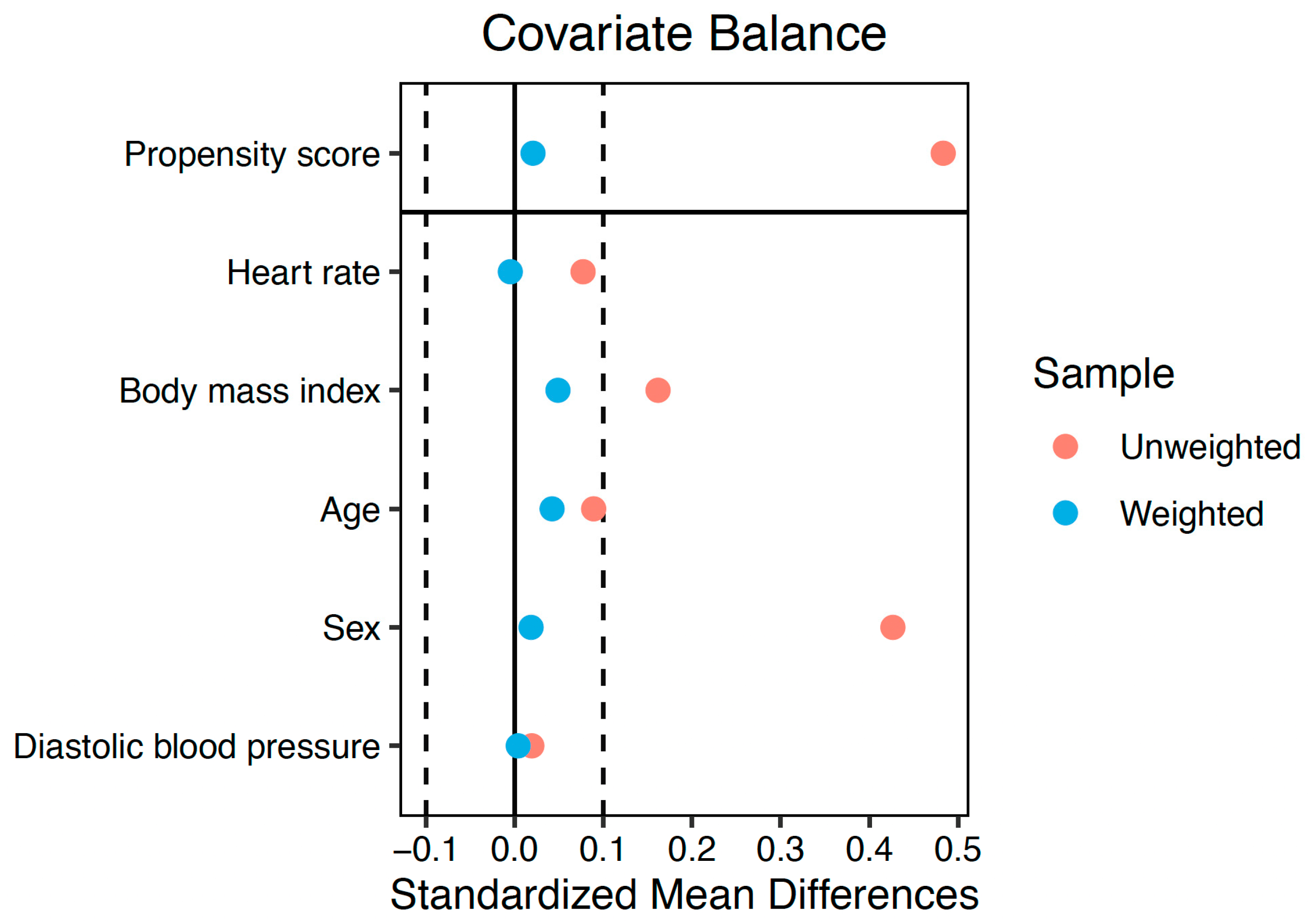
Between-group comparisons of echocardiographic measurements were performed using linear regression models. The inverse probability of treatment weighting (IPW) was used to address confounding [17]. Propensity scores were calculated with a logistic regression model that included the group as the dependent variable and age, sex, body mass index, heart rate, and diastolic blood pressure as covariates. To assess covariate balance, standardized mean differences were computed both before and after applying the weighting. A difference of less than 0.1 was considered indicative of good balance. Standard errors of the IPW linear regression coefficients were obtained using robust sandwich-type variance estimators [18].
Multivariable linear regression analyses were conducted to determine significant predictors for TAPSE, FAC, and RVFWS in the CLS. Covariates included sex, age, age at diagnosis, time since diagnosis, heart rate, systolic and diastolic blood pressure, hypertension, hypercholesterolemia, diabetes mellitus, obesity, smoking, sedentarism, cumulative anthracycline dose, radiotherapy, and HSCT. The models were tested for collinearity and were built using backward stepwise elimination, initially including clinically relevant variables and those with a p < 0.100 in the univariable models.
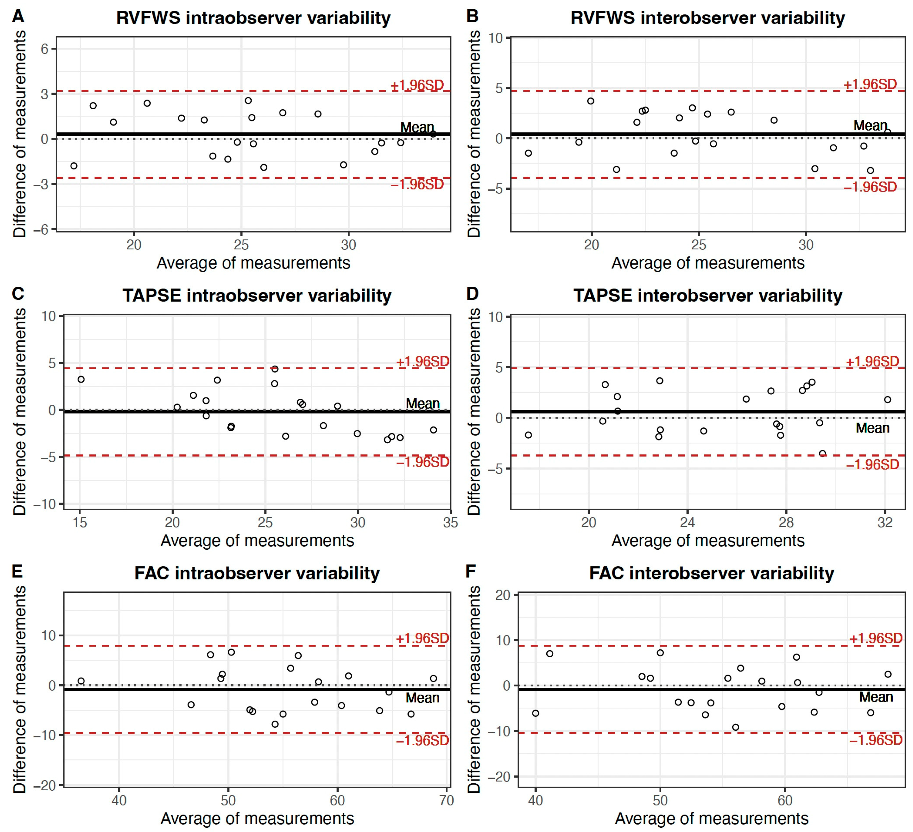
Intraobserver and interobserver variability were assessed through intraclass correlation coefficients and the Bland–Altman method, plotting the differences of 2 measurements (y-axis) against their mean (y-axis) for each subject. The limit of agreement was computed as the mean difference ± 1.96 standard deviations [19].
Statistical analyses were performed using the SPSS software (version 24; IBM Corp., Armonk, NY, USA) and R software (version 4.2.1; R Foundation for Statistical Computing, Vienna, Austria).

3. Results

3.1. Baseline Characteristics

For the present study, all 148 participants from the original CTOXLAL study were analyzed: 90 CLS and a control group comprising 58 healthy siblings. The CLS had a mean age at inclusion of 24.6 ± 9.7, and 34 (37.8%) were female. Table 1 displays the clinical characteristics of the groups. Both groups were similar in terms of age, body measurements, and the prevalence of risk factors. However, the control group had a higher proportion of female participants (56.6%; p = 0.018). Sedentarism was prevalent in both groups (41.1% vs. 34.5%; p = 0.525), as was obesity (10.0% vs. 12.1%, p = 0.901). Smoking was more common in the CLS group (15.6% vs. 1.7%; p = 0.005).

3.2. Cardiotoxic Treatment Exposure

The median age at diagnosis was 4 (3–8) years, and the median time elapsed from diagnosis to recruitment was 18 (11–26) years. All the survivors were exposed to anthracyclines, but only three (3.3%) received radiotherapy. Treatment details are shown in Table 1. According to the treatment risk categories for childhood and adolescent cancer survivors proposed in the European Society of Cardiology Cardio-Oncology guidelines [20], 23 (25.6%), 61 (67.7%), 3 (3.3%), and 3 (3.3%) CLS could be classified as low, moderate, high risk, and very high risk, respectively.

3.3. Prevalence of Right Ventricular Systolic Dysfunction

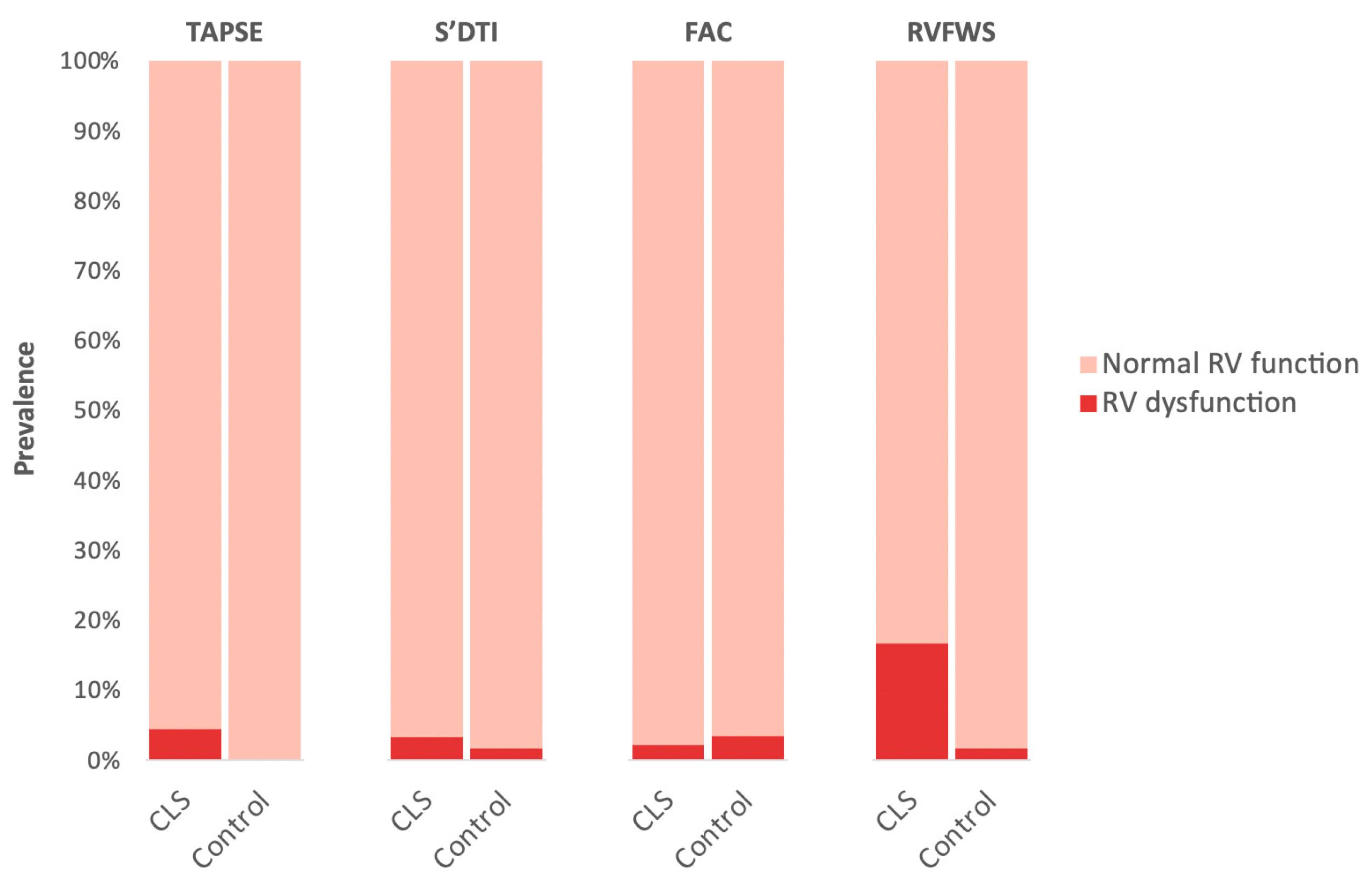
The prevalence of right ventricular systolic dysfunction in CLS was relatively low, ranging from 2.2 to 4.4%, and did not significantly differ from the rates observed in the control group when considering conventional right ventricular systolic parameters (TAPSE, FAC, S’DTI). However, the prevalence of subclinical right ventricular systolic dysfunction, as determined using the RVFWS, was significantly higher in the CLS cohort (16.7% vs. 1.7%, p = 0.005), especially in those with left ventricular systolic dysfunction (n = 11) (36.4% vs. 13.9%, p = 0.082) or subclinical left ventricular systolic dysfunction (n = 24) (37.5% vs. 9.1%, p = 0.001). Detailed prevalence rates for each parameter are presented in Figure 2.

3.4. Comparison of Echocardiographic Parameters between the Groups

On average, the CLS group exhibited lower values for the majority of right ventricular systolic function measurements, including TAPSE, RVFWS, and RV4CS. Conversely, FAC and S’DTI were comparable to the control group. The peak systolic RV-RA gradient was slightly higher in the CLS group. Of note, average right ventricular systolic function parameters were within the normal range in both groups. Right ventricular systolic function echocardiographic parameters, along with additional echocardiographic measurements, are summarized in Table 2. In addition to the unadjusted comparisons, the estimated differences in the IPW population are shown in the table. The covariables included in the propensity score model were well balanced after the weighting, with standardized mean differences <0.1 for all of them (Figure 3). In the IPW-adjusted comparisons, TAPSE, RVFWS, and RV4CS were also decreased compared to the control group. Moreover, CLS RV had lower values of RV end-diastolic area (beta: −1.43 cm2, p = 0.042).

3.5. Predictors of Conventional Right Ventricular Systolic Function Measurements in the Survivors

The results of univariable and multivariable linear regression models for TAPSE are presented in Table 3. In the univariable analysis, female sex, age at examination, time since diagnosis, heart rate, and HSCT history were associated with TAPSE. In the multivariable model, the only factors that were associated with TAPSE were HSCT history (Beta: −2.72, 95% CI: 4.70 to −0.65, p = 0.01) and age at examination (Beta: 0.10, 95% CI: 0.02 to 0.18, p = 0.01).
Univariable and multivariable linear regression models for FAC are shown in Table 4. In the univariable analysis, diabetes mellitus was the only factor associated with FAC (Beta: −9.71, 95% CI: −19.04 to −0.42, p = 0.41). In the multivariable model, which included other clinically relevant variables, this association was not significant (p = 0.74).

3.6. Predictors of Right Ventricular Free-Wall Strain in the Survivors

Univariable and multivariable linear regression models for RVFWS are presented in Table 5. In the univariable analysis, obesity, sedentarism, and smoking habits were associated with RVFWS. In the multivariable model, obesity (Beta: −5.12, 95% CI: −9.41 to −0.76, p = 0.02) and smoking (Beta: −3.17, 95% CI: −5.80 to −0.34, p = 0.02) were independently associated with RVFWS.

3.7. Intraobserver and Interobserver Variability Analysis

The variability analysis revealed a high level of agreement in the measured RV systolic function parameters, with particular robustness observed in RVFWS. The intraclass correlation coefficients for intraobserver agreement were 0.95 (95% CI: 0.89 to 0.98) for RVFWS, 0.89 (95% CI: 0.74 to 0.96) for TAPSE, and 0.85 (95% CI: 0.66 to 0.94) for FAC. For interobserver agreement, the intraclass correlation coefficients values were 0.90 (95% CI: 0.77 to 0.96) for RVFWS, 0.85 (95% CI: 0.67 to 0.94) for TAPSE, and 0.81 (95% CI: 0.58 to 0.92) for FAC. Bland–Altman plots (Figure 4) were consistent with these findings, showing acceptable agreement intervals and no relevant bias.

4. Discussion

In this study, we evaluated RV function in a cohort of long-term survivors of childhood ALL using conventional echocardiographic measurements and RVS. The main findings were as follows: (1) the prevalence of RVSD was notably higher when assessed with RVS compared to conventional parameters; (2) most RV systolic parameters showed significant reductions compared to the control group; (3) modifiable cardiovascular risk factors were frequent and inversely associated with RVS.

4.1. Prevalence of Right Ventricle Dysfunction

While historically overlooked, there has recently been a renewed interest in the study of the RV and its significance in cardiovascular disease [21]. In the context of cardio-oncology, although far less explored than the left ventricle, there is evidence supporting the susceptibility of the RV to anthracycline-induced acute cardiotoxicity [22]. However, scarce data are available regarding RV involvement in long-term survivors of childhood cancer with prior anthracycline exposure.
In the present study, we found a relatively low prevalence of RVSD using conventional 2D echocardiographic parameters, ranging from 2.2 to 4.4%. Nevertheless, when RV function was assessed with RVFWS, the prevalence of RVSD increased to 16.7%. In line with our findings, previous studies have reported a prevalence of RVSD that varied between 6 and 28%, depending on the diagnostic tool, the cut-off values, and the characteristics of the study sample, especially those related to treatment exposure (age, anthracycline dose, and proportion of patients exposed to radiotherapy). Massey et al. found a prevalence of RVSD of 13.6% in a cohort of survivors of HSCT treated in their youth (17.6 ± 9.5 years at HSCT) [23]. The prevalence rate is in line with our findings, and the findings of this study support the observed association between HSCT and TAPSE reduction in the multivariable model. Murbraech et al. reported a prevalence of RVSD of 6.2% in a cohort of adult long-term lymphoma survivors, with a mean age at diagnosis of 42 years [24]. Despite the higher cumulative anthracycline dose and more frequent exposure to radiotherapy, the lower prevalence rate observed in their study could be attributed to the application of stricter criteria for RVSD (requiring impairment in at least two parameters) or, potentially, to the different age profiles of the cohorts, since myocardia have been proposed to be especially vulnerable to anthracycline-induced injury during childhood. On the other hand, Christiansen et al. found a prevalence of RVSD of 28.2% [8]. The underlying reason for this noticeable high prevalence may lie in the more intensive treatment received by the recruited survivors: the cumulative anthracycline dose was 150 (40–485) mg/m2, and 24% of them were exposed to radiotherapy, which appears to exert a particularly cardiotoxic effect on the RV due to its anterior anatomical position [25]. In addition, the lower limits of normal RV function were based on the distribution of the control group values, resulting in more lenient criteria for RV impairment. Consistent with these results, Ylanen et al. reported a RVSD prevalence of 27% in a cohort of childhood cancer survivors using cardiac magnetic resonance (CMR), the gold standard for RV assessment [26]. The median cumulative anthracycline dose was remarkably high, i.e., 222 mg/m2 (80–419 mg/m2), and 11% received radiotherapy. Interestingly, no myocardial fibrosis, as assessed via late gadolinium enhancement, was detected. Although CMR was not available in our study, RVFWS has been previously demonstrated to be strongly correlated with CMR-based RV function [11,27,28].

4.2. Comparison of RV Echocardiographic Parameters between Survivors and Healthy Siblings

The CLS group presented lower values for the majority of right ventricular systolic function parameters, including TAPSE, RVFWS, and RV4CS, even after adjusting for potential confounders by means of IPW models. These findings are similar to those reported in other previously detailed cohorts of long-term survivors that included a control group [8,24]. Consistent with our results, the reported average values, although reduced compared to controls, were within the normal range. These data support the causal effect of anthracyclines on RV function in long-term survivors of childhood cancers, who might present subtle alterations in RV systolic function that may not reach the threshold for a RVSD diagnosis. In this context, RVFWS, capable of detecting RVSD at earlier stages, appears to be of paramount importance [16].

4.3. Association of RV Subclinical Dysfunction with Cardiovascular Risk Factors

In long-term childhood cancer survivors, the presence of modifiable cardiovascular risk factors is frequent and can significantly potentiate the risk of cardiovascular disease associated with cardiotoxic therapies [29]. The reason behind the increased frequency of cardiovascular risk factors is not clearly established, but it appears to be related to psychological factors such as stress and emotional distress [30]. Among participants in the CTOXALL cohort, a considerable prevalence of obesity (10%), sedentary lifestyle (41.1%), and smoking habits (15.6%) was observed. Furthermore, all three variables were associated with RVFWS in the univariable analysis, and obesity and smoking were independently associated with RVFWS in the multivariable model. These findings align with the recommendations outlined in the 2022 European Society of Cardiology Guidelines on cardio-oncology, emphasizing the importance of promoting healthy lifestyle habits and advocating for the annual screening of modifiable cardiovascular risk factors in these patients [20]. Such preventive measures take on particular significance given the observed association between these factors and RVFWS.

4.4. Limitations

The current study has several limitations. Firstly, RV was not evaluated with CMR to complement our findings with tissue characterization. Secondly, the cross-sectional design prevents us from identifying the exact moment of the RVSD onset. Thirdly, the conformation of the control group with healthy siblings resulted in sex and smoking habit differences between the two groups. Smoking was not included in the PS model because it was only present in one participant in the control group. However, sex was well balanced in the weighted population, while the inclusion of siblings as controls may have helped to reduce unmeasured confounding related to demographic, sociocultural, environmental, and genetic factors. Lastly, echocardiographic measurements were not analyzed by a core laboratory team.

5. Conclusions

In this cohort of long-term ALL survivors, right ventricular subclinical dysfunction was present in one out of every six patients. When compared to healthy siblings, most RV systolic parameters showed reductions. Notably, obesity and smoking were associated with greater reductions in RVFWS in the survivors.

Author Contributions

Conceptualization, G.H., R.G.-M. and J.C.C.; data curation, G.H., R.G.-M., J.R.M., C.F.-A. and F.H.; formal analysis, G.H. and R.G.-M.; funding acquisition, S.O., D.M., M.A., J.C.C. and M.P.; investigation, G.H., R.G.-M., J.R.M., C.F.-A. and M.C.; methodology, R.G.-M. and J.L.-A.; resources, S.O., D.M., M.A. and M.P.; supervision, R.G.-M., S.O., J.R.M., J.C.C. and M.P.; writing—original draft, G.H. and R.G.-M.; writing—review and editing, R.G.-M., S.O., J.R.M., F.H., J.L.-A., M.C., D.M., M.A., J.C.C. and M.P. All authors have read and agreed to the published version of the manuscript.

Funding

The CTOXALL study received research grants from Sociedad Española de Cardiología (Madrid, Spain) and Sociedad Andaluza de Cardiología (Granada, Spain) in 2019.

Institutional Review Board Statement

This study was conducted in accordance with the Declaration of Helsinki and approved by the Ethics Committee of Córdoba (Spain) with code 0189-N-19 on 27 March 2019.

Informed Consent Statement

Informed consent was obtained from all subjects involved in this study.

Data Availability Statement

The data presented in this study are available on request from the corresponding author.

Conflicts of Interest

The authors declare no conflict of interest.

References

  1. Greaves, M. A causal mechanism for childhood acute lymphoblastic leukaemia. Nat. Rev. Cancer 2018, 18, 471–484, Correction in Nat. Rev. Cancer 2018, 18, 526. [Google Scholar] [CrossRef] [PubMed]
  2. Armenian, S.H.; Armstrong, G.T.; Aune, G.; Chow, E.J.; Ehrhardt, M.J.; Ky, B.; Moslehi, J.; Mulrooney, D.A.; Nathan, P.C.; Ryan, T.D.; et al. Cardiovascular Disease in Survivors of Childhood Cancer: Insights into Epidemiology, Pathophysiology, and Prevention. J. Clin. Oncol. 2018, 36, 2135–2144. [Google Scholar] [CrossRef] [PubMed]
  3. Zhang, S.; Liu, X.; Bawa-Khalfe, T.; Lu, L.-S.; Lyu, Y.L.; Liu, L.F.; Yeh, E.T.H. Identification of the molecular basis of doxorubicin-induced cardiotoxicity. Nat. Med. 2012, 18, 1639–1642. [Google Scholar] [CrossRef] [PubMed]
  4. Henriksen, P.A. Anthracycline cardiotoxicity: An update on mechanisms, monitoring and prevention. Heart 2018, 104, 971–977. [Google Scholar] [CrossRef]
  5. Bhatia, S. Genetics of Anthracycline Cardiomyopathy in Cancer Survivors: JACC: CardioOncology State-of-the-Art Review. Cardio Oncol. 2020, 2, 539–552. [Google Scholar] [CrossRef]
  6. Gonzalez-Manzanares, R.; Castillo, J.C.; Molina, J.R.; Ruiz-Ortiz, M.; Mesa, D.; Ojeda, S.; Anguita, M.; Pan, M. Automated Global Longitudinal Strain Assessment in Long-Term Survivors of Childhood Acute Lymphoblastic Leukemia. Cancers 2022, 14, 1513. [Google Scholar] [CrossRef]
  7. Bottinor, W.J.; Deng, X.; Bandyopadhyay, D.; Coburn, G.; Havens, C.; Carr, M.; Saurers, D.; Judkins, C.; Gong, W.; Yu, C.; et al. Myocardial Strain during Surveillance Screening Is Associated with Future Cardiac Dysfunction among Survivors of Childhood, Adolescent and Young Adult-Onset Cancer. Cancers 2023, 15, 2349. [Google Scholar] [CrossRef]
  8. Christiansen, J.R.; Massey, R.; Dalen, H.; Kanellopoulos, A.; Hamre, H.; Ruud, E.; Kiserud, C.E.; Fosså, S.D.; Aakhus, S. Right ventricular function in long-term adult survivors of childhood lymphoma and acute lymphoblastic leukaemia. Eur. Heart J. Cardiovasc. Imaging 2016, 17, 735–741. [Google Scholar] [CrossRef]
  9. Edward, J.; Banchs, J.; Parker, H.; Cornwell, W. Right ventricular function across the spectrum of health and disease. Heart 2023, 109, 349–355. [Google Scholar] [CrossRef]
  10. Anastasiou, V.; Papazoglou, A.S.; Moysidis, D.V.; Daios, S.; Tsalikakis, D.; Giannakoulas, G.; Karamitsos, T.; Delgado, V.; Ziakas, A.; Kamperidis, V. The prognostic value of right ventricular longitudinal strain in heart failure: A systematic review and meta-analysis. Heart Fail. Rev. 2023, 28, 1383–1394. [Google Scholar] [CrossRef]
  11. Focardi, M.; Cameli, M.; Carbone, S.F.; Massoni, A.; De Vito, R.; Lisi, M.; Mondillo, S. Traditional and innovative echocardiographic parameters for the analysis of right ventricular performance in comparison with cardiac magnetic resonance. Eur. Heart J. Cardiovasc. Imaging 2015, 16, 47–52. [Google Scholar] [CrossRef] [PubMed]
  12. Fernández-Avilés, C.; González-Manzanares, R.; Ojeda, S.; Molina, J.R.; Heredia, G.; Resúa, A.; Hidalgo, F.; López-Aguilera, J.; Mesa, D.; Anguita, M.; et al. Diastolic function assessment with left atrial strain in long-term survivors of childhood acute lymphoblastic leukemia. Rev. Española De Cardiol. 2023; ahead of print. [Google Scholar] [CrossRef]
  13. Feijen, E.A.M.; Leisenring, W.M.; Stratton, K.L.; Ness, K.K.; van der Pal, H.J.H.; van Dalen, E.C.; Armstrong, G.T.; Aune, G.J.; Green, D.M.; Hudson, M.M.; et al. Derivation of Anthracycline and Anthraquinone Equivalence Ratios to Doxorubicin for Late-Onset Cardiotoxicity. JAMA Oncol. 2019, 5, 864–871. [Google Scholar] [CrossRef] [PubMed]
  14. Mitchell, C.; Rahko, P.S.; Blauwet, L.A.; Canaday, B.; Finstuen, J.A.; Foster, M.C.; Horton, K.; Ogunyankin, K.O.; Palma, R.A.; Velazquez, E.J. Guidelines for Performing a Comprehensive Transthoracic Echocardiographic Examination in Adults: Recommendations from the American Society of Echocardiography. J. Am. Soc. Echocardiogr. 2019, 32, 1–64. [Google Scholar] [CrossRef] [PubMed]
  15. Lang, R.M.; Badano, L.P.; Mor-Avi, V.; Afilalo, J.; Armstrong, A.; Ernande, L.; Flachskampf, F.A.; Foster, E.; Goldstein, S.A.; Kuznetsova, T.; et al. Recommendations for Cardiac Chamber Quantification by Echocardiography in Adults: An Update from the American Society of Echocardiography and the European Association of Cardiovascular Imaging. J. Am. Soc. Echocardiogr. 2015, 28, 1–39.e14. [Google Scholar] [CrossRef]
  16. Muraru, D.; Haugaa, K.; Donal, E.; Stankovic, I.; Voigt, J.U.; E Petersen, S.; A Popescu, B.; Marwick, T. Right ventricular longitudinal strain in the clinical routine: A state-of-the-art review. Eur. Heart J. Cardiovasc. Imaging 2022, 23, 898–912. [Google Scholar] [CrossRef]
  17. Austin, P.C.; Stuart, E.A. Moving towards best practice when using inverse probability of treatment weighting (IPTW) using the propensity score to estimate causal treatment effects in observational studies. Stat. Med. 2015, 34, 3661–3679. [Google Scholar] [CrossRef]
  18. Austin, P.C. Variance estimation when using inverse probability of treatment weighting (IPTW) with survival analysis. Stat. Med. 2016, 35, 5642–5655. [Google Scholar] [CrossRef]
  19. Giavarina, D. Understanding Bland Altman analysis. Biochem. Med. 2015, 25, 141–151. [Google Scholar] [CrossRef]
  20. Lyon, A.R.; López-Fernández, T.; Couch, L.S.; Asteggiano, R.; Aznar, M.C.; Bergler-Klein, J.; Boriani, G.; Cardinale, D.; Cordoba, R.; Cosyns, B.; et al. 2022 ESC Guidelines on cardio-oncology developed in collaboration with the European Hematology Association (EHA), the European Society for Therapeutic Radiology and Oncology (ESTRO) and the International Cardio-Oncology Society (IC-OS). Eur. Heart J. 2022, 43, 4229–4361, Correction in Eur. Heart J. 2023, 44, 1621. [Google Scholar] [CrossRef]
  21. Amsallem, M.; Mercier, O.; Kobayashi, Y.; Moneghetti, K.; Haddad, F. Forgotten No More: A Focused Update on the Right Ventricle in Cardiovascular Disease. JACC Heart Fail. 2018, 6, 891–903. [Google Scholar] [CrossRef]
  22. Zhao, R.; Shu, F.; Zhang, C.; Song, F.; Xu, Y.; Guo, Y.; Xue, K.; Lin, J.; Shu, X.; Hsi, D.H.; et al. Early Detection and Prediction of Anthracycline-Induced Right Ventricular Cardiotoxicity by 3-Dimensional Echocardiography. JACC CardioOncology 2020, 2, 13–22. [Google Scholar] [CrossRef] [PubMed]
  23. Massey, R.J.; Diep, P.P.; Burman, M.M.; Kvaslerud, A.B.; Brinch, L.; Aakhus, S.; Gullestad, L.; Ruud, E.; Beitnes, J.O. Impaired right ventricular function in long-term survivors of allogeneic haematopoietic stem-cell transplantation. Open Heart 2021, 8, e001768. [Google Scholar] [CrossRef]
  24. Murbraech, K.; Holte, E.; Broch, K.; Smeland, K.B.; Holte, H.; Rösner, A.; Lund, M.B.; Dalen, H.; Kiserud, C.; Aakhus, S. Impaired Right Ventricular Function in Long-Term Lymphoma Survivors. J. Am. Soc. Echocardiogr. 2016, 29, 528–536. [Google Scholar] [CrossRef]
  25. Tadic, M.; Kersten, J.; Buckert, D.; Rottbauer, W.; Cuspidi, C. Right Ventricle and Radiotherapy: More Questions than Answers. Diagnostics 2023, 13, 164. [Google Scholar] [CrossRef]
  26. Ylänen, K.; Poutanen, T.; Savikurki-Heikkilä, P.; Rinta-Kiikka, I.; Eerola, A.; Vettenranta, K. Cardiac Magnetic Resonance Imaging in the Evaluation of the Late Effects of Anthracyclines Among Long-Term Survivors of Childhood Cancer. J. Am. Coll. Cardiol. 2013, 61, 1539–1547. [Google Scholar] [CrossRef]
  27. Keramida, K.; Farmakis, D. Right ventricular involvement in cancer therapy–related cardiotoxicity: The emerging role of strain echocardiography. Heart Fail. Rev. 2021, 26, 1189–1193. [Google Scholar] [CrossRef] [PubMed]
  28. Sławiński, G.; Hawryszko, M.; Liżewska-Springer, A.; Nabiałek-Trojanowska, I.; Lewicka, E. Global Longitudinal Strain in Cardio-Oncology: A Review. Cancers 2023, 15, 986. [Google Scholar] [CrossRef] [PubMed]
  29. Armstrong, G.T.; Oeffinger, K.C.; Chen, Y.; Kawashima, T.; Yasui, Y.; Leisenring, W.; Stovall, M.; Chow, E.J.; Sklar, C.A.; Mulrooney, D.A.; et al. Modifiable Risk Factors and Major Cardiac Events Among Adult Survivors of Childhood Cancer. J. Clin. Oncol. 2013, 31, 3673–3680. [Google Scholar] [CrossRef]
  30. Lubas, M.M.; Wang, M.; Jefferies, J.L.; Ness, K.K.; Ehrhardt, M.J.; Krull, K.R.; Mulrooney, D.A.; Srivastava, D.K.; Howell, R.M.; Robison, L.L.; et al. The Contribution of Stress and Distress to Cardiovascular Health in Adult Survivors of Childhood Cancer. Cancer Epidemiol. Biomark. Prev. 2021, 30, 286–294. [Google Scholar] [CrossRef]
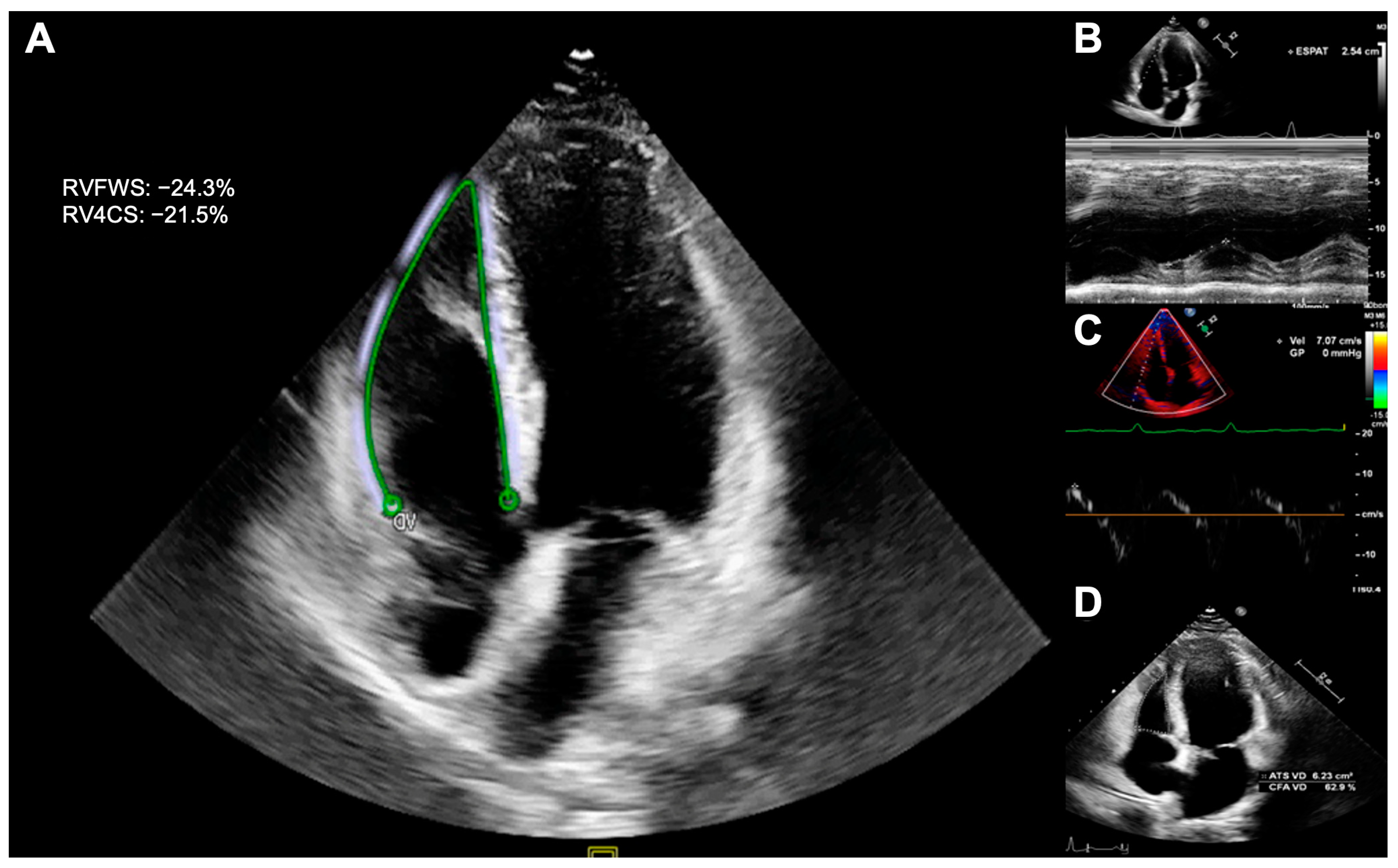
Figure 1. Echocardiographic assessment of right ventricular systolic function. Right ventricular strain (A), tricuspid annular plane systolic excursion (B), s’DTI (C), and fractional area change (D).
Cancers 15 05158 g001
Figure 2. Prevalence of right ventricular systolic dysfunction. Cut-off points: TAPSE < 17 mm, FAC < 35%, s’DTI < 9.5 cm/s, and RVFWS < 20%. FAC: fractional area change. RVFWS: right ventricular free-wall strain; TAPSE: tricuspid annular plane systolic excursion.
Cancers 15 05158 g002
Figure 3. Covariate balance of the covariables included in the propensity score model. In the weighted pseudo-population (blue), all the variables showed an excellent balance with standardized mean differences <0.1.
Cancers 15 05158 g003
Figure 4. Bland–Altman plots: RVFWS intraobserver variability (A); RVFWS interobserver agreement (B); TAPSE intraobserver agreement (C); TAPSE interobserver agreement (D); FAC intraobserver agreement (E); FAC interobserver agreement (F). FAC: fractional area change. RVFWS: right ventricular free-wall strain; TAPSE: tricuspid annular plane systolic excursion.
Cancers 15 05158 g004
Table 1. Characteristics of childhood leukemia survivors and controls.
CLSs
(n = 90)
Control Group
(n = 58)
p-Value
Clinical characteristics
Age at diagnosis (years)4 (3–7)--
Age at exam (years)24.6 ± 9.723.6 ± 10.80.593
Time since diagnosis (years)18 (11–26)--
Sex (% female)34 (37.8%)34 (58.6%)0.018
Weight (kg)64.8 ± 18.361.9 ± 17.20.333
Height (cm)165.6 ± 13.3164.2 ± 13.80.539
Body mass index (kg/m2)23.3 ± 5.122.6 ± 4.40.346
Body surface area (m2)1.7 ± 0.31.7 ± 0.30.366
Systolic blood pressure (mmHg)116.2 ± 11.3115.6 ± 11.10.768
Diastolic blood pressure (mmHg)69.6 ± 7.969.4 ± 7.90.908
Heart rate (bpm)72.5 ± 11.171.7 ± 11.50.646
Current smoker (%)14 (15.6%)1 (1.7%)0.005
Hypertension (%)3 (3.3%)0 (0.0%)0.280
Hypercholesterolemia (%)12 (13.3%)4 (6.9%)0.283
Diabetes mellitus (%)4 (4.4%)1 (1.7%)0.649
Obesity9 (10.0%)7 (12.1%)0.901
Sedentarism (%)37 (41.1%)20 (34.5%)0.525
Cardiotoxic therapies
Anthracycline dose (mg/m2)138 (72–192)--
Radiotherapy (%)3 (3.3%)--
ESC guidelines risk category
Low risk23 (25.6%)
Moderate risk61 (67.7%)
High risk3 (3.3%)
Very high risk3 (3.3%)
ALL: acute lymphoblastic leukemia; CLSs: childhood acute lymphoblastic leukemia survivors. ESC: The European Society of Cardiology. HSCT: hematopoietic stem cell transplantation.
Table 2. Echocardiographic parameters of childhood leukemia survivors and controls.
CLSs
(n = 90)
Control Group
(n = 58)
p-ValueIPW Beta (RSE)PIPW-Adjusted
Right ventricle
  End-diastolic area (cm2)16.5 ± 4.017.4 ± 3.90.185−1.43 (0.69)0.042
  End-systolic area (cm2)8.1 ± 3.18.7 ± 2.90.277−0.94 (0.52)0.071
  Fractional area change (%)52.0 ± 9.351.2 ± 6.90.5701.45 (1.38)0.293
  TAPSE (mm)23.3 ± 4.025.2 ± 3.40.0042.08 (0.64)0.001
  S’ DTI (cm/s)13.8 ± 2.414.4 ± 2.60.1960.73 (0.49)0.136
  RVFWS (-%)24.9 ± 4.626.8 ± 4.70.0321.92 (0.85)0.025
  RV4CS (-%)21.7 ± 3.323.1 ± 3.40.0171.57 (0.64)0.016
  RV-RA gradient (mmHg)19.2 ± 4.916.1 ± 3.90.0243.15 (1.39)0.027
Left ventricle
  LVDD (mm)45.6 ± 6.844.9 ± 6.10.5950.09 (1.14)0.935
  LVSD (mm)28.9 ± 6.026.2 ± 4.60.0052.27 (0.92)0.015
  IVS (mm)7.6 ± 1.57.7 ± 1.30.8240.17 (0.23)0.457
  LVEF 2D (%)56.2 ± 5.862.4 ± 5.5<0.0015.45 (0.95)<0.001
  GLS (-%)20.4 ± 2.822.9 ± 2.3<0.0012.28 (0.45)<0.001
CLSs: childhood acute lymphoblastic leukemia survivors; IPW: inverse probability weighting; RSE: robust standard error; Gradient RV-RA: pressure gradient between the right ventricle and the right atrium; TAPSE: tricuspid annular plane systolic excursion; S’ DTI: tissue Doppler-derived right ventricular systolic excursion velocity S’. RVFWS: right ventricular free-wall strain; RV4CS: right ventricular four-chamber strain; LVDD: left ventricular diastolic diameter; LVSD: left ventricular systolic diameter; IVS: interventricular septum; LVEF: left ventricular ejection fraction; GLS: global longitudinal strain.
Table 3. Univariable and multivariable regression models for TAPSE in the survivors.
UnivariableMultivariable
Beta95% CIp-ValueBeta95% CIp-Value
Sex (female)1.760.05 to 3.480.0441.41−0.28 to 3.030.102
Age at diagnosis (years)0.02−0.16 to 0.210.815
Age at exam (years)0.090.00 to 0.170.0420.100.02 to 0.180.017
Time since diagnosis (years)0.100.01 to 0.190.032
HR (bpm)−0.08−0.157 to −0.010.029
SBP (mmHg)−0.02−0.09 to 0.060.634
DBP (mmHg)0.01−0.09 to 0.120.808
Hypertension 3.36−1.27 to 7.990.153
Hypercholesterolemia0.65−1.82 to 3.130.602
Diabetes mellitus−0.73−4.81 to 3.350.723
Obesity1.04−1.76 to 3.840.462
Sedentarism−0.08−1.80 to 1.650.928
Current smoker−0.93−3.25 to 1.380.425
Anthracycline dose−0.01−0.02 to 0.010.239
Radiotherapy−4.02−8.63 to 0.580.086
HSCT−2.42−4.52 to −0.330.024−2.72−4.70 to −0.650.010
CI: confidence interval, HR: heart rate, SBP: systolic blood pressure, DBP: diastolic blood pressure, HSCT: hematopoietic stem-cell transplantation.
Table 4. Univariable and multivariable regression models for FAC in the survivors.
UnivariableMultivariable
Beta95% CIp-ValueBeta95% CIp-Value
Sex (female)−0.43−4.47 to 3.620.835−0.38−4.66 to 3.890.856
Age at diagnosis (years)0.05−0.38 to 0.4830.811
Age at exam (years)−0.02−0.23 to 0.1780.8180.01−0.22 to 0.230.961
Time since diagnosis (years)−0.05−0.27 to 0.1800.690
HR (bpm)0.04−0.14 to 0.2190.645
SBP (mmHg)0.10−0.07 to 0.2730.254
DBP (mmHg)−0.05−0.29 to 0.1970.687
Hypertension −8.70−19.51 to 2.080.112
Hypercholesterolemia−0.86−6.63 to 4.910.768
Diabetes mellitus−9.71−19.04 to −0.420.041−9.34−20.35 to 0.930.074
Obesity−3.70−10.2 to 2.790.261−2.57−9.51 to 4.490.470
Sedentarism0.04−3.95 to 4.030.984
Current smoker3.51−1.85 to 8.870.1973.28−2.54 to 8.920.264
Anthracycline dose0.01−0.02 to 0.040.3780.02−0.01 to 0.050.248
Radiotherapy−3.78−14.72 to 7.120.493−2.12−14.12 to 9.430.723
HSCT−2.79−7.77 to 2.180.267
CI: confidence interval, HR: heart rate, SBP: systolic blood pressure, DBP: diastolic blood pressure, HSCT: hematopoietic stem-cell transplantation.
Table 5. Univariable and multivariable regression models for RVFWS in the survivors.
UnivariableMultivariable
Beta95% CIp-ValueBeta95% CIp-Value
Sex (female)−0.13−2.23 to 1.970.9000.40−1.70 to 2.500.703
Age at diagnosis (years)−0.05−0.29 to 0.200.710
Age at exam (years)−0.07−0.17 to 0.040.2230.01−0.10 to 0.120.831
Time since diagnosis (years)−0.07−0.19 to 0.050.257
HR (bpm)−0.04−0.13 to 0.050.363
SBP (mmHg)0.00−0.09 to 0.090.940
DBP (mmHg)0.09−0.05 to 0.220.196
Hypertension −1.18−7.76 to 5.400.722
Hypercholesterolemia−1.16−4.13 to 1.820.441
Diabetes mellitus1.39−5.19 to 7.960.676
Obesity−5.09−9.18 to −1.000.015−5.12−9.41 to −0.760.022
Sedentarism−2.46−4.46 to −0.450.017
Current smoker−3.25−5.85 to −0.640.015−3.17−5.80 to −0.340.028
Anthracycline dose−0.01−0.02 to 0.010.527−0.01−0.02 to 0.010.312
Radiotherapy−2.70−8.07 to 2.680.321−3.01−8.32 to 2.330.264
HSCT−1.73−4.26 to 0.810.179
CI: confidence interval, HR: heart rate, SBP: systolic blood pressure, DBP: diastolic blood pressure, HSCT: hematopoietic stem-cell transplantation.
Disclaimer/Publisher’s Note: The statements, opinions and data contained in all publications are solely those of the individual author(s) and contributor(s) and not of MDPI and/or the editor(s). MDPI and/or the editor(s) disclaim responsibility for any injury to people or property resulting from any ideas, methods, instructions or products referred to in the content.