Article

Cytoreduction of Residual Tumor Burden Is Decisive for Prolonged Survival in Patients with Recurrent Brain Metastases—Retrospective Analysis of 219 Patients

1
Department of Neurosurgery, School of Medicine, Klinikum Rechts der Isar, Technical University Munich, 81675 Munich, Germany
2
Department of Neuroradiology, School of Medicine, Klinikum Rechts der Isar, Technical University Munich, 81675 Munich, Germany
3
Department of Radiation Oncology, School of Medicine, Klinikum Rechts der Isar, Technical University Munich, 81675 Munich, Germany
4
German Cancer Consortium (DKTK), Partner Site Munich, 80336 Munich, Germany
5
Department of Radiation Sciences (DRS) Helmholtz Zentrum Munich, Institute of Innovative Radiotherapy (iRT), 81675 Munich, Germany
6
Department of Neuropathology, Institute of Pathology, School of Medicine, Klinikum Rechts der Isar, Technical University Munich, 81675 Munich, Germany
7
Department of Neurosurgery, University Medical Center Hamburg-Eppendorf, 20251 Hamburg, Germany
*
Author to whom correspondence should be addressed.
Cancers 2023, 15(20), 5067; https://doi.org/10.3390/cancers15205067
Submission received: 16 September 2023 / Revised: 10 October 2023 / Accepted: 18 October 2023 / Published: 20 October 2023
(This article belongs to the Special Issue Advances in Research, Diagnosis and Treatment of Brain Metastases)

Abstract

:

Simple Summary

Appropriate therapies for brain metastases are still lacking and need further research, especially in terms of recurrent brain metastases. This is why we decided to highlight the importance of complete cytoreduction by addressing the impact of postoperative tumor burden as displayed by postoperative MRI. Our findings showed that residual tumor burden is a strong prognostic factor for survival in patients with recurrent brain metastases; operated patients showed longer survival independent of age and systemic progression.

Abstract

Background: Despite advances in treatment for brain metastases (BMs), the prognosis for recurrent BMs remains poor and requires further research to advance clinical management and improve patient outcomes. In particular, data addressing the impact of tumor volume and surgical resection with regard to survival remain scarce. Methods: Adult patients with recurrent BMs between December 2007 and December 2022 were analyzed. A distinction was made between operated and non-operated patients, and the residual tumor burden (RTB) was determined by using (postoperative) MRI. Survival analysis was performed and RTB cutoff values were calculated using maximally selected log-rank statistics. In addition, further analyses on systemic tumor progression and (postoperative) tumor therapy were conducted. Results: In total, 219 patients were included in the analysis. Median age was 60 years (IQR 52–69). Median preoperative tumor burden was 2.4 cm3 (IQR 0.8–8.3), and postoperative tumor burden was 0.5 cm3 (IQR 0.0–2.9). A total of 95 patients (43.4%) underwent surgery, and complete cytoreduction was achieved in 55 (25.1%) patients. Median overall survival was 6 months (IQR 2–10). Cutoff RTB in all patients was 0.12 cm3, showing a significant difference (p = 0.00029) in overall survival (OS). Multivariate analysis showed preoperative KPSS (HR 0.983, 95% CI, 0.967–0.997, p = 0.015), postoperative tumor burden (HR 1.03, 95% CI 1.008–1.053, p = 0.007), and complete vs. incomplete resection (HR 0.629, 95% CI 0.420–0.941, p = 0.024) as significant. Longer survival was significantly associated with surgery for recurrent BMs (p = 0.00097), and additional analysis demonstrated the significant effect of complete resection on survival (p = 0.0027). In the subgroup of patients with systemic progression, a cutoff RTB of 0.97 cm3 (p = 0.00068) was found; patients who had received surgery also showed prolonged OS (p = 0.036). Single systemic therapy (p = 0.048) and the combination of radiotherapy and systemic therapy had a significant influence on survival (p = 0.036). Conclusions: RTB is a strong prognostic factor for survival in patients with recurrent BMs. Operated patients with recurrent BMs showed longer survival independent of systemic progression. Maximal cytoreduction should be targeted to achieve better long-term outcomes.

1. Introduction

The prognosis for patients with brain metastases (BMs) has improved due to enhanced medical care and new treatment options in a multidisciplinary setting [1,2,3]. In this context, the role of surgical intervention of primary BMs has already been described [4,5,6]. Nevertheless, surgical resection increasingly fails to achieve local control [7,8,9]. Improved prognosis on the one hand and lack of intracranial tumor control on the other have contributed to the growing incidence of recurrent BMs. Through ongoing research, a variety of treatment modalities such as systemic therapy and radiotherapy have been established [10,11,12,13]. However, the treatment of recurrent brain metastases remains challenging due to sparse data on surgical resection. Previous studies have only suggested that surgery can improve outcomes [14,15,16], but there are little objective data on the impact of residual tumor volume on survival, an aspect currently only described for primary BMs [17,18].
The association between residual tumor presence, as evidenced by using postoperative magnetic resonance imaging (MRI), and an increased risk of intracranial progression is recognized [8,19]; nevertheless, early postoperative MRI imaging has not yet been established as a neurosurgical standard in the management of BMs [20].
One of the objectives of this work is to investigate the significance of residual tumor volume/burden (RTB) as assessed by using MRI imaging and its impact on survival in patients with recurrent BMs.
In addition, it was analyzed whether surgical resection in a multidisciplinary approach for the treatment of recurrent BMs provides benefits to patient outcomes and overall survival (OS).

2. Materials and Methods

2.1. Patient Population and Data Collection

A total of 231 patients with newly diagnosed recurrent BMs presented to the neurosurgical department of the Technical University of Munich between December 2007 and December 2022, and 95 (43.4%) of these cases underwent surgical treatment. A total of 219 patients fulfilled the inclusion criteria of histopathological diagnosis of a recurrent BM and MRI imaging. Twelve patients did not meet the inclusion criteria and did not receive (postoperative) MRI. A distinction was made between 124 non-operated patients (56.6%) and 95 operated patients (43.4%). The operated patient cohort was divided into the following categories: (a) Resection of recurrent BMs but with residual tumor elsewhere in the brain. (b) Resection with no residual tumor left in the brain. Patients’ medical records, including age at diagnosis, sex, tumor location, number of BMs, date of surgery, preoperative and postoperative Karnofsky Performance Status (KPS), preoperative and postoperative tumor burden, date of death, or date of last contact (in living patients), were evaluated. Data from (postoperative) systemic therapy and radiotherapy were registered and analyzed as well.

2.2. Surgery

The surgical approach aimed to achieve extensive tumor removal with a focus on protecting the eloquent areas of the brain. The consistent use of intraoperative neuronavigation was the standard procedure. When required, neuromonitoring and fiber tracking were used. The decision of surgical intervention was made by the multidisciplinary neuro-oncology board. Guiding factors for these decisions included (1) symptomatic lesions, (2) the presence of mass effects, (3) intratumoral hemorrhage, (4) cases with an unclear diagnosis, and (5) larger posterior fossa tumors with a potential risk of herniation and hydrocephalus.

2.3. Residual Tumor Burden

Volumetric measurements were obtained through early (postoperative) MRIs (within 72 h), specifically employing T1-weighted sequences with gadolinium contrast media, to identify the volume of remaining tumor remnants. An experienced neuroradiologist (BW, 11 years of experience) and neurosurgeon (AA, 7 years of experience) performed volumetric measurements. The volumes of the contrast-enhancing tumor parts were manually segmented using the Origin® software (Origin®, Brainlab, version 3.1, Brainlab AG, Munich, Germany).

2.4. Statistical Analysis

Statistical analyses were performed using R version 4.1.1 (© The R Foundation, https://www.r-project.org/, accessed on 17 October 2023). Logistic regression analyses were used to detect potential risk factors for outcome changes. A difference with a probability of error less than 0.05 was considered statistically significant. Descriptive statistics with means and standard deviations or medians with interquartile ranges were obtained for demographic variables. Survival analyses were performed using Kaplan–Meier estimates for univariate analysis and the Cox regression proportional hazards model for multivariate analysis. To identify the optimal cutoff for survival curve differences, the maximally selected log-rank statistics were determined. Survival curves separated by the resulting cutoff were then compared.

2.5. Standard Protocol Approvals, Registrations, and Patient Consent

This study was approved by the local ethics committee (no. 5626:12) and was conducted in alignment with the ethical standards of Helsinki and later amendments [21]. The ethics committee waived the requirement for written consent.

3. Results

3.1. Patient Population

A total of 219 patients were included. The median age at diagnosis of recurrent BMs was 60.0 years (IQR 52–69). Out of the 219 patients, 121 (55.3%) were female and 98 were (44.7%) male. Median pre- and postoperative KPS was 80 (IQR 70–80). A total of 82/219 (37.4%) patients presented with a single BM, 54/219 (24.7%) with two, 37/219 (16.9%) with three, and 46/219 (21.0%) with more than three recurrent BMs. In total, 45/219 (20.5%) patients did not receive any further adjuvant therapy, 74/219 (33.8%) underwent (postoperative) radiotherapy, 20/219 (9.2%) underwent systemic therapy, and 80/219 (36.5%) received both.

3.2. Survival Analysis and the Influence of Residual Tumor Volume

The mean preoperative tumor burden was 7.43 cm3 (SD 14.92), and postoperative tumor burden (RTB) was 3.45 cm3 (SD 9.159) (Table 1). A total of 124/219 (56.6%) patients were not operated on. A total of 40/219 (18.3%) patients had one or more BMs resected, but there was residual tumor in one or more sites. Complete cytoreduction was achieved in 55/219 (25.1%) patients. In 27/40 (67.5%) patients who underwent surgery, the targeted metastasis was completely resected, but residual tumor was still present at other sites in the brain. A total of 56/95 (59.0%) patients with a single recurrent BM underwent surgery, 19/95 (20.0%) with two, 10/95 (10.5%) with three, and 10/95 (10.5%) with more than three BMs. Complete resection was achieved in 48/55 (87.3%) cases with one BM, in 6/55 (10.9%) with two, and in 1/55 (1.8%) with three BMs (Table 2).
The mean postoperative tumor volume of the targeted BM was 0.34 cm3 (SD 1.91; range 0.00–15.60 cm3). Median overall survival was 6 months (IQR 2–10) (Figure 1A). A significant cutoff value for an RTB of 0.12 cm3 (p = 0.00029) was found by using maximally selected log-rank statistics for all patients, regardless of the number of BMs (Figure 1B).
Multivariate analysis showed preoperative KPSS (HR 0.983, 95% CI, 0.967–0.997, p = 0.015), postoperative tumor burden in cm3 (HR 1.03, 95% CI 1.008–1.053, p = 0.007), and complete vs. incomplete resection (HR 0.629, 95% CI 0.420–0.941, p = 0.024) to be significant (Table 3).
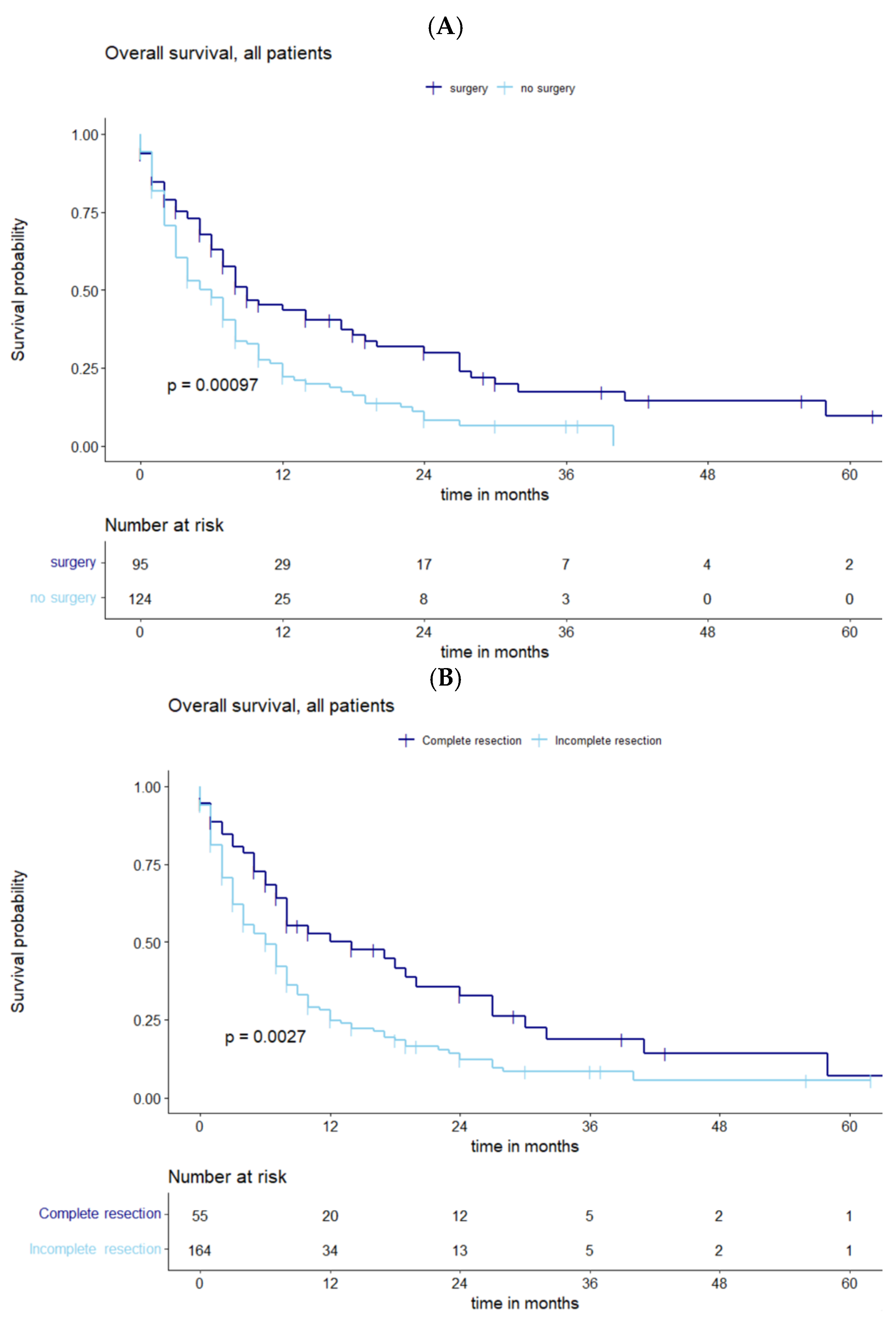
Comparing operated and non-operated patient collectives, longer survival was significantly associated with surgery for recurrent BMs (p = 0.00097) (Figure 2A). Additional analysis showed the significant impact of complete vs. incomplete resection on OS (p = 0.0027) (Figure 2B).
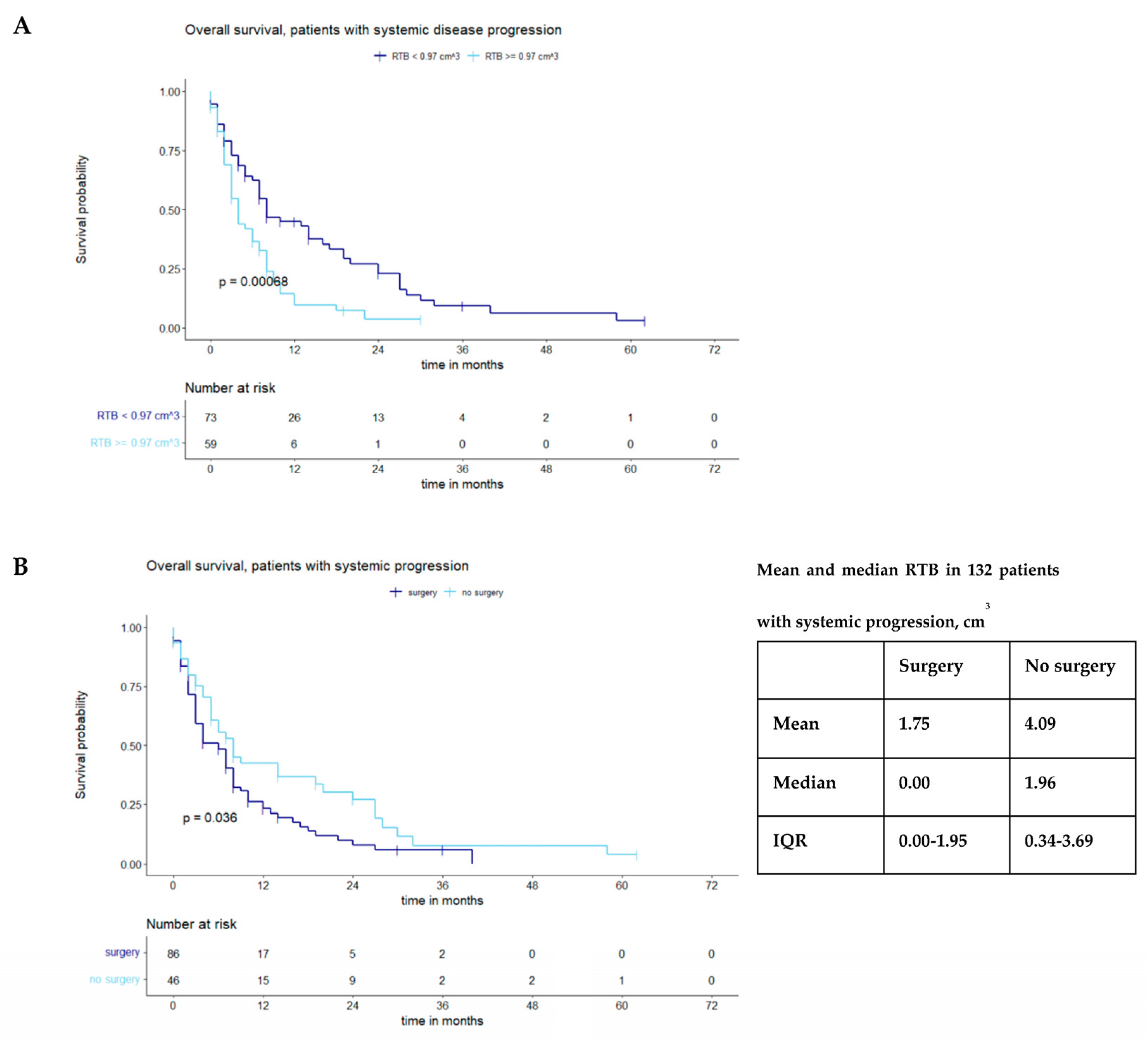
A subgroup analysis was performed to differentiate between patients with and without systemic progression; 132/219 (60.3%) patients presented with systemic progression. In this subgroup, a cutoff RTB of 0.97 cm3 (p = 0.00068) was identified (Figure 3A); patients who underwent surgery again showed prolonged OS (p = 0.036) (Figure 3B).

3.3. Impact of Adjuvant Therapy on Survival

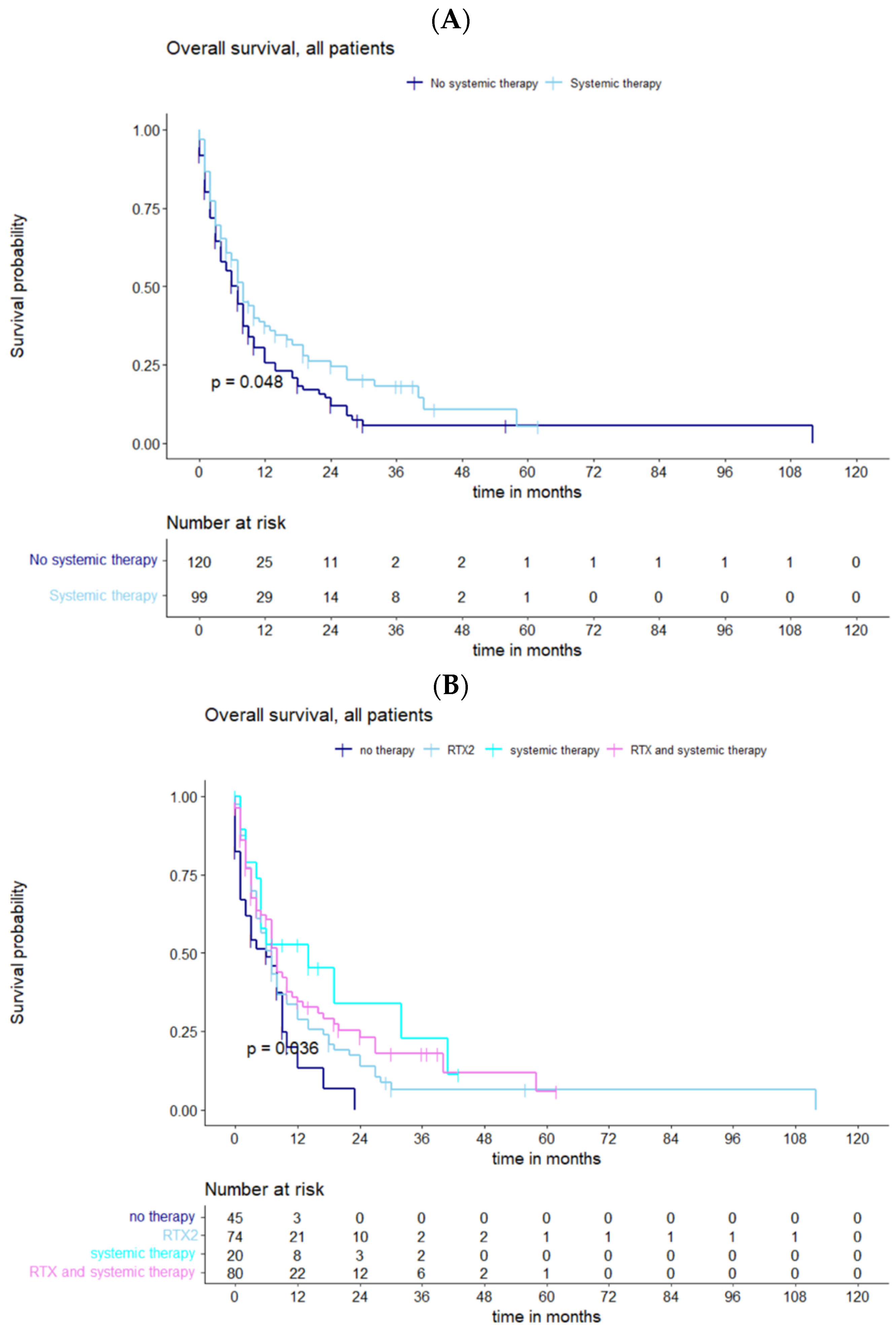
Regarding the different therapeutic regimens, only systemic therapy after relapse (p = 0.048) had a significant effect on survival (Figure 4A). However, combination therapy was associated with a better outcome than single therapy (p = 0.036) (Figure 4B).

4. Discussion

4.1. Prognostic Factors for Survival and Influence of Residual Tumor Volume

The extent of resection (EOR), initial functional status (KPS), and progression-free survival (PFS) are key factors that influence median OS for recurrent BMs [15,22]. In the present study, median OS was 6 months (IQR 2–10). When matching these prognostic factors, preoperative KPSS (HR 0.983, 95% CI, 0.967–0.997, p = 0.015), complete resection (HR 0.629, 95% CI 0.420–0.941, p = 0.024), and postoperative tumor burden (HR 1.03, 95% CI 1.008–1.053, p = 0.007) had a significant impact on OS. These results highlight the relevance of comprehensive cytoreduction, which is confirmed by a significant cutoff for a residual tumor volume (RTB) of 0.12 cm3 (p = 0.00029).
Prior studies have suggested that patients who face recurrent BMs alongside uncontrolled systemic disease may be less likely to benefit from surgery [14,16,23].
However, the benefits of surgical resection are particularly apparent in patients with symptomatic recurrent BMs. As previously mentioned, surgery results in improved functional outcomes and prolonged functional independence [11,24]. Furthermore, it should be added that median OS after neurosurgical resection of recurrent BMs is significantly influenced by baseline functional status, as already described by the prognostic factors mentioned above.
When comparing the groups of patients who underwent surgery and those who did not, it is evident that surgical intervention in the recurrence setting has an essential influence on survival (p = 0.00097). Regarding surgical intervention, it can further be added that complete resection provides a significant survival advantage (p = 0.0027). Recently, the extent of surgical resection has been shown to play a crucial role in the local progression rate of brain metastases, underscoring what has just been highlighted. Extensive surgical resection and evidence of residual tumor demonstrate a significant correlation with the rate of cerebral progression [7]. In this context, it is important to consider the efforts made in medical technology, particularly the availability and advancement of high-resolution microscopes [25,26,27] and the application of molecular biology methods, such as the use of fluorescence-guided resection [4,28,29]. These technologies have helped to improve the capabilities of BM resection.

4.2. Impact of Systemic Tumor Progression

In addition, the aspect of systemic progression should be considered. Most patients (60.3%) showed systemic progression, which included both recurrent BMs and the occurrence of at least one extracranial spread. According to recent research, it is controversial whether patients with advanced oncological disease with (extra-)cranial metastases would still benefit from further surgical intervention [4,10,23]. For the subgroup with systemic progression, a critical cutoff RTB of 0.97 cm3 (p = 0.00068) was again determined by using log-rank statistics, resulting in significant differences in the survival curves. Among the 132 patients with systemic progression, those who underwent surgery showed significantly prolonged OS, consistent with the results of the overall population (p = 0.036). These results reinforce the consensus that a progressive lesion in a patient demonstrating systemic disease and good functional status (Karnofsky performance status [KPS] > 60) presents a favorable profile for undergoing surgical intervention [7,11,15,30]. An infiltrative growth pattern observed in cerebral metastases could contribute as a potential reason for their elevated rate of local recurrence, thereby influencing individual OS [31,32]. For this reason, it is essential to verify the extent of resection (EOR) using postoperative MRI, as complete resection cannot always be justified through intraoperative estimates, as demonstrated in previous reports [8,19]. Thus, residual tumor is a well-established risk factor for local metastatic recurrences, although further investigation is needed to accurately assess the oncologic consequences of unintentionally incomplete metastatic resections [7,31].

4.3. Further Treatment and Outlook

An isolated surgical approach may be effective locally, but it neglects potentially present microscopic residuals and systemic metastases [31]. Radiotherapy and systemic therapy represent additional cornerstones within the interdisciplinary treatment for recurrent BMs and are proving essential not only for minimizing the local recurrence risk but also to more effectively control the overall disease [11,23,33].
In prospective randomized trials of the treatment of primary BMs, analyses have shown that WBRT and stereotactic radiosurgery of the cavity significantly improve local control compared with resection alone [34,35,36]. Studies investigating postoperative irradiation of BMs have also identified EOR as an important prognostic factor for OS [37,38]. However, our study showed that in patients who have already undergone radiotherapy and are in a recurrent situation, additional single radiotherapy does not provide a measurable prolongation of survival (p = 0.36).
Interestingly, another therapeutic approach, single systemic therapy, has been shown to be effective in this context (p = 0.048). Controlling the systemic burden of disease can help slow or stop the growth of BMs [12,33]. As this study specifically addresses the question of the benefits of surgical cytoreduction, two aspects can be highlighted. First, re-evaluating tumor molecular biology could lead to novel systemic treatment strategies, and second, resecting symptomatic recurrent BMs might restore clinical condition, enhancing the chance of subsequent adjuvant treatment and consequently increasing OS, as observed [14,24]. Taking into consideration the patient cohort without additional therapy, which presents inferior survival in comparison, reinforces this assertion.
Furthermore, the combined use of radio and systemic therapy was found to be superior to single therapeutic approaches (p = 0.036). Radiation therapy and systemic treatment can generate synergistic effects in terms of both local and systemic control, helping to control and eliminate the residual tumor and micrometastases [2,39,40]. Advances in combining surgical resection, radiotherapy, and systemic therapy have the potential to improve the quality of life and survival of patients with BMs. These patient-centered therapeutic approaches benefit from the close interdisciplinary collaborations between neurosurgeons, radiation oncologists, and medical oncologists that are now considered an established standard of care [1,10,23].
In this context, close interdisciplinary cooperation becomes an integral part of modern neuro-oncology practice, contributing significantly to the further development and improvement of patient care. However, in the setting of BM recurrence, the available data are limited, and further research is needed.
In summary, the present study illustrates that RTB has a significant impact on OS in cases of recurrence and particularly emphasizes the importance of performing cytoreductive surgery as comprehensively as possible. Thus, findings that have already been observed for first-time BMs [17] could also be confirmed for recurrent brain metastases.

4.4. Study Limitations

The retrospective study design at a single center limits the results of our analysis. The subsequent selection of patients, as well as varying treatment depending on functional status, has resulted in a heterogenous patient cohort. In addition, these patients also have differences in the type of primary tumors and tumor stages. Only patients who had MRI follow-up examinations were included. Special attention was paid to patients who were in a favorable oncologic condition, whereas CT imaging was not included for patients who were unable to obtain MRI imaging.
Additionally, the number of patients included in this current study is limited. A more extensive population would be required to obtain results of higher significance. The patient population was also diverse with respect to different adjuvant and systemic therapeutic approaches prior to the diagnosis of recurrent metastases. Adjuvant therapy plans after initial and repeat craniotomy are still a matter of debate and need to be further explored in future research [34,41,42,43].
Since a relatively substantial proportion (60.3%) of patients had an uncontrolled systemic health status, and metastatic tumor burden in the brain can be considered an expression of systemic disease itself, it is recommended that future research analyze the potential impact of systemic disease status on the rate of local recurrences [7].

5. Conclusions

RTB is a strong prognostic factor for survival in patients with recurrent brain metastases. The routine identification of residual tumors through conventional (postoperative) MRI imaging as well as the advancement of more aggressive surgical methods present significant neuro-oncological hurdles. The goal should always be to achieve maximal cytoreduction, even in recurrent BMs, regardless of age and systemic progression.
Addressing these challenges holds the potential to enhance local tumor control and consequently improve patient outcomes.

Author Contributions

Conceptualization, J.L.; methodology, J.L. and Y.K.; formal analysis, J.L. and Y.K.; investigation, J.L. and Y.K.; resources, J.L.; data curation, J.L.; writing—original draft preparation, J.L.; writing—review and editing, J.L., Y.K., B.W., D.B., A.K.A., S.E.C., C.D., B.M. and J.G.; visualization, J.L. and Y.K.; supervision, B.W., A.K.A., B.M. and J.G.; project administration, A.K.A. and J.G. All authors have read and agreed to the published version of the manuscript.

Funding

This research received no external funding.

Institutional Review Board Statement

Our study was approved by the local ethics committee of the Technical University of Munich (no. 5626:12). It was conducted in accordance with the ethical standards of the 1964 Declaration of Helsinki and its later amendments.

Informed Consent Statement

The requirement for written informed consent was waived by the ethics committee due to retrospective nature of this study.

Data Availability Statement

The data that support the findings of this study are available from the corresponding author, [A.K.A], upon reasonable request.

Conflicts of Interest

Jens Gempt and Bernhard Meyer (BM) work as consultants for Brainlab (Brainlab AG, Feldkirchen). In addition, BM works as a consultant for Medtronic, Spineart, Icotec, Relievant, and Depuy/Synthes, as a member of their advisory boards. Furthermore, BM reports having financial relationships with Medtronic, Ulrich Medical, Brainlab, Spineart, Icotec, Relievant, and Depuy/Synthes. He received personal fees and research grants for clinical studies from Medtronic, Ulrich Medical, Brainlab, Icotec, and Relievant. All of this happened independently of the submitted work. BM receives royalties and holds the patent for Spineart. All the named potential conflicts of interest are unrelated to this study. There are no further conflicts of interest regarding the authors.

References

  1. Schmieder, K.; Keilholz, U.; Combs, S. The Interdisciplinary Management of Brain Metastases. Dtsch. Arztebl. Int. 2016, 113, 415–421. [Google Scholar] [CrossRef]
  2. Shen, C.J.; Lim, M.; Kleinberg, L.R. Controversies in the Therapy of Brain Metastases: Shifting Paradigms in an Era of Effective Systemic Therapy and Longer-Term Survivorship. Curr. Treat. Options Oncol. 2016, 17, 46. [Google Scholar] [CrossRef]
  3. Kavouridis, V.K.; Harary, M.; Hulsbergen, A.F.C.; Lo, Y.T.; Reardon, D.A.; Aizer, A.A.; Iorgulescu, J.B.; Smith, T.R. Survival and prognostic factors in surgically treated brain metastases. J. Neuro-Oncol. 2019, 143, 359–367. [Google Scholar] [CrossRef]
  4. Proescholdt, M.A.; Schodel, P.; Doenitz, C.; Pukrop, T.; Hohne, J.; Schmidt, N.O.; Schebesch, K.M. The Management of Brain Metastases-Systematic Review of Neurosurgical Aspects. Cancers 2021, 13, 1616. [Google Scholar] [CrossRef]
  5. Olson, J.J.; Kalkanis, S.N.; Ryken, T.C. Congress of Neurological Surgeons Systematic Review and Evidence-Based Guidelines for the Treatment of Adults With Metastatic Brain Tumors: Executive Summary. Neurosurgery 2019, 84, 550–552. [Google Scholar] [CrossRef]
  6. Ferguson, S.D.; Wagner, K.M.; Prabhu, S.S.; McAleer, M.F.; McCutcheon, I.E.; Sawaya, R. Neurosurgical management of brain metastases. Clin. Exp. Metastasis 2017, 34, 377–389. [Google Scholar] [CrossRef]
  7. Kamp, M.A.; Fischer, I.; Dibue-Adjei, M.; Munoz-Bendix, C.; Cornelius, J.F.; Steiger, H.J.; Slotty, P.J.; Turowski, B.; Rapp, M.; Sabel, M. Predictors for a further local in-brain progression after re-craniotomy of locally recurrent cerebral metastases. Neurosurg. Rev. 2018, 41, 813–823. [Google Scholar] [CrossRef]
  8. Kamp, M.A.; Rapp, M.; Buhner, J.; Slotty, P.J.; Reichelt, D.; Sadat, H.; Dibue-Adjei, M.; Steiger, H.J.; Turowski, B.; Sabel, M. Early postoperative magnet resonance tomography after resection of cerebral metastases. Acta Neurochir. 2015, 157, 1573–1580. [Google Scholar] [CrossRef]
  9. Munoz-Bendix, C.; Rapp, M.; Mijderwijk, H.J.; von Sass, C.; Dibue-Adjei, M.; Cornelius, J.F.; Steiger, H.J.; Turowski, B.; Sabel, M.; Kamp, M.A. Risk factors for in-brain local progression in elderly patients after resection of cerebral metastases. Sci. Rep. 2019, 9, 7431. [Google Scholar] [CrossRef]
  10. Ammirati, M.; Cobbs, C.S.; Linskey, M.E.; Paleologos, N.A.; Ryken, T.C.; Burri, S.H.; Asher, A.L.; Loeffler, J.S.; Robinson, P.D.; Andrews, D.W.; et al. The role of retreatment in the management of recurrent/progressive brain metastases: A systematic review and evidence-based clinical practice guideline. J. Neuro-Oncol. 2010, 96, 85–96. [Google Scholar] [CrossRef]
  11. Diehl, C.D.; Giordano, F.A.; Grosu, A.L.; Ille, S.; Kahl, K.H.; Onken, J.; Rieken, S.; Sarria, G.R.; Shiban, E.; Wagner, A.; et al. Opportunities and Alternatives of Modern Radiation Oncology and Surgery for the Management of Resectable Brain Metastases. Cancers 2023, 15, 3670. [Google Scholar] [CrossRef] [PubMed]
  12. Langer, C.J.; Mehta, M.P. Current management of brain metastases, with a focus on systemic options. J. Clin. Oncol. 2005, 23, 6207–6219. [Google Scholar] [CrossRef] [PubMed]
  13. Skribek, M.; Rounis, K.; Makrakis, D.; Agelaki, S.; Mavroudis, D.; De Petris, L.; Ekman, S.; Tsakonas, G. Outcome of Patients with NSCLC and Brain Metastases Treated with Immune Checkpoint Inhibitors in a ‘Real-Life’ Setting. Cancers 2020, 12, 3707. [Google Scholar] [CrossRef]
  14. Heßler, N.; Jünger, S.T.; Meissner, A.K.; Kocher, M.; Goldbrunner, R.; Grau, S. Recurrent brain metastases: The role of resection of in a comprehensive multidisciplinary treatment setting. BMC Cancer 2022, 22, 275. [Google Scholar] [CrossRef] [PubMed]
  15. Schackert, G.; Schmiedel, K.; Lindner, C.; Leimert, M.; Kirsch, M. Surgery of recurrent brain metastases: Retrospective analysis of 67 patients. Acta Neurochir. 2013, 155, 1823–1832. [Google Scholar] [CrossRef]
  16. Hulsbergen, A.F.C.; Abunimer, A.M.; Ida, F.; Kavouridis, V.K.; Cho, L.D.; Tewarie, I.A.; Mekary, R.A.; Schucht, P.; Phillips, J.G.; Verhoeff, J.J.C.; et al. Neurosurgical resection for locally recurrent brain metastasis. Neuro-Oncology 2021, 23, 2085–2094. [Google Scholar] [CrossRef]
  17. Aftahy, A.K.; Barz, M.; Lange, N.; Baumgart, L.; Thunstedt, C.; Eller, M.A.; Wiestler, B.; Bernhardt, D.; Combs, S.E.; Jost, P.J.; et al. The Impact of Postoperative Tumor Burden on Patients With Brain Metastases. Front. Oncol. 2022, 12, 869764. [Google Scholar] [CrossRef]
  18. Baumgart, L.; Aftahy, A.K.; Anetsberger, A.; Thunstedt, D.; Wiestler, B.; Bernhardt, D.; Combs, S.E.; Meyer, B.; Meyer, H.S.; Gempt, J. Brain metastases in the elderly—Impact of residual tumor volume on overall survival. Front. Oncol. 2023, 13, 1149628. [Google Scholar] [CrossRef]
  19. Olesrud, I.C.; Schulz, M.K.; Marcovic, L.; Kristensen, B.W.; Pedersen, C.B.; Kristiansen, C.; Poulsen, F.R. Early postoperative MRI after resection of brain metastases-complete tumour resection associated with prolonged survival. Acta Neurochir. 2019, 161, 555–565. [Google Scholar] [CrossRef]
  20. Kiesel, B.; Thome, C.M.; Weiss, T.; Jakola, A.S.; Darlix, A.; Pellerino, A.; Furtner, J.; Kerschbaumer, J.; Freyschlag, C.F.; Wick, W.; et al. Perioperative imaging in patients treated with resection of brain metastases: A survey by the European Association of Neuro-Oncology (EANO) Youngsters committee. BMC Cancer 2020, 20, 410. [Google Scholar] [CrossRef]
  21. Association, W.M. World Medical Association Declaration of Helsinki: Ethical Principles for Medical Research Involving Human Subjects. JAMA 2013, 310, 2191–2194. [Google Scholar] [CrossRef]
  22. Kennion, O.; Holliman, D. Outcome after craniotomy for recurrent cranial metastases. Br. J. Neurosurg. 2017, 31, 369–373. [Google Scholar] [CrossRef]
  23. Vogelbaum, M.A.; Brown, P.D.; Messersmith, H.; Brastianos, P.K.; Burri, S.; Cahill, D.; Dunn, I.F.; Gaspar, L.E.; Gatson, N.T.N.; Gondi, V.; et al. Treatment for Brain Metastases: ASCO-SNO-ASTRO Guideline. J. Clin. Oncol. 2022, 40, 492–516. [Google Scholar] [CrossRef]
  24. Schodel, P.; Junger, S.T.; Wittersheim, M.; Reinhardt, H.C.; Schmidt, N.O.; Goldbrunner, R.; Proescholdt, M.; Grau, S. Surgical resection of symptomatic brain metastases improves the clinical status and facilitates further treatment. Cancer Med. 2020, 9, 7503–7510. [Google Scholar] [CrossRef] [PubMed]
  25. Hong, C.S.; Prevedello, D.M.; Elder, J.B. Comparison of endoscope- versus microscope-assisted resection of deep-seated intracranial lesions using a minimally invasive port retractor system. J. Neurosurg. 2016, 124, 799–810. [Google Scholar] [CrossRef] [PubMed]
  26. Ng, P.R.; Choi, B.D.; Aghi, M.K.; Nahed, B.V. Surgical advances in the management of brain metastases. Neuro-Oncol. Adv. 2021, 3, v4–v15. [Google Scholar] [CrossRef] [PubMed]
  27. Phang, I.; Leach, J.; Leggate, J.R.S.; Karabatsou, K.; Coope, D.; D’Urso, P.I. Minimally Invasive Resection of Brain Metastases. World Neurosurg. 2019, 130, e362–e367. [Google Scholar] [CrossRef]
  28. Kamp, M.A.; Munoz-Bendix, C.; Mijderwijk, H.J.; Turowski, B.; Dibue-Adjei, M.; von Sass, C.; Cornelius, J.F.; Steiger, H.J.; Rapp, M.; Sabel, M. Is 5-ALA fluorescence of cerebral metastases a prognostic factor for local recurrence and overall survival? J. Neuro-Oncol. 2019, 141, 547–553. [Google Scholar] [CrossRef]
  29. Kofoed, M.S.; Pedersen, C.B.; Schulz, M.K.; Kristensen, B.W.; Hansen, R.W.; Markovic, L.; Halle, B.; Poulsen, F.R. Fluorescein-guided resection of cerebral metastases is associated with greater tumor resection. Acta Neurochir. 2022, 164, 451–457. [Google Scholar] [CrossRef]
  30. Mitsuya, K.; Nakasu, Y.; Hayashi, N.; Deguchi, S.; Oishi, T.; Sugino, T.; Yasui, K.; Ogawa, H.; Onoe, T.; Asakura, H.; et al. Retrospective analysis of salvage surgery for local progression of brain metastasis previously treated with stereotactic irradiation: Diagnostic contribution, functional outcome, and prognostic factors. BMC Cancer 2020, 20, 331. [Google Scholar] [CrossRef]
  31. Kamp, M.A.; Slotty, P.J.; Cornelius, J.F.; Steiger, H.J.; Rapp, M.; Sabel, M. The impact of cerebral metastases growth pattern on neurosurgical treatment. Neurosurg. Rev. 2018, 41, 77–86. [Google Scholar] [CrossRef] [PubMed]
  32. Berghoff, A.S.; Rajky, O.; Winkler, F.; Bartsch, R.; Furtner, J.; Hainfellner, J.A.; Goodman, S.L.; Weller, M.; Schittenhelm, J.; Preusser, M. Invasion patterns in brain metastases of solid cancers. Neuro-Oncology 2013, 15, 1664–1672. [Google Scholar] [CrossRef] [PubMed]
  33. Chamberlain, M.C.; Baik, C.S.; Gadi, V.K.; Bhatia, S.; Chow, L.Q. Systemic therapy of brain metastases: Non-small cell lung cancer, breast cancer, and melanoma. Neuro-Oncology 2017, 19, i1–i24. [Google Scholar] [CrossRef]
  34. Brown, P.D.; Ballman, K.V.; Cerhan, J.H.; Anderson, S.K.; Carrero, X.W.; Whitton, A.C.; Greenspoon, J.; Parney, I.F.; Laack, N.N.I.; Ashman, J.B.; et al. Postoperative stereotactic radiosurgery compared with whole brain radiotherapy for resected metastatic brain disease (NCCTG N107C/CEC.3): A multicentre, randomised, controlled, phase 3 trial. Lancet Oncol. 2017, 18, 1049–1060. [Google Scholar] [CrossRef] [PubMed]
  35. Brown, P.D.; Jaeckle, K.; Ballman, K.V.; Farace, E.; Cerhan, J.H.; Anderson, S.K.; Carrero, X.W.; Barker, F.G., 2nd; Deming, R.; Burri, S.H.; et al. Effect of Radiosurgery Alone vs Radiosurgery With Whole Brain Radiation Therapy on Cognitive Function in Patients With 1 to 3 Brain Metastases: A Randomized Clinical Trial. JAMA 2016, 316, 401–409. [Google Scholar] [CrossRef]
  36. Mahajan, A.; Ahmed, S.; McAleer, M.F.; Weinberg, J.S.; Li, J.; Brown, P.; Settle, S.; Prabhu, S.S.; Lang, F.F.; Levine, N.; et al. Post-operative stereotactic radiosurgery versus observation for completely resected brain metastases: A single-centre, randomised, controlled, phase 3 trial. Lancet Oncol. 2017, 18, 1040–1048. [Google Scholar] [CrossRef]
  37. El Shafie, R.A.; Dresel, T.; Weber, D.; Schmitt, D.; Lang, K.; Konig, L.; Hone, S.; Forster, T.; von Nettelbladt, B.; Eichkorn, T.; et al. Stereotactic Cavity Irradiation or Whole-Brain Radiotherapy Following Brain Metastases Resection-Outcome, Prognostic Factors, and Recurrence Patterns. Front. Oncol. 2020, 10, 693. [Google Scholar] [CrossRef]
  38. Kalkanis, S.N.; Kondziolka, D.; Gaspar, L.E.; Burri, S.H.; Asher, A.L.; Cobbs, C.S.; Ammirati, M.; Robinson, P.D.; Andrews, D.W.; Loeffler, J.S.; et al. The role of surgical resection in the management of newly diagnosed brain metastases: A systematic review and evidence-based clinical practice guideline. J. Neuro-Oncol. 2010, 96, 33–43. [Google Scholar] [CrossRef]
  39. Aly, Z.; Peereboom, D.M. Combination of Radiotherapy and Targeted Agents in Brain Metastasis: An Update. Curr. Treat. Options Neurol. 2016, 18, 32. [Google Scholar] [CrossRef]
  40. Di Lorenzo, R.; Ahluwalia, M.S. Targeted therapy of brain metastases: Latest evidence and clinical implications. Ther. Adv. Med. Oncol. 2017, 9, 781–796. [Google Scholar] [CrossRef]
  41. Soffietti, R.; Kocher, M.; Abacioglu, U.M.; Villa, S.; Fauchon, F.; Baumert, B.G.; Fariselli, L.; Tzuk-Shina, T.; Kortmann, R.D.; Carrie, C.; et al. A European Organisation for Research and Treatment of Cancer phase III trial of adjuvant whole-brain radiotherapy versus observation in patients with one to three brain metastases from solid tumors after surgical resection or radiosurgery: Quality-of-life results. J. Clin. Oncol. 2013, 31, 65–72. [Google Scholar] [CrossRef] [PubMed]
  42. Kocher, M.; Soffietti, R.; Abacioglu, U.; Villa, S.; Fauchon, F.; Baumert, B.G.; Fariselli, L.; Tzuk-Shina, T.; Kortmann, R.D.; Carrie, C.; et al. Adjuvant whole-brain radiotherapy versus observation after radiosurgery or surgical resection of one to three cerebral metastases: Results of the EORTC 22952-26001 study. J. Clin. Oncol. 2011, 29, 134–141. [Google Scholar] [CrossRef] [PubMed]
  43. Sperduto, P.W.; Chao, S.T.; Sneed, P.K.; Luo, X.; Suh, J.; Roberge, D.; Bhatt, A.; Jensen, A.W.; Brown, P.D.; Shih, H.; et al. Diagnosis-specific prognostic factors, indexes, and treatment outcomes for patients with newly diagnosed brain metastases: A multi-institutional analysis of 4,259 patients. Int. J. Radiat. Oncol. Biol. Phys. 2010, 77, 655–661. [Google Scholar] [CrossRef] [PubMed]
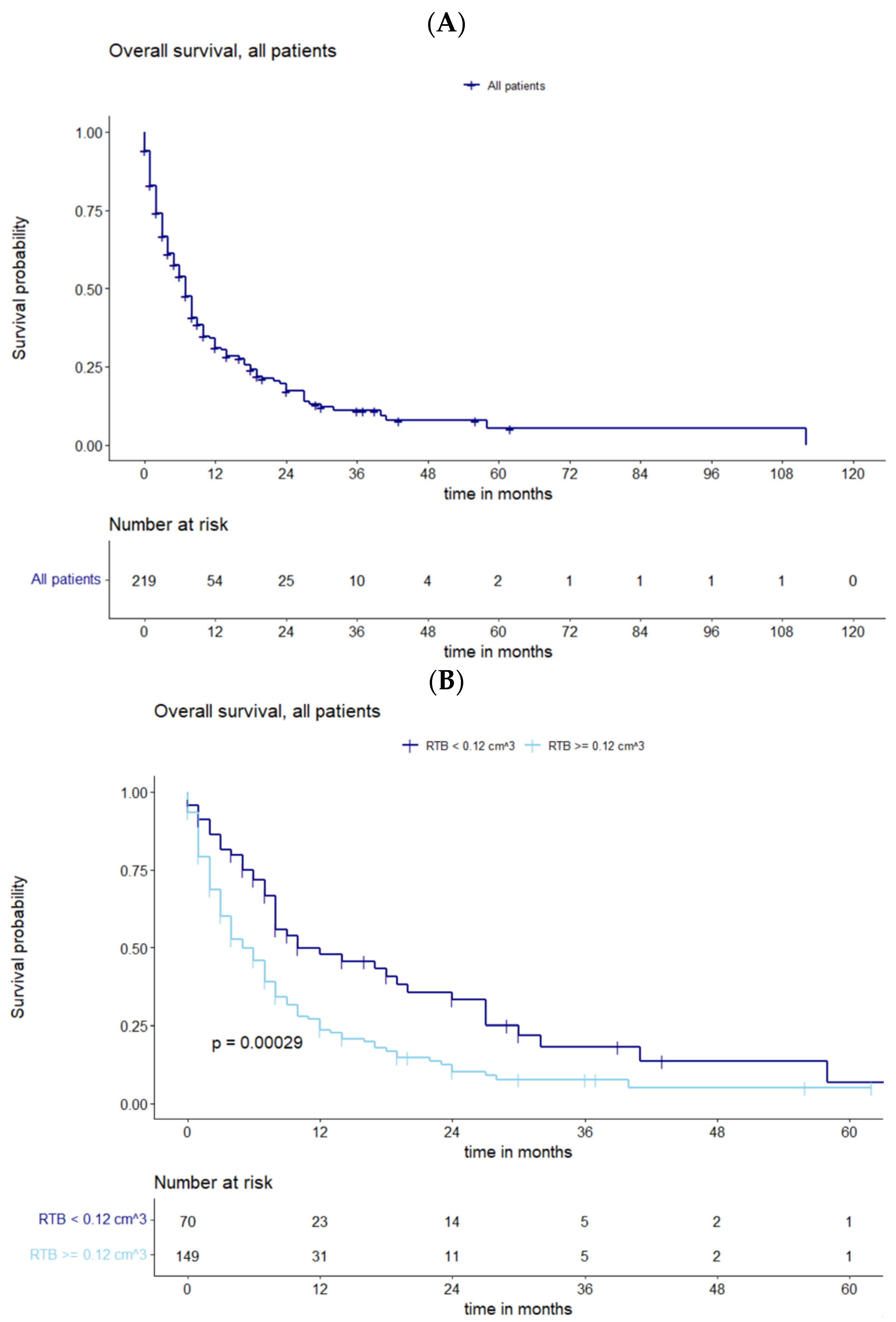
Figure 1. (A) Overall survival of all patients with recurrent BMs. (B) Survival analysis in all patients for subgroups of cutoff residual tumor showing statistically significant deviations in survival curves.
Cancers 15 05067 g001
Figure 2. (A) Kaplan–Meier curves representing OS of all patients with recurrent BMs stratified by surgery vs. no surgery, showing a statistically significant correlation. (B) Functions of OS demonstrating significantly divergent survival curves after complete vs. incomplete resection.
Cancers 15 05067 g002
Figure 3. (A) Maximally selected log-rank statistics displaying the cutoff of postoperative tumor volume in a subgroup with systemic tumor progression. (B) Divergence in survival in this subgroup stratified by surgery vs. no surgery.
Cancers 15 05067 g003
Figure 4. (A) Functions of OS demonstrating significantly divergent survival curves after systemic vs. no systemic therapy. (B) Survival estimates in subgroups which underwent different regimes of therapy demonstrating significantly divergent survival curves in favor of combined therapy modalities.
Cancers 15 05067 g004
Table 1. Demographics, tumor data, and adjuvant therapy.
Demographics, N (%) or Median (Range/IQR).
SexF 121/219 (55.3)
M 98/219 (44.7)
Age60.0 (IQR 52–69)
Karnofsky Performance Status Scale (KPSS)
Preoperative KPSS80% (IQR 70–80)
Postoperative KPSS80% (IQR 70–80)
Number of metastases, N (%)
182/219 (37.4)
254/219 (24.7)
337/219 (16.9)
>346/219 (21.0)
Adjuvant therapy, N (%)
No therapy45/219 (20.5)
Radiotherapy74/219 (33.8)
Systemic therapy20/219 (9.2)
Combined80/219 (36.5)
Tumor burden (cm3)
Preoperative
Median2.4 cm3 (0.8–8.3 cm3)
Range0.02–184.33 cm3
Mean7.43 cm3 (SD 14.92)
Postoperative
Median0.5 cm3 (0.0–2.9 cm3)
Range0.00–98.34 cm3
Mean3.45 cm3 (SD 9.159)
Table 2. Management and surgical outcomes in recurrent BMs.
Recurrent Brain Metastasis Management, N (%)
No resection of BMs124/219 (56.6)
Surgery with RTB left
at other locations
40/219 (18.3)
Surgery with no RTB left
Complete resection
55/219 (25.1)
Resection Outcomes with Targeted Recurrent BMs, N (%)
Complete resection of targeted BMs with RTB left at other locations27/40 (67.5)
Incomplete resection of targeted BMs with RTB left at other locations13/40 (32.5)
Surgery Based on the Number of BMs, N (%)
156/95 (59.0)
219/95 (20.0)
310/95 (10.5)
>310/95 (10.5)
Complete Resection Based on the Number of BMs, N (%)
148/55 (87.3)
26/55 (10.9)
31/55 (1.8)
>30/55 (0.0)
Table 3. Multivariate Cox regression analysis.
Multivariate Analysis ParametersHazard Ratio (IQR)p-Value
Sex0.8046 (0.5824–1.1117)0.18753
Age (year)1.002092 (0.9889–1.0155)0.75745
KPSS at admission0.982465 (0.9686–0.9965)0.01466
Preoperative tumor volume (cm3)0.9966 (0.9828–1.0106)0.63142
RTB (cm3)1.030376 (1.0081–1.0532)0.00736
RTB = 00.628666 (0.4202–0.9405)0.02391
Disclaimer/Publisher’s Note: The statements, opinions and data contained in all publications are solely those of the individual author(s) and contributor(s) and not of MDPI and/or the editor(s). MDPI and/or the editor(s) disclaim responsibility for any injury to people or property resulting from any ideas, methods, instructions or products referred to in the content.