Article

Optimal Time Interval between Neoadjuvant Platinum-Based Chemotherapy and Interval Debulking Surgery in High-Grade Serous Ovarian Cancer

by
Angeliki Andrikopoulou
1,*,†,
Charalampos Theofanakis
2,
Christos Markellos
1,
Maria Kaparelou
1,
Konstantinos Koutsoukos
1,
Kleoniki Apostolidou
1,
Nikolaos Thomakos
2,
Dimitrios Haidopoulos
2,
Alexandros Rodolakis
2,
Meletios-Athanasios Dimopoulos
1,
Flora Zagouri
1 and
Michalis Liontos
1
1
Department of Clinical Therapeutics, Alexandra Hospital, Medical School, 11528 Athens, Greece
2
1st Department of Obstetrics and Gynecology, Alexandra Hospital, Medical School, 11528 Athens, Greece
*
Author to whom correspondence should be addressed.
Current address: Department of Oncology, Medical School, National and Kapodistrian University of Athens, 80 Vasilissis Sofias Avenue, 11528 Athens, Greece.
Cancers 2023, 15(13), 3519; https://doi.org/10.3390/cancers15133519
Submission received: 28 May 2023 / Revised: 18 June 2023 / Accepted: 26 June 2023 / Published: 6 July 2023
(This article belongs to the Special Issue Gynecologic Cancer: From Diagnosis to Treatment)

Abstract

:

Simple Summary

The optimal time interval between the completion of neoadjuvant chemotherapy (NACT) and interval debulking surgery (IDS) in high-grade serous ovarian carcinoma (HGSC) is not well defined. We conducted a retrospective study of patients with HGSC stage IIIC/IV who had received NACT followed by IDS during a 15-year period (January 2003–December 2018) in our Institution. Performing IDS within four weeks after NACT was associated with better survival outcomes. On multivariate analysis, the performance of IDS within four weeks after NACT was an independent factor of both PFS (p = 0.004) and OS (p = 0.003). Our study provides evidence that surgical intervention should not be significantly delayed after neoadjuvant chemotherapy.

Highlights

What are the main findings?
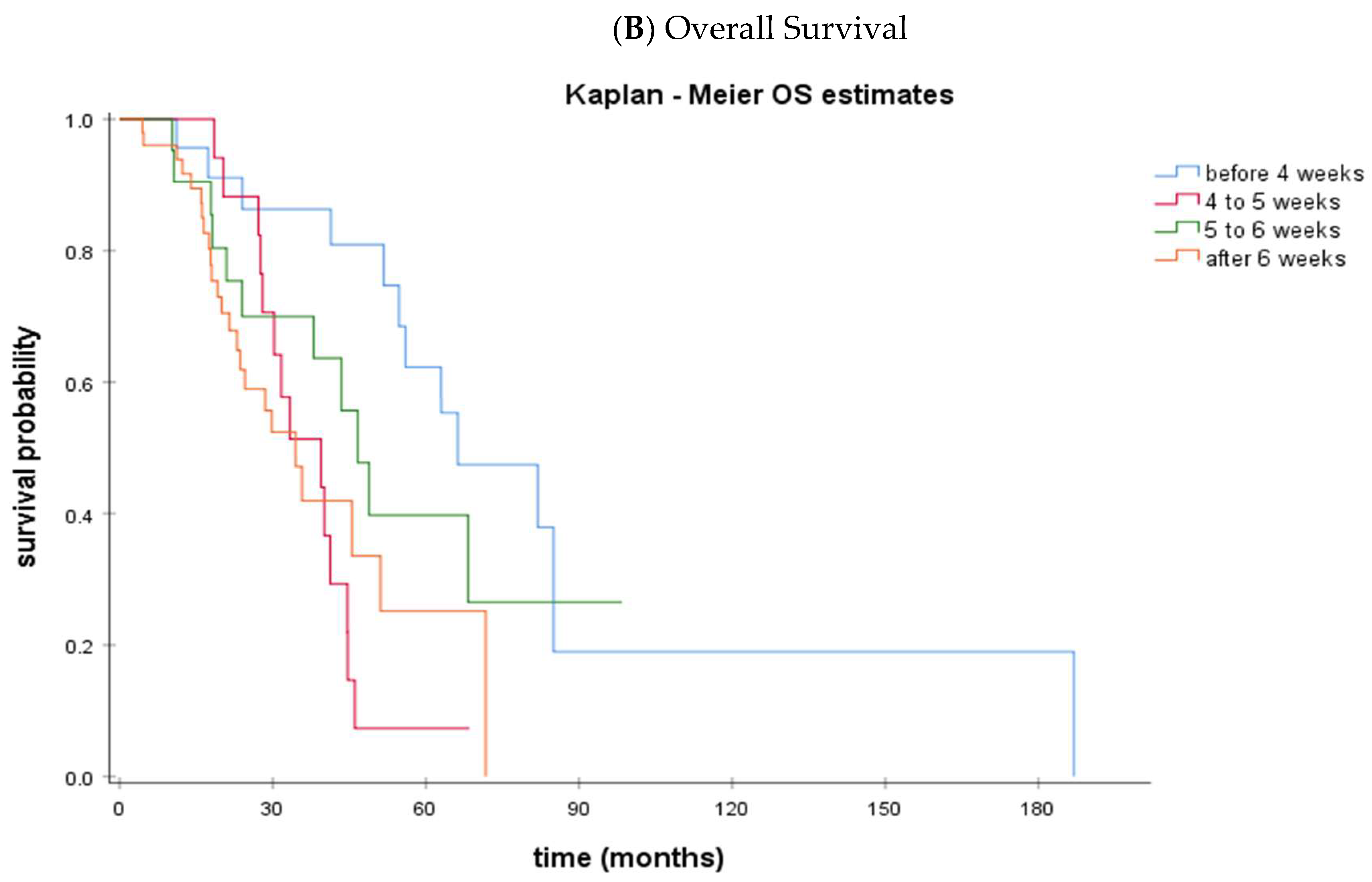
  • The time interval NACT to IDS < 4 weeks was significantly associated with a prolonged PFS (p = 0.004) and OS (p = 0.002).
  • Median OS was 66.3 months (95% CI: 39.1–93.4) vs. 39.4 months (95% CI: 31.8–47.0) in the <4 week vs. ≥4 week time interval NACT to IDS groups (p = 0.002)
  • On multivariate analysis, the performance of IDS within 4 weeks after NACT and optimal debulking were independent factors for both PFS and OS
What is the implication of the main finding?
  • Performing IDS early after NACT proved to be a good prognostic factor among ovarian cancer patients
  • Multidisciplinary coordination is required so as to avoid any unnecessary delays

Abstract

Background: There is limited data on the optimal time interval between the last dose of neoadjuvant chemotherapy (NACT) and interval debulking surgery (IDS) in high-grade serous ovarian carcinoma (HGSC). Methods: We retrospectively identified patients with stage IIIC/IV HGSC who received NACT followed by IDS during a 15-year period (January 2003–December 2018) in our Institution. Results: Overall, 115 patients with stage IIIC/IV HGSC were included. The median age of diagnosis was 62.7 years (IQR: 14.0). A total of 76.5% (88/115) of patients were diagnosed with IIIC HGSC and 23.5% (27/115) with IV HGSC. Median PFS was 15.7 months (95% CI: 13.0–18.5), and median OS was 44.7 months (95% CI: 38.8–50.5). Patients were categorized in groups according to the time interval from NACT to IDS: <4 weeks (group A); 4–5 weeks (group B); 5–6 weeks (group C); >6 weeks (group D). Patients with a time interval IDS to NACT ≥4 weeks had significantly shorter PFS (p = 0.004) and OS (p = 0.002). Median PFS was 26.6 months (95% CI: 24–29.2) for patients undergoing IDS <4 weeks after NACT vs. 14.4 months (95% CI: 12.6–16.2) for those undergoing IDS later (p = 0.004). Accordingly, median OS was 66.3 months (95% CI: 39.1–93.4) vs. 39.4 months (95% CI: 31.8–47.0) in the <4 week vs. >4 week time interval NACT to IDS groups (p = 0.002). On multivariate analysis, the short time interval (<4 weeks) from NACT to IDS was an independent factor of PFS (p = 0.004) and OS (p = 0.003). Conclusion: We have demonstrated that performing IDS within four weeks after NACT may be associated with better survival outcomes. Multidisciplinary coordination among ovarian cancer patients is required to avoid any unnecessary delays.

1. Background

Ovarian cancer is the eighth leading cause of cancer mortality among women, accounting for over 200,000 deaths in 2020 worldwide [1]. Although most patients initially respond to platinum-based chemotherapy, the majority of patients eventually relapse, and only 25% of patients with stage III/IV disease remain alive at five years [2].
Primary cytoreductive surgery (PDS), followed by platinum-based chemotherapy, is the current standard of care for advanced ovarian cancer. However, for those unsuitable for optimal debulking with no residual tumor, neoadjuvant platinum-based chemotherapy (NACT) for 3–4 cycles followed by interval debulking surgery (IDS) constitutes a valuable alternative. Four phase III trials have also provided evidence that NACT followed by IDS is a non-inferior approach for patients also suitable for PDS [3,4,5,6]. Platinum-based combination therapy with paclitaxel is the preferred regimen for NACT [3,4,5,6]. The addition of bevacizumab to the NACT regimen is feasible and safe according to the GEICO 1205/NOVA and ALTHALYA trials, but showed no difference in complete macroscopic response (CR) rates or progression-free survival (PFS) [7,8].
Delays in adjuvant chemotherapy initiation or surgery post-neoadjuvant chemotherapy are associated with impaired survival in several neoplasms. In ovarian cancer, large retrospective studies have indicated an association between the timing of adjuvant treatment and survival [9,10,11,12]. In addition, delays in postoperative treatment initiation (more than six or seven weeks after IDS) were associated with poor prognosis, especially in patients with no residual disease after surgery [9,10]. However, data regarding the optimal timing of IDS after NACT remains limited, and previous studies have not taken into consideration the molecular biology of the disease. Recent phase III trials recommend performing IDS within six weeks after NACT completion [13,14]. In clinical practice, though, reasons related either to the patient, such as hematological toxicity and performance status, or to the health system and the timely scheduling of the surgical procedure may result in IDS delays. Taking into account that the percentage of advanced ovarian cancer patients treated with NACT constantly increases, the accumulation of data regarding the significance of the ideal time frame within which IDS should be performed is necessary.
Considering the lack of data regarding the optimal interval between the last dose of NACT and IDS, we conducted this retrospective analysis of patients with high-grade serous ovarian cancer (HGSC) of advanced stage (IIIC/IV) treated with NACT and IDS in the Oncology Department of Alexandra University Hospital. We aim to define the impact of delaying IDS after NACT on overall prognosis by using a cutoff of four weeks as defined by previous data.

2. Methods

We retrospectively identified patients with stage IIIC/IV ovarian/fallopian tube/primary peritoneal cancer who had received NACT followed by IDS during a 15-year period (January 2003–December 2018) in our institutional database. Our institution has been certified by the European Society of Gyenocologic Oncology (ESGO) as a center of excellence for the treatment of ovarian cancer. The study has been performed in accordance with the 1964 Helsinki Declaration and has been approved by the Institutional Review Board of Alexandra University Hospital (Protocol Number: 513/15-07-2020). Patients were selected for NACT and IDS if it was judged by the experienced gynecologic oncologists that they could not be debulked upfront with no residual tumor. Assessment involved imaging studies and/or laparoscopic evaluation. All subjects received 3 cycles of NACT with carboplatin and paclitaxel according to existing guidelines. All women had provided informed consent for their treatment as well as for the use of their medical records for research purposes. Clinicopathological characteristics, including age at diagnosis, stage, histology, grade, debulking status, BRCA mutation status, type of chemotherapy administered, progression of disease, and overall survival, were collected from the medical records of the patients. Optimal debulking was defined as a maximum residual tumor of <1 cm in diameter after IDS.

Statistical Analysis

Continuous variables were summarized with the use of descriptive statistical measures [median (IQR; 25–75)], and categorical variables were displayed as frequency tables (N, %). The outcome of the debulking surgery was classified as optimal (residual disease below 1 cm) or suboptimal. Overall survival (OS) was defined as the time between the start of chemotherapy and the date of death from any cause. Progression-free survival (PFS) was defined as the time between the start of chemotherapy and the date of progression. Alive patients were censored at the date of last contact. Kaplan-Meier estimates were used to describe and visualize the effect of categorical variables on OS and PFS. For the analysis, patients were divided into two groups according to the time interval between NACT and IDS: the short (<4 weeks) and long (≥4 weeks) interval groups [15,16,17]. The association of these factors with PFS and OS was assessed through HRs and their 95% confidence intervals estimated from univariate Cox proportional hazard models. Interactions between covariates and time-varying effects were studied. All statistical analyses were performed using SPSS 24.0 statistical software.

3. Results

3.1. Study Population

Overall, 115 patients with HGSC stage IIIC/IV that underwent NACT followed by IDS were included in our analysis (Table 1). The median age of diagnosis was 62.7 years (IQR: 58.1–71.8). A total of 76.5% (88/115) of patients were diagnosed with stage IIIC HGSC and 23.5% of patients (27/115) with stage IV disease. The result of the debulking surgery was available in 101 patients, among whom 69 (68.3%) were optimally debulked. Performance status was ECOG 0 (53.2%; 59/111) and 1 (34.3%; 38/111) in most cases. Patients were categorized into two groups: 23 patients underwent IDS within 4 weeks from the end of NACT (20%; 23/115), while 92 patients (80%; 92/115) underwent the operation after 4 weeks. The median time interval between the last dose of NACT and IDS was 5.6 weeks (IQR: 4.1–7.0). Specifically, IDS was performed within 4 weeks from NACT completion in 23 patients (20%; 23/115), between weeks 4 and 5 in 20 patients (17.4%; 20/115), between weeks 5 and 6 in 22 patients (19.1%; 22/115), and after week 6 in 50 patients (43.5%; 50/115). BRCA1/2 mutation status was known in 79 patients. A total of 22.8% (18/79) of patients harbored BRCA1/2 somatic mutations, whereas 77.2% (61/79) were BRCA1/2 wild-type. Overall, 81.7% of patients (94/115) had experienced disease progression until the time of the analysis, while 53% (61/115) were deceased. Median PFS was 15.7 months (95% CI: 13.0–18.5) (Supplemental Figure S1), and median OS was 44.7 months (95% CI: 38.8–50.5) (Supplemental Figure S2).

3.2. Subgroup Analysis

Patients were categorized in groups according to the interval from the last dose of NACT to IDS: <4 weeks (group A); ≥4 to <5 weeks (group B); ≥5 to <6 weeks (group C); ≥6 weeks (group D). Table 2 presents the clinicopathological characteristics of patients undergoing IDS within 4 weeks after NACT vs. those undergoing IDS after 4 weeks after NACT. No statistically significant difference was noted for known prognostic characteristics such as age, stage, and performance status among the four groups of patients. In addition, the outcome of the IDS was similar among groups of patients, while the percentage of patients with known BRCA1/2 mutations also did not differ significantly (Table 2). Patients with a long-time interval from NACT to IDS (≥4 weeks) had significantly poorer PFS (p = 0.004) and OS (p = 0.002) than those in the short interval (<4 weeks). Median PFS was 26.6 months (95% CI: 24.0–29.2) for patients undergoing IDS <4 weeks after NACT vs. 14.4 months (95% CI: 12.6–16.3) for those undergoing IDS later (p = 0.004) (Figure 1). Accordingly, median OS was 66.3 months (95% CI: 39.1–93.4) vs. 39.4 months (95% CI: 31.8–47.0) in the <4 week vs. ≥4 week time interval NACT to IDS groups (p = 0.002) (Figure 1).
We then evaluated the impact of delaying IDS after NACT (group A: < 4 weeks; group B: ≥4 to <5; group C: ≥5 to <6; group D: ≥6 weeks). Table 3 summarizes the clinicopathological characteristics of the four subgroups (group A, group B, group C, and group D). The median time interval from NACT to IDS was 3.1 weeks (IQR: 3.0–3.7) for group A, 4.4 weeks (IQR: 4.1–4.7) for group B, 5.4 weeks (IQR: 5.1–5.7) for group C, and 7.1 weeks (IQR: 6.4–7.9) for group D. Kaplan-Meier curves of PFS and OS for each of the subgroups are shown in Figure 2. We observe again that both PFS and OS are significantly higher in group A compared to groups B, C, and D. Among the patients undergoing IDS after week 4, no differences were observed in terms of PFS or OS.

3.3. Multivariate Analysis

We performed a Cox regression analysis of the factors that influence PFS and OS in HGSC patients undergoing IDS. Initial status of disease (IIIC or IV), debulking status (optimal vs. suboptimal), ECOG performance status, and interval from NACT to IDS (<4 weeks vs. ≥4 weeks) were included in the analysis. Again, the performance of IDS within 4 weeks after the last dose of NACT retained its statistical significance in terms of PFS (p = 0.004) and OS (p = 0.003). Optimal debulking was also an independent factor in both PFS (p = 0.008) and OS (p = 0.001) (Table 4).

4. Discussion

There is no consensus regarding the optimal timing of interval debulking surgery after the completion of neoadjuvant platinum-based chemotherapy. We investigated the impact of performing IDS early (within four weeks) after neoadjuvant chemotherapy on PFS and OS. The cut-off of four weeks has been proposed by previous studies as the optional time interval for cytoreductive surgery in terms of safety and efficacy [15,16,17]. Indeed, performing the surgery within four weeks after the last dose of NACT improved both PFS and OS in univariate and multivariate analyses in patients with stage IIIC/IV HGSC.
Previous studies have shown that time to initiation of postoperative chemotherapy is significantly associated with OS [10,11,12]. Time off chemotherapy, defined as the time from the last dose of NACT to the initiation of adjuvant chemotherapy, has also been associated with disease prognosis [16,17,18]. Indeed, postoperative complications, including extended resections, wound healing, and bleeding, may often delay the initiation of adjuvant treatment. However, it is thought that chemotherapy should be started as soon as possible after surgery, especially if debulking was suboptimal. Early initiation of chemotherapy may prevent the distant dissemination of the remaining tumor cells and decrease the tumor burden. It is unclear whether these limitations apply to neoadjuvant chemotherapy as well.
There are several factors that could delay the performance of IDS after NACT completion in a real-world setting. First of all, patients should have recovered from treatment-related toxicities, especially hematologic toxicity, so that surgical intervention can be safely performed. This is of utmost importance for older patients with comorbidities who do not tolerate NACT well. In addition, patients should perform laboratory and imaging tests after chemotherapy completion and be evaluated for their eligibility to undergo IDS by a multidisciplinary team. Finally, logistical reasons related to the availability of operating rooms and the priority lists among ovarian cancer patients in each hospital may also account for delays in performing IDS after NACT.
Data emerging from other solid tumors imply that the time interval between NACT and surgery could affect prognosis [19,20]. In breast cancer, there are studies supporting the idea that breast surgery within three weeks after the completion of NACT would be of maximal benefit [19]. In rectal cancer, a longer time interval between neoadjuvant chemoradiotherapy and surgical excision in locally advanced rectal cancer could lead to higher pCR rates [21]. In non-small cell lung cancer, surgery should not be performed immediately but within a time frame of six weeks after the completion of neoadjuvant chemoradiation [22]. The timing of surgery after neoadjuvant treatment should be carefully balanced in solid tumors to overcome chemotherapy-related toxicities but also to prevent recurrence of residual disease before surgery.
Our data indicate that performing IDS early after NACT (within a month’s timeframe) is an independent prognostic factor for advanced ovarian cancer. Of note, this result seems independent from the biology of the disease since the percentage of BRCA1/2 mutant patients was equally distributed among the groups analyzed. Considering the efficacy of platinum-based chemotherapy in BRCA1/2 mutant patients, one would expect that merely biology would determine prognosis in these patients that receive NACT. However, this is not the case in our cohort of patients. On the other hand, our study result could be attributed to the residual disease post-NACT in ovarian cancer patients. As shown previously by us and others, approximately one third of patients receiving NACT have a very good pathological response to chemotherapy (CRS3) [23,24]. However, this is not equal to pathological complete response (pCR), as detected post-NACT in breast cancer patients. For breast cancer patients with pCR, the timing of surgery may be of minor importance. In ovarian cancer patients, though, delay of IDS may allow expansion of residual disease even in patients with major pathological responses, which could again allow microscopic intraperitoneal dissemination of the disease, hampering the outcome of the debulking surgery. In addition, the four-week timepoint revealed by our data may be considered a statistical cut-off value, but there is also a biological rationale since it coincides with the re-propagation of residual disease post-last chemotherapy cycle.
Our study is characterized by certain limitations. First, this is a retrospective analysis and not a prospective randomized study. In addition, other factors that could influence the overall prognosis were taken into account in the multivariate analysis. However, it is well known that BRCA mutation status is associated with PFS and OS, conferring a survival benefit in BRCA-mutated HGSC patients. Since our analysis goes back to 2003, when BRCA1/2 genetic testing was not a routine test for ovarian cancer patients in Greece, data regarding BRCA1/2 mutation status are limited and therefore not included in the multivariate analysis. In addition, delayed surgery is often the result of a more extensive intraabdominal disease or a worse performance status. Indeed, there are many ranking scores evaluating the likelihood of performing optimal cytoreduction. The peritoneal cancer index (PCI) was developed to evaluate the peritoneal dissemination of intraabdominal or intrapelvic malignant tumors. A PCI score >10 was associated with worse survival in ovarian tumors, even though complete cytoreduction was achieved [25]. Fagotti is a laparoscopy-based score for predicting surgical resectability after evaluating omental, peritoneal, and diaphragmatic involvement, bowel and stomach infiltration, and liver metastases [26]. This score was further modified to identify the patients more likely to achieve optimal cytoreduction [27]. Given the retrospective nature of our study, the evaluation of the disease extent at baseline based on these scores was not feasible. Hence, there is a possible bias toward delaying surgery in patients with an extensive disease spread who would have an adverse prognosis anyway. Finally, this is a single-institution study, including only a small number of patients. Large trials should be designed to address the question of the optimal timing of IDS after NACT.

5. Conclusions

To date, the importance of timely surgery after NACT remains under discussion in ovarian cancer. We have demonstrated that performing IDS within four weeks after NACT may be a good prognostic factor. Multidisciplinary coordination among ovarian cancer patients is required so as to avoid any unnecessary delays.

Supplementary Materials

The following supporting information can be downloaded at: https://www.mdpi.com/article/10.3390/cancers15133519/s1, Figure S1: Kaplan Meier curve for PFS in the overall population; Figure S2: Kaplan Meier curve for OS in the overall population.

Author Contributions

Conceptualization: M.L., F.Z., M.-A.D. and A.R.; Data curation: M.L., A.A., C.M., M.K., K.K., C.T., D.H., N.T. and A.R.; Investigation: A.A., C.M., M.K., D.H., N.T., K.K. and C.T.; Formal analysis: A.A. and K.A.; Methodology: M.L., A.A. and K.A.; Project administration: M.L., F.Z., M.-A.D., D.H. and A.R.; Supervision: M.L., F.Z.; Writing—original draft: A.A., C.M., N.T., M.K. and K.K.; Writing—review & editing: M.L., F.Z., A.R. and M.-A.D. All authors have read and agreed to the published version of the manuscript.

Funding

This research received no external funding.

Institutional Review Board Statement

Informed consent was obtained from all individual participants included in the study. The study has been performed in accordance with the 1964 Helsinki Declaration and has been approved by the Institutional Review Board of Alexandra University Hospital (Protocol Number: 513/15-07-2020).

Informed Consent Statement

Not applicable.

Data Availability Statement

Data presented in our study can be found in the patients’ archives that are safely stored in our Institution. The datasets generated during the current study are available from the corresponding author upon request.

Acknowledgments

We are indebted to patients and their families for their participation in the study.

Conflicts of Interest

M.L. has received honoraria from Roche, Astra Zeneca, Astellas, MSD, Janssen, Bristol-Myers-Squibb and IPSEN. K.K. has received honoraria by Roche, BMS, MSD and IPSEN. M.-A.D. has received honoraria from participation in advisory boards from Amgen, Bristol-Myers-Squibb, Celgene, Janssen, Takeda. F.Z. has received honoraria for lectures and has served in an advisory role for Astra-Zeneca, Daiichi, Eli-Lilly, Merck, Novartis, Pfizer, and Roche. The remaining authors declare no conflict of interest.

References

  1. Cancer Today. Available online: https://gco.iarc.fr/today/online-analysis-multi-bars?v=2018&mode=cancer&mode_population=countries&population=900&populations=900&key=total&sex=0&cancer=39&type=0&statistic=5&prevalence=0&population_group=0&ages_group%5B%5D=0&ages_group%5B%5D=17&nb_items=10&group_cancer=1&include_nmsc=1&include_nmsc_other=1&type_multiple=%257B%2522inc%2522%253Atrue%252C%2522mort%2522%253Afalse%252C%2522prev%2522%253Afalse%257D&orientation=horizontal&type_sort=0&type_nb_items=%257B%2522top%2522%253Atrue%252C%2522bottom%2522%253Afalse%257D&population_group_globocan_id= (accessed on 16 November 2020).
  2. Matulonis, U.A.; Sood, A.K.; Fallowfield, L.; Howitt, B.E.; Sehouli, J.; Karlan, B.Y. Ovarian cancer. Nat. Rev. Dis. Prim. 2016, 2, 16061. [Google Scholar] [CrossRef]
  3. Kehoe, S.; Hook, J.; Nankivell, M.; Jayson, G.C.; Kitchener, H.; Lopes, T.; Luesley, D.; Perren, T.; Bannoo, S.; Mascarenhas, M.; et al. Primary chemotherapy versus primary surgery for newly diagnosed advanced ovarian cancer (CHORUS): An open-label, randomised, controlled, non-inferiority trial. Lancet 2015, 386, 249–257. [Google Scholar] [CrossRef] [PubMed]
  4. Fagotti, A.; Ferrandina, G.; Vizzielli, G.; Fanfani, F.; Gallotta, V.; Chiantera, V.; Costantini, B.; Margariti, P.A.; Gueli Alletti, S.; Cosentino, F.; et al. Phase III randomised clinical trial comparing primary surgery versus neoadjuvant chemotherapy in advanced epithelial ovarian cancer with high tumour load (SCORPION trial): Final analysis of peri-operative outcome. Eur. J. Cancer 2016, 59, 22–33. [Google Scholar] [CrossRef] [PubMed]
  5. Vergote, I.; Tropé, C.G.; Amant, F.; Kristensen, G.B.; Ehlen, T.; Johnson, N.; Verheijen, R.H.M.; van der Burg, M.E.L.; Lacave, A.J.; Panici, P.B.; et al. Neoadjuvant chemotherapy or primary surgery in stage IIIC or IV ovarian cancer. N. Engl. J. Med. 2010, 363, 943–953. [Google Scholar] [CrossRef] [PubMed] [Green Version]
  6. Onda, T.; Satoh, T.; Saito, T.; Kasamatsu, T.; Nakanishi, T.; Nakamura, K.; Wakabayashi, M.; Takehara, K.; Saito, M.; Ushijima, K.; et al. Comparison of treatment invasiveness between upfront debulking surgery versus interval debulking surgery following neoadjuvant chemotherapy for stage III/IV ovarian, tubal, and peritoneal cancers in a phase III randomised trial: Japan Clinical Oncology Group Study JCOG0602. Eur. J. Cancer 2016, 64, 22–31. [Google Scholar] [CrossRef]
  7. Rouzier, R.; Gouy, S.; Selle, F.; Lambaudie, E.; Floquet, A.; Fourchotte, V.; Pomel, C.; Colombo, P.E.; Kalbacher, E.; Martin-Francoise, S.; et al. Efficacy and safety of bevacizumab-containing neoadjuvant therapy followed by interval debulking surgery in advanced ovarian cancer: Results from the ANTHALYA trial. Eur. J. Cancer 2017, 70, 133–142. [Google Scholar] [CrossRef] [Green Version]
  8. Garcia, Y.G.; De Juan Ferré, A.; Mendiola, C.; Barretina-Ginesta, M.P.; Gaba Garcia, L.; Santaballa Bertrán, A.; Bover Barcelo, I.; Gil-Martin, M.; Manzano, A.; Pérez, M.J.R.; et al. Efficacy and safety results from GEICO 1205, a randomized phase II trial of neoadjuvant chemotherapy with or without bevacizumab for advanced epithelial ovarian cancer. Int. J. Gynecol. Cancer 2019, 29, 1050–1056. [Google Scholar] [CrossRef]
  9. Timmermans, M.; van der Aa, M.A.; Lalisang, R.I.; Witteveen, P.O.; Van de Vijver, K.K.; Kruitwagen, R.F.; Sonke, G.S. Interval between debulking surgery and adjuvant chemotherapy is associated with overall survival in patients with advanced ovarian cancer. Gynecol. Oncol. 2018, 150, 446–450. [Google Scholar] [CrossRef]
  10. Mahner, S.; Eulenburg, C.; Staehle, A.; Wegscheider, K.; Reuss, A.; Pujade-Lauraine, E.; Harter, P.; Ray-Coquard, I.; Pfisterer, J.; Du Bois, A. Prognostic impact of the time interval between surgery and chemotherapy in advanced ovarian cancer: Analysis of prospective randomised phase III trials. Eur. J. Cancer 2013, 49, 142–149. [Google Scholar] [CrossRef]
  11. Alencar, V.; Pirolli, R.; Estati, F.L.; Ribeiro, A.R.G.; Guimaraes, A.P.; Baiocchi, G.; Costa, A.A.B.A. Da Association of time interval from neoadjuvant chemotherapy to interval cytoreduction and interval cytoreduction to adjuvant chemotherapy with survival outcomes and risk of platinum resistance. J. Clin. Oncol. 2020, 38, e18043. [Google Scholar] [CrossRef]
  12. Wang, D.F.; Zhang, G.N.; Peng, C.R.; Shi, Y.; Shi, X.W. Analysis of factors related to the prognostic benefit of neoadjuvant chemotherapy followed by interval debulking surgery in patients with advanced ovarian cancer. Zhonghua Fu Chan Ke Za Zhi 2021, 56, 385–392. [Google Scholar] [CrossRef] [PubMed]
  13. Jiang, R.; Zhu, J.; Kim, J.W.; Liu, J.; Kato, K.; Kim, H.S.; Zhang, Y.; Zhang, P.; Zhu, T.; Aoki, D.; et al. Study of upfront surgery versus neoadjuvant chemotherapy followed by interval debulking surgery for patients with stage IIIC and IV ovarian cancer, SGOG SUNNY (SOC-2) trial concept. J. Gynecol. Oncol. 2020, 31, e86. [Google Scholar] [CrossRef] [PubMed]
  14. Clamp, A.R.; James, E.C.; McNeish, I.A.; Dean, A.; Kim, J.W.; O’Donnell, D.M.; Hook, J.; Coyle, C.; Blagden, S.; Brenton, J.D.; et al. Weekly dose-dense chemotherapy in first-line epithelial ovarian, fallopian tube, or primary peritoneal carcinoma treatment (ICON8): Primary progression free survival analysis results from a GCIG phase 3 randomised controlled trial. Lancet 2019, 394, 2084–2095. [Google Scholar] [CrossRef] [PubMed] [Green Version]
  15. Chen, M.; Chen, Z.; Xu, M.; Liu, D.; Liu, T.; He, M.; Yao, S. Impact of the Time Interval from Neoadjuvant Chemotherapy to Surgery in Primary Ovarian, Tubal, and Peritoneal Cancer Patients. J. Cancer 2018, 9, 4087–4091. [Google Scholar] [CrossRef] [PubMed] [Green Version]
  16. Lee, Y.J.; Chung, Y.S.; Lee, J.Y.; Nam, E.J.; Kim, S.W.; Kim, S.; Kim, Y.T. Impact of the time interval from completion of neoadjuvant chemotherapy to initiation of postoperative adjuvant chemotherapy on the survival of patients with advanced ovarian cancer. Gynecol. Oncol. 2018, 148, 62–67. [Google Scholar] [CrossRef]
  17. Wang, D.; Zhang, G.; Peng, C.; Shi, Y.; Shi, X. Choosing the right timing for interval debulking surgery and perioperative chemotherapy may improve the prognosis of advanced epithelial ovarian cancer: A retrospective study. J. Ovarian Res. 2021, 14, 1–9. [Google Scholar] [CrossRef]
  18. Clark, M.; Lee, Y.; Xu, W.; Brown, T.; May, T. Does the time interval off neoadjuvant chemotherapy before and after interval debulking surgery affect the overall survival of women with advanced-epithelial ovarian cancer? Gynecol. Oncol. 2018, 149, 92. [Google Scholar] [CrossRef]
  19. Omarini, C.; Guaitoli, G.; Noventa, S.; Andreotti, A.; Gambini, A.; Palma, E.; Papi, S.; Tazzioli, G.; Balduzzi, S.; Dominici, M.; et al. Impact of time to surgery after neoadjuvant chemotherapy in operable breast cancer patients. Eur. J. Surg. Oncol. 2017, 43, 613–618. [Google Scholar] [CrossRef]
  20. Sanford, R.A.; Lei, X.; Barcenas, C.H.; Mittendorf, E.A.; Caudle, A.S.; Valero, V.; Tripathy, D.; Giordano, S.H.; Chavez-MacGregor, M. Impact of Time from Completion of Neoadjuvant Chemotherapy to Surgery on Survival Outcomes in Breast Cancer Patients. Ann. Surg. Oncol. 2016, 23, 1515–1521. [Google Scholar] [CrossRef]
  21. Yu, M.; Wang, D.C.; Li, S.; Huang, L.Y.; Wei, J. Does a long interval between neoadjuvant chemoradiotherapy and surgery benefit the clinical outcomes of locally advanced rectal cancer? A systematic review and meta analyses. Int. J. Colorectal Dis. 2022, 37, 855–868. [Google Scholar] [CrossRef]
  22. Gao, S.J.; Corso, C.D.; Wang, E.H.; Blasberg, J.D.; Detterbeck, F.C.; Boffa, D.J.; Decker, R.H.; Kim, A.W. Timing of Surgery after Neoadjuvant Chemoradiation in Locally Advanced Non-Small Cell Lung Cancer. J. Thorac. Oncol. 2017, 12, 314–322. [Google Scholar] [CrossRef] [Green Version]
  23. Liontos, M.; Sotiropoulou, M.; Kaparelou, M.; Tzannis, K.; Tsironis, G.; Kyriazoglou, A.; Tsiara, A.; Zakopoulou, R.; Koutsoukos, K.; Zagouri, F.; et al. Lymphocytic infiltration and Chemotherapy Response Score as prognostic markers in ovarian cancer patients treated with Neoadjuvant chemotherapy. Gynecol. Oncol. 2020, 157, 599–605. [Google Scholar] [CrossRef] [PubMed]
  24. Böhm, S.; Faruqi, A.; Said, I.; Lockley, M.; Brockbank, E.; Jeyarajah, A.; Fitzpatrick, A.; Ennis, D.; Dowe, T.; Santos, J.L.; et al. Chemotherapy response score: Development and validation of a system to quantify histopathologic response to neoadjuvant chemotherapy in tubo-ovarian high-grade serous carcinoma. J. Clin. Oncol. 2015, 33, 2457–2463. [Google Scholar] [CrossRef] [PubMed]
  25. Tentes, A.A.K.; Tripsiannis, G.; Markakidis, S.K.; Karanikiotis, C.N.; Tzegas, G.; Georgiadis, G.; Avgidou, K. Peritoneal Cancer Index: A Prognostic Indicator of Survival in Advanced Ovarian Cancer. Eur. J. Surg. Oncol. 2003, 29, 69–73. [Google Scholar] [CrossRef] [PubMed]
  26. Fagotti, A.; Ferrandina, G.; Fanfani, F.; Ercoli, A.; Lorusso, D.; Rossi, M.; Scambia, G. A Laparoscopy-Based Score to Predict Surgical Outcome in Patients with Advanced Ovarian Carcinoma: A Pilot Study. Ann. Surg. Oncol. 2006, 13, 1156–1161. [Google Scholar] [CrossRef]
  27. Brun, J.L.; Rouzier, R.; Uzan, S.; Daraï, E. External Validation of a Laparoscopic-Based Score to Evaluate Resectability of Advanced Ovarian Cancers: Clues for a Simplified Score. Gynecol. Oncol. 2008, 110, 354–359. [Google Scholar] [CrossRef]
Figure 1. Kaplan-Meier curves of PFS (A) and OS (B) according to the time interval from NACT to IDS: <4 weeks (blue line) vs. ≥4 weeks (red line).
Cancers 15 03519 g001
Figure 2. Kaplan-Meier curves of PFS (A) and OS (B) according to the time interval from NACT to IDS: <4 weeks (Group A) (blue line), ≥4 to <5 weeks (Group B) (red line), ≥5 to <6 weeks (Group C) (green line), ≥6 weeks (Group D) (orange line).
Cancers 15 03519 g002aCancers 15 03519 g002b
Table 1. Clinicopathological characteristics of the overall population.
CharacteristicTotal
N (%)
Age at diagnosis, median (IQR: 25–75), years62.7 (14.0; 58.1–71.8)
Initial stage (FIGO)
IIIC88 (76.5%)
IV 27 (23.5%)
Debulking status
Optimal69 (60%)
Suboptimal32 (27.8%)
Unknown14 (12.2%)
ECOG performance status
0/197 (84.3%)
2/314 (12.2%)
Unknown4 (3.5%)
Time interval NACT to IDS, median (IQR: 25–75), weeks5.6 (2.9; 4.1–7.0)
Time interval NACT to IDS, weeks
<423 (20%)
≥4 to <5 20 (17.4%)
≥5 to <6 22 (19.1%)
≥6 50 (43.5%)
BRCA1/2 somatic mutation
YES18 (15.7%)
NO61 (53%)
Unknown36 (31.3%)
PFS, median (range), months15.7 (13.0–18.5)
OS, median (range), months44.7 (38.8–50.5)
Table 2. Clinicopathological characteristics of patients performing IDS within 4 weeks of NACT vs. after 4 weeks.
Characteristic<4 Weeks
N (%)
≥4 Weeks
N (%)
Age at diagnosis, median (IQR: 25–75), years61.6 (15.0; 57.0—71.8)64.0 (14.0; 58.2–71.7)
Initial stage (FIGO)
IIIC18 (78.3%)70 (76.1%)
IV5 (21.7%)22 (23.9%)
Debulking status
Optimal14 (60.9%)55 (59.8%)
Suboptimal8 (34.8%)24 (26.1%)
Unknown1 (4.3%)13 (14.1%)
ECOG performance status
0/119 (82.6%)78 (84.8%)
2/33 (13%)11 (12%)
Unknown1 (4.3%)3 (3.3%)
BRCA1/2 somatic mutation
Yes3 (13%)15 (16.3%)
No14 (60.9%)47 (51.1%)
Unknown6 (26.1%)30 (32.6%)
Interval NACT to IDS, median (IQR: 25–75), weeks3.1 (0.7; 3.0–3.7)6.0 (2.2; 5.0–7.3)
PFS, median (95% CI), months26.6 (24.0–29.2)14.4 (12.6–16.3)
OS, median (95% CI), months66.3 (39.1–93.4)39.4 (31.8–47.0)
Table 3. Clinicopathological characteristics of patients performing IDS within 4 weeks (Group A), between weeks 4–5 (Group B), between weeks 5–6 (Group C), and after 6 weeks (Group D) following NACT.
Characteristic<4 Weeks
Group A
N (%)
≥4 to <5 Weeks
Group B
N (%)
≥5 to <6 Weeks
Group C
N (%)
≥6 Weeks
Group D
N (%)
Age at diagnosis, median
(IQR: 25–75), years
61.6 (15.0; 57.0–71.8)60.8 (10.0; 56.1–65.8)61.6 (19.0; 51.8–71.3)67.0 (17.0; 59.1–76.1)
Initial stage (FIGO)
IIIC18 (78.3%)14 (70.0%)17 (77.3%)39 (78.0%)
IV5 (21.7%)6 (30.0%)5 (22.7%)11 (22.0%)
Debulking status
Optimal14 (60.9%)11 (55.0%)16 (72.7%)28 (56.0%)
Suboptimal8 (34.8%)5 (25.0%)5 (22.7%)14 (28.0%)
Unknown1 (4.3%)4 (20.0%)1 (4.5%)8 (16.0%)
ECOG performance status
0/119 (82.6%)19 (95.0%)17 (77.3%)42 (84.0%)
2/33 (13.0%)1 (5.0%)3 (13.6%)7 (14.0%)
Unknown1 (4.3%)0 (0%)2 (9.1%)1 (2.0%)
BRCA1/2 somatic mutation
Yes3 (13.0%)4 (20.0%)3 (13.6%)8 (16.0%)
No14 (60.9%)9 (45.0%)10 (45.5%)28 (56.0%)
Unknown6 (26.1%)7 (35.0%)9 (40.9%)14 (28.0%)
Interval NACT to IDS, median (IQR: 25–75), weeks3.1 (0.7; 3.0–3.7)4.4 (0.6; 4.1–4.7)5.4 (0.6; 5.1–5.7)7.1 (1.5; 6.4–7.9)
PFS, median (95% CI), months26.6 (24.0–29.2)12.8 (12.2–13.3)14.6 (12.7–16.4)16.6 (13.9–19.2)
OS, median (95% CI), months66.3 (39.1–93.4)39.4 (25.4–53.5)46.6 (38.1–55.2)34.5 (25.3–43.7)
Table 4. Multivariate analyses for PFS and OS using a Cox proportional hazards model.
VariablesCategoryPFSOS
HR (95% CI)p-ValueHR (95% CI)p-Value
Disease stageIIIC vs. IV0.97 (0.85–1.11)0.6890.89 (0.75–1.05)0.175
DebulkingOptimal vs. Suboptimal1.96 (1.19–3.23)0.0083.09 (1.63–5.87)0.001
Performance status0/1 vs. 2/31.41 (0.67–2.95)0.3702.35 (0.99–5.60)0.053
Time interval NACT to IDS<4 weeks vs. ≥4 weeks2.33 (1.31–4.17)0.0043.23 (1.48–7.05)0.003
Disclaimer/Publisher’s Note: The statements, opinions and data contained in all publications are solely those of the individual author(s) and contributor(s) and not of MDPI and/or the editor(s). MDPI and/or the editor(s) disclaim responsibility for any injury to people or property resulting from any ideas, methods, instructions or products referred to in the content.

