Article

GALNT1 Expression Is Associated with Angiogenesis and Is a Prognostic Biomarker for Breast Cancer in Adolescents and Young Adults (AYA)

1
Department of Surgical Oncology, Roswell Park Comprehensive Cancer Center, Buffalo, NY 14263, USA
2
Department of Gastroenterological Surgery, Yokohama City University Graduate School of Medicine, Yokohama 236-0004, Japan
3
Department of Breast Surgery and Oncology, Tokyo Medical University, Tokyo 160-8402, Japan
4
Department of Breast and Thyroid Surgery, Yokohama City University Medical Center, Yokohama 232-0024, Japan
5
Department of Biostatistics & Bioinformatics, Roswell Park Comprehensive Cancer Center, Buffalo, NY 14263, USA
6
Division of Digestive and General Surgery, Niigata University Graduate School of Medical and Dental Sciences, Niigata 951-8520, Japan
7
Department of Breast Surgery, Fukushima Medical University School of Medicine, Fukushima 960-1295, Japan
8
Department of Surgery, Jacobs School of Medicine and Biomedical Sciences, State University of New York, Buffalo, NY 14263, USA
*
Author to whom correspondence should be addressed.
Cancers 2023, 15(13), 3489; https://doi.org/10.3390/cancers15133489
Submission received: 1 June 2023 / Revised: 2 July 2023 / Accepted: 3 July 2023 / Published: 4 July 2023
(This article belongs to the Special Issue Tumor Microenvironment of Breast Cancer)

Abstract

:

Simple Summary

Breast cancer (BC) in adolescents and young adults (AYA) has unique biological qualities, but the details of these features remain unclear, and treatment options are comparable to those of other age groups. In this study, we identified a gene that demonstrates a strong association with patient prognosis solely in AYA patients, despite the fact that its expression does not differ by age group. Although this gene, GALNT1, has no prognostic significance in whole BC, when examined in AYA BC, high GALNT1 was significantly associated with worse survival. Furthermore, high GALNT1 expression was found to be an independent factor for survival among several clinical features of AYA patients. Finally, high GALNT1 BC is associated with increased EMT, angiogenesis, and protein secretion in AYA patients, but not in the elderly. We report the clinical relevance of the GALNT1 gene in AYA BC.

Abstract

It is well established that genetic information differs amongst the adolescent and young adult population (AYA) and older patients. Although several studies on genetic information have been conducted, no current prognostic biomarker exists to help differentiate survival outcomes amongst AYA patients. The GALNT family of genes have been associated with several cancer etiologies, such as the Tn antigen and epithelial-mesenchymal transition (EMT); however, the clinical significance of GALNT1 expression in breast cancer (BC) remains unclear. We investigated the clinical relevance of GALNT1 expression in BC using two large independent cohorts. We found that, although triple-negative BC (TNBC) had the highest GALNT1 expression compared to ER-positive/HER2-negative BC, GALNT1 levels in BC were not associated with clinical aggressiveness, including histological grade, AJCC stage and N-category, and patient survival, consistently in both the METABRIC and GSE96058 cohorts. There was also no biological difference between low- and high-GALNT1 expression BC, as analyzed by hallmark gene sets via gene set enrichment analysis (GSEA). Further, no significant difference was found in GALNT1 expression levels among AYAs and older patients. However, high GALNT1 expression was associated with significantly worse survival in AYA patients, in both cohorts. Furthermore, high GALNT1 expression was found to be an independent factor among several clinical features, including subtype, histological grade, AJCC T and N-category, in AYA patients. In both cohorts, BC with high GALNT1 expression demonstrated low levels of CD8+ T-cell infiltration, but not other anti-cancerous or pro-cancerous immune cells. Finally, high levels of GALNT1 BC demonstrated increased EMT, angiogenesis, and protein secretion in the AYA population, but not in older patients. In conclusion, our findings demonstrate that GALNT1 expression was found to be associated with angiogenesis and EMT, and may have potential as prognostic biomarker, specifically in AYA patients.

1. Introduction

“AYA” is a frequently utilized acronym for the adolescent and young adult population, which is commonly defined as patients younger than 40 years old. Approximately 89,000 AYA are diagnosed with thyroid, skin, colorectal, and breast cancer in the United States each year, accounting for about 5% of all cancers diagnosed in the country. Although the incidence of breast cancer increases with age, AYA patients account for 5% of all breast cancer cases in the United States yearly, equivalent to about 12,000 cases per year and 4000–5000 per year in Japan [1,2]. Further, breast cancer remains the leading cause of cancer incidence and mortality among AYA, according to SEER [3]. Breast cancer in AYA patients is known to be clinically aggressive, with larger tumor size, higher likelihood of lymph node metastases, advanced stages, and aggressive subtype, TNBC or HER2-positive [4]. Breast cancer in this population is strongly associated with family history and genetic germline alterations, and also presents unique social challenges, such as fertility preservation [5]. Survival outcomes are often worse in AYA patients compared to older patients [3], and thus require more intensive therapies. Given these differences, there have been multiple studies examining gene expression amongst AYA and older patients. However, there is currently no biomarker that can identify prognosis and survival outcomes in AYA patients.
One of the methods in this biomarker research is investigation of gene expression profile to study the root cause of cancer; however, methods in approaching the analysis of huge amounts of data has been difficult. In recent years, the adaptation of machine learning and artificial intelligence-based approaches have made remarkable breakthroughs in analyzing big data and identifying biomarkers using high-throughput genomic data [6]. Furthermore, several bioinformatical algorithms, such as gene set enrichment analysis [7,8], xCell [9], CIBERSORT [10], TIMER [11], and ESTIMATE [12], helps us better understand a new perspective of tumor biology.
GALNT1 belongs to a family of polypeptide GalNAc transferases (GALNTs), and is the enzyme that initiates GalNAc-type O-glycosylation by transferring GalNAc to threonine or serine polypeptide residues of various glycoproteins, including the UMC1, Sonic hedgehog protein (Shh), and osteopontin [13,14]. O-glycosylation stabilizes the MUC1 gene, which plays a role in breast carcinogenesis [15]. Additionally, O-glycosylation of Osteopontin—an extracellular glycoprotein implicated in tumor development—advances its phosphorylation, cell-adhesion properties, and cancer progression [16]. Lastly, aberrant GalNAc-type O-glycosylation by GALNT1 in the endoplasmic reticulum drives accumulation of truncated O-glycans, such as the Tn antigen.
Cancer cells have been demonstrated to express much higher levels of the Tn antigen compared to surrounding non-cancer cells [17,18]. The Tn antigen, well-recognized in its role in cancer progression, has been implicated in stimulating cell adhesion, driving cell migration and invasion, and promoting the formation of metastases [19]. Most notably, it has been found in about 90% of breast cancer [20,21]. It has also been found to play a role in other cancers. The Tn antigen suppresses the tumor immune microenvironment and drives colorectal cancer growth [22], and its expression has been correlated with colorectal cancer metastases and prognosis [23]. A similar correlation has been evidenced in lung cancer [24]. Yet, the role of the Tn antigen remains controversial, with some studies reporting its association with a poorer prognosis amongst breast cancer patients [25], with other studies demonstrating no significant relationship [26].
Although there was a report that high GALNT1 expression was significantly associated with decreased survival in breast cancer [14], this study used optimal cut-off and the results were not validated. In our current study, we investigated the clinical significance of GALNT1 expression in 5176 breast cancer patients from the METABRIC (n = 1903) and GSE96058 (n = 3273) cohorts using multiple computational algorithms.

2. Materials and Methods

2.1. Data Acquisition of Breast Cancer

A total of 5176 breast cancer patients with clinical information and corresponding mRNA transcriptomic data were extracted from the Molecular Taxonomy of Breast Cancer International Consortium (METABRIC) [27] and GSE96058 [28] cohorts. The clinical features of patients including age, subtype, pathological grade, AJCC stage, topography (T), and lymph node (N) were downloaded. The Roswell Park Institutional Review Board waived approval due to the deidentified nature of the data. Overall survival (OS) was recorded from the time of study enrollment to the day of death due to any cause or last follow-up. Disease-specific survival (DSS) was defined as the time between the day of diagnosis or initiation of treatment for breast cancer and the day of death due to breast cancer.

2.2. Biological Function Analysis

To investigate the biological function of GALNT1 expression, we used a gene set variation analysis (GSEA) algorithm [7] with gene sets from the Molecular Signatures Database (MSigDB) hallmark gene set collection [29], as we reported previously [30,31,32,33,34]. The significantly enriched pathways were analyzed based on a false discovery rate (FDR) < 25%, as recommended by GSEA.

2.3. Other Scores

Cytolytic activity score (CYT) was defined as the geometric mean of granzyme A (GZMA) and perforin (PRF1) expression values in Transcripts Per Milion (TPM), as we previously reported [35,36,37]. An xCell algorithm was used to estimate the fraction of sixty-four immune and stromal cell types in each tumor tissue to evaluate intra-tumor microenvironment composition. These cell fractions were calculated via their online calculator, as we previously reported [38,39,40,41,42].

2.4. Statistical Analysis

The threshold of dichotomization of low and high GALNT1 expression groups was determined by the top one-fourth points within each cohort. The correlations between the expression of GALNT1 and clinic pathologic features were conducted using the Kruskal–Wallis and Mann–Whitney U tests. Comparisons of several immune infiltrating cells, including CD8+ T cells, CD4+ T cells, Th1 and Th2 cells, M1 and M2 macrophages, as well as CYT score, between low and high GALNT1 expression groups were performed with the Mann–Whitney U test. The association between GALNT1 expression and patient survival, along with other clinicopathological features, was analyzed utilizing univariate and multivariate analysis with Cox regression analysis. All statistical analyses and visualization were performed using R software (ver4.1.0) and Microsoft Excel (ver16), with a p value < 0.05 indicating statistical significance.

3. Results

3.1. GALNT1 Expression Levels Were Not Associated with Clinicopathological Aggressiveness, Patient Outcomes, or Biological Function in the Whole Breast Cancer Cohort

Since GALNT1 generates the Tn antigen that is widely reported to be predominantly expressed in cancer cells [17,18], there is clinical interest in investigating cellular expression levels in the tumor microenvironment (TME) of breast cancer patients, utilizing single-cell sequence cohorts. We found that the cancer cells, along with some other cells such as immune and stromal cells, were the main sources accounting for the observed GALNT1 expression in bulk tumors (Figure 1A). We investigated GALNT1 expression in tumors by subtype, Nottingham histological grade, and lymph node metastasis, and enrichment of the hallmark gene sets in the METABRIC and GSE96058 cohorts. We found that GALNT1 expression was increased in triple-negative breast cancer (TNBC) and HER2-positive subtypes compared to ER-positive/HER2-negative BC, consistently in both cohorts, although this difference was minor. There was no consistent trend in GALNT1 expression by histological grade although it appeared lower in G2 in the METABRIC cohort (Figure 1B) and higher in G1 in the GSE96058 cohort (Supplementary Figure S1). Furthermore, GALNT1 expression level was not significantly different by lymph node metastases in the METABRIC cohort, whereas it was in the GSE96058 cohort (Figure 1B and Supplementary Figure S1). Despite significant differences in GALNT1 expression levels among subtypes, there was no difference in patient overall survival (OS) GALNT1 expression levels divided by the top quarter (Figure 1C). Further, none of the 50-hallmark gene sets, except for KRAS signaling pathway, enriched to high GALNT1 expression tumors by gene set enrichment analysis (GSEA) (Figure 1D). These results demonstrated that GALNT1 expression level was not strongly associated with clinicopathological or biological features in breast cancer as a whole cohort.

3.2. High Levels of GALNT1 Expression in Breast Cancer Was Significantly Associated with Decreased Survival in Adolescent and Young Adults (AYA), but Not in Older Patients

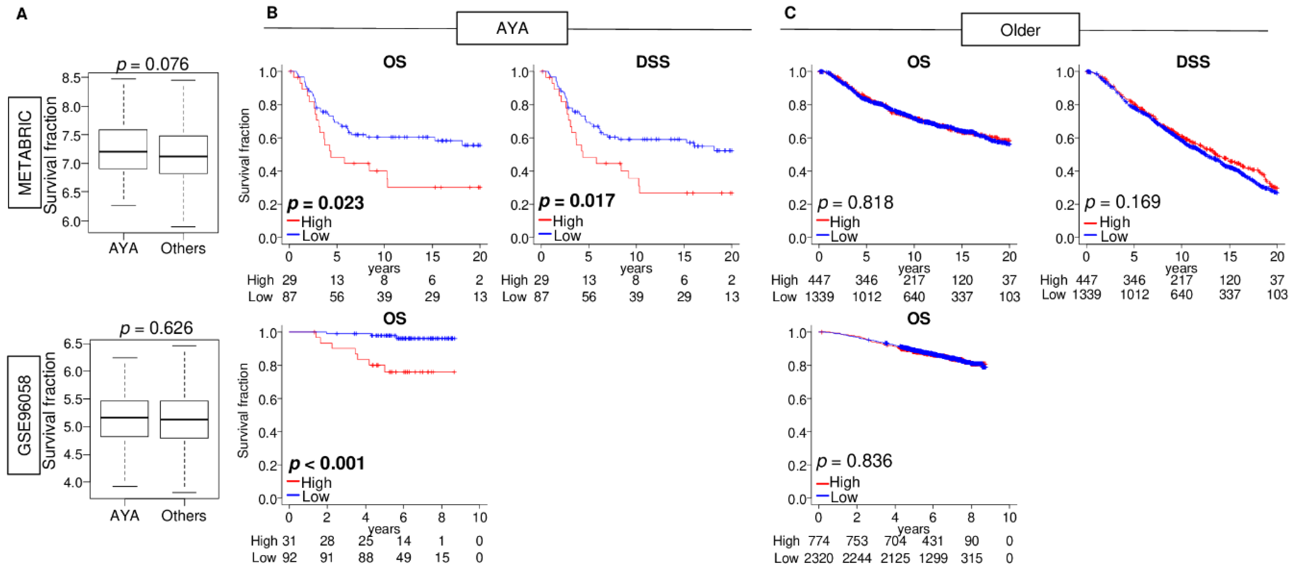
Since it is well established that breast cancer in Adolescents and Young Adults (AYA: age less than 40 years old) is typically more aggressive than in older patients, it was of interest whether GALNT1 expression differed by age. There was no difference in GALNT1 expression between AYA patients and older patients, which is consistent with the report on the Tn antigen [26] (Figure 2A). Interestingly, although though there was no survival difference by GALNT1 expression in the whole breast cancer cohort, as shown in Figure 1, we found that increased GALNT1 expression was significantly associated with worse OS and disease-specific survival (DSS) in AYA patients, whereas there was no association in the older patients in the METABRIC cohort (Figure 2B,C; p = 0.023 and 0.017 in AYA patients, and p = 0.818 and 0.169 in older patients, respectively). This association was validated in the GSE96058 cohort (p < 0.001). These results suggest that high levels of GALNT1 expression is significantly associated with worse patient survival in AYA patients, but not in older patients.

3.3. GALNT1 Expression Was an Independent Prognostic Biomarker among AYA Patients

We next investigated the association of GALNT1 expression with clinical features in AYA patients, but there were no associations in the two cohorts (Supplementary Figure S2). We next investigated the clinical relevance of GALNT1 expression with clinical features in AYA patients. We found that large tumors, lymph node metastasis, and high GALNT1 expression were all significantly associated with worse OS by univariate analysis in the AYA METABRIC cohort. Further, GALNT1 expression and lymph node metastasis was shown to be an independent factor by multivariate analysis (Table 1; hazard ratio (HR) = 2.05 and 1.78, 95%CI 1.03–4.09 and 1.06–3.29, p = 0.041 and 0.029, respectively). In the AYA GSE96058 cohort, there was no statistical significance in any of the clinical features; breast cancer subtype, Nottingham histological grade, T- and N-categories of American Joint Committee on Cancer staging, whereas GALNT1 expression was significantly associated with worse OS (Table 1; HR = 8.08, 95%CI 2.09–31.3, p = 0.002). These results indicated that GALNT1 expression was an independent prognostic biomarker for breast cancer in AYA patients.

3.4. High GALNT1 Breast Cancer Was Significantly Associated with Decreased CD8+ Cell Infiltration in AYA

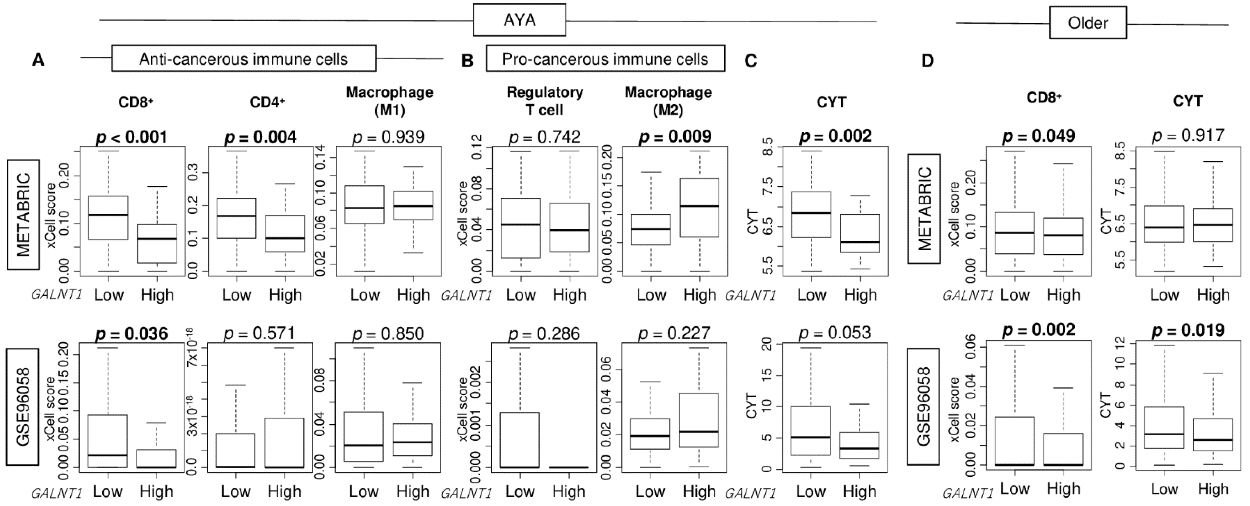
Given that the TN antigen has been found to suppress the tumor immune microenvironment and promote colorectal cancer growth [22], we examined the relationship between GALNT1 expression levels and the tumor immune microenvironment in breast cancer. In AYA patients, we found that high GALNT1 expression was significantly associated with a decreased number of CD8+ T cells in both the METABRIC and GSE96058 cohorts (Figure 3A; p < 0.001 and = 0.036, respectively). Other major immune cells, such as CD4+ T cells, M1 macrophages, regulatory T cells, and M2 macrophages, did not show significant difference between low and high GALNT1 expression in the GSE96058 cohort (Figure 3A,B). Cytolytic activity, which encapsulates overall immune cell killing, was decreased in GALNT1 high expression tumors (Figure 3C; p = 0.002 and = 0.053, respectively). In older breast cancer patients, high GALNT1 expression was also significantly associated with a decreased amount of CD8+ T cells in both cohorts, however, the difference was smaller than that in AYA patients (Figure 3D; p = 0.049 and = 0.002, respectively). Further, cytolytic activity trended lower in GALNT1 high expression tumors in older patients as well (Figure 3D; p = 0.917 and = 0.019, respectively). Furthermore, we investigated the relationship between GALNT1 expression and other cells in the tumor microenvironment, including fibroblasts, microvascular and lymphatic endothelial cells, and pericytes, in AYA breast cancer. We found that only pericytes were significantly highly infiltrated in the high GALNT1 group compared to the low group, consistently in both cohorts (Supplementary Figure S3A). There was no significant difference in mutation count between low and high GALNT1 group in AYA patients (Supplementary Figure S3B).

3.5. Cancer Cell Proliferation Was Not Associated with High GALNT1 Expressed Breast Cancer in Either AYA or Older Breast Cancer Patients

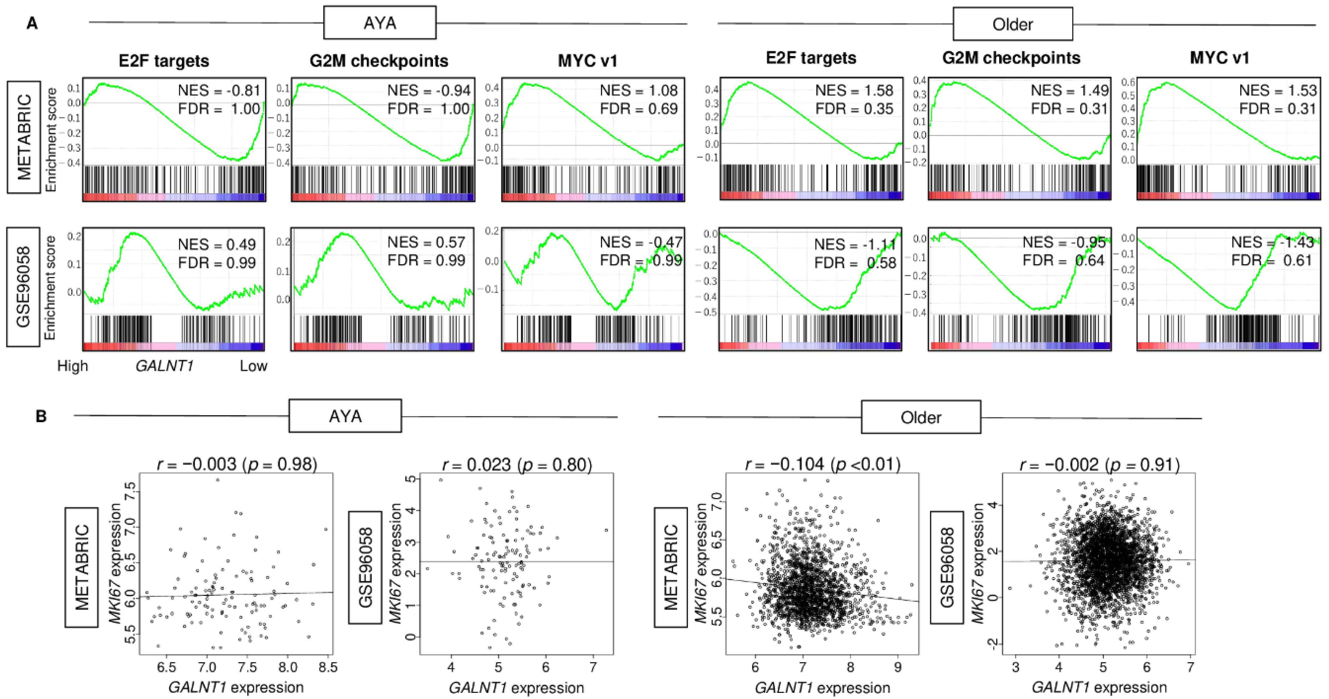
Given that GALNT1 expression was associated with patient survival in AYA patients but not in older patients, we hypothesized that GALNT1 expression may have different biological roles by age group. We investigated the association between GALNT1 expression and cell proliferation-related gene sets, which were significantly associated with breast cancer patient outcomes, as we had previously reported [43,44,45]. We found that cell proliferation-related gene sets, including E2F targets, G2M checkpoints, and MYC v1, did not demonstrate significant enrichment to high GALNT1 expression in either AYA patients or older patients (Figure 4A). Furthermore, GALNT1 expression was not correlated with Ki67 gene expression (MKI67), a cell proliferation marker used in clinics, in either age groups (Figure 4B; all Spearman’s rank correlation coefficient <0.150).

3.6. High GALNT1 Breast Cancer Enriched Angiogenesis, Epithelial Mesenchymal Transition (EMT), and Protein Secretion in the AYA Patient Group, but Not in the Older Patient Group

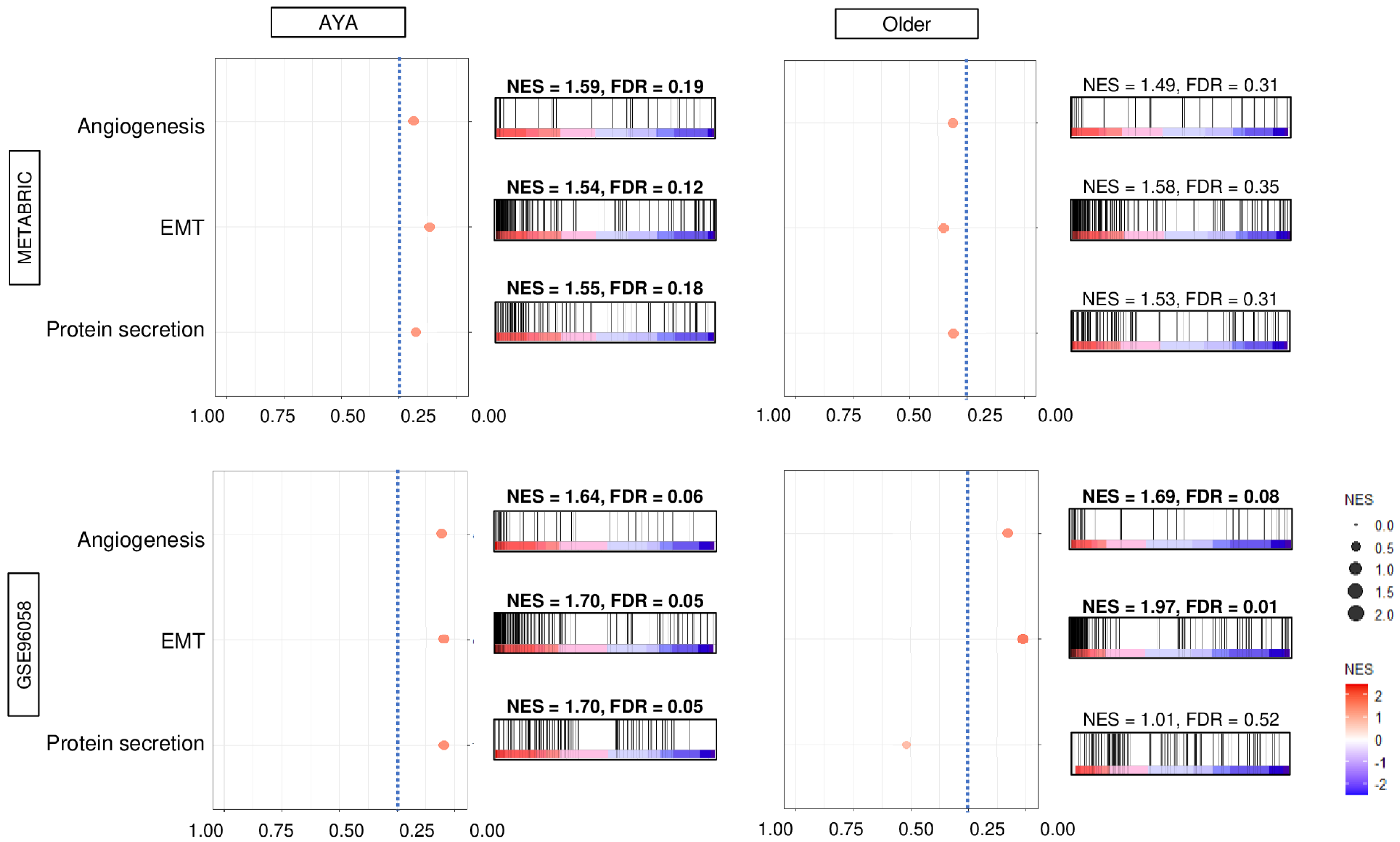
Finally, we investigated the biological functions using a hallmark gene set enrichment analysis. We found that breast cancer with high GALNT1 expression in AYA patients significantly enriched angiogenesis, EMT, and protein secretion gene sets, consistently in both the METABRIC and GSE96058 cohorts (Figure 5). In older patients; however, there were no hallmark gene sets that were significantly enriched to high GALNT1 expression consistently in both cohorts.

4. Discussion

GALNT1 expression, which generates the Tn antigen, was associated with decreased infiltration of CD8 T cells, enhanced angiogenesis, EMT, and protein secretion, and was a prognostic biomarker for breast cancer in AYA patients, but not older patients. Amongst both breast cancer cohorts, GALNT1 expression level was highest in TNBC, but was not associated with patient survival or clinical aggressiveness, including histological grade and metastasis. None of the hallmark cancer pathways, except for KRAS, were significantly enriched to high GALNT1 expression breast cancer by GSEA. Although there was no significant difference in GALNT1 expression levels between AYA patients and older patients, high GALNT1 expression was associated with significantly worse patient survival in AYA patients but not in older patients consistently in two independent cohorts. We found that high GALNT1 expression was an independent prognostic factor among several clinical features, including subtype, histological grade, AJCC T and N-category, in the AYA generation of both cohorts. Further, breast cancer with high GALNT1 expression was associated with less infiltration of CD8+ T cells, but not other immune cells. Increased levels of GALNT1 expression in breast cancer amongst AYA patients enriched angiogenesis, EMT, and protein secretion but not in older patients.
Altered levels of GALNT expression or distribution have been extensively reported in a wide range of cancer types [14]. GALNT expression results in marked alterations in GalNAc O-linked glycosylation, including exposure of truncated O-glycans such as the Tn antigen [13,46]. Altered levels of specific GALNTs were shown to have prognostic significance in cancers and were associated with changes in cell behavior, including cell proliferation, migration, invasion and metastasis in animal models [47]. In breast cancer, GALNT4 [48], GALNT6 [49], and GALNT8 [50], have been reported to be associated with patient outcomes, whereas no other study examines the clinical role of GALNT1. GALNT1 initiates O-glycosylation of glycoproteins; Sonic hedgehog protein (Shh), osteopontin, and MUC1, which contributes to the generation of breast cancer [14]. Although the role of GALNT1 in breast carcinogenesis is known, the clinical significance of its expression in breast cancer progression remains unclear. To our knowledge, our study is the first to report that GALNT1 mRNA expression levels are associated with decreased CD8 T-cell infiltration, angiogenesis and EMT, and poorer survival outcomes in AYA patients.
The Tn antigen is generated by GALNT and is present in 50–90% of all types of cancer, where it is associated with metastasis and decreased survival. Breast cancer was reported to have high levels of the Tn antigen [51]. Maria Florencia Festari et al. reported that the Tn antigen enhances breast tumor growth and lung metastasis by immunosuppression and suggested the Tn antigen-based immunotherapy approach [52]. Our study showed no significant association of GALNT1 expression with cell proliferation or metastasis. Further, GALNT1 expression was not associated with high infiltration of pro-cancerous immune cells, but with low infiltration of CD8+ T cells, which was evident in both the AYA generation and older patients, with the difference more pronounced in the AYA generation.
Data remains controversial regarding biological characteristics amongst breast tumors in AYA patients and older women. Although there have been several studies on genes that are differentially expressed between AYA patients and older patients, GALNT1, which has clinical significance in the AYA generation—with no difference in gene expression levels between generations—is unique. As the number of drug therapies for breast cancer has been rapidly expanding in recent years, the association between GALNT1 expression and response for other drugs warrants further exploration, particularly for drugs that target angiogenesis or EMT. For instance, the anti-angiogenesis approach clinical trial which failed for breast cancer may turn out to be effective for AYA patients. We cannot help but speculate that our findings on GALNT1 expression demonstrate its potential to become a predictive biomarker and a novel therapeutic target for breast cancer in the AYA generation [53].
Although intriguing, the data presented is still preliminary and requires additional validation through future research. Once validated, this information may prove beneficial to AYA breast cancer patients, and their physicians. As genotyping becomes increasingly more common in clinical decision making, this information may be especially vital in treatment decision making for early stage and/or advanced breast cancer. Remaining unanswered questions include how GALNT1 interacts with various pathways and immune cells.

5. Conclusions

GALNT1 expression was significantly associated with worse survival and associated with cell proliferation signaling in the AYA generation but not in older patients. GALNT1 expression may have potential as a predictive biomarker amongst AYA patients with breast cancer.

Supplementary Materials

Supplementary materials can be found at https://www.mdpi.com/article/10.3390/cancers15133489/s1. Supplementary Figure S1: Association of GALNT1 expression with clinicopathlogical features in the GSE96058 cohort. Figure S2: Association of GALNT1 expression with clinical aggressiveness in AYA patients. Figure S3: Association of GALNT1 expression with stromal fraction and mutation count in breast cancer of AYA patients.

Author Contributions

Conceptualization, I.E., K.T., M.O. and T.I.; methodology, K.T., M.O. and R.W.; formal analysis, M.O.; writing—original draft preparation, M.O.; writing—review and editing, A.Y., D.Z., K.C., K.N., K.T., L.Y. and S.Y.; supervision, K.T.; project administration, K.T. All authors have read and agreed to the published version of the manuscript.

Funding

This work was supported by National Institutes of Health, USA grant R01CA160688, R01CA250412, R37CA248018, as well as US Department of Defense BCRP grant W81XWH-19-1-0674 to K.T., and National Cancer Institute, USA cancer center support grant P30-CA016056 to Roswell Park Comprehensive Cancer Center (RPCCC).

Institutional Review Board Statement

Not applicable.

Informed Consent Statement

This study examined human data that had been generated in the past by other studies. Informed consent was therefore not obtained in this study.

Data Availability Statement

All the cohorts/datasets used in this study; Molecular Taxonomy of Breast Cancer International Consortium (METABRIC) and GSE96058 are all publicly available without any restrictions via cBioportal or Gene Expression Omnibus (GEO).

Conflicts of Interest

The authors declare no conflict of interest.

Abbreviations

AJCCAmerican Joint Committee on Cancer
DFSdisease-free survival
DSSdisease-specific survival
ERestrogen receptor
FDRfalse discovery rate
GSEAgene set enrichment analysis
HER2human epidermal growth factor receptor 2
METABRICMolecular Taxonomy of Breast Cancer International Consortium
NESnormalized enrichment score
OSoverall survival
TNBCtriple-negative breast cancer

References

  1. Cathcart-Rake, E.J.; Ruddy, K.J.; Bleyer, A.; Johnson, R.H. Breast Cancer in Adolescent and Young Adult Women under the Age of 40 Years. JCO Oncol. Pract. 2021, 17, 305–313. [Google Scholar] [CrossRef]
  2. Futamura, M.; Yoshida, K. Current status of AYA-generation breast cancer: Trends worldwide and in Japan. Int. J. Clin. Oncol. 2022, 27, 16–24. [Google Scholar] [CrossRef]
  3. Lewis, D.R.; Siembida, E.J.; Seibel, N.L.; Smith, A.W.; Mariotto, A.B. Survival outcomes for cancer types with the highest death rates for adolescents and young adults, 1975–2016. Cancer 2021, 127, 4277–4286. [Google Scholar] [CrossRef]
  4. Murphy, B.L.; Day, C.N.; Hoskin, T.L.; Habermann, E.B.; Boughey, J.C. Adolescents and Young Adults with Breast Cancer have More Aggressive Disease and Treatment than Patients in Their Forties. Ann. Surg. Oncol. 2019, 26, 3920–3930. [Google Scholar] [CrossRef] [PubMed]
  5. Johnson, R.H.; Anders, C.K.; Litton, J.K.; Ruddy, K.J.; Bleyer, A. Breast cancer in adolescents and young adults. Pediatr Blood Cancer 2018, 65, e27397. [Google Scholar] [CrossRef]
  6. Mirza, Z.; Ansari, M.S.; Iqbal, M.S.; Ahmad, N.; Alganmi, N.; Banjar, H.; Al-Qahtani, M.H.; Karim, S. Identification of Novel Diagnostic and Prognostic Gene Signature Biomarkers for Breast Cancer Using Artificial Intelligence and Machine Learning Assisted Transcriptomics Analysis. Cancers 2023, 15, 3237. [Google Scholar] [CrossRef]
  7. Subramanian, A.; Tamayo, P.; Mootha, V.K.; Mukherjee, S.; Ebert, B.L.; Gillette, M.A.; Paulovich, A.; Pomeroy, S.L.; Golub, T.R.; Lander, E.S.; et al. Gene set enrichment analysis: A knowledge-based approach for interpreting genome-wide expression profiles. Proc. Natl. Acad. Sci. USA 2005, 102, 15545–15550. [Google Scholar] [CrossRef]
  8. Mootha, V.K.; Lindgren, C.M.; Eriksson, K.F.; Subramanian, A.; Sihag, S.; Lehar, J.; Puigserver, P.; Carlsson, E.; Ridderstråle, M.; Laurila, E.; et al. PGC-1alpha-responsive genes involved in oxidative phosphorylation are coordinately downregulated in human diabetes. Nat. Genet. 2003, 34, 267–273. [Google Scholar] [CrossRef] [PubMed]
  9. Aran, D.; Hu, Z.; Butte, A.J. xCell: Digitally portraying the tissue cellular heterogeneity landscape. Genome Biol. 2017, 18, 220. [Google Scholar] [CrossRef] [PubMed] [Green Version]
  10. Newman, A.M.; Liu, C.L.; Green, M.R.; Gentles, A.J.; Feng, W.; Xu, Y.; Hoang, C.D.; Diehn, M.; Alizadeh, A.A. Robust enumeration of cell subsets from tissue expression profiles. Nat. Methods 2015, 12, 453–457. [Google Scholar] [CrossRef] [PubMed] [Green Version]
  11. Li, B.; Severson, E.; Pignon, J.C.; Zhao, H.; Li, T.; Novak, J.; Jiang, P.; Shen, H.; Aster, J.C.; Rodig, S.; et al. Comprehensive analyses of tumor immunity: Implications for cancer immunotherapy. Genome Biol. 2016, 17, 174. [Google Scholar] [CrossRef] [Green Version]
  12. Yoshihara, K.; Shahmoradgoli, M.; Martínez, E.; Vegesna, R.; Kim, H.; Torres-Garcia, W.; Treviño, V.; Shen, H.; Laird, P.W.; Levine, D.A.; et al. Inferring tumour purity and stromal and immune cell admixture from expression data. Nat. Commun. 2013, 4, 2612. [Google Scholar] [CrossRef] [PubMed] [Green Version]
  13. Li, C.; Du, Y.; Yang, Z.; He, L.; Wang, Y.; Hao, L.; Ding, M.; Yan, R.; Wang, J.; Fan, Z. GALNT1-Mediated Glycosylation and Activation of Sonic Hedgehog Signaling Maintains the Self-Renewal and Tumor-Initiating Capacity of Bladder Cancer Stem Cells. Cancer Res. 2016, 76, 1273–1283. [Google Scholar] [CrossRef] [PubMed] [Green Version]
  14. Fang, R.; Xu, F.; Shi, H.; Wu, Y.; Cao, C.; Li, H.; Ye, K.; Zhang, Y.; Liu, Q.; Zhang, S.; et al. LAMTOR5 raises abnormal initiation of O-glycosylation in breast cancer metastasis via modulating GALNT1 activity. Oncogene 2020, 39, 2290–2304. [Google Scholar] [CrossRef]
  15. Park, J.H.; Nishidate, T.; Kijima, K.; Ohashi, T.; Takegawa, K.; Fujikane, T.; Hirata, K.; Nakamura, Y.; Katagiri, T. Critical roles of mucin 1 glycosylation by transactivated polypeptide N-acetylgalactosaminyltransferase 6 in mammary carcinogenesis. Cancer Res. 2010, 70, 2759–2769. [Google Scholar] [CrossRef] [Green Version]
  16. Kariya, Y.; Kanno, M.; Matsumoto-Morita, K.; Konno, M.; Yamaguchi, Y.; Hashimoto, Y. Osteopontin O-glycosylation contributes to its phosphorylation and cell-adhesion properties. Biochem. J. 2014, 463, 93–102. [Google Scholar] [CrossRef] [PubMed]
  17. Pinho, S.S.; Reis, C.A. Glycosylation in cancer: Mechanisms and clinical implications. Nat. Rev. Cancer 2015, 15, 540–555. [Google Scholar] [CrossRef] [PubMed]
  18. Parameswaran, R.; Tan, W.B.; Nga, M.E.; Soon, G.S.T.; Ngiam, K.Y.; Brooks, S.A.; Sadler, G.P.; Mihai, R. Binding of aberrant glycoproteins recognizable by Helix pomatia agglutinin in adrenal cancers. BJS Open 2018, 2, 353–359. [Google Scholar] [CrossRef]
  19. Ju, T.; Aryal, R.P.; Kudelka, M.R.; Wang, Y.; Cummings, R.D. The Cosmc connection to the Tn antigen in cancer. Cancer Biomark 2014, 14, 63–81. [Google Scholar] [CrossRef]
  20. Taylor-Papadimitriou, J.; Epenetos, A.A. Exploiting altered glycosylation patterns in cancer: Progress and challenges in diagnosis and therapy. Trends Biotechnol. 1994, 12, 227–233. [Google Scholar] [CrossRef]
  21. Konska, G.; Guerry, M.; Caldefie-Chezet, F.; De Latour, M.; Guillot, J. Study of the expression of Tn antigen in different types of human breast cancer cells using VVA-B4 lectin. Oncol. Rep. 2006, 15, 305–310. [Google Scholar] [CrossRef] [PubMed] [Green Version]
  22. Cornelissen, L.A.M.; Blanas, A.; Zaal, A.; van der Horst, J.C.; Kruijssen, L.J.W.; O’Toole, T.; van Kooyk, Y.; van Vliet, S.J. Tn Antigen Expression Contributes to an Immune Suppressive Microenvironment and Drives Tumor Growth in Colorectal Cancer. Front. Oncol. 2020, 10, 1622. [Google Scholar] [CrossRef]
  23. Konno, A.; Hoshino, Y.; Terashima, S.; Motoki, R.; Kawaguchi, T. Carbohydrate expression profile of colorectal cancer cells is relevant to metastatic pattern and prognosis. Clin. Exp. Metastasis 2002, 19, 61–70. [Google Scholar] [CrossRef]
  24. Laack, E.; Nikbakht, H.; Peters, A.; Kugler, C.; Jasiewicz, Y.; Edler, L.; Hossfeld, D.K.; Schumacher, U. Lectin histochemistry of resected adenocarcinoma of the lung: Helix pomatia agglutinin binding is an independent prognostic factor. Am. J. Pathol. 2002, 160, 1001–1008. [Google Scholar] [CrossRef]
  25. Troup, S.; Njue, C.; Kliewer, E.V.; Parisien, M.; Roskelley, C.; Chakravarti, S.; Roughley, P.J.; Murphy, L.C.; Watson, P.H. Reduced expression of the small leucine-rich proteoglycans, lumican, and decorin is associated with poor outcome in node-negative invasive breast cancer. Clin. Cancer Res. 2003, 9, 207–214. [Google Scholar] [PubMed]
  26. Tsuchiya, A.; Kanno, M.; Kawaguchi, T.; Endo, Y.; Zhang, G.J.; Ohtake, T.; Kimijima, I.I. Prognostic Relevance of Tn Expression in Breast Cancer. Breast Cancer 1999, 6, 175–180. [Google Scholar] [CrossRef] [PubMed]
  27. Curtis, C.; Shah, S.P.; Chin, S.F.; Turashvili, G.; Rueda, O.M.; Dunning, M.J.; Speed, D.; Lynch, A.G.; Samarajiwa, S.; Yuan, Y.; et al. The genomic and transcriptomic architecture of 2,000 breast tumours reveals novel subgroups. Nature 2012, 486, 346–352. [Google Scholar] [CrossRef]
  28. Brueffer, C.; Gladchuk, S.; Winter, C.; Vallon-Christersson, J.; Hegardt, C.; Häkkinen, J.; George, A.M.; Chen, Y.; Ehinger, A.; Larsson, C.; et al. The mutational landscape of the SCAN-B real-world primary breast cancer transcriptome. EMBO Mol. Med. 2020, 12, e12118. [Google Scholar] [CrossRef]
  29. Liberzon, A.; Birger, C.; Thorvaldsdóttir, H.; Ghandi, M.; Mesirov, J.P.; Tamayo, P. The Molecular Signatures Database (MSigDB) hallmark gene set collection. Cell Syst. 2015, 1, 417–425. [Google Scholar] [CrossRef] [Green Version]
  30. Oshi, M.; Patel, A.; Le, L.; Tokumaru, Y.; Yan, L.; Matsuyama, R.; Endo, I.; Takabe, K. G2M checkpoint pathway alone is associated with drug response and survival among cell proliferation-related pathways in pancreatic cancer. Am. J. Cancer Res. 2021, 11, 3070–3084. [Google Scholar]
  31. Oshi, M.; Satyananda, V.; Angarita, F.A.; Kim, T.H.; Tokumaru, Y.; Yan, L.; Matsuyama, R.; Endo, I.; Nagahashi, M.; Takabe, K. Angiogenesis is associated with an attenuated tumor microenvironment, aggressive biology, and worse survival in gastric cancer patients. Am. J. Cancer Res. 2021, 11, 1659–1671. [Google Scholar] [PubMed]
  32. Oshi, M.; Tokumaru, Y.; Angarita, F.A.; Lee, L.; Yan, L.; Matsuyama, R.; Endo, I.; Takabe, K. Adipogenesis in triple-negative breast cancer is associated with unfavorable tumor immune microenvironment and with worse survival. Sci. Rep. 2021, 11, 12541. [Google Scholar] [CrossRef] [PubMed]
  33. Oshi, M.; Tokumaru, Y.; Angarita, F.A.; Yan, L.; Matsuyama, R.; Endo, I.; Takabe, K. Degree of Early Estrogen Response Predict Survival after Endocrine Therapy in Primary and Metastatic ER-Positive Breast Cancer. Cancers 2020, 12, 3557. [Google Scholar] [CrossRef] [PubMed]
  34. Oshi, M.; Tokumaru, Y.; Patel, A.; Yan, L.; Matsuyama, R.; Endo, I.; Katz, M.H.G.; Takabe, K. A Novel Four-Gene Score to Predict Pathologically Complete (R0) Resection and Survival in Pancreatic Cancer. Cancers 2020, 12, 3635. [Google Scholar] [CrossRef]
  35. Oshi, M.; Asaoka, M.; Tokumaru, Y.; Yan, L.; Matsuyama, R.; Ishikawa, T.; Endo, I.; Takabe, K. CD8 T Cell Score as a Prognostic Biomarker for Triple Negative Breast Cancer. Int. J. Mol. Sci. 2020, 21, 6968. [Google Scholar] [CrossRef]
  36. Oshi, M.; Kawaguchi, T.; Yan, L.; Peng, X.; Qi, Q.; Tian, W.; Schulze, A.; McDonald, K.A.; Narayanan, S.; Young, J.; et al. Immune cytolytic activity is associated with reduced intra-tumoral genetic heterogeneity and with better clinical outcomes in triple negative breast cancer. Am. J. Cancer Res. 2021, 11, 3628–3644. [Google Scholar]
  37. Oshi, M.; Newman, S.; Tokumaru, Y.; Yan, L.; Matsuyama, R.; Kalinski, P.; Endo, I.; Takabe, K. Plasmacytoid Dendritic Cell (pDC) Infiltration Correlate with Tumor Infiltrating Lymphocytes, Cancer Immunity, and Better Survival in Triple Negative Breast Cancer (TNBC) More Strongly than Conventional Dendritic Cell (cDC). Cancers 2020, 12, 3342. [Google Scholar] [CrossRef]
  38. Oshi, M.; Gandhi, S.; Huyser, M.R.; Tokumaru, Y.; Yan, L.; Yamada, A.; Matsuyama, R.; Endo, I.; Takabe, K. MELK expression in breast cancer is associated with infiltration of immune cell and pathological compete response (pCR) after neoadjuvant chemotherapy. Am. J. Cancer Res. 2021, 11, 4421–4437. [Google Scholar] [CrossRef]
  39. Oshi, M.; Asaoka, M.; Tokumaru, Y.; Angarita, F.A.; Yan, L.; Matsuyama, R.; Zsiros, E.; Ishikawa, T.; Endo, I.; Takabe, K. Abundance of Regulatory T Cell (Treg) as a Predictive Biomarker for Neoadjuvant Chemotherapy in Triple-Negative Breast Cancer. Cancers 2020, 12, 3038. [Google Scholar] [CrossRef]
  40. Oshi, M.; Angarita, F.A.; Tokumaru, Y.; Yan, L.; Matsuyama, R.; Endo, I.; Takabe, K. A Novel Three-Gene Score as a Predictive Biomarker for Pathologically Complete Response after Neoadjuvant Chemotherapy in Triple-Negative Breast Cancer. Cancers 2021, 13, 2401. [Google Scholar] [CrossRef]
  41. Oshi, M.; Gandhi, S.; Tokumaru, Y.; Yan, L.; Yamada, A.; Matsuyama, R.; Ishikawa, T.; Endo, I.; Takabe, K. Conflicting roles of EGFR expression by subtypes in breast cancer. Am. J. Cancer Res. 2021, 11, 5094–5110. [Google Scholar] [PubMed]
  42. Oshi, M.; Newman, S.; Tokumaru, Y.; Yan, L.; Matsuyama, R.; Endo, I.; Takabe, K. Inflammation Is Associated with Worse Outcome in the Whole Cohort but with Better Outcome in Triple-Negative Subtype of Breast Cancer Patients. J. Immunol. Res. 2020, 2020, 5618786. [Google Scholar] [CrossRef] [PubMed]
  43. Schulze, A.; Oshi, M.; Endo, I.; Takabe, K. MYC Targets Scores Are Associated with Cancer Aggressiveness and Poor Survival in ER-Positive Primary and Metastatic Breast Cancer. Int. J. Mol. Sci. 2020, 21, 8127. [Google Scholar] [CrossRef] [PubMed]
  44. Oshi, M.; Takahashi, H.; Tokumaru, Y.; Yan, L.; Rashid, O.M.; Nagahashi, M.; Matsuyama, R.; Endo, I.; Takabe, K. The E2F Pathway Score as a Predictive Biomarker of Response to Neoadjuvant Therapy in ER+/HER2- Breast Cancer. Cells 2020, 9, 1643. [Google Scholar] [CrossRef]
  45. Oshi, M.; Takahashi, H.; Tokumaru, Y.; Yan, L.; Rashid, O.M.; Matsuyama, R.; Endo, I.; Takabe, K. G2M Cell Cycle Pathway Score as a Prognostic Biomarker of Metastasis in Estrogen Receptor (ER)-Positive Breast Cancer. Int. J. Mol. Sci. 2020, 21, 2921. [Google Scholar] [CrossRef] [Green Version]
  46. Song, K.H.; Park, M.S.; Nandu, T.S.; Gadad, S.; Kim, S.C.; Kim, M.Y. GALNT14 promotes lung-specific breast cancer metastasis by modulating self-renewal and interaction with the lung microenvironment. Nat. Commun. 2016, 7, 13796. [Google Scholar] [CrossRef]
  47. Nguyen, A.T.; Chia, J.; Ros, M.; Hui, K.M.; Saltel, F.; Bard, F. Organelle Specific O-Glycosylation Drives MMP14 Activation, Tumor Growth, and Metastasis. Cancer Cell 2017, 32, 639–653. [Google Scholar] [CrossRef] [Green Version]
  48. Wu, Q.; Zhang, C.; Zhang, K.; Chen, Q.; Wu, S.; Huang, H.; Huang, T.; Zhang, N.; Wang, X.; Li, W.; et al. ppGalNAc-T4-catalyzed O-Glycosylation of TGF-β type II receptor regulates breast cancer cells metastasis potential. J. Biol. Chem. 2021, 296, 100119. [Google Scholar] [CrossRef]
  49. Liu, C.; Li, Z.; Xu, L.; Shi, Y.; Zhang, X.; Shi, S.; Hou, K.; Fan, Y.; Li, C.; Wang, X.; et al. GALNT6 promotes breast cancer metastasis by increasing mucin-type O-glycosylation of α2M. Aging Albany NY 2020, 12, 11794–11811. [Google Scholar] [CrossRef]
  50. Huang, T.; Wu, Q.; Huang, H.; Zhang, C.; Wang, L.; Wang, L.; Liu, Y.; Li, W.; Zhang, J.; Liu, Y. Expression of GALNT8 and O-glycosylation of BMP receptor 1A suppress breast cancer cell proliferation by upregulating ERα levels. Biochim. Biophys. Acta Gen. Subj. 2022, 1866, 130046. [Google Scholar] [CrossRef]
  51. Rømer, T.B.; Aasted, M.K.M.; Dabelsteen, S.; Groen, A.; Schnabel, J.; Tan, E.; Pedersen, J.W.; Haue, A.D.; Wandall, H.H. Mapping of truncated O-glycans in cancers of epithelial and non-epithelial origin. Br. J. Cancer 2021, 125, 1239–1250. [Google Scholar] [CrossRef] [PubMed]
  52. Festari, M.F.; da Costa, V.; Rodríguez-Zraquia, S.A.; Costa, M.; Landeira, M.; Lores, P.; Solari-Saquieres, P.; Kramer, M.G.; Freire, T. The tumor-associated Tn antigen fosters lung metastasis and recruitment of regulatory T cells in triple negative breast cancer. Glycobiology 2022, 32, 366–379. [Google Scholar] [CrossRef] [PubMed]
  53. Bear, H.D.; Tang, G.; Rastogi, P.; Geyer, C.E., Jr.; Robidoux, A.; Atkins, J.N.; Baez-Diaz, L.; Brufsky, A.M.; Mehta, R.S.; Fehrenbacher, L.; et al. Bevacizumab added to neoadjuvant chemotherapy for breast cancer. N. Engl. J. Med. 2012, 366, 310–320. [Google Scholar] [CrossRef] [PubMed] [Green Version]
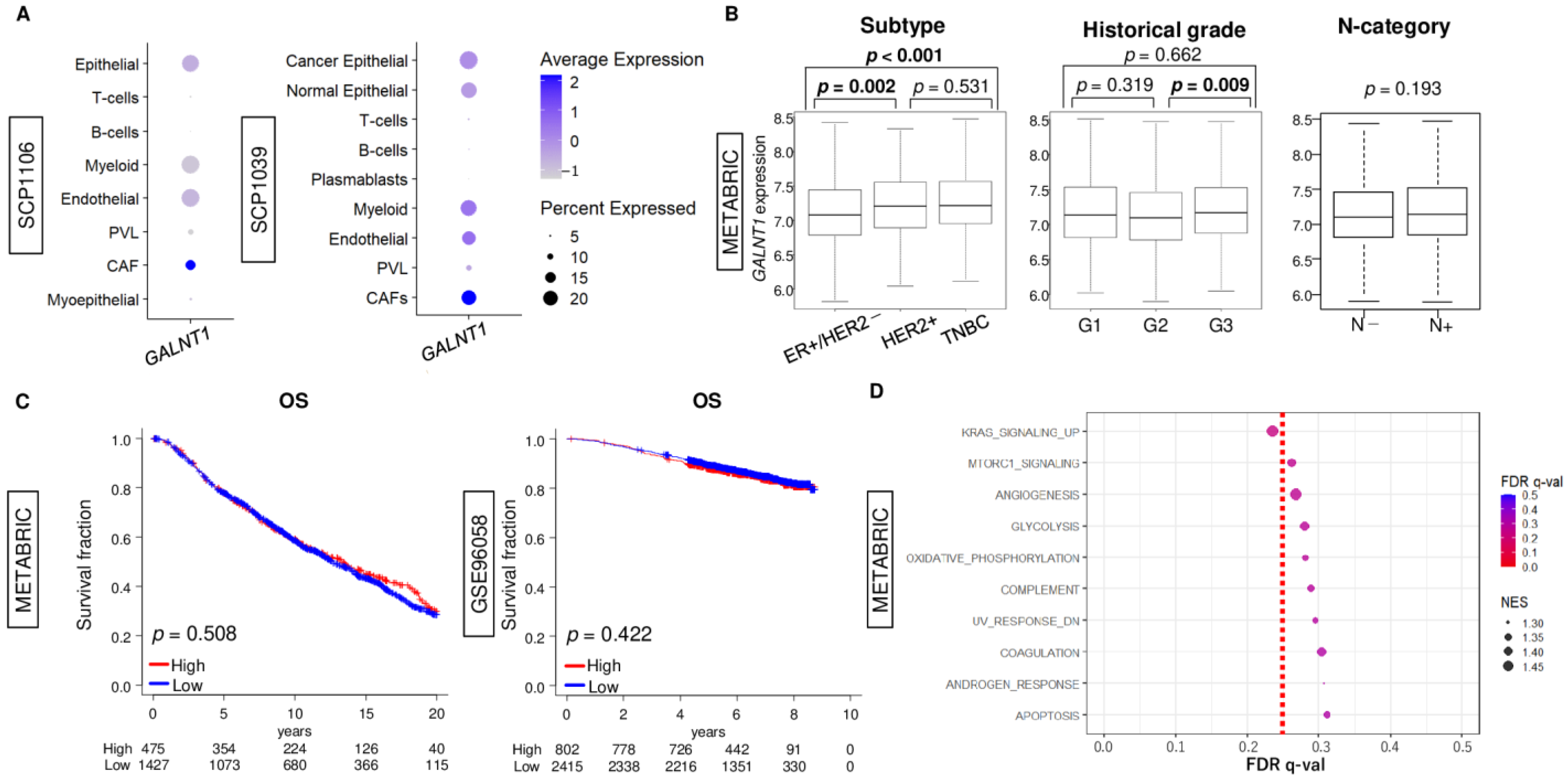
Figure 1. Association of GALNT1 expression with clinicopathological features, breast cancer patient survival, and biological function. (A) Boxplots of GALNT1 expression by cancer cells, immune cells; T cells, B cells, Myeloid, and stromal cells; epithelial, endothelial, cancer-associated fibroblasts (CAF), plasma blasts, and perivascular-like (PVL) cells, in the SCP1106 and SCP1039 single-cell sequence cohorts. (B) Boxplots of GALNT1 expression by subtype (ER-positive/HER2-negative, HER2-positive, and triple-negative breast cancer (TNBC)), histological grade (G1–G3), and lymph node metastasis (N-negative and N-positive) in METABRIC cohort. Significant difference among groups was calculated by the Kruskal–Wallis and Mann–Whitney U tests. (C) Kaplan–Meier plots of overall survival (OS) in breast cancer patients in the METABRIC, and OS in GSE96058 cohorts by low (blue) and high (red) GALNT1 expression groups. Significant difference of survival between groups was calculated by log-rank test. The top one-fourth was used as a cut-off to divide low and high groups for each cohort. (D) Dot plots of the ten gene sets with the lowest FDR q-value in the enrichment analysis between low and high GALNT1 low and high breast cancer groups using the GSEA algorithm. The top one-fourth was used as a cut-off to divide low and high groups.
Cancers 15 03489 g001
Figure 2. Association of GALNT1 expression with patient survival in AYA patients and older patients. (A) Boxplots of GALNT1 expression by age (AYA (less than 40 years old) vs. older (more than 40 years old)) in both METABRIC and GSE96058 cohorts. Significant difference among groups was calculated by the Mann–Whitney U tests. Kaplan–Meier plots of disease-specific survival (DSS) and overall survival (OS) in (B) AYA patients and (C) older patients in the METABRIC, and OS in GSE96058 cohorts by low (blue) and high (red) GALNT1 expression groups. Significant difference of survival between groups was calculated by log-rank test. The top one-fourth was used as a cut-off to divide low and high groups for each cohort.
Cancers 15 03489 g002
Figure 3. The association of the GALNT1 expression with immunity in the tumor immune microenvironment of breast cancer. Boxplots of infiltration fraction of (A) anti-cancerous immune cells; CD8+ T cells, CD4+ T cells, M1 macrophages, and Th1 cells, and (B) pro-cancerous immune cells; regulatory T cells, M2 macrophages, and Th2 cells, and (C) cytolytic activity (CYT) by low and high GALNT1 expression groups in METABRIC and GSE96058 cohorts in AYA. (D) CD8+ T cells and CYT by low and high GALNT1 expression groups in both cohorts in older patients. Significant difference between two groups was calculated by the Mann–Whitney U test.
Cancers 15 03489 g003
Figure 4. The association of high GALNT1 breast cancer with cell proliferation signaling in AYA and older patients of the METABRIC and GSE96058 cohorts. (A) Enrichment plots of cell proliferation-related gene sets; E2F targets, G2M checkpoints, and MYC target v1, in high GALNT1 expression groups of AYA and older patient in both cohorts. (B) Correlation plots of gene expression between GALNT1 and MKI67 of AYA and older patient in both cohorts. Spearman’s correlation coefficient (r) was used to the analysis. NES, normalized enrichment score.
Cancers 15 03489 g004
Figure 5. Biological feature of high GALNT1 breast cancer in AYA patients and older patients of the METABRIC and GSE96058 cohorts. Dot plots with distribution map of the three gene sets, which showed significant differences consistently in both cohorts, with the lowest FDR q-value in the enrichment analysis between GALNT1 low- and high-expression breast cancer groups using the GSEA algorithm. The top one-fourth was used as a cut-off to divide low and high groups. FDR, false discovery rate; NES, normalized enrichment score.
Cancers 15 03489 g005
Table 1. Uni- and multivariate analysis in AYA of the METABRIC and GSE96058 cohorts.
METABRIC (OS)Univariate Multivariate GSE96058 (OS)Univariate
HR95%CIp HR95%CIp HR95%CIp
SubtypeTNBC vs. others1.110.65–1.890.708 SubtypeTNBC vs. others1.350.15–12.10.790
GradeG3 vs. G1/21.730.85–3.530.134 GradeG3 vs. G1/22.190.46–10.30.322
TT3/4 vs. T1/20.520.29–0.920.026*0.690.37–1.270.23 TT3/4 vs. T1/2--
NN+ vs. N−2.631.39–4.990.003*2.051.03–4.090.041*NN+ vs. N−0.780.16–3.730.751
GALNT1High vs. Low2.361.32–4.200.004*1.871.06–3.290.029*GALNT1High vs. Low8.082.09–31.30.002*
*; significant difference.
Disclaimer/Publisher’s Note: The statements, opinions and data contained in all publications are solely those of the individual author(s) and contributor(s) and not of MDPI and/or the editor(s). MDPI and/or the editor(s) disclaim responsibility for any injury to people or property resulting from any ideas, methods, instructions or products referred to in the content.