
Evaluation of Staging Systems for Cancer of the Nasal Vestibule

 , , , ,
  , , and
Abstract
:Simple Summary
Abstract
1. Introduction
2. Materials and Methods
3. Results
4. Discussion
Pitfalls and Benefits of Available Staging Systems
5. Conclusions
Author Contributions
Funding
Institutional Review Board Statement
Informed Consent Statement
Data Availability Statement
Conflicts of Interest
References
- Agger, A.; von Buchwald, C.; Madsen, A.R.; Yde, J.; Lesnikova, I.; Christensen, C.B.; Foghsgaard, S.; Christensen, T.B.; Hansen, H.S.; Larsen, S.; et al. Squamous cell carcinoma of the nasal vestibule 1993–2002: A nationwide retrospective study from DAHANCA. Head Neck 2009, 31, 1593–1599. [Google Scholar] [CrossRef] [PubMed]
- Wong, C.S.; Cummings, B.J. The place of radiation therapy in the treatment of squamous cell carcinoma of the nasal vestibule. A review. Acta Oncol. 1988, 27, 203–208. [Google Scholar] [CrossRef] [PubMed]
- Zaoui, K.; Plinkert, P.K.; Federspil, P.A. Primary surgical treatment of nasal vestibule cancer—Therapeutic outcome and reconstructive strategies. Rhinology 2018, 56, 393–399. [Google Scholar] [CrossRef]
- Bussu, F.; Tagliaferri, L.; Mattiucci, G.; Parrilla, C.; Dinapoli, N.; Micciche, F.; Artuso, A.; Galli, J.; Almadori, G.; Valentini, V.; et al. Comparison of interstitial brachytherapy and surgery as primary treatments for nasal vestibule carcinomas. Laryngoscope 2016, 126, 367–371. [Google Scholar] [CrossRef]
- Lipman, D.; Verhoef, L.C.; Takes, R.P.; Kaanders, J.H.; Janssens, G.O. Outcome and toxicity profile after brachytherapy for squamous cell carcinoma of the nasal vestibule. Head Neck 2015, 37, 1297–1303. [Google Scholar] [CrossRef] [PubMed]
- Vital, D.; Morand, G.; Huber, G.F.; Studer, G.; Holzmann, D. Outcome in squamous cell carcinoma of the nasal vestibule: A single center experience. Head Neck 2015, 37, 46–51. [Google Scholar] [CrossRef]
- Langendijk, J.A.; Poorter, R.; Leemans, C.R.; de Bree, R.; Doornaert, P.; Slotman, B.J. Radiotherapy of squamous cell carcinoma of the nasal vestibule. Int. J. Radiat. Oncol. Biol. Phys. 2004, 59, 1319–1325. [Google Scholar] [CrossRef]
- Wallace, A.; Morris, C.G.; Kirwan, J.; Amdur, R.J.; Werning, J.W.; Mendenhall, W.M. Radiotherapy for squamous cell carcinoma of the nasal vestibule. Am. J. Clin. Oncol. 2007, 30, 612–616. [Google Scholar] [CrossRef]
- Lambertoni, A.; Cherubino, M.; Battaglia, P.; De Col, A.; Giovannardi, M.; Antognoni, P.; Valdatta, L.; Karligkiotis, A.; Bignami, M.; Castelnuovo, P.; et al. Squamous Cell Carcinoma of Nasal Vestibule and Pyramid: Outcomes and Reconstructive Strategies. Laryngoscope 2021, 131, E1198–E1208. [Google Scholar] [CrossRef]
- Chabrillac, E.; Talawdekar, A.; Garikipati, S.; Varley, I.; Sionis, S.; Beasley, N.; Jackson, R. A single centre’s experience of 23 cases of total rhinectomy for the treatment of squamous cell carcinoma involving the nasal vestibule. Eur. Arch. Oto-Rhino-Laryngol. 2022, 279, 2069–2075. [Google Scholar] [CrossRef]
- Levendag, P.C.; Nijdam, W.M.; van Moolenburgh, S.E.; Tan, L.; Noever, I.; van Rooy, P.; Mureau, M.A.; Jansen, P.P.; Munte, K.; Hofer, S.O. Interstitial radiation therapy for early-stage nasal vestibule cancer: A continuing quest for optimal tumor control and cosmesis. Int. J. Radiat. Oncol. Biol. Phys. 2006, 66, 160–169. [Google Scholar] [CrossRef]
- Vanneste, B.G.; Lopez-Yurda, M.; Tan, I.B.; Balm, A.J.; Borst, G.R.; Rasch, C.R. Irradiation of localized squamous cell carcinoma of the nasal vestibule. Head Neck 2016, 38 (Suppl. 1), E1870–E1875. [Google Scholar] [CrossRef] [PubMed]
- Czerwinski, M.D.; van Leeuwen, R.G.H.; Kaanders, J.; Zwijnenburg, E.M.; Lipman, D.; Takes, R.P.; Verhoef, C.G. Image Guided Brachytherapy for Cancer of the Nasal Vestibule: Local Control and Cosmesis. Int. J. Radiat. Oncol. Biol. Phys. 2019, 103, 913–921. [Google Scholar] [CrossRef] [PubMed]
- Czerwinski, M.D.; Jansen, P.P.; Zwijnenburg, E.M.; Al-Mamgani, A.; Vergeer, M.R.; Langendijk, J.A.; Wesseling, F.W.R.; Kaanders, J.; Verhoef, C.G. Radiotherapy as nose preservation treatment strategy for cancer of the nasal vestibule: The Dutch experience. Radiother. Oncol. 2021, 164, 20–26. [Google Scholar] [CrossRef] [PubMed]
- Bacorro, W.; Escande, A.; Temam, S.; Dumas, I.; Routier, E.; Gensse, M.C.; Blanchard, P.; Janot, F.; Mateus, C.; Tao, Y.; et al. Clinical outcomes after interstitial brachytherapy for early-stage nasal squamous cell carcinoma. Brachytherapy 2017, 16, 1021–1027. [Google Scholar] [CrossRef]
- Scheurleer, W.F.J.; Dehnad, H.; Braunius, W.W.; Janssen, L.M.; Tijink, B.M.; Breimer, G.E.; Smid, E.J.; Devriese, L.A.; Bree, R.; Ridder, M.; et al. Long-term oncological follow-up after mold-based pulsed dose rate brachytherapy for early stage squamous cell carcinoma of the nasal vestibule: A single center experience of 68 patients over a 17-year period. Brachytherapy 2022, 22, 221–230. [Google Scholar] [CrossRef]
- Tagliaferri, L.; Carra, N.; Lancellotta, V.; Rizzo, D.; Casa, C.; Mattiucci, G.; Parrilla, C.; Fionda, B.; Deodato, F.; Cornacchione, P.; et al. Interventional radiotherapy as exclusive treatment for primary nasal vestibule cancer: Single-institution experience. J. Contemp. Brachyther. 2020, 12, 413–419. [Google Scholar] [CrossRef]
- Brierly, J.D.; Gospodarowicz, M.K.; Wittekind, C. The TNM Classification of Malignant Tumours, 8th ed.; Wiley-Blackwell: Hoboken, NJ, USA, 2016. [Google Scholar]
- Wang, C.C. Treatment of carcinoma of the nasal vestibule by irradiation. Cancer 1976, 38, 100–106. [Google Scholar] [CrossRef]
- Filtenborg, M.V.; Lilja-Fischer, J.K.; Sharma, M.B.; Primdahl, H.; Kjems, J.; Plaschke, C.C.; Charabi, B.W.; Kristensen, C.A.; Andersen, M.; Andersen, E.; et al. Nasal vestibule squamous cell carcinoma: A population-based cohort study from DAHANCA. Acta Oncol. 2022, 61, 127–133. [Google Scholar] [CrossRef] [PubMed]
- Jeannon, J.P.; Riddle, P.J.; Irish, J.; O’Sullivan, B.; Brown, D.H.; Gullane, P. Prognostic indicators in carcinoma of the nasal vestibule. Clin. Otolaryngol. 2007, 32, 19–23. [Google Scholar] [CrossRef]
- Wray, J.; Morris, C.G.; Kirwan, J.M.; Amdur, R.J.; Werning, J.W.; Dziegielewski, P.T.; Mendenhall, W.M. Radiation therapy for nasal vestibule squamous cell carcinoma: A 40-year experience. Eur. Arch. Oto-Rhino-Laryngol. 2016, 273, 661–669. [Google Scholar] [CrossRef] [PubMed]
- Bussu, F.; Tagliaferri, L.; Piras, A.; Rizzo, D.; Tsatsaris, N.; De Corso, E.; Parrilla, C.; Paludetti, G. Multidisciplinary approach to nose vestibule malignancies: Setting new standards. Acta Otorhinolaryngol. Ital. 2021, 41, S158–S165. [Google Scholar] [CrossRef] [PubMed]
- Bussu, F.; Tagliaferri, L.; Crescio, C.; Rizzo, D.; Gallus, R.; Parrilla, C.; Fionda, B.; Lancellotta, V.; Mattiucci, G.; Galli, J. New standards for the management of nose vestibule malignancies. Acta Oto-Laryngol. 2023, 143, 215–222. [Google Scholar] [CrossRef]
- Bussu, F.; Tagliaferri, L.; De Corso, E.; Passali, G.C.; Lancellotta, V.; Mattiucci, G.; Gambacorta, M.A.; Rizzo, D.; Di Cintio, G.; Salvati, A.; et al. Functional results of exclusive interventional radiotherapy (brachytherapy) in the treatment of nasal vestibule carcinomas. Brachytherapy 2021, 20, 178–184. [Google Scholar] [CrossRef] [PubMed]
- Bussu, F.; Tagliaferri, L.; Mattiucci, G.; Parrilla, C.; Rizzo, D.; Gambacorta, M.A.; Lancellotta, V.; Autorino, R.; Fonnesu, C.; Kihlgren, C.; et al. HDR interventional radiotherapy (brachytherapy) in the treatment of primary and recurrent head and neck malignancies. Head Neck 2019, 41, 1667–1675. [Google Scholar] [CrossRef]


| UICC 8th ed. Nasal Cavity [18] | UICC 8th ed. SCHN [18] | Wang [19] | Bussu et al. [23] | |
|---|---|---|---|---|
| T1 | Tumor restricted to any one subsite, with or without bony invasion. | Tumor 2 cm or less in greatest dimension. | The lesion is limited to the nasal vestibule and is relatively superficial, involving one or more subsites within. | Tumor limited to the internal lining of the nasal vestibule (skin and/or mucosa). |
| T2a | Tumor invading two subsites in a single region or extending to involve an adjacent region within the nasoethmoidal complex, with or without bony invasion. | Tumor > 2 cm and ≤4 cm in greatest dimension. | The lesion has extended from the nasal vestibule to adjacent structures, such as the upper nasal septum, upper lip, philtrum, skin of the nose and/or nasolabial fold, but is not fixed to the underlying bone. | Tumor invading superficial structures (cutis, subcutis) beyond the nasal cavity, in particular the external surface of the nose, the nasolabial fold, philtrum, or upper lip, without invasion of cartilage, bone, or structures beyond the plane of the pyriform aperture. |
| T2b | n.a. | n.a. | n.a. | Tumor invading cartilage (quadrangular, triangular, alar), without invasion of bony structures or structures beyond the plane of the pyriform aperture. |
| T3 | Tumor extends to invade the medial wall or floor of the orbit, maxillary sinus, palate, or cribriform plate. | Tumor > 4 cm in greatest dimension or minor bone erosion or peri- neural invasion or deep invasion. * | The lesion has become massive with extension to the hard palate, buccogingival sulcus, including a large portion of the upper lip, upper nasal septum, turbinate and/or adjacent paranasal sinuses, fixed with deep muscle and bone involvement. | Tumor extends internally beyond the plane of the pyriform aperture, with or without cartilage invasion, but without bone invasion. |
| T4a | Moderately advanced local disease. Tumor invades any of the following: anterior orbital contents, skin of nose or cheek, minimal extension to anterior cranial fossa, pterygoid plates, sphenoid or frontal sinuses. | Tumor with gross cortical bone/marrow invasion. | n.a. | Tumor invades bony structures (e.g., hard palate, nasal bones, frontal process of the maxilla, ethmoid, or orbit). |
| T4b | Very advanced local disease. Tumor invades any of the following: orbital apex, dura, brain, middle cranial fossa, cranial nerves other than (V2), nasopharynx, or clivus. | Tumor with skull base or axial skeleton invasion including foraminal involvement and/or vertebral foramen involvement in the epidural space. | n.a. | Tumor invades any of the following: orbital apex, dura, brain, middle cranial fossa, cranial nerves other than (V2), nasopharynx, or clivus. |
| Sex | N | % |
| Total | 148 | 100 |
| Male | 95 | 64.2 |
| Female | 53 | 35.8 |
| Age | N | IQR |
| Median age in years | 70 | 63–77 |
| Imaging studies | N | % |
| MRI-head/neck | 106 | 71.6 |
| CT-head/neck | 90 | 60.8 |
| Neck ultrasound | 125 | 84.5 |
| Chest X-ray/CT | 113 | 76.4 |
| PET/CT | 28 | 18.9 |
| None | 2 | 1.4 |
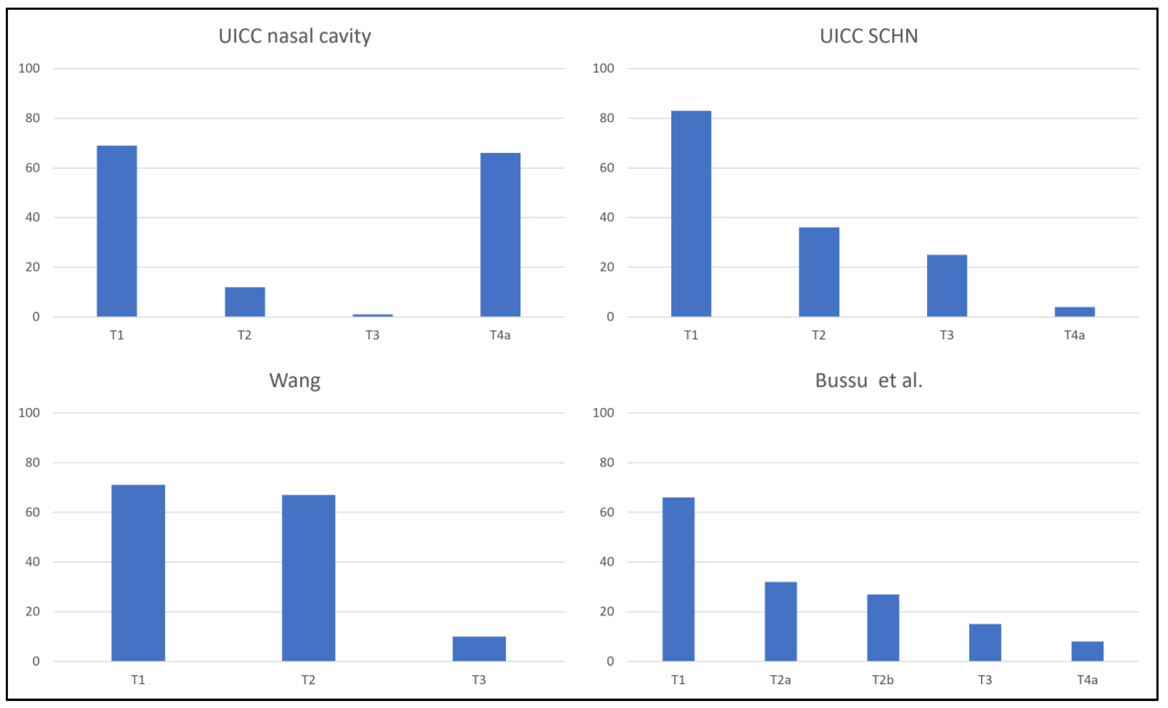
| T-stage at diagnosis | N | % |
| UICC nasal cavity | ||
| T1 | 69 | 46.6 |
| T2 | 12 | 8.1 |
| T3 | 1 | 0.7 |
| T4a | 66 | 44.6 |
| T4b | 0 | 0 |
| UICC SCHN | ||
| T1 | 83 | 56.1 |
| T2 | 36 | 24.3 |
| T3 | 25 | 16.9 |
| T4a | 4 | 2.7 |
| T4b | 0 | 0 |
| Wang | ||
| T1 | 71 | 48.0 |
| T2 | 67 | 45.3 |
| T3 | 10 | 6.8 |
| Bussu et al. | ||
| T1 | 66 | 44.6 |
| T2a | 32 | 21.6 |
| T2b | 27 | 18.2 |
| T3 | 15 | 10.1 |
| T4a | 8 | 5.4 |
| T4b | 0 | 0 |
| N-stage at diagnosis | N | % |
| N0 | 139 | 93.9 |
| N+ | 9 | 6.1 |
| Tumor diameter | N | % |
| <15 mm | 46 | 31.1 |
| ≥15 mm | 52 | 35.1 |
| Unknown | 50 | 33.8 |
| Primary tumor treatment modality | N | % |
| Brachytherapy | 113 | 76.4 |
| EBRT | 15 | 10,1 |
| Brachytherapy + EBRT | 3 | 2.0 |
| Surgery | 8 | 5.4 |
| Surgery + EBRT | 6 | 4.1 |
| Chemoradiotherapy | 1 | 0.7 |
| Best supportive care | 2 | 1.4 |
| Neck treatment modality | N | % |
| EBRT | 2 | 1.4 |
| Neck dissection | 7 | 4.7 |
| Neck dissection + EBRT | 1 | 0.7 |
| UICC Nasal Cavity | UICC SCHN | Bussu et al. | ||||
|---|---|---|---|---|---|---|
| Stage Migration | N | % | N | % | N | % |
| Upstaged | 61 | 41.2 | 26 | 17.6 | 19 | 12.8 |
| Downstaged | 3 | 2.0 | 18 | 12.2 | 3 | 2.0 |
| No stage migration | 84 | 56.8 | 104 | 70.3 | 126 | 85.1 |
Disclaimer/Publisher’s Note: The statements, opinions and data contained in all publications are solely those of the individual author(s) and contributor(s) and not of MDPI and/or the editor(s). MDPI and/or the editor(s) disclaim responsibility for any injury to people or property resulting from any ideas, methods, instructions or products referred to in the content. |
© 2023 by the authors. Licensee MDPI, Basel, Switzerland. This article is an open access article distributed under the terms and conditions of the Creative Commons Attribution (CC BY) license (https://creativecommons.org/licenses/by/4.0/).
Share and Cite
Scheurleer, W.F.J.; Tagliaferri, L.; Rijken, J.A.; Crescio, C.; Rizzo, D.; Mattiucci, G.C.; Pameijer, F.A.; de Bree, R.; Fionda, B.; de Ridder, M.; et al. Evaluation of Staging Systems for Cancer of the Nasal Vestibule. Cancers 2023, 15, 3028. https://doi.org/10.3390/cancers15113028
Scheurleer WFJ, Tagliaferri L, Rijken JA, Crescio C, Rizzo D, Mattiucci GC, Pameijer FA, de Bree R, Fionda B, de Ridder M, et al. Evaluation of Staging Systems for Cancer of the Nasal Vestibule. Cancers. 2023; 15(11):3028. https://doi.org/10.3390/cancers15113028
Chicago/Turabian StyleScheurleer, Willem Frederik Julius, Luca Tagliaferri, Johannes A. Rijken, Claudia Crescio, Davide Rizzo, Gian Carlo Mattiucci, Frank A. Pameijer, Remco de Bree, Bruno Fionda, Mischa de Ridder, and et al. 2023. "Evaluation of Staging Systems for Cancer of the Nasal Vestibule" Cancers 15, no. 11: 3028. https://doi.org/10.3390/cancers15113028
APA StyleScheurleer, W. F. J., Tagliaferri, L., Rijken, J. A., Crescio, C., Rizzo, D., Mattiucci, G. C., Pameijer, F. A., de Bree, R., Fionda, B., de Ridder, M., & Bussu, F. (2023). Evaluation of Staging Systems for Cancer of the Nasal Vestibule. Cancers, 15(11), 3028. https://doi.org/10.3390/cancers15113028