Article

The Genetic and Molecular Analyses of RAD51C and RAD51D Identifies Rare Variants Implicated in Hereditary Ovarian Cancer from a Genetically Unique Population

by
Wejdan M. Alenezi
1,2,3,
Larissa Milano
4,5,
Caitlin T. Fierheller
1,2,
Corinne Serruya
2,
Timothée Revil
1,6,
Kathleen K. Oros
7,
Supriya Behl
1,2,
Suzanna L. Arcand
2,
Porangana Nayar
2,8,
Dan Spiegelman
1,9,
Simon Gravel
1,6,
Anne-Marie Mes-Masson
10,11,
Diane Provencher
10,12,
William D. Foulkes
1,2,7,13,14,15,
Zaki El Haffaf
10,16,
Guy Rouleau
1,9,
Luigi Bouchard
17,18,19,
Celia M. T. Greenwood
1,7,15,20,
Jean-Yves Masson
4,5,
Jiannis Ragoussis
1,6 and
Patricia N. Tonin
1,2,14,*
add Show full author list remove Hide full author list
1
Department of Human Genetics, McGill University, Montreal, QC H3A0C7, Canada
2
Cancer Research Program, Centre for Translational Biology, The Research Institute of McGill University Health Centre, Montreal, QC H4A3J1, Canada
3
Department of Medical Laboratory Technology, Taibah University, Medina 42353, Saudi Arabia
4
Department of Molecular Biology, Medical Biochemistry and Pathology, Laval University, Quebec City, QC G1V0A6, Canada
5
Genome Stability Laboratory, Centre de Recherche du Centre Hospitalier de l’Université de Québec, HDQ Pavilion, Oncology Axis, Quebec City, QC G1R2J6, Canada
6
McGill Genome Centre, McGill University, Montreal, QC H3A0G1, Canada
7
Lady Davis Institute for Medical Research, Jewish General Hospital, Montreal, QC H3T1E2, Canada
8
Institute of Parasitology, McGill University, Montreal, QC H9X3V9, Canada
9
Montreal Neurological Institute, McGill University, Montreal, QC H3A2B4, Canada
10
Institut du Cancer de Montréal, Centre de Recherche du Centre Hospitalier de l’Université de Montréal, Montreal, QC H2X0A9, Canada
11
Département de Médecine, Université de Montréal, Montreal, QC H3T1J4, Canada
12
Division of Gynecologic Oncology, Université de Montréal, Montreal, QC H4A3J1, Canada
13
Department of Medical Genetics, McGill University Health Centre, Montreal, QC H4A3J1, Canada
14
Department of Medicine, McGill University, Montreal, QC H3G2M1, Canada
15
Gerald Bronfman Department of Oncology, McGill University, Montreal, QC H3A1G5, Canada
16
Service de Médecine Geénique, Centre Hospitalier de l’Université de Montréal, Montreal, QC H2X0A9, Canada
17
Department of Biochemistry and Functional Genomics, Université de Sherbrooke, Sherbrooke, QC J1K2R1, Canada
18
Department of Medical Biology, Centres Intégrés Universitaires de Santé et de Services Sociaux du Saguenay-Lac-Saint-Jean Hôpital Universitaire de Chicoutimi, Saguenay, QC G7H7K9, Canada
19
Centre de Recherche du Centre Hospitalier l’Université de Sherbrooke, Sherbrooke, QC J1H5N4, Canada
20
Department of Epidemiology, Biostatistics and Occupational Health, McGill University, Montreal, QC H3A1A2, Canada
*
Author to whom correspondence should be addressed.
Cancers 2022, 14(9), 2251; https://doi.org/10.3390/cancers14092251
Submission received: 18 March 2022 / Revised: 13 April 2022 / Accepted: 20 April 2022 / Published: 30 April 2022
(This article belongs to the Special Issue New Insights into Hereditary Cancer Syndromes)

Abstract

:

Simple Summary

We have investigated RAD51C and RAD51D, hereditary ovarian cancer risk genes, in French Canadians of Quebec, Canada. This population of Western European origins exhibits a unique genetic landscape as shown by the frequency of carriers of specific rare pathogenic variants. As studying French Canadians could facilitate the identification and interpretation of clinically relevant variants, we performed genetic analyses of RAD51C and RAD51D in this population comprised of cases with a family history of ovarian cancer or those who developed it at a young age. We identified candidate variants and then investigated them in other French Canadian study groups. We performed biological assays and revealed possible mechanisms that would affect gene function. Using engineered cells expressing one of our protein variants, we also showed that they were more sensitive to a recently approved treatment for ovarian cancer. Our findings support the role of inherited variants in RAD51C and RAD51D in ovarian cancer.

Abstract

To identify candidate variants in RAD51C and RAD51D ovarian cancer (OC) predisposing genes by investigating French Canadians (FC) exhibiting unique genetic architecture. Candidates were identified by whole exome sequencing analysis of 17 OC families and 53 early-onset OC cases. Carrier frequencies were determined by the genetic analysis of 100 OC or HBOC families, 438 sporadic OC cases and 1025 controls. Variants of unknown function were assayed for their biological impact and/or cellular sensitivity to olaparib. RAD51C c.414G>C;p.Leu138Phe and c.705G>T;p.Lys235Asn and RAD51D c.137C>G;p.Ser46Cys, c.620C>T;p.Ser207Leu and c.694C>T;p.Arg232Ter were identified in 17.6% of families and 11.3% of early-onset cases. The highest carrier frequency was observed in OC families (1/44, 2.3%) and sporadic cases (15/438, 3.4%) harbouring RAD51D c.620C>T versus controls (1/1025, 0.1%). Carriers of c.620C>T (n = 7), c.705G>T (n = 2) and c.137C>G (n = 1) were identified in another 538 FC OC cases. RAD51C c.705G>T affected splicing by skipping exon four, while RAD51D p.Ser46Cys affected protein stability and conferred olaparib sensitivity. Genetic and functional assays implicate RAD51C c.705G>T and RAD51D c.137C>G as likely pathogenic variants in OC. The high carrier frequency of RAD51D c.620C>T in FC OC cases validates previous findings. Our findings further support the role of RAD51C and RAD51D in hereditary OC.

1. Introduction

Ovarian cancer (OC) has a high estimated heritable component (39%, 95% confidence interval [CI]: 23–55) [1]. Harbouring loss-of-function (LoF) variants in BRCA1 [2] or BRCA2 [3] confers significant lifetime risk of developing OC, which accounts for 40–85% of OC cases in hereditary breast and ovarian cancer (HBOC) syndrome families and 10–15% of those not selected for age at OC diagnosis and/or family history of cancer [4]. Carrying rare LoF variants in RAD51C [5] and RAD51D [6] has been associated with OC predisposition in different populations [7], though the penetrance has yet to be determined. These genes encode RAD51 paralog proteins that are structurally similar to the RAD51 recombinase, which allows faithful DNA double-strand break repair along with BRCA1 and BRCA2 by the homologous recombination (HR) pathway [8]. RAD51C [9] and RAD51D [6] deficient cells or those expressing pathogenic variants in these genes [6,10] have been shown to render sensitivity to poly (ADP-ribose) polymerase (PARP) inhibitors such as olaparib, which is the first to be approved for OC treatment [11]. Indeed, four PARP inhibitors are currently approved for clinical use: olaparib, rucaparib, niraparib, and talazoparib for the treatment of BRCA1 and BRCA2 pathogenic variant-positive OC, breast, pancreatic and prostate cancers (recently reviewed [11]). PARP inhibitors, including olaparib, have been proven effective in the treatment of OC in the context of harbouring pathogenic BRCA1 and BRCA2 variants [12,13,14,15,16]. Thus, identifying carriers of pathogenic variants in RAD51C and RAD51D might be useful for identifying those who might benefit from management of OC with protocols using novel PARP inhibitors. Less than 2% of OC cases have been reported to harbour LoF pathogenic variants in RAD51C or RAD51D [7,17,18,19]. However, the role of pathogenic rare missense variants in these genes has not been fully explored, although approximately 39% are predicted to be damaging by in silico tools [17].
Investigating populations exhibiting unique genetic architecture due to common ancestors has the potential to facilitate the characterization of pathogenic variants in known or candidate cancer predisposing genes [20,21,22]. Our research on the French Canadians (FC) of Quebec (Canada), has shown that a small number of specific pathogenic variants account for the majority of BRCA1 or BRCA2 implicated HBOC and hereditary breast cancer (HBC) syndrome families, whereas a vast spectrum of variants in these genes has been reported in the general population [21,23,24]. Likewise, only one pathogenic variant accounts for all PALB2 implicated FC HBC syndrome families [21,25,26]. The unique genetic architecture of FCs has been attributed to genetic drift as result of multiple waves of localized population expansion in Quebec of European (France) settlers since 1608 [20,21,27]. Given the expectation that a higher frequency of carriers of rare variants would be observed in cancer cases versus controls in populations exhibiting genetic drift, the genetic analyses of FCs has the potential to identify clinically relevant pathogenic variants in new risk genes [20,21,22]. For example, recently, we reported that RAD51D c.620C>T; p.Ser207Leu, initially identified by genetic panel testing of familial OC cases in clinical settings, was found to occur at a significantly higher frequency in FC OC cases versus controls [28]. At the time of discovery, this RAD51D variant was classified as a missense variant of unknown clinical significance (VUS). Its classification has since been revised to pathogenic or likely pathogenic, especially as the same study reported that RAD51D p.Ser207Leu impaired HR function and rendered cells sensitive to olaparib. Although the role of RAD51C and RAD51D have yet to be fully explored in the FC population [21,29,30], it is clear from previous work that investigating the FC population can assist in characterizing new cancer risk genes (reviewed in [21]).
The main aim of this study was to identify and investigate candidate variants in RAD51C and RAD51D in FCs with OC by: (1) performing whole exome sequencing (WES) and bioinformatic analyses of the germline of familial and sporadic early-onset OC cases; (2) determining the carrier (harbouring the variant in the heterozygous state) frequencies of candidate variants by genotyping or surveying available genetic data in OC cases and population-matched controls; (3) assaying available tumour DNA from carriers for loss of heterozygosity (LOH) of RAD51C and RAD51D loci; (4) describing associated clinico-pathological features of OC in carriers; and (5) using biological assays involving cancer cell line models to assess the impact of missense variants with unknown biological function, including sensitivity to PARP inhibitors.

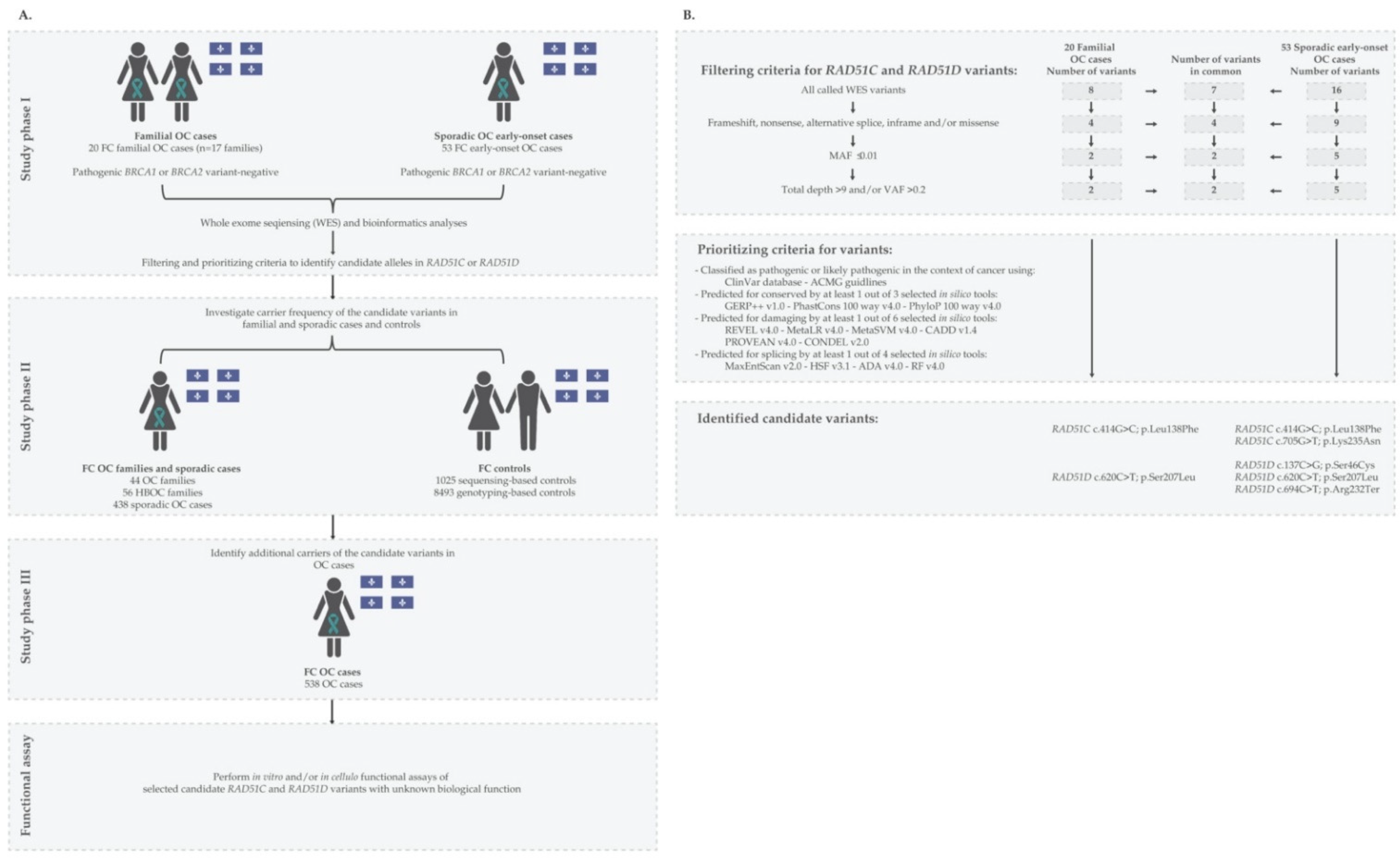
2. Materials and Methods

2.1. FC Study Participants

The FC participants investigated in this study were selected from independently established biobank resources as described in Table S1. All participants were recruited independently to these biobanks in accordance with ethical guidelines of their respective Institutions Research Ethics Boards, including those participants from adult hereditary cancer clinics in Quebec. The participants that had provided their associated biological specimens (DNA), genetic data, pedigree information and clinical metrics, where appropriate, were anonymized at source by the respective biobanks. To further protect the anonymity of study subjects, all samples were assigned a unique identifier and pedigrees modified. This project was conducted with approval and in accordance with the guidelines of The McGill University Health Centre Research Ethics Board (MP-37-2019-4783).
As described in Figure 1A, candidate variants in RAD51C and RAD51D were discovered (phase I) in peripheral blood lymphocyte (PBL) DNA from 20 familial (from 17 OC families) and 53 sporadic early-onset OC cases known to be negative for pathogenic BRCA1 and BRCA2 variants by FC mutation-panel or clinical multi-gene panel testing from information provided by adult hereditary cancer clinics in Quebec or the Banque de tissus et données of the Réseau de recherche sur le cancer of the Fond de recherche du Québec—Santé (RRCancer biobank) (rrcancer.ca).
Carrier frequencies of RAD51C and RAD51D candidate variants were determined (phase II) by genotyping PBL DNA from index cancer cases from three different FC cancer groups: 44 OC and 56 HBOC families, and 438 sporadic OC cases regardless of the carrier status of BRCA1 and BRCA2 pathogenic variants (RRCancer biobank). Carrier frequencies were also determined by investigating genetic data available from population-matched controls from three established biobanks: (1) CARTaGENE (cartagene.qc.ca) [31]; (2) McGill University—Montreal Neurological Institute (MNI) [28,32]; and (3) Sherbrook University—glucose regulation in gestation and growth (Gen3G) [33].
To increase the pool of carriers of our candidate variants (phase III), we genotyped another 538 PBL or tumour DNA samples from recently available OC cases that were provided by the RRCancer biobank.
Age at diagnosis, tumour grade, stage of the disease, histopathology subtype as confirmed by a gynecologic pathologist, personal history of other cancers, chemotherapy treatment and/or overall outcome were provided for selected OC cases from the RRCancer biobank if available. For comparative purposes, clinical data was also provided by the RRCancer biobank from carriers of the pathogenic BRCA1 c.4327C>T; p.Arg1443Ter from the sporadic OC case study group, investigated previously by our group for BRCA1 and BRCA2 carrier status [34].
All study groups described in this report were selected from participants that had been recruited from the province of Quebec to participate in various biobanking projects: familial OC or invasive breast cancer (BC) cases self-reported FC ancestry [23,24]; the majority of sporadic OC (at least 88%) self-reported FC ancestry [34]; additional OC cases self-reported FC ancestry by RRCancer biobank; Gen3G project included mothers that self-reported FC ancestry [33]; MNI controls were self-reported as FC ancestry [28,32]; and CARTaGENE resource defined FC status of controls based on being self-reported as FC, born in Quebec, having parents and all four grandparents born in Canada and having French as first language learned [31].

2.2. Identification and Verification of Candidate Variants

To identify candidate variants, PBL DNA from a total of 73 OC phase I cases (Figure 1A) were subjected to WES at the McGill Genome Centre using NimbleGen SeqCap EZ Exome v3.0 library kit (Roche, NJ, USA), followed by paired-end sequencing of 150 bp reads on different Illumina HiSeq platforms. Reads were aligned to the human reference genome assembly GRCh37/hg19 using Burrows-Wheeler aligner v0.7.17, followed by PCR deduplication using Picard v2.9.0. Realignment around small insertions and deletions was performed and then, germline variants were called using HaplotypeCaller using Genome Analysis Toolkit (GATK) v3.5. Variants were then filtered for base sequencing quality score ≥ 30 and annotated using Ensembl Variant Effect Predictor (VEP) and GEMINI v0.19.1.
Variants in RAD51C and RAD51D were extracted from the annotated variant call files (VCF) for filtering and prioritization (Figure 1B). Silent and intronic variants and those with a minor allele frequency (MAF) ≥ 0.01 in the Genome Aggregation Database of non-cancer population (gnomAD) v2.1.1 (gnomad.broadinstitute.org) [35,36], low coverage (<10 reads) and variant allele frequency (VAF) <0.20 and >0.80 were filtered out [37]. Manual inspection was performed to confirm variants using Integrative Genomics Viewer (IGV) v2.4.10.
We prioritized for further investigation (see Figure 1B) LoF variants and missense variants classified as pathogenic, likely-pathogenic or VUS in ClinVar [38,39] and the American College of Medical Genetics and Genomics (ACMG) guidelines [40]. We prioritized variants predicted to overlap conserved residues or be damaging at the RNA or protein level by at least one of several in silico tools selected based on their best performance [41,42,43]. Briefly, we selected variants having a conserved prediction score ≥ 2.0 by Genomic Evolutionary Rate Profiling v1.0 (GERP++ v1.0) [44], ≥0.2 Phylogenetic p value v4.0 of 100 vertebrates (PhyloP 100 way v4.0) and ≥0.4 by PHAST Conservation v4.0 of 100 vertebrates (PhastCons 100 way v4.0) [45]; damaging prediction score ≥ 0.4 by Rare Exome Variant Ensemble Learner v4.0 (REVEL v4.0) [46], Meta-analytic Logistic Regression v4.0 (MetaLR v4.0) [47] and CONsensus DELeteriousness v2.0 (CONDEL v2.0) [48], ≥0.0 by Meta-analytic Support Vector Machine v4.0 (MetaSVM v4.0) [47]; ≥−2.5 by Protein Variation Effect Analyzer v4.0 (PROVEAN v4.0) [49] and ≥20 (Phred score = −10 log10 P) by Combined Annotation Dependent Depletion v1.4 (CADD v1.4) [50]. Prediction performance of these in silico tools was tested based on germline variants submitted to the ClinVar database and classified by ACMG guidelines [41,43]. Variants were predicted to affect splicing if the score was ≥0.4 by two of Database Splicing Consensus Single Nucleotide Variant (dbscSNV) in silico tools [45]: AdaBoost v1.1 (ADA v1.1) or Random Forest v1.1 (RF v1.1); or classified to affect splicing by Maximum Entropy Estimates of Splice Junction Strengths v2.0 (MaxEntScan v2.0) [51] and Human Splicing Finder v3.1 (HSF v3.1) [52]. Prediction performance of these splicing predictor in silico tools was tested on somatic variants submitted to the catalogue of somatic mutations in cancer (COSMIC) database [42].
Candidate variants were verified in PBL DNA by bidirectional Sanger sequencing of PCR products using customized primers (Table S2) at the McGill Genome Centre as previously described [28]. Sequencing chromatograms were visually inspected for variant heterozygosity using 4Peaks v1.8. (nucleobytes.com/4peaks/) (The Netherlands Cancer institute, Amsterdam, The Netherlands).

2.3. Investigating Carrier Frequencies of Candidate Variants in FC OC Cases and Controls

Carrier frequencies of candidate variants were determined by genotyping PBL DNA from OC or BC cases from defined FC study groups (Table S1) using customized TaqMan assays [53] (Table S2). Carriers were verified by bidirectional Sanger sequencing as described above. Corresponding tumour DNA from the index case was genotyped where PBL DNA was not available.
Carrier frequencies of candidate variants were determined in FC controls by surveying 1025 sequencing-based: 433 from Gen3G, 422 from MNI and 170 from CARTaGENE [28,31,33] and 8493 SNP genotyping-based [31] data available from CARTaGENE resources (Table S1). Carrier counts were extracted from VCF files or PLINK files. If the variant probes were not represented on the SNP array, pre-phasing and imputation were performed using Eagle2 with the Burrows-Wheeler transformation through Sanger Imputation Services (sanger.ac.uk/tool/sanger-imputation-service/, accessed on 1 February 2019), where Haplotype Reference Consortium release v1.1 (HRC.r1) was used as the reference [54,55].
Fisher’s exact test was used to compare carrier or allele frequencies in appropriate OC study groups versus controls using two-tailed p values where <0.05 was considered significant. A permutation analysis using Fisher’s exact test was also performed of cases and controls to address the possibility that carriers could have been recruited to more than one FC study group (Table S1).

2.4. Surveying Allele and Carrier Frequencies of Candidate Variants in Genetic Databases of Non-FC Populations

Carrier or allele frequencies of candidate variants were surveyed in genetic data that was derived from cancer-free individuals of European ancestry, as the original founders of FCs of Quebec were of Western European (France) origin [20,21,27,56]. Carrier or allele frequencies were determined by querying data available from three resources (Table S3). Allele frequencies for the non-cancer of non-Finnish European ancestry were extracted from WES or whole genome sequencing (WGS) data deposited in the Genome Aggregation Database (gnomAD) v2.1.1 (gnomad.broadinstitute.org, accessed on 1 October 2021). For comparative purposes, we also extracted data derived from other populations from this resource. Carrier frequencies for 7325 women of European ancestry regardless of family history of cancer were extracted from genetic data from the Fabulous Ladies Over Seventy (FLOSSIES) project (whi.color.com, accessed on 1 October 2021), which included data from a 27-gene panel sequencing study.
Candidate variants were also queried in genetic data from 25,509 OC cases and 40,491 controls using the Ovarian Cancer Association Consortium (OCAC) database (ocac.ccge.medschl.cam.ac.uk/data-projects/, accessed on 15 June 2020). Summary statistics provided included odd ratios (ORLog2) with p values comparing all epithelial OC histopathological subtypes with OCAC controls.
For comparison purposes, we also queried BRCA1 c.4327C>T; p.Arg1443Ter in all of these resources, as this variant is the most frequently reported pathogenic BRCA1 variant in the FC and European populations [21].

2.5. LOH Analysis of RAD51C and RAD51D Loci in OC Tumour DNA from Candidate Variant Carriers

LOH analysis of RAD51C and RAD51D loci was performed by Sanger sequencing of OC tumour DNA from variant carriers, where possible, using customized primers as described above (Table S2). Extracted DNA from fresh-frozen (FF) or histopathological sections from formalin-fixed paraffin-embedded (FFPE) tumour tissues were provided from the RRCancer biobank for DNA extraction based on the manufacturer’s instructions (Promega, Canada). Sequencing chromatograms were visually inspected for complete or partial loss of the wild-type RAD51C or RAD51D alleles from carriers using 4Peaks v1.8. (nucleobytes.com/4peaks/) (The Netherlands Cancer institute, Amsterdam, The Netherlands).

2.6. RNA Extraction and Reverse Transcription Analyses of RAD51C

An Epstein–Barr virus transformed lymphoblastoid cell line (LCL) was established from PBLs from RAD51C c.705G>T carriers and non-carriers of this variant as previously described [57]. Approximately 5,000,000 cells were treated with 28 µg/mL of cycloheximide or DMSO for 3 h. RNA was extracted from cell pellets treated with 1000 µL of TRIzol (Invitrogen, Canada) for reverse transcription [58]. cDNA was amplified and purified for Sanger sequencing using customized primers as described above (Table S4). Sequencing chromatograms were visually inspected for splicing impact using 4Peaks v1.8. (nucleobytes.com/4peaks/) (The Netherlands Cancer institute, Amsterdam, The Netherlands).

2.7. Cell Lines

We used three cell lines in our assays, HeLa (cervical carcinoma), U2OS (sarcoma) and OVCAR-3 (epithelial ovarian adenocarcinoma). HeLa cells were obtained from American Type Culture Collection (atcc.org) and maintained in Dulbecco’s Modified Eagle Media supplemented with 10% Fetal Bovine Serum and 1% Penicillin/Streptomycin, at 37 °C, 5% CO2, and 20% O2. OVCAR-3 cells were grown in RPMI supplemented with 0.01 mg/mL bovine insulin and 20% foetal bovine serum, at 37 °C, 5% CO2. U2OS cells RAD51D knock-out (KO) [59] were purchased from the Deutsche Sammlung von Mikroorganismen und Zellkulturen GmbH (U2OS#18-RAD51D-4, DSMZ, Braunschweig, Germany) maintained in Dulbecco’s Modified Eagle Media supplemented with 10% Fetal Bovine Serum and 1% Penicillin/Streptomycin, at 37  °C, 5% CO2, and 20% O2. OVCAR-3 cells were a kind gift from Dimcho Batchvarov (CHU de Québec).

2.8. Complementation Assays and siRNA Transfections

For in cellulo experiments in HeLa and OVCAR-3 cells, the RAD51D p.Ser46Cys protein variant was obtained via site-directed mutagenesis using the Q5 Site-Directed Mutagenesis Kit (NEB) (New England biolabs, Canada) using the pcDNA3-RAD51D wild-type (WT) as a template and primers listed on Table S4. The mammalian expression vector pcDNA3-RAD51D that was used as a template had been previously modified for RAD51D expression with a FLAG tag at the N-terminus and for resistance to RAD51D siRNA with the Q5 Site-Directed Mutagenesis Kit (NEB) and primers listed on Table S4. The siRNA target sequence used to silence RAD51D was siRAD51D #7 GGCCAAAUCUUCCCGACAGdTdT and the non-specific siRNA used as control was UUCGAACGUGUCACGUCAAdTdT.
Approximately 240,000 cells were seeded into one well of a six-well plate before being double transfected 24 and 48 h later with 50 nM control or RAD51D siRNA using Lipofectamine RNAiMAX (Invitrogen, Canada). For Hela, cells were then complemented by transfection of 800 ng of the pcDNA3 empty vector or the indicated siRNA-resistant FLAG-tagged RAD51D construct using Lipofectamine 2000 (Invitrogen, Canada) for four hours. Transient transfections in OVCAR3 cells were performed with 300 or 700 ng of the pcDNA3 empty vector or the indicated siRNA-resistant FLAG-tagged RAD51D construct using Lipofectamine 3000 (Invitrogen, Canada) for four hours according to the manufacturer’s protocol. U2OS RAD51D KO cells were stably complemented using the AAVS1 integration system [60]. The AAVS1 RAD51D WT or p.Ser46Cys constructs were generated by amplification using the pcDNA3 plasmids previously described as a template and primers listed on Table S4; both products were cloned into the AAVS1 vector in NotI/PspXI sites. Briefly, cells were transfected with the 4 μg of the AAVS1 construct containing either the WT or the RAD51D p.Ser46Cys variant, along with the 0.4 μg of the pZFN plasmid, using Lipofectamine 2000 (Invitrogen) for 4 h. 24 h later, transfected cells were selected with G418 for 7 days. To confirm genomic integration, genomic DNA was extracted from stable cells using PureLink Genomic DNA Mini Kit (Invitrogen) and used as a template to amplify the integrated cDNA in the AAVS1 locus using primers from Table S4. Complementation was then confirmed by Sanger sequencing.

2.9. Olaparib and Talazoparib Sensitivity Assays

PARP inhibitors, olaparib and talazoparib, sensitivity assay and cell imaging were performed as described previously [61]. Cells were treated for four days with concentrations ranging from 0 (DMSO) to 2.5 μM of olaparib or 0 (DMSO) to 40 nM of talazoparib. Cell viability was expressed as percentage of survival in olaparib-treated or talazoparib-treated cells relative to vehicle (DMSO)-treated cells. Results represent the mean ± standard error of the mean (SEM) of at least three independent experiments, each performed in triplicate.

2.10. Immunofluorescence Analysis

U2OS RAD51D cells stably complemented with either RAD51D WT or the p.Ser46Cys were seeded into Corning 96-Well Half Area High Content Imaging Film Bottom Microplate at 7000 cells per well. Then, 18h later, cells were irradiated with 5 Gray and processed for immunofluorescence 4 h post-irradiation. Unless otherwise stated, all immunofluorescence dilutions were prepared in PBS and incubations performed at room temperature with intervening washes in PBS. Cell fixation was carried out by incubation with 4% paraformaldehyde for 10 min followed by 100% ice-cold methanol for 5 min at −20 °C. Cells were permeabilised in 0.2% Triton X-100 for 5 min followed by a quenching step using 0.1% sodium borohydride for 5 min. After blocking for 1 h in a solution containing 10% goat serum and 1% BSA, cells were incubated for 1 h with primary antibody anti-RAD51 (1:5000, Bioacademia #70-001) or anti-phospho-Histone H2A.X (Ser139) (1:5000, Millipore, #05-636), combined with anti-Geminin (1:7000, Abcam #ab104306 or Proteintech #10802-1-AP) all diluted in 1% BSA. Secondary antibody labelling used Alexa Fluor 488 goat anti-rabbit (Invitrogen, #A-11008) and Alexa Fluor 647 goat anti-mouse (Invitrogen, #A21235) or Alexa Fluor 488 goat anti-mouse (Invitrogen, #A-11001) and Alexa Fluor 647 goat anti-rabbit (Invitrogen, #A21244), diluted at 1:1000 in 1% BSA for 1 h. Nuclei were stained for 10 min with 1 mg/mL 4,6-diamidino-2-phenylindole (DAPI). Z-stack images were acquired on a ZEISS Celldiscoverer 7 automated microscope using a 50× water immersion objective and analysed for RAD51 or gH2AX foci formation with ZEN Blue software 3.2 (ZEISS). Data from three independent trials were analysed for outliers using the ROUT method (Q = 1.0%) in GraphPad Prism v8.0 and the remaining were reported in a violin plot.

2.11. Protein Expression and Immunoblotting Analyses of RAD51D

Total soluble protein extraction and immunoblotting were performed as previously described [62]. For RAD51D detection, a polyclonal antibody (#ab202063, Abcam, US) was used at a 1:1000 dilution. Mouse monoclonal anti-vinculin (#V9131, Sigma, US) at 1:200,000 dilution was used as the loading control. Horseradish peroxidase-conjugated anti-rabbit IgG or anti-mouse at 1:10,000 dilution (Jackson Immuno Research, US) was used as secondary antibodies.

3. Results

3.1. Identification and Characteristics of Candidate Variants

By extracting all variants located in protein encoding and splice-site regions of RAD51C and RAD51D in WES data, we identified a total of 8 variants in 20 familial cases and 16 variants in 53 sporadic cases of OC (Figure 1 and Table S5). From this list, we identified five candidate variants in these genes that fulfilled our selection criteria (Figure 1B): two missense variants in RAD51C c.414G>C; p.Leu138Phe and c.705G>T; p.Lys235Asn, two missense variants in RAD51D c.137C>G; p.Ser46Cys and c.620C>T; p.Ser207Leu and a nonsense variant RAD51D c.694C>T; p.Arg232Ter.
The variants were identified in nine OC cases: 17.6% (3/17) of OC families and 11.3% (6/53) of sporadic early-onset cases (Table 1). Our results include the identification of newly reported variants in FCs: two carriers of RAD51C c.414G>C; p.Leu138Phe, one of RAD51C c.705G>T; p.Lys235Asn, one of RAD51D c.137C>G; p.Ser46Cys and one of RAD51D c.694C>T; p.Arg232Ter. In addition, our results include four carriers of RAD51D c.620C>T; p.Ser207Leu, a variant previously reported to occur in more than one FC OC case by our group [28].
Our candidate variants are rare in various cancer-free populations of non-FC and European ancestry based on surveying available genetic data (Table 1 and Table S5). All candidate variants have a MAF ≤ 0.0001 in the non-cancer population represented in the gnomAD database and in cancer-free women in the FLOSSIES database.
All variants were predicted to affect highly conserved loci and the four missense variants to be damaging by at least one of our in silico tools that we selected based on their best performance (Table 1). Four different in silico tools also predicted that the RAD51C c.705G>T; p.Lys235Asn located at the 5′ splice-donor site would affect transcript splicing.
The ClinVar database and/or ACMG guidelines classified RAD51C c.414G>C; p.Leu138Phe and c.705G>T; p.Lys235Asn and RAD51D c.694C>T; p.Arg232Ter as likely pathogenic or pathogenic. In contrast, there were conflicting classifications reported for RAD51D c.137C>G; p.Ser46Cys, such as VUS and likely benign in the ClinVar database and VUS by ACMG guidelines and for RAD51D c.620C>T; p.Ser207Leu, such as pathogenic/likely pathogenic in the ClinVar database and VUS by ACMG guidelines.
None of the five candidate variants were found to co-occur in the nine carriers of these variants. We also reviewed WES data of these nine carriers for the presence of pathogenic variants in any of the known risk genes for OC based on the National Comprehensive Cancer Network (NCCN) for clinical practice in oncology guidelines (version 2021–Genetic/Familial High-Risk Assessment: Breast, Ovarian and Pancreatic): BRCA1, BRCA2, MLH1, MSH2, MSH6, PMS2, BRIP1, RAD51C, RAD51D, PALB2, ATM or STK11. All carriers were found not to carry a pathogenic or likely pathogenic variant in any of these genes based on ClinVar or ACMG guidelines, with the exception of the RAD51D c.694C>T; p.Arg232Ter carrier who also harboured a pathogenic variant in BRCA1 c.1462dupA; p.Thr488AsnfsTer2. Interestingly, this known BRCA1 pathogenic variant has not been previously reported in the FC populations.

3.2. Carrier Frequency of Candidate Variants in OC Cases and Cancer-Free Controls of FC Ancestry

We compared the carrier frequencies of our candidate variants in different FC groups comprised of cancer cases, regardless of BRCA1 or BRCA2 pathogenic variant status, and cancer-free controls (Table 2). Pair-wise comparisons of carrier frequencies were performed using data from each cancer group and sequencing-based controls (see Table S1). The highest overall carrier frequency was among carriers of RAD51D c.620C>T found in the sporadic group. Frequencies of this variant ranged from 2.3% (1/44) in OC families having at least two OC cases to 3.4% (15/438) in sporadic cases. Notably, all carriers were among the pathogenic BRCA1 or BRCA2 variant-negative cases as determined by previous studies which included whole gene or targeted genotyping of FC variants in these study groups (see Table S1). The carriers in the sporadic cases included the previously identified carriers of this variant (3.8%; 13/341) [28]. In contrast, the carrier frequencies of each of the other variants were lower in the cancer groups. The carrier frequencies of these variants ranged from 0% to 2.3% (1/44) in OC families harbouring RAD51C c.414G>C, where the carrier was from a pathogenic BRCA1 or BRCA2 variant-negative family, and from 0% to 0.2% (1/438) for those harbouring RAD51C c.705G>T, RAD51D c.137C>G or c.694C>T variants. None of the index OC or BC cases from 56 HBOC families were found to harbour any of our candidate variants. Our targeted genotyping assays or review of available WES data revealed that none of the carriers identified in the cancer study groups (Table 2) also carried another one of our RAD51C or RAD51D candidate variants.
It was not possible to perform pair-wise comparisons to further assess carrier frequency of our variants in cancer study groups and genotyping-based controls. No carriers of RAD51C c.414G>C or RAD51D c.620C>T were identified in the genotyping-based data of 8493 population-matched controls. Carriers of RAD51C c.705G>T, RAD51D c.137C>G or RAD51D c.694C>T could not be identified in the same data and were not available in the HRC.r1 haplotype reference panel used for imputation from SNP array data.
Although the cancer study groups were independently derived for previous research purposes [21,23,24,34,63], we cannot exclude the possibility that individuals were recruited to multiple study groups. Based on the unique RRCancer biobank sample reference number, we are only aware of nine samples where the same case was included in two different study groups (Figure S1). We therefore performed a permutation analysis with 5000 random allocations of the observed variants to the participants across the three cancer groups (44 OC families, 56 HBOC families and 438 sporadic OC cases) and the two control groups (1025 sequencing-based and 8493 genotyping-based controls) investigated this study. When compared to sequencing-based controls, the permutation analysis presented evidence for a higher variant rate among all cases (p = 0.026), OC families (p = 0.015) and all families (p = 0.026). When examining only RAD51D c.620C>T, which was captured in data from both sequencing-based and genotyping-based controls, permutation analysis provided evidence for a higher frequency of this variant in all cancer cases versus all controls (p = 0.0098) and in all familial cases versus all controls (p = 0.014). The permutation analysis also allowed us to estimate the family-wise error rate for all tests performed: in 6.7% of the permutations, we found that at least one of the five tests demonstrated significance at p < 0.05, reflecting a type-1 error rate of potential concern. However, our permutation analysis also demonstrated that it was highly unlikely for all five comparisons to result in a naïve p-value < 0.05 simultaneously (permutation study p = 0.0002).

3.3. Clinico-Pathological Characteristics of OC Variant Carriers

The histopathological and clinical characteristics available for the 6 RAD51C and 28 RAD51D variant carriers are shown in Table S6, which also includes known personal history of cancer. The pedigrees of selected carriers are shown in Figure S2, anonymized to only show information relevant to this study to protect the identity of participants. Thirteen of 28 RAD51D c.620C>T carriers from a previous study were also included for comparative purposes as their associated clinical features had not been reported [28]. Features of carriers of RAD51C c.705G>T (n = 2) and RAD51D c.137C>G (n = 1) and c.620C>T (n = 7) that were identified by targeted screening of an additional 538 cases of OC of FC ancestry were also included in Table S6.
Most OC carriers of our candidate variants had HGSC (31/34), which is the most common subtype of epithelial OC [64]. Three other carriers had either a high-grade endometrioid adenocarcinoma, serous carcinoma of unknown grade or OC of undisclosed histopathology. A query of the OCAC data, which only revealed summary statistics for one of our candidates, showed statistical differences in the frequency of RAD51D c.620C>T carriers having HGSC (ORLog2 = 17.2; p = 0.00001) versus controls (Table S7). This observation is consistent with our query of BRCA1 c.4327C>T in OCAC data (ORLog2 = 1.211; p = 0.009051), the most common pathogenic FC OC risk allele as a comparator, where we found statistical differences in the frequency of carriers of this BRCA1 variant having the high-grade serous subtype OC versus controls.
The average age of OC diagnosis in carriers of 58.5 years (median 59 years [age range = 42–77; SD ± 9.0 years]) was comparable to the average age of OC diagnosis in the general population being 60 years of age [28]. Fifty-three percent (18/34) of carriers were diagnosed before the age of 60 years, where 21% (7/34) were diagnosed before the age of 50 years.
Given the high frequency of RAD51D c.620C>T carriers in our OC cases, it was possible to compare clinical data of carriers (15/438) of this RAD51D variant with carriers (15/438) [21,28] of a frequently occurring variant in BRCA1 c.4327C>T [21,23,24,34,65], previously reported in our investigation of the same sporadic OC study group [34]. The average and median ages at diagnosis of RAD51D variant carriers was approximately 59 years (age range = 46–74; SD ± 8.4 years). This was older than the average and median ages of diagnosis of 54 years observed in BRCA1 variant carriers (age range = 36–76; SD ± 11 years (p = 0.15; 95% CI: −1.96 to 12.49; two tailed p value, unpaired t-test). The average survival among the 15 RAD51D c.620C>T carriers was 81.9 months (median 69 months [range = 1–195 months]) which was longer than the average survival of 67.1 months among BRCA1 c.4327C>T carriers, though not significantly different (median 52 months [range = 10–168 months]; p = 0.46; 95% CI: −25.86 to 55.33, two-tailed p value, unpaired t-test). There were 46.7% (7/15) of RAD51D c.620C>T carriers and 60% (9/15) of BRCA1 c.4327C>T carriers who had died of OC.

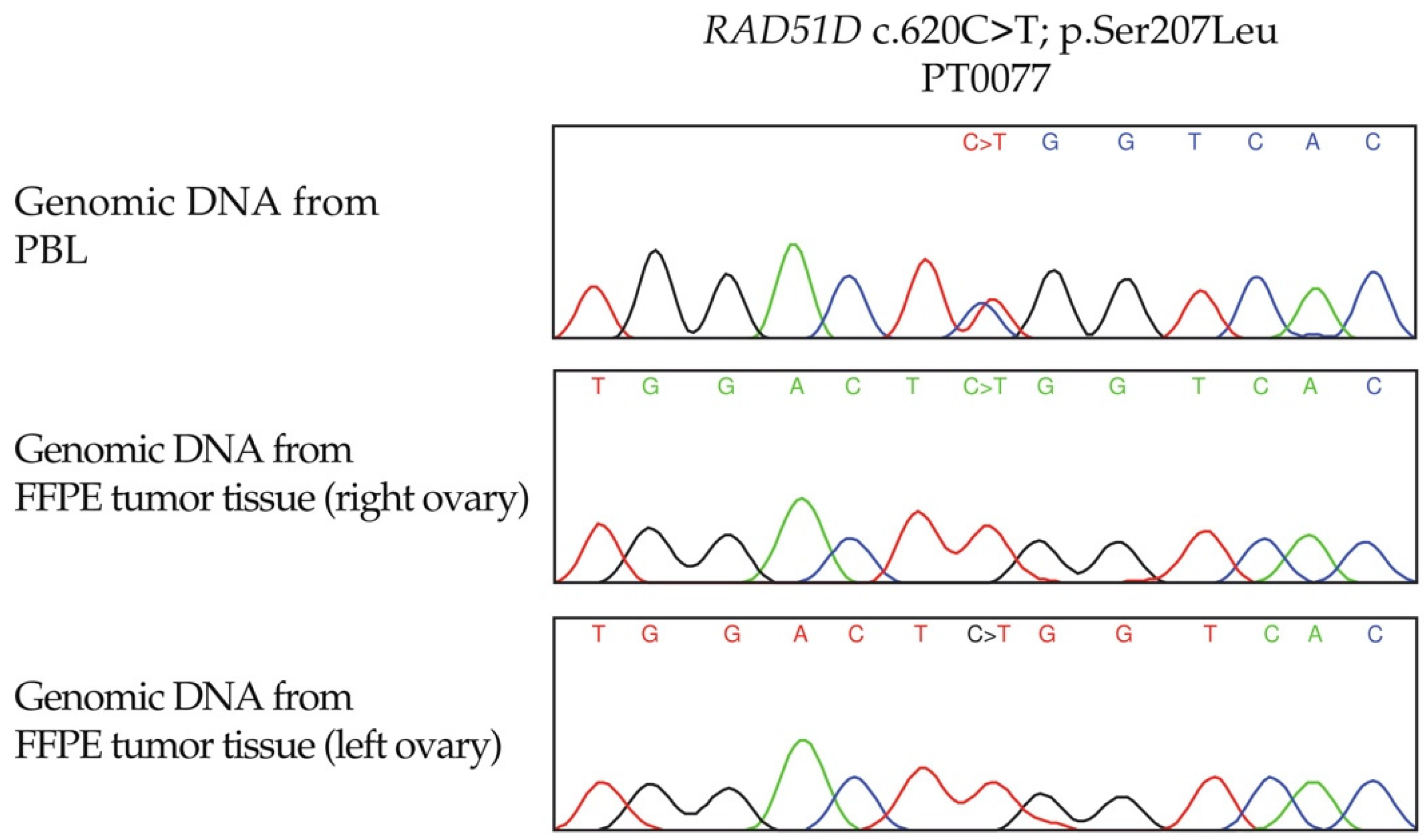
3.4. LOH Analyses of RAD51C and RAD51D Loci in OC Tumour DNA from Candidate Variant Carriers

Evidence of partial or complete loss of the wild-type allele was observed in tumour DNA from at least one carrier of each type of missense candidate variant (Table 3) as tumour DNA was not available for all variant carriers. Although our assays were not performed in DNA samples extracted from sections enriched for cancer cells, in five cases harbouring RAD51D c.137C>G or c.620C>T (PT0058, PT0071, PT0075, PT0076 and PT0077), there was clear evidence of loss wild-type allele in the analysis of tumour DNA extracted from FFPE. These findings suggest that partial loss or allelic imbalance observed with some samples may be an indication of contaminating normal stromal cells, although OC tumour specimens are often abundant in cancer cells [66]. Interestingly, only the RAD51D c.620C>T germline allele was detectable in both OC tumours from a bilateral OC case by Sanger sequencing of tumour DNA (Figure 2). This observation suggests the possibility that somatic loss of the wild-type allele preceded clonal expansion in the tumourigenesis of OC in this carrier.

3.5. In Vitro Investigation of Aberrant Splicing of RAD51C c.705G>T

We established RAD51C c.705G>T carrier- and non-carrier-derived LCLs and performed RT-PCR on the extracted RNA to determine if the genomic position of the variant affected splicing consistent with predictions based on the application of our selected in silico tools (Table 1). RT-PCR analyses showed two different size bands from the c.705G>T carrier-derived LCLs but not in controls (Figure 3A), suggesting that this variant affected splicing of the transcript. Although we could not verify the presence of exon 4 in the non-carrier as cDNA no longer available, Sanger sequencing verified the absence of the entire exon four in the variant carrier-derived cDNA (Figure 3B,C), suggesting that exon skipping had occurred.

3.6. In Cellulo Investigation of RAD51D p.Ser46Cys

We performed in cellulo assays of RAD51D p.Ser46Cys due to conflicting reports of its clinical significance and paucity of data concerning its effect on biological function. We selected this candidate variant for further study as the biological impact of our other remaining missense candidate variants, RAD51C p.Leu138Phe [5] and RAD51D p.Ser207Leu [28] have been reported, or have been inferred as biologically relevant as described above for the effects of splicing on RAD51C c.705G>T.
Given the role of RAD51D in HR function, we investigated sensitivity to the PARP inhibitor, olaparib, taking advantage of the synthetic lethal interaction between loss of HR function and PARP inhibition [62]. Using HeLa cells, RAD51D-knock-down cells were more sensitive to olaparib (Figure S4A). Complementation with RAD51D WT siRNA resistant construct restored sensitivity to endogenous levels, while RAD51D p.Ser46Cys siRNA resistant construct failed to rescue the viability of RAD51D knock-down cells, showing olaparib sensitivity similar to cells complemented with the empty vector.
Immunoblotting 24 h post-transfection showed that RAD51D p.Ser46Cys protein was weakly expressed compared to WT (Figure S4B). As the expression of RAD51D p.Ser46Cys was lower than the WT, we then investigated if the reduced expression of the variant protein could be due to protein instability by examining the protein’s half-life in RAD51D knock-down cells transfected with either FLAG-RAD51D WT or p.Se46Cys. Cells were then exposed to cycloheximide (CHX) to inhibit protein synthesis and pellets for protein extraction were collected at the indicated time points. Over the time course RAD51D protein levels were reduced for both isoforms, however, the effect was more pronounced in cells expressing the p.Ser46Cys variant when compared to the WT (Figure S4C). RAD51D WT protein starts to reduce at after six hours, while the RAD51D p.Ser46Cys protein is hardly visible at four hours (Figure S4C). We were able to recapitulate our findings in an OC cell line background (Figure S5). In OVCAR-3 cells, the p.Ser46Cys variant protein is also expressed at a reduced level. These observations suggest that RAD51D p.Ser46Cys is unstable and affects cellular sensitivity to olaparib.
To further investigate the functionality of the p.Ser46Cys variant and eliminate the effect of endogenous RAD51D, we used U2OS RAD51D KO cells stably complemented with either the WT or the p.Ser46Cys variant using the AAVS1 genomic editing system (Figure 4A) [59,60]. Although RAD51D KO cells were successfully complemented with either WT and p.Ser46Cys, as confirmed by Sanger sequencing, the p.Ser46Cys variant was also weakly expressed in this cell line when compared to the WT (Figure 4B,C).This is in agreement with the observation that U2OS RAD51D cells harbouring the p.Ser46Cys variant failed to complement survival when exposed to olaparib and talazoparib (Figure 4D,E).
RAD51D is required to facilitate RAD51 filament formation and proper repair of damage-induced double-strand breaks [67]. Therefore, we evaluated both RAD51 and γH2AX foci formation after treatment with 5 Gray of ionizing radiation in S/G2-cells (Figure S6). As expected, a decrease in the mean number of RAD51 foci per cell was observed in RAD51D deficient cells and expression of RAD51D WT partially rescued this phenotype, while rescue was less obvious in cells expressing the p.Ser46Cys (Figure S6A,B). Moreover, after ionizing radiation, the p.Ser46Cys variant also exhibited elevated levels of γH2AX foci, while RAD51D WT cells were able to rescue the increased γH2AX foci formation observed in the RAD51D deficient cells (Figure S6C,D). Altogether, these results indicate an impaired HR functionality leading to increased DNA double-strand breaks in cells expressing the RAD51D p.Ser46Cys variant.

4. Discussion

Our WES analysis of 73 familial and sporadic early-onset OC FCs of Quebec identified five candidate variants in RAD51C and RAD51D. The genetic analyses of additional FC OC study groups confirmed that RAD51D c.620C>T, previously reported in sporadic OC cases by our group [28], occurs at a high frequency in FCs with this disease. This observation is likely due to the unique genetic architecture of FCs of Quebec that has been attributed to common founders of this population [20,21,22]. OC cases harbouring other variants were found once for the nonsense RAD51D c.694C>T and twice for the missense RAD51D c.137C>G or RAD51C c.414G>C, suggesting that FCs are more genetically heterogenous population than other populations with common founders where few frequently occurring variants have been reported [21]. We identified a total of four OC cases harbouring RAD51C c.705G>T suggesting that these individuals might also share common ancestors in FCs. Indeed, another OC case harbouring this RAD51C variant was identified in a woman diagnosed with a HGSC of unknown origin (likely upper genital tract) in a hereditary cancer clinic by medical genetic panel testing and was provided to us at the conclusion of this study (Figure S8).
The differing carrier frequencies of our variants likely reflect genetic drift due to the waves of localized expansion of the FC population that occurred in Quebec since the founding of this European population in 1608 [21,27]. This change in genetic architecture of the FC population over time has been proposed to account for the varying frequencies of carriers of different pathogenic variants in BRCA1 and BRCA2 in this population [20,21,22]. We cannot exclude the possibility that those that harbour the same variant are closely related as familial associations were not available nor known for all OC cases investigated in this study. We have not determined identity by descent of the more frequently occurring variants as for RAD51D c.620C>T [28], due to paucity of cases harbouring such variants in our investigation. It is plausible given the history of the FC population in Quebec that individuals harbouring these variants share common ancestors as we have shown in our studies of the most frequently occurring pathogenic variants in BRCA1, BRCA2, and PALB2 [21,23,24,25,26,63,68,69].
All variants except RAD51C c.705G>T have been reported in OC cases from other populations in the published literature. Our literature review of OC or BC cases with pedigrees (Figure S9) showed that those harbouring our candidate variants had a family history of OC (Figure S10). It has been shown that pathogenic BRCA1 or BRCA2 variant-negative families with a family history of at least two OC cases are more likely to harbour a pathogenic variant in RAD51C or RAD51D though the overall carrier frequency is lower than that found for pathogenic BRCA1 or BRCA2 variant-carriers [5,6,7,17,70]. Our findings showed that the average ages at diagnosis of all cases harbouring RAD51C and RAD51D variants are 58.0 and 58.6 years, respectively, which is comparable with a recent population-based study [71]. These observations are consistent with age at diagnosis of cases harbouring pathogenic variants as reported in the NCCN guidelines [72]. However, our study showed that 21% (7/34) of women harbouring our candidate variants developed OC before the age of 50 years, where the youngest was diagnosed at age of 42. We also showed a higher frequency of the sporadic early-onset OC cases harbouring RAD51D variants which is consistent with a previous report [73]. Although we are not able to estimate risk, our data suggests that penetrance might vary in those harbouring pathogenic variants in RAD51C or RAD51D.
Candidate variants were prioritized for genetic analyses in our study groups based on results from high performance in silico tools for missense variants [41,43]. RAD51D p.Arg232Ter is predicted to affect RAD51D protein production due to premature amino acid termination eliciting nonsense-mediated mRNA decay, rendering this LoF variant compatible with conferring risk for OC [70,74,75] and its classification as pathogenic. The aberrant function of the RAD51C p.Leu138Phe and RAD51D p.Ser207Leu protein variants have been reported independently, where there was a significant reduction of RAD51 foci affecting HR function in the complemented cell lines [5,28]. RAD51D p.Ser207Leu has been shown to disrupt the direct interaction of RAD51D and XRCC2 in RAD51B-RAD51C-RAD51D-XRCC2 (BCDX2) complex reducing HR function [28,76] rendering cells sensitive to PARP inhibitor, olaparib [28] in RAD51D KO cell lines [6]. Our investigation clearly demonstrates that cancer cell lines, including an epithelial ovarian adenocarcinoma cell line characteristic of HGSC OC disease, complemented with the RAD51D p.Ser46Cys have impaired protein expression. Two of the cell lines tested are also sensitive to olaparib (see Figure 4 and Figure S4). Sensitivity may be explained by the weak expression of the protein variant which would impact HR function as was observed by the reduced RAD51 foci formation and increased in γH2AX foci. However, further assays are required to elucidate the underlying mechanism of HR deficiency that resulted in olaparib and talazoparib sensitivity. RAD51C c.705G>T; p.Lys235Asn is an interesting missense variant as the nucleotide alteration occurs at the 5′ splice-donor site of the coding region which is predicted to affect splicing by skipping exon four, as was demonstrated by our assays of cDNA from carrier-derived LCLs. We were unable to confirm the presence of exon 4 in the cDNA from non-carrier derived LCLs as cDNA was no longer available. However, our results are consistent with a recent report, which was published during the course of this study, showing that the entirety of exon four of RAD51C was excluded from the transcript using a splicing reporter minigene system of this variant [77]. Interestingly, this report showed compelling evidence that only the transcript lacking exon 4 was only transcribed. Although we cannot exclude the possibility that the exon skipping occurs 100% of the time in cell line model systems applied to assay transcription of this variant, it is difficult to demonstrate that exon skipping also occurs in progenitor cells biologically relevant to the development of OC in carriers. Future studies investigating such variants in cancer predisposition models might be helpful. Notable is that exon 4 of RAD51C encodes the Walker-B ATPase motif (see Figure 3C) that is critical for RAD51C function in the HR pathway [78]. Although the mechanisms of aberrant RAD51C and RAD51D in conferring risk to OC is unknown, our LOH analyses of tumour DNA from carriers are consistent with independent studies that have demonstrated loss of the protein function in tumour cells. Moreover, our LOH analyses of one of the RAD51D c.620C>T carriers with bilateral disease suggests that the loss of wild-type RAD51D allele was an early event in ovarian tumourigenesis. Collectively, our findings support the application of our bioinformatic pipeline of WES data and selected predictive tools to identify candidate missense variants in RAD51C and RAD51D suitable for functional validation.

5. Conclusions

We were able to identify RAD51C and RAD51D candidate variants implicated in familial and sporadic OC using our strategy of investigating the germline DNA of the genetically unique FC population that may also be relevant to non-FC populations. Our filtering and prioritizing criteria allowed us to focus on the role of missense variants as candidate OC risk alleles, variants that are more difficult to assess using genetic strategies due to inferences in their role in abrogating gene function. To the best of our knowledge, this is the first report describing RAD51C c.705G>T; p.Lys235Asn in the context of hereditary OC, and purporting the clinical relevance of RAD51D p.Ser46Cys by our in cellulo assays including olaparib sensitivity. Collectively, our findings suggest that our variants are all likely pathogenic, further supporting the role of RAD51C and RAD51D in conferring risk to OC.

Supplementary Materials

The following are available online at https://www.mdpi.com/article/10.3390/cancers14092251/s1, Table S1: Description of French Canadian study groups investigated in this study; Table S2: List of customized primers for Sanger sequencing or TaqMan genotyping assays; Table S3: Description of non-French Canadian ovarian cancer cases and controls; Table S4: List of customized primers for reverse transcription and site-directed mutagenesis; Table S5: All variants identified in RAD51C and RAD51D by whole exome sequencing of 73 OC cases in phase I of the study; Table S6: Clinico-pathological characteristics of ovarian cancer cases harbouring a candidate RAD51C or RAD51D variant; Table S7: Investigation of RAD51D c.620C>T and BRCA1 c.4327C>T variants in the Ovarian Cancer Association Consortium (OCAC) database; Figure S1: Venn diagram depicting relationship between carriers of candidate RAD51C or RAD51D variants in study groups investigated in phase II; Figure S2: Pedigrees of selected index ovarian cancer cases carrying a candidate RAD51C or RAD51D variant; Figure S3: Uncropped gel electrophoresis of carrier- and non-carrier-derived lymphoblastoid cell lines (LCLs) of RAD51C c.705G>T.; Figure S4: In cellulo functional characterization of the RAD51D p.Ser46Cys variant in the Hela cells; Figure S5: The RAD51D p.Ser46Cys variant impairs protein stability in the ovarian cancer cell line OVCAR-3.; Figure S6: The RAD51D p.Ser46Cys variant impacts RAD51 and γH2AX foci formation in U2OS RAD51D knock-out (KO) cells.; Figure S7: Uncropped western blot of U2OS RAD51D knock-out (KO) cells stably and complemented with wild-type (WT) or RAD51D p.Ser46Cys; Figure S8: Pedigrees of ovarian cancer cases carrying a candidate RAD51C or RAD51D variant received from adult hereditary cancer clinics of Quebec; Figure S9: Schematic diagram of the criteria used for selecting rare RAD51C or RAD51D variants implicated in familial ovarian cancer from the published literature; Figure S10: Lollipop figure of RAD51C and RAD51D functional protein domains with reported germline variants.

Author Contributions

Conceptualization, W.M.A. and P.N.T.; methodology, W.M.A., L.M., T.R., K.K.O., W.D.F., C.M.T.G., J.-Y.M., J.R. and P.N.T.; investigation and formal analyses, W.M.A., L.M., S.B., C.S., C.T.F., S.L.A., P.N., W.D.F., C.M.T.G., J.-Y.M., J.R. and P.N.T.; cases resources: A.-M.M.-M., D.P., W.D.F. and Z.E.H.; control recourses: D.S., G.R., S.G. and L.B.; writing—original draft preparation, W.M.A. and P.N.T.; writing—reviewing and editing, W.M.A., T.R., K.K.O., L.M., C.T.F., C.S., S.L.A., A.-M.M.-M., D.P., W.D.F., Z.E.H., D.S., G.R., S.G., L.B., C.M.T.G., J.R., J.-Y.M. and P.N.T.; and supervision, P.N.T. All authors have read and agreed to the published version of the manuscript.

Funding

The work was supported in part by the Department of Medicine, McGill University to P.N.T.; Saudi Arabian Cultural Bureau to P.N.T. and W.M.A.; The Canadian Institute for Health Research (CIHR) operating grants (PCC-156736 to P.N.T., C.M.T.G. and J.R.) and (PJT-156124 to P.N.T. and J.R.); Cancer Research Society and Ovarian Cancer Canada partnership grant (21123 to P.N.T.); Department of Medicine, McGill University Grant to P.N.T.; the Fond de la Recherche du Québec en Santé (FRQS) and Quebec Breast Cancer Foundation network grants to P.N.T.; Compute Canada Resource Allocation project wst-164 and Genome Canada Genome Technology Platform Award to J.R.; CIHR Foundation grant to J.Y.M (FDN-388879); Gen3G has been supported over the time by FRQS grant (20697 to M.F.H.); CIHR grant (MOP-115071 to M.F.H.) and (PJT-152989 to L.B.); American Diabetes Association (ADA) Accelerator Award (1-15-ACE-26 to M.F.H.). L.B. is a senior research scholar from FRQS. The sequencing of the Gen3G offspring has been sponsored by Fonds de Recherche du Québec, McGill University and Université de Sherbrooke. Ovarian tumour banking was supported by the Banque de Tissus et de Données of the Réseau de Recherche sur le Cancer of the FRQS affiliated with the Canadian Tumour Repository Network (CTRNet). The RI-MUHC and CRCHUM receives support from the FRQS. J.-Y.M. is a Tier I Canada Research Chair in DNA repair and cancer therapeutics. W.M.A. was supported by Taibah University Scholarship Award and C.T.F. was supported in part by RI-MUHC Scholarship Award and James O and Maria Meadows Award.

Institutional Review Board Statement

This project was conducted and approved according to the guidelines of The McGill University Health Centre Research Ethics Board (MP-37-2019-4783).

Informed Consent Statement

Patient consent was waived for this study as study subjects were not directly recruited for this study. The study subjects investigated in this study had been recruited with informed consent to participate in independently established biobanks for research purposes in accordance with ethical guidelines of their respective Institutions Research Ethics Boards. Access to biobanked materials and data are in accordance with their distribution upon request from their respective Institutions Research Ethics Boards and approval for use from this project from The McGill University Health Centre Research Ethics Board.

Data Availability Statement

WES data for familial and sporadic OC cases, CARTaGENE, MNI and Gen3G will be returned to their respective biobanks at the conclusion of our study of OC predisposing genes which is still ongoing. For more information concerning these data contact Patricia N. Tonin at [email protected]. The data from the analyses of investigation of OCAC, FLOSSIES and gnomAD are available from each of these data resource banks.

Acknowledgments

The Banque de tissus et données of the Réseau de recherche sur le cancer of the Fond de recherche du Québec—Santé (RRCancer biobank) is affiliated with the Canadian Tissue Repository Network (CTRNet). We acknowledge Patrice Peron, Pierre-Étienne Jacques and Marie-France Hivert who established the Gen3G cohort and sequencing. We also thank Yannick Doyon for the AAVS1 complementation system. We acknowledge Liliane Meunier for preparing histopathological blocks, Manon De Ladurantaye and Lise Portelance for providing all clinical data and DNA from the RRCancer biobank, Nancy Hamel for providing data from RAD51C experiments and Javad Nadaf, Amanda Lovato and Mary Fujiwara for reviewing the final draft of the manuscript.

Conflicts of Interest

The authors declare no conflict of interest.

References

  1. Mucci, L.A.; Hjelmborg, J.B.; Harris, J.R.; Czene, K.; Havelick, D.J.; Scheike, T.; Graff, R.E.; Holst, K.; Möller, S.; Unger, R.H.; et al. Familial Risk and Heritability of Cancer Among Twins in Nordic Countries. JAMA J. Am. Med. Assoc. 2016, 315, 68–76. [Google Scholar] [CrossRef] [PubMed] [Green Version]
  2. Miki, Y.; Swensen, J.; Shattuck-Eidens, D.; Futreal, P.A.; Harshman, K.; Tavtigian, S.; Liu, Q.; Cochran, C.; Bennett, L.M.; Ding, W.; et al. A strong candidate for the breast and ovarian cancer susceptibility gene BRCA1. Science 1994, 266, 66–71. [Google Scholar] [CrossRef] [PubMed] [Green Version]
  3. Wooster, R.; Bignell, G.; Lancaster, J.; Swift, S.; Seal, S.; Mangion, J.; Collins, N.; Gregory, S.; Gumbs, C.; Micklem, G.; et al. Identification of the breast cancer susceptibility gene BRCA2. Nature 1995, 378, 789–792. [Google Scholar] [CrossRef] [PubMed]
  4. Chen, S.; Parmigiani, G. Meta-Analysis of BRCA1 and BRCA2 Penetrance. J. Clin. Oncol. 2007, 25, 1329–1333. [Google Scholar] [CrossRef] [Green Version]
  5. Meindl, A.; Hellebrand, H.; Wiek, C.; Erven, V.; Wappenschmidt, B.; Niederacher, D.; Freund, M.; Lichtner, P.; Hartmann, L.; Schaal, H.; et al. Germline mutations in breast and ovarian cancer pedigrees establish RAD51C as a human cancer susceptibility gene. Nat. Genet. 2010, 42, 410–414. [Google Scholar] [CrossRef] [PubMed]
  6. Loveday, C.; Turnbull, C.; Ramsay, E.; Hughes, D.; Ruark, E.; Frankum, J.R.; Bowden, G.; Kalmyrzaev, B.; Warren-Perry, M.; Snape, K.; et al. Germline mutations in RAD51D confer susceptibility to ovarian cancer. Nat. Genet. 2011, 43, 879–882. [Google Scholar] [CrossRef] [PubMed]
  7. Pavanello, M.; Chan, I.H.; Ariff, A.; Pharoah, P.D.; Gayther, S.A.; Ramus, S.J. Rare Germline Genetic Variants and the Risks of Epithelial Ovarian Cancer. Cancers 2020, 12, 3046. [Google Scholar] [CrossRef]
  8. Nielsen, F.C.; van Overeem Hansen, T.; Sørensen, C.S. Hereditary breast and ovarian cancer: New genes in confined pathways. Nat. Rev. Cancer 2016, 16, 599–612. [Google Scholar] [CrossRef]
  9. Min, A.; Im, S.-A.; Yoon, Y.-K.; Song, S.-H.; Nam, H.-J.; Hur, H.-S.; Kim, H.-P.; Lee, K.-H.; Han, S.-W.; Oh, D.-Y.; et al. RAD51C-Deficient Cancer Cells Are Highly Sensitive to the PARP Inhibitor Olaparib. Mol. Cancer Ther. 2013, 12, 865–877. [Google Scholar] [CrossRef] [Green Version]
  10. Gayarre, J.; Martin-Gimeno, P.; Osorio, A.; Paumard, B.; Barroso, A.; Fernández, V.; de la Hoya, M.; Rojo-Sebastián, A.; Caldés, T.; Palacios, J.; et al. Characterisation of the novel deleterious RAD51C p.Arg312Trp variant and prioritisation criteria for functional analysis of RAD51C missense changes. Br. J. Cancer 2017, 117, 1048–1062. [Google Scholar] [CrossRef]
  11. Curtin, N.J.; Szabo, C. Poly(ADP-ribose) polymerase inhibition: Past, present and future. Nat. Rev. Drug Discov. 2020, 19, 711–736. [Google Scholar] [CrossRef]
  12. Fong, P.C.; Yap, T.A.; Boss, D.S.; Carden, C.P.; Mergui-Roelvink, M.; Gourley, C.; De Greve, J.; Lubinski, J.; Shanley, S.; Messiou, C.; et al. Poly(ADP)-Ribose Polymerase Inhibition: Frequent Durable Responses in BRCA Carrier Ovarian Cancer Correlating With Platinum-Free Interval. J. Clin. Oncol. 2010, 28, 2512–2519. [Google Scholar] [CrossRef]
  13. Audeh, M.W.; Carmichael, J.; Penson, R.T.; Friedlander, M.; Powell, B.; Bell-McGuinn, K.M.; Scott, C.; Weitzel, J.N.; Oaknin, A.; Loman, N.; et al. Oral poly(ADP-ribose) polymerase inhibitor olaparib in patients with BRCA1 or BRCA2 mutations and recurrent ovarian cancer: A proof-of-concept trial. Lancet 2010, 376, 245–251. [Google Scholar] [CrossRef]
  14. Kaufman, B.; Shapira-Frommer, R.; Schmutzler, R.K.; Audeh, M.W.; Friedlander, M.; Balmaña, J.; Mitchell, G.; Fried, G.; Stemmer, S.M.; Hubert, A.; et al. Olaparib Monotherapy in Patients With Advanced Cancer and a Germline BRCA1/2 Mutation. J. Clin. Oncol. 2015, 33, 244–250. [Google Scholar] [CrossRef]
  15. Pujade-Lauraine, E.; Ledermann, J.A.; Selle, F.; Gebski, V.; Penson, R.T.; Oza, A.M.; Korach, J.; Huzarski, T.; Poveda, A.; Pignata, S.; et al. Olaparib tablets as maintenance therapy in patients with platinum-sensitive, relapsed ovarian cancer and a BRCA1/2 mutation (SOLO2/ENGOT-Ov21): A double-blind, randomised, placebo-controlled, phase 3 trial. Lancet Oncol. 2017, 18, 1274–1284. [Google Scholar] [CrossRef] [Green Version]
  16. Ledermann, J.; Harter, P.; Gourley, C.; Friedlander, M.; Vergote, I.; Rustin, G.; Scott, C.L.; Meier, W.; Shapira-Frommer, R.; Safra, T.; et al. Olaparib maintenance therapy in patients with platinum-sensitive relapsed serous ovarian cancer: A preplanned retrospective analysis of outcomes by BRCA status in a randomised phase 2 trial. Lancet Oncol. 2014, 15, 852–861. [Google Scholar] [CrossRef]
  17. Song, H.; Dicks, E.; Ramus, S.; Tyrer, J.P.; Intermaggio, M.P.; Hayward, J.; Edlund, C.K.; Conti, D.; Harrington, P.; Fraser, L.; et al. Contribution of Germline Mutations in the RAD51B, RAD51C, and RAD51D Genes to Ovarian Cancer in the Population. J. Clin. Oncol. 2015, 33, 2901–2907. [Google Scholar] [CrossRef] [Green Version]
  18. Suszynska, M.; Ratajska, M.; Kozlowski, P. BRIP1, RAD51C, and RAD51D mutations are associated with high susceptibility to ovarian cancer: Mutation prevalence and precise risk estimates based on a pooled analysis of ~30,000 cases. J. Ovarian Res. 2020, 13, 50. [Google Scholar] [CrossRef]
  19. Lilyquist, J.; LaDuca, H.; Polley, E.; Davis, B.T.; Shimelis, H.; Hu, C.; Hart, S.N.; Dolinsky, J.S.; Couch, F.J.; Goldgar, D.E. Frequency of mutations in a large series of clinically ascertained ovarian cancer cases tested on multi-gene panels compared to reference controls. Gynecol. Oncol. 2017, 147, 375–380. [Google Scholar] [CrossRef]
  20. Scriver, C.R. Human genetics: Lessons from Quebec populations. Annu. Rev. Genomics Hum. Genet. 2001, 2, 69–101. [Google Scholar] [CrossRef]
  21. Fierheller, C.; Alenezi, W.; Tonin, P. The Genetic Analyses of French Canadians of Quebec Facilitate the Characterization of New Cancer Predisposing Genes Implicated in Hereditary Breast and/or Ovarian Cancer Syndrome Families. Cancers 2021, 13, 3406. [Google Scholar] [CrossRef]
  22. Laberge, A.-M.; Michaud, J.; Richter, A.; Lemyre, E.; Lambert, M.; Brais, B.; Mitchell, G. Population history and its impact on medical genetics in Quebec. Clin. Genet. 2005, 68, 287–301. [Google Scholar] [CrossRef]
  23. Tonin, P.N.; Mes-Masson, A.-M.; Futreal, P.A.; Morgan, K.; Mahon, M.; Foulkes, W.; Cole, D.E.; Provencher, D.; Ghadirian, P.; Narod, S.A. Founder BRCA1 and BRCA2 Mutations in French Canadian Breast and Ovarian Cancer Families. Am. J. Hum. Genet. 1998, 63, 1341–1351. [Google Scholar] [CrossRef] [Green Version]
  24. Oros, K.K.; Ghadirian, P.; Greenwood, C.; Perret, C.; Shen, Z.; Paredes, Y.; Arcand, S.L.; Mes-Masson, A.-M.; Narod, S.A.; Foulkes, W.; et al. Significant proportion of breast and/or ovarian cancer families of French Canadian descent harbor 1 of 5BRCA1 andBRCA2 mutations. Int. J. Cancer 2004, 112, 411–419. [Google Scholar] [CrossRef]
  25. Foulkes, W.D.; Ghadirian, P.; Akbari, M.R.; Hamel, N.; Giroux, S.; Sabbaghian, N.; Darnel, A.; Royer, R.; Poll, A.; Fafard, E.; et al. Identification of a novel truncating PALB2mutation and analysis of its contribution to early-onset breast cancer in French-Canadian women. Breast Cancer Res. 2007, 9, R83. [Google Scholar] [CrossRef] [Green Version]
  26. Tischkowitz, M.; Sabbaghian, N.; Hamel, N.; Pouchet, C.; Foulkes, W.D.; Mes-Masson, A.-M.; Provencher, D.M.; Tonin, P.N. Contribution of the PALB2 c.2323C>T [p.Q775X] Founder mutation in well-defined breast and/or ovarian cancer families and unselected ovarian cancer cases of French Canadian descent. BMC Med. Genet. 2013, 14, 5. [Google Scholar] [CrossRef] [Green Version]
  27. Charbonneau, H.; Desjardins, B.; Légaré, J.; Denis, H. The Population of the St. Lawrence Valley, 1608–1760 | Bibliographie sur l’histoire de Montréal. In A Population History of North America; Haines, M.R., Steckel, R.H., Eds.; Cambridge University Press: Cambridge, UK, 2000; pp. 99–142. [Google Scholar]
  28. Rivera, B.; Di Iorio, M.; Frankum, J.; Nadaf, J.; Fahiminiya, S.; Arcand, S.L.; Burk, D.L.; Grapton, D.; Tomiak, E.; Hastings, V.; et al. Functionally Null RAD51D Missense Mutation Associates Strongly with Ovarian Carcinoma. Cancer Res. 2017, 77, 4517–4529. [Google Scholar] [CrossRef] [Green Version]
  29. Akbari, M.R.; Tonin, P.; Foulkes, W.D.; Ghadirian, P.; Tischkowitz, M.; Narod, S.A. RAD51C germline mutations in breast and ovarian cancer patients. Breast Cancer Res. 2010, 12, 404. [Google Scholar] [CrossRef]
  30. Osher, D.J.; De Leeneer, K.; Michils, G.; Hamel, N.; Tomiak, E.; Poppe, B.; Leunen, K.; Legius, E.; Shuen, A.; Smith, E.; et al. Mutation analysis of RAD51D in non-BRCA1/2 ovarian and breast cancer families. Br. J. Cancer 2012, 106, 1460–1463. [Google Scholar] [CrossRef] [Green Version]
  31. Awadalla, P.; Boileau, C.; Payette, Y.; Idaghdour, Y.; Goulet, J.-P.; Knoppers, B.; Hamet, P.; Laberge, C. Cohort profile of the CARTaGENE study: Quebec’s population-based biobank for public health and personalized genomics. Int. J. Epidemiol. 2012, 42, 1285–1299. [Google Scholar] [CrossRef]
  32. Gan-Or, Z.; Zhou, S.; Ambalavanan, A.; Leblond, C.S.; Xie, P.; Johnson, A.; Spiegelman, D.; Allen, R.P.; Earley, C.J.; Desautels, A.; et al. Analysis of functional GLO1 variants in the BTBD9 locus and restless legs syndrome. Sleep Med. 2015, 16, 1151–1155. [Google Scholar] [CrossRef] [PubMed]
  33. Guillemette, L.; Allard, C.; Lacroix, M.; Patenaude, J.; Battista, M.-C.; Doyon, M.; Moreau, J.; Ménard, J.; Bouchard, L.; Ardilouze, J.-L.; et al. Genetics of Glucose regulation in Gestation and Growth (Gen3G): A prospective prebirth cohort of mother–child pairs in Sherbrooke, Canada. BMJ Open 2016, 6, e010031. [Google Scholar] [CrossRef] [PubMed]
  34. Belanger, M.H.; Dolman, L.; Arcand, S.L.; Shen, Z.; Chong, G.; Mes-Masson, A.-M.; Provencher, D.; Tonin, P.N. A targeted analysis identifies a high frequency of BRCA1 and BRCA2 mutation carriers in women with ovarian cancer from a founder population. J. Ovarian Res. 2015, 8, 1. [Google Scholar] [CrossRef] [PubMed] [Green Version]
  35. Karczewski, K.J.; Francioli, L.C.; Tiao, G.; Cummings, B.B.; Alfoldi, J.; Wang, Q.; Collins, R.L.; Laricchia, K.M.; Ganna, A.; Birnbaum, D.P.; et al. The mutational constraint spectrum quantified from variation in 141,456 humans. Nature 2020, 581, 434–443. [Google Scholar] [CrossRef]
  36. Koch, L. Exploring human genomic diversity with gnomAD. Nat. Rev. Genet. 2020, 21, 448. [Google Scholar] [CrossRef]
  37. Pedersen, B.S.; Brown, J.M.; Dashnow, H.; Wallace, A.D.; Velinder, M.; Tristani-Firouzi, M.; Schiffman, J.D.; Tvrdik, T.; Mao, R.; Best, D.H.; et al. Effective variant filtering and expected candidate variant yield in studies of rare human disease. NPJ Genom. Med. 2021, 6, 60. [Google Scholar] [CrossRef]
  38. Landrum, M.J.; Lee, J.M.; Benson, M.; Brown, G.R.; Chao, C.; Chitipiralla, S.; Gu, B.; Hart, J.; Hoffman, D.; Jang, W.; et al. ClinVar: Improving access to variant interpretations and supporting evidence. Nucleic Acids Res. 2018, 46, D1062–D1067. [Google Scholar] [CrossRef] [Green Version]
  39. Landrum, M.J.; Lee, J.M.; Benson, M.; Brown, G.; Chao, C.; Chitipiralla, S.; Gu, B.; Hart, J.; Hoffman, D.; Hoover, J.; et al. ClinVar: Public archive of interpretations of clinically relevant variants. Nucleic Acids Res. 2016, 44, D862–D868. [Google Scholar] [CrossRef] [Green Version]
  40. Richards, S.; Aziz, N.; Bale, S.; Bick, D.; Das, S.; Gastier-Foster, J.; Grody, W.W.; Hegde, M.; Lyon, E.; Spector, E.; et al. Standards and guidelines for the interpretation of sequence variants: A joint consensus recommendation of the American College of Medical Genetics and Genomics and the Association for Molecular Pathology. Genet. Med. 2015, 17, 405–423. [Google Scholar] [CrossRef] [Green Version]
  41. Ghosh, R.; Oak, N.; Plon, S.E. Evaluation of in silico algorithms for use with ACMG/AMP clinical variant interpretation guidelines. Genome Biol. 2017, 18, 225. [Google Scholar] [CrossRef] [Green Version]
  42. Jian, X.; Boerwinkle, E.; Liu, X. In silico prediction of splice-altering single nucleotide variants in the human genome. Nucleic Acids Res. 2014, 42, 13534–13544. [Google Scholar] [CrossRef] [Green Version]
  43. Gunning, A.C.; Fryer, V.; Fasham, J.; Crosby, A.H.; Ellard, S.; Baple, E.L.; Wright, C.F. Assessing performance of pathogenicity predictors using clinically relevant variant datasets. J. Med. Genet. 2021, 58, 547–555. [Google Scholar] [CrossRef]
  44. Davydov, E.V.; Goode, D.; Sirota, M.; Cooper, G.M.; Sidow, A.; Batzoglou, S. Identifying a High Fraction of the Human Genome to be under Selective Constraint Using GERP++. PLoS Comput. Biol. 2010, 6, e1001025. [Google Scholar] [CrossRef] [Green Version]
  45. Liu, X.; Wu, C.; Li, C.; Boerwinkle, E. dbNSFP v3.0: A One-Stop Database of Functional Predictions and Annotations for Human Nonsynonymous and Splice-Site SNVs. Hum. Mutat. 2016, 37, 235–241. [Google Scholar] [CrossRef] [Green Version]
  46. Ioannidis, N.M.; Rothstein, J.H.; Pejaver, V.; Middha, S.; McDonnell, S.K.; Baheti, S.; Musolf, A.; Li, Q.; Holzinger, E.; Karyadi, D.; et al. REVEL: An Ensemble Method for Predicting the Pathogenicity of Rare Missense Variants. Am. J. Hum. Genet. 2016, 99, 877–885. [Google Scholar] [CrossRef] [Green Version]
  47. Dong, C.; Wei, P.; Jian, X.; Gibbs, R.; Boerwinkle, E.; Wang, K.; Liu, X. Comparison and integration of deleteriousness prediction methods for nonsynonymous SNVs in whole exome sequencing studies. Hum. Mol. Genet. 2015, 24, 2125–2137. [Google Scholar] [CrossRef] [Green Version]
  48. González-Pérez, A.; López-Bigas, N. Improving the Assessment of the Outcome of Nonsynonymous SNVs with a Consensus Deleteriousness Score, Condel. Am. J. Hum. Genet. 2011, 88, 440–449. [Google Scholar] [CrossRef] [Green Version]
  49. Choi, Y.; Chan, A.P. PROVEAN web server: A tool to predict the functional effect of amino acid substitutions and indels. Bioinformatics 2015, 31, 2745–2747. [Google Scholar] [CrossRef] [Green Version]
  50. Rentzsch, P.; Witten, D.; Cooper, G.M.; Shendure, J.; Kircher, M. CADD: Predicting the deleteriousness of variants throughout the human genome. Nucleic Acids Res. 2019, 47, D886–D894. [Google Scholar] [CrossRef]
  51. Shamsani, J.; Kazakoff, S.H.; Armean, I.; McLaren, W.; Parsons, M.T.; A Thompson, B.; A O’Mara, T.; Hunt, S.; Waddell, N.; Spurdle, A.B. A plugin for the Ensembl Variant Effect Predictor that uses MaxEntScan to predict variant spliceogenicity. Bioinformatics 2019, 35, 2315–2317. [Google Scholar] [CrossRef]
  52. Desmet, F.-O.; Hamroun, D.; Lalande, M.; Collod-Beroud, G.; Claustres, M.; Béroud, C. Human Splicing Finder: An online bioinformatics tool to predict splicing signals. Nucleic Acids Res. 2009, 37, e67. [Google Scholar] [CrossRef] [Green Version]
  53. Fedick, A.; Su, J.; Jalas, C.; Northrop, L.; Devkota, B.; Ekstein, J.; Treff, N.R. High-Throughput Carrier Screening Using TaqMan Allelic Discrimination. PLoS ONE 2013, 8, e59722. [Google Scholar] [CrossRef] [Green Version]
  54. Loh, P.-R.; Danecek, P.; Palamara, P.F.; Fuchsberger, C.; Reshef, Y.A.; Finucane, H.K.; Schoenherr, S.; Forer, L.; McCarthy, S.; Abecasis, C.F.G.R.; et al. Reference-based phasing using the Haplotype Reference Consortium panel. Nat. Genet. 2016, 48, 1443–1448. [Google Scholar] [CrossRef] [Green Version]
  55. Durbin, R. Efficient haplotype matching and storage using the positional Burrows-Wheeler transform (PBWT). Bioinformatics 2014, 30, 1266–1272. [Google Scholar] [CrossRef] [Green Version]
  56. Gagnon, A.; Heyer, E. Fragmentation of the Québec population genetic pool (Canada): Evidence from the genetic contribution of founders per region in the 17th and 18th centuries. Am. J. Phys. Anthr. 2001, 114, 30–41. [Google Scholar] [CrossRef]
  57. Shuen, A.Y.; Lanni, S.; Panigrahi, G.B.; Edwards, M.; Yu, L.; Campbell, B.B.; Mandel, A.; Zhang, C.; Zhukova, N.; Alharbi, M.; et al. Functional Repair Assay for the Diagnosis of Constitutional Mismatch Repair Deficiency From Non-Neoplastic Tissue. J. Clin. Oncol. 2019, 37, 461–470. [Google Scholar] [CrossRef]
  58. Elkholi, I.E.; Di Iorio, M.; Fahiminiya, S.; Arcand, S.L.; Han, H.; Nogué, C.; Behl, S.; Hamel, N.; Giroux, S.; de Ladurantaye, M.; et al. Investigating the causal role of MRE11A p.E506* in breast and ovarian cancer. Sci. Rep. 2021, 11, 2409. [Google Scholar] [CrossRef]
  59. Garcin, E.B.; Gon, S.; Sullivan, M.R.; Brunette, G.J.; De Cian, A.; Concordet, J.-P.; Giovannangeli, C.; Dirks, W.G.; Eberth, S.; Bernstein, K.A.; et al. Differential Requirements for the RAD51 Paralogs in Genome Repair and Maintenance in Human Cells. PLoS Genet. 2019, 15, e1008355. [Google Scholar] [CrossRef] [Green Version]
  60. Hockemeyer, D.; Soldner, F.; Beard, C.; Gao, Q.; Mitalipova, M.; DeKelver, R.C.; Katibah, G.E.; Amora, R.; Boydston, E.A.; Zeitler, B.; et al. Efficient targeting of expressed and silent genes in human ESCs and iPSCs using zinc-finger nucleases. Nat. Biotechnol. 2009, 27, 851–857. [Google Scholar] [CrossRef] [Green Version]
  61. Rodrigue, A.; Margaillan, G.; Gomes, T.T.; Coulombe, Y.; Montalban, G.; Carvalho, S.D.C.E.S.; Milano, L.; Ducy, M.; De-Gregoriis, G.; Dellaire, G.; et al. A global functional analysis of missense mutations reveals two major hotspots in the PALB2 tumor suppressor. Nucleic Acids Res. 2019, 47, 10662–10677. [Google Scholar] [CrossRef]
  62. Castroviejo-Bermejo, M.; Cruz, C.; Llop-Guevara, A.; Gutiérrez-Enríquez, S.; Ducy, M.; Ibrahim, Y.H.; Oliver, A.G.; Pellegrino, B.; Bruna, A.; Guzmán, M.; et al. A RAD 51 assay feasible in routine tumor samples calls PARP inhibitor response beyond BRCA mutation. EMBO Mol. Med. 2018, 10, e9172. [Google Scholar] [CrossRef] [PubMed]
  63. Cavallone, L.; Arcand, S.L.; Maugard, C.M.; Nolet, S.; Gaboury, L.A.; Mes-Masson, A.-M.; Ghadirian, P.; Provencher, D.; Tonin, P.N. Comprehensive BRCA1 and BRCA2 mutation analyses and review of French Canadian families with at least three cases of breast cancer. Fam. Cancer 2010, 9, 507–517. [Google Scholar] [CrossRef] [PubMed]
  64. Matulonis, U.A.; Sood, A.K.; Fallowfield, L.; Howitt, B.; Sehouli, J.; Karlan, B.Y. Ovarian cancer. Nat. Rev. Dis. Prim. 2016, 2, 16061. [Google Scholar] [CrossRef] [PubMed]
  65. Tonin, P.N.; Mes-Masson, A.-M.; A Narod, S.; Ghadirian, P.; Provencher, D. Founder BRCA1 and BRCA2 mutations in French Canadian ovarian cancer cases unselected for family history. Clin. Genet. 1999, 55, 318–324. [Google Scholar] [CrossRef] [PubMed]
  66. Manderson, E.N.; Provencher, D.; Mes-Masson, A.-M.; Tonin, P.N. Comparative analysis of loss of heterozygosity of specific chromosome 3, 13, 17, and X loci andTP53 mutations in human epithelial ovarian cancer. Mol. Carcinog. 2002, 34, 78–90. [Google Scholar] [CrossRef] [PubMed]
  67. Chun, J.; Buechelmaier, E.S.; Powell, S.N. Rad51 Paralog Complexes BCDX2 and CX3 Act at Different Stages in the BRCA1-BRCA2-Dependent Homologous Recombination Pathway. Mol. Cell. Biol. 2013, 33, 387–395. [Google Scholar] [CrossRef] [PubMed] [Green Version]
  68. Simard, J.; Dumont, M.; Moisan, A.-M.; Gaborieau, V.; Vezina, H.; Durocher, F.; Chiquette, J.; Plante, M.; Avard, D.; Bessette, P.; et al. Evaluation of BRCA1 and BRCA2 mutation prevalence, risk prediction models and a multistep testing approach in French-Canadian families with high risk of breast and ovarian cancer. J. Med. Genet. 2007, 44, 107–121. [Google Scholar] [CrossRef]
  69. Ghadirian, P.; Robidoux, A.; Zhang, P.; Royer, R.; Akbari, M.; Zhang, S.; Fafard, E.; Costa, M.; Martin, G.; Potvin, C.; et al. The contribution of founder mutations to early-onset breast cancer in French-Canadian women. Clin. Genet. 2009, 76, 421–426. [Google Scholar] [CrossRef]
  70. Yang, X.; Song, H.; Leslie, G.; Engel, C.; Hahnen, E.; Auber, B.; Horváth, J.; Kast, K.; Niederacher, D.; Turnbull, C.; et al. Ovarian and Breast Cancer Risks Associated With Pathogenic Variants in RAD51C and RAD51D. JNCI J. Natl. Cancer Inst. 2020, 112, 1242–1250. [Google Scholar] [CrossRef] [Green Version]
  71. Cummings, S.; Roman, S.S.; Saam, J.; Bernhisel, R.; Brown, K.; Lancaster, J.M.; Usha, L. Age of ovarian cancer diagnosis among BRIP1, RAD51C, and RAD51D mutation carriers identified through multi-gene panel testing. J. Ovarian Res. 2021, 14, 61. [Google Scholar] [CrossRef]
  72. Daly, M.B.; Pilarski, R.; Yurgelun, M.B.; Berry, M.P.; Buys, S.S.; Dickson, P.; Domchek, S.M.; Elkhanany, A.; Friedman, S.; Garber, J.E.; et al. NCCN Guidelines Insights: Genetic/Familial High-Risk Assessment: Breast, Ovarian, and Pancreatic, Version 1.2020. J. Natl. Compr. Cancer Netw. 2020, 18, 380–391. [Google Scholar] [CrossRef]
  73. Bernards, S.S.; Norquist, B.M.; Harrell, M.I.; Agnew, K.J.; Lee, M.K.; Walsh, T.; Swisher, E.M. Genetic characterization of early onset ovarian carcinoma. Gynecol. Oncol. 2016, 140, 221–225. [Google Scholar] [CrossRef] [Green Version]
  74. Wickramanyake, A.; Bernier, G.; Pennil, C.; Casadei, S.; Agnew, K.J.; Stray, S.M.; Mandell, J.; Garcia, R.L.; Walsh, T.; King, M.-C.; et al. Loss of function germline mutations in RAD51D in women with ovarian carcinoma. Gynecol. Oncol. 2012, 127, 552–555. [Google Scholar] [CrossRef] [Green Version]
  75. Kurosaki, T.; Maquat, L.E. Nonsense-mediated mRNA decay in humans at a glance. J. Cell Sci. 2016, 129, 461–467. [Google Scholar] [CrossRef] [Green Version]
  76. Masson, J.-Y.; Tarsounas, M.C.; Stasiak, A.Z.; Stasiak, A.; Shah, R.; McIlwraith, M.J.; Benson, F.E.; West, S.C. Identification and purification of two distinct complexes containing the five RAD51 paralogs. Genes Dev. 2001, 15, 3296–3307. [Google Scholar] [CrossRef] [Green Version]
  77. Sanoguera-Miralles, L.; Valenzuela-Palomo, A.; Bueno-Martínez, E.; Llovet, P.; Díez-Gómez, B.; Caloca, M.J.; Pérez-Segura, P.; Fraile-Bethencourt, E.; Colmena, M.; Carvalho, S.; et al. Comprehensive Functional Characterization and Clinical Interpretation of 20 Splice-Site Variants of the RAD51C Gene. Cancers 2020, 12, 3771. [Google Scholar] [CrossRef]
  78. Wiese, C.; Hinz, J.M.; Tebbs, R.S.; Nham, P.B.; Urbin, S.S.; Collins, D.W.; Thompson, L.H.; Schild, D. Disparate requirements for the Walker A and B ATPase motifs of human RAD51D in homologous recombination. Nucleic Acids Res. 2006, 34, 2833–2843. [Google Scholar] [CrossRef] [Green Version]
Figure 1. Study design and criteria for identifying candidate variants in RAD51C and RAD51D. (A) Schematic diagram summarizing phase I of the study for identifying candidate variants in ovarian cancer (OC) cases, phase II of the study for determining the carrier frequency of the candidate variants in familial and sporadic OC cases, Hereditary Breast and Ovarian Cancer (HBOC) syndrome families and controls from the French Canadian (FC) population of Quebec, and phase III of the study for identifying additional carriers; teal ribbon signifies women with OC; and diagrams contain provincial flag of Quebec denoting geographic ascertainment of cases and controls; and (B) Schematic diagram presenting the filtering and prioritizing criteria applied to identify candidate variants (see Table S5). Variants were prioritized using different in silico tools for conservation: Genomic Evolutionary Rate Profiling v1.0 (GERP++ v1.0 (score ≥ 2.0)); Phylogenetic p value v4.0 of 100 vertebrates (PhyloP 100 way v4.0 (score ≥ 0.2)) and PHAST Conservation v4.0 of 100 vertebrates (PhastCons 100 way v4.0 (score ≥ 0.4)); for predicting damaging effects based on their best predictive performance: Rare Exome Variant Ensemble Learner v4.0 (REVEL v4.0 (score ≥ 0.4)); Meta-analytic Logistic Regression v4.0 (MetaLR v4.0 (score ≥ 0.4)); Meta-analytic support Vector Machine v4.0 (MetaSVM v4.0 (score ≥ 0.0)); Combined Annotation Dependent Depletion v1.3 (CADD v1.4 (Phred score ≥ 20)); Protein Variation Effect Analyzer v4.0 (PROVEAN v4.0 (score ≥ −2.5)); and CONsensus DELeteriousness v2.0 (CONDEL v2.0 (score ≥ 0.4)); and for affecting alternative splicing: Maximum Entropy Estimates of Splice junction strengths v2.0 (MaxEntScan v2.0); Human Splicing Finder v3.1 (HSF v3.1); and two different Database Splicing Consensus Single Nucleotide Variant (dbscSNV) in silico tools: AdaBoost v4.0 (ADA v4.0 (score ≥ 0.4)) and Random Forest v4.0 (RF v4.0 (score ≥ 0.4)) (see Materials and Methods Section 2.2.).
Cancers 14 02251 g001
Figure 2. Loss of heterozygosity (LOH) analysis of a RAD51D c.620C>T carrier. Sanger sequencing chromatograms showing evidence of a complete loss of the wild-type variant in genomic DNA from formalin-fixed paraffin embedded (FFPE) tumour tissues from both ovaries.
Cancers 14 02251 g002
Figure 3. RAD51C c.705G>T effect on transcript splicing in carrier- and non-carrier-derived lymphoblastoid cell lines (LCLs) (see Figure S3). (A) Agarose gel of cDNA analysis showing different sized bands from carrier- compared to non-carrier-derived LCLs; (B) Sanger sequencing chromatograms of genomic and cDNA from carrier-derived LCLs, confirming the variant status as indicated with * in genomic DNA and showing skipping of exon four in cDNA; and (C) The upper panel depicting the wild-type RAD51C at the genomic, mRNA and protein level whereas the lower panel depicting the predicted effect of RAD51C c.705G>T at mRNA and protein level, resulting in skipping of exon four (44 amino acids) annotated according to genomic (NC_000017.10), mRNA (NM_058216.1) and protein (NP_478123.1) NCBI Reference Sequence (RefSeq) Database (ncbi.nlm.nih.gov/refseq/, accessed on 1 October 2021).
Cancers 14 02251 g003
Figure 4. The RAD51D p.Ser46Cys variant impairs protein stability and function in U2OS RAD51D knock-out (KO) cells. (A) Scheme representing the AAVS1 genomic integration system used to complement the RAD51D KO U2OS cell line. (B) U2OS RAD51D KO cells complemented with the AAVS1 system were confirmed by Sanger sequencing. (C) Western blots of U2OS RAD51D KO cells stably complemented with wild-type (WT) or the p.Ser46Cys variant (see Figure S7); CTL was used as non-edited control and Vinculin was used as a loading control. (D,E) Survival curves of U2OS RAD51D KO cells stably complemented with the WT RAD51D, the RAD51D p.Ser46Cys variant or empty vector (EV) and plated in triplicate in a 96 well plate. Cell viability was monitored following (D) olaparib or (E) talazoparib treatments for 96 h and was assessed by counting remaining nuclei. Experiments were performed in three biological replicates.
Cancers 14 02251 g004
Table 1. Characteristics of candidate variants identified in RAD51C and RAD51D.
GeneRAD51CRAD51CRAD51DRAD51DRAD51D
Genomic features (GRCh37/hg19)
RefSeq transcript no.NM_058216.3NM_058216.3NM_002878NM_002878NM_002878
Genome changeg.56774063G>Cg.56780690G>Tg.33446137G>Cg.33430520G>Ag.33430317G>A
Coding changec.414G>Cc.705G>Tc.137C>Gc.620C>Tc.694C>T
Protein changep.Leu138Phep.Lys235Asnp.Ser46Cysp.Ser207Leup.Arg232Ter
Number of carriers discovered (Phase I)
Familial OC cases (n = 20)10020
Sporadic OC early-onset cases (n = 53)11121
Allele frequencies in gnomAD 1
Non-Finish European0.00001
(1/102,736)
0.00001
(1/102,610)
0.0001
(16/118,138)
0.0001
(6/118,136)
0.00003
(4/126,578)
Carrier frequencies in FLOSSIES 2
European0
(0/7325)
0
(0/7325)
0.0002
(2/7325)
0.0003
(3/7325)
0.0001
(1/7325)
Clinical classification 3
ClinVar (number of submissions)Pathogenic/Likely pathogenic (7)Conflicting (7): Likely pathogenic (1); Uncertain significance (6)Conflicting (8):
Uncertain significance (7); Likely benign (1)
Conflicting (11): Pathogenic (2); Likely pathogenic (6); Uncertain significance (3)Pathogenic (15)
ACMG guidelines
(classification codes)
Likely pathogenic
(PS1; PM2; PP3; PP5)
Pathogenic
(PS3; PM2)
Uncertain
significance
(PM2; PP3)
Uncertain
significance
(PS3; M2; PP3)
Pathogenic
(PVS1; PM2; PP3; PP5)
Predictions by in silico tools 4
GERP++ v1.0ConservedConservedConservedConservedConserved
PhyloP 100 way v4.0ConservedConservedConservedConservedConserved
PhastCons 100 way v4.0ConservedConservedConservedConservedConserved
REVEL v4.0PathogenicBenignPathogenicPathogenic-
MetaLR v4.0ToleratedToleratedToleratedDamaging-
MetaSVM v4.0ToleratedToleratedToleratedDamaging-
CONDEL v2.0DamagingToleratedDamagingDamaging-
PROVEAN v4.0DamagingToleratedDamagingDamaging-
CADD v1.4DamagingDamagingDamagingDamagingDamaging
ADA v1.1-Affecting splicing---
RF v1.1-Affecting splicing---
HSF v3.1-Affecting splicing---
MaxEntScan v2.0-Affecting splicing---
1 Allele frequencies in non-cancer controls from gnomAD v2.1.1 database (gnomad.broadinstitute.org). Allele frequencies in non-cancer controls from different populations from gnomAD v2.1.1 database are presented in Table S5. 2 Carrier frequencies from non-cancer controls from Fabulous Ladies Over Seventy (FLOSSIES) database (whi.color.com/about) (see Table S4). 3 Clinical classifications from ClinVar (ncbi.nlm.nih.gov/clinvar/) [38,39] and American College of Medical Genetics and Genomics (ACMG) guidelines and associated codes [40] based on last revision reviewed in October 2021 as PS1: Pathogenic Strong Level 1; PS3; Pathogenic Strong Level 3; PM2: Pathogenic Moderate Level 2; PP3: Pathogenic Supporting Level 3; PP5: Pathogenic Supporting Level 5; and PVS1: Pathogenic Very Strong Level 1. 4 Details of in silico tools applied: ADA v1.1: AdaBoost v1.1; CADD v1.4: Combined Annotation Dependent Depletion v1.4; CONDEL v2.0: CONsensus DELeteriousness v2.0; GERP++ v1.0: Genomic Evolutionary Rate Profiling v1.0; HSF v3.1; Human Splicing Finder v3.1; MaxEntScan v2.0: Maximum Entropy Estimates of Splice Junction Strengths v2.0; MetaLR v4.0: Meta-analytic Logistic Regression v4.0; MetaSVM v4.0: Meta-analytic Support Vector Machine v4.0; PhyloP 100 way v4.0: phylogenetic p value v4.0 of 100 vertebrates; PhastCons 100 way v4.0: PHAST Conservation v4.0 of 100 vertebrates; PROVEAN v4.0: Protein Variation Effect Analyzer v4.0; RF v1.1: Random Forest v1.1; REVEL v4.0: Rare Exome Variant Ensemble Learner v4.0. OC: ovarian cancer; RefSeq: reference sequence; and (-): Not applicable/available.
Table 2. Carrier frequency of candidate variants in French Canadian study groups and comparison of cancer cases to controls.
VariantStudy GroupsCancer Case
Tested
Number of Participants or
(Families) per Group
Number of
Carriers (%)
p Value 1
RAD51C c.414G>COC familiesOC49 (44)1/44 (2.3)0.081
HBOC familiesOC or BC56 (56)0-
Sporadic OC casesOC4380-
Sequencing-based controls-10251/1025 (0.1)-
RAD51C c.705G>TOC familiesOC49 (44)0-
HBOC familiesOC or BC56 (56)0-
Sporadic OC casesOC4381/438 (0.2)0.299
Sequencing-based controls-10250-
RAD51D c.137C>GOC familiesOC49 (44)0-
HBOC familiesOC or BC56 (56)0-
Sporadic OC casesOC4381/438 (0.2) 20.299
Sequencing-based controls-10250-
RAD51D c.694C>TOC familiesOC49 (44)0-
HBOC familiesOC or BC56 (56)0-
Sporadic OC casesOC4381/438 (0.2) 20.299
Sequencing-based controls-10250-
RAD51D c.620C>TOC familiesOC49 (44)1/44 (2.3)0.081
HBOC familiesOC or BC56 (56)0-
Sporadic OC casesOC43815/438 (3.4) 3<0.0001
Sequencing-based controls-10251/1025 (0.1)-
1 Two-tailed p values calculated using Fisher’s exact test in pair-wise comparisons between carriers in cancer study groups and controls; not adjusted for multiple testing. 2 Carriers known to also have been part of the sporadic early-onset OC cases phase I study group (see Table S6). 3 Thirteen of 15 RAD51D c.620C>T carriers were previously reported [28] (see Table S6). BC: Breast cancer; HBOC: Hereditary breast and ovarian cancer syndrome; OC: Ovarian cancer; and (-): Not applicable.
Table 3. Loss of heterozygosity (LOH) analyses of tumour DNA from ovarian cancer carriers by Sanger sequencing.
Carrier ID 1GeneCoding Change 2Protein ChangeGermline StatusLaterality of DiseaseLOH Analyses of Available
DNA from Fresh Frozen Tumour
LOH Analyses of Available DNA from Formalin-Fixed Paraffin-Embedded Tumour
Right ovaryLeft ovaryLaterality unknown or alternative tissueRight ovaryLeft ovary
PT0095RAD51Cc.414G>Cp.Leu138PheHeterozygousUnilateral (Left)-----
PT0094RAD51Cc.414G>Cp.Leu138PheHeterozygousBilateral--Partial loss in ascites--
PT0124RAD51Cc.705G>Tp.Lys235AsnHeterozygousBilateralPartial loss----
PT0125RAD51Cc.705G>Tp.Lys235AsnHeterozygousBilateral-Complete loss---
PT0126 3RAD51Cc.705G>Tp.Lys235AsnHeterozygousBilateralHeterozygous----
PT0127RAD51Cc.705G>Tp.Lys235AsnHeterozygousUnknown-----
PT0143RAD51Dc.694C>Tp.Arg232TerHeterozygousBilateral-----
PT0058RAD51Dc.137C>Gp.Ser46CysHeterozygousBilateral--HeterozygousPartial loss-
PT0145RAD51Dc.137C>Gp.Ser46CysHeterozygousBilateralPartial loss----
PT0080RAD51Dc.620C>Tp.Ser207LeuHeterozygousBilateral--Partial loss in omentum--
PT0071RAD51Dc.620C>Tp.Ser207LeuHeterozygousBilateralPartial loss--Partial loss-
PT0073RAD51Dc.620C>Tp.Ser207LeuHeterozygousUnilateral (Left)-----
PT0090RAD51Dc.620C>Tp.Ser207LeuHeterozygousBilateral-----
PT0078RAD51Dc.620C>Tp.Ser207LeuHeterozygousBilateral-----
PT0079RAD51Dc.620C>Tp.Ser207LeuHeterozygousUnilateral (Left)-----
PT0089RAD51Dc.620C>Tp.Ser207LeuHeterozygousBilateral-----
PT0059RAD51Dc.620C>Tp.Ser207LeuHeterozygousBilateral--Complete loss in ovary--
PT0065 3RAD51Dc.620C>Tp.Ser207LeuHeterozygousBilateral--Heterozygous in ovary--
PT0075 3RAD51Dc.620C>Tp.Ser207LeuHeterozygousUnilateral (Right)Partial loss--Complete loss-
PT0076RAD51Dc.620C>Tp.Ser207LeuHeterozygousBilateral---Complete lossPartial loss
PT0077RAD51Dc.620C>Tp.Ser207LeuHeterozygousBilateral---Complete lossComplete loss
PT0074 3RAD51Dc.620C>Tp.Ser207LeuHeterozygousBilateralPartial loss----
PT0144RAD51Dc.620C>Tp.Ser207LeuHeterozygousBilateral--Heterozygous--
1 The 13 carriers with high-grade serous ovarian carcinomas (HGSC) previously reported are not included in this table. 2 Transcripts of RAD51C (NM_058216.3) and RAD51D (NM_002878.4) are based on the NCBI Reference Sequence (RefSeq) database (ncbi.nlm.nih.gov/refseq/). 3 The DNA was extracted from tumour samples post-chemotherapy treatment. (-): Tumour DNA not available or failed analyses.
Publisher’s Note: MDPI stays neutral with regard to jurisdictional claims in published maps and institutional affiliations.

