Article

KRAS Mutations Impact Clinical Outcome in Metastatic Non-Small Cell Lung Cancer

1
Sahlgrenska Center for Cancer Research, Department of Surgery, Institute of Clinical Sciences, University of Gothenburg, 40530 Gothenburg, Sweden
2
Wallenberg Centre for Molecular and Translational Medicine, University of Gothenburg, 40530 Gothenburg, Sweden
3
Department of Oncology, Sahlgrenska University Hospital, 41345 Gothenburg, Sweden
4
Department of Laboratory Medicine, Institute of Biomedicine, University of Gothenburg, 40530 Gothenburg, Sweden
5
Department of Clinical Pathology, Sahlgrenska University Hospital, 41345 Gothenburg, Sweden
6
Department of Microbiology and Immunology, Institute for Biomedicine, Sahlgrenska Academy, University of Gothenburg, 40530 Gothenburg, Sweden
7
Department of Oncology, Institute of Clinical Sciences, University of Gothenburg, 40530 Gothenburg, Sweden
*
Author to whom correspondence should be addressed.
Cancers 2022, 14(9), 2063; https://doi.org/10.3390/cancers14092063
Submission received: 17 March 2022 / Revised: 8 April 2022 / Accepted: 18 April 2022 / Published: 20 April 2022
(This article belongs to the Section Cancer Biomarkers)

Abstract

:

Simple Summary

In this retrospective study including 580 patients with metastatic (Stage IV) non-small cell lung cancer, we investigated whether KRAS mutational status had any impact on clinical outcome. First, we analyzed overall survival of patients grouped based on absence (KRASWT) or presence (KRASMUT) of mutations in KRAS. Next, we assessed the effect of first-line therapies on both groups: platinum doublet chemotherapy (PT), the backbone treatment for most patients with metastatic non-small cell lung cancer, and immune checkpoint blockade (ICB) given to Stage IV patients with high PD-L1 expressing tumors. We found that KRASMUT patients had better response to ICB, but worse response to PT compared to KRASWT patients and that KRASWT patients with high PD-L1 expressing tumors responded better to PT than ICB. Our findings will have immediate clinical value, as KRAS mutations and PD-L1 expression are routinely assessed in most patients diagnosed with lung cancer.

Abstract

There is an urgent need to identify new predictive biomarkers for treatment response to both platinum doublet chemotherapy (PT) and immune checkpoint blockade (ICB). Here, we evaluated whether treatment outcome could be affected by KRAS mutational status in patients with metastatic (Stage IV) non-small cell lung cancer (NSCLC). All consecutive patients molecularly assessed and diagnosed between 2016–2018 with Stage IV NSCLC in the region of West Sweden were included in this multi-center retrospective study. The primary study outcome was overall survival (OS). Out of 580 Stage IV NSCLC patients, 35.5% harbored an activating mutation in the KRAS gene (KRASMUT). Compared to KRAS wild-type (KRASWT), KRASMUT was a negative factor for OS (p = 0.014). On multivariate analysis, KRASMUT persisted as a negative factor for OS (HR 1.478, 95% CI 1.207–1.709, p < 0.001). When treated with first-line platinum doublet (n = 195), KRASMUT was a negative factor for survival (p = 0.018), with median OS of 9 months vs. KRASWT at 11 months. On multivariate analysis, KRASMUT persisted as a negative factor for OS (HR 1.564, 95% CI 1.124–2.177, p = 0.008). KRASMUT patients with high PD-L1 expression (PD-L1high) had better OS than PD-L1high KRASWT patients (p = 0.036). In response to first-line ICB, KRASMUT patients had a significantly (p = 0.006) better outcome than KRASWT patients, with a median OS of 23 vs. 6 months. On multivariable Cox analysis, KRASMUT status was an independent prognostic factor for better OS (HR 0.349, 95% CI 0.148–0.822, p = 0.016). kRAS mutations are associated with better response to treatment with immune checkpoint blockade and worse response to platinum doublet chemotherapy as well as shorter general OS in Stage IV NSCLC.

1. Background

Non-small cell lung cancer (NSCLC) is the most commonly diagnosed cancer worldwide with 2.1 million new cases and 1.8 million deaths annually [1]. NSCLC can be treated effectively with local management of the primary tumor in early stages, but the 5-year survival for patients with advanced metastatic (Stage IV) NSCLC is below 10% [2,3]. NSCLC generally tends to have a high overall mutational burden mainly due to exposure to exogenous mutagens such as tobacco smoke but also air pollution, causing high genomic instability and inter-patient heterogeneity [4].
The treatment options for Stage IV NSCLC patients have become dependent on molecular profiling because of the introduction of small molecule kinase inhibitors (SMKIs) targeting activating mutations in EGFR, ALK, BRAF, RET and ROS1 oncogenes [5]. Unfortunately, the majority of Stage IV NSCLC patients (in non-SE Asian populations) lack a targetable driving mutation, for which platinum-based chemotherapy doublets (PT) have been the only available treatment option until recently [4]. The introduction of immune checkpoint blockade (ICB) targeting programmed death ligand 1 (PD-L1) or programmed cell death 1 (PD-1) proteins led to impressive clinical results in a subset of patients [6,7,8]. Consequently, all Stage IV patients without any clinically actionable mutations are assessed for ICB, single or in combination with chemotherapy, as a first-line treatment option [9,10,11,12]. PD-L1 expression is the only validated predictive marker for response to immunotherapy. However, its accuracy as an individual prediction tool is not as good as initially assumed since on one hand, PD-L1 negative patients have been reported as ICB responders, while on the other, many patients with tumors expressing high levels of PD-L1 are being reported as non-responders to ICB therapy [9,13]. Along these lines, a meta-analysis of six randomized controlled trials with ICB showed that PD-L1 expression levels were neither prognostic nor predictive for OS [14].
The most frequent oncogenic driver in NSCLC is the Kirsten rat sarcoma viral oncogene (KRAS) present in up to 40% of all cases, and the most common mutations are G12C, G12V and G12D [15,16]. KRAS mutations have been considered to negatively influence the prognosis of NSCLC. Accordingly, KRAS mutations have been associated with a shorter OS following first-line PT treatment in more recent studies [17,18,19,20,21]. General attempts to target mutant KRAS and its downstream mediators in cancer therapy remain largely unsuccessful [4,22]. More recently, several inhibitors specifically binding mutant KRAS-G12C have been investigated in clinical trials; among these, Sotorasib is the first treatment to gain approval as second-line therapy for adults with NSCLC whose tumors have KRAS-G12C mutation [23,24,25,26,27]. However, like other SMKIs, a heterogeneous pattern of resistance to KRAS-G12C inhibition has already been observed [28,29]. Concurrently, it has been suggested that NSCLC harboring KRAS mutations might benefit from ICB therapy compared to KRAS wild-type tumors [30]. Nevertheless, retrospective real-world data have so far shown inconclusive results [30,31,32,33,34,35].
The gradual introduction of ICB treatment in recent years brought a new era with high hopes for patients with Stage IV NSCLC. Clinical trials have shown ICB as being superior to chemotherapy in patients harboring tumors with a high tumor proportion score (TPS) ≥ 50% for PD-L1. These results were confirmed in a recent network meta-analysis [7,8,10,36]. However, the impact of mutant KRAS was not addressed in these analyses. In addition, multiple real-world studies have reported conflicting results regarding the impact of mutant KRAS on ICB response. In the early days of immunotherapy, only PD-L1 ≥ 50% patients were included in first-line ICB treatment. We know now that PD-L1 negative patients may also respond to ICB [9,13]. Interestingly, some studies suggest that PD-L1 expression per se might have a prognostic impact on NSCLC [37,38].
Importantly, as of today and for the coming years, tumors from patients are being routinely assessed for only a few targetable genomic alterations in KRAS, EGFR, ALK, BRAF, RET and ROS1 genes. Broad panel sequencing for guided treatment decisions is currently not clinically implemented, as the required infrastructure and expertise are, in most cases, still beyond clinical healthcare means. Thus, we ask whether KRAS mutational status could become a diagnostic biomarker to direct therapy choice.
By including all consecutive patients diagnosed with Stage IV NSCLC and molecularly assessed between 2016–2018 in West Sweden, the current retrospective cohort study provided a unique real-world dataset for assessing the impact of KRAS mutations and PD-L1 expression on OS following first-line standard of care, including platinum doublet chemotherapy and immunotherapy.

2. Materials and Methods

2.1. Patient Population

We conducted a multi-center retrospective study including all consecutive NSCLC patients diagnosed with Stage IV NSCLC and having molecular assessment performed between 2016–2018 in the Region Västra Götaland (region of West Sweden), Sweden (n = 580). Our study focused on patients diagnosed with Stage IV NSCLC, who, at the time, were the only NSCLC patients receiving first-line ICB therapy. During this period, patients diagnosed with squamous cell carcinoma were molecularly assessed to a lesser extent. Patient demographics (including age, gender, Eastern Cooperative Oncology Group (ECOG) performance status and smoking history), cancer stage, number of metastasis locations, pathological details (histology, mutation status including KRAS mutational status and subtype), first-line treatment and outcome data were retrospectively collected from patient charts and the Swedish Lung Cancer Registry. Approval from the Swedish Ethical Review Authority (Dnr 2019-04771) was obtained prior to study commencement.

2.2. Mutational Status

Patients were assessed with NGS for mutational status on DNA from FFPE blocks or cytological smears using the Ion AmpliSeq™ Colon and Lung Cancer Panel v2 from Thermo Fisher Scientific, Waltham, MA, USA, as a part of the diagnostic workup process at the Department of Clinical Pathology at Sahlgrenska University Hospital, Gothenburg, Sweden, assessing hotspot mutations in EGFR, BRAF, KRAS and NRAS. Until June 2017, ALK fusions were assessed with immunohistochemistry (IHC), and with fluorescence in situ hybridization (FISH) if positive or inconclusive IHC; ROS1 was analyzed upon request with FISH. Thereafter, ALK, ROS1 and RET fusions were assessed on RNA using the Oncomine Solid Tumor Fusion Panel from Thermo Fisher Scientific.

2.3. PD-L1 Expression

Programmed death ligand 1 (PD-L1) expression was determined based on percentage of tumor cells with positive membranous staining and was reported as the tumor proportion score (TPS): PD-L1 negative TPS < 1%, low TPS 1–49%, high TPS ≥ 50%. PD-L1 expression was detected using the PD-L1 IHC 28-8 pharmDx system during routine diagnostic workup, and staining was assessed by lung pathologists.

2.4. ICB Treatment

During the time period of this study, the only ICB treatment approved for first-line treatment was Pembrolizumab, a humanized antibody targeting PD-1. The criteria were PD-L1high TPS ≥ 50% for first-line and PD-L1low TPS ≥ 1% for second-line treatment.

2.5. PT Treatment

Platinum doublet treatment consists of carboplatin or cisplatin in combination with one more non-platinum chemotherapy agent such as pemetrexed, vinorelbin, gemcitabine, paclitaxel, etoposide or vincristine.

2.6. Study Objectives

The primary outcome of this study was OS, defined as the interval between the date of diagnostic sample collection and the date of death from any cause. Patients alive or lost to follow-up at data collection were censored at last contact. Median follow-up time was 35 months (95% CI 31.1–38.9) and was estimated using the reverse Kaplan–Meier method.
We compared OS stratified on KRASWT and KRASMUT for the entire cohort, for all patients receiving life-extending treatment excluding patients receiving best supportive care or palliative radiotherapy (e.g., single tumor radiation for pain relief), and for all patients receiving PT or ICB as first-line treatment. We also investigated the impact of TPS score (negative, low and high) on OS, stratified on KRAS mutational status. Finally, we assessed the clinical impact of KRASG12C mutations compared to KRASnon-G12C mutations for the entire cohort, including OS and response to first-line therapies.

2.7. Statistical Analysis
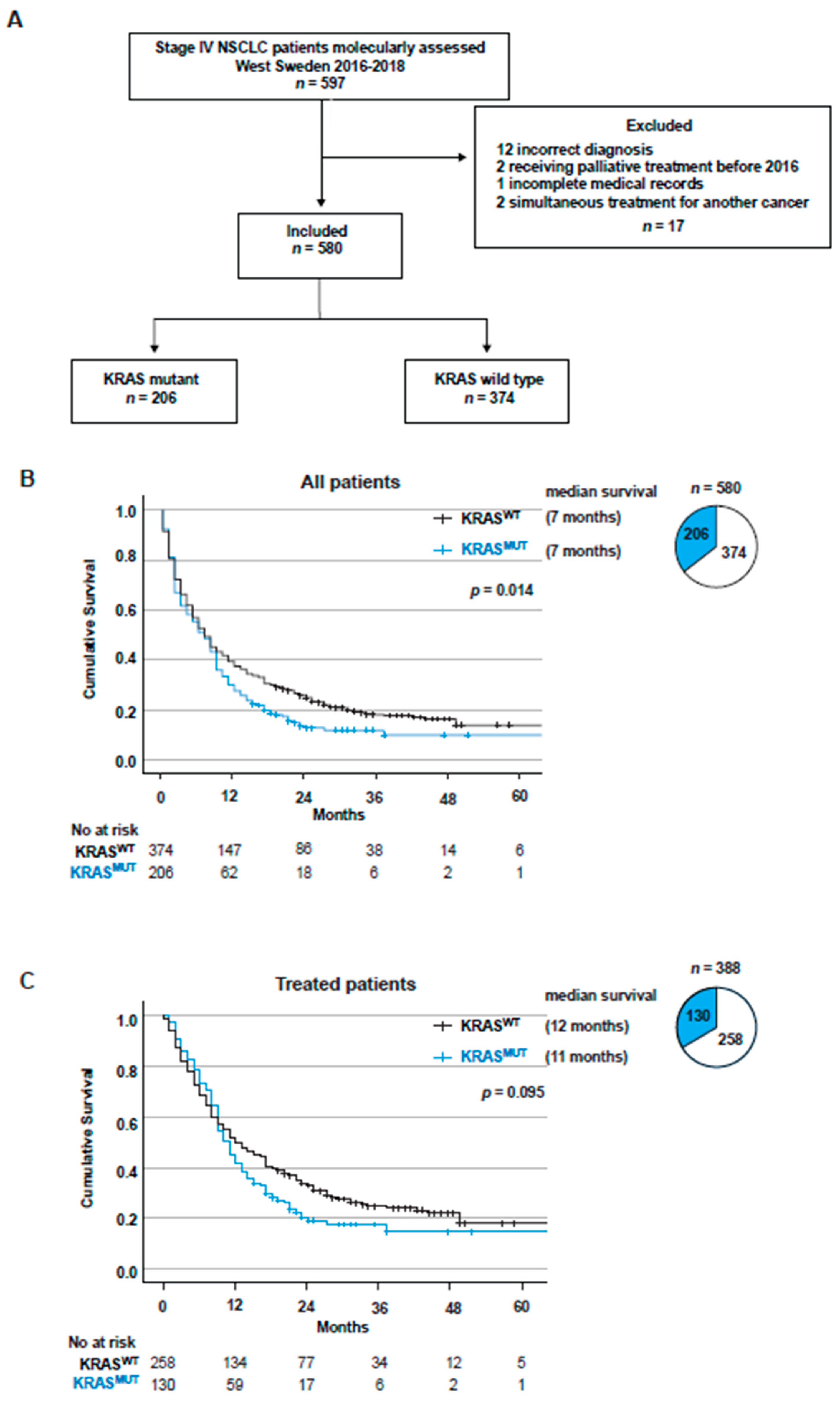
Clinical characteristics were summarized using descriptive statistics and evaluated with univariate analysis in table form. Survival was estimated using the Kaplan–Meier method. Log-rank test was used to assess significant differences in OS between groups. Multivariable Cox regression analyses were conducted to compensate for potential confounders. Statistical significance was set at p < 0.05, and no adjustments were made for multiple comparisons. One-sided Fisher’s exact test was used to determine the connection between KRAS status and being alive in the ICB-treated group. Data analysis was conducted using IBM SPSS Statistics version 27 and GraphPad Prism version 9.

3. Results

3.1. Patients and Tumor Characteristics

A total of 597 consecutive patients diagnosed with Stage IV NSCLC were molecularly assessed during 2016–2018 in West Sweden. A total of 17 patients were excluded for having incomplete medical records, for receiving palliative treatment before 2016, for incorrect diagnosis or for receiving simultaneous treatment for another type of cancer (Figure 1A). Among the 580 included patients, more than a third harbored a KRAS mutation (206, 35.5%), the majority were female (326, 56.2%), the median age was 71 years, and 80% were current or former smokers (Table 1). The majority of patients (322, 55.5%) had a good performance status (PS) with low ECOG 0–1 at diagnosis (Table 1). Histologically, the vast majority of NSCLCs were adenocarcinoma of the lung (498, 85.9%), while squamous cell carcinoma cases were relatively low, which was expected due to the selection of histological type for NGS assessment (32, 5.5%) (Table 1). In line with earlier studies, patients diagnosed with a low ECOG score had a significantly better overall survival (OS) compared to patients diagnosed with a higher ECOG score (p = 0.0001) (Figure S1A) [3,39]. In addition, higher numbers of metastasis locations at diagnosis correlated with poor OS (p = 0.0002) (Figure S1B), which was expected [3,39]. Furthermore, there was a trend towards better OS for females compared to males (p = 0.064) (Figure S1C) [39,40]. When comparing the baseline characteristics of KRASWT with KRASMUT patients, there were more females and a higher proportion of current and former smokers in the KRASMUT population (Table 1).

3.2. KRAS Mutations Are a Negative Factor for Overall Survival in Stage IV NSCLC

Survival estimates for the whole study cohort displayed that KRAS mutations were a significant negative factor for overall survival (p = 0.014) (Figure 1B). On multivariate Cox regression analysis, KRASMUT status and ECOG 2 or higher were independent prognostic factors for worse OS (HR 1.478, 95% CI 1.207–1.709, p < 0.001 and HR 3.175, 95% CI 2.622–3.845, p = 0.038). When comparing KRASG12C and KRASnon-G12C, no difference in OS was observed (p = 0.222) (Figure S2A). The population that received any type of life-extending treatment (n = 388), excluding patients only receiving best supportive care or palliative radiotherapy (e.g., single tumor radiation for pain relief), did not show a significant difference in OS between KRASMUT and KRASWT (p = 0.095) (Figure 1C). Similarly, when comparing KRASG12C and KRASnon-G12C, no difference in OS was observed among treated patients (p = 0.250) (Figure S2B).
When treated with first-line PT, one of the standard treatments at the time (n = 219), KRASMUT was a significant negative factor for survival (p = 0.001), with median OS of 9 months vs. KRASWT of 14 months (Figure S2C). Baseline characteristics of the patients showed that around 20% of KRASWT patients displayed druggable genomic alterations in ALK or EGFR (Table S1). To eliminate the risk of these patients driving any difference between KRASMUT and KRASWT groups due to potentially receiving targeted therapies in the second line of treatment, we excluded all patients with EGFR mutations and ALK fusions in the KRASWT group from further analysis. Nevertheless, in patients receiving first-line PT chemotherapy (n = 195), KRASMUT was a significant (p = 0.018) negative factor for survival (Figure 2A), with median OS of 9 months for KRASMUT vs. 11 months for KRASWT. In multivariate Cox regression analysis, KRASMUT status and ECOG 2 or higher were independent prognostic factors for worse OS (HR 1.564, 95% CI 1.124–2.177, p = 0.008 and HR 1.906, 95% CI 1.307–7779, p < 0.001). When comparing KRASG12C and KRASnon-G12C, there was no difference in OS (p = 0.313) (Figure 2B).

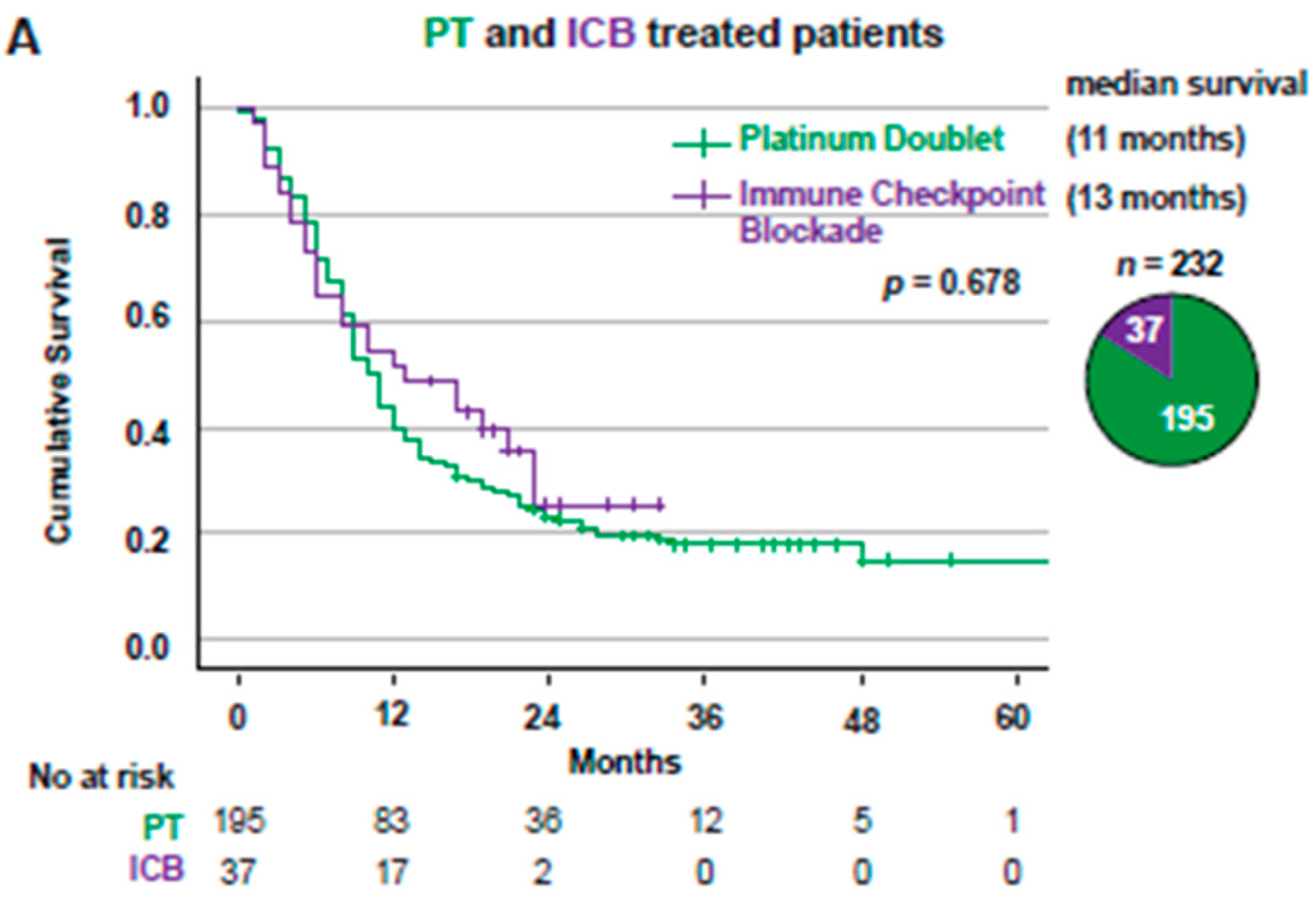
3.3. Overall Survival for Stage IV NSCLC Patients Treated with First-Line PT or ICB Stratified Based on KRAS Mutational Status

When comparing OS for all Stage IV patients receiving PT (n = 195) or ICB (n = 37) in a first-line setting, there was no difference between the two treatment groups (p = 0.678), with median OS of 11 months for PT vs. 13 months for ICB (Figure 3A). KRASMUT patients treated with ICB (n = 20) had a significantly (p = 0.003) better outcome compared to patients treated with PT (n = 104), with median OS of 23 vs. 9 months (Figure 3B). KRASWT patients treated with ICB (n = 17) had significantly (p = 0.023) worse survival than patients treated with PT (n = 91) with a median OS of 6 vs. 11 months (Figure 3C).

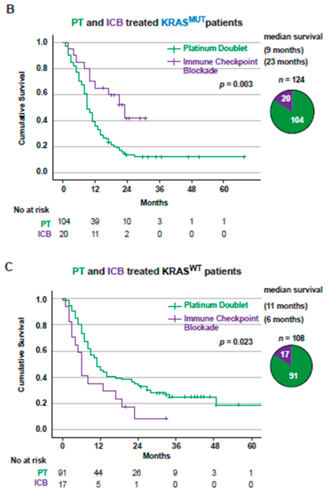
3.4. PD-L1 Expression in Tumors from Stage IV Patients Is a Positive Factor for Overall Survival in KRASMUT but Not KRASWT Patients

Currently, tumoral PD-L1 expression is the only biomarker for ICB treatment widely used in the clinic for NSCLC [41,42]. When analyzing all treated Stage IV NSCLC patients with known PD-L1 status (n = 261) stratified on KRAS mutational status and PD-L1 expression levels (negative, low or high) (Figure 4A), we observed that increased PD-L1 expression correlated with significantly better OS in KRASMUT patients (p = 0.036, Figure 4B). KRASMUT patients were clearly separated on OS between PD-L1 negative, low and high groups with median OS of 6, 11 and 17 months, respectively (Figure 4B). In multivariate Cox regression analysis, PD-L1 was an independent positive factor, and ECOG 2 or higher were independent factors for worse OS (HR 0.993 95% CI 0.986–0.999, p = 0.028 and HR 2.470, 95% CI 1.470–4.273, p = 0.001) for KRASMUT patients (Table S3). No correlation between PD-L1 expression and OS in KRASWT patients was observed (Figure 4C). No factor was significant in multivariate analysis (Table S3). There was a larger proportion of PD-L1high in the KRASMUT population compared to KRASWT (43.0% vs. 32.7%).

3.5. KRAS Mutations Are a Positive Factor for Overall Survival in Patients with PD-L1high Tumors Receiving Immunotherapy

We next analyzed the specific impact of PD-L1high expression on OS, comparing all PT-treated vs. ICB-treated patients. Outcome was significantly improved for KRASMUT patients on ICB treatment (p = 0.028), with median OS of 9 vs. 23 months (Figure 5A). KRASWT PD-L1high patients displayed a worse outcome among patients on ICB treatment compared to those on PT treatment (p = 0.010), with median OS of 6 months vs. 28 months for each respective treatment (Figure 5B). Importantly, the KRASMUT group had a significantly (p = 0.006) better outcome on ICB treatment compared to the KRASWT group, with a median OS of 23 vs. 6 months (Figure 5C, Table S2). On multivariate Cox regression analysis, KRASMUT status and ECOG PS 0–1 were independent prognostic factors for better OS following first-line ICB (HR 0.349, 95% CI 0.148–0.822, p = 0.016 and HR 0.398, 95% CI 0.165–0.963, p = 0.041). Finally, when looking at all patients treated with ICB individually (n = 37), there were significantly more KRASMUT patients alive at last follow-up compared to KRASWT patients (50% vs. 11.8%, p = 0.0152) (Figure 5D).

4. Discussion

The predictive value of KRAS mutations for survival in Stage IV NSCLC patients is still not clearly defined. While some studies have reported KRAS mutations to be a negative factor for survival estimates [3,22,43], others have shown this not to be the case [44]. The current retrospective cohort study showed a clear difference in OS for Stage IV NSCLC patients when stratified on KRAS mutational status. Previous studies have shown that KRAS mutations were associated with a shorter OS in response to chemotherapy [18,19]. In line with these studies, our data showed that KRASMUT is a potentially negative predictive factor for PT treatment. This is of importance, especially in light of the new treatment options now available, such as immunotherapy. Indeed, when stratifying Stage IV NSCLC patients receiving first-line ICB on KRAS mutational status, there was a clear increase in survival for KRASMUT patients.
Translational studies have suggested that KRASMUT patients might respond well to ICB due to a larger proportion of smokers and a higher immunologically active tumor environment as a consequence of constitutive activation of KRAS and downstream signaling pathways [30,31,32,33,34,39,45,46]. In agreement, our data showed that first-line ICB led to a clinically meaningful improvement in OS for KRASMUT patients when compared to first-line platinum doublet treatment. Furthermore, PD-L1 status had a clear and significant impact on stratifying OS in the KRASMUT group receiving treatment, whereas no difference was observed for the KRASWT group, which is in line with a previous study reporting a similar trend for KRASMUT NSCLC patients receiving ICB treatment [35].
Interestingly, KRASWT patients showed a better response to PT treatment than ICB. This might be explained by KRASWT being a heterogeneous group, with the majority of patients not having an identified driving mutation, especially as patients with targetable alterations in ALK and EGFR were excluded from further analysis.
There are several retrospective studies showing different outcomes when comparing KRASMUT and KRASWT response to ICB. These discrepancies could be explained by major study design differences, such as inclusion of several different stages at diagnosis, multiple treatment lines considered, and inclusion of patients with no known PD-L1 status [30,31,32,33,34,35,39]. A recent registry study from the Netherlands reported no difference in OS for PD-L1high patients receiving first-line ICB monotherapy, but the fraction of patients where KRAS mutations status had been assessed and reported to the registry was uncertain and could only be approximated (at around 75%) [39]. However, a meta-analysis of randomized controlled trials showed a significant OS benefit for KRASMUT patients in first-line immunotherapy with or without chemotherapy vs. chemotherapy alone [46], which is in line with the current study. Among these, the retrospective analysis of the phase 3 study KEYNOTE042 indicated a clear trend towards better OS in the KRASMUT group when comparing first-line ICB versus platinum-containing chemotherapy (28 vs. 11 months) than for the patients with KRASWT (15 vs. 12 months) [45], albeit only 301 out of 782 patients were molecularly assessed for KRAS status, among which 69 KRASMUT cases were identified. Importantly, KEYNOTE042 did not address the impact of KRAS mutations on PT treatment outcome. In our cohort, 580 patients were assessed, and 206 KRASMUT patients were identified. To the best of our knowledge, our retrospective cohort study is the largest to date comparing response to first-line ICB and PT therapy in Stage IV NSCLC patients where the KRAS mutational status was known for all patients.
Currently, first-line ICB is an option for all NSCLC patients with PD-L1high tumors. However, only a subset of patients benefit from durable response to immunotherapy, which does not correlate with tumoral PD-L1 expression. In line with other studies [9,13,14], our findings suggest that additional biomarkers for ICB response are clearly needed. Indeed, KRASWT patients with PD-L1high tumoral expression responded better to PT than to ICB. In contrast, KRASMUT patients clearly benefited more from ICB treatment (50% alive at last follow-up). Our results suggest that tumoral PD-L1high expression in combination with KRAS mutations is a better predictive biomarker for ICB in Stage IV NSCLC than PD-L1high expression alone. Our findings will have immediate clinical value, as assessing both KRAS mutational status and PD-L1 expression is currently performed routinely for most NSCLC patents.
Although this study provides real-world evidence of the impact of KRAS mutational status on first-line therapy in a large and well-controlled group of Stage IV NSCLC patients, it is obviously limited by its retrospective nature. Despite the fact that all Stage IV NSCLC patients molecularly assessed in the region of West Sweden between 2016–2018 were included in this cohort study, a relatively small number of patients were treated with first-line ICB therapy when compared to first-line PT therapy. Hence, pooled analyses from multi-cohort studies will be important to validate and expand our findings.

5. Conclusions

Here we reported that KRAS mutations are associated with a better response to treatment with immunotherapy and worse response to platinum doublet chemotherapy as well as shorter general overall survival in Stage IV NSCLC. Our findings suggest that KRAS mutations combined with PD-L1high expression were associated with better response to first-line immunotherapy than PD-L1high alone in patients with Stage IV NSCLC.

Supplementary Materials

The following supporting information can be downloaded at: https://www.mdpi.com/article/10.3390/cancers14092063/s1. Figure S1: A. Kaplan–Meier estimates comparing overall survival for the full cohort stratified on ECOG PS. Pie chart showing patient distribution between ECOG stages. B. Kaplan–Meier estimates comparing overall survival for the full cohort stratified on number of metastasis locations. Pie chart showing patient distribution between number of metastasis locations. C. Kaplan–Meier estimates comparing overall survival for the full cohort stratified on gender. Pie chart showing patient distribution between males and females. ECOG: Eastern Cooperative Oncology Group; PS: Performance status, Figure S2: A. Kaplan–Meier estimates comparing overall survival for all KRASMUT stratified on KRASG12C vs. KRASnon G12C. Pie chart showing patient distribution between KRASG12C and KRASnonG12C. B. Kaplan–Meier estimates comparing overall survival for all KRASMUT patients receiving treatment with platinum doublet stratified on KRASG12C vs. KRASnon G12C. Pie chart showing patient distribution between KRASG12C and KRASnon G12C. C. Kaplan–Meier estimates comparing overall survival for all receiving treatment with platinum doublet stratified on KRASWT vs. KRASMUT. Pie chart showing patient distribution between KRASWT and KRASMUT, Table S1 (Related to Figure S2C), Characteristics of all PT treated patients, Table S2 (Related to Figure 5A), Characteristics of all patients treated with first-line ICB, Table S3: Multivariate analyses.

Author Contributions

Conceptualization, E.A.E., H.F., J.N., A.H. and V.I.S.; Data curation, E.A.E. and H.F.; Formal analysis, E.A.E. and C.W.; Funding acquisition, E.A.E., C.W., H.F., L.M.A., S.R., J.N., A.H. and V.I.S.; Methodology, E.A.E.; Resources, H.F. and L.M.A.; Supervision, A.H. and V.I.S.; Visualization, E.A.E., C.W., L.M.A. and V.I.S.; Writing—original draft, E.A.E. and V.I.S.; Writing—review & editing, C.W., H.F., S.R., A.H. and V.I.S., Project coordination, V.I.S. All authors have read and agreed to the published version of the manuscript.

Funding

This work was supported by the Swedish Society for Medical Research (2018; S18-034), the Swedish Cancer Society, the Medical Research Council (2018; 2018-02318), AG Fond (2020), the Knut and Alice Wallenberg Foundation and the Wallenberg Centre for Molecular and Translational Medicine (to V.I.S.); the Gothenburg Society of Medicine (2019; 19/889991) and Department of Oncology, Sahlgrenska University Hospital (to E.A.E. and A.H.); the Swedish Cancer Society (2021; 21-1721) and The Healthcare Board for the Region of West Sweden (to S.R); Sahlgrenska Academy, Gothenburg University (2019; GU2019-3467) (to C.W.); the Swedish government under the LUA/ALF agreement (to H.F.); the Swedish Cancer Society (2017; 17-0171) and the Swedish government under the LUA/ALF agreement (ALFGBG-495961) (to L.M.A.).

Institutional Review Board Statement

Approval from the Swedish Ethical Review Authority (Dnr 2019-04771, approved 8 November 2019) was obtained prior to the commencement of the study.

Informed Consent Statement

Patient statement was not required due to the retrospective nature of this study.

Data Availability Statement

Data from this study are available upon reasonable request.

Acknowledgments

We thank Sayin lab members for critical reading of the manuscript. In addition, we thank members of the of the Swedish Lung Cancer Registry, and are grateful for the continuous reporting by Swedish healthcare employees.

Conflicts of Interest

The authors declare no conflict of interest.

References

  1. World Health Organization, International Agency for Research on Cancer. Globocan 2020: Lung Cancer. International Agency for Research on Cancer. Available online: http://gco.iarc.fr/today/data/factsheets/cancers/15-Lung-fact-sheet.pdf (accessed on 2 March 2021).
  2. Swedish Lung Cancer Registry. Available online: https://cancercentrum.se/samverkan/cancerdiagnoser/lunga-och-lungsack/vardprogram (accessed on 1 February 2022).
  3. El Osta, B.; Behera, M.; Kim, S.; Berry, L.D.; Sica, G.; Pillai, R.N.; Owonikoko, T.K.; Kris, M.G.; Johnson, B.E.; Kwiatkowski, D.J.; et al. Characteristics and Outcomes of Patients with Metastatic KRAS-Mutant Lung Adenocarcinomas: The Lung Cancer Mutation Consortium Experience. J. Thorac. Oncol. Off. Publ. Int. Assoc. Study Lung Cancer 2019, 14, 876–889. [Google Scholar] [CrossRef] [PubMed]
  4. Herbst, R.S.; Morgensztern, D.; Boshoff, C. The biology and management of non-small cell lung cancer. Nature 2018, 553, 446–454. [Google Scholar] [CrossRef] [PubMed]
  5. König, D.; Savic Prince, S.; Rothschild, S.I. Targeted Therapy in Advanced and Metastatic Non-Small Cell Lung Cancer. An Update on Treatment of the Most Important Actionable Oncogenic Driver Alterations. Cancers 2021, 13, 804. [Google Scholar] [CrossRef] [PubMed]
  6. Herbst, R.S.; Giaccone, G.; de Marinis, F.; Reinmuth, N.; Vergnenegre, A.; Barrios, C.H.; Morise, M.; Felip, E.; Andric, Z.; Geater, S.; et al. Atezolizumab for First-Line Treatment of PD-L1-Selected Patients with NSCLC. N. Engl. J. Med. 2020, 383, 1328–1339. [Google Scholar] [CrossRef]
  7. Reck, M.; Rodríguez-Abreu, D.; Robinson, A.G.; Hui, R.; Csőszi, T.; Fülöp, A.; Gottfried, M.; Peled, N.; Tafreshi, A.; Cuffe, S.; et al. Five-Year Outcomes with Pembrolizumab versus Chemotherapy for Metastatic Non-Small-Cell Lung Cancer with PD-L1 Tumor Proportion Score ≥ 50. J. Clin. Oncol. Off. J. Am. Soc. Clin. Oncol. 2021, 39, 2339–2349. [Google Scholar] [CrossRef]
  8. Reck, M.; Rodríguez-Abreu, D.; Robinson, A.G.; Hui, R.; Csőszi, T.; Fülöp, A.; Gottfried, M.; Peled, N.; Tafreshi, A.; Cuffe, S.; et al. Updated Analysis of KEYNOTE-024: Pembrolizumab versus Platinum-Based Chemotherapy for Advanced Non-Small-Cell Lung Cancer with PD-L1 Tumor Proportion Score of 50% or Greater. J. Clin. Oncol. Off. J. Am. Soc. Clin. Oncol. 2019, 37, 537–546. [Google Scholar] [CrossRef]
  9. Borghaei, H.; Langer, C.J.; Paz-Ares, L.; Rodríguez-Abreu, D.; Halmos, B.; Garassino, M.C.; Houghton, B.; Kurata, T.; Cheng, Y.; Lin, J.; et al. Pembrolizumab plus chemotherapy versus chemotherapy alone in patients with advanced non-small cell lung cancer without tumor PD-L1 expression: A pooled analysis of 3 randomized controlled trials. Cancer 2020, 126, 4867–4877. [Google Scholar] [CrossRef]
  10. Ferrara, R.; Imbimbo, M.; Malouf, R.; Paget-Bailly, S.; Calais, F.; Marchal, C.; Westeel, V. Single or combined immune checkpoint inhibitors compared to first-line platinum-based chemotherapy with or without bevacizumab for people with advanced non-small cell lung cancer. Cochrane Database Syst. Rev. 2021, 4, Cd013257. [Google Scholar] [CrossRef]
  11. Frederickson, A.M.; Arndorfer, S.; Zhang, I.; Lorenzi, M.; Insinga, R.; Arunachalam, A.; Burke, T.A.; Simon, G.R. Pembrolizumab plus chemotherapy for first-line treatment of metastatic nonsquamous non-small-cell lung cancer: A network meta-analysis . Immunotherapy 2019, 11, 407–428. [Google Scholar] [CrossRef] [Green Version]
  12. Gandhi, L.; Rodríguez-Abreu, D.; Gadgeel, S.; Esteban, E.; Felip, E.; De Angelis, F.; Domine, M.; Clingan, P.; Hochmair, M.J.; Powell, S.F.; et al. Pembrolizumab plus Chemotherapy in Metastatic Non-Small-Cell Lung Cancer. N. Engl. J. Med. 2018, 378, 2078–2092. [Google Scholar] [CrossRef]
  13. Rittmeyer, A.; Barlesi, F.; Waterkamp, D.; Park, K.; Ciardiello, F.; von Pawel, J.; Gadgeel, S.M.; Hida, T.; Kowalski, D.M.; Dols, M.C.; et al. Atezolizumab versus docetaxel in patients with previously treated non-small-cell lung cancer (OAK): A phase 3, open-label, multicentre randomised controlled trial. Lancet 2017, 389, 255–265. [Google Scholar] [CrossRef]
  14. Zhang, B.; Liu, Y.; Zhou, S.; Jiang, H.; Zhu, K.; Wang, R. Predictive effect of PD-L1 expression for immune checkpoint inhibitor (PD-1/PD-L1 inhibitors) treatment for non-small cell lung cancer: A meta-analysis. Int. Immunopharmacol. 2020, 80, 106214. [Google Scholar] [CrossRef] [PubMed]
  15. Adderley, H.; Blackhall, F.H.; Lindsay, C.R. KRAS-mutant non-small cell lung cancer: Converging small molecules and immune checkpoint inhibition. EBioMedicine 2019, 41, 711–716. [Google Scholar] [CrossRef] [PubMed] [Green Version]
  16. Ferrer, I.; Zugazagoitia, J.; Herbertz, S.; John, W.; Paz-Ares, L.; Schmid-Bindert, G. KRAS-Mutant non-small cell lung cancer: From biology to therapy. Lung Cancer 2018, 124, 53–64. [Google Scholar] [CrossRef] [PubMed] [Green Version]
  17. Brady, A.K.; McNeill, J.D.; Judy, B.; Bauml, J.; Evans, T.L.; Cohen, R.B.; Langer, C.; Vachani, A.; Aggarwal, C. Survival outcome according to KRAS mutation status in newly diagnosed patients with stage IV non-small cell lung cancer treated with platinum doublet chemotherapy. Oncotarget 2015, 6, 30287–30294. [Google Scholar] [CrossRef] [PubMed]
  18. Hames, M.L.; Chen, H.; Iams, W.; Aston, J.; Lovly, C.M.; Horn, L. Correlation between KRAS mutation status and response to chemotherapy in patients with advanced non-small cell lung cancer. Lung Cancer 2016, 92, 29–34. [Google Scholar] [CrossRef] [Green Version]
  19. Marabese, M.; Ganzinelli, M.; Garassino, M.C.; Shepherd, F.A.; Piva, S.; Caiola, E.; Macerelli, M.; Bettini, A.; Lauricella, C.; Floriani, I.; et al. KRAS mutations affect prognosis of non-small-cell lung cancer patients treated with first-line platinum containing chemotherapy. Oncotarget 2015, 6, 34014–34022. [Google Scholar] [CrossRef] [Green Version]
  20. Mellema, W.W.; Dingemans, A.M.; Thunnissen, E.; Snijders, P.J.; Derks, J.; Heideman, D.A.; Van Suylen, R.; Smit, E.F. KRAS mutations in advanced nonsquamous non-small-cell lung cancer patients treated with first-line platinum-based chemotherapy have no predictive value. J. Thorac. Oncol. Off. Publ. Int. Assoc. Study Lung Cancer 2013, 8, 1190–1195. [Google Scholar] [CrossRef] [Green Version]
  21. Rodenhuis, S.; Boerrigter, L.; Top, B.; Slebos, R.J.; Mooi, W.J.; van’t Veer, L.; van Zandwijk, N. Mutational activation of the K-ras oncogene and the effect of chemotherapy in advanced adenocarcinoma of the lung: A prospective study. J. Clin. Oncol. Off. J. Am. Soc. Clin. Oncol. 1997, 15, 285–291. [Google Scholar] [CrossRef]
  22. Meng, D.; Yuan, M.; Li, X.; Chen, L.; Yang, J.; Zhao, X.; Ma, W.; Xin, J. Prognostic value of K-RAS mutations in patients with non-small cell lung cancer: A systematic review with meta-analysis. Lung Cancer 2013, 81, 1–10. [Google Scholar] [CrossRef]
  23. Addeo, A.; Banna, G.L.; Friedlaender, A. KRAS G12C Mutations in NSCLC: From Target to Resistance. Cancers 2021, 13, 2541. [Google Scholar] [CrossRef] [PubMed]
  24. Burns, T.F.; Borghaei, H.; Ramalingam, S.S.; Mok, T.S.; Peters, S. Targeting KRAS-Mutant Non-Small-Cell Lung Cancer: One Mutation at a Time, with a Focus on KRAS G12C Mutations. J. Clin. Oncol. Off. J. Am. Soc. Clin. Oncol. 2020, 38, 4208–4218. [Google Scholar] [CrossRef] [PubMed]
  25. Indini, A.; Rijavec, E.; Ghidini, M.; Cortellini, A.; Grossi, F. Targeting KRAS in Solid Tumors: Current Challenges and Future Opportunities of Novel KRAS Inhibitors. Pharmaceutics 2021, 13, 653. [Google Scholar] [CrossRef]
  26. Mathieu, M.; Steier, V.; Fassy, F.; Delorme, C.; Papin, D.; Genet, B.; Duffieux, F.; Bertrand, T.; Delarbre, L.; Le-Borgne, H.; et al. KRAS G12C fragment screening renders new binding pockets. Small GTPases 2021, 13, 1–14. [Google Scholar] [CrossRef] [PubMed]
  27. Skoulidis, F.; Li, B.T.; Dy, G.K.; Price, T.J.; Falchook, G.S.; Wolf, J.; Italiano, A.; Schuler, M.; Borghaei, H.; Barlesi, F.; et al. Sotorasib for Lung Cancers with KRAS p.G12C Mutation. N. Engl. J. Med. 2021, 384, 2371–2381. [Google Scholar] [CrossRef] [PubMed]
  28. Zhang, J.; Zhang, J.; Liu, Q.; Fan, X.X.; Leung, E.L.; Yao, X.J.; Liu, L. Resistance looms for KRAS G12C inhibitors and rational tackling strategies. Pharm. Ther. 2022, 229, 108050. [Google Scholar] [CrossRef]
  29. Zhao, Y.; Murciano-Goroff, Y.R.; Xue, J.Y.; Ang, A.; Lucas, J.; Mai, T.T.; Da Cruz Paula, A.F.; Saiki, A.Y.; Mohn, D.; Achanta, P.; et al. Diverse alterations associated with resistance to KRAS(G12C) inhibition. Nature 2021, 599, 679–683. [Google Scholar] [CrossRef] [PubMed]
  30. Liu, C.; Zheng, S.; Jin, R.; Wang, X.; Wang, F.; Zang, R.; Xu, H.; Lu, Z.; Huang, J.; Lei, Y.; et al. The superior efficacy of anti-PD-1/PD-L1 immunotherapy in KRAS-mutant non-small cell lung cancer that correlates with an inflammatory phenotype and increased immunogenicity. Cancer Lett. 2020, 470, 95–105. [Google Scholar] [CrossRef]
  31. Cinausero, M.; Laprovitera, N.; De Maglio, G.; Gerratana, L.; Riefolo, M.; Macerelli, M.; Fiorentino, M.; Porcellini, E.; Buoro, V.; Gelsomino, F.; et al. KRAS and ERBB-family genetic alterations affect response to PD-1 inhibitors in metastatic nonsquamous NSCLC. Ther. Adv. Med. Oncol. 2019, 11, 1758835919885540. [Google Scholar] [CrossRef] [Green Version]
  32. Gianoncelli, L.; Spitaleri, G.; Passaro, A.; Radice, D.; Fumagalli, C.; Del Signore, E.; Stati, V.; Catania, C.M.; Guerini-Rocco, E.; Barberis, M.; et al. Efficacy of Anti-PD1/PD-L1 Therapy (IO) in KRAS Mutant Non-small Cell Lung Cancer Patients: A Retrospective Analysis. Anticancer Res. 2020, 40, 427–433. [Google Scholar] [CrossRef]
  33. Karatrasoglou, E.A.; Chatziandreou, I.; Sakellariou, S.; Stamopoulos, K.; Kavantzas, N.; Lazaris, A.C.; Korkolopoulou, P.; Saetta, A.A. Association between PD-L1 expression and driver gene mutations in non-small cell lung cancer patients: Correlation with clinical data. Virchows Arch. Int. J. Pathol. 2020, 477, 207–217. [Google Scholar] [CrossRef]
  34. Torralvo, J.; Friedlaender, A.; Achard, V.; Addeo, A. The Activity of Immune Checkpoint Inhibition in KRAS Mutated Non-small Cell Lung Cancer: A Single Centre Experience. Cancer Genom. Proteom. 2019, 16, 577–582. [Google Scholar] [CrossRef] [PubMed] [Green Version]
  35. Jeanson, A.; Tomasini, P.; Souquet-Bressand, M.; Brandone, N.; Boucekine, M.; Grangeon, M.; Chaleat, S.; Khobta, N.; Milia, J.; Mhanna, L.; et al. Efficacy of Immune Checkpoint Inhibitors in KRAS-Mutant Non-Small Cell Lung Cancer (NSCLC). J. Thorac. Oncol. Off. Publ. Int. Assoc. Study Lung Cancer 2019, 14, 1095–1101. [Google Scholar] [CrossRef]
  36. Herbst, R.; Jassem, J.; Abogunrin, S.; James, D.; McCool, R.; Belleli, R.; Giaccone, G.; De Marinis, F. A Network Meta-Analysis of Cancer Immunotherapies versus Chemotherapy for First-Line Treatment of Patients with Non-Small Cell Lung Cancer and High Programmed Death-Ligand 1 Expression. Front. Oncol. 2021, 11, 676732. [Google Scholar] [CrossRef] [PubMed]
  37. Arak, H.; Aytekin, A.; Canoz, O.; Ozkan, M. Prognostic and Predictive Significance of PD-L1 Expression in Non-Small Cell Lung Cancer Patients: A Single-Center Experience. Turk. Patoloji Derg. 2021, 37, 239–248. [Google Scholar] [CrossRef]
  38. Wang, A.; Wang, H.Y.; Liu, Y.; Zhao, M.C.; Zhang, H.J.; Lu, Z.Y.; Fang, Y.C.; Chen, X.F.; Liu, G.T. The prognostic value of PD-L1 expression for non-small cell lung cancer patients: A meta-analysis. Eur. J. Surg. Oncol. J. Eur. Soc. Surg. Oncol. Br. Assoc. Surg. Oncol. 2015, 41, 450–456. [Google Scholar] [CrossRef]
  39. Noordhof, A.L.; Damhuis, R.A.M.; Hendriks, L.E.L.; de Langen, A.J.; Timens, W.; Venmans, B.J.W.; van Geffen, W.H. Prognostic impact of KRAS mutation status for patients with stage IV adenocarcinoma of the lung treated with first-line pembrolizumab monotherapy. Lung Cancer 2021, 155, 163–169. [Google Scholar] [CrossRef] [PubMed]
  40. Dogan, S.; Shen, R.; Ang, D.C.; Johnson, M.L.; D’Angelo, S.P.; Paik, P.K.; Brzostowski, E.B.; Riely, G.J.; Kris, M.G.; Zakowski, M.F.; et al. Molecular Epidemiology of EGFR and KRAS Mutations in 3,026 Lung Adenocarcinomas: Higher Susceptibility of Women to Smoking-Related KRAS Mutant. Cancers 2012, 18, 6169–6177. [Google Scholar] [CrossRef] [Green Version]
  41. Darabi, S.; Braxton, D.R.; Eisenberg, B.L.; Demeure, M.J. Predictive Biomarkers for Immunotherapy Response Beyond PD-1/PD-L1. Oncology 2020, 34, 321–327. [Google Scholar] [CrossRef]
  42. Niu, M.; Yi, M.; Li, N.; Luo, S.; Wu, K. Predictive biomarkers of anti-PD-1/PD-L1 therapy in NSCLC. Exp. Hematol. Oncol. 2021, 10, 18. [Google Scholar] [CrossRef]
  43. Hallqvist, A.; Enlund, F.; Andersson, C.; Sjögren, H.; Hussein, A.; Holmberg, E.; Nyman, J. Mutated KRAS Is an Independent Negative Prognostic Factor for Survival in NSCLC Stage III Disease Treated with High-Dose Radiotherapy. Lung Cancer Int. 2012, 2012, 587424. [Google Scholar] [CrossRef] [PubMed] [Green Version]
  44. Sebastian, M.; Eberhardt, W.E.E.; Hoffknecht, P.; Metzenmacher, M.; Wehler, T.; Kokowski, K.; Alt, J.; Schütte, W.; Büttner, R.; Heukamp, L.C.; et al. KRAS G12C-mutated advanced non-small cell lung cancer: A real-world cohort from the German prospective, observational, nation-wide CRISP Registry (AIO-TRK-0315). Lung Cancer 2021, 154, 51–61. [Google Scholar] [CrossRef] [PubMed]
  45. Herbst, R.S.; Lopes, G.; Kowalski, D.M.; Kasahara, K.; Wu, Y.L.; De Castro, G.; Cho, B.C.; Turna, H.Z.; Cristescu, R.; Aurora-Garg, D.; et al. LBA4 Association of KRAS mutational status with response to pembrolizumab monotherapy given as first-line therapy for PD-L1-positive advanced non-squamous NSCLC in Keynote-042. Ann. Oncol. 2019, 30, xi63–xi64. [Google Scholar] [CrossRef]
  46. Landre, T.; Justeau, G.; Assié, J.B.; Chouahnia, K.; Davoine, C.; Taleb, C.; Chouaïd, C.; Duchemann, B. Anti-PD-(L)1 for KRAS-mutant advanced non-small-cell lung cancers: A meta-analysis of randomized-controlled trials. Cancer Immunol. Immunother. 2022, 71, 719–726. [Google Scholar] [CrossRef]
Figure 1. KRAS mutations are a negative factor for overall survival. (A) Flow chart showing the patient selection for the study. (B) Kaplan–Meier estimates comparing overall survival and median survival stratified on KRASWT vs. KRASMUT for the full cohort (n = 580). Pie chart showing patient distribution between KRASWT and KRASMUT. (C) Kaplan–Meier estimates comparing overall survival for all receiving life-extending treatment (no treatment and only palliative radiotherapy excluded) stratified on KRASWT vs. KRASMUT. Pie chart showing patient distribution between KRASWT and KRASMUT.
Cancers 14 02063 g001
Figure 2. KRASMUT is a negative factor for overall survival when treated with platinum doublet independently of EGFR and ALK mutations. (A) Kaplan–Meier estimates comparing overall survival for all receiving treatment with platinum doublet stratified on KRASWT vs. KRASMUT. Patients with EGFR and ALK alterations were excluded from KRASWT group. Pie chart showing patient distribution between KRASWT and KRASMUT. (B) Kaplan–Meier estimates comparing overall survival for all KRASMUT receiving treatment with platinum doublet stratified on KRASG12C vs. KRASnon G12C. Pie chart showing patient distribution between KRASG12C and KRASnon G12C.
Cancers 14 02063 g002aCancers 14 02063 g002b
Figure 3. KRASMUT, but not KRASWT, had a better outcome with ICB than with platinum doublet treatment. (A) Kaplan–Meier estimates comparing overall survival for all patients, excluding patients with ALK or EGFR alterations, receiving treatment with PT vs. ICB. Pie chart showing patient distribution between PT and ICB. (B) Kaplan–Meier estimates comparing overall survival for all KRASMUT receiving treatment with PT vs. ICB. Pie chart showing patient distribution between platinum doublet and ICB. (C) Kaplan–Meier estimates comparing overall survival for KRASWT, excluding patients with ALK or EGFR alterations, receiving treatment with platinum doublet vs. ICB. Pie chart showing patient distribution between PT and ICB. ICB: Immune checkpoint blockade; PT: Platinum doublet.
Cancers 14 02063 g003aCancers 14 02063 g003b
Figure 4. PD-L1 status has an impact on overall survival for treated patients with KRASMUT. (A) IHC depicting examples of PD-L1 staining used to assess PD-L1 status in both KRASWT and KRASMUT NSCLC patients. PD-L1 expression is classified as negative, <50% or ≥50% based on tumor proportion score. (B) Kaplan–Meier estimates comparing overall survival for KRASMUT treated patients stratified on PD-L1 status. Pie chart showing patient distribution between PD-L1 negative, <50% and ≥50%. (C) Kaplan–Meier estimates comparing overall survival for KRASWT treated patients stratified on PD-L1 status. Pie chart showing patient distribution between PD-L1 negative, <50% and ≥50%. IHC: Immunohistochemistry; PD-L1: Programmed death ligand 1. Scale bar: 100 μm.
Cancers 14 02063 g004aCancers 14 02063 g004b
Figure 5. KRASMUT patients have a better outcome on ICB treatment than KRASWT patients. (A) Kaplan–Meier estimates comparing overall survival for KRASMUT PD-L1 ≥ 50% treated patients receiving PD or ICB. Pie chart showing patient distribution receiving PT or ICB treatment. (B) Kaplan–Meier estimates comparing overall survival for KRASWT PD-L1 ≥ 50% treated patients receiving PT or ICB. Pie chart showing patient distribution receiving PT or ICB treatment. (C) Kaplan–Meier estimates comparing overall survival for all patients receiving ICB treatment in a first-line setting. Pie chart showing patient distribution between KRASWT and KRASMUT. (D) Swimmer plot showing survival time for both KRASWT and KRASMUT ICB-treated patients. Patients with KRASG12C mutation are marked with G12C. Status alive at last follow-up is shown. ICB: Immune checkpoint blockade; PT: Platinum doublet; PD-L1: Programmed death ligand 1.
Cancers 14 02063 g005
Table 1. Characteristics of the total cohort as well as stratified on KRASWT and KRASMUT. Data are presented as n (%).
Characteristics of All Patients Stage IV NSCLC TotalKRAS WTKRAS MUT
n (%)n (%)n (%)
Total580 (100)374 (64, 5)206 (35, 5)
Age in years, median (range)71 (24–94)70,5 (24–94)71 (46–90)
Sex
Male254 (43, 8)173 (46, 3)81 (39, 3)
Female 326 (56, 2)201 (53, 7)125 (60, 7)
Smoking history
Current smoker 192 (33, 1)117 (31, 4)75 (36, 4)
Former smoker270 (46, 6)152 (40, 6)118 (57, 3)
Never smoker116 (20)103 (27, 5)13 (6, 3)
Missing2 (0, 3)2 (0, 5)0
Performance status
ECOG 079 (13, 6)48 (12, 8)31 (15, 0)
ECOG 1243 (41, 9)142 (38, 0)101 (49, 0)
ECOG 2154 (26, 6)99 (26, 5)55 (26, 7)
ECOG 374 (12, 8)57 (15, 2)17 (8, 3)
ECOG 419 (3, 3)17 (4, 5)2 (1, 0)
Missing11 (1, 9)11 (2, 9)0
Histology
Adenocarcinoma498 (85, 9) 316 (84, 5)128 (85, 9)
NSCLC NOS50 (8, 6)28 (7, 5)19 (12, 8)
Squamous cell carcinoma32 (5, 5)30 (8, 0)2 (1, 3)
Mutation status
None known231 (39, 8)231 (71, 8)
ALK19 (3, 3)19 (5, 1)
EGFR85 (14, 7)85 (22, 7)
BRAF20 (3, 4)20 (5, 3)
Other15 (2, 6)15 (4)
ROS12 (0, 3)2 (0, 5)
RET2 (0, 3)2 (0, 5)
KRAS206 (35, 5)
KRAS submutation
G12A 14 (6, 8)
G12C 83 (40, 3)
G12D 23 (11, 2)
G12V 43 (20, 9)
Q61H 10 (4, 9)
Others 33 (16)
At last follow-up
Alive97 (16, 7)69 (18, 4)28 (13, 6)
Deceased483 (83, 3)305 (81, 6)178 (86, 4)
Survival
Median survival (months)777
No. of metastatic locations at diagnosis
1361 (62, 2)234 (62, 6)127 (61, 7)
2153 (26, 4)101 (27, 0)52 (25, 2)
347 (8, 1)30 (8, 017 (8, 3)
>311 (1, 9)3 (0, 8)8 (3, 9)
Missing8 (1, 4)6 (1, 6)2 (1, 0)
ECOG PS, Eastern Cooperative Oncology Group Performance Status.
Publisher’s Note: MDPI stays neutral with regard to jurisdictional claims in published maps and institutional affiliations.

