Article

Germline Variants in Angiogenesis-Related Genes Contribute to Clinical Outcome in Head and Neck Squamous Cell Carcinoma

by
Dorota Butkiewicz
1,*,
Agnieszka Gdowicz-Kłosok
1,
Małgorzata Krześniak
1,
Tomasz Rutkowski
2,
Barbara Łasut-Szyszka
1 and
Krzysztof Składowski
2
1
Center for Translational Research and Molecular Biology of Cancer, Maria Skłodowska-Curie National Research Institute of Oncology, Gliwice Branch, 44-102 Gliwice, Poland
2
I Radiation and Clinical Oncology Department, Maria Skłodowska-Curie National Research Institute of Oncology, Gliwice Branch, 44-102 Gliwice, Poland
*
Author to whom correspondence should be addressed.
Cancers 2022, 14(7), 1844; https://doi.org/10.3390/cancers14071844
Submission received: 26 January 2022 / Revised: 28 March 2022 / Accepted: 1 April 2022 / Published: 6 April 2022
(This article belongs to the Special Issue Chemoradiotherapy for Head and Neck Cancer)

Abstract

:

Simple Summary

A high risk of relapse and treatment resistance are among the major challenges in locally advanced head and neck squamous cell carcinoma (HNSCC). Data show that common germline alterations in genes regulating angiogenesis may modulate treatment sensitivity, cancer progression, and prognosis, but relatively little is known about their role in HNSCC. Thus, our goal was to examine the effect of variation in these genes on survival outcomes in HNSCC patients receiving radiotherapy and cisplatin-based chemoradiotherapy. We identified genetic variants significantly affecting therapy results, constituting independent prognostic factors in these patients. Our results suggest that some polymorphisms in angiogenesis genes may be determinants of treatment efficacy and tumor aggressiveness in HNSCC, which may be of importance in standard therapy. These findings emphasize the potential value of the host genetic profile related to angiogenesis in assessing the risk of treatment failure.

Abstract

Fibroblast growth factor (FGF)/FGF receptor (FGFR), and platelet-derived growth factor (PDGF)/PDGF receptor (PDGFR) systems, as well as some matrix metalloproteinases (MMPs) and their tissue inhibitors (TIMPs), are involved in various steps of angiogenesis. Data indicate that common germline variations in angiogenesis-regulating genes may modulate therapy results and cancer progression. However, whether these variants affect clinical outcome in head and neck squamous cell carcinoma (HNSCC) is unclear. Hence, we assessed the relationship between FGF/FGFR, PDGF/PDGFR, MMP, and TIMP genetic variants and treatment outcomes in HNSCC patients receiving radiotherapy (RT) alone or combined with cisplatin-based chemotherapy. In multivariate analysis, FGF2 rs1048201 CC homozygotes showed a higher risk of death (p = 0.039), while PDGFRA rs2228230 T was strongly associated with an increased risk of locoregional relapse (HR 2.49, p = 0.001) in the combination treatment subgroup. In the RT alone subset, MMP2 rs243865 TT carriers had a higher risk of locoregional recurrence (HR 2.92, p = 0.019), whereas PDGFRB rs246395 CC homozygotes were at increased risk of metastasis (HR 3.06, p = 0.041). The MMP2 rs7201 C and TIMP2 rs7501477 T were associated with a risk of locoregional failure in the entire cohort (p = 0.032 and 0.045, respectively). Furthermore, rs1048201, rs2228230, rs246395, rs243865, rs7201, and rs7201/rs7501477 were independent indicators of an unfavorable outcome. This study demonstrates that the FGF2, PDGFRA, PDGFRB, MMP2, and TIMP2 variants may contribute to treatment failure and poor prognosis in HNSCC.

1. Introduction

Head and neck squamous cell carcinoma (HNSCC) is one of the most common malignant neoplasms in the world, very often diagnosed in an advanced stage [1]. Although progress has been made in the treatment of this cancer, locoregional and distant relapse occurring in a large number of patients is still a serious problem leading to poor survival outcomes. In HNSCC, decreased sensitivity, or resistance to treatment, is one of the major challenges in terms of patient prognosis. In locally advanced and unresectable HNSCC, radiotherapy (RT) and cisplatin-based chemotherapy (CT) are the mainstays of treatment [2]. Their effectiveness is highly influenced by hypoxia and dysregulated angiogenesis [3,4].
Angiogenesis is recognized as playing a crucial role in the development and progression of solid tumors [5]. Several growth factors are involved in different steps of new blood vessel formation. Angiogenesis is primarily mediated by the vascular endothelial growth factor (VEGF)/VEGF receptor (VEGFR) system; however, other proangiogenic growth factors, such as fibroblast growth factor 2 (FGF2, also known as basic FGF, or bFGF), are also very potent regulators of the process. FGF/FGF receptor (FGFR) signaling leads to proliferation, migration, and differentiation of endothelial cells and fibroblasts [6]. The FGF pathway may indirectly control angiogenesis by coordinating other growth factor signaling (e.g., VEGF) and various cell–cell interactions [7]. Platelet-derived growth factor (PDGF) is another angiogenesis-inducing cytokine that exerts its effects by interacting with PDGF receptors α (PDGFRA) and β (PDGFRB). The PDGF/PDGFR system is critical for the proliferation, migration, and recruitment of mesenchymal cells, including vascular smooth muscle cells, pericytes, and fibroblasts [8]. Both networks, FGF/FGFR and PDGF/PDGFR, are implicated in embryogenesis, tissue regeneration, and wound healing, and when deregulated, are also involved in tumor growth, survival, and metastasis [9,10]. High levels of these proteins have been associated with a worse prognosis in various cancers [6,8,11,12].
The structure and composition of the extracellular matrix (ECM) are important factors in the regulation of angiogenesis. Angiogenesis is accompanied by the degradation of the vascular basement membrane and ECM components by matrix metalloproteinases (MMPs). These calcium-dependent zinc endopeptidases, produced by stromal and tumor cells, are involved in inflammation, tumor invasion, and metastasis. MMPs are essential for tumor angiogenesis, as they participate in vascular remodeling, cell migration, and sprout formation [13]. Two gelatinases, MMP2 and MMP9, are believed to play particularly important roles in this process. They are known to activate and release proangiogenic growth factors (e.g., VEGF and FGF2) from ECM, as well as generate antiangiogenic molecules [14,15]. The enzymatic activity of MMPs is regulated by the family of endogenous tissue inhibitors of metalloproteinases (TIMPs). In addition to the inhibitory role against MMPs, TIMPs may participate in the MMP activation. Moreover, TIMP2 is able to suppress endothelial cell proliferation in response to angiogenic factors, while TIMP3 has the ability to interact with VEGFR2 and block VEGF binding [16,17]. Growth factors such as VEGF, FGF, and PDGF can stimulate production of MMPs [10]. The overexpression of MMPs, observed in many cancers, has been found to correlate with tumor aggressiveness and poor prognosis [18,19].
Growing evidence suggests that common germline alterations, such as single nucleotide polymorphisms (SNPs), in angiogenesis-regulating genes may not only increase individual susceptibility to cancer, but may also be implicated in modulating sensitivity to anticancer treatment, thereby affecting therapy results and patient survival [17,20,21,22]. In HNSCC, very few studies have so far addressed the role of SNPs in angiogenesis genes in the context of treatment outcome and prognosis. In a previous report, we demonstrated the predictive and prognostic potential of inherited genetic variants in the ANGPT2/TEK and VEGF/VEGFR2 systems in HNSCC [21]. In the present study, we aimed to evaluate the possible association between a panel of 19 variants in the FGF2, FGFR2, PDGFB, PDGFRA, PDGFRB, MMP2, MMP9, TIMP1, TIMP2, and TIMP3 genes and the clinical outcomes in non-surgically treated HNSCC patients who received radical RT alone or in combination with cisplatin-based CT.

2. Materials and Methods

2.1. Patients

The study group comprised 422 Caucasian patients diagnosed with primary T1–4N0–3M0 HNSCC of the larynx (LSCC), oropharynx (OPSCC) or hypopharynx (HPSCC). The patient characteristics are shown in Table S1. There were 290 (69%) patients with stage III–IVB. Most of the patients were males (80%), cigarette smokers (80%), and alcohol users (77%). All subjects had a WHO performance status of 0 or 1. The treatment and follow-up details have been previously described [21]. Briefly, all patients were treated with curative intent with RT alone (n = 219, 52%) or combined with cisplatin-based CT given as induction treatment (docetaxel/cisplatin/5-fluorouracil or cisplatin/5-fluorouracil; n = 72, 17%) or administered concurrently (n = 131, 31%). Patients who received surgery were excluded from the study. Clinical and demographic data were obtained from medical records and the Silesian Cancer Registry. The study endpoints were overall survival (OS), locoregional recurrence-free survival (LRFS), and metastasis-free survival (MFS). OS was calculated from the date of diagnosis to the date of death from any cause, or the last known date alive. LRFS and MFS were defined as the time from treatment completion to clinically detectable local and/or regional recurrence (for LRFS) or distant metastasis (for MFS), or the last examination without evidence of disease.

2.2. SNP Identification

A total of 19 candidate SNPs were analyzed in this study, including rs5757573, rs2285094 in PDGFB, rs2228230, rs1800812 in PDGFRA, rs2302273, rs246395 in PDGFRB, rs1449683, rs1048201 in FGF, rs2981582 in FGFR2, rs243865, rs7201 in MMP2, rs17576, rs17577 in MMP9, rs4898, rs2070584 in TIMP1, rs2277698, rs7501477 in TIMP2, and rs9862, rs9619311 in TIMP3 (Table S2). These were SNPs that had a minor allele frequency (MAF) ≥10% in the European Caucasian population [23] and functional significance, and/or were located in coding or regulatory regions, and/or were reported as associated with cancer risk or outcome for other solid cancers [22,24,25,26,27,28,29,30,31,32,33,34,35,36,37,38,39,40,41]. Genomic DNA was isolated from frozen peripheral blood with Genomic Maxi AX kit (A&A Biotechnology, Gdynia, Poland). The SNPs were determined using TaqMan SNP Genotyping Assays (Applied Biosystems, Foster City, CA, USA), according to the manufacturer’s standard protocol. Genotyping was repeated in 50 randomly selected samples, and the concordance was 100%.

2.3. Statistical Analysis

The associations between SNPs and survival endpoints were examined using the Kaplan–Meier method and log-rank test. All SNPs were tested under dominant, recessive, and codominant genetic models, and the model with the most significant p value was selected for the final analysis. The Cox proportional hazards regression method was used in univariate and multivariate analysis. Multivariate models were adjusted for the following variables: median age at diagnosis (<59 versus ≥59 years), sex (male versus female), cigarette smoking or alcohol use (ever versus never), T stage (T1–2 versus T3–4), N stage (N0 versus N1–3), primary tumor site (LSCC versus OPSCC versus HPSCC), CT use (yes versus no), local and regional relapse (for OS and MFS only; yes versus no), as well as metastasis and second primary cancer, SPC (for OS only; yes versus no). Backward stepwise regression was performed to identify independent risk factors. The proportional hazards assumption was examined using Schoenfeld residuals. Since the assumption was not met in some cases, all hazard ratios should be interpreted as weighted averages of the true values over the follow-up period [42]. A Spearman’s correlation and Pearson’s chi-square test were used to evaluate the associations between variables. To account for multiple testing, the Bonferroni correction was applied, with the significance level set at ≤0.003. However, given the exploratory character of this study, uncorrected p values were presented, and p ≤ 0.05 was considered statistically significant. All tests were two-sided and Statistica 13.1 (TIBCO Software Inc., Palo Alto, CA, USA) was used for calculations.

3. Results

During the median follow-up period of 72 months, 125 patients (30%) had locoregional recurrence, 48 patients (11%) developed metastasis, and 198 died (47%). The median OS was 105 months in the group treated with RT alone and 70 months in the combination treatment subgroup. The median LRFS and MFS were not reached. Patient baseline characteristics are shown in Table S1. The genotype frequencies were consistent with the Hardy-Weinberg equilibrium (HWE) except for the rs2285094, rs1800812, rs4898 and rs2070584 SNPs, which were excluded from further analysis (Table S2).
For greater homogeneity in terms of treatment, the data analysis was carried out separately in the subgroup treated with RT alone (n = 219) and in patients receiving combined therapy (RT + CT, n = 203), as well as in the entire group of patients. In the univariate analysis, TIMP3 rs9619311, MMP2 rs243865, and PDGFRB rs246395 SNPs were associated with clinical outcome in the RT alone subgroup. Both the rs9619311 TT and rs243865 TT homozygotes showed shorter LRFS than C allele carriers (p log-rank = 0.013, hazard ratio (HR) 1.86, p = 0.019 and p log-rank = 0.050, HR 2.17, p = 0.072, respectively; Figure 1A,B). Patients with PDGFRB rs246395 CC genotype were at nearly three-fold higher risk of metastasis compared to those with the T allele (p log-rank = 0.032, HR 2.88, p = 0.041; Figure 1C). In the RT + CT subgroup, MMP2 rs7201 C and FGF2 rs1048201 CC were associated with an unfavorable OS (p log-rank = 0.030, HR 1.63, p = 0.035 and p log-rank = 0.032, HR 1.61, p = 0.036, respectively; Figure 1D,E). The PDGFRA rs2228230 T variant demonstrated a strong association with an increased risk of locoregional relapse (p log-rank = 0.004, HR 2.07, p = 0.006; Figure 1F). The TIMP3 rs9862 C carriers showed decreased LRFS (p log-rank = 0.029, HR 2.08, p = 0.053), while FGFR2 rs2981582 CC homozygotes were at elevated risk of distant failure (p log-rank = 0.022, HR 2.36, p = 0.024) (Figure 1G,H). In the whole group, the MMP2 rs7201 C variant conferred an increased risk of locoregional recurrence (p log-rank = 0.025, HR 1.55, p = 0.037; Figure 1I), while the association of TIMP2 rs7501477 T allele with poor LRFS was only marginally significant (p log-rank = 0.068, HR 1.40, 95% confidence interval (CI) 0.95–2.07, p = 0.085. None of these associations remained statistically significant after the Bonferroni correction.
Subsequently, the effect of six of the above SNPs on the outcome was confirmed in multivariate models integrating genetic, clinical, and demographic factors (Table 1). In the RT alone subset, MMP2 rs243865 TT homozygotes had an almost three-fold higher risk of locoregional recurrence compared to variant C carriers (HR 2.92, p = 0.019), while the PDGFRB rs246395 CC genotype was associated with an over three-fold increase in the risk of metastasis (HR 3.06, p = 0.041). In the combination treatment subgroup, patients with FGF2 rs1048201 CC showed a higher risk of death (HR 1.66, p = 0.039), while PDGFRA rs2228230 T allele carriers were at a significantly increased risk of locoregional relapse (HR 2.49, p = 0.001). The MMP2 rs7201 C and TIMP2 rs7501477 T alleles were associated with elevated risk of locoregional failure in the entire cohort (HR 1.59, p = 0.032 and HR 1.49, p = 0.045, respectively). Only the effect of the PDGFRA rs2228230 T variant on LRFS survived the correction for multiple testing (Bonferroni adjusted p = 0.015).
The final multivariate models for OS, LRFS, and MFS are presented in Table 2. The analysis identified five SNPs as independent risk factors affecting clinical outcome in the studied HNSCC cohort. In patients treated with RT alone, MMP2 rs243865 TT was a predictor of poor LRFS, together with T3–4, N > 0 and non-oropharyngeal primary site, whereas the PDGFRB rs246395 CC genotype and regional recurrence were independent risk factors for shorter MFS. In the RT + CT subgroup, FGF2 rs1048201 CC, in addition to HPSCC local and regional recurrence, SPC and alcohol use independently predicted unfavorable OS. The PDGFRA rs2228230 T variant and non-OPSCC were independent risk factors for poor LRFS in these patients. In all patients, only MMP2 rs7201 C was found to be an indicator of shorter LRFS, together with T3–4, N > 0 and non-OPSCC.
Next, the cumulative effect of unfavorable genotypes on treatment outcomes was assessed. The SNPs with p ≤ 0.05 in multivariate analysis were included; therefore, the only combination to be studied comprised rs7201 and rs7501477 in relation to LRFS in the whole group (see Table 1). The rs7201 C and rs7501477 T were assumed to be risk alleles. Patients with both unfavorable variants (i.e., rs7201 AC/CC + rs7501477 GT/TT) had shorter LRFS than carriers of other variant combinations (Figure 2). The rs7201 AC/CC + rs7501477 GT/TT combination was associated with an over two-fold increased genetic risk of locoregional recurrence (HR 2.21, 95% CI 1.25–3.91, p = 0.006). The combination was also an independent genetic predictor of unfavorable LRFS (HR 1.67, 95% CI 1.09–2.57, p = 0.020), together with clinical features such as T3–4, N > 0, and non-OPSCC. Furthermore, when we tested the rs7201/rs7501477 combination in both treatment subgroups, the AC/CC + GT/TT elevated the risk of locoregional relapse (HR 2.43, 95% CI 1.11–5.29, p = 0.026), and it was an independent risk factor for LRFS in the RT + CT subset (HR 1.79, 95% CI 1.00–3.19, p = 0.050).

4. Discussion

The individual angiogenic potential of the patient may be of great importance for the natural course of the disease and the effectiveness of radiotherapy, as well as systemic, treatment [20]. To date, however, relatively little is known about the impact of common germline variation in genes regulating angiogenesis on therapy outcomes in cancer, especially in HNSCC, and the existing data are often inconsistent. In this study, we identified FGF2 rs1048201, PDGFRB rs246395, PDGFRA rs2228230, MMP2 rs243865, rs7201, and TIMP2 rs7501477 as predictors of the clinical outcome in HNSCC patients receiving radiotherapy alone or combined with chemotherapy. In multivariate analysis, four of these SNPs were associated with LRFS, one with MFS, and one with OS. Furthermore, the rs1048201 CC, rs2228230 T, rs246395 CC, rs243865 TT, and rs7201 C showed an independent negative effect on the outcome in the final models.
In our HNSCC group, FGF2 rs1048201 was found to be the only SNP related to OS as observed in the subset treated with combination therapy. The rs1048201 CC genotype increased the risk of death in these patients in a multivariate model. To the best of our knowledge, this SNP has not been studied in cancer before. However, several reports have shown its importance in other pathological conditions such as osteoporosis, non-syndromic orofacial cleft, or diabetic peripheral neuropathy [24,43,44]. The rs1048201 C>T is located in the 3′ untranslated region (UTR) involved in controlling mRNA stability, localization, and translation. The SNP may alter the potential target site for several microRNAs (miRNAs), e.g., hsa-miR-496 [24], hsa-miR-196a-3p [44], and hsa-miR-545 [43], implying its role in the regulation of gene expression. FGF2 aberrant expression has been found in a variety of human malignancies [12]. For example, in head and neck [45,46,47] and lung [48,49] cancers, elevated FGF2 levels in tumor or serum have been correlated with an aggressive phenotype and unfavorable prognosis. Deregulated FGF/FGFR signaling promotes disease progression by increasing angiogenesis and driving the growth, migration, and invasion of cancer cells. Thus, the impact of the rs1048201 SNP on a long-term endpoint such as OS, found in the combination treatment group, could be supported by the direct effect of FGF2 on tumor survival and metastasis. Moreover, it has been shown that upregulated FGF2 confers resistance to anticancer drugs, including cisplatin [50,51,52]. It is therefore plausible that this SNP is of some importance in response to systemic therapy, and in-depth functional studies in the context of cancer treatment would be warranted.
In the present study, the effect on MFS was only noted for PDGFRB rs246395 SNP. The CC genotype was an independent prognostic factor associated with an almost three-fold increase in the risk of distant failure after RT alone. Although rs246395 is a synonymous SNP at codon 867 (L867L) in exon 19 and therefore, should not directly affect the amino acid sequence of the protein, it may nevertheless influence mRNA splicing, stability, and structure, as well as protein translation and folding [53]. Similar to our observations, the only study on this SNP demonstrated shorter survival in colorectal cancer patients carrying the C variant [27]. In addition, the CC genotype was correlated with increased PDGFRB protein levels and pathway activation in colorectal cancer cell lines. PDGFRB upregulation has been linked to poor outcome, treatment resistance, and metastasis in several cancers [54,55,56]. Recently, in a large study on early-stage breast cancer, high PDGFRB expression was associated with the risk of recurrence after RT [57]. In oral cancer, a positive correlation was found between elevated PDGFRB levels and lymph node metastases [58]. PDGFRB signaling is implicated in covering new blood vessels with pericytes, providing their remodeling, stabilization, and maturation, as well as the regulation of vascular perfusion, contributing to tumor growth [59,60]. It can be speculated that rs246395 SNP (and/or other variants in linkage disequilibrium, LD) may influence this process by modifying PDGFRB protein function, resulting, for example, in perturbed pericyte–endothelial cell–cell interactions and an increased likelihood of metastatic spread.
Another interesting finding of our study was the strong effect of PDGFRA rs2228230 on the risk of locoregional recurrence after combination treatment. Importantly, this effect remained significant even after adjusting for multiple comparisons using the conservative Bonferroni method. The magnitude of risk was also larger than that of the independent clinical risk factor in the model. The rs2228230 V824V is located in exon 18, encoding the tyrosine kinase domain II. It is the second synonymous SNP relevant for predicting treatment outcomes in our HNSCC cohort, although data on its functional significance and prognostic role in cancer are very limited and contradictory. Consistent with our findings, in a Spanish study, the rs2228230 TT genotype was correlated with unfavorable disease-free survival rates in patients with renal cell carcinoma [26]. In contrast, a Chinese report showed a protective effect of variant T on OS and progression-free survival in acral melanoma [25]. In the same study, the T allele was associated with decreased stability and expression of PDGFRA mRNA and protein, as well as reduced downstream signaling activity. Nevertheless, PDGFRA overexpression was observed in many cancers, which correlated with malignant progression [11,61,62]. For example, high levels of PDGFRA have been associated with regional metastasis and decreased survival in oral carcinoma [58,63]. Therefore, it seems that the sparse data obtained so far on rs2228230 suggest that the effect of this synonymous SNP is likely context-dependent and may vary according to e.g., the ethnic origin of the population and/or type of cancer.
Furthermore, we demonstrated that the MMP2 gene variants were independently associated with locoregional failure in our HNSCC group. The MMP2 SNPs have been studied fairly extensively in the context of susceptibility to various cancers and non-malignant pathologies, but the results were inconclusive [64,65]. In contrast, there is little data on these SNPs as risk factors for cancer progression and clinical outcome, especially in HNSCC. The rs243865 SNP causes -1306C>T transition in the gene promoter region that abolishes the Sp1 binding site, reducing its transcriptional activity [28]. In the current study, the TT genotype conferred a three-fold increase in the risk of recurrence after RT alone. This corresponds to our previous report on inoperable NSCLC patients receiving RT with or without chemotherapy, in which we found a significant association between the rs243865 T and earlier progression [22]. The T allele also correlated with an unfavorable prognosis in colorectal [66], ER negative breast [67], and cervical cancer [68], while in bladder cancer, the rs243865 T carriers were at an increased risk of recurrence [69]. However, oral cancer patients with T variant showed lower metastasis rates after surgery [70]. In addition, it was shown that the MMP2 expression levels in HNSCC cell lines and tumors with the CC genotype were higher compared to those carrying the CT genotype [71]. At the same time, variant T was found to be protective with respect to head and neck cancer susceptibility [70,71,72]. Thus, the present study indicates, for the first time, that the rs243865 TT may be a risk factor for HNSCC recurrence. Our findings support the functional importance of this SNP; however, the direction of its effect in terms of different types of cancer and treatments remains to be elucidated. Moreover, the complexity of the MMP2 role in cancer should be mentioned here, since MMP2 may be involved in blocking angiogenesis by cleaving plasminogen and producing angiostatin [15,73].
Finally, we identified MMP2 rs7201 C and TIMP2 rs7501477 T variants as predictors of locoregional recurrence in the whole group, both individually and in combination. The rs7201/rs7501477 combination also showed an independent effect on the risk of recurrence in all patients and in the combination treatment subset. The rs7201 is 3′UTR SNP in the miRNA binding site, suggesting its regulatory effect on gene expression, while rs7501477 -4804G>T in the gene promoter region is predicted to create binding sites for several transcription factors that may act as activators or repressors of target genes [74]. It has been found that the rs7201 C variant reduced the silencing effect of miRNA-520 g and was associated with an increased expression level in the reporter assay [29]. Unfortunately, there is no data on the predictive or prognostic value of this SNP in cancer. The only available studies concerned the risk of laryngeal and nasopharyngeal carcinomas and showed no association [75,76]. In turn, the rs7501477 TT was identified as a risk factor for breast cancer in a single study, but with no effect on survival [30]. Both MMP2 and TIMP2 are known to interact with each other in modulating the angiogenic response. By regulating the MMP2 catalytic activity, TIMP2 not only inactivates the active form of the enzyme, but it is also required for the pro-MMP2 activation [77]. It can therefore be assumed that rs7201 and rs7501477 SNPs, by leading to the MMP/TIMP imbalance, as well as possibly affecting MMP2 and TIMP2 functions, may partially contribute to locoregional relapse in HNSCC patients.
In summary, our data demonstrate that common germline alterations in some angiogenesis-related genes may constitute determinants of treatment efficacy and tumor aggressiveness in HNSCC relevant to standard therapy, such as curative RT alone or combined with cisplatin-based CT. The observed effects may be specific to a particular modality of standard treatment. Our findings may also be of some importance in anti-angiogenic therapy and immunotherapy. Moreover, recent data show beneficial effects of combining immune checkpoint inhibitors with anti-angiogenic agents in several cancers [78]. Given the complex interplay between the vasculature and immune systems, and the immunosuppressive role of VEGF and other angiogenic molecules (e.g., FGF2) in the tumor microenvironment [79], it cannot be excluded that variation in angiogenesis genes may contribute to the modulation of these processes, affecting the response to the above-mentioned therapies. As the FGF2 rs1048201, PDGFRB rs246395, PDGFRA rs2228230, MMP2 rs243865, rs7201, and TIMP2 rs7501477 have not been previously examined in HNSCC in terms of survival and treatment outcome, this is most likely the first report describing their prognostic and predictive value in this type of cancer. Nevertheless, our study has limitations, including a moderate size of the patient group and currently little understanding of the biological mechanisms explaining the observed associations. In addition, it cannot be ruled out that the SNPs we identified are not true causal variants. Hence, their potential clinical relevance should be investigated in large datasets, and functional studies are also required.

5. Conclusions

In conclusion, the present study shows that the FGF2 rs1048201, PDGFRB rs246395, PDGFRA rs2228230, MMP2 rs243865, and rs7201 variants may independently predict therapy failure and poor survival in non-surgically treated HNSCC patients receiving radical RT alone or combined with cisplatin-based CT. Information on individual host genetic risk factors could be a valuable complement to the classical clinical factors used to assess the risk of locoregional and distant relapse in HNSCC, ultimately contributing to improved prognosis.

Supplementary Materials

The following supporting information can be downloaded at: https://www.mdpi.com/article/10.3390/cancers14071844/s1, Table S1: General characteristics of the studied patients; Table S2: SNPs examined in the study and the genotype distribution in all patients.

Author Contributions

Conceptualization, D.B.; methodology, D.B., A.G.-K. and M.K.; formal analysis, D.B.; investigation, A.G.-K., M.K., B.Ł.-S., T.R. and D.B.; validation, A.G.-K., M.K., B.Ł.-S. and D.B.; resources, D.B., T.R. and K.S.; data curation, D.B. and T.R.; writing—original draft preparation, D.B.; writing—review and editing, D.B. and M.K.; visualization, D.B.; supervision and project administration, D.B.; funding acquisition, D.B. All authors have read and agreed to the published version of the manuscript.

Funding

This research was funded by the National Science Centre (NCN), Poland, OPUS grant number 2016/23/B/NZ5/03470 (to D.B.).

Institutional Review Board Statement

The study was conducted in accordance with the Declaration of Helsinki and approved by the Institutional Ethics Committee of Maria Skłodowska-Curie National Research Institute of Oncology, Gliwice Branch (protocol code KB/430-37/18).

Informed Consent Statement

Informed consent was obtained from all subjects involved in the study.

Data Availability Statement

The data are available from the corresponding author on reasonable request.

Acknowledgments

The authors wish to thank Iwona Domińczyk and Zofia Kołosza for their assistance in the clinical and epidemiological data acquisition, as well as Iwona Matuszczyk, Teresa Stępień, and Małgorzata Żmuda for their technical help. We are also deeply grateful to Monika Pietrowska and Piotr Widłak for their valuable contribution to the collection of research material.

Conflicts of Interest

The authors declare no conflict of interest.

References

  1. Sung, H.; Ferlay, J.; Siegel, R.L.; Laversanne, M.; Soerjomataram, I.; Jemal, A.; Bray, F. Global Cancer Statistics 2020: Globocan Estimates of Incidence and Mortality Worldwide for 36 Cancers in 185 Countries. CA Cancer J. Clin. 2021, 71, 209–249. [Google Scholar] [CrossRef] [PubMed]
  2. Seiwert, T.Y.; Cohen, E.E. State-of-the-art management of locally advanced head and neck cancer. Br. J. Cancer 2005, 92, 1341–1348. [Google Scholar] [CrossRef] [PubMed]
  3. Vaupel, P.; Kelleher, D.K.; Höckel, M. Oxygen status of malignant tumors: Pathogenesis of hypoxia and significance for tumor therapy. Semin. Oncol. 2001, 28, 29–35. [Google Scholar] [CrossRef]
  4. Muz, B.; de la Puente, P.; Azab, F.; Azab, A.K. The role of hypoxia in cancer progression, angiogenesis, metastasis, and resistance to therapy. Hypoxia 2015, 3, 83–92. [Google Scholar] [CrossRef] [Green Version]
  5. Hanahan, D.; Folkman, J. Patterns and emerging mechanisms of the angiogenic switch during tumorigenesis. Cell 1996, 86, 353–364. [Google Scholar] [CrossRef] [Green Version]
  6. Xie, Y.; Su, N.; Yang, J.; Tan, Q.; Huang, S.; Jin, M.; Ni, Z.; Zhang, B.; Zhang, D.; Luo, F.; et al. FGF/FGFR signaling in health and disease. Signal Transduct. Target. Ther. 2020, 5, 181. [Google Scholar] [CrossRef]
  7. Murakami, M.; Simons, M. Fibroblast growth factor regulation of neovascularization. Curr. Opin. Hematol. 2008, 15, 215–220. [Google Scholar] [CrossRef] [Green Version]
  8. Östman, A. PDGF receptors in tumor stroma: Biological effects and associations with prognosis and response to treatment. Adv. Drug Deliv. Rev. 2017, 121, 117–123. [Google Scholar] [CrossRef]
  9. Jing, Q.; Wang, Y.; Liu, H.; Deng, X.; Jiang, L.; Liu, R.; Song, H.; Li, J. FGFs: Crucial factors that regulate tumour initiation and progression. Cell Prolif. 2016, 49, 438–447. [Google Scholar] [CrossRef] [Green Version]
  10. Andrae, J.; Gallini, R.; Betsholtz, C. Role of platelet-derived growth factors in physiology and medicine. Genes Dev. 2008, 22, 1276–1312. [Google Scholar] [CrossRef] [Green Version]
  11. Heldin, C.H.; Lennartsson, J.; Westermark, B. Involvement of platelet-derived growth factor ligands and receptors in tumorigenesis. J. Intern. Med. 2018, 283, 16–44. [Google Scholar] [CrossRef] [PubMed] [Green Version]
  12. Akl, M.R.; Nagpal, P.; Ayoub, N.M.; Tai, B.; Prabhu, S.A.; Capac, C.M.; Gliksman, M.; Goy, A.; Suh, K.S. Molecular and clinical significance of fibroblast growth factor 2 (FGF2 /bFGF) in malignancies of solid and hematological cancers for personalized therapies. Oncotarget 2016, 7, 44735–44762. [Google Scholar] [CrossRef] [PubMed] [Green Version]
  13. Deryugina, E.I.; Quigley, J.P. Pleiotropic roles of matrix metalloproteinases in tumor angiogenesis: Contrasting, overlapping and compensatory functions. Biochim. Et Biophys. Acta 2010, 1803, 103–120. [Google Scholar] [CrossRef] [PubMed] [Green Version]
  14. Rundhaug, J.E. Matrix metalloproteinases and angiogenesis. J. Cell. Mol. Med. 2005, 9, 267–285. [Google Scholar] [CrossRef] [PubMed]
  15. Kessenbrock, K.; Plaks, V.; Werb, Z. Matrix metalloproteinases: Regulators of the tumor microenvironment. Cell 2010, 141, 52–67. [Google Scholar] [CrossRef] [PubMed] [Green Version]
  16. Qi, J.H.; Ebrahem, Q.; Moore, N.; Murphy, G.; Claesson-Welsh, L.; Bond, M.; Baker, A.; Anand-Apte, B. A novel function for tissue inhibitor of metalloproteinases-3 (TIMP3): Inhibition of angiogenesis by blockage of VEGF binding to VEGF receptor-2. Nat. Med. 2003, 9, 407–415. [Google Scholar] [CrossRef]
  17. Bourboulia, D.; Jensen-Taubman, S.; Stetler-Stevenson, W.G. TIMP-2: An Endogenous Angiogenesis Inhibitor with Distinct Antitumoral Properties. Treat. Strateg. Hematol. 2012, 2, 31–35. [Google Scholar]
  18. Rosenthal, E.L.; Matrisian, L.M. Matrix metalloproteases in head and neck cancer. Head Neck 2006, 28, 639–648. [Google Scholar] [CrossRef] [PubMed] [Green Version]
  19. Chaudhary, A.K.; Pandya, S.; Ghosh, K.; Nadkarni, A. Matrix metalloproteinase and its drug targets therapy in solid and hematological malignancies: An overview. Mutat. Res. 2013, 753, 7–23. [Google Scholar] [CrossRef] [PubMed]
  20. Buysschaert, I.; Schmidt, T.; Roncal, C.; Carmeliet, P.; Lambrechts, D. Genetics, epigenetics and pharmaco-(epi)genomics in angiogenesis. J. Cell. Mol. Med. 2008, 12, 2533–2551. [Google Scholar] [CrossRef] [PubMed]
  21. Butkiewicz, D.; Gdowicz-Kłosok, A.; Krześniak, M.; Rutkowski, T.; Krzywon, A.; Cortez, A.J.; Domińczyk, I.; Składowski, K. Association of Genetic Variants in ANGPT/TEK and VEGF/VEGFR with Progression and Survival in Head and Neck Squamous Cell Carcinoma Treated with Radiotherapy or Radiochemotherapy. Cancers 2020, 12, 1506. [Google Scholar] [CrossRef] [PubMed]
  22. Butkiewicz, D.; Krześniak, M.; Drosik, A.; Giglok, M.; Gdowicz-Kłosok, A.; Kosarewicz, A.; Rusin, M.; Masłyk, B.; Gawkowska-Suwińska, M.; Suwiński, R. The VEGFR2, COX-2 and MMP-2 polymorphisms are associated with clinical outcome of patients with inoperable non-small cell lung cancer. Int. J. Cancer 2015, 137, 2332–2342. [Google Scholar] [CrossRef] [PubMed]
  23. Ensembl Database 103. Available online: http://www.ensembl.org/ (accessed on 25 February 2021).
  24. Li, D.; Zhang, H.; Ma, L.; Han, Y.; Xu, M.; Wang, Z.; Jiang, H.; Zhang, W.; Wang, L.; Pan, Y. Associations between microRNA binding site SNPs in FGFs and FGFRs and the risk of non-syndromic orofacial cleft. Sci. Rep. 2016, 6, 31054. [Google Scholar] [CrossRef] [PubMed]
  25. Dai, J.; Yang, L.; Xu, T.; Si, L.; Cui, C.; Sheng, X.; Chi, Z.; Mao, L.; Lian, B.; Tang, B.; et al. A Functional Synonymous Variant in PDGFRA Is Associated with Better Survival in Acral Melanoma. J. Cancer 2020, 11, 2945–2956. [Google Scholar] [CrossRef] [PubMed] [Green Version]
  26. Garrigós, C.; Espinosa, M.; Salinas, A.; Osman, I.; Medina, R.; Taron, M.; Molina-Pinelo, S.; Duran, I. Single nucleotide polymorphisms as prognostic and predictive biomarkers in renal cell carcinoma. Oncotarget 2017, 8, 106551–106564. [Google Scholar] [CrossRef] [Green Version]
  27. Estevez-Garcia, P.; Castaño, A.; Martin, A.C.; Lopez-Rios, F.; Iglesias, J.; Muñoz-Galván, S.; Lopez-Calderero, I.; Molina-Pinelo, S.; Pastor, M.D.; Carnero, A.; et al. PDGFRα/β and VEGFR2 polymorphisms in colorectal cancer: Incidence and implications in clinical outcome. BMC Cancer 2012, 12, 514. [Google Scholar] [CrossRef] [Green Version]
  28. Price, S.J.; Greaves, D.R.; Watkins, H. Identification of novel, functional genetic variants in the human matrix metalloproteinase-2 gene: Role of Sp1 in allele-specific transcriptional regulation. J. Biol. Chem. 2001, 276, 7549–7558. [Google Scholar] [CrossRef] [Green Version]
  29. Tsai, E.M.; Wang, Y.S.; Lin, C.S.; Lin, W.Y.; His, E.; Wu, M.T.; Juo, S.H. A microRNA-520 mirSNP at the MMP2 gene influences susceptibility to endometriosis in Chinese women. J. Hum. Genet. 2013, 58, 202–209. [Google Scholar] [CrossRef]
  30. Peterson, N.B.; Beeghly-Fadiel, A.; Gao, Y.T.; Long, J.; Cai, Q.; Shu, X.O.; Zheng, W. Polymorphisms in tissue inhibitors of metalloproteinases-2 and -3 and breast cancer susceptibility and survival. Int. J. Cancer 2009, 125, 844–850. [Google Scholar] [CrossRef] [Green Version]
  31. Liu, H.; Huang, P.Y.; Tang, L.Q.; Chen, Q.Y.; Zhang, Y.; Zhang, L.; Guo, L.; Luo, D.H.; Mo, H.Y.; Xiang, Y.Q.; et al. Functional polymorphisms of matrix metalloproteinase-9 and survival in patients with locoregionally advanced nasopharyngeal carcinoma treated with chemoradiotherapy. Med. Oncol. 2013, 30, 685. [Google Scholar] [CrossRef]
  32. Jin, G.; Miao, R.; Hu, Z.; Xu, L.; Huang, X.; Chen, Y.; Tian, T.; Wei, Q.; Boffetta, P.; Shen, H. Putative functional polymorphisms of MMP9 predict survival of NSCLC in a Chinese population. Int. J. Cancer 2009, 124, 2172–2178. [Google Scholar] [CrossRef] [PubMed]
  33. Wang, K.; Wang, G.; Huang, S.; Luo, A.; Jing, X.; Li, G.; Zhou, Y.; Zhao, X. Association between TIMP-2 gene polymorphism and breast cancer in Han Chinese women. BMC Cancer 2019, 19, 446. [Google Scholar] [CrossRef] [PubMed]
  34. Su, C.W.; Huang, Y.W.; Chen, M.K.; Su, S.C.; Yang, S.F.; Lin, C.W. Polymorphisms and Plasma Levels of Tissue Inhibitor of Metalloproteinase-3: Impact on Genetic Susceptibility and Clinical Outcome of Oral Cancer. Medicine 2015, 94, e2092. [Google Scholar] [CrossRef] [PubMed]
  35. Tsai, H.T.; Hsieh, M.J.; Chiou, H.L.; Lee, H.L.; Hsin, M.C.; Liou, Y.S.; Yang, C.C.; Yang, S.F.; Kuo, W.H. TIMP-3-1296 T>C and TIMP-4 -55 T>C gene polymorphisms play a role in the susceptibility of hepatocellular carcinoma among women. Tumor Biol. 2014, 35, 8999–9007. [Google Scholar] [CrossRef] [PubMed]
  36. Chang, W.S.; Liu, L.C.; Hsiao, C.L.; Su, C.H.; Wang, H.C.; Ji, H.X.; Tsai, C.W.; Maa, M.C.; Bau, D.T. The contributions of the tissue inhibitor of metalloproteinase-1 genotypes to triple negative breast cancer risk. Biomedicine 2016, 6, 4. [Google Scholar] [CrossRef] [PubMed]
  37. Luizon, M.R.; Palei, A.C.; Sandrim, V.C.; Amaral, L.M.; Machado, J.S.; Lacchini, R.; Cavalli, R.C.; Duarte, G.; Tanus-Santos, J.E. Tissue inhibitor of matrix metalloproteinase-1 polymorphism, plasma TIMP-1 levels, and antihypertensive therapy responsiveness in hypertensive disorders of pregnancy. Pharm. J. 2014, 14, 535–541. [Google Scholar] [CrossRef]
  38. Campbell, T.M.; Castro, M.A.A.; de Santiago, I.; Fletcher, M.N.C.; Halim, S.; Prathalingam, R.; Ponder, B.A.J.; Meyer, K.B. FGFR2 risk SNPs confer breast cancer risk by augmenting oestrogen responsiveness. Carcinogenesis 2016, 37, 741–750. [Google Scholar] [CrossRef] [Green Version]
  39. Schulz, S.; Köhler, K.; Schagdarsurengin, U.; Greiser, P.; Birkenmeier, G.; Müller-Werdan, U.; Werdan, K.; Gläser, C. The human FGF2 level is influenced by genetic predisposition. Int. J. Cardiol. 2005, 101, 265–271. [Google Scholar] [CrossRef]
  40. Kim, M.J.; Kim, S.K.; Park, H.J.; Chung, D.H.; Park, H.K.; Lee, J.S.; Kwon, K.H.; Chung, J.H. PDGFRA promoter polymorphisms are associated with the risk of papillary thyroid cancer. Mol. Med. Rep. 2012, 5, 1267–1270. [Google Scholar] [CrossRef] [Green Version]
  41. Duan, B.; Hu, J.; Liu, H.; Wang, Y.; Li, H.; Liu, S.; Xie, J.; Owzar, K.; Abbruzzese, J.; Hurwitz, H.; et al. Genetic variants in the platelet-derived growth factor subunit B gene associated with pancreatic cancer risk. Int. J. Cancer 2018, 142, 1322–1331. [Google Scholar] [CrossRef] [Green Version]
  42. Stensrud, M.J.; Hernán, M.A. Why Test for Proportional Hazards? JAMA 2020, 323, 1401–1402. [Google Scholar] [CrossRef] [PubMed]
  43. Lei, S.F.; Papasian, C.J.; Deng, H.W. Polymorphisms in predicted miRNA binding sites and osteoporosis. J. Bone Miner. Res. 2011, 26, 72–78. [Google Scholar] [CrossRef] [PubMed] [Green Version]
  44. Jiang, G.; Xiao, G.; Luo, C.; Tang, Z.; Teng, Z.; Peng, X. Correlation Between SNPs at the 3’UTR of the FGF2 Gene and Their Interaction with Environmental Factors in Han Chinese Diabetic Peripheral Neuropathy Patients. J. Mol. Neurosci. 2021, 71, 203–214. [Google Scholar] [CrossRef] [PubMed]
  45. Dietz, A.; Rudat, V.; Conradt, C.; Weidauer, H.; Ho, A.; Moehler, T. Prognostic relevance of serum levels of the angiogenic peptide bFGF in advanced carcinoma of the head and neck treated by primary radiochemotherapy. Head Neck 2000, 22, 666–673. [Google Scholar] [CrossRef]
  46. Rades, D.; Seibold, N.D.; Gebhard, M.P.; Noack, F.; Bruchhage, K.L.; Schild, S.E. Fibroblast growth factor 2 is of prognostic value for patients with locally advanced squamous cell carcinoma of the head and neck. Strahlenther. Onkol. 2014, 190, 68–74. [Google Scholar] [CrossRef]
  47. Mariz, B.A.L.A.; Soares, C.D.; de Carvalho, M.G.F.; Jorge-Júnior, J. FGF-2 and FGFR-1 might be independent prognostic factors in oral tongue squamous cell carcinoma. Histopathology 2019, 74, 311–320. [Google Scholar] [CrossRef]
  48. Bremnes, R.M.; Camps, C.; Sirera, R. Angiogenesis in non-small cell lung cancer: The prognostic impact of neoangiogenesis and the cytokines VEGF and bFGF in tumours and blood. Lung Cancer 2006, 51, 143–158. [Google Scholar] [CrossRef]
  49. Hu, M.; Hu, Y.; He, J.; Li, B. Prognostic Value of Basic Fibroblast Growth Factor (bFGF) in Lung Cancer: A Systematic Review with Meta-Analysis. PLoS ONE 2016, 11, e0147374. [Google Scholar] [CrossRef]
  50. He, L.; Meng, Y.; Zhang, Z.; Liu, Y.; Wang, X. Downregulation of basic fibroblast growth factor increases cisplatin sensitivity in A549 non-small cell lung cancer cells. J. Cancer Res. Ther. 2018, 14, 1519–1524. [Google Scholar] [CrossRef]
  51. McDermott, S.C.; Rodriguez-Ramirez, C.; McDermott, S.P.; Wicha, M.S.; Nör, J.E. FGFR signaling regulates resistance of head and neck cancer stem cells to cisplatin. Oncotarget 2018, 9, 25148–25165. [Google Scholar] [CrossRef] [Green Version]
  52. Zhou, Y.; Wu, C.; Lu, G.; Hu, Z.; Chen, Q.; Du, X. FGF/FGFR signaling pathway involved resistance in various cancer types. J. Cancer 2020, 11, 2000–2007. [Google Scholar] [CrossRef] [PubMed]
  53. Hunt, R.; Sauna, Z.E.; Ambudkar, S.V.; Gottesman, M.M.; Kimchi-Sarfaty, C. Silent (synonymous) SNPs: Should we care about them? Methods Mol. Biol. 2009, 578, 23–39. [Google Scholar] [CrossRef] [PubMed]
  54. Guo, Y.; Yin, J.; Zha, L.; Wang, Z. Clinicopathological significance of platelet-derived growth factor B, platelet-derived growth factor receptor-β, and E-cadherin expression in gastric carcinoma. Contemp. Oncol. 2013, 17, 150–155. [Google Scholar] [CrossRef]
  55. Steller, E.J.; Raats, D.A.; Koster, J.; Rutten, B.; Govaert, K.M.; Emmink, B.L.; Snoeren, N.; van Hooff, S.R.; Holstege, F.C.; Maas, C.; et al. PDGFRB promotes liver metastasis formation of mesenchymal-like colorectal tumor cells. Neoplasia 2013, 15, 204–217. [Google Scholar] [CrossRef] [PubMed] [Green Version]
  56. Avril, S.; Dincer, Y.; Malinowsky, K.; Wolff, C.; Gündisch, S.; Hapfelmeier, A.; Boxberg, M.; Bronger, H.; Becker, K.F.; Schmalfeldt, B. Increased PDGFR-beta and VEGFR-2 protein levels are associated with resistance to platinum-based chemotherapy and adverse outcome of ovarian cancer patients. Oncotarget 2017, 8, 97851–97861. [Google Scholar] [CrossRef] [PubMed] [Green Version]
  57. Strell, C.; Stenmark Tullberg, A.; Jetne Edelmann, R.; Akslen, L.A.; Malmström, P.; Fernö, M.; Holmberg, E.; Östman, A.; Karlsson, P. Prognostic and predictive impact of stroma cells defined by PDGFRb expression in early breast cancer: Results from the randomized SweBCG91RT trial. Breast Cancer Res. Treat. 2021, 187, 45–55. [Google Scholar] [CrossRef]
  58. Lin, L.H.; Lin, J.S.; Yang, C.C.; Cheng, H.W.; Chang, K.W.; Liu, C.J. Overexpression of Platelet-Derived Growth Factor and Its Receptor Are Correlated with Oral Tumorigenesis and Poor Prognosis in Oral Squamous Cell Carcinoma. Int. J. Mol. Sci. 2020, 21, 2360. [Google Scholar] [CrossRef] [Green Version]
  59. Zhang, J.; Cao, R.; Zhang, Y.; Jia, T.; Cao, Y.; Wahlberg, E. Differential roles of PDGFR-alpha and PDGFR-beta in angiogenesis and vessel stability. FASEB J. 2009, 23, 153–163. [Google Scholar] [CrossRef]
  60. Ribeiro, A.L.; Okamoto, O.K. Combined effects of pericytes in the tumor microenvironment. Stem Cells Int. 2015, 2015, 868475. [Google Scholar] [CrossRef] [Green Version]
  61. Sulzbacher, I.; Birner, P.; Träxler, M.; Marberger, M.; Haitel, A. Expression of platelet-derived growth factor-alpha alpha receptor is associated with tumor progression in clear cell renal cell carcinoma. Am. J. Clin. Pathol. 2003, 120, 107–112. [Google Scholar] [CrossRef]
  62. Carvalho, I.; Milanezi, F.; Martins, A.; Reis, R.M.; Schmitt, F. Overexpression of platelet-derived growth factor receptor alpha in breast cancer is associated with tumour progression. Breast Cancer Res. 2005, 7, R788–R795. [Google Scholar] [CrossRef] [PubMed] [Green Version]
  63. Ong, H.S.; Gokavarapu, S.; Tian, Z.; Li, J.; Xu, Q.; Zhang, C.P.; Cao, W. PDGFRA mRNA overexpression is associated with regional metastasis and reduced survival in oral squamous cell carcinoma. J. Oral Pathol. Med. 2018, 47, 652–659. [Google Scholar] [CrossRef] [PubMed]
  64. Chaudhary, A.K.; Singh, M.; Bharti, A.C.; Asotra, K.; Sundaram, S.; Mehrotra, R. Genetic polymorphisms of matrix metalloproteinases and their inhibitors in potentially malignant and malignant lesions of the head and neck. J. Biomed. Sci. 2010, 17, 10. [Google Scholar] [CrossRef] [PubMed] [Green Version]
  65. Peng, B.; Cao, L.; Ma, X.; Wang, W.; Wang, D.; Yu, L. Meta-analysis of association between matrix metalloproteinases 2, 7 and 9 promoter polymorphisms and cancer risk. Mutagenesis 2010, 25, 371–379. [Google Scholar] [CrossRef] [Green Version]
  66. Langers, A.M.; Sier, C.F.; Hawinkels, L.J.; Kubben, F.J.; van Duijn, W.; van der Reijden, J.J.; Lamers, C.B.; Hommes, D.W.; Verspaget, H.W. MMP-2 geno-phenotype is prognostic for colorectal cancer survival, whereas MMP-9 is not. Br. J. Cancer 2008, 98, 1820–1823. [Google Scholar] [CrossRef] [PubMed] [Green Version]
  67. Grieu, F.; Li, W.Q.; Iacopetta, B. Genetic polymorphisms in the MMP-2 and MMP-9 genes and breast cancer phenotype. Breast Cancer Res. Treat. 2004, 88, 197–204. [Google Scholar] [CrossRef]
  68. Xie, B.; Zhang, Z.; Wang, H.; Chen, Z.; Wang, Y.; Liang, H.; Yang, G.; Yang, X.; Zhang, H. Genetic polymorphisms in MMP 2, 3, 7, and 9 genes and the susceptibility and clinical outcome of cervical cancer in a Chinese Han population. Tumour Biol. 2016, 37, 4883–4888. [Google Scholar] [CrossRef]
  69. Srivastava, P.; Kapoor, R.; Mittal, R.D. Association of single nucleotide polymorphisms in promoter of matrix metalloproteinase-2, 8 genes with bladder cancer risk in Northern India. Urol. Oncol. 2013, 31, 247–254. [Google Scholar] [CrossRef]
  70. Tsai, C.W.; Hsu, H.M.; Wang, Y.C.; Chang, W.S.; Shih, L.C.; Sun, K.T.; Hung, Y.W.; Yang, Y.C.; Gong, C.L.; Bau, D.T. Contribution of MMP2 Promoter Genotypes to Oral Cancer Susceptibility, Recurrence and Metastasis in Taiwan. Anticancer Res. 2018, 38, 6821–6826. [Google Scholar] [CrossRef]
  71. Charoenrat, P.; Khantapura, P. The role of genetic polymorphisms in the promoters of the matrix metalloproteinase-2 and tissue inhibitor of metalloproteinase-2 genes in head and neck cancer. Oral Oncol. 2006, 42, 257–267. [Google Scholar] [CrossRef]
  72. Zhang, C.; Li, C.; Zhu, M.; Zhang, Q.; Xie, Z.; Niu, G.; Song, X.; Jin, L.; Li, G.; Zheng, H. Meta-analysis of MMP2, MMP3, and MMP9 promoter polymorphisms and head and neck cancer risk. PLoS ONE 2013, 8, e62023. [Google Scholar] [CrossRef] [PubMed] [Green Version]
  73. O’Reilly, M.S.; Wiederschain, D.; Stetler-Stevenson, W.G.; Folkman, J.; Moses, M.A. Regulation of angiostatin production by matrix metalloproteinase-2 in a model of concomitant resistance. J. Biol. Chem. 1999, 274, 29568–29571. [Google Scholar] [CrossRef] [PubMed] [Green Version]
  74. Rodríguez-Pérez, J.M.; Martínez-Rodríguez, N.; Vargas-Alarcón, G.; Vallejo, M.; Monroy-Muñoz, I.E.; Posadas-Romero, C.; Kimura-Hayama, E.; Juárez-Cedillo, T.; Fragoso, J.M.; Pérez-Hernández, N. TIMP2 gene polymorphisms are associated with hypertension in patients with myocardial infarction. J. Genet. 2014, 93, 517–522. [Google Scholar] [CrossRef] [PubMed]
  75. Zhu, Y.; Guo, L.; Wang, S.; Yu, Q.; Lu, J. Association of Smoking and XPG, CYP1A1, OGG1, ERCC5, ERCC1, MMP2, and MMP9 Gene Polymorphisms with the early detection and occurrence of Laryngeal Squamous Carcinoma. J. Cancer 2018, 9, 968–977. [Google Scholar] [CrossRef]
  76. Liu, Y.; Cai, H.; Liu, J.; Fan, H.; Wang, Z.; Wang, Q.; Shao, M.; Sun, X.; Diao, J.; Liu, Y.; et al. A miR-151 binding site polymorphism in the 3′-untranslated region of the cyclin E1 gene associated with nasopharyngeal carcinoma. Biochem. Biophys. Res. Commun. 2013, 432, 660–665. [Google Scholar] [CrossRef] [PubMed]
  77. Wang, Z.; Juttermann, R.; Soloway, P.D. TIMP-2 is required for efficient activation of proMMP-2 in vivo. J. Biol. Chem. 2000, 275, 26411–26415. [Google Scholar] [CrossRef] [Green Version]
  78. Guo, F.; Cui, J. Anti-angiogenesis: Opening a new window for immunotherapy. Life Sci. 2020, 258, 118163. [Google Scholar] [CrossRef]
  79. Solimando, A.G.; Summa, S.; Vacca, A.; Ribatti, D. Cancer-Associated Angiogenesis: The Endothelial Cell as a Checkpoint for Immunological Patrolling. Cancers 2020, 12, 3380. [Google Scholar] [CrossRef]
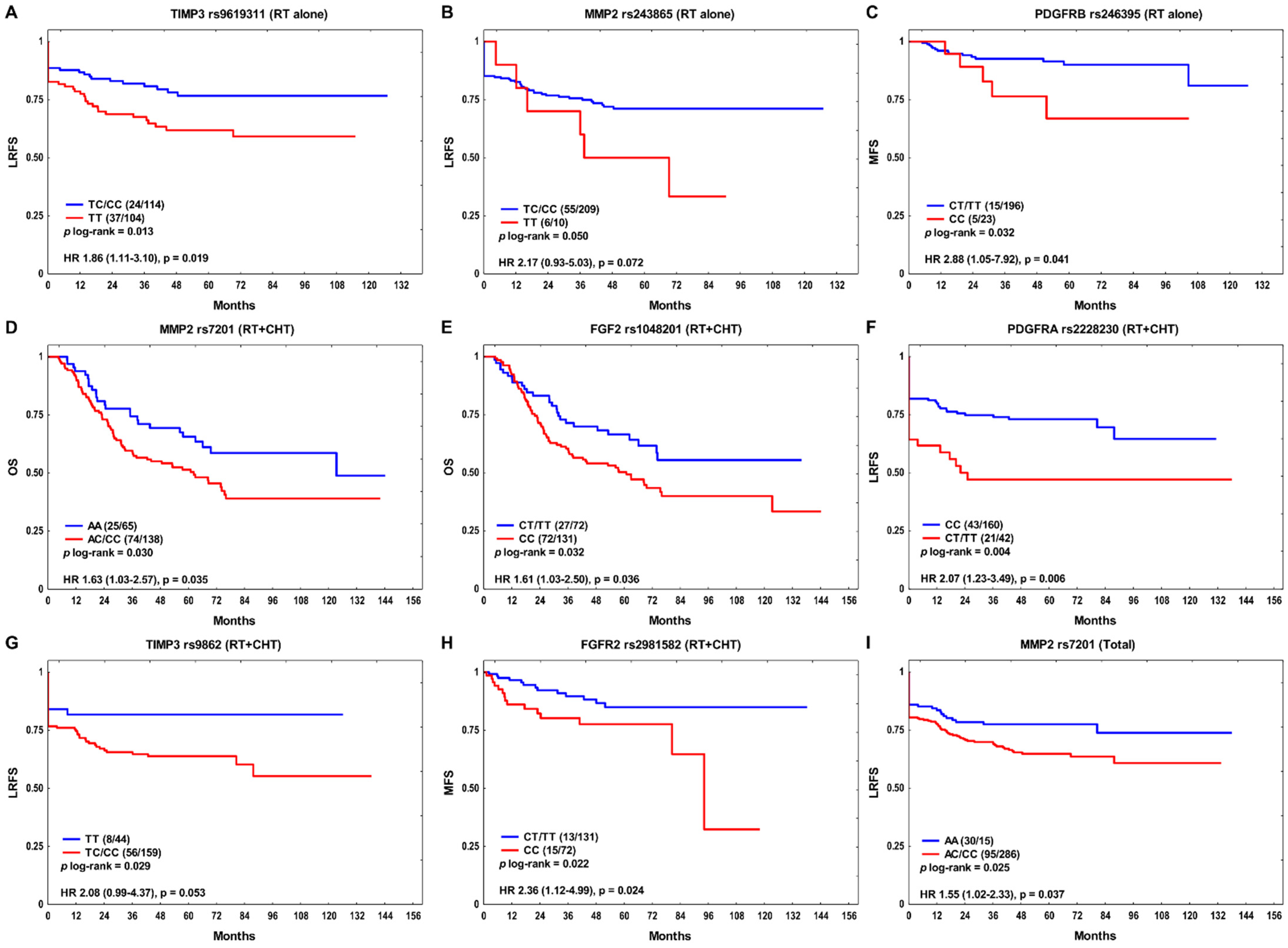
Figure 1. The Kaplan–Meier plots according to SNPs with p ≤ 0.05 in univariate analysis for: (A,B) locoregional recurrence-free survival (LRFS) and (C) metastasis-free survival (MFS) in the RT alone subgroup; (D,E) overall survival (OS); (F,G) LRFS and (H) MFS in the combination treatment subgroup (RT + CHT); and (I) LRFS in the whole group. Number of events and n are shown in the brackets.
Cancers 14 01844 g001
Figure 2. The Kaplan–Meier plot showing combined effects of MMP2 rs7201 and TIMP2 rs7501477 on locoregional recurrence-free survival (LRFS) in the studied group. Number of events and n are shown in the brackets.
Cancers 14 01844 g002
Table 1. Multivariate analysis for association of SNPs (only SNPs with p ≤ 0.100 are shown) with OS, LRFS, and MFS.
SNPGenotypeRT AloneRT + CTTotal
HR (95% CI)pHR (95% CI)pHR (95% CI)p
OS
rs1048201CC0.76 (0.49–1.17)0.2081.66 (1.03–2.68)0.0391.10 (0.81–1.48)0.544
LRFS
rs2228230CT/TT0.83 (0.44–1.57)0.5722.49 (1.42–4.36)0.0011.41 (0.93–2.14)0.106
rs243865TT2.92 (1.20–7.11)0.0190.74 (0.23–2.42)0.6201.38 (0.69–2.73)0.360
rs7201AC/CC1.50 (0.82–2.75)0.1911.54 (0.84–2.81)0.1591.59 (1.04–2.42)0.032
rs7501477GT/TT1.41 (0.79–2.52)0.2501.57 (0.91–2.72)0.1071.49 (1.01–2.21)0.045
rs9862TC/CC0.91 (0.51–1.63)0.7492.12 (0.98–4.57)0.0551.18 (0.76–1.82)0.459
MFS
rs246395CC3.06 (1.05–8.95)0.0410.36 (0.08–1.70)0.1981.29 (0.56–2.99)0.548
rs1048201CC3.08 (0.92–10.25)0.0671.31 (0.57–3.01)0.5191.70 (0.89–3.24)0.111
RT, radiotherapy; RT + CT, combination treatment; HR, hazard ratio; CI, confidence interval; OS, overall survival; LRFS, locoregional recurrence-free survival; MFS, metastasis free survival; p ≤ 0.050 shown in bold.
Table 2. Independent risk factors for OS, LRFS, and MFS—the final models.
RT AloneRT + CTTotal
VariablesHR (95% CI)pVariablesHR (95% CI)pVariablesHR (95% CI)p
OSStage N > 0
SPC
Metastasis
Local recurrence
2.24 (1.49–3.36)
2.29 (1.37–3.84)
1.89 (1.08–3.32)
3.95 (2.62–5.97)
0.0001
0.0016
0.026
<1 × 10⁻⁶
rs1048201 CC
Alcohol: ever
HPSCC
Local recurrence
Regional recurrence
SPC
1.61 (1.01–2.55)
2.11 (1.18–3.74)
2.01 (1.25–3.24)
5.51 (3.33–9.11)
1.73 (1.06–2.84)
2.07 (1.09–3.93)
0.044
0.011
0.004
<1 × 10⁻⁶
0.029
0.026
Alcohol: ever
Stage N > 0
Local recurrence
Regional recurrence
Metastasis
SPC
1.51 (1.06–2.16)
1.81 (1.31–2.49)
4.84 (3.52–6.67)
1.49 (1.01–2.18)
1.72 (1.17–2.54)
2.32 (1.56–3.46)
0.024
0.0003
<1 × 10⁻⁶
0.044
0.006
4 × 10⁻⁵
LRFSrs243865 TT
Stage T3–4
Stage N > 0
Non-OPSCC
2.92 (1.23–6.94)
2.97 (1.72–5.14)
2.19 (1.23–3.91)
2.29 (1.20–4.39)
0.015
0.0001
0.008
0.012
rs2228230 CT/TT
Non-OPSCC
2.26 (1.33–3.84)
1.74 (1.05–2.87)
0.003
0.032
rs7201 AC/CC
Stage T3–4
Stage N > 0
Non-OPSCC
1.56 (1.02–2.37)
1.69 (1.14–2.50)
1.68 (1.11–2.55)
1.66 (1.11–2.49)
0.038
0.008
0.015
0.013
MFSrs246395 CC
Regional recurrence
2.79 (1.01–7.69)
5.56 (1.71–18.13)
0.048
0.004
Regional recurrence3.65 (1.62–8.22)0.002HPSCC
Regional recurrence
2.36 (1.13–4.89)
4.60 (2.37–8.92)
0.021
6 × 10⁻⁶
RT, radiotherapy; RT + CT, combination treatment; HR, hazard ratio; CI, confidence interval; OS, overall survival; LRFS, locoregional recurrence-free survival; MFS, metastasis free survival; HPSCC, hypopharyngeal squamous cell carcinoma; Non-OPSCC, non-oropharyngeal squamous cell carcinoma; SPC, second primary cancer.
Publisher’s Note: MDPI stays neutral with regard to jurisdictional claims in published maps and institutional affiliations.

