
Biomarkers and Genetic Markers of Hepatocellular Carcinoma and Cholangiocarcinoma—What Do We Already Know

 ,
,  , , , , , ,
Abstract
:Simple Summary
Abstract
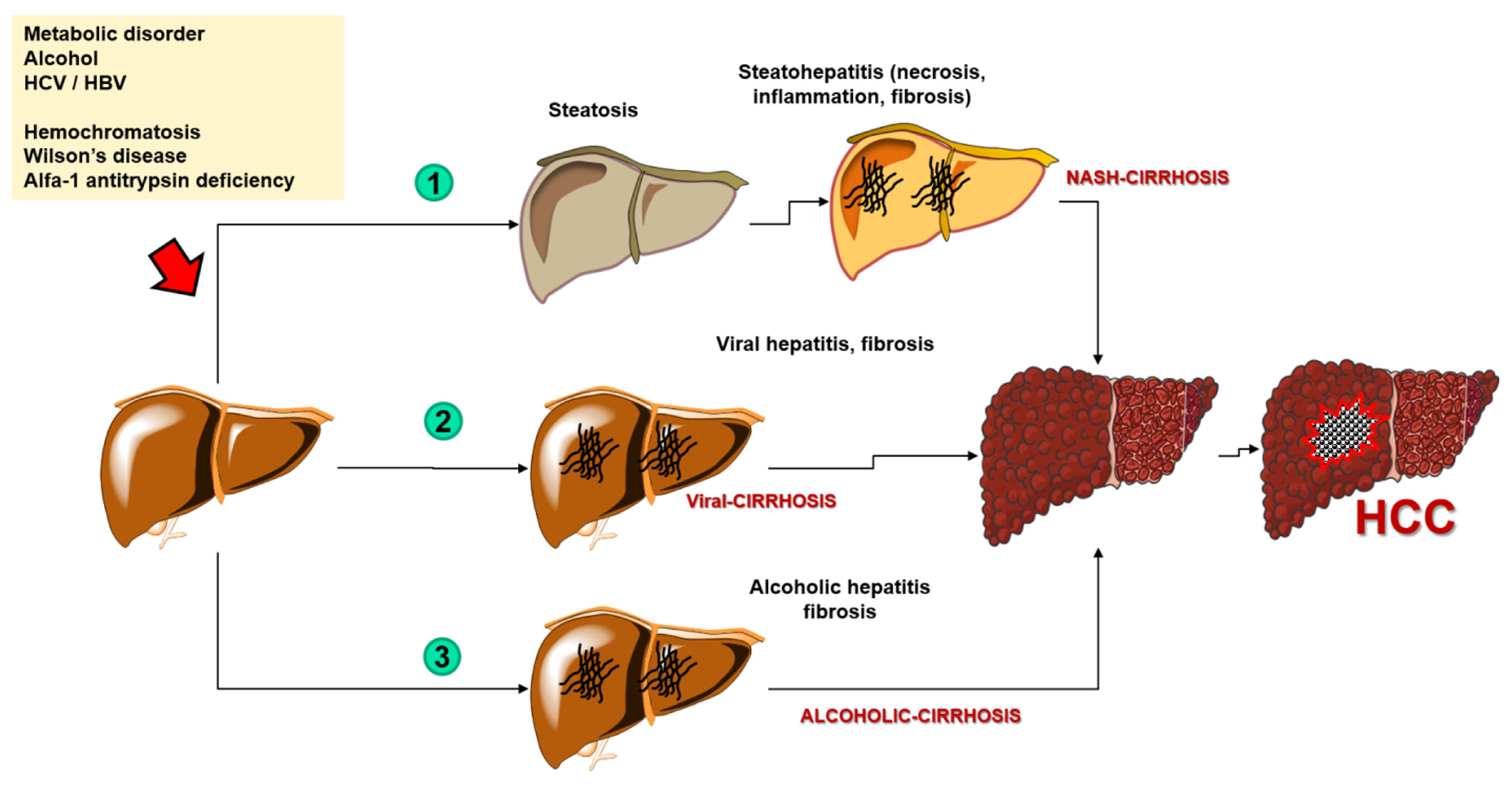
1. Introduction
2. Search Strategy
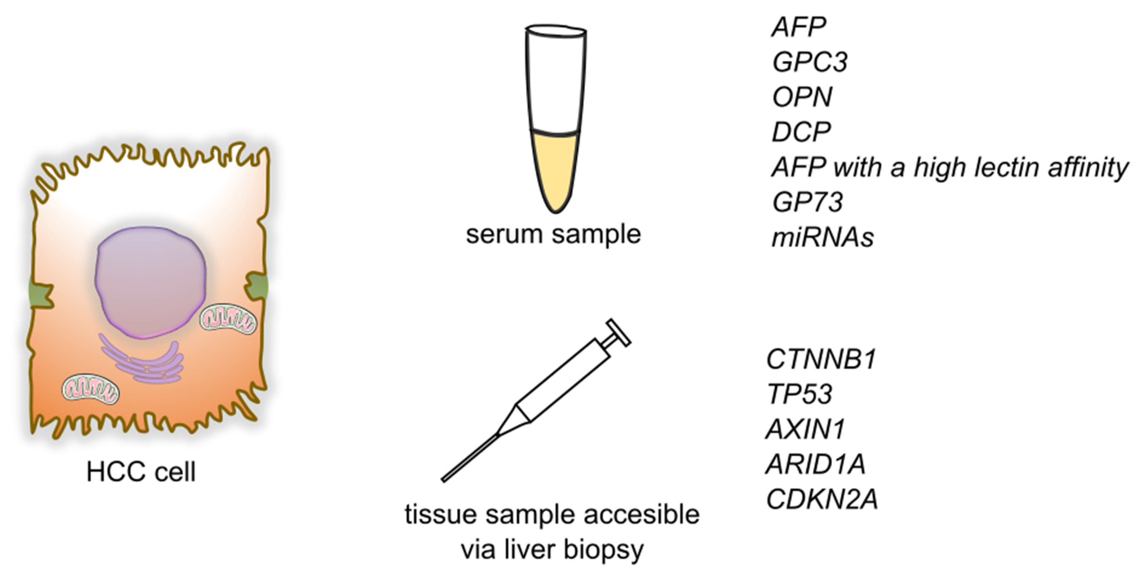
3. HCC Biomarkers
3.1. Alpha-Fetoprotein
3.2. Glypican-3
3.3. Osteopontin
3.4. Des-γ-Carboxy Prothrombin
3.5. AFP with a High Lectin Affinity
3.6. Golgi Protein-73
4. Genetic Markers for HCC
4.1. MicroRNAs
4.2. Genetic Markers
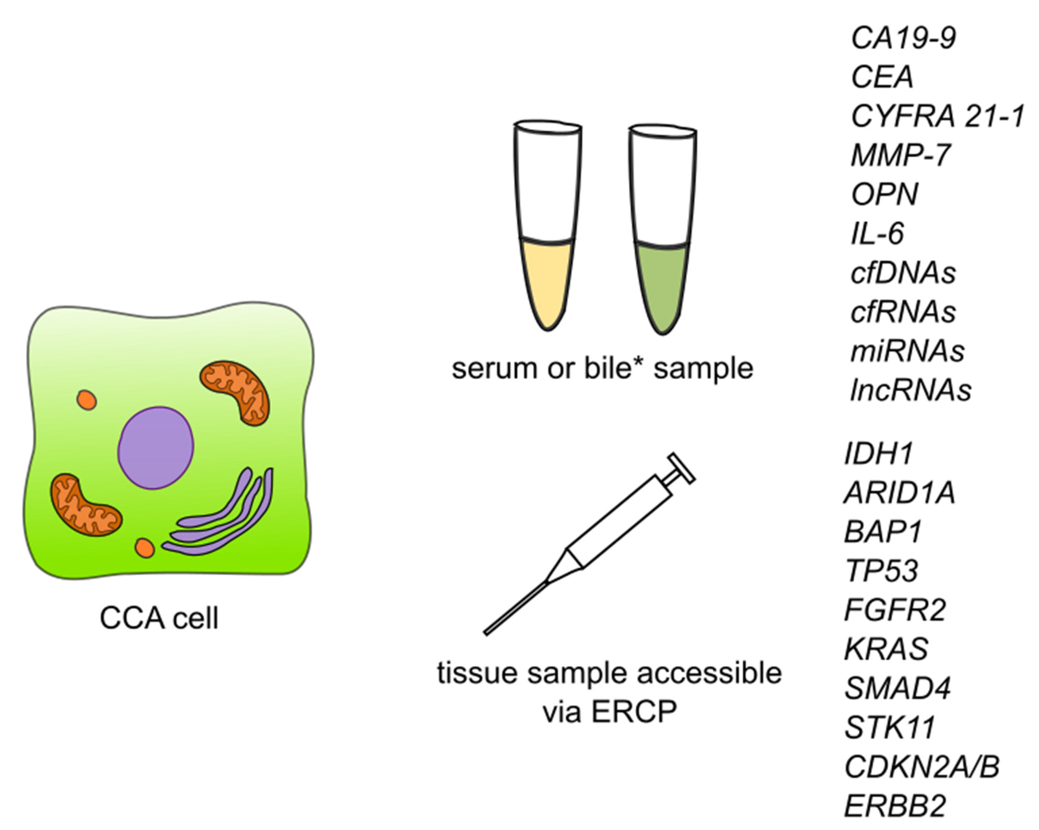
5. Biomarkers in CCA
5.1. Carbohydrate Antigen 19-9
5.2. Carcinoembryonic Antigen
5.3. CYFRA 21-1
5.4. Matrix Metalloproteinase 7
5.5. Osteopontin
5.6. Interleukin 6
5.7. New Potential Biomarkers of CCA
6. Genetic Markers of CCA
6.1. Circulating Nucleic Acids
6.1.1. Cell-Free DNA
6.1.2. Cell-Free RNA
6.1.3. Cell-Free Long Non-Coding RNA
6.1.4. Micro RNA
6.2. Genetic Markers
7. Conclusions
Author Contributions
Funding
Conflicts of Interest
Abbreviations
| ACOX-1 | Acyl-CoA oxidase 1 |
| AEG1 | Astrocyte elevated gene-1 |
| AFP | Alpha-fetoprotein |
| AFP-L3 | Alpha-fetoprotein with a high lectin affinity, Lens culinaris-agglutinin-reactive fraction of AFP |
| AKT1 | AKT Serine/threonine kinase 1 |
| ALT | Alanine transaminase |
| APOA2 | Apolipoprotein A2 |
| APOB | Apolipoprotein B |
| ARID1A | AT-Rich interaction domain 1A |
| AST | Aspartate transaminase |
| AXIN1 | Axin 1 |
| BAP1 | BRCA1 associated protein 1 |
| BRCA1/2 | BRCA1 DNA repair associated repair associated |
| BTLA | B- And T-Lymphocyte-associated protein |
| CA19-9 | Carbohydrate antigen 19-9/Cancer antigen 19-9 |
| CAA | Cholangiocarcinoma |
| CCAT2 | colon cancer-associated transcript 2 |
| CCND1 | Cyclin D1 |
| CDKN2A | Cyclin dependent kinase inhibitor 2A |
| CDKN2B | Cyclin dependent kinase inhibitor 2B |
| CDKN2B-AS1 | CDKN2B antisense RNA 1 |
| CEA | Carcinoembryonic antigen |
| cfDNA | Cell-free DNA |
| cfRNA | Cell free RNA |
| CK 19 | Cytokeratin 19 |
| CNV | copy number variation |
| COL18A1-AS2 | COL18A1 antisense RNA 2 |
| CT | computed tomography scan |
| CTNNB1 | Catenin beta-1 |
| CYFRA 21-1 | Cytokeratin fragment antigen 21-1 |
| dCCA | distal cholangiocarcinoma |
| DCP | Des-γ-carboxy prothrombin |
| ERBB2 | Erb-B2 receptor tyrosine kinase 2 |
| ERCP | endoscopic retrograde cholangiopancreatography |
| EZH2 | Enhancer of zeste 2 polycomb repressive complex 2 subunit |
| FGA | Fibrinogen alpha chain |
| FGF | Fibroblast growth factor |
| FGFR1 | Fibroblast growth factor receptor 1 |
| FGFR2 | Fibroblast growth factor receptor 2 |
| FGFR3 | Fibroblast growth factor receptor 3 |
| FGFR4 | Fibroblast growth factor receptor 4 |
| FGG | Fibrinogen gamma chain |
| FNH | focal nodular hyperplasia |
| GO | Gene ontology |
| GP73 | Golgi protein-73 |
| GPC3 | Glypican 3 |
| HBV | Hepatitis B virus |
| HCC | Hepatocellular carcinoma |
| HCCR-1 | human cervical cancer proto-oncogene 1 |
| HCV | Hepatitis C virus |
| HOXC13-AS | HOXC13 antisense RNA |
| iCCA | intrahepatic cholangiocarcinoma |
| IDH 1/2 | Isocitrate dehydrogenase (NADP(+)) 1/Isocitrate dehydrogenase (NADP(+)) 2 |
| IDUS | Intraductal ultrasonography |
| IL-6 | Interleukin 6 |
| IRF2 | Interferon regulatory factor 2 |
| KRAS | KRAS proto-oncogene, GTPase |
| lnc-CBLB-5 | long non-coding Cbl Proto-Oncogene B |
| lnc-CCHCR1-1 | long non-coding coiled-coil alpha-helical rod protein 1 |
| lnc-CDK9-1 | long non-coding cyclin dependent kinase 9 |
| lnc-RELL2-1 | long non-coding RELT like 2 |
| lncRNAs | long non-coding RNAs |
| MAPK | mitogen-activated protein kinase |
| miRNAs | MicroRNAs |
| MKI67 | Marker of proliferation Ki-67 |
| MMP-7 | Matrix metallopeptidase 7 |
| MRI | Magnetic resonance imaging |
| mTORC1 | mammalian target of rapamycin complex 1 |
| NASH | nonalcoholic steatohepatitis |
| NFE2L2 | NFE2 like BZIP transcription factor 2 |
| NK cells | Natural killer cells |
| OPN | Osteopontin |
| pCCA | perihilar cholangiocarcinoma |
| PD-1 | programmed death receptor 1 |
| PD-L2 | Programmed cell death 1 ligand 2 |
| PET | Positron emission tomography |
| piRNAs | piwi-interacting RNAs |
| PIVKA-II | protein induced by vitamin K absence or antagonist-II |
| PPARG | Peroxisome proliferator activated receptor gamma |
| PRC1 | Protein regulator of cytokinesis 1 |
| RNF24 | Ring finger protein 24 |
| RPS6KA3 | Ribosomal protein S6 kinase A3 |
| SIGLEC | Sialic acid binding Ig like lectin |
| SMAD4 | SMAD family member 4 |
| SMARCA2 | SWI/SNF related, matrix associated, actin dependent regulator of chromatin, subfamily A, member 2 |
| SNV | single nucleotide variation |
| STK11 | Serine/threonine kinase 11 |
| TERT | Telomerase reverse transcriptase |
| TET1 | Tet methylcytosine dioxygenase 1 |
| TOP2A | DNA topoisomerase II alpha |
| TP53 | Tumor protein P53 |
| WGS | Whole genome sequencing |
| WNT5B | Wnt family member 5B |
References
- Singal, A.G.; Lampertico, P.; Nahon, P. Epidemiology and surveillance for hepatocellular carcinoma: New trends. J. Hepatol. 2020, 72, 250–261. [Google Scholar] [CrossRef] [PubMed] [Green Version]
- Bertuccio, P.; Turati, F.; Carioli, G.; Rodriguez, T.; La Vecchia, C.; Malvezzi, M.; Negri, E. Global trends and predictions in hepatocellular carcinoma mortality. J. Hepatol. 2017, 67, 302–309. [Google Scholar] [CrossRef] [PubMed]
- Kulik, L.; El-Serag, H.B. Epidemiology and Management of Hepatocellular Carcinoma. Gastroenterology 2019, 156, 477–491. [Google Scholar] [CrossRef] [PubMed]
- De Stefano, F.; Chacon, E.; Turcios, L.; Marti, F.; Gedaly, R. Novel biomarkers in hepatocellular carcinoma. Dig. Liver Dis. 2018, 50, 1115–1123. [Google Scholar] [CrossRef] [PubMed]
- Khan, A.S.; Dageforde, L.A. Cholangiocarcinoma. Surg. Clin. N. Am. 2019, 99, 315–335. [Google Scholar] [CrossRef] [PubMed]
- Bertuccio, P.; Malvezzi, M.; Carioli, G.; Hashim, D.; Boffetta, P.; El-Serag, H.B.; La Vecchia, C.; Negri, E. Global trends in mortality from intrahepatic and extrahepatic cholangiocarcinoma. J. Hepatol. 2019, 71, 104–114. [Google Scholar] [CrossRef] [PubMed]
- Bergquist, A.; Von Seth, E. Epidemiology of cholangiocarcinoma. Best Pract. Res. Clin. Gastroenterol. 2015, 29, 221–232. [Google Scholar] [CrossRef] [PubMed]
- Intuyod, K.; Armartmuntree, N.; Jusakul, A.; Sakonsinsiri, C.; Thanan, R.; Pinlaor, S. Current omics-based biomarkers for cholangiocarcinoma. Expert Rev. Mol. Diagn. 2019, 19, 997–1005. [Google Scholar] [CrossRef] [PubMed]
- Macias, R.I.R.; Kornek, M.; Rodrigues, P.M.; Paiva, N.A.; Castro, R.E.; Urban, S.; Pereira, S.P.; Cadamuro, M.; Rupp, C.; Loosen, S.H.; et al. Diagnostic and prognostic biomarkers in cholangiocarcinoma. Liver Int. 2019, 39, 108–122. [Google Scholar] [CrossRef] [PubMed] [Green Version]
- Shen, N.; Zhang, D.; Yin, L.; Qiu, Y.; Liu, J.; Yu, W.; Fu, X.; Zhu, B.; Xu, X.; Duan, A.; et al. Bile cell-free DNA as a novel and powerful liquid biopsy for detecting somatic variants in biliary tract cancer. Oncol. Rep. 2019, 42, 549–560. [Google Scholar] [CrossRef] [PubMed]
- Zheng, Y.; Zhu, M.; Li, M. Effects of alpha-fetoprotein on the occurrence and progression of hepatocellular carcinoma. J. Cancer Res. Clin. Oncol. 2020, 146, 2439–2446. [Google Scholar] [CrossRef] [PubMed]
- Zhang, G.; Ha, S.A.; Kim, H.K.; Yoo, J.; Kim, S.; Lee, Y.S.; Hur, S.Y.; Kim, Y.W.; Kim, T.E.; Park, Y.G.; et al. Combined analysis of AFP and HCCR-1 as an useful serological marker for small hepatocellular carcinoma: A prospective cohort study. Dis. Markers 2012, 32, 265–271. [Google Scholar] [CrossRef] [PubMed]
- Jin, J.; Zhang, X.Y.; Shi, J.L.; Xue, X.F.; Lu, L.L.; Lu, J.H.; Jiang, X.P.; Hu, J.F.; Duan, B.S.; Yang, C.Q.; et al. Application of AFP whole blood one-step rapid detection kit in screening for HCC in Qidong. Am. J. Cancer Res. 2017, 7, 1384–1388. [Google Scholar] [PubMed]
- Ding, Y.; Liu, K.; Xu, Y.; Zhao, Q.; Lou, S.; Xiang, X.; Yan, L.; Cao, Z.; Xie, Q.; Zhu, C.; et al. Combination of inflammatory score/liver function and AFP improves the diagnostic accuracy of HBV-related hepatocellular carcinoma. Cancer Med. 2020, 9, 3057–3069. [Google Scholar] [CrossRef] [PubMed] [Green Version]
- Liu, X.; Meng, J.; Xu, H.; Niu, J. Alpha-fetoprotein to transaminase ratio is related to higher diagnostic efficacy for hepatocellular carcinoma. J. Med. 2019, 98, e15414. [Google Scholar] [CrossRef] [PubMed]
- Park, S.J.; Jang, J.Y.; Jeong, S.W.; Cho, Y.K.; Lee, S.H.; Kim, S.G.; Cha, S.W.; Kim, Y.S.; Cho, Y.D.; Kim, H.S.; et al. Usefulness of AFP, AFP-L3, and PIVKA-II, and their combinations in diagnosing hepatocellular carcinoma. J. Med. 2017, 96, e5811. [Google Scholar] [CrossRef]
- Wang, X.; Zhang, Y.; Yang, N.; He, H.; Tao, X.; Kou, C.; Jiang, J. Evaluation of the Combined Application of AFP, AFP-L3%, and DCP for Hepatocellular Carcinoma Diagnosis: A Meta-analysis. Biomed Res. Int. 2020, 2020, 1–10. [Google Scholar] [CrossRef]
- Notarpaolo, A.; Layese, R.; Magistri, P.; Gambato, M.; Colledan, M.; Magini, G.; Miglioresi, L.; Vitale, A.; Vennarecci, G.; Ambrosio, C.D.; et al. Validation of the AFP model as a predictor of HCC recurrence in patients with viral hepatitis-related cirrhosis who had received a liver transplant for HCC. J. Hepatol. 2017, 66, 552–559. [Google Scholar] [CrossRef]
- Ma, W.J.; Wang, H.Y.; Teng, L.S. Correlation analysis of preoperative serum alpha-fetoprotein (AFP) level and prognosis of hepatocellular carcinoma (HCC) after hepatectomy. World J. Surg. Oncol. 2013, 11, 212. [Google Scholar] [CrossRef] [Green Version]
- Centonze, L.; De Carlis, R.; Vella, I.; Carbonaro, L.; Incarbone, N.; Palmieri, L.; Sgrazzutti, C.; Ficarelli, A.; Valsecchi, M.G.; Iacono, U.D.; et al. From LI-RADS Classification to HCC Pathology: A Retrospective Single-Institution Analysis of Clinico-Pathological Features Affecting Oncological Outcomes after Curative Surgery. Diagnostics 2022, 12, 160. [Google Scholar] [CrossRef] [PubMed]
- Nakatsura, T.; Ofuji, K.; Saito, K.; Yoshikawa, T. Critical analysis of the potential of targeting GPC3 in hepatocellular carcinoma. J. Hepatocell. Carcinoma 2014, 1, 35. [Google Scholar] [CrossRef] [PubMed] [Green Version]
- Liu, H.; Yang, C.; Lu, W.; Zeng, Y. Prognostic significance of glypican-3 expression in hepatocellular carcinoma. J. Med. 2018, 97, e9702. [Google Scholar] [CrossRef] [PubMed]
- Attallah, A.M.; El-Far, M.; Omran, M.M.; Abdelrazek, M.A.; Attallah, A.A.; Saeed, A.M.; Farid, K. GPC-HCC model: A combination of glybican-3 with other routine parameters improves the diagnostic efficacy in hepatocellular carcinoma. Tumor Biol. 2016, 37, 12571–12577. [Google Scholar] [CrossRef] [PubMed]
- Wu, Y.; Liu, H.; Ding, H.-G. GPC-3 in hepatocellular carcinoma: Current perspectives. J. Hepatocell. Carcinoma 2016, 3, 63–67. [Google Scholar] [CrossRef] [PubMed] [Green Version]
- Sun, B.; Huang, Z.; Wang, B.; Yu, Y.; Lin, S.; Luo, L.; Wang, Y.; Huang, Z. Significance of glypican-3 (GPC3) expression in hepatocellular cancer diagnosis. Med. Sci. Monit. 2017, 23, 850–855. [Google Scholar] [CrossRef] [PubMed] [Green Version]
- Li, Y.; Zhang, J.; Gu, J.; Hu, K.; Huang, S.; Conti, P.S.; Wu, H.; Chen, K. Radiofluorinated GPC3-Binding Peptides for PET Imaging of Hepatocellular Carcinoma. Mol. Imaging Biol. 2020, 22, 134–143. [Google Scholar] [CrossRef] [PubMed]
- Taniguchi, M.; Mizuno, S.; Yoshikawa, T.; Fujinami, N.; Sugimoto, M.; Kobayashi, S.; Takahashi, S.; Konishi, M.; Gotohda, N.; Nakatsura, T. Peptide vaccine as an adjuvant therapy for glypican-3-positive hepatocellular carcinoma induces peptide-specific CTLs and improves long prognosis. Cancer Sci. 2020, 111, 2747–2759. [Google Scholar] [CrossRef] [PubMed]
- Gao, H.; Li, K.; Tu, H.; Pan, X.; Jiang, H.; Shi, B.; Kong, J.; Wang, H.; Yang, S.; Gu, J.; et al. Development of T cells redirected to glypican-3 for the treatment of hepatocellular carcinoma. Clin. Cancer Res. 2014, 20, 6418–6428. [Google Scholar] [CrossRef] [Green Version]
- Fu, Y.; Urban, D.J.; Nani, R.R.; Zhang, Y.F.; Li, N.; Fu, H.; Shah, H.; Gorka, A.P.; Guha, R.; Chen, L.; et al. Glypican-3-Specific Antibody Drug Conjugates Targeting Hepatocellular Carcinoma. Hepatology 2019, 70, 563–576. [Google Scholar] [CrossRef]
- Tang, X.; Chen, L.; Li, A.; Cai, S.; Zhang, Y.; Liu, X.; Jiang, Z.; Liu, X.; Liang, Y.; Ma, D. Anti-gpc3 antibody-modified sorafenib-loaded nanoparticles significantly inhibited hepg2 hepatocellular carcinoma. Drug Deliv. 2018, 25, 1484–1494. [Google Scholar] [CrossRef] [Green Version]
- Yu, M.; Luo, H.; Fan, M.; Wu, X.; Shi, B.; Di, S.; Liu, Y.; Pan, Z.; Jiang, H.; Li, Z. Development of GPC3-Specific Chimeric Antigen Receptor-Engineered Natural Killer Cells for the Treatment of Hepatocellular Carcinoma. Mol. Ther. 2018, 26, 366–378. [Google Scholar] [CrossRef] [PubMed] [Green Version]
- Shevde, L.A.; Samant, R.S. Role of osteopontin in the pathophysiology of cancer. Matrix Biol. 2014, 37, 131–141. [Google Scholar] [CrossRef]
- Tsuchiya, N.; Sawada, Y.; Endo, I.; Saito, K.; Uemura, Y.; Nakatsura, T. Biomarkers for the early diagnosis of hepatocellular carcinoma. World J. Gastroenterol. 2015, 21, 10573–10583. [Google Scholar] [CrossRef] [PubMed]
- Kawashima, R.; Mochida, S.; Matsui, A.; Youlutuz, Y.; Ishikawa, K.; Toshima, K.; Yamanobe, F.; Inao, M.; Ikeda, H.; Ohno, A.; et al. Expression of osteopontin in Kupffer cells and hepatic macrophages and stellate cells in rat liver after carbon tetrachloride intoxication: A possible factor for macrophage migration into hepatic necrotic areas. Biochem. Biophys. Res. Commun. 1999, 256, 527–531. [Google Scholar] [CrossRef] [PubMed]
- Loosen, S.H.; Roderburg, C.; Kauertz, K.L.; Pombeiro, I.; Leyh, C.; Benz, F.; Vucur, M.; Longerich, T.; Koch, A.; Braunschweig, T.; et al. Elevated levels of circulating osteopontin are associated with a poor survival after resection of cholangiocarcinoma. J. Hepatol. 2017, 67, 749–757. [Google Scholar] [CrossRef] [PubMed]
- Sun, T.; Tang, Y.; Sun, D.; Bu, Q.; Li, P. Osteopontin versus alpha-fetoprotein as a diagnostic marker for hepatocellular carcinoma: A meta-analysis. Onco. Targets. Ther. 2018, 11, 8925–8935. [Google Scholar] [CrossRef] [PubMed] [Green Version]
- Zhu, M.; Zheng, J.; Wu, F.; Kang, B.; Liang, J.; Heskia, F.; Zhang, X.; Shan, Y. OPN is a promising serological biomarker for hepatocellular carcinoma diagnosis. J. Med. Virol. 2020, 92, 3596–3603. [Google Scholar] [CrossRef]
- Wang, H.; Guo, D.; Li, J.; Wei, B.; Zheng, H. Increased expression of osteopontin indicates poor prognosis in hepatocellular carcinoma. Int. J. Clin. Exp. Pathol. 2018, 11, 5916–5922. [Google Scholar] [PubMed]
- Byeon, H.; Lee, S.D.; Hong, E.K.; Lee, D.E.; Kim, B.H.; Seo, Y.; Joo, J.; Han, S.S.; Kim, S.H.; Park, S.J. Long-term prognostic impact of osteopontin and Dickkopf-related protein 1 in patients with hepatocellular carcinoma after hepatectomy. Pathol. Res. Pract. 2018, 214, 814–820. [Google Scholar] [CrossRef]
- Ding, K.; Fan, L.; Chen, S.; Wang, Y.; Yu, H.; Sun, Y.; Yu, J.; Wang, L.; Liu, X.; Liu, Y. Resistance to cisplatin treatment in HCC. Oncol. Rep. 2015, 34, 3297–3303. [Google Scholar] [CrossRef] [PubMed] [Green Version]
- Zhang, Y.S.; Chu, J.H.; Cui, S.X.; Song, Z.Y.; Qu, X.J. Des-γ-carboxy prothrombin (DCP) as a potential autologous growth factor for the development of hepatocellular carcinoma. Cell. Physiol. Biochem. 2014, 34, 903–915. [Google Scholar] [CrossRef] [PubMed] [Green Version]
- Inagaki, Y.; Tang, W.; Makuuchi, M.; Hasegawa, K.; Sugawara, Y.; Kokudo, N. Clinical and molecular insights into the hepatocellular carcinoma tumour marker des-γ-carboxyprothrombin. Liver Int. 2011, 31, 22–35. [Google Scholar] [CrossRef] [PubMed]
- Cui, S.X.; Yu, X.F.; Qu, X.J. Roles and Signaling Pathways of Des-γ-Carboxyprothrombin in the Progression of Hepatocellular Carcinoma. Cancer Investig. 2016, 34, 459–464. [Google Scholar] [CrossRef] [PubMed]
- Sumi, A.; Akiba, J.; Ogasawara, S.; Nakayama, M.; Nomura, Y.; Yasumoto, M.; Sanada, S.; Nakashima, O.; Abe, T.; Yano, H. Des-γ-carboxyprothrombin (DCP) and NXDCP expressions and their relationship with clinicopathological features in hepatocellular carcinoma. PLoS ONE 2015, 10, 1–12. [Google Scholar] [CrossRef] [PubMed] [Green Version]
- Koike, Y.; Shiratori, Y.; Sato, S.; Obi, S.; Teratani, T.; Imamura, M.; Yoshida, H.; Shiina, S.; Omata, M. Des-γ-carboxy prothrombin as a useful predisposing factor for the development of portal venous invasion in patients with hepatocellular carcinoma: A prospective analysis of 227 patients. Cancer 2001, 91, 561–569. [Google Scholar] [CrossRef]
- Choi, J.; Kim, G.; Han, S.; Lee, W.; Chun, S.; Lim, Y. Longitudinal Assessment of Three Serum Biomarkers to Detect Very Early-Stage Hepatocellular Carcinoma. Hepatology 2019, 69, 1983–1994. [Google Scholar] [CrossRef] [PubMed]
- Yang, M.; Zhang, X.; Liu, J. Prognostic value of des-γ-carboxy prothrombin in patients with hepatocellular carcinoma treated with transarterial chemotherapy: A systematic review and meta-analysis. PLoS ONE 2019, 14, 1–14. [Google Scholar] [CrossRef] [PubMed]
- Zhang, Y.S.; Chu, J.H.; Song, Z.Y.; Cui, S.X.; Qu, X.J. Des-gamma-carboxy prothrombin (DCP) antagonizes the effects of gefitinib on human hepatocellular carcinoma cells. Cell. Physiol. Biochem. 2015, 35, 201–212. [Google Scholar] [CrossRef] [PubMed]
- Lai, Q.; Iesari, S.; Levi Sandri, G.B.; Lerut, J. Des-gamma-carboxy prothrombin in hepatocellular cancer patients waiting for liver transplant: A systematic review and meta-analysis. Int. J. Biol. Markers 2017, 32, e370–e374. [Google Scholar] [CrossRef] [PubMed] [Green Version]
- Cheng, J.; Wang, W.; Zhang, Y.; Liu, X.; Li, M.; Wu, Z.; Liu, Z.; Lv, Y.; Wang, B. Prognostic role of pre-treatment serum AFP-L3% in hepatocellular carcinoma: Systematic review and meta-analysis. PLoS ONE 2014, 9, e87011. [Google Scholar] [CrossRef] [Green Version]
- Zhao, J.; Guo, L.-Y.; Yang, J.-M.; Jia, J.-W. Sublingual vein parameters, AFP, AFP-L3, and GP73 in patients with hepatocellular carcinoma. Genet. Mol. Res. 2015, 14, 7062–7067. [Google Scholar] [CrossRef] [PubMed]
- Lim, T.S.; Kim, D.Y.; Han, K.H.; Kim, H.S.; Shin, S.H.; Jung, K.S.; Kim, B.K.; Kim, S.U.; Park, J.Y.; Ahn, S.H. Combined use of AFP, PIVKA-II, and AFP-L3 as tumor markers enhances diagnostic accuracy for hepatocellular carcinoma in cirrhotic patients. Scand. J. Gastroenterol. 2016, 51, 344–353. [Google Scholar] [CrossRef] [PubMed]
- Cheng, H.T.; Chang, Y.H.; Chen, Y.Y.; Lee, T.H.; Tai, D.I.; Lin, D.Y. AFP-L3 in chronic liver diseases with persistent elevation of alpha-fetoprotein. J. Chin. Med. Assoc. 2007, 70, 310–317. [Google Scholar] [CrossRef] [Green Version]
- Kobayashi, M.; Hosaka, T.; Ikeda, K.; Seko, Y.; Kawamura, Y.; Sezaki, H.; Akuta, N.; Suzuki, F.; Suzuki, Y.; Saitoh, S.; et al. Highly sensitive AFP-L3% assay is useful for predicting recurrence of hepatocellular carcinoma after curative treatment pre- and postoperatively. Hepatol. Res. 2011, 41, 1036–1045. [Google Scholar] [CrossRef] [PubMed]
- Gatselis, N.K.; Tornai, T.; Shums, Z.; Zachou, K.; Saitis, A.; Gabeta, S.; Albesa, R.; Norman, G.L.; Papp, M.; Dalekos, G.N. Golgi protein-73: A biomarker for assessing cirrhosis and prognosis of liver disease patients. World J. Gastroenterol. 2020, 26, 5130–5145. [Google Scholar] [CrossRef] [PubMed]
- Qiao, Y.; Chen, J.; Li, X.; Wei, H.; Xiao, F.; Chang, L.; Zhang, R.; Hao, X.; Wei, H. Serum gp73 is also a biomarker for diagnosing cirrhosis in population with chronic HBV infection. Clin. Biochem. 2014, 47, 216–222. [Google Scholar] [CrossRef]
- Bröker, M.E.E.; Ijzermans, J.N.M.; Witjes, C.D.M.; Van Vuuren, H.J.; De Man, R.A. The predictive value of Golgi Protein 73 in differentiating benign from malignant liver tumors. PLoS ONE 2014, 9, 7–10. [Google Scholar] [CrossRef]
- Sai, W.L.; Yao, M.; Shen, S.J.; Zheng, W.J.; Sun, J.Y.; Wu, M.N.; Wang, L.; Yao, D.F. Dynamic expression of hepatic GP73 mRNA and protein and circulating GP73 during hepatocytes malignant transformation. Hepatobiliary Pancreat. Dis. Int. 2020, 19, 449–454. [Google Scholar] [CrossRef]
- Chen, X.; Wang, Y.; Tao, J.; Shi, Y.; Gai, X.; Huang, F.; Ma, Q.; Zhou, Z.; Chen, H.; Zhang, H.; et al. MTORC1 Up-Regulates GP73 to Promote Proliferation and Migration of Hepatocellular Carcinoma Cells and Growth of Xenograft Tumors in Mice. Gastroenterology 2015, 149, 741.e14–752.e14. [Google Scholar] [CrossRef] [Green Version]
- Hann, H.W.; Wang, M.; Hafner, J.; Long, R.E.; Kim, S.H.; Ahn, M.; Park, S.; Comunale, M.A.; Block, T.M.; Mehta, A. Analysis of GP73 in patients with HCC as a function of anti-cancer treatment. Cancer Biomarkers 2010, 7, 269–273. [Google Scholar] [CrossRef] [Green Version]
- Ke, M.Y.; Wu, X.N.; Zhang, Y.; Wang, S.; Lv, Y.; Dong, J. Serum GP73 predicts posthepatectomy outcomes in patients with hepatocellular carcinoma. J. Transl. Med. 2019, 17, 1–10. [Google Scholar] [CrossRef] [PubMed]
- Liu, C.H.; Huang, Q.; Jin, Z.Y.; Xie, F.; Zhu, C.L.; Liu, Z.; Wang, C. Circulating microRNA-21 as a prognostic, biological marker in cholangiocarcinoma. J. Cancer Res. Ther. 2018, 14, 220–225. [Google Scholar] [CrossRef] [PubMed]
- Lixin, S.; Wei, S.; Haibin, S.; Qingfu, L.; Tiemin, P. miR-885-5p inhibits proliferation and metastasis by targeting IGF2BP1 and GALNT3 in human intrahepatic cholangiocarcinoma. Mol. Carcinog. 2020, 59, 1371–1381. [Google Scholar] [CrossRef] [PubMed]
- Shen, S.; Lin, Y.; Yuan, X.; Shen, L.; Chen, J.; Chen, L.; Qin, L.; Shen, B. Biomarker MicroRNAs for Diagnosis, Prognosis and Treatment of Hepatocellular Carcinoma: A Functional Survey and Comparison. Sci. Rep. 2016, 6, 1–21. [Google Scholar] [CrossRef] [PubMed] [Green Version]
- Cui, Y.; Xu, H.F.; Liu, M.Y.; Xu, Y.J.; He, J.C.; Zhou, Y.; Cang, S.D. Mechanism of exosomal microRNA-224 in development of hepatocellular carcinoma and its diagnostic and prognostic value. World J. Gastroenterol. 2019, 25, 1890–1898. [Google Scholar] [CrossRef] [PubMed]
- Yang, C.; Ma, X.; Guan, G.; Liu, H.; Yang, Y.; Niu, Q.; Wu, Z.; Jiang, Y.; Bian, C.; Zang, Y.; et al. MicroRNA-766 promotes cancer progression by targeting NR3C2 in hepatocellular carcinoma. FASEB J. 2019, 33, 1456–1467. [Google Scholar] [CrossRef] [PubMed]
- Liu, Y.; Tan, J.; Ou, S.; Chen, J.; Chen, L. Adipose-derived exosomes deliver miR-23a/b to regulate tumor growth in hepatocellular cancer by targeting the VHL/HIF axis. J. Physiol. Biochem. 2019, 75, 391–401. [Google Scholar] [CrossRef] [PubMed]
- Lin, H.; Huang, Z.P.; Liu, J.; Qiu, Y.; Tao, Y.P.; Wang, M.C.; Yao, H.; Hou, K.Z.; Gu, F.M.; Xu, X.F. MiR-494-3p promotes PI3K/AKT pathway hyperactivation and human hepatocellular carcinoma progression by targeting PTEN. Sci. Rep. 2018, 8, 1–9. [Google Scholar] [CrossRef] [PubMed]
- Jiang, L.; Cheng, Q.; Zhang, B.H.; Zhang, M.Z. Circulating micrornas as biomarkers in hepatocellular carcinoma screening a validation set from China. J. Med. 2015, 94, 1–10. [Google Scholar] [CrossRef]
- Schulze, K.; Imbeaud, S.; Letouzé, E.; Alexandrov, L.B.; Calderaro, J.; Rebouissou, S.; Couchy, G.; Meiller, C.; Shinde, J.; Soysouvanh, F.; et al. Exome sequencing of hepatocellular carcinomas identifies new mutational signatures and potential therapeutic targets. Nat. Genet. 2015, 47, 505–511. [Google Scholar] [CrossRef]
- Guichard, C.; Amaddeo, G.; Imbeaud, S.; Ladeiro, Y.; Pelletier, L.; Maad, I.B.; Calderaro, J.; Bioulac-Sage, P.; Letexier, M.; Degos, F.; et al. Integrated analysis of somatic mutations and focal copy-number changes identifies key genes and pathways in hepatocellular carcinoma. Nat. Genet. 2012, 44, 694–698. [Google Scholar] [CrossRef] [PubMed]
- Wang, X.W.; Hussain, S.P.; Huo, T.I.; Wu, C.G.; Forgues, M.; Hofseth, L.J.; Brechot, C.; Harris, C.C. Molecular pathogenesis of human hepatocellular carcinoma. Toxicology 2002, 181–182, 43–47. [Google Scholar] [CrossRef] [Green Version]
- Long, J.; Wang, A.; Bai, Y.; Lin, J.; Yang, X.; Wang, D.; Yang, X.; Jiang, Y.; Zhao, H. Development and validation of a TP53-associated immune prognostic model for hepatocellular carcinoma. EBioMedicine 2019, 42, 363–374. [Google Scholar] [CrossRef] [PubMed] [Green Version]
- Ozen, C.; Yildiz, G.; Dagcan, A.T.; Cevik, D.; Ors, A.; Keles, U.; Topel, H.; Ozturk, M. Genetics and epigenetics of liver cancer. N. Biotechnol. 2013, 30, 381–384. [Google Scholar] [CrossRef] [PubMed]
- Abitbol, S.; Dahmani, R.; Coulouarn, C.; Ragazzon, B.; Mlecnik, B.; Senni, N.; Savall, M.; Bossard, P.; Sohier, P.; Drouet, V.; et al. AXIN deficiency in human and mouse hepatocytes induces hepatocellular carcinoma in the absence of β-catenin activation. J. Hepatol. 2018, 68, 1203–1213. [Google Scholar] [CrossRef] [PubMed]
- He, F.; Li, J.; Xu, J.; Zhang, S.; Xu, Y.; Zhao, W.; Yin, Z.; Wang, X. Decreased expression of ARID1A associates with poor prognosis and promotes metastases of hepatocellular carcinoma. J. Exp. Clin. Cancer Res. 2015, 34. [Google Scholar] [CrossRef] [PubMed] [Green Version]
- Yang, H.; Huo, J.; Li, X. Identification and validation of a five-gene prognostic signature for hepatocellular carcinoma. World J. Surg. Oncol. 2021, 19, 1–13. [Google Scholar] [CrossRef]
- Nahon, P.; Nault, J.C. Constitutional and functional genetics of human alcohol-related hepatocellular carcinoma. Liver Int. 2017, 37, 1591–1601. [Google Scholar] [CrossRef] [Green Version]
- Piao, Z.; Park, C.; Lee, J.S.; Yang, C.H.; Choi, K.Y.; Kim, H. Homozygous deletions of the CDKN2 gene and loss of heterozygosity of 9p in primary hepatocellular carcinoma. Cancer Lett. 1998, 122, 201–207. [Google Scholar] [CrossRef]
- Lee, T.; Teng, T.Z.J.; Shelat, V.G. Carbohydrate antigen 19-9—Tumor marker: Past, present, and future. World J. Gastrointest. Surg. 2020, 12, 468–490. [Google Scholar] [CrossRef]
- Scarà, S.; Bottoni, P. The Role of Human Chorionic Gonadotropin as Tumor Marker: Biochemical and Clinical Aspects. In Advances in Cancer Biomarkers; Springer: Berlin/Heidelberg, Germany, 2015; pp. 247–260. [Google Scholar] [CrossRef] [Green Version]
- Parra-Robert, M.; Santos, V.M.; Canis, S.M.; Pla, X.F.; Fradera, J.M.A.; Porto, R.M. Relationship between CA 19.9 and the lewis phenotype: Options to improve diagnostic efficiency. Anticancer Res. 2018, 38, 5883–5888. [Google Scholar] [CrossRef] [PubMed]
- Kim, S.; Park, B.K.; Seo, J.H.; Choi, J.; Choi, J.W.; Lee, C.K.; Chung, J.B.; Park, Y.; Kim, D.W. Carbohydrate antigen 19-9 elevation without evidence of malignant or pancreatobiliary diseases. Sci. Rep. 2020, 10, 1–9. [Google Scholar] [CrossRef] [PubMed]
- Szekanecz, É.; Śandor, Z.; Antal-Szalmás, P.; Soós, L.; Lakos, G.; Besenyei, T.; Szentpétery, Á.; Simkovics, E.; Szántó, J.; Kiss, E.; et al. Increased production of the soluble tumor-associated antigens CA19-9, CA125, and CA15-3 in rheumatoid arthritis: Potential adhesion molecules in synovial inflammation? Ann. N. Y. Acad. Sci. 2007, 1108, 359–371. [Google Scholar] [CrossRef] [PubMed] [Green Version]
- Liang, B.; Zhong, L.; He, Q.; Wang, S.; Pan, Z.; Wang, T. Diagnostic Accuracy of Serum CA19-9 in Patients with Cholangiocarcinoma: A Systematic Review and Meta-Analysis. Med. Sci. Monit. 2015, 21, 3555–3563. [Google Scholar] [CrossRef] [PubMed] [Green Version]
- Coelho, R.; Silva, M.; Rodrigues-Pinto, E.; Cardoso, H.; Lopes, S.; Pereira, P.; Vilas-Boas, F.; Santos-Antunes, J.; Costa-Maia, J.; MacEdo, G. CA 19-9 as a Marker of Survival and a Predictor of Metastization in Cholangiocarcinoma. GE Port. J. Gastroenterol. 2017, 24, 114–121. [Google Scholar] [CrossRef] [PubMed]
- Lee, B.S.; Lee, S.H.; Son, J.H.; Jang, D.K.; Chung, K.H.; Paik, W.H.; Ryu, J.K.; Kim, Y.T. Prognostic value of CA 19-9 kinetics during gemcitabine-based chemotherapy in patients with advanced cholangiocarcinoma. J. Gastroenterol. Hepatol. 2016, 31, 493–500. [Google Scholar] [CrossRef] [PubMed]
- Ali, C.W.; Kaye, T.F.; Adamson, D.J.A.; Tait, I.S.; Polignano, F.M.; Highley, M.S. CA 19-9 and survival in advanced and unresectable pancreatic adenocarcinoma and cholangiocarcinoma. J. Gastrointest. Cancer 2007, 38, 108–114. [Google Scholar] [CrossRef] [PubMed]
- Juntermanns, B.; Kaiser, G.M.; Itani Gutierrez, S.; Heuer, M.; Buechter, M.; Kahraman, A.; Reis, H.; Kasper, S.; Paul, A.; Fingas, C.D. CA19-9 in intrahepatic cholangiocarcinoma: A diagnostic and prognostic armamentarium? Chirurg 2018, 89, 466–471. [Google Scholar] [CrossRef] [PubMed]
- Li, Y.; Li, D.J.; Chen, J.; Liu, W.; Li, J.W.; Jiang, P.; Zhao, X.; Guo, F.; Li, X.W.; Wang, S.G. Application of joint detection of AFP, CA19-9, CA125 and CEA in identification and diagnosis of cholangiocarcinoma. Asian Pac. J. Cancer Prev. 2015, 16, 3451–3455. [Google Scholar] [CrossRef] [Green Version]
- Li, Y.; Huang, Y.; Chen, J. Diagnostic value of serum biomarkers for intrahepatic cholangiocarcinoma. J. Coll. Physicians Surg. Pak. 2019, 29, 962–966. [Google Scholar] [CrossRef]
- Bates, P.A.; Luo, J.; Sternberg, M.J.E. A predicted three-dimensional structure for the carcinoembryonic antigen (CEA). FEBS Lett. 1992, 301, 207–214. [Google Scholar] [CrossRef] [Green Version]
- Kelleher, M.; Singh, R.; O’Driscoll, C.M.; Melgar, S. Carcinoembryonic antigen (CEACAM) family members and Inflammatory Bowel Disease. Cytokine Growth Factor Rev. 2019, 47, 21–31. [Google Scholar] [CrossRef]
- Hall, C.; Clarke, L.; Pal, A.; Buchwald, P.; Eglinton, T.; Wakeman, C.; Frizelle, F. A review of the role of carcinoembryonic antigen in clinical practice. Ann. Coloproctol. 2019, 35, 294–305. [Google Scholar] [CrossRef] [PubMed]
- Lumachi, F.; Re, G.L.O.; Tozzoli, R.; Aurizio, F.D.; Facomer, F.; Chiara, G.B.; Basso, S.M.M. Lumachi. Measurement of panel biomarkers. Anticancer Res. 2014, 6668, 6663–6667. [Google Scholar]
- Brito, A.F.; Abrantes, A.M.; Encarnação, J.C.; Tralhão, J.G.; Botelho, M.F. Cholangiocarcinoma: From molecular biology to treatment. Med. Oncol. 2015, 32, 1–8. [Google Scholar] [CrossRef] [PubMed] [Green Version]
- Nakeeb, A.; Lipsett, P.A.; Lillemoe, K.D.; Fox-Talbot, M.K.; Coleman, J.A.; Cameron, J.L.; Pitt, H.A. Biliary carcinoembryonic antigen levels are a marker for cholangiocarcinoma. Am. J. Surg. 1996, 171, 147–153. [Google Scholar] [CrossRef]
- Malaguarnera, G.; Paladina, I.; Giordano, M.; Malaguarnera, M.; Bertino, G.; Berretta, M. Serum markers of intrahepatic cholangiocarcinoma. Dis. Markers 2013, 34, 219–228. [Google Scholar] [CrossRef]
- Hyun, J.W.; Shin, H.S.; Kim, S.H.; Kong, S.Y.; Yoo, H.; Gwak, H.S.; Kim, H.J. CYFRA 21-1 levels in cerebrospinal fluid as a putative therapeutic monitoring biomarker for patients with leptomeningeal carcinomatosis: A pilot study. Cancer Biomark. 2020, 28, 81–89. [Google Scholar] [CrossRef]
- Wu, Y.J.; Rheinwald, J.G. A new small (40 kd) keratin filament protein made by some cultured human squamous cell carcinomas. Cell 1981, 25, 627–635. [Google Scholar] [CrossRef]
- Huang, L.; Chen, W.; Liang, P.; Hu, W.; Zhang, K.; Shen, S.; Chen, J.; Zhang, Z.; Chen, B.; Han, Y.; et al. Serum CYFRA 21-1 in Biliary Tract Cancers: A Reliable Biomarker for Gallbladder Carcinoma and Intrahepatic Cholangiocarcinoma. Dig. Dis. Sci. 2015, 60, 1273–1283. [Google Scholar] [CrossRef]
- Muraki, M.; Tohda, Y.; Iwanaga, T.; Uejima, H.; Nagasaka, Y.; Nakajima, S. Assessment of serum CYFRA 21-1 in lung cancer. Cancer 1996, 77, 1274–1277. [Google Scholar] [CrossRef]
- Paganuzzi, M.; Onetto, M.; Marroni, P.; Filiberti, R.; Tassara, E.; Parodi, S.; Felletti, R. Diagnostic value of CYFRA 21-1 tumor marker and CEA in pleural effusion due to mesothelioma. Chest 2001, 119, 1138–1142. [Google Scholar] [CrossRef] [PubMed]
- Brockmann, J.G.; Nottberg, H.S.; Glodny, B.; Heinecke, A.; Senninger, N.J. CYFRA 21-1 serum analysis in patients with esophageal cancer. Clin. Cancer Res. 2000, 6, 4249–4252. [Google Scholar] [PubMed]
- Wang, Y.X.; Hu, D.; Yan, X. Diagnostic accuracy of Cyfra 21-1 for head and neck squamous cell carcinoma: A meta-analysis. Eur. Rev. Med. Pharmacol. Sci. 2013, 17, 2383–2389. [Google Scholar] [PubMed]
- Fu, L.; Wang, R.; Yin, L.; Shang, X.; Zhang, R.; Zhang, P. CYFRA21-1 tests in the diagnosis of non-small cell lung cancer: A meta-analysis. Int. J. Biol. Markers 2019, 34, 251–261. [Google Scholar] [CrossRef] [PubMed] [Green Version]
- Sarwar, M.; Tomiyoshi, K.; Inoue, T.; Fukazawa, K.; Endo, K. CYFRA 21-1 as a tumor marker used in measuring the serum fragment of cytokeratin subunit 19 by immunoradiometric assay. Ann. Nucl. Med. 1994, 8, 301–306. [Google Scholar] [CrossRef] [PubMed]
- Chapman, M.H.; Sandanayake, N.S.; Andreola, F.; Dhar, D.K.; Webster, G.J.; Dooley, J.S.; Pereira, S.P. Circulating CYFRA 21-1 is a specific diagnostic and prognostic biomarker in biliary tract cancer. J. Clin. Exp. Hepatol. 2011, 1, 6–12. [Google Scholar] [CrossRef]
- Guowei, H.; Yuan, L.; Ma, L.; Zhongyang, L.; Zhixing, S.; Lin, L.; Minqi, L. The diagnostic efficacy of CYFRA21-1 on intrahepatic cholangiocarcinoma: A meta-analysis. Clin. Res. Hepatol. Gastroenterol. 2019, 43, 266–272. [Google Scholar] [CrossRef] [PubMed]
- Itatsu, K.; Zen, Y.; Yamaguchi, J.; Ohira, S.; Ishikawa, A.; Ikeda, H.; Sato, Y.; Harada, K.; Sasaki, M.; Sasaki, M.; et al. Expression of matrix metalloproteinase 7 is an unfavorable postoperative prognostic factor in cholangiocarcinoma of the perihilar, hilar, and extrahepatic bile ducts. Hum. Pathol. 2008, 39, 710–719. [Google Scholar] [CrossRef]
- Leelawat, K.; Sakchinabut, S.; Narong, S.; Wannaprasert, J. Detection of serum MMP-7 and MMP-9 in cholangiocarcinoma patients: Evaluation of diagnostic accuracy. BMC Gastroenterol. 2009, 9, 1–8. [Google Scholar] [CrossRef] [Green Version]
- Leelawat, K.; Narong, D.S.; Wannaprasert, J.; Ratanashu-ek, T. Prospective study of MMP7 serum levels in the diagnosis of cholangiocarcinoma. World J. Gastroenterol. 2010, 16, 4697–4703. [Google Scholar] [CrossRef] [PubMed]
- Miwa, S.; Miyagawa, S.I.; Soeda, J.; Kawasaki, S. Matrix metalloproteinase-7 expression and biologic aggressiveness of cholangiocellular carcinoma. Cancer 2002, 94, 428–434. [Google Scholar] [CrossRef] [PubMed]
- Itatsu, K.; Zen, Y.; Ohira, S.; Ishikawa, A.; Sato, Y.; Harada, K.; Ikeda, H.; Sasaki, M.; Nimura, Y.; Nakanuma, Y. Immunohistochemical analysis of the progression of flat and papillary preneoplastic lesions in intrahepatic cholangiocarcinogenesis in hepatolithiasis. Liver Int. 2007, 27, 1174–1184. [Google Scholar] [CrossRef] [PubMed]
- Laohaviroj, M.; Chamgramol, Y.; Pairojkul, C.; Mulvenna, J.; Sripa, B. Clinicopathological Significance of Osteopontin in Cholangiocarcinoma Cases. Asian Pac. J. Cancer Prev. 2016, 17, 201–205. [Google Scholar] [CrossRef] [PubMed] [Green Version]
- Zhou, K.-Q.; Liu, W.-F.; Yang, L.-X.; Sun, Y.-F.; Hu, J.; Chen, F.-Y.; Zhou, C.; Zhang, X.-Y.; Peng, Y.-F.; Yu, L.; et al. Circulating osteopontin per tumor volume as a prognostic biomarker for resectable intrahepatic cholangiocarcinoma. Hepatobiliary Surg. Nutr. 2019, 8, 582–596. [Google Scholar] [CrossRef] [PubMed]
- Zheng, Y.; Zhou, C.; Yu, X.X.; Wu, C.; Jia, H.L.; Gao, X.M.; Yang, J.M.; Wang, C.Q.; Luo, Q.; Zhu, Y.; et al. Osteopontin promotes metastasis of intrahepatic cholangiocarcinoma through recruiting MAPK1 and mediating Ser675 phosphorylation of β-Catenin. Cell Death Dis. 2018, 9, 179. [Google Scholar] [CrossRef] [PubMed] [Green Version]
- Terashi, T.; Aishima, S.; Taguchi, K.; Asayama, Y.; Sugimachi, K.; Matsuura, S.; Shimada, M.; Maehara, S.; Maehara, Y.; Tsuneyoshi, M. Decreased expression of osteopantin is related to tumor aggressiveness and clinical outcome of intrahepatic cholangiocarcinoma. Liver Int. 2004, 24, 38–45. [Google Scholar] [CrossRef]
- Rose-John, S. Interleukin-6 family cytokines. Cold Spring Harb. Perspect. Biol. 2018, 10, 1–17. [Google Scholar] [CrossRef] [PubMed] [Green Version]
- Mott, J.L.; Gores, G.J. Targeting IL-6 in cholangiocarcinoma therapy. Am. J. Gastroenterol. 2007, 102, 2171–2172. [Google Scholar] [CrossRef] [PubMed]
- Goydos, J.S.; Brumfield, A.M.; Frezza, E.; Booth, A.; Lotze, M.T.; Carty, S.E. Marked elevation of serum interleukin-6 in patients with cholangiocarcinoma: Validation of utility as a clinical marker. Ann. Surg. 1998, 227, 398–404. [Google Scholar] [CrossRef]
- Cheon, Y.K.; Cho, Y.D.; Moon, J.H.; Jang, J.Y.; Kim, Y.S.; Kim, Y.S.; Lee, M.S.; Lee, J.S.; Shim, C.S. Diagnostic utility of interleukin-6 (IL-6) for primary bile duct cancer and changes in serum IL-6 levels following photodynamic therapy. Am. J. Gastroenterol. 2007, 102, 2164–2170. [Google Scholar] [CrossRef] [PubMed]
- Sugawara, H.; Yasoshima, M.; Katayanagi, K.; Kono, N.; Watanabe, Y.; Harada, K.; Nakanuma, Y. Relationship between interleukin-6 and proliferation and differentiation in cholangiocarcinoma. Histopathology 1998, 33, 145–153. [Google Scholar] [CrossRef] [PubMed]
- Rizzo, A.; Ricci, A.D.; Tavolari, S.; Brandi, G. Circulating tumor DNA in biliary tract cancer: Current evidence and future perspectives. Cancer Genom. Proteom. 2020, 17, 441–452. [Google Scholar] [CrossRef] [PubMed]
- Wasenang, W.; Chaiyarit, P.; Proungvitaya, S.; Limpaiboon, T. Serum cell-free DNA methylation of OPCML and HOXD9 as a biomarker that may aid in differential diagnosis between cholangiocarcinoma and other biliary diseases. Clin. Epigenet. 2019, 11, 39. [Google Scholar] [CrossRef] [Green Version]
- Driescher, C.; Fuchs, K.; Haeberle, L.; Goering, W.; Frohn, L.; Opitz, F.V.; Haeussinger, D.; Knoefel, W.T.; Keitel, V.; Esposito, I. Bile-based cell-free DNA analysis is a reliable diagnostic tool in pancreatobiliary cancer. Cancers 2021, 13, 39. [Google Scholar] [CrossRef]
- Wang, X.; Fu, X.H.; Qian, Z.L.; Zhao, T.; Duan, A.Q.; Ruan, X.; Zhu, B.; Yin, L.; Zhang, Y.J.; Yu, W.L. Non-invasive detection of biliary tract cancer by low-coverage whole genome sequencing from plasma cell-free DNA: A prospective cohort study. Transl. Oncol. 2021, 14, 100908. [Google Scholar] [CrossRef]
- Gu, X.; Wang, C.; Deng, H.; Qing, C.; Liu, R.; Liu, S.; Xue, X. Exosomal piRNA profiling revealed unique circulating piRNA signatures of cholangiocarcinoma and gallbladder carcinoma. Acta Biochim. Biophys. Sin. 2021, 52, 475–484. [Google Scholar] [CrossRef]
- Bai, J.G.; Tang, R.F.; Shang, J.F.; Qi, S.; Yu, G.D.; Sun, C. Upregulation of long non-coding RNA CCAT2 indicates a poor prognosis and promotes proliferation and metastasis in intrahepatic cholangiocarcinoma. Mol. Med. Rep. 2018, 17, 5328–5335. [Google Scholar] [CrossRef] [Green Version]
- Angenard, G.; Merdrignac, A.; Louis, C.; Edeline, J.; Coulouarn, C. Expression of long non-coding RNA ANRIL predicts a poor prognosis in intrahepatic cholangiocarcinoma. Dig. Liver Dis. 2019, 51, 1337–1343. [Google Scholar] [CrossRef] [PubMed]
- Yao, Y.; Jiao, D.; Liu, Z.; Chen, J.; Zhou, X.; Li, Z.; Li, J.; Han, X. Novel miRNA Predicts Survival and Prognosis of Cholangiocarcinoma Based on RNA-seq Data and in Vitro Experiments. Biomed Res. Int. 2020, 2020, 1–14. [Google Scholar] [CrossRef]
- Asukai, K.; Kawamoto, K.; Eguchi, H.; Konno, M.; Asai, A.; Iwagami, Y.; Yamada, D.; Asaoka, T.; Noda, T.; Wada, H.; et al. Micro-RNA-130a-3p Regulates Gemcitabine Resistance via PPARG in Cholangiocarcinoma. Ann. Surg. Oncol. 2017, 24, 2344–2352. [Google Scholar] [CrossRef] [PubMed]
- Jusakul, A.; Cutcutache, I.; Yong, C.H.; Lim, J.Q.; Huang, M.N.; Padmanabhan, N.; Nellore, V.; Kongpetch, S.; Ng, A.W.T.; Ng, L.M.; et al. Whole-genome and epigenomic landscapes of etiologically distinct subtypes of cholangiocarcinoma. Cancer Discov. 2017, 7, 1116–1135. [Google Scholar] [CrossRef] [PubMed] [Green Version]
- Lowery, M.A.; Ptashkin, R.; Jordan, E.; Berger, M.F.; Zehir, A.; Capanu, M.; Kemeny, N.E.; O’Reilly, E.M.; El-Dika, I.; Jarnagin, W.R.; et al. Comprehensive molecular profiling of intrahepatic and extrahepatic cholangiocarcinomas: Potential targets for intervention. Clin. Cancer Res. 2018, 24, 4154–4161. [Google Scholar] [CrossRef] [PubMed] [Green Version]
- Li, H.; Long, J.; Xie, F.; Kang, K.; Shi, Y.; Xu, W.; Wu, X.; Lin, J.; Xu, H.; Du, S.; et al. Transcriptomic analysis and identification of prognostic biomarkers in cholangiocarcinoma. Oncol. Rep. 2019, 42, 1833–1842. [Google Scholar] [CrossRef] [PubMed] [Green Version]

| Marker | Pros | Cons | Notes | Reference(s) |
|---|---|---|---|---|
| AFP | Good for screening patients from risk groups | Low sensitivity | Established | [12,13,14,15,16,17,18,19] |
| GCP3 | Negative prognostic value, Detection of GPC3 itself allows differentiation of HCC from healthy liver tissue, benign lesions, and liver cirrhosis, marker of malignant transformation | Specificity 72–81% | Established | [21,22,23,24,25,26,27,28] |
| OPN | Negative prognostic value, positive in AFP negative HCC | Experimental | [36,37,38,39,40] | |
| DCP | Negative prognostic value, allows differentiation form other chronic liver diseases | Experimental | [42,43,44,45,46,47,48,49] | |
| AFP-L3 | Good for screening, detecting recurrence, negative prognostic value | Elevated in Hepatitis | Experimental | [17,50,51,52,54] |
| GP73 | Negative prognostic value | Elevated in other diseases (HBV caused cirrhosis, focal nodular hyperplasia) | Experimental | [58,59,60,61] |
| miRNA | Negative prognostic value, possible screening value | Experimental | [64,65,66,67,68,69] |
| Marker | Pros | Cons | Notes | Reference(s) |
|---|---|---|---|---|
| CA19-9 | Negative prognostic value | Absent in Lewis(a-b-) patients, low specificity and sensitivity | Established | [80,81,82,85,86,87,88,89,90,91] |
| CEA | Negative prognostic value when used with other markers | Low specificity and sensitivity | Established. | [94,95,96,97,98] |
| CYFRA 21-1 | Negative prognostic value | Low specificity and sensitivity | Established | [95,96,97,101,108,109] |
| MMP-7 | Negative Predictive value, correlated with CAA stage | Low specificity and sensitivity | Experimental | [9,111,112,113,114] |
| OPN | High sensitivity | Experimental | [9,115,116,117,118] | |
| IL-6 | High specificity | Experimental | [120,121,122,123] | |
| cfDNA | Possible diagnostic marker, correlated with tumor grade | Experimental | [10,125,126,127,131] | |
| lncRNA | Shows prognostic value, correlated with tumor grade | Experimental | [129,130] | |
| miRNA | Negative prognostic value | Bad diagnostic marker | Experimental | [9,62,63,131,132] |
Publisher’s Note: MDPI stays neutral with regard to jurisdictional claims in published maps and institutional affiliations. |
© 2022 by the authors. Licensee MDPI, Basel, Switzerland. This article is an open access article distributed under the terms and conditions of the Creative Commons Attribution (CC BY) license (https://creativecommons.org/licenses/by/4.0/).
Share and Cite
Baj, J.; Bryliński, Ł.; Woliński, F.; Granat, M.; Kostelecka, K.; Duda, P.; Flieger, J.; Teresiński, G.; Buszewicz, G.; Furtak-Niczyporuk, M.; et al. Biomarkers and Genetic Markers of Hepatocellular Carcinoma and Cholangiocarcinoma—What Do We Already Know. Cancers 2022, 14, 1493. https://doi.org/10.3390/cancers14061493
Baj J, Bryliński Ł, Woliński F, Granat M, Kostelecka K, Duda P, Flieger J, Teresiński G, Buszewicz G, Furtak-Niczyporuk M, et al. Biomarkers and Genetic Markers of Hepatocellular Carcinoma and Cholangiocarcinoma—What Do We Already Know. Cancers. 2022; 14(6):1493. https://doi.org/10.3390/cancers14061493
Chicago/Turabian StyleBaj, Jacek, Łukasz Bryliński, Filip Woliński, Michał Granat, Katarzyna Kostelecka, Piotr Duda, Jolanta Flieger, Grzegorz Teresiński, Grzegorz Buszewicz, Marzena Furtak-Niczyporuk, and et al. 2022. "Biomarkers and Genetic Markers of Hepatocellular Carcinoma and Cholangiocarcinoma—What Do We Already Know" Cancers 14, no. 6: 1493. https://doi.org/10.3390/cancers14061493
APA StyleBaj, J., Bryliński, Ł., Woliński, F., Granat, M., Kostelecka, K., Duda, P., Flieger, J., Teresiński, G., Buszewicz, G., Furtak-Niczyporuk, M., & Portincasa, P. (2022). Biomarkers and Genetic Markers of Hepatocellular Carcinoma and Cholangiocarcinoma—What Do We Already Know. Cancers, 14(6), 1493. https://doi.org/10.3390/cancers14061493