Article

Patterns of Hearing Loss in Irradiated Survivors of Head and Neck Rhabdomyosarcoma

by
Franciscus A. Diepstraten
1,
Jan Wiersma
2,
Reineke A. Schoot
1,
Rutger R. G. Knops
1,
Charlotte L. Zuur
3,4,
Annelot J. M. Meijer
1,
Raquel Dávila Fajardo
1,5,
Bradley R. Pieters
2,6,
Brian V. Balgobind
2,6,
Henrike Westerveld
2,6,
Nicole Freling
7,
Harm van Tinteren
8,
Ludwig E. Smeele
1,3,9,
Arjan Bel
2,
Marry M. van den Heuvel-Eibrink
1,
Robert J. Stokroos
10,11,
Johannes H. M. Merks
1,
Alexander E. Hoetink
10,11 and
Marinka L. F. Hol
1,9,10,*
1
Princess Máxima Center for Pediatric Oncology, 3584 CS Utrecht, The Netherlands
2
Department of Radiation Oncology, Amsterdam University Medical Centers, University of Amsterdam, 1105 AZ Amsterdam, The Netherlands
3
Department of Head and Neck Surgery, Netherlands Cancer Institute, 1066 CX Amsterdam, The Netherlands
4
Department of Otorhinolaryngology Head and Neck Surgery, Leiden University Medical Center, 2333 ZA Leiden, The Netherlands
5
Department of Radiation Oncology, University Medical Center Utrecht, 3584 CX Utrecht, The Netherlands
6
Amsterdam Cancer Center, Cancer Treatment and Quality of Life, 1081 HV Amsterdam, The Netherlands
7
Department of Radiology, Amsterdam University Medical Centers, University of Amsterdam, 1105 AZ Amsterdam, The Netherlands
8
Trial and Data Center, Princess Máxima Center for Pediatric Oncology, 3584 CS Utrecht, The Netherlands
9
Department of Maxillofacial Surgery, Amsterdam University Medical Centers, 1105 AZ Amsterdam, The Netherlands
10
Department of Otorhinolaryngology-Head and Neck Surgery, University Medical Center, 3584 CX Utrecht, The Netherlands
11
University Medical Center Utrecht Brain Center, 3584 CX Utrecht, The Netherlands
*
Author to whom correspondence should be addressed.
Cancers 2022, 14(23), 5749; https://doi.org/10.3390/cancers14235749
Submission received: 14 October 2022 / Revised: 16 November 2022 / Accepted: 16 November 2022 / Published: 23 November 2022
(This article belongs to the Special Issue Rhabdomyosarcoma: Still Unresolved Questions but New Perspectives)

Abstract

:

Simple Summary

Hearing loss (HL) can be a side effect of paediatric cancer treatment and can be caused by chemotherapy but also local therapies such as radiotherapy and/or surgery of the head and neck region. In this study, the frequency and patterns of HL were assessed in survivors of head and neck rhabdomyosarcoma (HNRMS). Our secondary aim was to look into the dose–effect relationship between radiotherapy dose on the cochlea and the presence of HL. Forty-nine survivors of HNRMS were included in this study, forty-two of them underwent audiological evaluation. HL was found in up to 19% of the survivors. Four survivors had low frequencies HL with normal hearing or milder HL in the higher frequencies. In our series, HL (≥Muenster 2b) was significantly associated with the maximum cochlear irradiation dose (p = 0.047). More research is needed on HL patterns in HNRMS survivors and on the radiotherapy dose–effect relationship.

Abstract

Purpose: The frequency and patterns of HL in a HNRMS survivor cohort were investigated. A dose–effect relationship between the dose to the cochlea and HL was explored. Methods: Dutch survivors treated for HNRMS between 1993 and 2017 with no relapse and at least two years after the end of treatment were eligible for inclusion. The survivors were evaluated for HL with pure-tone audiometry. HL was graded according to the Muenster, Common Terminology Criteria for Adverse Events (CTCAE) v4.03 and International Society for Paediatric Oncology (SIOP) classification. We defined deleterious HL as Muenster ≥ 2b, CTCAE ≥ 2, and SIOP ≥ 2. Mixed-effects logistic regression was used to search for the dose–effect relationship between the irradiation dose to the cochlea and the occurrence of HL. Results: Forty-two HNRMS survivors underwent pure-tone audiometry. The Muenster, CTCAE and SIOP classification showed that 19.0% (n = 8), 14.2% (n = 6) and 11.9% (n = 5) of survivors suffered from HL, respectively. A low-frequency HL pattern with normal hearing or milder hearing loss in the higher frequencies was seen in four survivors. The maximum cochlear irradiation dose was significantly associated with HL (≥Muenster 2b) (p = 0.047). In our series, HL (≥Muenster 2b) was especially observed when the maximum dose to the cochlea exceeded 19 Gy. Conclusion: HL occurred in up to 19% of survivors of HNRMS. More research is needed on HL patterns in HNRMS survivors and on radiotherapy dose–effect relationships.

1. Introduction

Rhabdomyosarcoma (RMS) is a primitive malignant soft tissue sarcoma of the skeletal muscle phenotype that originates from mesenchymal cells [1]. It is the most common childhood and adolescent soft tissue sarcoma (3–5% of childhood tumours, 50% of paediatric soft tissue sarcomas) [1,2,3,4]. The median age at diagnosis of RMS patients is 6.5 years, and there is a slight male predominance (male/female ratio 1.3/1) [5]. Different subtypes exist, including embryonal (70%), alveolar (20%), and spindle cell/sclerosing (10%) RMS [1,2,3,4,5]. Local treatment options for RMS include external beam radiotherapy with photons (RT), external beam radiotherapy with protons (PT) [6], Ablative surgery followed by the MOuld technique after loading brachytherapy and surgical REconstruction (AMORE) [7], and the combination of RT or PT with conventional surgery [8]. With improvements in multimodality treatment, imaging, and supportive care, five-year overall survival has increased up to 70–90%, depending on the patient and tumour characteristics [9]. As survival rates increase, more attention is needed to inventory and prevent adverse effects of treatment such as dental and facial deformities [10,11,12], endocrine disorders [13], and hearing loss (HL) [14].
Due to the complex anatomy of the head and neck region and the extension of the tumour, radiotherapy and/or surgery can directly or indirectly affect the nasopharynx, middle ear, nerves, brain, and cochlea and consequently result in conductive, sensorineural, or mixed HL [14,15,16,17]. In addition, recurrent ear nose throat (ENT) infections might contribute to reversible HL in HNRMS patients [14]. It seems that radiotherapy has a dose-dependent effect on hearing outcomes [18,19,20]. Where chemotherapy mainly induces irreversible hearing loss in an early stage during treatment [21], radiation-induced HL is mainly described as a late effect in paediatric cancer patients, especially in those who receive a high cochlear cumulative radiation dose (>30 Gy) [15,22,23]. Pre-treatment hearing levels appeared to be a predictive factor for hearing capability after chemotherapy and radiotherapy [24]. Younger patients are more likely to develop HL. This is important, as HL is a serious problem which has negative consequences for speech and language development in children [25]. This can result in reduced communication skills with consequences for psychosocial and socioeconomic development. A lower quality of life is reported by children with HL compared to peers with normal hearing [26].
As survivors with HNRMS are treated with three different radiotherapy modalities in our cohort, we can investigate hearing function in these groups. The primary aim of the current study is to describe the frequency and pattern of hearing loss as measured by pure-tone audiometry and as systematically assessed by frequently used ototoxicity grading scales in HNRMS survivors treated with either RT, PT, or brachytherapy (BT) (BT as part of AMORE). The secondary aim of this study was to explore possible dose–effect relationships between the radiation dose to the cochlea and the development of HL.

2. Materials and Methods

2.1. Survivors

Survivors were selected through two different identification systems. First, survivors of HNRMS, treated between January 1993 and December 2017 and visiting the follow-up clinic at the Emma Children’s Hospital at least two years after the completion of treatment, were selected for inclusion. Secondly, HNRMS survivors treated in one of the paediatric oncology centres in the Netherlands who visited the Princess Máxima Center late effects department for audiological follow-up between 2018 and March 2021 were eligible for inclusion. From 2018 onwards, all paediatric oncology care, including follow-up, was centralised in a national centre, the Princess Máxima Center for Pediatric Oncology. Inclusion criteria were: (1) treated for primary HNRMS disease, and (2) no recurrence of disease at least two years after the completion of treatment. Survivors underwent an audiological examination in the Emma Children’s Hospital, Amsterdam Medical Center (AMC) and/or Princess Máxima Center, University Medical Center Utrecht (UMCU), during a follow-up multidisciplinary outpatient clinic consultation.
Audiological data were collected during regular follow-up visits. The institutional Review Board of the Amsterdam University Medical Center decided that the Act on Medical Research Involving Subjects did not apply.

2.2. Treatment

The included HNRMS survivors had been treated according to SIOP–MMT (International Society for Paediatrics Oncology–Malignant Mesenchymal Tumour group) 95 [27], SIOP–MMT 89 [28,29], EpSSG (European paediatric Soft tissue sarcoma Study Group) RMS 2005 [30], CWS (Cooperative WeichteilSarkom Studien Gruppe)-91 [31], or CWS-2007HR [32] protocols. In SIOP–MMT 95 and SIOP–MMT 89, children received ifosfamide, vincristine, actinomycin D (IVA), or IVA alternating with carboplatin, epirubicin, and etoposide. The maximum cumulative carboplatin dose was 3600 mg/m2. Survivors treated according to EpSSG RMS 2005 protocol received IVA or IVA with doxorubicin; high-risk patients were randomised to receive maintenance chemotherapy with vinorelbine and cyclophosphamide. Survivors treated according to CWS-91 received etoposide, vincristine, dactinomycin, ifosfamide, and doxorubicin. Survivors treated according to CWS-2007 received IVA. All survivors had received local treatment with RT, PT, or BT (as part of AMORE).

2.3. Radiotherapy Data

The original radiotherapy treatment data were retrieved for all survivors treated at the Emma Children’s Hospital. The cochlea was delineated by one researcher (MH) and checked by a head and neck radiologist (NF). The cochlea was delineated on a computed tomography scan (bone setting) according to a head and neck anatomy app [33]. This head and neck anatomy app is a fully interactive atlas of head and neck anatomy created by doctors for anyone else professionally involved in head and neck anatomy [33]. Dose to organs was exported for minimal, mean, and maximum doses (D0.1 cm3). To enable comparisons between treatment modalities, all radiotherapy parameters were recalculated as equivalent doses in 2 Gy fractions (EQD2) using the following formula; EQD2 = BED/[2 + (α/β)]. The BED depends on radiotherapy modality (RT, PT, BT) and is described by the following generic formula: BED = D*qlet*[grepair*d + (α/β)], with D representing total dose, d is the dose per fraction, glet is a factor representing the effective dose relative to photon therapy, with glet = 1 for RT and BT, and glet =1.1 for PT, and grepair is a factor describing the change in the dose–effect due to the biological response, i.e., “damage” repair during and in between pulsed (BT-PDR) or during (BT-LDR) dose delivery. The alpha–beta ratio, a parameter that can be assessed and which represents the measure of cell death and cell repair, for the cochlea was set at 2 Gy [34]. The parameter describing the influence of repair is the half-time for repair, and it was set to 1.5 h [35,36].

2.4. Audiometry

Hearing function was measured by pure-tone audiometry (PTA) during follow-up visits after HNRMS treatment. Air-conducted thresholds were measured between 0.125–8 kHz, and if elevated thresholds were found, bone-conducted thresholds were measured between 0.5–4 kHz. HL was evaluated by two researchers (M.L.F.H., F.A.D.) and an audiologist (A.E.H.). HL was graded according to the Muenster, International Society for Paediatric Oncology (SIOP) and U.S. National Cancer Institute Common Terminology Criteria for Adverse Events (CTCAE) v4.03 criteria (Table S1) [37,38,39]. Disagreements between the researchers (M.L.F.H., F.A.D.) and the audiologist (A.E.H.) were discussed. The audiologist (A.E.H) made the final decision. Deleterious HL was defined as a Muenster ≥ 2b grade, SIOP ≥ 2 grade, or CTCAE grade ≥ 2. Both ears were graded independently.

2.5. Statistics

Descriptive statistics were used, including the median and range for the quantitative variables of age and follow-up time, and the frequency and percentage for the qualitative variables of sex, RMS subtype, and treatment strategy. The characteristics between the groups with and without hearing loss were compared using Pearson’s Chi-square test for categorical variables, an independent Student’s t-test for normally distributed continuous variables, or a Mann–Whitney U test for abnormally distributed continuous variables. The dose–effect evaluation for radiotherapy data on HL was analysed using mixed-effects logistic regression with the patient as the random factor. A two-sided p-value less than 0.05 was considered statically significant. Statistical analyses were performed using IBM SPSS Statistics 25.0.0.2 (SPSS Inc. Chicago, IL, USA) and R version 4.2.1 (2022-06-23 ucrt).

3. Results

3.1. Clinical Characteristics

Forty-nine survivors (25 females, 24 males) of primary HNRMS were included in this study (Table 1). Thirty-nine HNRMS survivors initially treated at the Emma Children’s Hospital were identified, of whom 32 underwent audiological testing during follow-up visits. Additionally, 10 HNRMS survivors with audiological tests during follow-up at the Princess Máxima Center were identified (Figure S1): six of these patients were treated at the Emma Children’s Hospital, three at the Sophia Children’s Hospital and one patient at the University Medical Center Groningen. The survivors had a median age of 5.0 years (range 0.04–13.4) at the time of diagnosis and a median age of 16.3 years (range 5.2–33.7) at the time of follow-up. The median follow-up time was 10.1 years (range 2.0–26.6). Forty-three survivors had been treated for embryonal RMS, five survivors had alveolar RMS, and in one survivor, the RMS histology subtype was unknown. In total, 51% (25/49) of the survivors had a parameningeal tumour location, 37% (18/49) had an orbital location, and 12% (6/49) had a non-parameningeal localisation. Twenty-eight survivors had been treated according to the EpSSG RMS 2005 protocol, thirteen according to the SIOP–MMT-95 protocol, five according to SIOP–MMT-89, two according to CWS-2007HR, and one to CWS-91. Seventeen survivors were treated with RT, twenty-three with BT (as part of AMORE), and nine with PT (Table 1 and Table 2).

3.2. Frequency and Patterns of Hearing Loss

Based on Muenster, CTCAE, and SIOP grading, 19.0% (8/42), 14.3% (6/42), and 11.9% (5/42) of the survivors had deleterious HL, respectively (Table 1). There were no statistically significant differences regarding clinical characteristics such as age at diagnosis (p = 0.863) and follow-up time (p = 0.235) between the survivors with HL and those without HL. A combination of conductive and sensorineural HL was observed. Four survivors showed a specific recurrent HL pattern in the low frequencies with normal hearing function or milder hearing loss in the higher frequencies (Figure 1). None of the survivors with deleterious HL were treated with carboplatin.

3.3. Laterality of Hearing Loss

In five survivors (5/8) with a Muenster ≥ 2b grade, HL occurred ipsilateral of local RMS treatment. In three survivors (3/8), HL occurred in both ears. These survivors had been treated for a medial orbital tumour with BT (as part of AMORE), a midline mandibular tumour with RT, and a nasopharyngeal RMS with RT.

3.4. Radiotherapy and Hearing Loss

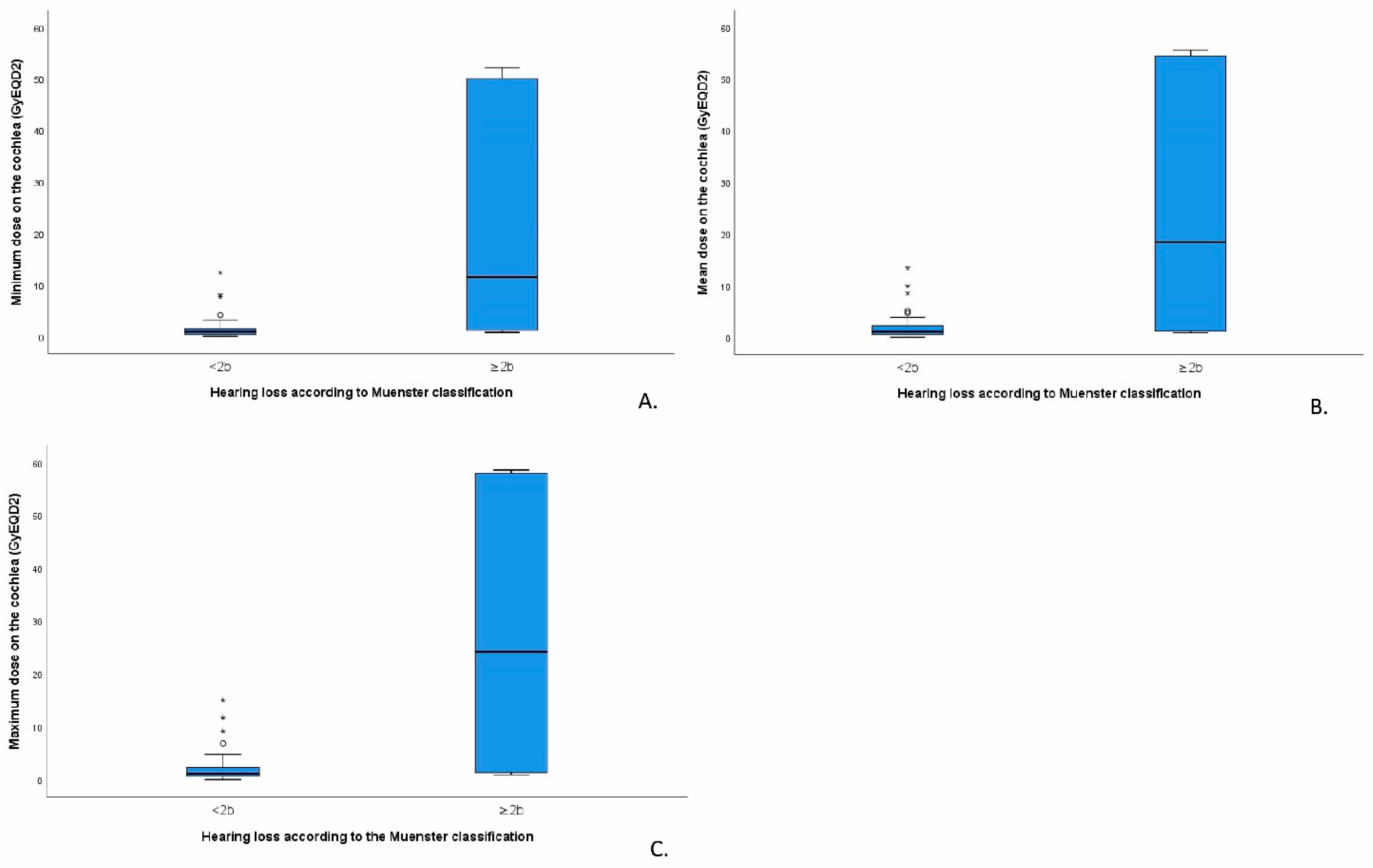
Of the eight survivors diagnosed with deleterious HL (≥Muenster 2b), five survivors had been treated with RT, two with BT (as part of AMORE), and one with PT. Tumour localisation was in the mandibular (n = 2), nasopharynx (n = 2), and orbit (n = 1) for the RT-treated survivors, ear canal (n = 1) and orbit (n = 1) for the BT-treated survivors, and the infratemporal fossa for the survivor treated with PT. The cochlear irradiation dose was available for 23 of the 39 survivors treated at the Emma Children’s Hospital. We found a statistical difference between the maximum, mean, and minimum doses of RT at the cochleae of patients with and without HL (p = 0.015; p = 0.021 and p = 0.011, respectively, Figure 2). The maximum cochlear dose was most strongly correlated with HL occurrence (Pearson’s correlation 0.662, p < 0.01). HL (≥Muenster 2b) was significantly associated with the maximum cochlear irradiation dose (p = 0.047). In our series, HL (≥Muenster 2b) was especially observed when the maximum dose to the cochlea exceeded 19 Gy.

4. Discussion

The overall purpose of this study was to evaluate the hearing status and HL patterns of HNRMS survivors. Additionally, dose–effect relationships for the radiation dose to the cochlea and the development of HL were explored. For the first time, to our knowledge, a radiotherapy dose–effect relationship was explored in survivors of paediatric HNRMS. Although our study describes a small cohort, it includes a unique patient population that survived HNRMS with multimodality treatment including tertiary innovative approaches such as the brachytherapy (AMORE) protocol.
In the current study, we observed that up to 19% of HNRMS survivors had deleterious HL based on the Muenster criteria, 14% based on the CTCAE criteria, and 12% based on the SIOP criteria. However, it was challenging to grade audiograms, as ototoxicity grading scales are developed to monitor HL progression from high-to-low frequencies because most cases of paediatric oncology patients suffer from drug-induced (platinum-related) high-frequency hearing loss [37,38,39].
The HL prevalence in this study is in accordance with other studies. A previous study on survivors of HNRMS showed that clinically relevant hearing loss at speech frequencies occurred in 19% of survivors [14]. Schoot et al. (2016) [14] concluded that AMORE-based treatment resulted in less HL compared to RT; our study found that 2 of the 23 survivors treated with AMORE and 5 of the 16 RT-treated survivors developed deleterious HL (Muenster ≥ 2b). Lockney et al. (2016) [11] studied late toxicities in 30 HNRMS survivors (7.7 (1.2–14.4) years follow-up time) and showed that 20% of them had HL after a median dose of 50.4 (36–50.4) Gy delivered with intensity-modulated radiation therapy. In addition, Bass et al. (2016) [40] studied the effect of cranial irradiation in 235 children with brain tumours including craniopharyngioma, ependymoma, and juvenile pilocytic astrocytoma and found that during a median follow-up period of 9 years and a median of 11 post-RT audiograms per patient, 14% developed relevant HL.
Interestingly, in our cohort, four survivors had a peculiar but consistent low-frequency HL pattern with normal hearing function in the higher frequencies. Bass et al. (2016) [40] observed a so-called “tent-shaped” (loss in the low and high frequencies but normal in the mid-to-high-frequency range) HL pattern in two patients with brain tumours. Hence, it seems that this HL pattern is related to radiotherapy and/or local surgery, and not to chemotherapy or co-administered medication. No clear explanation for this pattern is available yet. It is conceivable that a local infiltrative growth of the tumour itself nearby the middle ear and/or cochlea as well as local surgery can destruct essential hearing structures. Another explanation might be that the outer and middle ear structures are exposed to radiation, potentially leading to chronic otitis externa/media, deep ulcerations, and problems in the external ear canal (stenosis and osteoradionecrosis), tympanic membrane perforation, and fibrosis [17,41]. Vascular insufficiency in the inner ear can develop weeks to months after irradiation, leading to the progressive degeneration and atrophy of sensory structures and even the fibrosis and ossification of the cochlear fluid space [15,22,42]. Addressing low-frequency HL is therefore highly important as this may influence speech and language developmental, educational, and cognitive outcomes in children, just as HL in the higher frequencies does [25].
Our study shows that HL (≥Muenster 2b) is significantly associated with the maximum cochlear irradiation dose (p = 0.047). In our series, HL (≥Muenster 2b) was especially observed when the maximum dose to the cochlea exceeded 19 Gy. This observation is confirmed by previous studies [23,40,43,44]. Recently, Keilty et al. (2021) [43] showed in patients with a brain tumour that the cumulative incidence of high-frequency HL (>4 kHz) was 50% or higher at 5 years after radiotherapy if the mean cochlear dose was >30 Gy. Additionally, Hua et al. (2008) [23] observed that the incidence of HL increased when the mean cochlear dose was greater than 40–45 Gy. Merchant et al. (2004) [19] reported that hearing loss in cranial irradiated children with primary brain tumours is uncommon in the first 4 years after cranial irradiation, although patients with shunts and supratentorial tumours appear to be at risk for low- and intermediate-frequency HL when the cochlear RT dose exceeds 32 Gy. To our knowledge, no literature exists in which the delineation of the cochlear was performed in BT (as part of AMORE) and RT treatment plans of HNRMS survivors. Based on the available literature and our findings, we recommend that one should try to minimise the radiation dose on the cochlea as much as possible to avoid hearing loss and keep the dose on the cochlea at least below 20 Gy if possible. Currently used radiotherapy techniques such as Volumetric-Modulated Arc Therapy (VMAT) [45,46], Cone-beam computed tomography [47], and improved radiotherapy masks [48] can reduce inaccuracies and prevent the unnecessary irradiation of organs at risk, for example, the cochlea.
Our data confirm that it is important to pursue audiological monitoring before but also long after head and neck irradiation, as HL can occur months to years after treatment. Awareness is needed to monitor this adverse effect during long-term follow-up [49] for a minimum of 10 years post-RT [40]. Age-appropriate audiological testing is recommended along with vertigo [50] and tinnitus screening [51]. Classifying audiological tests according to current ototoxicity grading classifications is challenging in this patient group, as these classifications are not developed for HL starting in the low and intermediate frequencies. Therefore, it is recommended to develop a classification system for radiation-induced hearing loss. We advise the assessment of such aberrant HL patterns by ear, nose, and throat (ENT) specialists or specialised audiologists since HL warrants evaluation and physical examination when patterns cannot be explained by administered therapies. The early detection of HL is important to determine if interventions for hearing function optimisation are necessary, such as hearing aids, remote microphone technologies, or cochlear implants to increase the quality of life of those children [52].

Strengths and Limitations

Hearing function was studied in a relatively large Dutch HNRMS survivor cohort treated with different radiotherapy modalities. The survivors had a long follow-up time and were screened for late side effects on a regular basis. Their hearing status was evaluated by senior audiologists with experience in ototoxicity in the paediatric oncology field. For the first time, to our knowledge, cochleae were delineated in BT (as part of AMORE) and RT radiotherapy treatment plans, resulting in a more exact minimum, mean, and maximum cochlear radiotherapy dose.
Unfortunately, not all patients underwent audiological monitoring during follow-up but only patients complaining of HL or showing abnormalities at physical examination. Pre-treatment hearing evaluation was not performed in most patients due to lacking the recommendations for baseline testing in the treatment protocols. Hence, the presence of pre-existing hearing loss in a part of the patients cannot be excluded. In general, we recommend to always perform standardised audiological monitoring in patients with head and neck cancer that will undergo platinum treatment, radiotherapy, and/or ear, nose and throat surgery, including a baseline hearing assessment in future clinical practice. Dosimetry data were available for only a subset of the survivors (23/39, 59%) treated at the Emma Children’s Hospital. Furthermore, in contrast to chemotherapy data, data on ototoxic co-medication, like antibiotics and diuretics, were not available in all survivors. Although strong evidence is not available, we presume that the observed asymmetric HL is inflicted by local therapies. In the current study, the survivors were treated according to different treatment regimens. To our knowledge, the only potentially administered ototoxic chemotherapeutic agent in the survivors of our study could be carboplatin, but this had not been administered in the eight survivors with hearing loss (≥Muenster 2b). Furthermore, as shown in a study by Moke et al. (2021) [53], vincristine is a risk factor for HL in cisplatin-treated children. However, Riga et al. (2005) [54] and Lugassy et al. (1996) [55] found no evidence of the ototoxic effect of vincristine. To date, it is unknown whether the combination of cochlear irradiation and vincristine exposure has a detrimental effect on hearing. More research is needed in larger cohorts to study the effect of this treatment combination on HL. To our knowledge, no evidence exists for the relation between other chemotherapeutic agents received by the RMS survivors (ifosfamide, antracyclines, etoposide, vinorelbine, and/or cyclofosfamide) and hearing loss development.
Due to the limited number of patients with deleterious HL, it was not possible to perform multivariable analyses on dose–effect relationships. A larger sample size with complete cochlear irradiation data and audiological measurements during follow-up is necessary for a more accurate dose–effect analysis.

5. Conclusions

According to the Muenster grading, hearing loss occurred in up to 19% of HNRMS survivors at least 2 years had passed after treatment. Four survivors out of eight had peculiar low-frequency HL with normal/improved hearing function in higher frequencies. HL was significantly associated with the maximum cochlear irradiation dose. More research is needed on the dose–effect relationship of cochlear irradiation and the development of HL during long-term follow-up. Furthermore, the development of a classification scale to grade radiation-induced hearing loss is needed. Overall, audiological monitoring is recommended in HNRMS survivors before, during, and after radiotherapy to detect HL in an early state.

Supplementary Materials

The following supporting information can be downloaded at: https://www.mdpi.com/article/10.3390/cancers14235749/s1, Table S1: Ototoxicity classification systems; Figure S1: Subject identification flow diagram.

Author Contributions

M.L.F.H., J.H.M.M., L.E.S., R.R.G.K., R.A.S., C.L.Z. and M.M.v.d.H.-E. contributed to the inclusion and collection of clinical data on HNRMS survivors. J.W., R.D.F., B.R.P., B.V.B., H.W., N.F., A.B. and M.L.F.H. obtained radiotherapy data and contributed to the radiotherapy analysis. A.E.H., R.J.S., M.L.F.H. and F.A.D. classified and interpreted audiograms. A.J.M.M. and H.v.T. assisted with the statistical part. M.L.F.H. and F.A.D. wrote the manuscript. All authors proofread the manuscript. All authors have read and agreed to the published version of the manuscript.

Funding

The authors received no funding for the work reported in this study.

Institutional Review Board Statement

The study was conducted according to the guidelines of the Declaration of Helsinki and approved by the Institutional Review Board of the University Medical Center Amsterdam.

Informed Consent Statement

The Institutional Review Board of Amsterdam University Medical Centre decided that the Act on Medical Research Involving Subjects did not apply, as the treatment-related data and audiological data were collected during regular follow-up visits.

Data Availability Statement

Data are available upon reasonable request.

Acknowledgments

The authors thank Caroline Thomas for her textual and grammatical corrections in the manuscript.

Conflicts of Interest

The authors declare no conflict of interest.

References

  1. Caron, H.N.; Biondi, A.; Boterberg, T.; Doz, F. Soft tissue sarcomas. In Oxford Textbook of Cancer in Children; Oxford University Press: Oxford, UK, 2021. [Google Scholar]
  2. Glosli, H.; Bisogno, G.; Kelsey, A.; Chilolm, J.; Gaze, M.; Kolb, F.; Mchugh, K.; Shipley, J.; Gallego, S.; Merks, J. Non-parameningeal head and neck rhabdomyosarcoma in children, adolescents, and young adults: Experience of the European paediatric Soft tissue sarcoma Study Group (EpSSG)—RMS2005 study. Eur. J. Cancer 2021, 151, 84–93. [Google Scholar] [CrossRef]
  3. Merks, J.; De Salvo, G.; Bergeron, C.; Bisogno, G.; De Paoli, A.; Ferrari, A.; Rey, A.; Oberlin, O.; Stevens, M.; Kelsey, A.; et al. Parameningeal rhabdomyosarcoma in pediatric age: Results of a pooled analysis from North American and European cooperative groups. Ann. Oncol. Off. J. Eur. Soc. Med. Oncology 2014, 25, 231–236. [Google Scholar] [CrossRef]
  4. Oberlin, O.; Rey, A.; Anderson, J.; Carli, M.; Raney, R.B.; Treuner, J.; Stevens, M.C.; The German Collaborative Soft Tissue Sarcoma Group. Treatment of orbital rhabdomyosarcoma: Survival and late effects of treatment—Results of an international workshop. J. Clin. Oncol. 2001, 19, 197–204. [Google Scholar] [CrossRef] [PubMed]
  5. Kaseb, H.; Kuhn, J.; Babiker, H.M. Rhabdomyosarcoma. StatPearls; StatPearls Publishing LLC.: Treasure Island, FL, USA, 2022. [Google Scholar]
  6. Yock, T.; Schneider, R.; Friedmann, A.; Adams, J.; Fullerton, B.; Tarbell, N. Proton radiotherapy for orbital rhabdomyosarcoma: Clinical outcome and a dosimetric comparison with photons. Int. J. Radiat. Oncol. Biol. Phys. 2005, 63, 1161–1168. [Google Scholar] [CrossRef]
  7. Buwalda, J.; Schouwenburg, P.; Blank, L.; Merks, J.; Copper, M.; Strackee, S.; Voûte, P.; Caron, H. A novel local treatment strategy for advanced stage head and neck rhabdomyosarcomas in children: Results of the AMORE protocol. Eur. J. Cancer 2003, 39, 1594–1602. [Google Scholar] [CrossRef] [PubMed]
  8. Schalow, E.L.; Broecker, B.H. Role of surgery in children with rhabdomyosarcoma. Med. Pediatr. Oncol. 2003, 41, 1–6. [Google Scholar] [CrossRef] [PubMed]
  9. Darwish, C.; Shim, T.; Sparks, A.D.; Chillakuru, Y.; Strum, D.; Benito, D.A.; Monfared, A. Pediatric head and neck rhabdomyosarcoma: An analysis of treatment and survival in the United States (1975–2016). Int. J. Pediatr. Otorhinolaryngol. 2020, 139, 110403. [Google Scholar] [CrossRef] [PubMed]
  10. Schoot, R.A.; Slater, O.; Ronckers, C.M.; Zwinderman, A.H.; Balm, A.J.; Hartley, B.; Brekel, M.W.V.D.; Gupta, S.; Saeed, P.; Gajdosova, E.; et al. Adverse events of local treatment in long-term head and neck rhabdomyosarcoma survivors after external beam radiotherapy or AMORE treatment. Eur. J. Cancer 2015, 51, 1424–1434. [Google Scholar] [CrossRef] [PubMed]
  11. Lockney, N.A.; Friedman, D.N.; Wexler, L.H.; Sklar, C.A.; Casey, D.L.; Wolden, S.L. Late Toxicities of Intensity-Modulated Radiation Therapy for Head and Neck Rhabdomyosarcoma. Pediatr. Blood Cancer 2016, 63, 1608–1614. [Google Scholar] [CrossRef] [PubMed] [Green Version]
  12. De Mattos, V.D.; Ferman, S.; Magalhães, D.M.A.; Antunes, H.S.; Lourenço, S. Dental and craniofacial alterations in long-term survivors of childhood head and neck rhabdomyosarcoma. Oral Surg. Oral Med. Oral Pathol. Oral Radiol. 2019, 127, 272–281. [Google Scholar] [CrossRef] [PubMed]
  13. Clement, S.; Schoot, R.; Slater, O.; Chisholm, J.; Abela, C.; Balm, A.; Brekel, M.V.D.; Breunis, W.; Chang, Y.; Fajardo, R.D.; et al. Endocrine disorders among long-term survivors of childhood head and neck rhabdomyosarcoma. Eur. J. Cancer 2016, 54, 1–10. [Google Scholar] [CrossRef] [PubMed]
  14. Schoot, R.A.; Theunissen, E.A.R.; Slater, O.; Lopez-Yurda, M.; Zuur, C.L.; Gaze, M.N.; Chang, Y.-C.; Mandeville, H.; Gains, J.E.; Rajput, K.; et al. Hearing loss in survivors of childhood head and neck rhabdomyosarcoma: A long-term follow-up study. Clin. Otolaryngol. 2016, 41, 276–283. [Google Scholar] [CrossRef] [PubMed]
  15. Jereczek-Fossa, B.; Zarowski, A.; Milani, F.; Orecchia, R. Radiotherapy-induced ear toxicity. Cancer Treat. Rev. 2003, 29, 417–430. [Google Scholar] [CrossRef] [PubMed]
  16. Bhandare, N.; Antonelli, P.J.; Morris, C.G.; Malayapa, R.S.; Mendenhall, W.M. Ototoxicity after radiotherapy for head and neck tumors. Int. J. Radiat. Oncol. Biol. Phys. 2007, 67, 469–479. [Google Scholar] [CrossRef]
  17. Khan, A.; Budnick, A.; Barnea, D.; Feldman, D.R.; Oeffinger, K.C.; Tonorezos, E.S. Hearing Loss in Adult Survivors of Childhood Cancer Treated with Radiotherapy. Children 2018, 5, 59. [Google Scholar] [CrossRef] [Green Version]
  18. Rasmussen, R.; Claesson, M.; Stangerup, S.E.; Roed, H.; Christensen, I.J.; Cayé-Thomasen, P.; Juhler, M. Fractionated stereotactic radiotherapy of vestibular schwannomas accelerates hearing loss. Int. J. Radiat. Oncol. Biol. Phys. 2012, 83, e607–e611. [Google Scholar] [CrossRef]
  19. Merchant, T.E.; Gould, C.J.; Xiong, X.; Robbins, N.; Zhu, J.; Pritchard, D.L.; Khan, R.; Heideman, R.L.; Krasin, M.J.; Kun, L.E. Early neuro-otologic effects of three-dimensional irradiation in children with primary brain tumors. Int. J. Radiat. Oncol. Biol. Phys. 2004, 58, 1194–1207. [Google Scholar] [CrossRef]
  20. Zuur, C.L.; Simis, Y.J.; Lamers, E.A.; Hart, A.A.; Dreschler, W.A.; Balm, A.J.; Rasch, C.R. Risk factors for hearing loss in patients treated with intensity-modulated radiotherapy for head-and-neck tumors. Int. J. Radiat. Oncol. Biol. Phys. 2009, 74, 490–496. [Google Scholar] [CrossRef]
  21. Meijer, A.J.M.; Li, K.H.; Brooks, B.; Clemens, E.; Ross, C.J.; Rassekh, S.R.; Hoetink, A.E.; Grotel, M.; Heuvel-Eibrink, M.M.; Carleton, B.C. The cumulative incidence of cisplatin-induced hearing loss in young children is higher and develops at an early stage during therapy compared with older children based on 2052 audiological assessments. Cancer 2022, 128, 169–179. [Google Scholar] [CrossRef]
  22. Young, Y.-H.; Lu, Y.-C. Mechanism of hearing loss in irradiated ears: A long-term longitudinal study. Ann. Otol. Rhinol. Laryngol. 2001, 110, 904–906. [Google Scholar] [CrossRef]
  23. Hua, C.; Bass, J.K.; Khan, R.; Kun, L.E.; Merchant, T.E. Hearing loss after radiotherapy for pediatric brain tumors: Effect of cochlear dose. Int. J. Radiat. Oncol. Biol. Phys. 2008, 72, 892–899. [Google Scholar] [CrossRef]
  24. Zuur, C.L.; Simis, Y.J.; Lansdaal, P.E.; Hart, A.A.; Rasch, C.R.; Schornagel, J.H.; Dreschler, W.A.; Balm, A.J. Risk factors of ototoxicity after cisplatin-based chemo-irradiation in patients with locally advanced head-and-neck cancer: A multivariate analysis. Int. J. Radiat. Oncol. Biol. Phys. 2007, 68, 1320–1325. [Google Scholar] [CrossRef] [PubMed]
  25. Lieu, J.E.C.; Kenna, M.; Anne, S.; Davidson, L. Hearing Loss in Children: A Review. Jama 2020, 324, 2195–2205. [Google Scholar] [CrossRef] [PubMed]
  26. Ronner, E.A.; Benchetrit, L.; Levesque, P.; Basonbul, R.A.; Cohen, M.S. Quality of Life in Children with Sensorineural Hearing Loss. Otolaryngol. Head Neck Surg. 2020, 162, 129–136. [Google Scholar] [CrossRef] [PubMed]
  27. Stevens, M.; Rey, A.; Bouvet, N.; Ellershaw, C.; Toledo, J.S.; Oberlin, O. SIOP MMT 95: Intensified (6 drug) versus standard (IVA) chemotherapy for high risk non metastatic rhabdomyosarcoma (RMS). J. Clin. Oncol. 2004, 22, 8515. [Google Scholar] [CrossRef]
  28. Defachelles, A.S.; Rey, A.; Oberlin, O.; Spooner, D.; Stevens, M.C. Treatment of nonmetastatic cranial parameningeal rhabdomyosarcoma in children younger than 3 years old: Results from international society of pediatric oncology studies MMT 89 and 95. J. Clin. Oncol. 2009, 27, 1310–1315. [Google Scholar] [CrossRef]
  29. Stevens, M.C.; Rey, A.; Bouvet, N.; Bouvet, N.; Ellershaw, C.; Flamant, F.; Habrand, J.L.; Marsden, H.B.; Martelli, H.; de Toledo, J.S.; et al. Treatment of nonmetastatic rhabdomyosarcoma in childhood and adolescence: Third study of the International Society of Paediatric Oncology—SIOP Malignant Mesenchymal Tumor. J. Clin. Oncol. 2005, 23, 2618–2628. [Google Scholar] [CrossRef]
  30. Bisogno, G. European Paediatric Soft Tissue Sarcoma Study Group RMS 2005 a Protocol for Non Metastatic Rhabdomyosarcoma. 2005. Available online: https://www.skion.nl/workspace/uploads/Protocol-EpSSG-RMS-2005-1-3-May-2012_1.pdf (accessed on 1 January 2020).
  31. Dantonello, T.M.; Int-Veen, C.; Harms, D.; Leuschner, I.; Schmidt, B.F.; Herbst, M.; Juergens, H.; Scheel-Walter, H.-G.; Bielack, S.S.; Klingebiel, T.; et al. Cooperative trial CWS-91 for localized soft tissue sarcoma in children, adolescents, and young adults. J. Clin. Oncol. 2009, 27, 1446–1455. [Google Scholar] [CrossRef]
  32. Koscielniak, E.T.K. CWS-Guidance for Risk Adapted Treatment of Soft Tissue Sarcoma and Soft Tissue Tumours in Children, Adolescents, and Young Adults. Version 1.5. from 01.07. Available online: https://fnkc.ru/docs/CWS-2009.pdf (accessed on 1 July 2009).
  33. Freling, N.J.M.; Hol, M.; Velduis, W. Anatomy of the Head and Neck. Version 1.2. Available online: https://doradiology.com/product-anatomy-head-neck.html (accessed on 15 August 2021).
  34. Hasegawa, T.; Kida, Y.; Kato, T.; Iizuka, H.; Yamamoto, T. Factors associated with hearing preservation after Gamma Knife surgery for vestibular schwannomas in patients who retain serviceable hearing. J. Neurosurg. 2011, 115, 1078–1086. [Google Scholar] [CrossRef] [Green Version]
  35. Couto, J.G.; Bravo, I.; Pirraco, R. Biological equivalence between LDR and PDR in cervical cancer: Multifactor analysis using the linear-quadratic model. J. Contemp. Brachyther. 2011, 3, 134–141. [Google Scholar] [CrossRef]
  36. Pötter, R.; Haie-Meder, C.; Van Limbergen, E.; Barillot, I.; De Brabandere, M.; Dimopoulos, J.; Dumas, I.; Erickson, B.; Lang, S.; Nulens, A.; et al. Recommendations from gynaecological (GYN) GEC ESTRO working group (II): Concepts and terms in 3D image-based treatment planning in cervix cancer brachytherapy-3D dose volume parameters and aspects of 3D image-based anatomy, radiation physics, radiobiology. Radiother. Oncol. 2006, 78, 67–77. [Google Scholar] [CrossRef]
  37. Brock, P.R.; Knight, K.R.; Freyer, D.R.; Campbell, K.C.; Steyger, P.S.; Blakley, B.W.; Rassekh, S.R.; Chang, K.W.; Fligor, B.J.; Rajput, K.; et al. Platinum-induced ototoxicity in children: A consensus review on mechanisms, predisposition, and protection, including a new International Society of Pediatric Oncology Boston ototoxicity scale. J. Clin. Oncol. 2012, 30, 2408–2417. [Google Scholar] [CrossRef] [PubMed] [Green Version]
  38. Schmidt, C.M.; Bartholomäus, E.; Deuster, D.; Heinecke, A.; Dinnesen, A.G. The “Muenster classification” of high frequency hearing loss following cisplatin chemotherapy. HNO 2007, 55, 299–306. [Google Scholar] [CrossRef]
  39. Clemens, E.; Brooks, B.; De Vries, A.C.H.; Van Grotel, M.; Heuvel-Eibrink, M.M.V.D.; Carleton, B. A comparison of the Muenster, SIOP Boston, Brock, Chang and CTCAEv4.03 ototoxicity grading scales applied to 3,799 audiograms of childhood cancer patients treated with platinum-based chemotherapy. PLoS ONE 2019, 14, e0210646. [Google Scholar] [CrossRef] [PubMed]
  40. Bass, J.K.; Hua, C.-H.; Huang, J.; Onar-Thomas, A.; Ness, K.K.; Jones, S.; White, S.; Bhagat, S.P.; Chang, K.W.; Merchant, T.E. Hearing Loss in Patients Who Received Cranial Radiation Therapy for Childhood Cancer. J. Clin. Oncol. 2016, 34, 1248–1255. [Google Scholar] [CrossRef] [PubMed] [Green Version]
  41. Grewal, S.; Merchant, T.; Reymond, R.; McInerney, M.; Hodge, C.; Shearer, P. Auditory late effects of childhood cancer therapy: A report from the Children’s Oncology Group. Pediatrics 2010, 125, e938–e950. [Google Scholar] [CrossRef] [PubMed] [Green Version]
  42. Low, W.-K.; Tan, M.G.; Chua, A.W.; Sun, L.; Wang, D.-Y. 12th Yahya Cohen Memorial Lecture: The cellular and molecular basis of radiation-induced sensori-neural hearing loss. Ann. Acad. Med. Singap. 2009, 38, 91–94. [Google Scholar] [CrossRef] [PubMed]
  43. Keilty, D.; Khandwala, M.; Liu, Z.A.; Papaioannou, V.; Bouffet, E.; Hodgson, D.; Yee, R.; Cushing, S.; Laperriere, N.; Ahmed, S.; et al. Hearing Loss After Radiation and Chemotherapy for CNS and Head-and-Neck Tumors in Children. J. Clin. Oncol. 2021, 39, Jco2100899. [Google Scholar] [CrossRef]
  44. Bhandare, N.; Jackson, A.; Eisbruch, A.; Pan, C.C.; Flickinger, J.; Antonelli, P.; Mendenhall, W.M. Radiation therapy and hearing loss. Int. J. Radiat. Oncol. Biol. Phys. 2010, 76, S50–S57. [Google Scholar] [CrossRef] [Green Version]
  45. Nagarajan, M.; Banu, R.; Sathya, B.; Sundaram, T.; Chellapandian, T.P. Dosimetric Evaluation and Comparison Between Volumetric Modulated Arc Therapy (VMAT) and Intensity Modulated Radiation Therapy (IMRT) Plan in Head and Neck Cancers. Gulf J. Oncolog. 2020, 1, 45–50. [Google Scholar]
  46. Buciuman, N.; Marcu, L.G. Dosimetric justification for the use of volumetric modulated arc therapy in head and neck cancer-A systematic review of the literature. Laryngoscope Investig. Otolaryngol. 2021, 6, 999–1007. [Google Scholar] [CrossRef] [PubMed]
  47. Grover, D.; Laraque, M.; Debenham, B. Evaluation of Target Volume Location and Its Impact on Delivered Dose Using Cone-Beam Computed Tomography Scans for Patients with Head and Neck Cancer. J. Med. Imaging Radiat. Sci. 2019, 50, 387–397. [Google Scholar] [CrossRef] [PubMed]
  48. Fukao, M.; Okamura, K.; Sabu, S.; Akino, Y.; Arimura, T.; Inoue, S.; Kado, R.; Seo, Y. Repositioning accuracy of a novel thermoplastic mask for head and neck cancer radiotherapy. Phys. Med. 2020, 74, 92–99. [Google Scholar] [CrossRef] [PubMed]
  49. Clemens, E.; Heuvel-Eibrink, M.M.V.D.; Mulder, R.L.; Kremer, L.C.M.; Hudson, M.M.; Skinner, R.; Constine, L.S.; Bass, J.K.; Kuehni, C.E.; Langer, T.; et al. Recommendations for ototoxicity surveillance for childhood, adolescent, and young adult cancer survivors: A report from the International Late Effects of Childhood Cancer Guideline Harmonization Group in collaboration with the PanCare Consortium. Lancet Oncol. 2019, 20, e29–e41. [Google Scholar] [CrossRef] [PubMed]
  50. Humphriss, R.L.; Hall, A.J. Dizziness in 10 year old children: An epidemiological study. Int. J. Pediatr. Otorhinolaryngol. 2011, 75, 395–400. [Google Scholar] [CrossRef] [PubMed]
  51. Savastano, M. Characteristics of tinnitus in childhood. Eur. J. Pediatr. 2007, 166, 797–801. [Google Scholar] [CrossRef] [PubMed]
  52. Tharpe, A.M.; Gustafson, S. Management of Children with Mild, Moderate, and Moderately Severe Sensorineural Hearing Loss. Otolaryngol. Clin. N. Am. 2015, 48, 983–994. [Google Scholar] [CrossRef] [PubMed]
  53. Moke, D.J.; Luo, C.; Millstein, J.; Knight, K.R.; Rassekh, S.R.; Brooks, B.; Ross, C.J.D.; Wright, M.; Mena, V.; Rushing, T.; et al. Prevalence and risk factors for cisplatin-induced hearing loss in children, adolescents, and young adults: A multi-institutional North American cohort study. Lancet Child. Adolesc. Health 2021, 5, 274–283. [Google Scholar] [CrossRef]
  54. Riga, M.; Psarommatis, I.; Korres, S.; Lyra, C.; Papadeas, E.; Varvutsi, M.; Ferekidis, E.; Apostolopoulos, N. The effect of treatment with vincristine on transient evoked and distortion product otoacoustic emissions. Int. J. Pediatr. Otorhinolaryngol. 2006, 70, 1003–1008. [Google Scholar] [CrossRef]
  55. Lugassy, G.; Shapira, A. A prospective cohort study of the effect of vincristine on audition. Anticancer. Drugs 1996, 7, 525–526. [Google Scholar] [CrossRef]
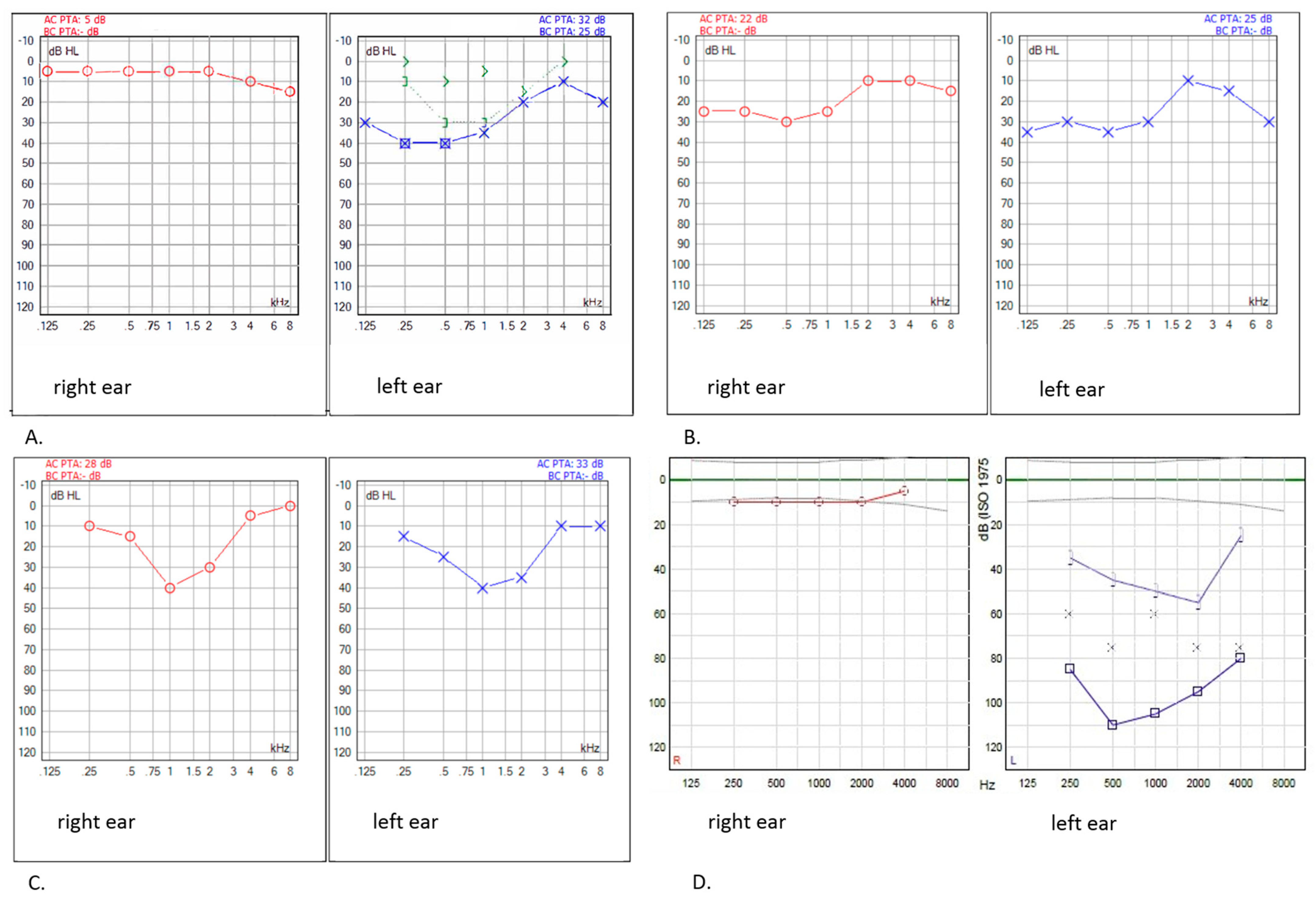
Figure 1. Patterns of hearing loss (HL) as shown per pure-tone audiogram in survivors with a left nasopharyngeal RMS (A), right nasopharyngeal RMS (B), right orbital RMS (C), and RMS in the left ear canal (D). Low-frequency HL with a mild HL/normal hearing function in higher frequencies is observed in survivor (A) left ear, survivors (B,C) both ears, and survivor (D) left ear.
Cancers 14 05749 g001
Figure 2. Boxplots showing minimum (A), mean (B) and maximum (C) irradiation dose on the cochlea (GyEQD2) in survivors without (Muenster < 2b) and with deleterious hearing loss (Muenster ≥ 2b). Outliers are indicated with *.
Cancers 14 05749 g002
Table 1. Patient characteristics and audiologic outcomes.
Patient IDRT TypeSexDiagnosisTumour LocationTumour SideSiteAge at Diagnosis (Years)Time to FU (Years)Treatment ProtocolTotal Radiotherapy Dose (Gy)Max. Cochlear Dose in Gy (Right)Max. Cochlear Dose in Gy (Left)Muenster Grade (Right)Muenster Grade (Left)SIOP Grade (Right)SIOP Grade (Left)CTCAE Grade (Right)CTCAE Grade (Left)
1AMOREmeRMSUpper
eyelid
rightNPM6.013.4MMT-95400.70.4110000
2 feRMSEar canalleftPM10.111.5RMS 2005401.036.7040404
3 mRMS nsParotid spaceleftNPM3.425.5MT-8946
4 feRMSSinus
maxillary,
orbit,
ethmoid
leftPM2.417.3MMT-95501.021.38000000
5 feRMSMasticator spaceleftNPM13.012.1MMT-95401.13.90 0 0
6 meRMSPterygoid spaceleftPM2.122.4CWS-914511.21.9
7 maRMSTemporalleftPM1.811.4RMS 2005401.14.8010000
8 meRMSOrbitrightPM7.711.6MMT-95451.30.7000001
9 feRMSNasal
cavity
rightPM1.321.0MMT-95451.11.0110000
10 feRMSOrbitleftorbit8.08.3RMS 2005400.61.0
11 meRMSOrbitrightorbit5.56.6RMS 2005400.80.0
12 meRMS/
botryoid
Nasal
cavity
leftPM3.22.0RMS 2005501.94.302a0101
13 faRMSOrbit,
ethmoidal sinus,
sinus
maxillary
leftPM13.412.5MMT-95451.72.7000000
14 meRMSOrbitleftorbit4.33.7RMS 2005400.40.5
15 feRMSOrbitleftorbit5.016.7MMT-95451.30.93a3a3333
16 meRMSParotidrightNPM11.23.4RMS 20054011.71.02a01010
17 feRMSParotid space, mandibularleftPM5.825.3MT-8940 02a0101
18 maRMSNostrilleftNPM7.515.5MMT-95400.40.5000000
19 meRMSOrbitrightorbit10.22.9RMS 200540 000000
20 meRMSOrbitleftorbit7.126.6MMT-89500.00.0100000
21 feRMSOrbit, pterygopalatine fossarightPM4.826.5MMT-8960 000000
22 meRMSNasal
cavity
leftPM3.22.9RMS 200568 12a0101
23 meRMSOrbitrightorbit5.59.4RMS 200540 000000
24ProtonfeRMSOrbitrightorbit6.39.0RMS 200550.4 110000
25 feRMSPara-pharyngealrightPM4.012.3MMT-9550 12a0101
26 meRMSMastoid /middle earleftPM2.76.4RMS 200555.8 010000
27 faRMSInfratemporal fossaleftPM4.611.2RMS 2005 03c0303
28 feRMSMaxillaleftPM4.03.6RMS 200550.4 2a01010
29 feRMSOrbitleftorbit2.07.3RMS 2005 000000
30 feRMSOrbitrightorbit10.07.2RMS 200545 000000
31 feRMSOrbitrightorbit8.28.7RMS 200554 000000
32 feRMSOrbitrightorbit6.210.1RMS 200550.4 000000
33PhotonmeRMSPara- pharyngealrightPM3.326.4MT-8950.75 2a01010
34 meRMSAngle of mandiblerightPM0.0413.5MMT-95
RMS 2005
45 2b01020
35 feRMSNasopharynxrightPM4.012.2RMS 200550.458.758.13a3a0101
36 feRMSOrbitleftorbit4.46.7CWS-2007HR50.4 000000
37 meRMSNasopharynx, oropharynxright/leftPM4.56.7RMS 200550.40.00.0000001
38 feRMS/ botryoidNasopharynxrightPM6.53.6RMS 200550.4 000000
39 meRMSOrbitrightorbit7.23.4RMS 20054518.417.0
40 meRMSSinus
maxillary
leftPM5.05.2RMS 200550.49.115.0000000
41 faRMSMandible, ethmoid, Sella
Turcica
orbit
leftPM4.416.2MMT-9554 3b3c4434
42 meRMSCheekrightNPM0.55.8RMS 200550.40.20.2Not classifiable *
43 feRMSOrbitleftorbit7.29.9RMS 200550.46.811.603a0303
44 meRMSSphenoidNSPM5.113.5MMT-9550.4 010000
45 feRMS/botryoid RMSNasopharynxrightPM6.56.0RMS 200550.4 000000
46 feRMSOrbitleftorbit4.49.7CWS-2007HR50.4 000000
47 meRMSOrbitrightorbit4.914.4MMT-9545 + 40 brachy 000000
48 feRMSOrbitleftOrbit3.712.6RMS 200545 100000
49 meRMSNasopharynxleftPM13.19.8RMS 2005 Avastin trial55.8 03a0000
* Free field visual reinforcement audiometry: Hearing threshold at 0.5–4.0 kHz between 20–30 dB; Abbreviations: AMORE = Ablative surgery, MOuld technique brachytherapy and surgical REconstruction; aRMS = alveolar rhabdomyosarcoma; CTCAE = Common Terminology Criteria for Adverse Events; CWS = Cooperative Weichteilsarkom Study; RT = External Beam Photon RadioTherapy; eRMS = embryonal rhabdomyosarcoma; F = female; FU = follow-up; M = male; MMT = Malignant Mesenchymal Tumour; NPM = non-parameningeal; NS = not specified/unknown; PM = parameningeal; RMS = rhabdomyosarcoma; RMS 2005 = European Paediatric Soft Tissue Sarcoma Study Group RMS 2005; SIOP = Société Internationale d’Oncologie Pédiatrique; VRA = visual reinforcement audiometry.
Table 2. Clinical characteristics of patients with HNRMS.
TotalAudiological Evaluation (n = 42)Unknown/
Not Classifiable
Muenster ≥ 2b HLMuenster < 2b HL
(n = 49)(n = 8)(n = 34)(n = 7)
Male (n)242175
Female256172
Age at diagnosis, median (range) (years)5.0 (0.04–13.4)4.8 (0.04–13.1)5.1 (1.3–13.4)4.3 (0.5–8.0)
Age at follow-up, median (range) (years)16.3 (5.2–33.7)18.9 (13.6–24.5)16.6 (5.2–33.7)12.1 (6.3–29.0)
Time to follow-up, median (ranges)10.1 (2.0–26.6)11.9 (9.8–16.7)9.9 (2.0–26.6)6.6 (3.4–25.5)
RMS histology subtype (n)
Alveolar5230
Embryonal436316
Not specified1001
Tumour site (n)
Parameningeal256181
Non-parameningeal6042
Orbital182124
Treatment protocol (n)
MMT-95133100
MMT-895041
CWS-911001
CWS-2007HR2020
EpSSG RMS 2005285185
Radiotherapy subgroup (n)
AMORE232165
RT16592
PT9180
Brachy + RT1010
Abbreviations: AMORE = Ablative surgery MOuld techniques after loading brachytherapy and surgical REconstruction; brachy = brachytherapy; HL = hearing loss; n = number; PT = proton radiotherapy; RT = photon radiotherapy.
Publisher’s Note: MDPI stays neutral with regard to jurisdictional claims in published maps and institutional affiliations.