Review

Intravascular NK/T-Cell Lymphoma: What We Know about This Diagnostically Challenging, Aggressive Disease

1
Pathology Unit, Azienda USL-IRCCS di Reggio Emilia, 42123 Reggio Emilia, Italy
2
Pathology Unit, Fondazione IRCCS Casa Sollievo della Sofferenza, 71013 San Giovanni Rotondo, Italy
3
Pathology Unit, Policlinico Riuniti, University of Foggia, 71122 Foggia, Italy
4
Surgical Oncology Unit, Azienda USL-IRCCS di Reggio Emilia, 42123 Reggio Emilia, Italy
5
Laboratory Oncology, Fondazione IRCCS Casa Sollievo della Sofferenza San Giovanni Rotondo, 71013 San Giovanni Rotondo, Italy
6
Anatomic Pathology, Ospedale Policlinico San Martino IRCCS Genova, 16132 Genova, Italy
7
Pathology Unit, Azienda Ospedaliera Santa Maria di Terni, University of Perugia, 05100 Terni, Italy
8
Pathology Unit, Humanitas University, Pieve Emanuele, 20072 Milan, Italy
9
Pathology Unit, Humanitas Research Hospital-IRCCS, Rozzano, 20089 Milan, Italy
10
Department of Medical and Surgical Sciences and Advanced Technologies “G.F. Ingrassia” Anatomic Pathology, University of Catania, 95123 Catania, Italy
*
Author to whom correspondence should be addressed.
Cancers 2022, 14(21), 5458; https://doi.org/10.3390/cancers14215458
Submission received: 24 October 2022 / Revised: 2 November 2022 / Accepted: 5 November 2022 / Published: 6 November 2022
(This article belongs to the Special Issue Advances in NK/T-cell Lymphoma, Epidemiology, Biology and Therapy)

Abstract

:

Simple Summary

Intravascular lymphoma is a neoplasm with tumor cells localized exclusively within blood vessel lumina. Most cases are of B-cell origin; only rare cases of NK or T-cell lineage. Unlike intravascular large B-cell lymphoma, which is a well-recognized entity in the WHO classification, intravascular NK/T-cell lymphoma is not considered a specific entity and there is still a debate about whether it is closer to aggressive NK leukemia or to extranodal NK/T-cell lymphoma. Our aim was to summarize the clinical, pathological, and molecular data on intravascular NK/T-cell lymphoma, which is a challenge both in terms of diagnosis and treatment. Recent molecular studies improved our understanding of the mechanism of lymphomagenesis, showing that multiple genetic events associated with EBV infection are required for the pathogenesis of this aggressive lymphoma. Promising therapeutic results may be offered by immune checkpoint inhibitors due to the high PD-L1 expression, which is possibly related to EBV infection.

Abstract

Intravascular lymphoma is a form of lymphoid malignancy characterized by neoplastic cells growing almost exclusively within the lumina of small- to medium-sized blood vessels. Most cases are of B-cell origin with rare cases of natural killer or T-cell lineage. Extranodal sites are affected, mainly the skin and central nervous system, although any organ may be involved. Intravascular NK/T-cell lymphoma deserves special attention because of its clinicopathologic features and the need for adequate immunophenotyping combined with clonality test for a proper diagnosis. Moreover, intravascular NK/T-cell lymphoma is strongly linked to Epstein–Barr virus (EBV), which is considered to play a role in tumorigenesis and to be responsible for the aggressive behavior of the disease. In this paper, we review the current knowledge on this rare lymphoma and, in particular, the most recent advances about its molecular landscape. The main distinguishing features with other EBV-related entities, such as extranodal NK/T-cell lymphoma, EBV-positive primary nodal T/NK-cell lymphoma, and aggressive NK-cell leukemia, are discussed to help pathologists obtain the correct diagnosis and consequently develop an adequate and prompt therapy response.

1. Introduction

Intravascular lymphoma (IVL) is a malignancy characterized by the presence of tumor cells localized within blood vessel lumina. Although IVL may involve any organ, the majority of cases are reported within the skin and central nervous system (CNS).
IVL is more often of B-cell nature. IVL of T and NK-cell lineage is extremely rare, with a limited number of cases with sufficient immunohistochemical and clonality data reported so far [1,2,3,4,5,6,7,8,9,10,11,12,13,14,15,16,17,18,19,20,21].
In the last World Health Organization (WHO) classification of lymphoid neoplasms, intravascular NK/T-cell lymphoma (IVNKTL) is not recognized as a specific entity, but as very close to extranodal NK/T-cell lymphoma (ENKTL), from which it differs for its typical intravascular nature [22].
ENKTL is a high-grade lymphoma involving at presentation, in the majority of cases, the nasal or upper aerodigestive tract and, more rarely, other non-nasal, extranodal sites, including the skin [23,24,25,26].
The disease is prevalent in adult males from Asia and Latin America. EBV infection and genetic predisposition are involved in the pathogenesis of ENKTL, therefore explaining the geographic distribution of the neoplasm [22,26,27,28].
Whereas ENKTL is a mass-forming malignancy, classically characterized by the presence of neoplastic cells infiltrating the tissues with an angioinvasive and angiodestructive pattern of growth, IVNKTL does not form any mass and is characteristically a blood-vessel lumina-restricted lymphoma, affecting predominantly skin and CNS [18].
In the recently published review article by Alaggio et al., IVNKTL, due to its unclear nosological nature, is now described under aggressive NK-cell leukemia (ANKL) rather than ENKTL, although further studies are required to define where it fits best [29].
The present paper highlights the clinicopathologic features of IVNKTL, focusing on the main differential diagnoses as well as on the molecular pathogenesis of this rare, highly aggressive disease. Due to its relation to EBV infection, a brief introductive paragraph on EBV biology is also included.

2. Overview on EBV Biology

EBV, also known as human herpes virus 4 (HHV4), is one of the most diffuse human viruses [30,31,32,33]. The majority of adults are estimated to be seropositive.
People generally get infected at an early age through saliva. The virus entries through the oropharynx and, during a lytic phase, it replicates in the oropharyngeal epithelial cells. During this initial lytic phase, the host lacks immunity against EBV, whereas in the following weeks, cellular and humoral immunity develop, and the viral genome remains in a latent phase within memory B-lymphocytes.
Primary infection may be either asymptomatic or lead to infectious mononucleosis (IM), often with a limited course. The EBV enters B lymphocytes through the binding between the major viral envelope glycoprotein gp350 and the complement receptor 2 (CR2/CD21) on the B-lymphocyte surface. Moreover, the viral glycoprotein (gp42) acts as a cofactor binding to human leucocyte antigen (HLA) class II molecules on B-lymphocytes.
Memory B-cells are the virus reservoir in healthy people; hence, through this hiding mechanism (latency), the virus may elude the immune system. EBV-infected cells express, in different latency phases, the following six types of EBV nuclear antigen (EBNA): EBNA1, EBNA2, EBNA3A, EBNA3B, EBNA3C, and EBNA leader protein and three types of latent membrane protein (LMP): LMP1, LMP2A, and LMP2B. The latency phase of EBV infection is sub-classified into types III, II, and I, the former progressing to the latter. Upon infection of resting naïve lymphoid B-cells, EBV enters into type-III latency, in which all viral gene products are expressed, leading to the production of immunogenic viral proteins and causing a strong reaction of the T-cell immunity. Subsequently, the virus enters type-II latency with restriction of gene viral expression; latency II is characterized by expression of to EBNA1, LMP1, and LMP2, and in this phase, B-lymphocytes differentiate into memory B-cells. Finally, in type-I latency, viral gene expression is further restricted, with all infected cells expressing only EBNA1, which is responsible for the maintenance and replication of the EBV viral genome. Since approximately 50 years ago, when EBV was discovered, the virus has been shown to play an oncogenic role in different neoplasms, both lymphomas, and epithelial tumors—for instance, gastric carcinoma and nasopharyngeal carcinoma.
Interestingly, EBV may infect not only B-lymphocytes but even T-lymphocytes and NK-cells, although the mechanism for virus entrance into T/NK-cells is not well-known [34,35,36,37,38].
EBV-associated diseases develop predominantly in immunocompromised individuals when the balance between the immune system of the host and the infection is altered. EBV plays a pathogenetic role in lymphoproliferative disorders (LPDs) of B-cell origin, as well as of T- and NK-cell origin, including IVNKTL [28,39,40].

3. Overview on the PD-1/PD-L1 Axis and Its Relation to EBV in Lymphomas

The PD-1 pathway, which includes the immune cell co-receptor programmed death 1 (PD-1) and its ligands, programmed cell death ligand 1 (PD-L1) and programmed cell death ligand 2 (PD-L2), acts on local immunosuppression in the tumor microenvironment (TME) [41,42,43].
PD-1 is expressed on immune cells such as T-, B-, and NK-cells; PD-L1 is expressed on neoplastic cells and antigen-presenting cells, and PD-L2 is displayed by activated monocytes and dendritic cells.
The survival of several neoplasms is favored by the PD-1/PD-L1 interaction; PD-L1 expressed by tumor cells binds with its receptor PD-1, displayed by immune cells, and favors the process of immune evasion by neoplastic cells.
The PD-1/PD-L1 interaction suppresses the activity and proliferation of cytotoxic tumor-infiltrating lymphocytes (TILs) and favors the immunosuppressive effects of regulatory T-lymphocytes. Additionally, the PD-1/PD-L1 binding has multiple effects on all the components of the TME that favor tumor immune evasion, for instance, regulating the secretion of cytokines and the inhibition of NK-cells, which may directly eliminate neoplastic cells [41,42,43].
PD-1/PD-L1 inhibitors represent a highly promising cancer treatment option because, by blocking PD-1–PD-L1 signaling, they can prevent tumor immune escape and facilitate a positive immune response to kill tumor cells.
Currently, checkpoint inhibitors represent a frontline therapy for several neoplasms such as non-small-cell lung cancer, metastatic melanoma, renal cell carcinoma, urothelial tumors, and head-and-neck squamous cell carcinoma, but they are also under active evaluation in many other solid cancers, as well as in some hematological neoplasms [44,45,46].
EBV is associated with a variety of lymphomas, and immunodeficiency has always been considered to play a critical role in the development of EBV-related lymphomas. However, the immune escape of tumor cells has recently been regarded as contributing to the development of some EBV-associated hematological malignancies, in which PD-L1 overexpression is observed [45].
Upregulation of PD-L1 is found in EBV-associated NK/T-cell lymphomas, which are the subject of the present review, and EBV infection is considered to play an important role in upregulating PD-1/PD-L1 expression through various mechanisms (better detailed in the paragraphs of the different entities). In ENKTL, both LMP1 overexpression and the JAK/STAT pathway contribute to PD-L1 overexpression and to tumor immune evasion.
Recently, promising results have been obtained with the use of checkpoint inhibitors in lymphomas; in particular, lymphomas characterized by immune escape of the tumor cells are considered good candidates for anti-PD-1/PD-L1 therapy [45,46,47].

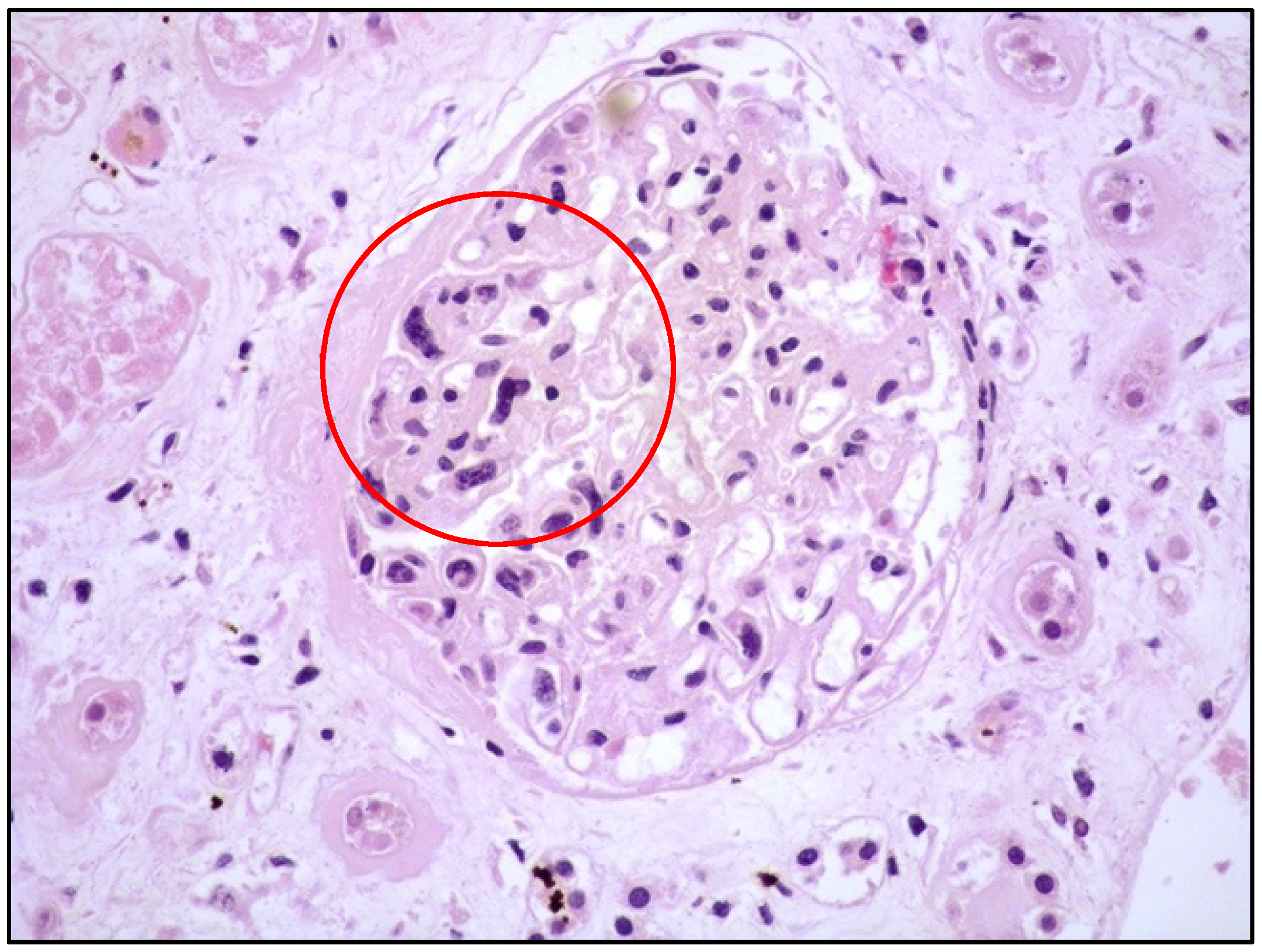
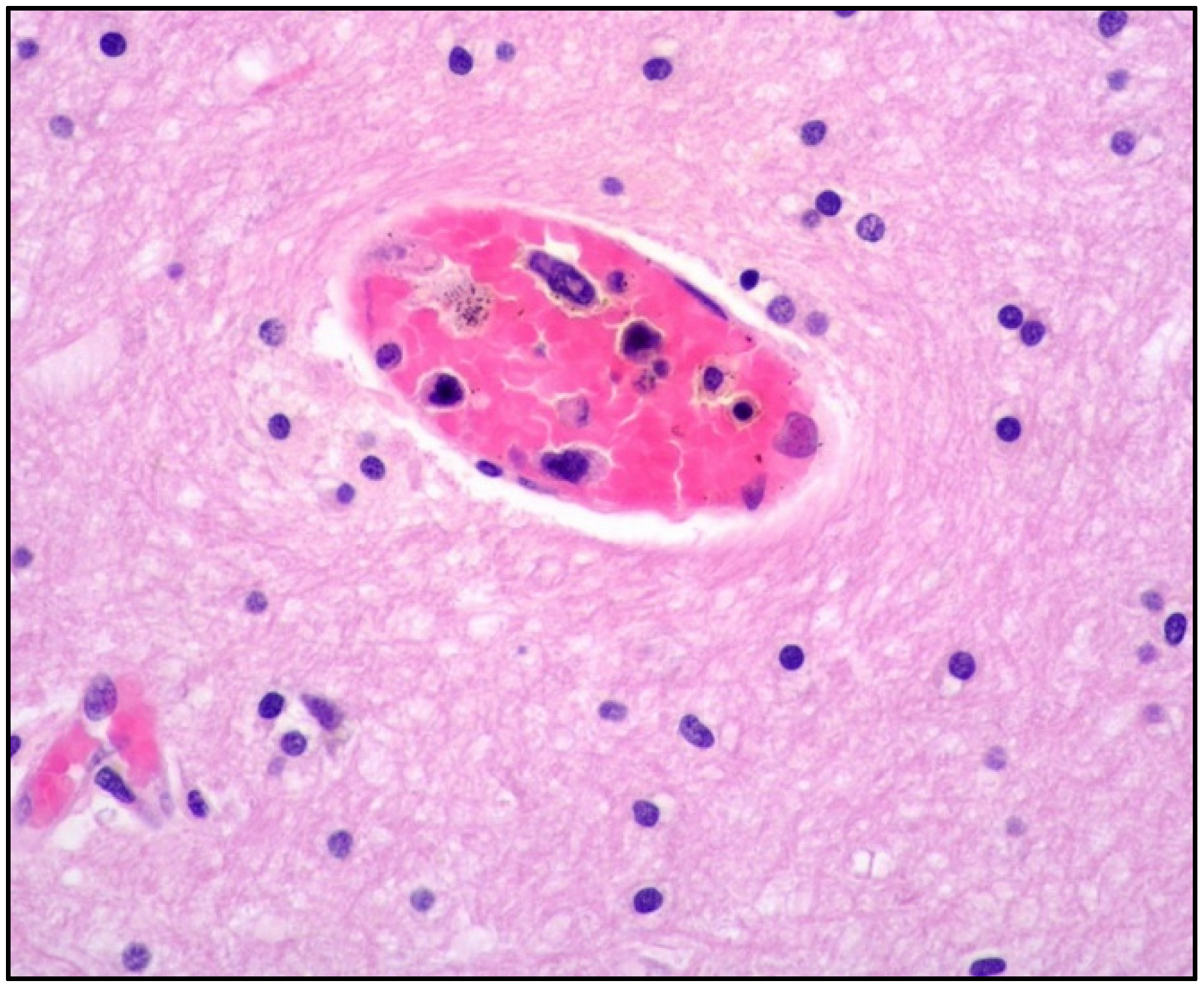
4. Clinicopathological Features of IVNKTL

IVNKTL is an exceedingly rare lymphoid malignancy with a limited number of cases (approximately 22) with sufficient immunophenotypic and molecular data reported in the literature [1,15,16,17,19,20,21]. The disease occurs predominantly in adults with a variable range of ages; the skin and CNS are the most frequently involved sites, with the skin being the most frequent and, in some cases, the unique site affected. Tender erythematous subcutaneous nodules are the most frequent clinical manifestation [14].
Notably, even in IVL of B-cell origin, which is far more frequent than IVNKTL, the skin is the main organ affected, being involved alone or in combination with other organs—the CNS predominating, in 40% of cases [7,22]. Intravascular large B-cell lymphoma (IVLBL), which is a well-recognized entity, has two distinct patterns of presentation: the Western variant, mainly affecting skin and CNS, and the Asian variant, usually involving multiple organs and often associated to hemophagocytic lymphohistiocytosis (HLH) and worse prognosis [48].
Nonetheless, considering the almost-exclusive blood-vessel lumina-restricted growth pattern of IVNKTL without mass lesions, prior to post-mortem evaluation, it can be presumed that almost any organ may be affected [18].
The clinical manifestations are multiple, including neurologic symptoms, fever, weight loss, malaise, arthralgia, jaundice, and night sweats. As the clinical picture is rather confusing and tumor masses are not detectable by imaging examination, making the correct diagnosis is particularly challenging. Bone marrow (BM) involvement may cause hematologic symptoms such as anemia, leucopenia, and thrombocytopenia; however, BM intravascular involvement can be subtle and difficult to detect in absence of adequate immunohistochemical and molecular evaluations [2,13,18].
Histologically, the tumor cells are usually discohesive, pleomorphic, and large-sized, with scant cytoplasm and irregular, enlarged nuclei showing hyperchromatic nucleoli; the cells grow inside the lumina of small- to medium-sized vessels without forming a mass (Figure 1 and Figure 2).
The majority of NK/T-cell IVL express either T-cell (Figure 3) or NK-cell markers (CD56) and cytotoxic molecules such as granzyme B and perforin (Figure 4).
In the majority of cases, there is a strong association with EBV infection and diffuse positive staining with in situ hybridization for EBV-encoded RNA (EBER) (Figure 5).
In IVNKTL, the precise lineage assignment requires accurate phenotypic analyses to differentiate cases with T-cells from those with NK-cell origin; however, differentiation may be difficult, as NK-cells can be positive for markers also expressed by T-lymphocytes, such as cytoplasmic CD3 (cCD3), CD2, CD7, CD8, CD56, and cytotoxic antigens. NK-cells are generally negative for CD5; T-cell clonality is the best way to define the cell of origin as the T-cell receptor (TCR) genes are not rearranged in true NK-cell proliferations [22].
Incorporating analysis from the literature of the immunophenotypic data for this lymphoma is complicated because of the different antibodies used, which are often insufficient for a precise judgement, particularly in older reports.
Although the outcome of patients with clinical manifestations limited to the skin appears better than that of patients with multiple organ involvement, according to the literature data, this difference is not statistically significant, and IVNKTL remains an aggressive disease with poor response to chemotherapy [14].
Yan et al. reported an improvement in survival of 11 months in patients who received chemotherapy compared with non-chemotherapy patients [14]. No standard chemotherapy regimen is currently defined for the disease. A traditional CHOP-based regimen (cyclophosphamide, doxorubicin, vincristine, prednisolone) seems insufficient for IVNKTL. CHOP plus stem cell transplantation, salvage chemotherapy with DHAP (dexamethasone, cytarabine, cisplastin), and proteasome inhibitor treatment have been found to be more effective in some patients [14]. As discussed in the following paragraph, the disease may be susceptible to immune checkpoint inhibitor drugs due to the strong expression of PD-L1 in IVNKTL [19].

5. Molecular Features and Pathogenesis of IVNKTL

In 2019, Fujikimura et al. analyzed the somatic mutations and copy number alterations (CNAs) of two cases of IVNKTL using whole-exome sequencing (WES) analysis [19] and found frequent alterations in genes encoding epigenetic regulators. Interestingly, four histone genes (HIST1H2AN, HIST1H2BE, HIST1H2BN, and H3F3A) were mutated in both cases, making histone genes as frequently mutated in this type of lymphoma.
In addition, mutations in TET2 or DNMT1, which generally defend against abnormal DNA methylation in the genome, were found, and it is well-known that DNA methylation alteration is a major oncogenic event in some lymphomas [49].
Fujikimura et al. also found a strong expression of PD-L1 in IVNKTL [19]. Recent studies support the role of EBV infection in upregulating PD-1/PD-L1 expression in various neoplasms [50]. Therefore, the high levels of PD-L1 expression identified in IVLNKL may be strictly related to EBV infection, as occurs in other neoplasms with EBV-infected lymphoid cells. The EBV oncoprotein LMP1 possibly drives PD-L1 overexpression in IVNKTL as in ENKTL [19].
It is now well-known that neoplasms that are strongly positive for PD-L1 show a high rate of response to PD-1/PD-L1 blockade, which may therefore represent a potentially effective treatment in this type of lymphoma [51,52,53,54,55].
This first study by Fujikimura et al. was mainly on somatic alterations occurring at the DNA level and supported the concept that multiple genetic events associated with EBV infection are required for the pathogenesis of this lymphoma [19].
More recently, in 2020, Fujikimura’s group addressed the complex issue of the IVNKTL transcriptome [21].
It is also well-known that the human genome contains about 20,000 protein-coding genes, whereas their transcriptome is much more complex, containing 83,666 distinct mRNA sequences—at least in the currently used version of GENCODE [56]. The relevant discrepancy between the number of protein-coding genes and the number of mRNA sequences is understandable, considering the role of the process of alternative splicing (AS).
AS is responsible for splicing single mRNA precursors into different arrangements, producing proteins with distinct structures and functions; therefore, AS has a remarkably important effect on the biological features of the final protein [57].
AS is frequently altered in malignancies, playing an important role in tumorigenesis as well as in tumor progression [58,59,60,61,62,63,64].
Using high-throughput RNA sequencing analysis, the authors evaluated two cases of IVNKTL for the tumor-related modifications that occur at the stages of transcription and splicing and identified transcript isoforms specific to this lymphoma [21].
In this study, the authors identified AS behavior that is known to be tumor-specific in cancer driver genes involved in IVNKTL origin, such as HRAS, MDM2, FGFR2, and VEGFA [21].
The whole transcriptome analysis supported the hypothesis that this lymphoma may be driven by multiple factors, including non-exonic transcription, defects in epigenetic regulators, AS, and EBV infection [21].

6. Differential Diagnoses of IVNKTL

6.1. ENKTL, ANKL, and EBV-Positive Nodal T/NK Lymphoma

ENKTL and ANKL are EBV-positive T- or NK-cell malignancies, with distinct clinical features. EBV-positive nodal T- (or, rarely, NK-) cell lymphoma is a recently recognized entity characterized by an EBV-positive tumor primarily involving the lymph nodes. These three entities have different clinical presentations but share EBV as an important etiologic factor and a cytotoxic phenotype of either T- or NK-cells. The main clinicopathologic features for the differential diagnoses are summarized in Table 1.

6.2. Extranodal NK/T-Cell Lymphoma (ENKTL)

6.2.1. General Features and Clinical Manifestations
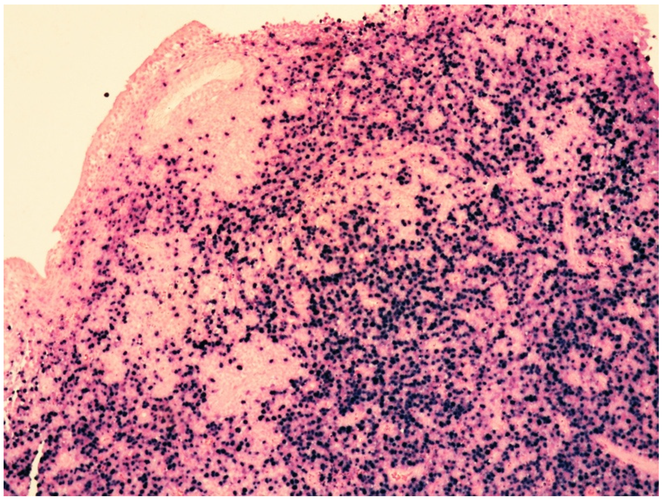
Extranodal NK/T-cell lymphoma (ENKTL) is an EBV-associated NK- or cytotoxic T-cell lymphoma occurring at extranodal sites [22,23,24,25,26,27,28].
The majority of cases (approximately 80%) are represented by what is called the nasal form, in which the sites of presentation are the nasal or upper aerodigestive tract; however, even in the nasal form, systemic involvement of other extranodal sites may occur at advanced stages of disease. Primary nodal involvement is extremely rare, whereas lymph nodes may be involved secondarily.
A minority of cases (approximately 20%) are represented by the non-nasal form, arising in extranodal, non-nasal sites [22,65,66].
ENKTL has a particular ethnic distribution, mainly affecting adult males from Asia and Latin America. EBV infection is strongly linked to ENKTL with virtually all cases positive for EBV. Hence, EBV-positivity is considered a diagnostic criterion, although it is not completely clear how the virus plays its oncogenic role; the overexpression of LMP1 protein, as well as the activation of the NF-kB signaling pathway, may be possible EBV-associated oncogenic mechanisms [26,27].
The most common clinical manifestations of the nasal form are nasal obstruction, epistaxis, and often destructive lesions involving the upper aerodigestive tract and midface.
As already mentioned, non-nasal sites such as skin, soft tissue, testis, salivary gland, liver, spleen, and gastrointestinal tract may be the primary sites of involvement, causing clinical manifestations varying according to the sites affected.
BM involvement may occur during the course of the disease, and HLH syndrome represents a serious complication often developing in cases with BM involvement [67]. Systemic symptoms such as fever and weight loss may be present.
Grossly, ENKTL usually causes an ulcerated mass.

6.2.2. Histology

The classic histological picture is characterized by a rich inflammatory background associated with necrosis sometimes overwhelming the neoplastic component and making difficult to distinguish ENKTL from an inflammatory condition.
The malignant lymphoid cells present as a diffuse proliferation with an angiocentric and angiodestructive pattern of growth and fibrinoid necrosis in the vessel wall.
Of note, when ENKTL involves skin or mucosal sites, the epithelium may show features of pseudoepitheliomatous hyperplasia, simulating squamous cell carcinoma [68].
The neoplastic elements are rather polymorphic and variable in size, although small- to medium-sized cells with scarce and clear cytoplasm, irregular nucleus, granular chromatin, and inconspicuous nucleolus are generally prevalent. Large-sized lymphoid cells may be present. Mitotic figures are frequent. (Figure 6).

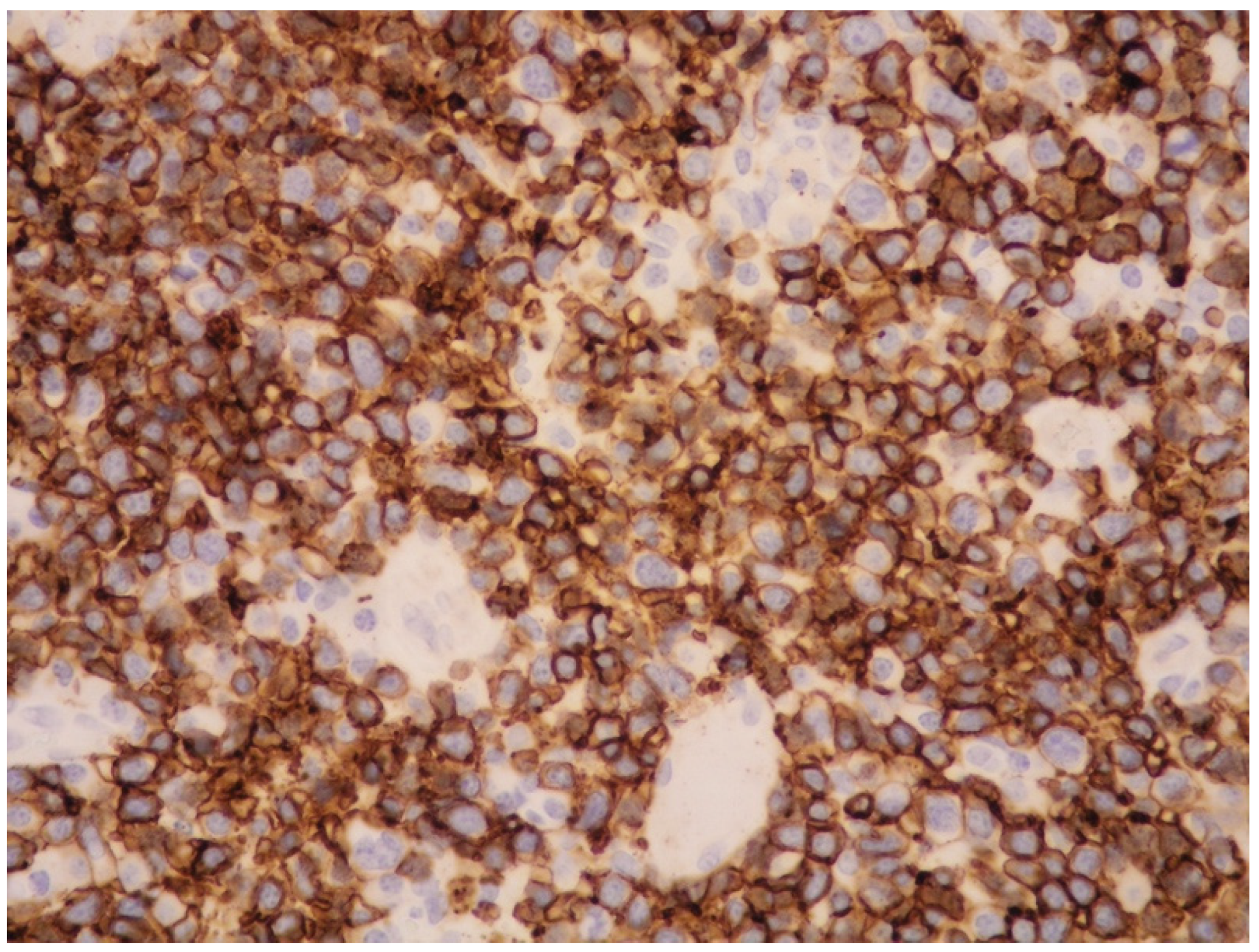
6.2.3. Immunophenotypic Features

The disease may be of NK- or T-cell nature [69], although it is more often of NK-cell origin with neoplastic elements positive for cytotoxic molecules (TIA1, perforin, and granzyme B), CD2, cCD3, CD56 (Figure 7).
CD7 and CD30 are rarely expressed, although, particularly in the non-nasal form, approximately 30% of neoplastic cells are positive for CD30 (Figure 8).
Surface CD3 (sCD3), CD4, CD8, CD5, CD16, and CD57 are usually negative, and the T-cell receptor gene (TCR) rearrangement is negative in the form of NK-cell origin.
A minority of cases (approximately 40%) belonging to the cytotoxic T-cell lineage are positive for sCD3, CD5, and CD8 and show T-cell clonality. Nuclear expression of megakaryocyte-associated tyrosine kinase (MATK) is frequent [69].
EBER is positive in the majority of viable neoplastic cells (Figure 9).
Type-II latency pattern with neoplastic cells often expressing LMP1, LMP2, and EBNA2 is often found.
CD56 is a useful, but not specific, marker for NK-cells, and most ENKTL are CD56-positive; however, CD56 can be negative in approximately 20% of cases, especially in ENKTL of T-cell origin [69]. To diagnose a CD56-negative lymphoma as ENKTL, the neoplasm must express a cytotoxic molecule and EBV.
As already mentioned, other NK-cell markers, such as CD16 or CD57, are commonly negative. As ENKTL of T and NK-cell origin may share the expression of some cell markers, the cell lineage cannot be distinguished only by immunophenotyping, but it is determining the identification or not of T-cell clonality. Rarely, the aberrant expression of B-cell markers such as CD20 may be found [70].
In ENKTL, PD-1 is rarely expressed, whereas PD-L1 is frequently positive [71]. PD-L1 is the ligand of the immune-checkpoint protein PD1 identified on T-lymphocytes. By binding to PD1 on T-lymphocytes and blocking their functions, PD-L1 favors the mechanism of immune evasion by neoplastic cells of ENKTL [72].
As previously mentioned, an association between EBV and the PD1/PD-L1 pathway has been noted in different neoplasms, and the overexpression of PD-L1 represents a mechanism of immune evasion in lymphoid cells infected by EBV [72].
In ENKTL, PD-L1 overexpression is possibly related to different factors. The EBV-related oncoprotein LMP1 is one of the mechanisms driving PD-L1 overexpression through the NF-kB pathway [72].
In ENKTL, both JAK/STAT and NF-kB are usually activated, and multiple recurrent genetic and epigenetic (hypermethylation) alterations have been identified [24,27,73,74,75,76]. The JAK/STAT pathway also contributes to PD-L1 overexpression by interacting with the interferon-stimulated response element in the promoter region of PD-L1 [72].

6.2.4. Genetic Features

ENKTL shows multiple genomic alterations. Deletion of 6q21-25 is the most identified abnormality, harboring several tumor suppressor genes (PRDM1, PTPRK, FOXO3, HACE1). PRDM1 is a tumor suppressor gene, often found to be inactivated by mutation, deletion, or hypermethylation, which plays an important role in ENKTL pathogenesis [77].
Mutations of tumor suppressor genes such as BCOR, DDX3X, and TP53 have also been identified [77].

6.2.5. Treatment

ENKTL is notoriously resistant to anthracycline-based chemotherapy, whereas the use of L-asparaginase-based chemotherapy combined with radiotherapy has improved the outcome for this aggressive lymphoma [78]. However, as chemotherapy is often unable to achieve long-term remissions in advanced stages of the disease, it is extremely important that novel therapies targeting different molecular pathways are currently available [79,80].
Despite still lacking a standardized expression cutoff for predicting the response to PD-1/PD-L1 inhibitors, several clinical trials are ongoing, and immune checkpoint inhibitor therapy is used in relapsed or refractory disease [81]. Due to the relevant role played by the activation of both JAK/STAT and NF-kB pathways in ENKTL pathogenesis, these pathways are potentially targetable for therapy [74,75,76,79,80]. Further studies are needed to evaluate the efficacy of the anti-CD30 antibody brentuximab vedotin, particularly in combination with chemotherapy, for the treatment of ENKTL [82].
In brief, the main clinicopathologic differences between IVNKTL and ENKTL are the sites of involvement, as the nasal and upper aerodigestive tract are the sites most involved in ENKTL, whereas IVNKTL frequently involves the skin and CNS with no nasal involvement; a further difference is the presence of mass lesions in ENKTL, whereas IVNKTL usually develops within vascular spaces in the absence of a clear-cut mass.

6.3. EBV-Positive Nodal T/NK-Cell Lymphoma

6.3.1. General Features and Clinical Manifestations

EBV-positive nodal T/NK-cell lymphoma is a primary nodal EBV-positive lymphoma that usually exhibits a cytotoxic T-cell phenotype or, more rarely, one of NK-cell nature. By definition, it is a systemic disease primarily affecting lymph nodes with the involvement of a limited number of extranodal sites and, in particular, lacking nasal involvement.
In the 2017 WHO classification, it was listed as an EBV-positive variant of peripheral T-cell lymphoma, not otherwise specified (PTCL, NOS), whereas in the 2022 International Consensus Classification of Mature Lymphoid Neoplasms (2022 ICC), as well as in the recent article by Alaggio et al., it is recognized as a new provisional entity distinct from PTCL, NOS [22,29,83,84,85].
Patients are mainly adults/elderly from East Asia, and the neoplasm may occur in the setting of immunosuppression or in association with autoimmune diseases. Patients present with multiple lymphadenopathy with or without extranodal involvement such as hepatosplenomegaly, B symptoms, and advanced stages. The outcome is poor.

6.3.2. Histology and Immunophenotypic Features

The morphological features are those of a high-grade lymphoma, either monomorphic or polymorphic. In the majority of cases (approximately more than 80%), this neoplasm is of T-cell lineage, based on T-cell clonality, with less than 20% of cases being of NK-cell origin [86,87].
The neoplastic cells are usually positive for T-cell markers and cytotoxic molecules. The typical phenotype is CD8-positive, CD56-negative, cytotoxic αβT cells. A minority of cases (0–13%) show γδT cells, whereas αβT cells are expressed in 40–60% of cases [87].
EBV positivity is a diagnostic criterion, and the virus plays an important pathogenetic role. Similarly, in ENKTL and other EBV-associated T/NK lymphoproliferative diseases, most cases of EBV-positive nodal T/NK-cell lymphoma have a type-II EBV latency pattern and EBNA1, LMP1, and LMP2A are expressed, although LMP1 protein is difficult to find [88,89].
Histologically, unlike ENKTL, this lymphoma usually shows a monomorphic pattern of growth, the absence of necrosis and angioinvasion, and infrequent CD56 expression.

6.3.3. Genetic Features

The genetic profile is different from that of ENKTL, with the most mutated gene being TET2 (64%) [89]. Other common mutated genes are PIK3CD (33%), DDX3 (20%), and STAT3 (19%).
Recently, this lymphoma has been found to be characterized by low genomic instability, upregulation of immune-related pathways favoring tumor evasion, and downregulation of EBV miRNA [89]. This rare type of lymphoma, despite its aggressive behavior, has been demonstrated to be more genomically stable than ENKTL and PTCL, NOS, supporting its consideration as a distinct entity [89]. The downregulation of EBV miRNA observed in EBV-positive nodal T/NK-cell lymphoma compared to ENKTL suggests that there are differences in the EBV biology between these diseases.
The main clinicopathological differences between EBV-positive nodal T/NK-cell lymphoma and IVNKTL are the sites of presentation, as IVNKTL mainly has an extra-nodal presentation and rarely occurs primarily in lymph nodes. In addition, in EBV-positive nodal T/NK-cell lymphoma, intravascular involvement is rare, cells are more often of T-cell origin, and CD56 is often negative.

6.4. Aggressive NK-Cell Leukemia (ANKL)

6.4.1. General Features and Clinical Manifestations

ANKL is a rare and aggressive leukemia, more prevalent in Asia, NK-cell in origin and characterized by a strong association with EBV, identified in about 90% of cases. EBV is considered to play an important role in the pathogenesis of ANKL and it is considered to be responsible of the aggressive nature of the disease.
Unlike IVNKTL, ENKTL, and EBV positive nodal T/NK-cell lymphoma, ANKL is a malignancy of young to middle-aged adults [90,91]. Particularly in young individuals, the disease may represent an aggressive evolution of chronic active EBV disease (CAEBV) [92]. Rare cases of EBV-negative ANKL have been described either developing de novo or deriving from NK-large granular lymphocyte leukemia (LGLL) [93,94].
Commonly involved sites are the BM, peripheral blood (PB), liver, and spleen, but any organ may be involved [90]. The number of leukemic cells in PB and BM smear is variable and may be low.
Patients present with fever, a leukemic blood picture, and symptoms of HLH syndrome. Hepatosplenomegaly and disseminated intravascular coagulation are frequently observed. Lymphadenopathy may be present, whereas skin involvement is rare. The lack of nasal involvement is a distinguishing feature from ENKTL. The disease usually follows a fulminant clinical course, although it is not clear whether the very rare EBV-negative cases have the same poor prognosis [94].

6.4.2. Histology and Immunophenotypic Features

Histologically, neoplastic elements vary from normal large granular lymphocytes (LGLs) to highly atypical cells, with large, irregular nuclei and evident nucleoli. The cells may have pale cytoplasm or contain azurophilic granules. Malignant cells can involve the bone marrow with an interstitial or sinusoidal pattern of growth, and the involvement can be either subtle or massive.
The immunophenotype is identical to that of ENKTL, except for CD16 expression, which is far more frequent (75%) in ANKL compared to ENKTL (22%).

6.4.3. Genetic Features

Some genetic abnormalities such as el(6)(q21q25), i(7)(q10) and 11q deletion have been found in ANKL [95]. Nakashima et al. found different genomic alteration patterns in ANKL and ENKTL, with loss in 6q being more frequent in ENKTL, unlike losses in 7p and 17p and gains in 1q more frequently present in ANKL [96]. ANKL has been found to harbor mutations in TP53 (34%), TET2 (28%), CREBBP (21%), and MLL2 genes (21%) [96]. Mutations in the JAK/STAT pathway have been detected in 48% of cases [97], and Huang et al. found that ANKL cells show increased expression of the transcription factor MYC, which is under the control of JAK and STAT genes [98].

6.4.4. Treatment

ANKL is a disease with a poor prognosis and is resistant to anthracycline-based chemotherapy. Treatments may consist of concurrent chemoradiation, sequential chemoradiation, or sandwich chemoradiation. The efficacy of L-asparaginase has led to its inclusion in different combination regimens. Treatments providing lower dose intensity of chemoradiation followed by ASCT have demonstrated some benefits. Relapse remains an issue in this disease, and novel treatments targeting molecular pathways such as the JAK/STAT pathway are currently under evaluation [97].
Regarding the relation between ANKL and IVNKTL, Alaggio et al. recently proposed that IVNKTL is more similar to ANKL than ENKTL in its intravascular presentation, however, the authors suggested that further studies are needed to better determine IVNKTL categorization [29].

7. Conclusions

IVNKTL is a rare lymphoma characterized by the exclusive growth of large cells within the lumen of small- and medium-sized vessels, involving mainly skin and CNS, although virtually any organ may be affected. It is strictly linked to EBV infection and needs an accurate differential diagnosis with other EBV-related NK/T-lymphomas. IVNKTL shows somatic mutations in epigenetic regulator genes. The high PD-L1 expression, probably due to EBV infection, suggests that checkpoint inhibitors may be effective treatments for this aggressive lymphoma.
Although WES analysis and transcriptome studies have provided new insight into molecular pathogenesis and possible treatment, the nature of IVNKTL is not completely elucidated. It has been recently postulated that IVNKTL may be more similar to ANKL than ENKTL.
Further studies on a larger number of cases with detailed immunophenotypic and molecular data are needed to clarify this issue.

Author Contributions

Conceptualization, M.Z. (Magda Zanelli); P.P. and S.A. methodology, M.Z. (Magda Zanelli), P.P. and S.A.; data curation, A.B.; A.C.; A.P.; D.T.; G.M.; F.S.; G.B.; I.C.; L.M.; L.R.; R.C.; M.Z. (Maurizio Zizzo) and S.P.; writing—original draft preparation, M.Z. (Magda Zanelli); writing—review and editing, M.Z. (Magda Zanelli); supervision, S.A. All authors have read and agreed to the published version of the manuscript.

Funding

This research received no external funding.

Acknowledgments

The study was partially supported by the Italian Ministry of Health—Ricerca Corrente Annual Program 2023.

Conflicts of Interest

The authors declare no conflict of interest.

References

  1. Santucci, M.; Pimpinelli, N.; Massi, D.; Kadin, M.E.; Meijer, C.J.L.M.; Muller-Hermelink, H.K.; Paulli, M.; Wechsler, J.; Willemze, R.; Audrig, H.; et al. EORTC cutaneous Lymphoma Task Force. Cytotoxic/natural killer cell cutaneous lymphomas. Report of EORTC cutaneous lymphoma Task Force workshop. Cancer 2003, 97, 610–627. [Google Scholar] [CrossRef] [PubMed]
  2. Wu, H.; Said, J.W.; Ames, E.D.; Chen, C.; McWorther, V.; Chen, P.; Ghali, V.; Pinkus, G.S. First reported cases of intravascular Large cell lymphoma of the NK cell type. Am. J. Clin. Pathol. 2005, 123, 603–611. [Google Scholar] [CrossRef] [PubMed]
  3. Kuo, T.-T.; Chen, M.-J.; Kuo, M.-C. Cutaneous intravascular NK-cell lymphoma: Report of a rare variant associated with Ep-stein-Barr virus. Am. J. Surg. Pathol. 2006, 30, 1197–1201. [Google Scholar] [CrossRef]
  4. Song, D.E.; Lee, M.-W.; Ryu, M.-H.; Kang, D.W.; Kim, S.J.; Huh, J. Intravascular Large Cell Lymphoma of the Natural Killer Cell Type. J. Clin. Oncol. 2007, 25, 1279–1282. [Google Scholar] [CrossRef] [PubMed]
  5. Nakamichi, N.; Fukuhara, S.; Aozasa, K.; Morii, E. NK-cell intravascular lymphomatosis—A mini-review. Eur. J. Haematol. 2008, 81, 1–7. [Google Scholar] [CrossRef] [PubMed]
  6. Gleason, B.C.; Brinster, N.K.; Granter, S.R.; Pinkus, G.S.; Lindeman, N.I.; Miller, D.M. Intravascular cytotoxic T-cell lymphoma: A case report and review of the literature. J. Am. Acad. Dermatol. 2008, 58, 290–294. [Google Scholar] [CrossRef]
  7. Cerroni, L.; Massoni, C.; Kutzner, H.; Mentzel, T.; Umbert, P.; Kerl, H. Intravascular large T-cell or NK-cell lymphoma: A rare variant of intravascular large cell lymphoma with frequent cytotoxic phenotype and association with Epstein-Barr virus infection. Am. J. Surg. Pathol. 2008, 32, 891–898. [Google Scholar] [CrossRef]
  8. Wu, C.; Liao, J.; Hsieh, P.; Hwang, Y.; Lin, S. Cutaneous Intravascular Natural Killer-cell Lymphoma: A Rare Case and Review of the Literature. Acta Derm. Venereol. 2011, 91, 472–473. [Google Scholar] [CrossRef] [Green Version]
  9. Yanning, X.; Chen, H.; Si, H.; Liu, Y.; Min, Z. Cutaneous intravascular NK-cell lymphoma. Eur. J. Dermatol. 2013, 23, 252–253. [Google Scholar] [CrossRef]
  10. Liu, Y.; Zhang, W.; An, J.; Li, H.; Liu, S. Cutaneous Intravascular Natural Killer–Cell Lymphoma. Am. J. Clin. Pathol. 2014, 142, 243–247. [Google Scholar] [CrossRef]
  11. Bi, Y.; Huo, Z.; Liang, Z.; Yunxiao, M.Y.; Jia, C.; Shi, X.; Song, L.; Luo, Y.; Ling, Q.; Liu, T. Intravascular NK-cell lymphoma: A case report and review of the literature. Diagn. Pathol. 2015, 10, 84. [Google Scholar] [CrossRef] [PubMed] [Green Version]
  12. Wang, L.; Chen, S.; Ma, H.; Shi, D.; Huang, C.; Lu, C.; Gao, T.; Wang, G. Intravascular NK/T-cell lymphoma: A report of five cases with cutaneous manifestation from China. J. Cutan. Pathol. 2015, 42, 610–617. [Google Scholar] [CrossRef] [PubMed]
  13. Sharma, T.L.; Yeaney, G.A.; Soltanzadeh, P.; Li, Y.; Cotta, C.V. Intravascular T-cell lymphoma: A rare, poorly characterized entity with cytotoxic phenotype. Neuropathology 2017, 37, 365–370. [Google Scholar] [CrossRef]
  14. Yan, J.; Zhang, F.; Luo, D.; Yao, S.; Chen, Y.; Xu, F.; Luo, X.; He, J.; Liu, Y. Intravascular NK/T-cell lymphoma: A series of four cases. Int. J. Clin. Exp. Pathol. 2017, 10, 9541–9550. [Google Scholar] [PubMed]
  15. Alegría-Landa, V.; Manzarbeitia, F.; Calderón, M.G.S.; Requena, L.; Rodríguez-Pinilla, S.M. Cutaneous intravascular natural killer/T cell lymphoma with peculiar immunophenotype. Histopathology 2017, 71, 994–1002. [Google Scholar] [CrossRef] [PubMed]
  16. Okonkwo, L.; Jaffe, E.S. Intravascular large cell lymphoma of NK/T-cell type, EBV positive. Blood 2017, 130, 837. [Google Scholar] [CrossRef] [Green Version]
  17. Elshiekh, M.; Naresh, K.N. A rare case of renal intravascular NK/T-cell lymphoma. Blood 2018, 132, 1354. [Google Scholar] [CrossRef] [Green Version]
  18. Zanelli, M.; Mengoli, M.C.; Del Sordo, R.; Cagini, A.; De Marco, L.; Simonetti, E.; Martino, G.; Zizzo, M.; Ascani, S. Intravascular NT/T-cell lymphoma, Epstein-Barr virus positive with multiorgan involvement: A clinical dilemma. BMC Cancer 2018, 18, 1115. [Google Scholar] [CrossRef]
  19. Fujikura, K.; Yamashita, D.; Sakamoto, R.; Ishikawa, T.; Chuang, S.-S.; Itoh, T.; Imai, Y. Intravascular NK/T-cell lymphoma: Clinicopathological and integrated molecular analysis of two cases provides a clue to disease pathogenesis. J. Clin. Pathol. 2019, 72, 642–646. [Google Scholar] [CrossRef]
  20. Wang, J.; Yang, X.; Song, Z.; You, Y. Cutaneous intravascular NK/T-cell lymphoma. Australas. J. Dermatol. 2019, 61, 61–63. [Google Scholar] [CrossRef]
  21. Fujikura, K.; Yoshida, M.; Uesaka, K. Transcriptome complexity in intravascular NK/T-cell lymphoma. J. Clin. Pathol. 2020, 73, 671–675. [Google Scholar] [CrossRef] [PubMed]
  22. WHO Classification of Tumours Editorial Board (Ed.) WHO Classification of Tumours Haematopoietic and Lymphoid Tissues, Revised 4th ed.; IARC: Lyon, France, 2017. [Google Scholar]
  23. Sánchez-Romero, C.; Bologna-Molina, R.; de Almeida, O.P.; Santos-Silva, A.R.; Prado-Ribeiro, A.C.; Brandão, T.B.; Carlos, R. Extranodal NK/T cell lymphoma, nasal type: An updated overview. Crit. Rev. Oncol. 2021, 159, 103237. [Google Scholar] [CrossRef] [PubMed]
  24. Huang, Y.; de Leval, L.; Gaulard, P. Molecular underpinning of extranodal NK/T-cell lymphoma. Best Pr. Res. Clin. Haematol. 2013, 26, 57–74. [Google Scholar] [CrossRef] [PubMed]
  25. Takata, K.; Hong, M.E.; Sitthinamsuwan, P.; Loong, F.; Tan, S.Y.; Liau, J.Y.; Hsieh, P.P.; Ng, S.B.; Yang, S.F.; Pongpruttipan, T.; et al. Primary cutaneous NK/T-cell lymphoma, nasal type and CD56-positive peripheral T-cell lymphoma: A cellular lineage and clinicopathologic study of 60 patients from Asia. Am. J. Surg. Pathol. 2015, 39, 1–12. [Google Scholar] [CrossRef]
  26. Syrykh, C.; Péricart, S.; Lamaison, C.; Escudié, F.; Brousset, P.; Laurent, C. Epstein–Barr Virus-Associated T- and NK-Cell Lymphoproliferative Diseases: A Review of Clinical and Pathological Features. Cancers 2021, 13, 3315. [Google Scholar] [CrossRef] [PubMed]
  27. Takada, H.; Imadome, K.I.; Shibayama, H.; Yoshimori, M.; Wang, L.; Saitoh, Y.; Uota, S.; Yamaoka, S.; Koyama, T.; Shimizu, N.; et al. EBV induces persistent NF-kB activation and contributes to survival of EBV-positive neoplastic T-or NK-cells. PLoS ONE 2017, 12, e0174136. [Google Scholar]
  28. Zanelli, M.; Sanguedolce, F.; Palicelli, A.; Zizzo, M.; Martino, G.; Caprera, C.; Fragliasso, V.; Soriano, A.; Gozzi, F.; Cimino, L.; et al. EBV-Driven Lymphoproliferative Disorders and Lymphomas of the Gastrointestinal Tract: A Spectrum of Entities with a Common Denominator (Part 3). Cancers 2021, 13, 6021. [Google Scholar] [CrossRef]
  29. Alaggio, R.; Amador, C.; Anagnostopoulos, I.; Ayoma, D.; Attygalle, A.D.; de Oliveira Araujo, I.B.; Borges, A.M.; Boyer, D.; Calaminici, M.; Chadburn, A.; et al. The 5th edition of the World Health Organization Classification of Haematolymphoid Tumours: Lymphoid Neoplasms. Leukemia 2022, 36, 1720–1748. [Google Scholar] [CrossRef]
  30. Taylor, G.S.; Long, H.M.; Brooks, J.M.; Rickinson, A.B.; Hislop, A.D. The Immunology of Epstein-Barr Virus–Induced Disease. Annu. Rev. Immunol. 2015, 33, 787–821. [Google Scholar] [CrossRef]
  31. Young, L.; Yap, L.-F.; Murray, P.G. Epstein–Barr virus: More than 50 years old and still providing surprises. Nat. Cancer 2016, 16, 789–802. [Google Scholar] [CrossRef]
  32. Rickinson, A. Co-infections, inflammation and oncogenesis: Future directions for EBV research. Semin. Cancer Biol. 2014, 26, 99–115. [Google Scholar] [CrossRef] [PubMed]
  33. Price, A.M.; Luftig, M.A. To Be or Not IIb: A Multi-Step Process for Epstein-Barr Virus Latency Establishment and Consequences for B Cell Tumorigenesis. PLOS Pathog. 2015, 11, e1004656. [Google Scholar] [CrossRef] [PubMed] [Green Version]
  34. Dojcinov, S.; Fend, F.; Quintanilla-Martinez, L. EBV-Positive Lymphoproliferations of B- T- and NK-Cell Derivation in Non-Immunocompromised Hosts. Pathogens 2018, 7, 28. [Google Scholar] [CrossRef] [Green Version]
  35. Fujiwara, S.; Kimura, H. Editorial: Epstein-Barr Virus-Associated T/NK-Cell Lymphoproliferative Diseases. Front. Pediatr. 2019, 7, 285. [Google Scholar] [CrossRef] [PubMed]
  36. Kim, W.Y.; Montes-Mojarro, I.A.; Fend, F.; Quintanilla-Martinez, L. Epstein-Barr virus-associated T and NK-cell lymphopro-liferative diseases. Front. Pediatr. 2019, 7, 71. [Google Scholar] [CrossRef] [PubMed] [Green Version]
  37. Kimura, H.; Fujiwara, S. Overview of EBV-Associated T/NK-Cell Lymphoproliferative Diseases. Front. Pediatr. 2019, 6, 417. [Google Scholar] [CrossRef]
  38. Kim, H.; Ko, Y.H. The Pathologic and Genetic Characteristics of Extranodal NK/T-Cell Lymphoma. Life 2022, 12, 73. [Google Scholar] [CrossRef] [PubMed]
  39. Zanelli, M.; Sanguedolce, F.; Palicelli, A.; Zizzo, M.; Martino, G.; Caprera, C.; Fragliasso, V.; Soriano, A.; Valle, L.; Ricci, S.; et al. EBV-driven lymphoproliferative disorders and lymphomas of the gastrointestinal tract: A spectrum of entities with a common denominator (Part 1). Cancers 2021, 13, 4578. [Google Scholar] [CrossRef]
  40. Zanelli, M.; Sanguedolce, F.; Palicelli, A.; Zizzo, M.; Martino, G.; Caprera, C.; Fragliasso, V.; Soriano, A.; Valle, L.; Ricci, S.; et al. EBV-driven lymphoproliferative disorders and lymphomas of the gastrointestinal tract: A spectrum of entities with a common denominator (Part 2). Cancers 2021, 13, 4527. [Google Scholar] [CrossRef]
  41. Patsoukis, N.; Wang, Q.; Strauss, L.; Boussiotis, V.A. Revisiting the PD-1 pathway. Sci. Adv. 2020, 6, eabd2712. [Google Scholar] [CrossRef]
  42. Lipson, E.J.; Forde, P.M.; Hammers, H.-J.; Emens, L.A.; Taube, J.M.; Topalian, S.L. Antagonists of PD-1 and PD-L1 in Cancer Treatment. Semin. Oncol. 2015, 42, 587–600. [Google Scholar] [CrossRef]
  43. Alsaab, H.O.; Sau, S.; Alzhrani, R.; Tatiparti, K.; Bhise, K.; Kashaw, S.K.; Iyer, A.K. PD-1 and PD-L1 chechpoint signaling inhibition for cancer immunotherapy: Mechanism, combinations and clinical outcome. Front. Pharmacol. 2017, 8, 561. [Google Scholar] [CrossRef] [PubMed] [Green Version]
  44. Palicelli, A.; Bonacini, M.; Croci, S.; Magi-Galluzzi, C.; Cañete-Portillo, S.; Chaux, A.; Bisagni, A.; Zanetti, E.; De Biase, D.; Melli, B.; et al. What Do We Have to Know about PD-L1 Expression in Prostate Cancer? A Systematic Literature Review. Part 1: Focus on Immunohistochemical Results with Discussion of Pre-Analytical and Interpretation Variables. Cells 2021, 10, 3166. [Google Scholar] [CrossRef]
  45. Ansell, S.M.; Lesokhin, A.M.; Borrello, I.; Halwani, A.; Scott, E.C.; Gutierrez, M.; Schuster, S.J.; Millenson, M.M.; Cattry, D.; Freeman, G.J.; et al. PD-1 Blockade with Nivolumab in Relapsed or Refractory Hodgkin’s Lymphoma. N. Engl. J. Med. 2015, 372, 311–319. [Google Scholar] [CrossRef] [PubMed] [Green Version]
  46. Lesokhin, A.M.; Ansell, S.M.; Armand, P.; Scott, E.C.; Halwani, A.; Gutierrez, M.; Millenson, M.M.; Cohen, A.D.; Schuster, S.J.; Lebovic, D.; et al. Nivolumab in patients with repalsed or refractory hematologic malignancy: Preliminary results of a phase Ib study. J. Clin. Oncol. 2016, 34, 2698–2704. [Google Scholar] [CrossRef] [PubMed] [Green Version]
  47. Satou, A.; Nakamura, S. EBV-positive B-cell lymphomas and lymphoproliferative disorders: Review from the perspective of immune escape and immunodeficiency. Cancer Med. 2021, 10, 6777–6785. [Google Scholar] [CrossRef]
  48. Ferreri, A.J.; Dognini, G.P.; Campo, E.; Willemze, R.; Seymour, J.F.; Bairey, O.; Martelli, M.; De Renz, A.O.; Doglioni, C.; Montalban, C.; et al. Variations in clinical presentation, frequency of hemophagocytosis and clinical behavior of in-travascular lymphoma diagnosed in different geographical regions. Haematologica 2007, 92, 486–492. [Google Scholar] [CrossRef] [Green Version]
  49. Pastor, W.A.; Aravind, L.; Rao, A. TETonic shift: Biological roles of TET proteins in DNA demethylation and transcription. Nat. Rev. Mol. Cell Biol. 2013, 14, 341–356. [Google Scholar] [CrossRef] [Green Version]
  50. Bi, X.-W.; Wang, H.; Zhang, W.-W.; Wang, J.-H.; Liu, W.-J.; Xia, Z.-J.; Huang, H.-Q.; Jiang, W.-Q.; Zhang, Y.-J.; Wang, L. PD-L1 is upregulated by EBV-driven LMP1 through NF-κB pathway and correlates with poor prognosis in natural killer/T-cell lym-phoma. J. Hematol. Oncol. 2016, 9, 3432–3433. [Google Scholar] [CrossRef] [Green Version]
  51. Scheel, A.H.; Baenfer, G.; Baretton, G.; Dietel, M.; Diezko, R.; Henkel, T.; Heukamp, L.C.; Jasani, B.; Johrens, K.; Kirchner, T.; et al. Interlaboratory concordance of PD- L1 immunohistochemistry for non-small-cell lung cancer. Histopathology 2018, 72, 449–459. [Google Scholar] [CrossRef]
  52. Batenchuk, C.; Albitar, M.; Zerba, K.; Sudarsanam, S.; Chizhevsky, V.; Jin, C.; Burns, V. A real-world, comparative study of FDA-approved diagnostic assays PD-L1 IHC 28-8 and 22C3 in lung cancer and other malignancies. J. Clin. Pathol. 2018, 71, 1078–1083. [Google Scholar] [CrossRef] [PubMed]
  53. Menguy, S.; Prochazkova-Carlotti, M.; Beylot-Barry, M.; Saltel, F.; Vergier, B.; Merlio, J.-P.; Pham-Ledard, A. PD-L1 and PD-L2 Are Differentially Expressed by Macrophages or Tumor Cells in Primary Cutaneous Diffuse Large B-Cell Lymphoma, Leg Type. Am. J. Surg. Pathol. 2018, 42, 326–334. [Google Scholar] [CrossRef] [PubMed]
  54. Mitteldorf, C.; Berisha, A.; Pfaltz, M.C.; Broekaert, S.M.; Schön, M.P.; Kerl, K.; Kempf, W. Tumor Microenvironment and Checkpoint Molecules in Primary Cutaneous Diffuse Large B-Cell Lymphoma—New Therapeutic Targets. Am. J. Surg. Pathol. 2017, 41, 998–1004. [Google Scholar] [CrossRef] [Green Version]
  55. Sakakibara, A.; Kohno, K.; E Eladl, A.; Klaisuwan, T.; Ishikawa, E.; Suzuki, Y.; Shimada, S.; Nakaguro, M.; Shimoyama, Y.; Takahara, T.; et al. Immunohistochemical assessment of the diagnostic utility of PD-L1: A preliminary analysis of anti-PD-L1 antibody (SP142) for lymphoproliferative diseases with tumour and non-malignant Hodgkin-Reed-Sternberg (HRS)-like cells. Histopathology 2018, 72, 1156–1163. [Google Scholar] [CrossRef]
  56. Harrow, J.; Frankish, A.; Gonzalez, J.M.; Tapanari, E.; Diekhans, M.; Kokocinski, F.; Aken, B.L.; Barrell, D.; Zadissa, A.; Searle, S.; et al. GENCODE: The reference human genome annotation for The ENCODE Project. Genome Res. 2012, 22, 1760–1774. [Google Scholar] [CrossRef] [Green Version]
  57. Lee, Y.; Rio, D.C. Mechanisms and regulation of alternative pre-mRNA splicing. Annu. Rev. Biochem. 2015, 84, 291–323. [Google Scholar] [CrossRef] [PubMed] [Green Version]
  58. Venables, J.P. Aberrant and Alternative Splicing in Cancer. Cancer Res. 2004, 64, 7647–7654. [Google Scholar] [CrossRef] [Green Version]
  59. Anczuków, O.; Krainer, A.R. Splicing-factor alterations in cancers. RNA 2016, 22, 1285–1301. [Google Scholar] [CrossRef] [Green Version]
  60. Clemente-González, H.; Porta-Pardo, E.; Godzik, A.; Eyra, E. The functional impact of alternative splicing in cancer. Cell. Rep. 2017, 20, 2215–2226. [Google Scholar] [CrossRef] [Green Version]
  61. Koedoot, E.; Smid, M.; Foekens, J.A.; Martens, J.W.M.; Le Dévédec, S.E.; Van De Water, B. Co-regulated gene expression of splicing factors as drivers of cancer progression. Sci. Rep. 2019, 9, 5484. [Google Scholar] [CrossRef] [Green Version]
  62. Escobar-Hoyos, L.; Knorr, K.; Abdel-Wahab, O. Aberrant RNA Splicing in Cancer. Annu. Rev. Cancer Biol. 2019, 3, 167–185. [Google Scholar] [CrossRef] [PubMed]
  63. Rodrigues-Peres, R.M.; de S Carvalho, B.; Anurag, M.; Lei, J.T.; Conz, L.; Goncalves, R.; Filho, C.C.; Ramalho, S.; de Paiva, G.R.; Derchain, S.F.M.; et al. Copy number alterations associated with clinical features in an underrepresented population with breast cancer. Mol. Genet. Genomic.Med. 2019, 7, e00750. [Google Scholar] [CrossRef] [PubMed] [Green Version]
  64. Foy, A.; McMullin, M.F. Somatic SF3B1 mutations in myelodysplastic syndrome with ring sideroblasts and chronic lympho-cytic leukaemia. J. Clin. Pathol. 2019, 72, 778–782. [Google Scholar] [CrossRef] [PubMed]
  65. Kim, T.M.; Lee, S.Y.; Jeon, Y.K.; Ryoo, B.Y.; Cho, G.J.; Hong, Y.S.; Kim, H.J.; Kim, S.Y.; Kim, C.S.; Kim, S.; et al. Lymphoma Subcommittee of the Korean Cancer Study Group. Clinical heterogeneity of extranodal NK/T-cell lymphoma, nasal type: A national survey of the Korean Cancer Study Group. Ann. Oncol. 2008, 19, 1477–1484. [Google Scholar] [CrossRef]
  66. Au, W.-Y.; Weisenburger, D.D.; Intragumtornchai, T.; Nakamura, S.; Kim, W.-S.; Sng, I.; Vose, J.; Armitage, J.O.; Liang, R. Clinical differences between nasal and extranasal natural killer/T-cell lymphoma: A study of 136 cases from the International Peripheral T-Cell Lymphoma Project. Blood 2009, 113, 3931–3937. [Google Scholar] [CrossRef] [Green Version]
  67. Li, N.; Jiang, M.; Wu, W.-C.; Wei, W.-W.; Zou, L.-Q. How to Identify Patients at High Risk of Developing Nasal-Type, Extranodal Nature Killer/T-Cell Lymphoma-Associated Hemophagocytic Syndrome. Front. Oncol. 2021, 11, 704962. [Google Scholar] [CrossRef]
  68. Ling, Y.-H.; Zhu, C.-M.; Wen, S.-H.; Luo, R.-Z.; Li, P.; Cao, Y.; Rao, H.-L.; Lin, S.-X.; Cai, M.-Y. Pseudoepitheliomatous hyperplasia mimicking invasive squamous cell carcinoma in extranodal natural killer/T-cell lymphoma: A report of 34 cases. Histopathology 2015, 67, 404–409. [Google Scholar] [CrossRef]
  69. Pongpruttipan, T.; Sukpanichnant, S.; Assanasen, T.; Wannakrairot, P.; Boonsakan, P.; Kanoksil, W.; Kayasut, K.; Mitarnun, W.; Khuhapinant, A.; Bunworasate, U.; et al. Extranodal NK/T-cell lymphoma, nasal type, includes cases of natural killer cell and αβ, γδ, and αβ/γδ T-cell origin: A comprehensive clinicopathologic and phenotypic study. Am. J. Surg. Pathol. 2012, 6, 481–499. [Google Scholar] [CrossRef]
  70. Huang, Y.; Chen, S.; Wei, R.; Guo, X.; Yang, X.; Cao, Q.; Yang, Y.; Yun, J. CD20-positive extranodal NK/T cell lymphoma: Clinicopathologic and prognostic features. Virchows Arch. 2020, 477, 873–883. [Google Scholar] [CrossRef]
  71. Jo, J.-C.; Kim, M.; Choi, Y.; Kim, H.-J.; Kim, J.E.; Chae, S.W.; Kim, H.; Cha, H.J. Expression of programmed cell death 1 and programmed cell death ligand 1 in extranodal NK/T-cell lymphoma, nasal type. Ann. Hematol. 2016, 96, 25–31. [Google Scholar] [CrossRef]
  72. Tse, E.; Kwong, Y.-L. Recent Advances in the Diagnosis and Treatment of Natural Killer Cell Malignancies. Cancers 2022, 14, 597. [Google Scholar] [CrossRef]
  73. Xiong, J.; Zhao, W.-L. Advances in multiple omics of natural-killer/T cell lymphoma. J. Hematol. Oncol. 2018, 11, 134. [Google Scholar] [CrossRef]
  74. Coppo, P.; Gouilleux-Gruart, V.; Huang, Y.; Bouhlal, H.; Bouamar, H.; Bouchet, S.; Perrot, C.; Vieillard, V.; Dartigues, P.; Gaulard, P.; et al. STAT3 transcription factor is constitutively activated and is oncogenic in nasal-type NK/T-cell lymphoma. Leukemia 2009, 23, 1667–1678. [Google Scholar] [CrossRef] [PubMed] [Green Version]
  75. Huang, Y.; De Reyniès, A.; de Leval, L.; Ghazi, B.; Martin-Garcia, N.; Travert, M.; Bosq, J.; Brière, J.; Petit, B.; Thomas, E.; et al. Gene expression profiling identifies emerging oncogenic pathways operating in extranodal NK/T-cell lymphoma, nasal type. Blood 2010, 115, 1226–1237. [Google Scholar] [CrossRef] [PubMed]
  76. Nairismägi, M.; Gerritsen, M.E.; Li, Z.M.; Wijaya, G.C.; Chia, B.K.H.; Laurensia, Y.; Lim, J.Q.; Yeoh, K.W.; Yao, X.S.; Pang, W.L.; et al. Oncogenic activation of JAK3-STAT signaling confers clinical sensitivity to PRN371, a novel selective and potent JAK3 inhibitor, in natural killer/T-cell lymphoma. Leukemia 2018, 32, 1147–1156. [Google Scholar] [CrossRef] [PubMed]
  77. Dong, G.; Li, Y.; Lee, L.; Liu, X.; Shi, Y.; Liu, X.; Bouska, A.; Gong, Q.; Kong, L.; Wang, J.; et al. Genetic manipulation of primary human natural killer cells to investigate the functional and oncogenic roles of PRDM1. Haematologica 2020, 106, 2427–2438. [Google Scholar] [CrossRef]
  78. Kwong, Y.L.; Kim, W.S.; Lim, S.T.; Kim, S.J.; Tang, T.; Tse, E.; Leung, A.Y.; Chim, C.S. SMILE for natural killer/T-cell lymphoma: Analysis of safety and efficacy from the Asia Lymphoma Study Group. Blood 2012, 120, 2973–2980. [Google Scholar] [CrossRef] [Green Version]
  79. Sun, K.-H.; Wong, Y.-T.; Cheung, K.-M.; Yuen, C.; Chan, Y.-T.; Lai, W.-Y.; Chao, C.; Fan, W.-S.; Chow, Y.-K.; Law, M.-F.; et al. Update on Molecular Diagnosis in Extranodal NK/T-Cell Lymphoma and Its Role in the Era of Personalized Medicine. Diagnostics 2022, 12, 409. [Google Scholar] [CrossRef]
  80. Tse, E.; Zhao, W.-L.; Xiong, J.; Kwong, Y.-L. How we treat NK/T-cell lymphomas. J. Hematol. Oncol. 2022, 15, 74. [Google Scholar] [CrossRef]
  81. Kwong, Y.-L.; Chan, T.S.Y.; Tan, D.; Kim, S.J.; Poon, L.-M.; Mow, B.; Khong, P.-L.; Loong, F.; Au-Yeung, R.; Iqbal, J.; et al. PD1 blockade with pembrolizumab is highly effective in relapsed or refractory NK/T-cell lymphoma failing l-asparaginase. Blood 2017, 129, 2437–2442. [Google Scholar] [CrossRef] [Green Version]
  82. Kim, S.J.; Yoon, D.H.; Kim, J.S.; Kang, H.J.; Lee, H.W.; Eom, H.S.; Hong, J.Y.; Cho, J.; Ko, Y.H.; Huh, J. Efficay of Brentuximab Vedotin in relapsed or refractory high-CD30-expressing non-Hodgkin lymphomas: Results of a multicenter, open-labeled Phase II trial. Cancer Res. Treat. 2020, 52, 374–387. [Google Scholar] [CrossRef] [PubMed]
  83. Campo, E.; Jaffe, E.S.; Cook, J.R.; Quintanilla-Martinez, L.; Swerdlow, S.H.; Anderson, K.C.; Brousset, P.; Cerroni, L.; de Leval, L.; Dirnhofer, S.; et al. The International Consensus Classification of Mature Lymphoid Neoplasms: A report from the Clinical Advisory Committee. Blood 2022, 140, 1229–1253. [Google Scholar] [CrossRef] [PubMed]
  84. Feldman, A.L.; Laurent, C.; Narbaitz, M.; Nakamura, S.; Chan, W.C.; de Leval, L.; Gaulard, P. Classification and diagnostic evaluation of nodal T- and NK-cell lymphomas. Virchows Arch. 2022, 1–15. [Google Scholar] [CrossRef] [PubMed]
  85. Quintanilla-Martinez, L.; Swerdlow, S.H.; Tousseyn, T.; Barrionuevo, C.; Nakamura, S.; Jaffe, E.S. New concepts in EBV-associated B, T, and NK cell lymphoproliferative disorders. Virchows Arch. 2022, 1–18. [Google Scholar] [CrossRef]
  86. Jeon, Y.K.; Kim, J.-H.; Sung, J.-Y.; Han, J.H.; Ko, Y.-H. Epstein-Barr virus–positive nodal T/NK-cell lymphoma: An analysis of 15 cases with distinct clinicopathological features. Hum. Pathol. 2015, 46, 981–990. [Google Scholar] [CrossRef]
  87. Kato, S.; Asano, N.; Miyata-Takata, T.; Takata, K.; Elsayed, A.A.; Satou, A.; Takahashi, E.; Kinoshita, T.; Nakamura, S. T-cell receptor (TCR) phenotype of nodal Epstein-Barr virus (EBV)-positive cytotoxic T-cell lymphoma (CTL): A clinicopathologic study of 39 cases. Am. J. Surg. Pathol. 2015, 39, 462–471. [Google Scholar] [CrossRef]
  88. Takahashi, E.; Asano, N.; Li, C.; Tanaka, T.; Shimada, K.; Shimada, S.; Yoshino, T.; Kojima, M.; Hara, K.; Eimoto, T.; et al. Nodal T/NK-cell lymphoma of nasal type: A clinicopathological study of six cases. Histopathology 2008, 52, 585–596. [Google Scholar] [CrossRef]
  89. Wai, C.M.M.; Chen, S.; Phyu, T.; Fan, S.; Leong, S.M.; Zheng, W.; Low, L.C.Y.; Choo, S.N.; Lee, C.K.; Chung, T.H.; et al. Immune pathway upregulation and lower genomic instability distinguish EBV-positive nodal T/NK-cell lymphoma from ENKTL and PTCL-NOS. Haematologica 2022, 107, 1864–1879. [Google Scholar] [CrossRef]
  90. Suzuki, R.; Suzumiya, J.; Nakamura, S.; Aoki, S.; Notoya, A.; Ozaki, S.; Gondo, H.; Hino, N.; Mori, H.; Sugimori, H.; et al. Aggressive natural killer-cell leukemia revisited: Large granular lymphocyte leukemia of cytotoxic NK cells. Leukemia 2004, 18, 763–770. [Google Scholar] [CrossRef] [Green Version]
  91. Tang, Y.-T.; Wang, D.; Luo, H.; Xiao, M.; Zhou, H.-S.; Liu, D.; Ling, S.-P.; Wang, N.; Hu, X.-L.; Luo, Y.; et al. Aggressive NK-cell leukemia: Clinical subtypes, molecular features, and treatment outcomes. Blood Cancer J. 2017, 7, 660. [Google Scholar] [CrossRef] [Green Version]
  92. Ishihara, S.; Okada, S.; Wakiguchi, H.; Kurashige, T.; Hirai, K.; Kawa-Ha, K. Clonal lymphoproliferation following chronic active Epstein-Barr virus infection and hypersensitivity to mosquito bites. Am. J. Hematol. 1997, 54, 276–281. [Google Scholar] [CrossRef]
  93. Ko, Y.H.; Park, S.; Kim, K.; Kim, S.J.; Kim, W.S. Aggressive natural killer cell leukemia: Is Epstein-Barr virus negativity an indicator of a favorable prognosis? Acta Haematol. 2008, 120, 199–206. [Google Scholar] [CrossRef] [PubMed]
  94. Nicolae, A.; Ganapathi, K.A.; Pham, T.H.; Xi, L.; Torres-Cabala, C.A.; Nanaji, N.M.; Zha, H.D.; Fan, Z.; Irwin, S.; Pittaluga, S.; et al. EBV-negative Aggressive NK-cell Leukemia/Lymphoma: Clinical, Pathologic, and Genetic Features. Am. J. Surg. Pathol. 2017, 41, 67–74. [Google Scholar] [CrossRef] [PubMed] [Green Version]
  95. Ryder, J.; Wang, X.; Bao, L.; Gross, S.A.; Hua, F.; Irons, R.D.; Ironsa, R. Aggressive Natural Killer Cell Leukemia: Report of a Chinese Series and Review of the Literature. Int. J. Hematol. 2007, 85, 18–25. [Google Scholar] [CrossRef]
  96. Nakashima, Y.; Tagawa, H.; Suzuki, R.; Karnan, S.; Karube, K.; Ohshima, K.; Muta, K.; Nawata, H.; Morishima, Y.; Nakamura, S.; et al. Genome-wide array-based comparative genomic hybridization of natural killer cell lymphoma/leukemia: Different genomic alteration patterns of aggressive NK-cell leukemia and extranodal Nk/T-cell lymphoma, nasal type. Genes Chromosom. Cancer 2005, 44, 247–255. [Google Scholar] [CrossRef]
  97. Sumbly, V.; Vest, M.; Landry, I. Aggressive Natural Killer Cell Leukemia: A Brief Overview of Its Genomic Landscape, Histological Features, and Current Management. Cureus 2022, 14, e22537. [Google Scholar] [CrossRef] [PubMed]
  98. Huang, L.; Liu, D.; Wang, N.; Ling, S.; Tang, Y.; Wu, J.; Hao, L.; Luo, H.; Hu, X.; Sheng, L.; et al. Integrated genomic analysis identifies deregulated JAK/STAT-MYC-biosynthesis axis in aggressive NK-cell leukemia. Cell Res. 2017, 28, 172–186. [Google Scholar] [CrossRef]
Figure 1. High-power view showing kidney parenchyma with large, atypical cells growing within vascular spaces (see within red circle; hematoxylin and eosin, 200× magnification, previously unpublished, original image from S.A.).
Cancers 14 05458 g001
Figure 2. High-power view showing central nervous system parenchyma with atypical cells within a vascular space (hematoxylin and eosin, 400× magnification, previously unpublished, original image from S.A.).
Cancers 14 05458 g002
Figure 3. Medium-power view showing bone marrow with CD3-positive atypical cells within sinusoidal spaces (CD3 immunostaining, 100× magnification, previously unpublished, original image from S.A.).
Cancers 14 05458 g003
Figure 4. High-power view showing lung parenchyma with perforin-positive atypical cells within vascular spaces (perforin immunostaining, 200× magnification, previously unpublished, original image from S.A.).
Cancers 14 05458 g004
Figure 5. High-power view showing lung parenchyma with EBER-positive atypical cells within vascular spaces (in situ hybridization for EBV-encoded RNA, 400× magnification, previously unpublished, original image from S.A.).
Cancers 14 05458 g005
Figure 6. High-power view showing a diffuse and polymorphic proliferation of atypical, medium- to large-sized lymphoid cells (hematoxylin and eosin, 200× magnification, previously unpublished, original image from S.A.).
Cancers 14 05458 g006
Figure 7. High-power view showing a diffuse proliferation of CD56-positive atypical cells (CD56 immunostaining, 200× magnification, previously unpublished, original image from S.A.).
Cancers 14 05458 g007
Figure 8. High-power view showing CD30-positive atypical cells (CD30 immunostaining, 400× magnification, previously unpublished, original image from S.A.).
Cancers 14 05458 g008
Figure 9. Medium-power view showing a diffuse proliferation of EBER-positive cells (in situ hybridization for EBV-encoded RNA, 100× magnification, previously unpublished, original image from S.A.).
Cancers 14 05458 g009
Table 1. Differential diagnoses of IVNKTL.
IVNKTLENKTLANKLEBV + Nodal T/NK Lymphoma
Median Age and SexAdult (middle-age, around 46 yrs) M > FAdults (elderly, around 60 yrs) M > FAdult (mainly young adult) M = FAdult (middle-age, often immunocompromised) M = F
EthnicityAsian and Latin AmericaAsianAsianAsian
SitesSkin and CNS (mainly); any organ may be involvedExtranodal sites (mainly nasal region and upper aerodigestive tract), very rare primary nodal sitesBM, blood, liver, spleen; very rare nodal presentationCommonly nodal presentation
Nasal involvementNoVery commonNoNo
Systemic symptomsProtean clinical manifestationsB-symptomsB-symptoms; often CID B-symptoms
Tumor massNoYes, necrotic/ulcerated massNoYes
HistologyLarge-sized cells within medium- to small-sized vesselsPolymorphic cells, clear cytoplasm; inflammatory background, necrosis. Angioinvasive and angiodestructive pattern. Variable in size from normal granular lymphocytes to large, atypical cellsHigh-grade morphology (either polymorphic or monomorphic)
Cell of originNK or cytotoxic T cellsNK > cytotoxic T cellNKCytotoxic T-cell > NK
EBV-associationCommonCommonCommonCommon
CD56 expressionOften positiveOften positiveOften positive (p16 generally positive)Often negative
T-cell clonalityPresent in cases of T-cell originPresent in cases of T-cell originNo Present in cases of T-cell origin
PrognosisPoorPoorPoorPoor
Legends: ANKL: aggressive NK leukemia; BM: bone marrow; CID: coagulation intravascular disseminated; CNS: central nervous system EBV: Epstein–Barr virus; ENKTL: extranodal NK/T-cell lymphoma; F: female; IVNKTL: intravascular NK/T-cell lymphoma; M: male; yrs: years.
Publisher’s Note: MDPI stays neutral with regard to jurisdictional claims in published maps and institutional affiliations.

