Review

Electromotive Enhanced Drug Administration in Oncology: Principles, Evidence, Current and Emerging Applications

1
Department of GI Surgery, Ghent University Hospital, 9000 Ghent, Belgium
2
Department of Electromechanical, Systems and Metal Engineering, Faculty of Engineering and Architecture, Ghent University, 9000 Ghent, Belgium
3
Cancer Research Institute Ghent (CRIG), 9000 Ghent, Belgium
4
Department of Chemistry, Faculty of Sciences, Ghent University, 9000 Ghent, Belgium
*
Author to whom correspondence should be addressed.
Cancers 2022, 14(20), 4980; https://doi.org/10.3390/cancers14204980
Submission received: 4 August 2022 / Revised: 5 October 2022 / Accepted: 8 October 2022 / Published: 11 October 2022
(This article belongs to the Section Cancer Therapy)

Abstract

:

Simple Summary

Since the 17th century, the use of electric currents to improve the transport of drugs into human tissues has been described. Currently, electrically driven drug transport is used in a variety of medicinal fields such as the urinary system, skin, eye and others. In this review, we summarize the principles and factors that govern electrically driven drug transport and discuss its current and emerging applications for the treatment of cancer patients.

Abstract

Local-regional administration of cytotoxic drugs is an important adjunct to systemic chemotherapy amongst cancer patients. It allows for targeted delivery of agents at high concentration to target sites while minimizing systemic side effects. Despite the pharmacokinetic advantages of the local–regional approach, drug transport into tumor nodules remains limited due to the biophysical properties of these tissues. Electromotive enhanced drug administration (EMDA) represents a potential solution to overcome challenges in local drug transport by applying electric currents. Through electrokinetic phenomena of electromigration, electroosmosis and electroporation, electric currents have been shown to improve drug penetration and distribution in a wide variety of clinical applications. Amongst patients with non-muscular invasive bladder cancer (NMIBC) and basal and squamous cell skin cancers, EMDA has been successfully adopted and proven efficacious in several pre-clinical and clinical studies. Its application in ophthalmological and other conditions has also been explored. This review provides an overview of the underlying principles and factors that govern EMDA and discusses its application in cancer patients. We also discuss novel EMDA approaches in pre-clinical studies and explore future opportunities of developments in this field.

1. Introduction

The ideal drug delivery model is safe, convenient, site-specific and can maximize therapeutic efficacy while ensuring minimal toxicity to unintended sites [1]. As such, a local–regional approach is logical and has pharmacokinetic (PK) advantages when compared with systemic drug administration [2]. This approach has been most widely adopted in bladder and peritoneal cancers, where intra-vesical and intra-peritoneal (IP) delivery of cytotoxic chemotherapeutic agents have been shown to have superior anti-tumoral effects [3,4,5]. However, both delivery modes rely heavily on passive diffusion for the transport of drugs into target tissues—a process hindered by the relative impermeability of both urothelial and peritoneal membranes [6,7,8]. Furthermore, the penetration of drugs into tumor nodules with elevated interstitial fluid pressures (IFP) represents an additional barrier to efficient drug transport [9].
Recognizing the challenges in local–regional drug delivery, the use of electric forces to enhance drug penetration has gained increasing popularity in the recent decade [10,11]. First described by Veratti in 1747, the concept of electricity-enhanced drug transport is not new [12]. In the early 20th century, Leduc, through a classical experiment using two rabbits connected in an electrical series circuit with strychnine sulfate and potassium cyanide solutions, proved that ionized drugs could penetrate the skin and exert a systemic effect. He also demonstrated the importance of polarity with respect to an ionized drug and its counter ion [13]. This phenomenon was termed iontophoresis, describing an accelerated delivery of charged ions into tissues when an electric current was applied through a drug solution [14,15,16].
By the 1930s, iontophoresis was frequently adopted in ophthalmologic and skin conditions and was found to be highly effective in the treatment of hyperhidrosis [17,18]. It was also used to enhance the effects of local anesthesia to the tympanic membrane, oral mucosa, skin and the eye [19]. The term electromotive drug administration (EMDA) was subsequently coined in 1994, recognizing electroporation as a secondary force at play with iontophoresis that resulted in an increased penetration of both ionized and unionized drugs into surrounding tissues [20,21,22]. Since then, EMDA has been frequently used to describe electricity-enhanced intra-vesical administration of cytotoxic and anesthetic agents for urological conditions.
In this review, we summarize the underlying principles and factors influencing EMDA-enhanced drug delivery. In addition, we examine pre-clinical and clinical data in its application in a diverse group of oncological conditions, ranging from urological, skin, ophthalmology and others, emerging technologies and explore future opportunities for development in this field.

2. Fundamental Principles in EMDA

EMDA refers to the use of a low intensity electrical current to drive drugs across various tissues. There are three main electrokinetic phenomena that govern EMDA: electromigration, electroosmosis and electroporation (Figure 1) [21,22,23]. The summation of electro-migratory and electro-osmotic forces is termed iontophoresis and describes the mechanism in which the delivery of ionized drugs across biological membranes is enhanced through the application of a mild electric current in an electrolyte drug solution [23]. In electromigration, the repulsion of cations by the anode and anions by the cathode result in ionic fluxes across tissues to maintain electrical neutrality. This is coupled with electroosmosis, where solvent flows in an anode to cathode direction, providing a secondary driving force in the transport of cationic drugs [24]. This convective solvent flow also represents the main mechanism by which unionized drug compounds are transported across membranes [21]. The two main electrokinetic phenomena (electromigration and electroosmosis) in EMDA-mediated drug transport can be described by the modified Nernst–Planck equation (File S1 in Supplementary Materials) [25,26]. Finally, electroporation describes the formation of aqueous pores by the application of an electric current, thereby increasing the permeability of membranes and facilitating drug delivery [22,27]. This should be differentiated from “high voltage electroporation” techniques where voltages of >100 volts over very short durations (microseconds to milliseconds) are used to permeabilize the skin [28].

2.1. EMDA Devices

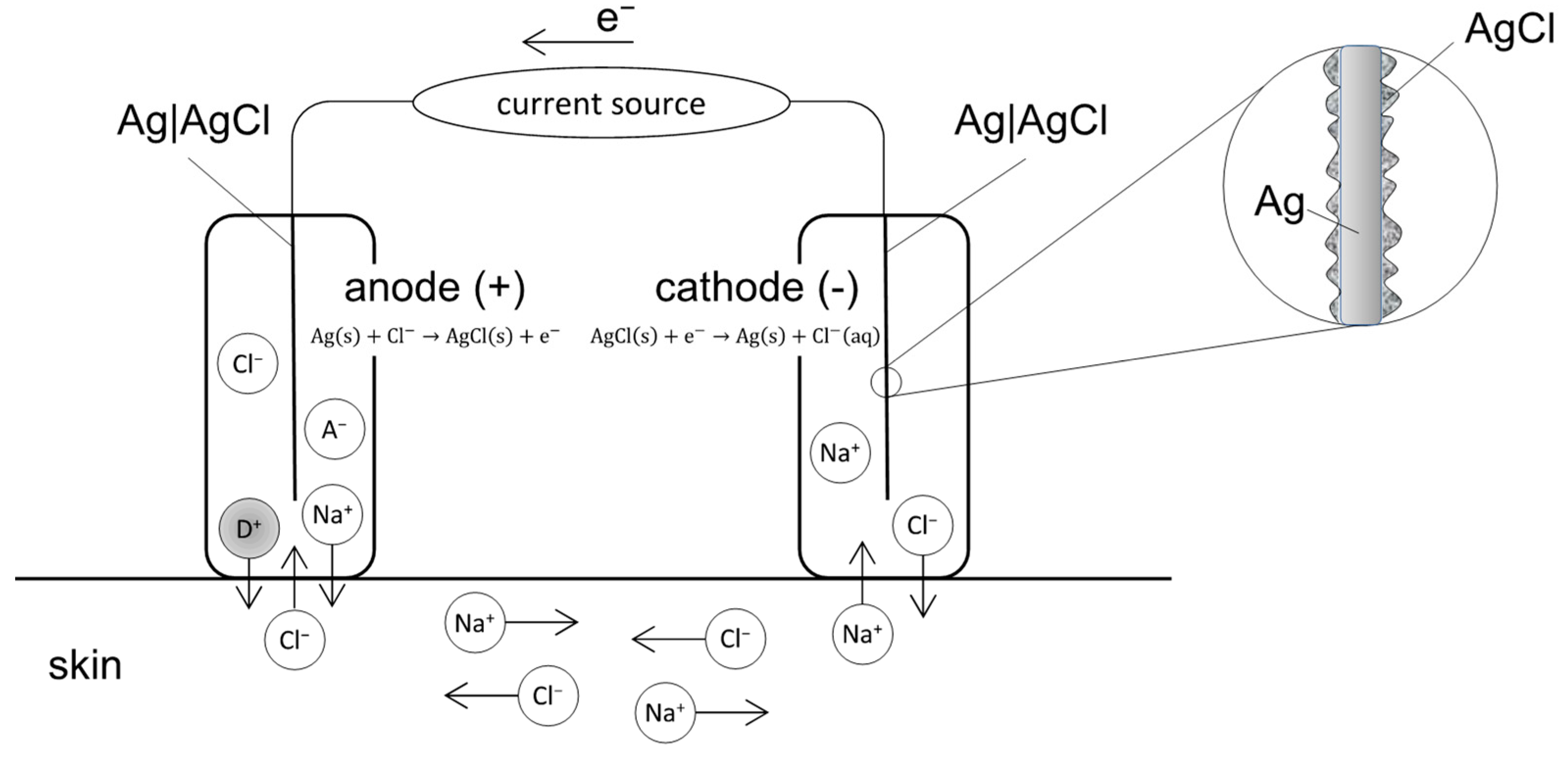
In transdermal applications, several Food and Drug Administration (FDA) approved EMDA devices exist; most use a current source and two electrode compartments, the latter consisting of an electrode immersed in an electrolyte (ionic conductor) solution or gel [29]. Both electrode compartments are placed at two distant sites on the skin while aiming for transdermal drug delivery [20]. The total set-up operates as an electrochemical cell (Figure 2):
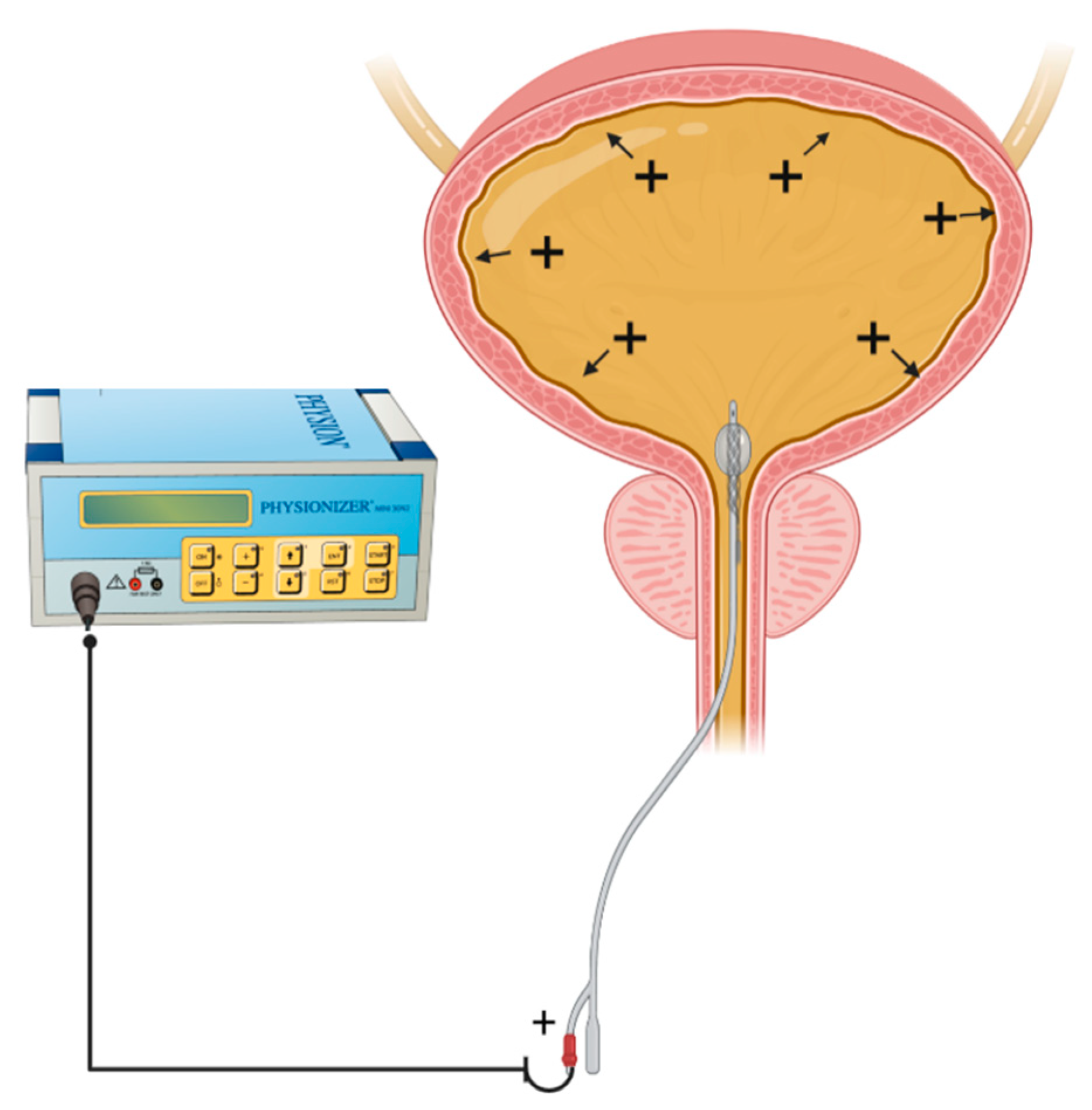
In intra-vesical EMDA, a catheter-electrode is connected to a current generator, the Physionizer®® Mini 30N2 (Medolla, MO, Italy) and a positive polarity applied (Figure 3). The bladder is filled with a drug containing electrolyte solution such that if a cationic drug ion is present, penetration across the bladder wall is enhanced predominantly through an electromigratory phenomenon [24]. This contrasts with unionized large drug molecules, whose transport is more electro-osmotically driven [21].

2.2. Relationship between Current Intensity, Ion Valency and EMDA

Faraday’s law of electrochemical reaction states that [30]:
Q = t i I t Z i F
where Q is the mass of drug delivered by electromotive transport, ti the transference number of the ionic species i, I the current applied (in amperes) and t the time duration (in seconds).
As such, the amount of drug delivered during EMDA is directly proportional to the current intensity and treatment time but inversely proportional to the charge of the drug ion. The transference number of a specific drug ion refers to its ability to carry electric current and is defined as the ratio of the electric current carried by the drug ion i to the total current carried by all ionic species within the electrolyte solution.
The linear relationship between applied current and electromotive transport was illustrated by Harding (1987), who found increasing rates of angiotensin release from Ringer’s solution with increasing amplitudes of electric current applied [31]. Amongst drug compounds with similar molecular weight, monovalent sodium ions were found to have a drug delivery efficiency that was more than twice that of divalent magnesium ions, reflecting the slower migration of drug ions with high valency when a constant electric current is applied [16]. This is also reflected by the inverse relationship between valency and drug delivery in Faraday’s equation above.
However, while increasing current can increase drug delivery in experimental models, thermal damage to healthy tissues is a significant concern in clinical applications. For example, in transdermal EMDA treatments, a current density exceeding 0.5 mA/cm2 induces skin irritation while a maximum of 15 to 20 mA is used in intravesical EMDA to prevent discomfort and tissue damage [10]. Other adjuncts adopted to reduce the likelihood of skin injury during transdermal EMDA include the use of well-saturated absorbent pads and ensuring that there is no direct contact between metallic components and the skin [32]. In addition, based on early transdermal experiments, pulsed direct current (DC) is preferred over continuous DC, as the latter has been found to cause skin polarization, which in turn results in skin irritation and likely a reduced drug delivery efficiency [33].

2.3. Relationship between Drug Physicochemical Properties and EMDA

The physicochemical properties of drug molecules and their carrier solution are important factors influencing EMDA [15]. The charge and molecular size of drug molecules and the pH and presence of buffer ions within its carrier solution affect iontophoresis. As only ionized or charged drugs may be delivered via electromotive forces, unionized drugs rely solely on electroosmosis for transport. Since the degree of drug ionization is often pH dependent, altering the pH of carrier solutions can have a significant impact during EMDA. In an experiment using lidocaine (a local anesthetic agent), Gangarosa et al. demonstrated that when an alkaline solution was added, conductivity was reduced by 15%, as an increase in pH drove the conversion of positively charged lidocaine ions to unionized molecules [34]. The impact of pH on EMDA delivery was further evaluated by Murthy et al., who found that iontophoretic flux of salicylic acid (SA) was significantly increased at higher pH due to the increased ionization of SA (at a pH of 1.2, SA is present solely in the unionized form and the only driving force for transport is electroosmosis. At a pH of 7.1, SA is completely ionized and electromigration drives drug transport) [35].
When present in carrier solutions, buffer ions act as competitors to charged drug ions, resulting in reduced iontophoretic delivery of the latter. As such, if EMDA is desired, the inclusion of small mobile ions in drug diluent solutions should be avoided.

2.4. Membrane or Barrier Properties and EMDA

The skin, urothelium and sclera are examples of biological barriers that can affect EMDA. In general, the porosity of membranes is affected by their thickness as well as pore size and charge. In transdermal EMDA, this membrane barrier is composed of up to 15 layers of corneocytes embedded within the intracellular lamellar lipid membrane, which make up the stratum corneum [36]. At physiological pH, the skin carries a net negative charge; hence, EMDA enhances the transport of positively charged drug ions with an anode while retarding the movement of negatively charged drug ions with a cathode. Neutral drug molecules whose transport is mainly driven by electroosmosis will experience enhanced transport with an anode due to the negatively charged skin barrier [32]. With transscleral applications, since the sclera also carries a net negative charge under physiological conditions, a similar pattern of iontophoretic flux is seen [37]. As the sclera is relatively porous when compared with the skin due to larger pore sizes, it allows for the easy penetration of macromolecules. An example is the fact that the transport of bevacizumab (molecular weight 149 kDa), an antibody targeting the vascular endothelial growth factor (VEGF), is enhanced 32-fold when 2 mA of current is applied during EMDA [38].
In intravesical oncological applications, the ideal penetration depth is to the lamina propria, which is at 1193 ± 26.9 µm [39]. When EMDA with mitomycin-C (MMC) is applied in an in vitro human bladder model, drug penetration at a constant current of 20 mA was highest at a depth between 80 and 200 µm and lowest at a depth between 2000 and 4000 µm [39,40]. This illustrates the importance of barrier thickness and the desired ‘target’ depth in oncological EMDA applications.

3. Pre-Clinical Studies and Clinical Applications of EMDA in Oncology

3.1. Urological Cancer

The first clinical application of EMDA in urology was in the delivery of local anesthetic agents for trans-urethral intra-vesical procedures [40]. The intact urothelium represents a highly impermeable barrier membrane that prevents the systemic absorption of ionized (Na+, K+, Cl) and unionized (urea) solutes from the urinary system [7]. As such, EMDA in urology was devised to enhance locoregional drug delivery due to the limitations in passive transport.
Following a series of EMDA feasibility studies with local anesthetic drugs, its uro-oncologic application was first evaluated by Di Stasi et al. in in vitro studies, which compared passive versus electromotive delivery of MMC in the human bladder wall in the late 1990s [39,41]. In the preceding decade, intra-vesical MMC had been widely utilized amongst urologists following transurethral resection of non-muscle invasive bladder cancer (NMIBC) to prevent local recurrence, which can occur in up to 50% of patients [42]. By combining conventional locoregional MMC treatment with EMDA, Di Stasi’s group found that the application of 20 mA of pulsed direct current (DC) during 30 min resulted in increased penetration of MMC into all layers of the ex vivo bladder wall [39]. When EMDA (15 mA pulsed DC over 40 min) was applied to the bladders of adult mongrel dogs using an intravesical anode inserted through a Foley’s catheter, a similar EMDA-enhanced transport of dye materials into the submucosa and muscularis of the bladder wall was observed [43].
Subsequently, the applicability of EMDA with MMC was evaluated in a series of clinical studies (Table 1). Amongst them, three randomized controlled trials conducted by Di Stasi’s Italian group provided the highest quality of evidence supporting the use of EMDA in the adjuvant and neoadjuvant treatment of NMIBC [44,45,46]. In the first study (2003), 108 patients with early bladder cancer (Tis or T1) were randomized into three groups of 36 patients each following transurethral resection of bladder tumor (TURBT) and underwent (1) EMDA with MMC with 20 mA of electric current for 30 min or (2) passive MMC with a 1 h dwell time or (3) Bacillus Calmette-Guerin (BCG) with a 2 h dwell time, as part of adjuvant therapy [44]. Local and systemic adverse effects were monitored and included symptoms such as urinary frequency, fever, malaise and allergic reactions. The incidence of post-procedure bacterial or drug induced cystitis, prostatitis and epididymitis was also recorded. Comparing the three study arms, patients undergoing BCG treatment were more likely to experience local and systemic side effects. There was no difference in safety and toxicity profiles between passive and EMDA MMC groups. Treatment efficacy measured in terms of complete response and recurrence free rates were comparable in the BCG and EMDA MMC arms, but these were superior to passive MMC instillation. This led to the conduct of a second RCT in the adjuvant setting comparing the use of BCG alone with sequential BCG and EMDA with MMC [45]. Patients allocated to sequential BCG and EMDA with MMC experienced lower recurrence rates and had longer disease-free intervals.
In the neoadjuvant setting, EMDA with MMC was administered immediately prior to TURBT of NMIBC. The 3rd Italian RCT compared three patient groups: (1) TURBT alone, (2) immediate post-TURBT passive MMC and (3) pre-TURBT EMDA with MMC [46]. Irritative bladder symptoms were more common in groups 2 and 3 when compared with group 1 alone, and the use of EMDA immediately before TURBT did not result in higher rates of intra-operative complications. Recurrence and disease-free intervals were superior in the pre-TURBT EMDA group, although there was no difference in overall survival.
EMDA protocols adopted by the various centers show slight variations, with earlier studies using a lower current amplitude of 15 mA over shorter time periods of 20 min, while most subsequent studies utilize a maximum of 20 to 23 mA of pulsed DC applied over a duration of 30 min [47,48,49,50,51,52,53,54,55]. No differences in tolerability and adverse side effects were observed with the higher amplitude and longer durations. Amongst adjuvant studies, reported rates of complete response post-EMDA with MMC range from 40% to 82% while recurrence rates range from 14 to 52%. In addition, it was also found to have a potential role as salvage therapy in BCG refractory high grade NMIBC [55].

3.2. Skin Cancer

The skin is a highly heterogenous membrane and represents the largest organ in the human body. Its outermost layer, the stratum corneum (SC), is key to its barrier function that protects from the external environment [36]. The SC is approximately 15–20 µm thick and has a ‘brick-and-mortar’ structure comprising corneocytes supported by an inter-cellular matrix acting as an effective barrier that prevents the loss of water and the penetration of harmful compounds [56]. Beyond the SC, intercellular junctions, such as tight junctions and adherens junctions, also act as barriers to transdermal drug delivery [57].
In transdermal EMDA (often referred to as transdermal iontophoresis), a low amplitude of direct current, i.e., 0.5 mA/cm2, is used to enhance the penetration of a wide variety of drugs for the treatment of benign skin and sweat gland conditions, ulcers, scars and infections [36]. This should be differentiated from transdermal electroporation studies where high voltage pulses over short durations are applied to increase the permeability of the skin [22].
The use of EMDA in skin cancer has been described since the 1980s for the treatment of squamous cell carcinoma (SCC), basal cell carcinoma (BCC) and other skin tumors (Table 2). In these clinical studies, a variety of common chemotherapeutic agents such as cisplatin, 5-fluorouracil (FU), bleomycin and vinblastine were applied during EMDA. In general, a maximum of 4 mA (range 0.5 to 4 mA) of direct current is applied over the skin lesions during 10 to 30 min. Complete or partial response are common, especially after consecutive EMDA treatment of small, less aggressive lesions (e.g., BCC) [58,59,60,61,62,63].
In recent years, several preclinical studies have explored the use of novel anti-cancer agents and carriers in both melanoma and non-melanocytic skin cancers, i.e., BCC and SCC (Table 3). Doxorubicin, for example, is known to interact strongly with the SC, limiting its ability to be administered transdermally. However, when loaded in positively charged gel carriers during EMDA, improved penetration was achieved compared to water-based formulations. This may be explained by the interactions between the positively charged carrier or its degradation products with the negatively charged sites in the skin, such that the cationic doxorubicin is liberated from its SC binding sites and can penetrate into deeper layers of the skin [64]. Additionally, the use of nano-carriers such as liposomes, gold nanoparticles, dendrimers and lipid nanoparticles in conjunction with EMDA is increasingly explored to enhance the penetration of hydrophilic macromolecules [65,66,67,68,69,70,71,72,73]. When doxorubicin was encapsulated in solid lipid nanoparticles (SLNs) and EMDA applied, a 50-fold increase in penetration depth compared to passive diffusion of unformulated doxorubicin was achieved [65]. The doxorubicin-SLN treatment was also effective in inhibiting tumor cell survival and growth and was associated with increased keratinization and cell death in SCC murine models [65]. In a series of three studies by Labala and coworkers, gold nanoparticle carriers were used to optimize the transport of imatinib mesylate, anti-STAT3 SiRNAs and a combination of both using EMDA [68,71,72]. The co-delivery of both drugs was found to be feasible and resulted in greater suppression in STAT3 and a corresponding reduction in tumor cell viability in melanoma mice models as compared to single agent treatment alone. With increasing recognition of the efficacy of immunotherapy and anti-cancer vaccines in skin cancer, the application of EMDA with CpG-ODN (oligodeoxynucleotides containing unmethylated cytosine–phosphate–guanosine motifs) and cancer antigen gp-100 peptide KVPRNQDWL loaded in nanogels were also reported [66,67]. In both instances, improved penetration and a significant inhibition of tumor growth in melanoma mouse models were observed after EMDA. Similarly, in SCC xenograft models, transdermal EDMA application of EGFR-targeted immunoliposomes loaded with 5-FU was more effective than subcutaneous injection of the drug compound [73].
Photodynamic therapy (PDT) is another common treatment modality in skin cancers. It involves the topical application of a photosensitizer drug followed by illumination with visible light to activate the drug, which selectively destroys cancerous cells [74]. Common photosensitizing agents include 5-aminolevulinic acid (ALA, drug precursor), which penetrates the skin and is converted to protoporphyrin IX (active form) after exogenous application. In vitro studies evaluating ALA with EMDA have found that when 0.47 to 0.5 mA/cm2 of constant current is applied, the transport of ALA into the skin is significantly increased [75,76,77]. This effect was further enhanced when erbium: yttrium-aluminum-garnet (Erb:YAG) laser or microdermabrasion techniques were used to ablate the SC layer prior to EMDA [78]. However, as PDT was not applied in these experiments, the impact on cancer treatment is not known. However, when tetrasulfonated zinc phthalocyanine (ZnPcS4), a 2nd generation photosensitizer, was used in an EMDA experiment with PDT in SCC mouse models, a 15-fold improvement in tumor drug uptake and a 2.8-fold reduction in tumor volume were achieved [70].

3.3. Ophthalmic Cancer

Ocular EMDA has a long history and has been investigated since the early 1900s for the treatment of benign eye conditions such as corneal ulcers, keratitis and episcleritis and eye infections [17]. Trans-corneal or trans-scleral applications have been described. In the former, the EMDA device is placed over the cornea, and in the latter, the device is placed directly on the anterior sclera [79].
Currently, the evidence on the application of EMDA in ophthalmic cancer is limited to preclinical animal studies. The treatment of intra-ocular retinoblastoma with trans-scleral EMDA has been evaluated in mice and rabbits and a dose-dependent inhibition of tumor growth was observed with carboplatin [80,81]. During the experiments, electric currents ranging between 2.57 and 5.14 mA/cm2 were applied during 2 to 10 min. Optimal ocular drug transport was achieved with current densities between 2.57 and 3.85 mA/cm2, while a density of 5.14 mA/cm2 was associated with corneal–limbal toxicity [80]. However, when a hydroxyethyl methacrylate (HEMA) hydrogel carrier was used for carboplatin delivery during EMDA, no advantage over passive diffusion of the drug was seen [82].

3.4. Other Cancer Types

Trans-buccal EMDA in head and neck cancer, intra-ductal in breast tissue, and peri-pancreatic applications have been described [83,84,85,86,87]. In trans-buccal experiments, when bovine buccal mucosa was treated with 5-FU and leucovorin at 1 mA/cm2 during 20 min, EMDA resulted in a significant increase in mucosal penetration of both drugs compared to passive transport [80]. In a later study using nanoparticles as carriers for oxaliplatin, further improvements in penetration were observed, and this was accompanied by cytotoxic effects on SCC cells [84].
In canine and murine breast cancer models, a novel EMDA device was developed to administer an anti-estrogen drug (miproxifen phosphate, TAT-59) into mammary ducts via the nipple. The technique was feasible and resulted in significantly higher local drug concentrations when compared with oral administration [85]. In another series of studies evaluating the use of EMDA for the local delivery of cytotoxic therapies to both breast and pancreatic xenograft tumor models, Byrne et al. designed an implantable device that could be either implanted intra-abdominally or applied trans-dermally. Amongst orthotopic breast cancer mice, EMDA with cisplatin when directly applied to implanted tumors resulted in significant inhibition to tumor growth rates and extended life expectancy by 2-fold. A combination of intravenous with EMDA device cisplatin achieved the best outcomes [85]. Peri-pancreatic applications of gemcitabine and FOLFIRNOX (combination of 5FU, leucovorin, irinotecan and oxaliplatin) using the same EMDA device yielded significant tumor regression outperforming intravenous administration [87,88].

4. Future Outlook

4.1. Cellular Pathways Mediating EMDA

Traditionally, reported mechanisms for EMDA-mediated drug transport include electromigration, electroosmosis and electroporation (as described in Section 2). However, recent studies have found that cellular signaling pathways may play a role during EMDA. Susumu et al. evaluated the effect of faint electrical stimulus on intracellular events, including protein phosphorylation and changes in intracellular signaling factors and found that iontophoretic treatment induces Ca2+ influx into skin cells via a change in membrane potential. This in turn leads to a reduction in the expression of jap junction proteins and depolymerization of tight junction associated actin filaments, which enhances drug penetration [89].
In patients with glioblastoma, an emerging treatment is the use of tumor treating fields (TTFs), where low intensity, alternating currents are delivered to the tumor using electrodes placed in its vicinity [90]. TTFs have been shown to halt the cell cycle by disrupting spindle formation during metaphase leading to tumor cell death [91]. The use of TTFs in newly diagnosed glioblastoma was endorsed by the National Comprehensive Cancer Network (NCCN), with multiple ongoing clinical trials extending its use to other solid tumors [90,91,92,93]. Though conceptually different from EMDA-mediated drug transport, the success of TTFs in brain tumors highlights the potential for the use of electrical fields in various forms of anti-cancer treatment. Future studies should focus on evaluating the molecular and cellular mechanisms of EMDA and its anti-tumoral efficacy.

4.2. Extrapolating Current Evidence for Intra-Peritoneal Applications

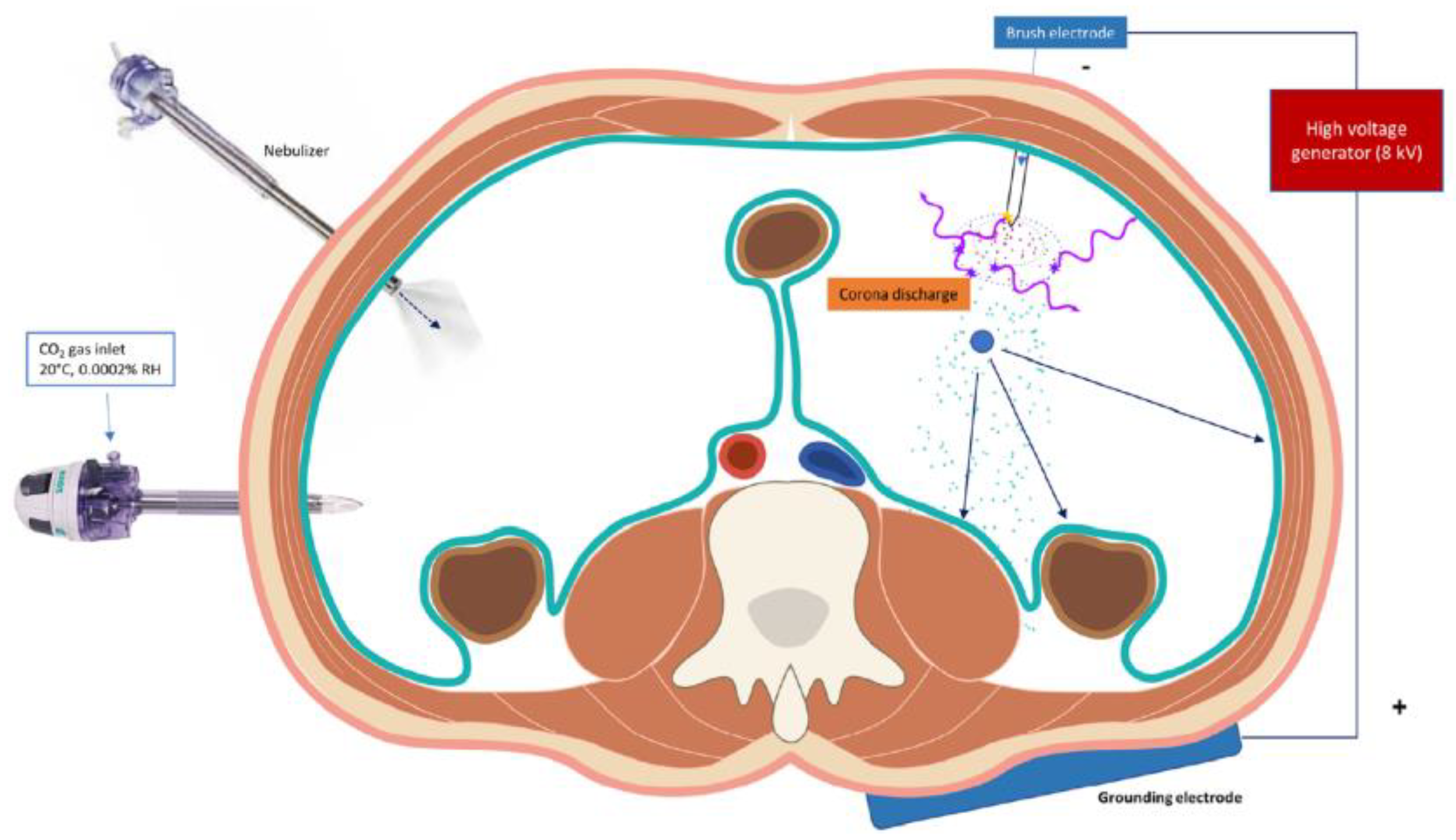
The efficacy of intra-peritoneal (IP) drug delivery for the treatment of peritoneal cancers is limited by high intra-tumoral interstitial fluid pressure (IFP) that prevents effective drug penetration via diffusion and convection phenomena [5]. Current strategies aimed at improving IP drug delivery include the use of hyperthermia (i.e., hyperthermic intra-peritoneal chemotherapy, HIPEC), metronomic IP dosing, the use of novel drug carriers such as albumin, nanoparticles and hydrogels, and aerosolization of chemotherapeutic agents (i.e., pressurized intra-peritoneal aerosolized chemotherapy, PIPAC) [4,94,95,96]. The addition of electromotive forces during PIPAC in the form of electrostatic-PIPAC (e-PIPAC) has also gained increasingly popularity, with evidence suggesting improved spatial distribution and drug penetration (Figure 4). During e-PIPAC, up to 7.5 kV (current intensity < 10 µA) is applied to a stainless-steel brush electrode and results in a stream of negatively charged aerosolized drug particles that show accelerated delivery to the positively charged abdominal wall [94].
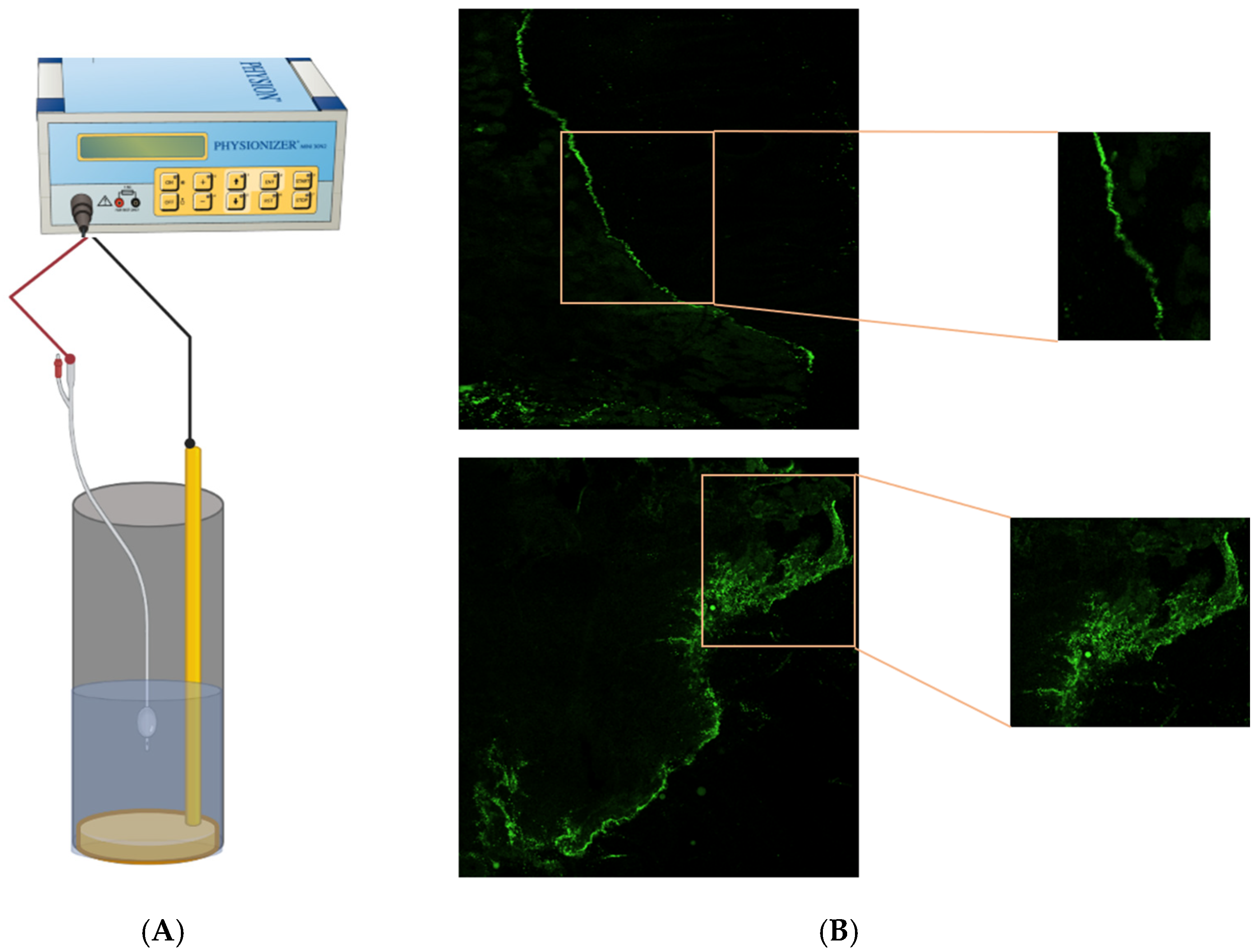
Given the success of EMDA in enhancing loco-regional drug delivery in the context of the urothelium, skin, sclera and tumoral tissues of varying origins, it may represent a promising solution to current challenges encountered during IP drug delivery. Drawing from our experience with e-PIPAC, we have established an in vitro EMDA IP model and evaluated the impact of applying a pulsed DC current to porcine peritoneal samples (Figure 5). Further in vitro and in vivo studies will be needed to confirm the safety, tolerability and efficacy in the context of peritoneal malignancies.

5. Conclusions

EMDA as a modality to improve loco-regional drug delivery is a rapidly growing field. Oncological applications are diverse and include the treatment of skin, eye and bladder cancer. Preclinical studies have also demonstrated its efficacy when combined with nanocarriers and novel drug delivery devices. Future efforts should focus on the clinical translation of EMDA in these fields as well as expanding its applicability to intraperitoneal drug delivery systems.

Supplementary Materials

The following supporting information can be downloaded at: https://www.mdpi.com/article/10.3390/cancers14204980/s1, File S1: Elaboration on the Fundamental Principles of EMDA.

Author Contributions

J.W.S.M., concept and writing; A.C., concept and writing; M.A., concept and writing; N.S., concept and writing, W.C., concept and writing. All authors have read and agreed to the published version of the manuscript.

Funding

This research received no external funding. W.C. is a senior clinical researcher from the Research Foundation—Flanders (FWO) and A.C. is supported by FWO as well through a senior postdoctoral research fellowship (No. 12Z4722N).

Conflicts of Interest

The authors declare no conflict of interest.

References

  1. Petrak, K. Essential properties of drug-targeting delivery systems. Drug Discov. Today 2005, 10, 1667–1673. [Google Scholar] [CrossRef]
  2. Collins, J.M. Pharmacologic rationale for regional drug delivery. J. Clin. Oncol. 1984, 2, 498–504. [Google Scholar] [CrossRef] [PubMed]
  3. Joice, G.A.; Bivalacqua, T.J.; Kates, M. Optimizing pharmacokinetics of intravesical chemotherapy for bladder cancer. Nat. Rev. Urol. 2019, 16, 599–612. [Google Scholar] [CrossRef] [PubMed]
  4. Ceelen, W.; Demuytere, J.; de Hingh, I. Hyperthermic Intraperitoneal Chemotherapy: A Critical Review. Cancers 2021, 13, 3114. [Google Scholar] [CrossRef] [PubMed]
  5. Ceelen, W.P.; Flessner, M.F. Intraperitoneal therapy for peritoneal tumors: Biophysics and clinical evidence. Nat. Rev. Clin. Oncol. 2010, 7, 108–115. [Google Scholar] [CrossRef] [PubMed]
  6. Mishina, T.; Watanabe, H.; Kobayashi, T.; Maegawa, M.; Nakao, M.; Nakagawa, S. Absorption of anticancer drugs through bladder epithelium. Urology 1986, 27, 148–157. [Google Scholar] [CrossRef]
  7. Lasič, E.; Višnjar, T.; Kreft, M.E. Properties of the Urothelium that Establish the Blood-Urine Barrier and Their Implications for Drug Delivery. Rev. Physiol. Biochem. Pharmacol. 2015, 168, 1–29. [Google Scholar]
  8. Dedrick, R.L. Theoretical and experimental bases of intraperitoneal chemotherapy. Semin. Oncol. 1985, 12 (Suppl. S4), 1–6. [Google Scholar]
  9. Heldin, C.H.; Rubin, K.; Pietras, K.; Ostman, A. High interstitial fluid pressure—An obstacle in cancer therapy. Nat. Rev. Cancer 2004, 4, 806–813. [Google Scholar] [CrossRef]
  10. Hashemi, S.; Sahai, A.; Malde, S. Applications of electromotive drug administration in urology. Urol. Ann. 2020, 12, 301–308. [Google Scholar]
  11. Charoo, N.A.; Rahman, Z.; Repka, M.A.; Murthy, S.N. Electroporation: An avenue for transdermal drug delivery. Curr. Drug Deliv. 2010, 7, 125–136. [Google Scholar] [CrossRef] [PubMed]
  12. Turnell, W.J. Therapeutic action of constant current. Proc. R. Soc. Med. 1921, 14, 41–52. [Google Scholar]
  13. Leduc, S. Introduction of medicinal substances into the depth of tissues by electric current. Ann. D’Eleetrobiol. 1900, 3, 545–560. [Google Scholar]
  14. Leduc, S. Electric Ions and Their Use in Medicine; Rebman Ltd.: London, UK, 1908.
  15. Banga, A.K.; Chien, Y.W. Iontophoretic delivery of drugs: Fundamentals, developments and biomedical application. J. Control. Release 1988, 7, 1–24. [Google Scholar] [CrossRef]
  16. Phipps, J.B.; Padmanabhan, R.B.; Lattin, G.A. Iontophoretic delivery of model inorganic and drug ions. J. Pharm. Sci. 1989, 48, 365. [Google Scholar] [CrossRef] [PubMed]
  17. Hughes, L.; Maurice, D.M. A fresh look at iontophoresis. Arch. Ophthalmol. 1984, 102, 1825–1829. [Google Scholar] [CrossRef]
  18. Shelley, W.B.; Horwath, P.; Weidrnan, F.; Pillsbury, D.M. Experimental milaria in man. Production of sweat retention anhidrosis and vesicles by means of iontophoresis. J. Investig. Dermatol. 1948, 11, 275–291. [Google Scholar] [CrossRef] [Green Version]
  19. Harris, R. Treatment of post-therapeutic neuralgia. Lancet 1957, 269, 378–379. [Google Scholar] [CrossRef]
  20. Stephen, R.; Miotti, D.; Bettaglio, R.; Rossi, C.; Bonezzi, C. Electromotive administration of a new morphine formulation: Morphine citrate. Artif. Organs 1994, 18, 461–465. [Google Scholar] [CrossRef]
  21. Gangarosa, L.P.; Park, N.H.; Wiggins, C.A.; Hill, J.M. Increased penetration of nonelectrolytes into mouse skin during iontophoretic water transport (iontohydrokinesis). J. Pharmacol. Exp. Ther. 1980, 212, 377–381. [Google Scholar]
  22. Prausnitz, M.R.; Bose, V.G.; Langer, R.; Weaver, J.C. Electroporation of mammalian skin: A mechanism to enhance transdermal drug delivery. Proc. Natl. Acad. Sci. USA 1993, 90, 10504–10508. [Google Scholar] [CrossRef] [PubMed] [Green Version]
  23. Guy, R.H.; Kalia, Y.N.; Delgado-Charro, M.B.; Merino, V.; López, A.; Marro, D. Iontophoresis: Electrorepulsion and electroosmosis. J. Control. Release 2000, 64, 129–132. [Google Scholar] [CrossRef]
  24. Kalia, Y.N.; Naik, A.; Garrison, J.; Guy, R.H. Iontophoretic drug delivery. Adv. Drug Deliv. Rev. 2004, 56, 619–658. [Google Scholar] [CrossRef] [PubMed]
  25. Bard, A.J.; Faulkner, L.R. Electrochemical Methods: Fundamentals and Applications, 1st ed.; Wiley: New York, NY, USA, 1980. [Google Scholar]
  26. Fick, A.V. On liquid diffusion. Lond. Edinb. Dublin Philos. Mag. J. Sci. 1855, 63, 30–39. [Google Scholar] [CrossRef]
  27. Li, S.K.; Hao, J.; Liddell, M.R. Electrotransport across membranes in biological media: Electrokinetic theories and applications in drug delivery. In Transport in Biological Media; Becker, S., Kuznetsoy, A., Eds.; Elsevier: Philadelphia, PA, USA, 2013; pp. 417–454. [Google Scholar]
  28. Banga, A.K.; Bose, S.; Ghosh, T.K. Iontophoresis and electroporation: Comparisons and contrasts. Int. J. Pharm. 1999, 179, 1–19. [Google Scholar] [CrossRef]
  29. Available online: https://www.federalregister.gov/documents/2016/07/26/2016-17609/physical-medicine-devices-reclassification-of-iontophoresis-device-intended-for-any-other-purposes (accessed on 7 October 2022).
  30. Stillwell, G.K. Electrical stimulation and iontophoresis. In Handbook of Physical Medicine and Rehabilitation, 2nd ed.; Krussen, F.H., Ed.; W.B. Saunders Company: St. Louis, MO, USA, 1971; Chapter 14. [Google Scholar]
  31. Harding, J.W.; Felix, D. Quantification of angiotensin iontophoresis. J. Neurosci. Methods 1987, 19, 209–215. [Google Scholar] [CrossRef]
  32. Sloan, J.B.; Soltani, K. Iontophoresis in dermatology. A review. J. Am. Acad. Dermatol. 1986, 15 Pt 1, 671–684. [Google Scholar] [CrossRef]
  33. Sun, Y.; Siddiqui, O.; Liu, J.C.; Chien, Y.W. Transdermal modulated delivery of polypeptides: Effect of DC pulse wave-form on enhancement. In Proceedings of the 13th International Symposium on Controlled Release of Bioactive Materials, Controlled Release Society, Lincolnshire, IL, USA, 23–25 July 1986. [Google Scholar]
  34. Gangarosa, L.P.; Park, N.H.; Fong, B.C.; Scott, D.F.; Hill, J.M. Conductivity of drugs used for iontophoresis. J. Pharm. Sci. 1978, 67, 1439–1443. [Google Scholar] [CrossRef]
  35. Narasimha Murthy, S.; Wiskirchen, D.E.; Bowers, C.P. Iontophoretic drug delivery across human nail. J. Pharm. Sci. 2007, 96, 305–311. [Google Scholar] [CrossRef]
  36. Hadgraft, J. Skin, the final frontier. Int. J. Pharm. 2001, 224, 1–18. [Google Scholar] [CrossRef]
  37. Chopra, P.; Hao, J.; Li, S.K. Iontophoretic transport of charged macromolecules across human sclera. Int. J. Pharm. 2010, 388, 107–113. [Google Scholar] [CrossRef] [PubMed] [Green Version]
  38. Li, S.K.; Liddell, M.R.; Wen, H. Effective electrophoretic mobilities and charges of anti-VEGF proteins determined by capillary zone electrophoresis. J. Pharm. Biomed. Anal. 2011, 55, 603–607. [Google Scholar] [CrossRef] [PubMed] [Green Version]
  39. Di Stasi, S.M.; Giannantoni, A.; Massoud, R.; Dolci, S.; Navarra, P.; Vespasiani, G.; Stephen, R.L. Electromotive versus passive diffusion of mitomycin C into human bladder wall: Concentration-depth profiles studies. Cancer Res. 1999, 59, 4912–4918. [Google Scholar] [PubMed]
  40. Lugnani, F.; Mazza, G.; Cerulli, N.; Rossi, C.; Stephen, R. Iontophoresis of drugs in the bladder wall: Equipment and preliminary studies. Artif. Organs 1993, 17, 8–17. [Google Scholar] [CrossRef]
  41. Di Stasi, S.M.; Vespasiani, G.; Giannantoni, A.; Massoud, R.; Dolci, S.; Micali, F. Electromotive delivery of mitomycin C into human bladder wall. Cancer Res. 1997, 57, 875–880. [Google Scholar]
  42. Volpe, A.; Racioppi, M.; D’Agostino, D.; Cappa, E.; Filianoti, A.; Bassi, P.F. Mitomycin C for the treatment of bladder cancer. Minerva Urol. Nefrol. 2010, 62, 133–144. [Google Scholar]
  43. Gürpinar, T.; Truong, L.D.; Wong, H.Y.; Griffith, D.P. Electromotive drug administration to the urinary bladder: An animal model and preliminary results. J. Urol. 1996, 156, 1496–1501. [Google Scholar] [CrossRef]
  44. Di Stasi, S.M.; Giannantoni, A.; Stephen, R.L.; Capelli, G.; Navarra, P.; Massoud, R.; Vespasiani, G. Intravesical electromotive mitomycin C versus passive transport mitomycin C for high risk superficial bladder cancer: A prospective randomized study. J. Urol. 2003, 170, 777–782. [Google Scholar] [CrossRef]
  45. Di Stasi, S.M.; Giannantoni, A.; Giurioli, A.; Valenti, M.; Zampa, G.; Storti, L.; Attisani, F.; De Carolis, A.; Capelli, G.; Vespasiani, G.; et al. Sequential BCG and electromotive mitomycin versus BCG alone for high-risk superficial bladder cancer: A randomised controlled trial. Lancet Oncol. 2006, 7, 43–51. [Google Scholar] [CrossRef] [Green Version]
  46. Di Stasi, S.M.; Valenti, M.; Verri, C.; Liberati, E.; Giurioli, A.; Leprini, G.; Masedu, F.; Ricci, A.R.; Micali, F.; Vespasiani, G. Electromotive instillation of mitomycin immediately before transurethral resection for patients with primary urothelial non-muscle invasive bladder cancer: A randomised controlled trial. Lancet Oncol. 2011, 12, 871–879. [Google Scholar] [CrossRef] [Green Version]
  47. Brausi, M.; Campo, B.; Pizzocaro, G.; Rigatti, P.; Parma, A.; Mazza, G.; Vicini, A.; Stephen, R. Intravesical electromotive administration of drugs for treatment of superficial bladder cancer: A comparative Phase II study. Urology 1998, 51, 506–509. [Google Scholar] [CrossRef]
  48. Riedl, C.R.; Knoll, M.; Plas, E.; Pflüger, H. Intravesical electromotive drug administration technique: Preliminary results and side effects. J. Urol. 1998, 159, 1851–1856. [Google Scholar] [CrossRef]
  49. Gan, C.; Amery, S.; Chatterton, K.; Khan, M.S.; Thomas, K.; O’Brien, T. Sequential bacillus Calmette-Guérin/Electromotive Drug Administration of Mitomycin C as the Standard Intravesical Regimen in High Risk Nonmuscle Invasive Bladder Cancer: 2-Year Outcomes. J. Urol. 2016, 195, 1697–1703. [Google Scholar] [CrossRef] [PubMed]
  50. Carando, R.; Zazzara, M.; Cotrufo, S.; Ludovico, G.M. Intravesical Treatment with Electro-Mediated Administration of Mytomicin C as Prophylaxis for Intermediate and High-Risk Nonmuscle-Invasive Bladder Cancer: A Retrospective Multicenter Study. Urol. Int. 2019, 103, 285–290. [Google Scholar] [CrossRef]
  51. Carando, R.; Soldini, E.; Cotrufo, S.; Zazzara, M.; Ludovico, G.M. Electro-mediated drug administration of mitomycin C in preventing non-muscle-invasive bladder cancer recurrence and progression after transurethral resection of the bladder tumour in intermediate- and high-risk patients. Arab. J. Urol. 2020, 19, 71–77. [Google Scholar] [CrossRef]
  52. Zazzara, M.; Nazaraj, A.; Scarcia, M.; Cardo, G.; Carando, R.; Ludovico, G.M. Electromotive Drug Administration of Mitomycin C (EMDA/MMC) versus Intravesical Immunotherapy with Bacillus Calmette-Guérin (BCG) in Intermediate and High Risk Non Muscle Invasive Bladder Cancer. Urol. Int. 2021, 1–8. [Google Scholar] [CrossRef] [PubMed]
  53. Colombo, R.; Brausi, M.; Da Pozzo, L.; Salonia, A.; Montorsi, F.; Scattoni, V.; Roscigno, M.; Rigatti, P. Thermo-chemotherapy and electromotive drug administration of mitomycin C in superficial bladder cancer eradication. a pilot study on marker lesion. Eur. Urol. 2001, 39, 95–100. [Google Scholar] [CrossRef]
  54. Decaestecker, K.; Lumen, N.; Van Camp, C.; Oosterlinck, W. Single ablative intravesical electromotive mitomycin C administration for small non-muscle-invasive bladder cancer: A prospective study. Acta Clin. Belg. 2018, 73, 324–327. [Google Scholar] [CrossRef]
  55. Racioppi, M.; Di Gianfrancesco, L.; Ragonese, M.; Palermo, G.; Sacco, E.; Bassi, P.F. ElectroMotive drug administration (EMDA) of Mitomycin C as first-line salvage therapy in high risk "BCG failure" non muscle invasive bladder cancer: 3 years follow-up outcomes. BMC Cancer 2018, 18, 1224. [Google Scholar] [CrossRef] [Green Version]
  56. Marks, R. The stratum corneum barrier: The final frontier. J. Nutr. 2004, 134, 2017S–2021S. [Google Scholar] [CrossRef] [Green Version]
  57. Yokouchi, M.; Kubo, A. Maintenance of tight junction barrier integrity in cell turnover and skin diseases. Exp. Dermatol. 2018, 27, 876–883. [Google Scholar] [CrossRef] [PubMed] [Green Version]
  58. Luxenberg, M.N.; Guthrie, T.H., Jr. Chemotherapy of basal cell and squamous cell carcinoma of the eyelids and periorbital tissues. Ophthalmology 1986, 93, 504–510. [Google Scholar] [CrossRef]
  59. Chang, B.K.; Guthrie, T.H., Jr.; Hayakawa, K.; Gangarosa, L.P., Sr. A pilot study of iontophoretic cisplatin chemotherapy of basal and squamous cell carcinomas of the skin. Arch. Dermatol. 1993, 129, 425–427. [Google Scholar] [CrossRef] [PubMed]
  60. Bacro, T.R.; Holladay, E.B.; Stith, M.J.; Maize, J.C.; Smith, C.M. Iontophoresis treatment of basal cell carcinoma with cisplatin: A case report. Cancer Detect. Prev. 2000, 24, 610–619. [Google Scholar]
  61. Welch, M.L.; Grabski, W.J.; McCollough, M.L.; Skelton, H.G.; Smith, K.J.; Menon, P.A.; Anderson, L.L. 5-fluorouracil iontophoretic therapy for Bowen’s disease. J. Am. Acad. Dermatol. 1997, 36 Pt 1, 956–958. [Google Scholar] [CrossRef]
  62. Tsuji, T. Bleomycin iontophoretic therapy for verrucous carcinoma. Arch. Dermatol. 1991, 127, 973–975. [Google Scholar] [CrossRef]
  63. Smith, K.J.; Konzelman, J.L.; Lombardo, F.A.; Skelton, H.G., 3rd; Holland, T.T.; Yeager, J.; Wagner, K.F.; Oster, C.N.; Chung, R. Iontophoresis of vinblastine into normal skin and for treatment of Kaposi’s sarcoma in human immunodeficiency virus-positive patients. The Military Medical Consortium for Applied Retroviral Research. Arch. Dermatol. 1992, 128, 1365–1370. [Google Scholar] [CrossRef]
  64. Taveira, S.F.; Nomizo, A.; Lopez, R.F. Effect of the iontophoresis of a chitosan gel on doxorubicin skin penetration and cytotoxicity. J. Control. Release 2009, 134, 35–40. [Google Scholar] [CrossRef]
  65. Huber, L.A.; Pereira, T.A.; Ramos, D.N.; Rezende, L.C.; Emery, F.S.; Sobral, L.M.; Leopoldino, A.M.; Lopez, R.F. Topical Skin Cancer Therapy Using Doxorubicin-Loaded Cationic Lipid Nanoparticles and lontophoresis. J. Biomed. Nanotechnol. 2015, 11, 1975–1988. [Google Scholar] [CrossRef]
  66. Kigasawa, K.; Kajimoto, K.; Nakamura, T.; Hama, S.; Kanamura, K.; Harashima, H.; Kogure, K. Noninvasive and efficient transdermal delivery of CpG-oligodeoxynucleotide for cancer immunotherapy. J. Control. Release 2011, 150, 256–265. [Google Scholar] [CrossRef]
  67. Toyoda, M.; Hama, S.; Ikeda, Y.; Nagasaki, Y.; Kogure, K. Anti-cancer vaccination by transdermal delivery of antigen peptide-loaded nanogels via iontophoresis. Int. J. Pharm. 2015, 483, 110–114. [Google Scholar] [CrossRef] [PubMed]
  68. Labala, S.; Mandapalli, P.K.; Kurumaddali, A.; Venuganti, V.V. Layer-by-layer polymer coated gold nanoparticles for topical delivery of imatinib mesylate to treat melanoma. Mol. Pharm. 2015, 12, 878–888. [Google Scholar] [CrossRef] [PubMed]
  69. Venuganti, V.V.; Saraswathy, M.; Dwivedi, C.; Kaushik, R.S.; Perumal, O.P. Topical gene silencing by iontophoretic delivery of an antisense oligonucleotide-dendrimer nanocomplex: The proof of concept in a skin cancer mouse model. Nanoscale 2015, 7, 3903–3914. [Google Scholar] [CrossRef] [PubMed]
  70. Lemos, C.N.; de Souza, J.G.; Simão, P.S.; Lopez, R.F. Iontophoresis Improved Growth Reduction of Invasive Squamous Cell Carcinoma in Topical Photodynamic Therapy. PLoS ONE 2016, 11, e0145922. [Google Scholar] [CrossRef] [PubMed]
  71. Labala, S.; Jose, A.; Venuganti, V.V. Transcutaneous iontophoretic delivery of STAT3 siRNA using layer-by-layer chitosan coated gold nanoparticles to treat melanoma. Colloids Surf. B Biointerfaces 2016, 146, 188–197. [Google Scholar] [CrossRef]
  72. Labala, S.; Jose, A.; Chawla, S.R.; Khan, M.S.; Bhatnagar, S.; Kulkarni, O.P.; Venuganti, V.V.K. Effective melanoma cancer suppression by iontophoretic co-delivery of STAT3 siRNA and imatinib using gold nanoparticles. Int. J. Pharm. 2017, 525, 407–417. [Google Scholar] [CrossRef]
  73. Petrilli, R.; Eloy, J.O.; Saggioro, F.P.; Chesca, D.L.; de Souza, M.C.; Dias, M.V.S.; da Silva, L.L.P.; Lee, R.J.; Lopez, R.F.V. Skin cancer treatment effectiveness is improved by iontophoresis of EGFR-targeted liposomes containing 5-FU compared with subcutaneous injection. J. Control. Release 2018, 283, 151–162. [Google Scholar] [CrossRef]
  74. Hasan, T.; Ortel, B.; Moor, A.C.; Pogue, B.W. Photodynamic therapy of cancer. In Cancer Medicine, 9th ed.; Kufe, D.W., Pollock, R.E., Weichselbaum, R.R., Bast, R.C., Gansler, T.S., Holland, J.F., Frei, E., Eds.; B.C. Decker: Hamilton, ON, USA, 2017; p. 537. [Google Scholar]
  75. Lopez, R.F.R.; Bentley, M.V.; Begoña Delgado-Charro, M.; Guy, R.H. Optimization of aminolevulinic acid delivery by iontophoresis. J. Control. Release 2003, 88, 65–70. [Google Scholar] [CrossRef]
  76. HBodde, E.; Roemele, P.E.H.; Star, W.M. Quantification of topically delivered 5-aminolevulinic acid by iontophoresis across ex vivo human stratum corneum. Photochem. Photobiol. 2002, 75, 418–423. [Google Scholar]
  77. Merclin, N.; Bramer, T.; Edsman, K. Iontophoretic delivery of 5-aminolevulinic acid and its methyl ester using a carbopol gel as vehicle. J. Control. Release 2004, 98, 57–65. [Google Scholar] [CrossRef]
  78. Fang, J.Y.; Lee, W.R.; Shen, S.C.; Fang, Y.P.; Hu, C.H. Enhancement of topical 5-aminolaevulinic acid delivery by erbium:YAG laser and microdermabrasion: A comparison with iontophoresis and electroporation. Br. J. Dermatol. 2004, 151, 132–140. [Google Scholar] [CrossRef] [PubMed]
  79. Perez, V.L.; Wirostko, B.; Korenfeld, M.; From, S.; Raizman, M. Ophthalmic Drug Delivery Using Iontophoresis: Recent Clinical Applications. J. Ocul. Pharmacol. Ther. 2020, 36, 75–87. [Google Scholar] [CrossRef] [PubMed] [Green Version]
  80. Hayden, B.; Jockovich, M.E.; Murray, T.G.; Kralinger, M.T.; Voigt, M.; Hernandez, E.; Feuer, W.; Parel, J.M. Iontophoretic delivery of carboplatin in a murine model of retinoblastoma. Investig. Ophthalmol. Vis. Sci. 2006, 47, 3717–3721. [Google Scholar] [CrossRef]
  81. Hayden, B.C.; Jockovich, M.E.; Murray, T.G.; Voigt, M.; Milne, P.; Kralinger, M.; Feuer, W.J.; Hernandez, E.; Parel, J.M. Pharmacokinetics of systemic versus focal Carboplatin chemotherapy in the rabbit eye: Possible implication in the treatment of retinoblastoma. Investig. Ophthalmol. Vis. Sci. 2004, 45, 3644–3649. [Google Scholar] [CrossRef]
  82. Eljarrat-Binstock, E.; Domb, A.J.; Orucov, F.; Dagan, A.; Frucht-Pery, J.; Pe’Er, J. In vitro and in vivo evaluation of carboplatin delivery to the eye using hydrogel-iontophoresis. Curr. Eye Res. 2008, 33, 269–275. [Google Scholar] [CrossRef]
  83. Gratieri, T.; Kalia, Y.N. Targeted local simultaneous iontophoresis of chemotherapeutics for topical therapy of head and neck cancers. Int. J. Pharm. 2014, 460, 24–27. [Google Scholar] [CrossRef]
  84. Matos, B.N.; Pereira, M.N.; Bravo, M.O.; Cunha-Filho, M.; Saldanha-Araújo, F.; Gratieri, T.; Gelfuso, G.M. Chitosan nanoparticles loading oxaliplatin as a mucoadhesive topical treatment of oral tumors: Iontophoresis further enhances drug delivery ex vivo. Int. J. Biol. Macromol. 2020, 154, 1265–1275. [Google Scholar] [CrossRef] [PubMed]
  85. Komuro, M.; Suzuki, K.; Kanebako, M.; Kawahara, T.; Otoi, T.; Kitazato, K.; Inagi, T.; Makino, K.; Toi, M.; Terada, H. Novel iontophoretic administration method for local therapy of breast cancer. J. Control. Release 2013, 168, 298–306. [Google Scholar] [CrossRef]
  86. Byrne, J.D.; Jajja, M.R.; O’Neill, A.T.; Bickford, L.R.; Keeler, A.W.; Hyder, N.; Wagner, K.; Deal, A.; Little, R.E.; Moffitt, R.A.; et al. Local iontophoretic administration of cytotoxic therapies to solid tumors. Sci. Trans. Med. 2015, 7, 273ra14. [Google Scholar] [CrossRef] [Green Version]
  87. Byrne, J.D.; Jajja, M.R.; Schorzman, A.N.; Keeler, A.W.; Luft, J.C.; Zamboni, W.C.; DeSimone, J.M.; Yeh, J.J. Iontophoretic device delivery for the localized treatment of pancreatic ductal adenocarcinoma. Proc. Natl. Acad. Sci. USA 2016, 113, 2200–2205. [Google Scholar] [CrossRef]
  88. Byrne, J.D.; Jajja, M.R.; O’Neill, A.T.; Schorzman, A.N.; Keeler, A.W.; Luft, J.C.; Zamboni, W.C.; Desimone, J.M.; Yeh, J.J. Impact of formulation on the iontophoretic delivery of the FOLFIRINOX regimen for the treatment of pancreatic cancer. Cancer Chemother. Pharmacol. 2018, 81, 991–998. [Google Scholar] [CrossRef] [PubMed]
  89. Hama, S.; Kimura, Y.; Mikami, A.; Shiota, K.; Toyoda, M.; Tamura, A.; Nagasaki, Y.; Kanamura, K.; Kajimoto, K.; Kogure, K. Electric stimulus opens intercellular spaces in skin. J. Biol. Chem. 2014, 289, 2450–2456. [Google Scholar] [CrossRef] [PubMed] [Green Version]
  90. Rominiyi, O.; Vanderlinden, A.; Clenton, S.J.; Bridgewater, C.; Al-Tamimi, Y.; Collis, S.J. Tumour treating fields therapy for glioblastoma: Current advances and future directions. Br. J. Cancer. 2021, 124, 697–709, Epub 2020, 125, 623. [Google Scholar] [CrossRef] [PubMed]
  91. Mun, E.J.; Babiker, H.M.; Weinberg, U.; Kirson, E.D.; Von Hoff, D.D. Tumor-Treating Fields: A Fourth Modality in Cancer Treatment. Clin. Cancer Res. 2018, 24, 266–275. [Google Scholar] [CrossRef] [Green Version]
  92. Tan, A.C.; Ashley, D.M.; López, G.Y.; Malinzak, M.; Friedman, H.S.; Khasraw, M. Management of glioblastoma: State of the art and future directions. CA Cancer J. Clin. 2020, 70, 299–312. [Google Scholar] [CrossRef]
  93. Aguilar, A.A.; Ho, M.C.; Chang, E.; Carlson, K.W.; Natarajan, A.; Marciano, T.; Bomzon, Z.; Patel, C.B. Permeabilizing Cell Membranes with Electric Fields. Cancers 2021, 13, 2283. [Google Scholar] [CrossRef] [PubMed]
  94. Rahimi-Gorji, M.; Van de Sande, L.; Debbaut, C.; Ghorbaniasl, G.; Braet, H.; Cosyns, S.; Remaut, K.; Willaert, W.; Ceelen, W. Intraperitoneal aerosolized drug delivery: Technology, recent developments, and future outlook. Adv. Drug Deliv. Rev. 2020, 160, 105–114. [Google Scholar] [CrossRef] [PubMed]
  95. Van de Sande, L.; Cosyns, S.; Willaert, W.; Ceelen, W. Albumin-based cancer therapeutics for intraperitoneal drug delivery: A review. Drug Deliv. 2020, 27, 40–53. [Google Scholar] [CrossRef] [Green Version]
  96. Van de Sande, L.; Rahimi-Gorji, M.; Giordano, S.; Davoli, E.; Matteo, C.; Detlefsen, S.; D’Herde, K.; Braet, H.; Shariati, M.; Remaut, K.; et al. Electrostatic Intraperitoneal Aerosol Delivery of Nanoparticles: Proof of Concept and Preclinical Validation. Adv. Healthc. Mater. 2020, 9, e2000655. [Google Scholar] [CrossRef]
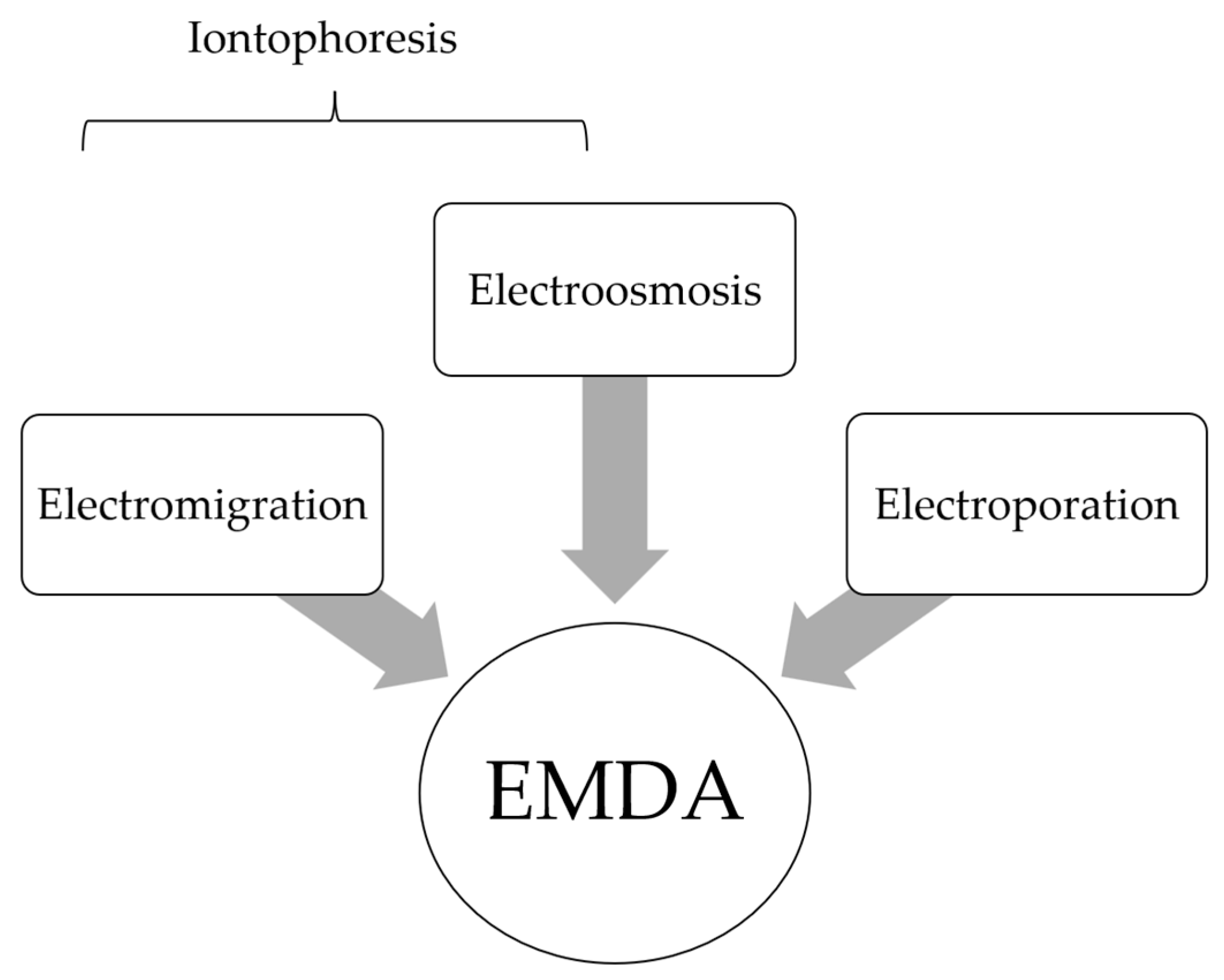
Figure 1. Electromotive drug administration (EMDA) encompasses the electrokinetic phenomena of electromigration (EM), electroosmosis (EO) and electroporation (EP). Conventionally, iontophoresis alone was commonly used to describe electric-driven drug transport.
Cancers 14 04980 g001
Figure 2. Schematic diagram of an iontophoretic device consisting of a current source and two Ag|AgCl electrodes. During EMDA, D+ is placed inside the electrode compartment bearing the same charge (i.e., the anode). Cations, including D+, are transported from the anode into the skin. At the same time, anions from the skin move into the anode. In the cathode, anions leave the cathode towards the skin, while cations move into the cathode.
Cancers 14 04980 g002
Figure 3. Schematic depiction of EMDA of a cationic drug molecule (+) within a filled bladder. The foley catheter contains a spiral Ag electrode at its tip and is connected to a current generator. During EMDA, grounding skin electrodes are placed over the anterior abdominal wall and connected to the cathode component of the generator.
Cancers 14 04980 g003
Figure 4. Schematic overview of electrostatic precipitation combined with pressurized intraperitoneal aerosol chemotherapy (e-PIPAC) where electromotive forces are used to improve IP drug distribution and penetration. (Adapted from Rahimi-Gorji et al., 2020).
Cancers 14 04980 g004
Figure 5. (A) Schema of in vitro IP model: custom-made plastic cylinder that mimics the abdominal cavity. A Physionizer® Mini 30N2 current generator is connected to a Foley catheter housing a silver spiral electrode and a grounding metallic plate. The setup aims to replicate the delivery of IP therapeutics. (B) Confocal microscopy images of porcine peritoneum tissue treated with fluorescent nanoparticles before (top) and after (bottom) EMDA, demonstrating improved penetration after EMDA. Experimental surgery lab, Ghent University, unpublished data.
Cancers 14 04980 g005
Table 1. Clinical Studies on EMDA and treatment of non-muscle invasive bladder cancer (NMIBC).
Study/YDesign Clinical ContextControl GroupIntervention GroupOncological Outcomes
(Compare Control vs. Intervention)
No. of
Patients
Treatment RegimeNo. of
Patients
Treatment RegimeHistological pCRRecurrence and
Survival
Adjuvant Treatment in NMIBC
Brausi 1998 [47] Multi-center CohortPrimary or recurrent stage Ta or T1, Grade 1 or 2 TCC1240 mg PD MMC
(2 h dwell time)
weekly over 8 weeks
1540 mg EMDA-MMC 15 mA over 20 min wkly over 8 wks41.6% vs. 40%RR: 60% vs. 33%
DFI: 10.5 vs. 14.5 months
Riedl
1998 [48]
Single arm
prospective
Ta or T1 TCCNANA2240 mg EMDA-MMC 15 mA 20 min weekly over 4 weeks 56.6%
(no control,
at 14 months)
RR: 44%
Di Stasi 2003 [44] Multi-center
RCT
Ta or T1 TCC 36
36
A: 40 mg PD MMC
(1 h dwell time)
weekly over 6 weeks +/− additional course for non-responders
B: BCG alone
(2 h dwell time)
3640 mg EMDA-MMC 20 mA 30 min weekly over 6 wks +/− additional course for non-responders 31%
(Control A) vs.
58% (at 6 months)
RR: 75% and 52.8 (Controls A and B) vs. 52.8%
DFI: 19.5 and 26 (Controls A and B) vs. 35 months *
Di Stasi
2006 [45]
Multi-center
RCT
Primary or recurrent stage Ta or T1 TCC 105BCG alone (2 h dwell time) weekly over 6 wks
In CR, followed by BCG maintenance (to 10 months)
107BCG (2 h dwell time) weekly over 2 weeks + 40 mg EMDA-MMC 20 mA 30 min weekly over 3 weeks
In CR, followed by BCG + EMDA-MMC maintenance (to 10 months)
57.1% vs. 69%RR: 57.9% vs. 41.9% *
DFI: 21 vs. 69 months *
DSS: 83.8% vs. 94.4% *
Gan
2016 [49]
Single arm
prospective
High risk NMIBC NANA107BCG (2 h dwell time) + 40 mg EMDA-MMC 20 mA 30 min weekly over 3 wks71%
(No control, at 1 year)
NR
Carando 2019# [50] Retrospective single-arm Intermediate and high risk NMIBCNANA6540 mg EMDA-MMC 20 to 23 mA 30 min weekly over 8 weeks 82%
(at 6 months)
NR
Carando 2020# [51] Retrospective single-armAdjuvant setting
Intermediate and high risk NMIBC
NANA10140 mg EMDA-MMC 20 to 23 mA 30 min weekly over 8 weeks
Followed by EMDA-MMC maintenance (up to 12 months)
75%
(at 6 months)
PFS (at 3 months): 94%
PFS (at 6 months): 90%
Zazzara 2021# [52] Retrospective cohortAdjuvant setting
Intermediate and high risk NMIBC
104BCG alone (1.5 to 2 h dwell time) weekly over 6-weeks
Followed by BCG maintenance (up to 12 months)
14040 mg EMDA-MMC
20 to 23 mA 30 min weekly over 8 weeks
Followed by EMDA-MMC maintenance (up to 12 months)
NRRR: 20 vs. 14%
(at maintenance phase)
Neoadjuvant treatment in NMIBC
Colombo 2001 [53]Single center Cohort Ta or T1, Grade 1 or 2, <2 cm tumors 3640 mg PD MMC
(1 h dwell time)
weekly over 4 wks
1540 mg EMDA-MMC 15 mA 20 min weekly over 4 wks27.7% vs. 40%NR
Di Stasi
2011 [46]
Multi-center
RCT
Neoadjuvant setting
Ta or T1 TCC
116
119
A: TURBT alone
B: TURBT + 40 mg PD MMC (1 h dwell time)
117Pre-TURBT 40 mg EMDA-MMC 20 mA 30 minNARR: 64% and 59% (Controls A and B) vs. 44% *
DFI: 2 and 16
(Controls A and B) vs. 52 months *
Decae-
Stecker 2018 [54]
Single-arm
prospective
Neoadjuvant setting
Primary or recurrent stage Ta or T1, tumor <2 cm
NANA36Pre-TURBT 60 mg EMDA-MMC 25 mA 25 min25%
(no control,1 year)
NR
Other
Racioppi 2018 [55]Single-arm
prospective
Salvage bladder-sparing
BCG-refractory High grade NMIBC
NANA2640 mg EMDA-MMC 20 mA 30 min weekly over 6 wks
Followed by maintenance over 6 months
NAHigh-grade disease-free rate 61.5%
PD passive diffusion; TCC Transitional Cell Carcinoma of the bladder; pCR pathological complete response; RR Recurrence rate; DFI Disease free interval; DSS Disease specific survival; NR not reported; NA Not applicable; RCT Randomized controlled trial; * significant p value; # Likely repeated cohort–same institution study with overlapping time periods.
Table 2. Clinical Studies on EMDA and treatment of skin cancer.
Study/YearDesignClinical ContextNo. of
Patients
DrugEMDA ProtocolOncological Outcomes
Luxenberg 1986 [58] Case
series
BCC or SCC
(non-surgical candidates treated with systemic chemotherapy)
Tumor site: Peri-orbital
EMDA applied to recurrent or residual lesions after systemic chemotherapy
5Cisplatin 0.5 to 1.5 mA DC over
20 min
PR in all patients
Chang
1993 [59]
Case
series
BCC or SCC
(non-surgical candidates)
Tumor site: Face and others
12Cisplatin 0.5 to 1.5 mA DC over
20 to 30 min
Daily for 3–5 days or once/week
CR 26.7% of lesions, PR 46.7%, minimal response 26.7%
Factors associated with CR:
(1)
Lesion <3 cm
(2)
BCC
(3)
Daily administration
Bacro
2000 [60]
Case
report
BCC
Tumor site: Lower extremity
1Cisplatin Daily for 5 days over 4 weeks CR was achieved
Welch
1997 [61]
Case
series
SCC-in-situ
Tumor site: Head and Neck, Trunk, Upper and lower extremities
265-FU 4 mA DC over 10 min
Twice/week over 4 weeks
CR in 96% (25 out of 26) patients
Tsuji
1991 [62]
Case
study
Verrucous carcinoma (not surgical candidate)
Tumor site: Lips
1Bleomycin2 mA DC over 30 min
Trice/week over 2 weeks
CR was achieved
Recurrence-free at 6 months
Smith
1992 [63]
Case
series
Kaposi’s sarcoma in HIV patients
Tumor site: Upper and lower extremities, face, tongue
4Vinblastine sulfate4 mA DC over 10 min onceCR in 29% of lesions, PR in 71%
DC Direct Current BCC basal cell carcinoma SCC squamous cell carcinoma PR partial response CR complete response FU Fluorouracil HIV Human immunodeficiency virus.
Table 3. Pre-clinical studies on EMDA and treatment of skin cancer.
Study/YearStudy TypeExperimental Tissue/Skin Cancer TypeDrug UsedCarrier (if Any) or Carrier SolutionEMDA ProtocolOutcomes
Taveira 2009 [64]In vitroPorcine skin
Melanoma (Murine cells)
Doxorubicin
(cationic)
Chitosan gel (cationic), water, HEC gel (non-ionic)0.5 mA/cm2
over 6 hrs
Doxorubicin with chitosan gel carrier result in improved epidermal penetration (in porcine skin) with EMDA when compared with passive diffusion (PD)
3-fold increase in doxorubicin cytotoxicity with EMDA in cancer cells over PD
Huber 2015 [65]In vitro and In vivo Porcine skin
SCC
(Murine model)
Doxorubicin
(cationic)
Solid lipid nanoparticles (SLN), water 0.5 mA/cm2
over 6 h
2-fold improvement of doxorubicin penetration (in porcine skin) with SLN carrier
Significantly improved SCC tumour inhibition using doxorubicin-SLN with EMDA over PD
Kigasawa 2011 [66]In vivoMelanoma
(Murine model)
CpG-ODNNaCl0.3 mA/cm2
over 1 h
Improved penetration and distribution of drug in epidermis and dermis with EMDA over PD
Significant inhibition of tumour growth with local or distant application of drug with EMDA compared with non-treated controls
Toyoda 2015 [67]In vitro and In vivoPorcine Skin
Melanoma
(Murine model)
Cancer-antigen gp-100 peptide PEG modified Nanogels 0.4 mA/cm2
over 1 h
Significant increase in no. of Langerhans cells in epidermis with gp100 and EMDA over non-treated controls
Significant tumour growth suppression with antigen peptide-loaded nanogels and EMDA over non-treated controls
Labala 2015 [68]In vitro Porcine skin
Melanoma
(Murine cells)
Imatinib mesylate Gold nanoparticles 0.47 mA/cm2
over 4 h
6.2-fold improvement of drug-nanocomplex penetration with EMDA over PD
Significant inhibition of tumour cell viability by nanocomplex over non-treated controls
Venuganti 2015 [69]In vitro and In vivo Porcine skin
Skin tumor (Murine model)
Anti-sense Oligonucleotide (ASO) PAMAM Dendrimer0.3 mA/cm2
over 4 h
Increased penetration of ASO-Dendrimer complex into epidermis with EMDA over PD
Significant apoptosis of skin tumour with drug complex over non-treated controls
Lemos 2016 [70] In vivoSCC
(Murine model)
ZnPcS4 *
(anionic)
HEC Gel (Non-ionic) 0.5 mA/cm2
over 30 min
15-fold improvement of drug uptake by tumour with EMDA over PD
2.8-fold reduction in tumour volume in the group of mice treated with photosensitizer with EMDA followed by photodynamic therapy compared with non-treated controls
Labala 2016 [71]In vitro Porcine skin
Melanoma
(Murine cells)
anti-STAT3 SiRNAGold nanoparticles (Chitosan capped) 0.47 mA/cm2
over 4 h
Complex was able to successfully penetrate skin with EMDA
Nanoparticle complex inhibited cell growth by 50%
Labala 2017 [72]In vitro and In vivo Melanoma
(Murine cells)
Melanoma
(Murine model)
Co-delivery of anti-STAT3 SiRNA and Imatinib mesylateGold nanoparticles 0.5 mA/cm2
over 2 h
Combination of 2 drugs resulted in greater suppression of STAT3, reduction in tumour cell viability when compared with single drug delivery
Transdermal EMDA delivery of nanocomplex resulted in significant reduction in tumour volume, weight, and STAT3 suppression (similar to direct intra-tumoral injection) over non-treated controls
Petrilli 2018 [73] In vitro and In vivoPorcine skin
SCC
(Murine model)
5-FU EGFR-targeted immunoliposome0.5 mA/cm2
over 6 h
2-fold penetration of drug in skin with immunoliposomes with EMDA compared with control liposomes
2-fold reduction in tumour volume with transdermal 5-FU containing liposomes and EMDA compared with subcutaneous injection
SCC squamous cell carcinoma; PD passive diffusion; HEG Hydroxyethyl cellulose; ALA aminolaevulinic acid; HEPES (4-(2-hydroxyethyl)-1-piperazineethanesulfonic acid)–zwitterionic sulfonic acid buffering agent; Erb:YAG erbium:yttrium-aluminium-garnet; PEG Polyethylene glycol; CpG-ODN unmethylated; CpG motifs 5-FU Fluorouracil; EGFR epidermal growth receptor; SiRNA small interfering RNA PAMAM poly (amidoamine). ZnPcS4 tetrasulfonated zinc phthalocyanine * photosensitizer.
Publisher’s Note: MDPI stays neutral with regard to jurisdictional claims in published maps and institutional affiliations.