
Staging of Endometrial Cancer Using Fusion T2-Weighted Images with Diffusion-Weighted Images: A Way to Avoid Gadolinium?

Abstract
:Simple Summary
Abstract
1. Introduction
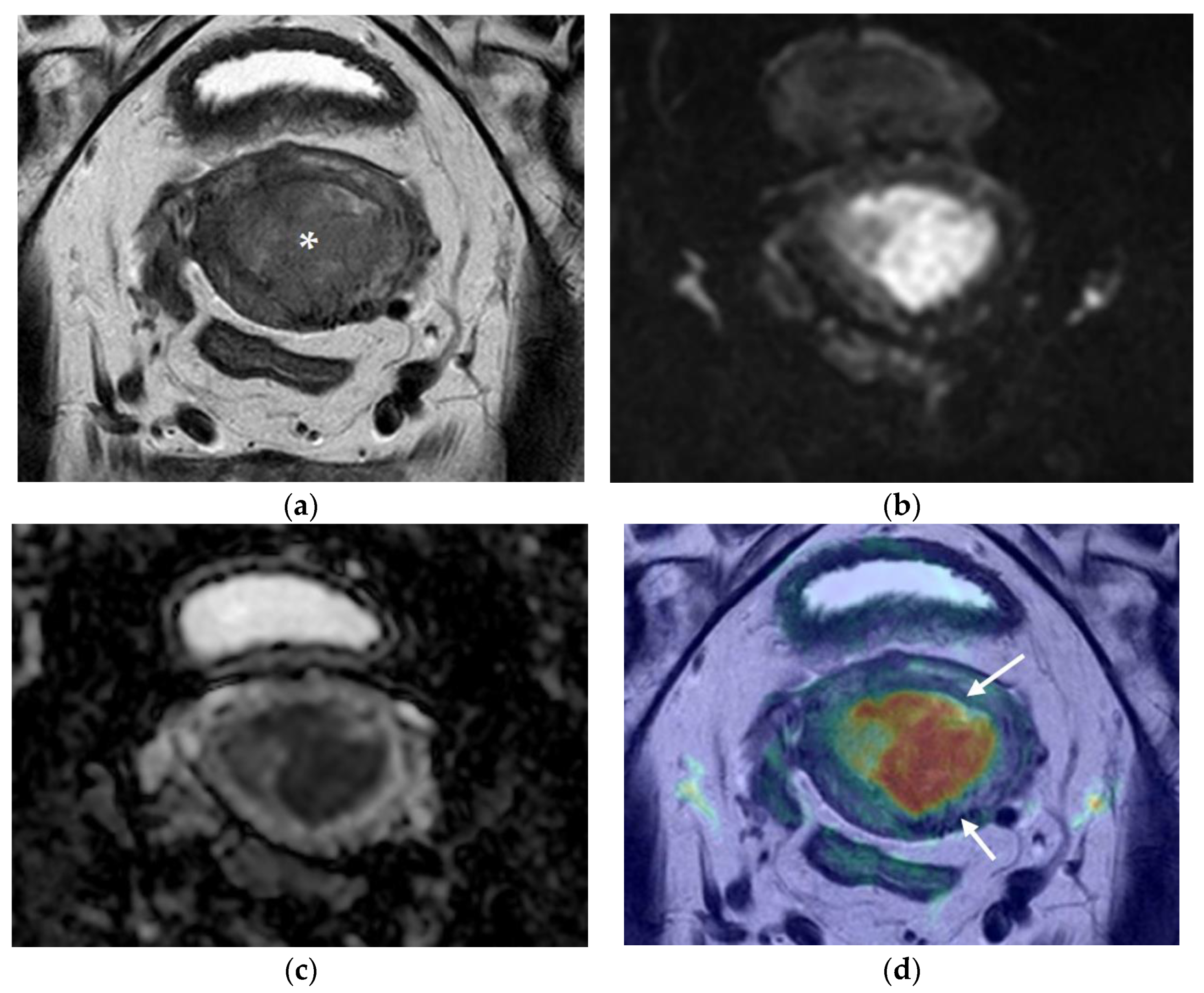
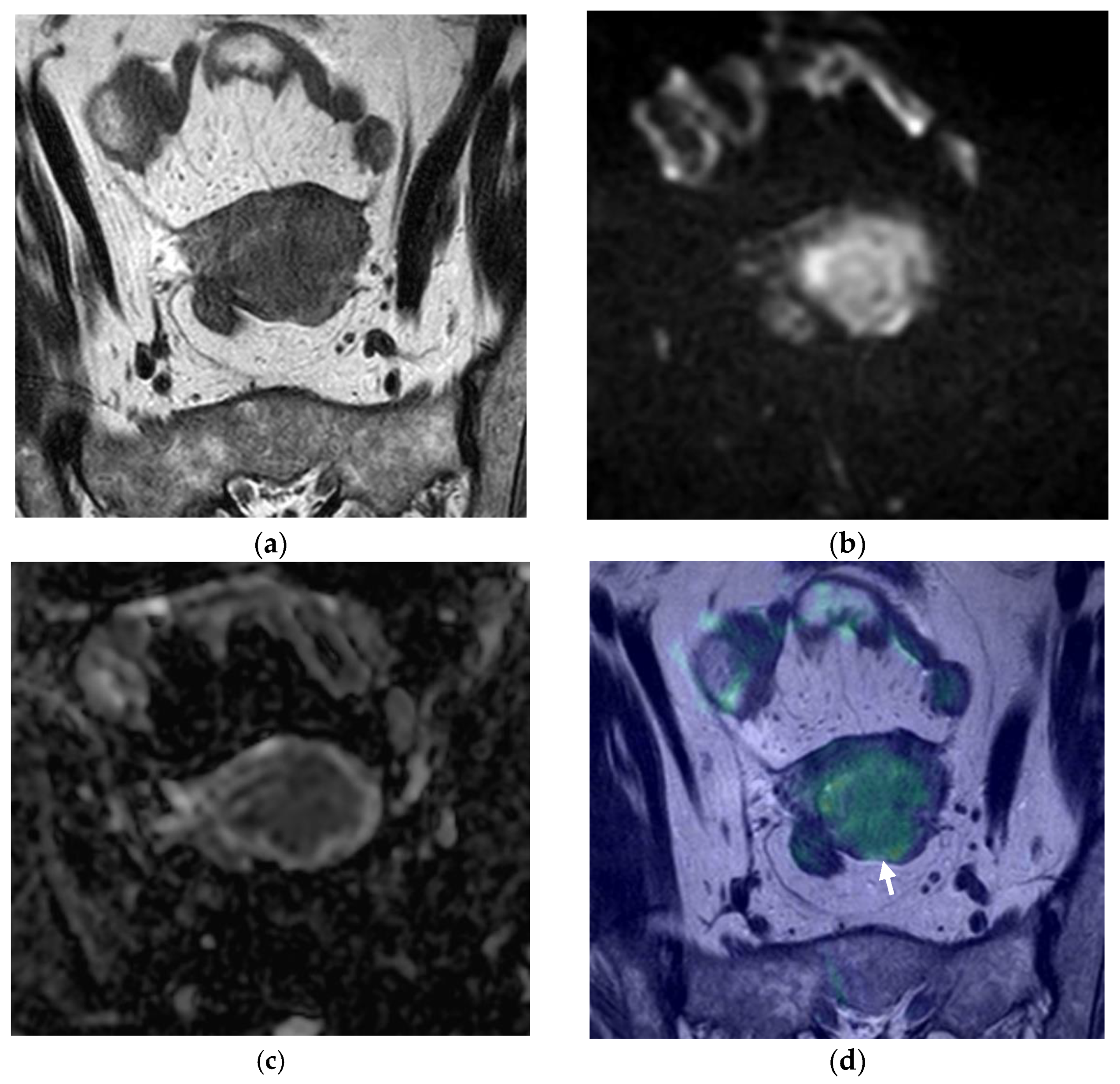
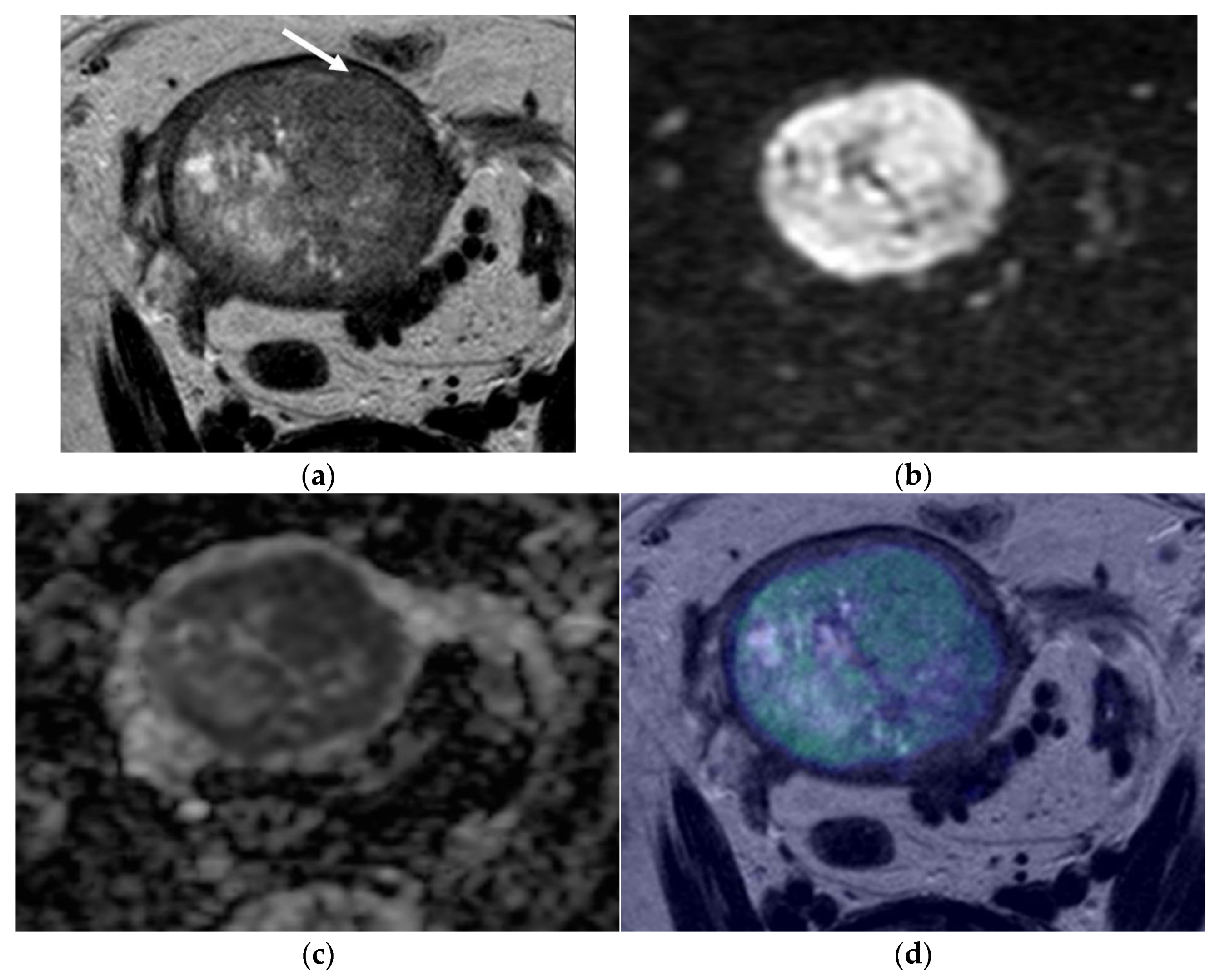
2. Results
3. Discussion
4. Materials and Methods
4.1. Patient Selection
4.2. MRI Protocol
4.3. MRI Analysis
4.4. Histological Evaluation
4.5. Statistical Analysis
5. Conclusions
Author Contributions
Funding
Institutional Review Board Statement
Informed Consent Statement
Data Availability Statement
Conflicts of Interest
References
- Observatory, G.C. Estimated Age-Standardized Incidence Rates (World) in 2020, Worldwide, Both Sexes, all Ages 2020. Available online: https://gco.iarc.fr/today/online-analysis-multi-bars?v=2020&mode=cancer&mode_population=countries&population=900&populations=900&key=asr&sex=0&cancer=39&type=0&statistic=5&prevalence=0&population_group=0&ages_group%5B%5D=0&ages_group%5B%5D=17&nb_items=10&group_cancer=1&include_nmsc=1&include_nmsc_other=1&type_multiple=%257B%2522inc%2522%253Atrue%252C%2522mort%2522%253Afalse%252C%2522prev%2522%253Afalse%257D&orientation=horizontal&type_sort=0&type_nb_items=%257B%2522top%2522%253Atrue%252C%2522bottom%2522%253Afalse%257D (accessed on 10 October 2021).
- Observatory, G.C. Estimated Age-Standardized Incidence Rates (World) in 2020, Worldwide, Females, all Ages 2020. Available online: https://gco.iarc.fr/today/online-analysis-multi-bars?v=2020&mode=cancer&mode_population=countries&population=900&populations=900&key=asr&sex=2&cancer=39&type=0&statistic=5&prevalence=0&population_group=0&ages_group%5B%5D=0&ages_group%5B%5D=17&nb_items=10&group_cancer=1&include_nmsc=1&include_nmsc_other=1&type_multiple=%257B%2522inc%2522%253Atrue%252C%2522mort%2522%253Afalse%252C%2522prev%2522%253Afalse%257D&orientation=horizontal&type_sort=0&type_nb_items=%257B%2522top%2522%253Atrue%252C%2522bottom%2522%253Afalse%257D (accessed on 10 October 2021).
- Gil, R.; Cunha, T.M.; Horta, M.; Alves, I. The added value of diffusion-weighted imaging in the preoperative assessment of endometrial cancer. Radiol Bras. 2019, 52, 229–236. [Google Scholar] [CrossRef] [PubMed] [Green Version]
- Expert Panel on GYN and OB Imaging; Reinhold, C.; Ueno, Y.; Akin, E.A.; Bhosale, P.R.; Dudiak, K.M.; Jhingran, A.; Kang, S.K.; Kilcoyne, A.; Lakhman, Y.; et al. ACR Appropriateness Criteria(R) Pretreatment Evaluation and Follow-Up of Endometrial Cancer. J. Am. Coll. Radiol. 2020, 17, S472–S486. [Google Scholar] [CrossRef] [PubMed]
- Meissnitzer, M.; Forstner, R. MRI of endometrium cancer—How we do it. Cancer Imaging 2016, 16, 11. [Google Scholar] [CrossRef] [PubMed] [Green Version]
- Lu, K.H.; Broaddus, R.R. Endometrial Cancer. N. Engl. J. Med. 2020, 383, 2053–2064. [Google Scholar] [CrossRef] [PubMed]
- Rizzo, S.; Femia, M.; Buscarino, V.; Franchi, D.; Garbi, A.; Zanagnolo, V.; Del Grande, M.; Manganaro, L.; Alessi, S.; Giannitto, C.; et al. Endometrial cancer: An overview of novelties in treatment and related imaging keypoints for local staging. Cancer Imaging 2018, 18, 45. [Google Scholar] [CrossRef] [PubMed]
- Santoro, A.; Angelico, G.; Travaglino, A.; Inzani, F.; Arciuolo, D.; Valente, M.; D’Alessandris, N.; Scaglione, G.; Fiorentino, V.; Raffone, A.; et al. New Pathological and Clinical Insights in Endometrial Cancer in View of the Updated ESGO/ESTRO/ESP Guidelines. Cancers 2021, 13, 2623. [Google Scholar] [CrossRef] [PubMed]
- Amant, F.; Mirza, M.R.; Koskas, M.; Creutzberg, C.L. Cancer of the corpus uteri. Int. J. Gynaecol. Obstet. 2018, 143 (Suppl. S2), 37–50. [Google Scholar] [CrossRef] [PubMed] [Green Version]
- Concin, N.; Matias-Guiu, X.; Vergote, I.; Cibula, D.; Mirza, M.R.; Marnitz, S.; Ledermann, J.; Bosse, T.; Chargari, C.; Fagotti, A.; et al. ESGO/ESTRO/ESP guidelines for the management of patients with endometrial carcinoma. Int. J. Gynecol. Cancer 2021, 31, 12–39. [Google Scholar] [CrossRef] [PubMed]
- Baiden-Amissah, R.E.M.; Annibali, D.; Tuyaerts, S.; Amant, F. Endometrial Cancer Molecular Characterization: The Key to Identifying High-Risk Patients and Defining Guidelines for Clinical Decision-Making? Cancers 2021, 13, 3988. [Google Scholar] [CrossRef] [PubMed]
- Sala, E.; Rockall, A.; Kubik-Huch, R.A. Advances in magnetic resonance imaging of endometrial cancer. Eur. Radiol. 2011, 21, 468–473. [Google Scholar] [CrossRef] [PubMed] [Green Version]
- Deng, L.; Wang, Q.P.; Chen, X.; Duan, X.Y.; Wang, W.; Guo, Y.M. The Combination of Diffusion- and T2-Weighted Imaging in Predicting Deep Myometrial Invasion of Endometrial Cancer: A Systematic Review and Meta-Analysis. J. Comput. Assist. Tomogr. 2015, 39, 661–673. [Google Scholar] [CrossRef] [PubMed]
- Rechichi, G.; Galimberti, S.; Signorelli, M.; Perego, P.; Valsecchi, M.G.; Sironi, S. Myometrial invasion in endometrial cancer: Diagnostic performance of diffusion-weighted MR imaging at 1.5-T. Eur. Radiol. 2010, 20, 754–762. [Google Scholar] [CrossRef] [PubMed]
- Ascher, S.M.; Reinhold, C. Imaging of cancer of the endometrium. Radiol. Clin. N. Am. 2002, 40, 563–576. [Google Scholar] [CrossRef]
- Larson, D.M.; Connor, G.P.; Broste, S.K.; Krawisz, B.R.; Johnson, K.K. Prognostic significance of gross myometrial invasion with endometrial cancer. Obstet. Gynecol. 1996, 88, 394–398. [Google Scholar] [CrossRef]
- Otero-García, M.M.; Mesa-Álvarez, A.; Nikolic, O.; Blanco-Lobato, P.; Basta-Nikolic, M.; De Llano-Ortega, R.M.; Paredes-Velázquez, L.; Nikolic, N.; Szewczyk-Bieda, M. Role of MRI in staging and follow-up of endometrial and cervical cancer: Pitfalls and mimickers. Insights Imaging. 2019, 10, 19. [Google Scholar] [CrossRef] [PubMed]
- Nougaret, S.; Horta, M.; Sala, E.; Lakhman, Y.; Thomassin-Naggara, I.; Kido, A.; Masselli, G.; Bharwani, N.; Sadowski, E.; Ertmer, A.; et al. Endometrial Cancer MRI staging: Updated Guidelines of the European Society of Urogenital Radiology. Eur. Radiol. 2019, 29, 792–805. [Google Scholar] [CrossRef] [PubMed]
- Guo, Y.; Wang, P.; Wang, P.; Gao, W.; Li, F.; Yang, X.; Ni, H.; Shen, W.; Guo, Z. Myometrial invasion and overall staging of endometrial carcinoma: Assessment using fusion of T2-weighted magnetic resonance imaging and diffusion-weighted magnetic resonance imaging. Onco Targets Ther. 2017, 10, 5937–5943. [Google Scholar] [CrossRef] [PubMed] [Green Version]
- Mohamed Shatat, O.M. Fusion of T2 Weighted MRI and diffusion weighted imaging in the evaluation of myometrial invasion in endometrial cancer. Erciyes Med. J. 2019, 41, 375–381. [Google Scholar] [CrossRef]


| Variable | Percentage (n) |
|---|---|
| Histological Subtype | |
| Endometrioid | 59.8 (52) |
| Nonendometrioid | 40.2 (35) |
| Serous carcinoma | 34.3 (12) |
| Clear cell carcinoma | 11.4 (4) |
| Mixed cell carcinoma | 42.9 (15) |
| Carcinossarcoma | 5.7 (2) |
| Non differentiated | 5.7 (2) |
| Tumor Grade | |
| 1 | 28.1 (16) |
| 2 | 38.6 (22) |
| 3 | 33.3 (19) |
| Myometrial invasion | |
| Superficial (<50%) | 52.9 (46) |
| Deep (≥50%) | 47.1 (41) |
| 2018 FIGO staging | |
| IA | 39.1 (34) |
| IB | 10.3 (9) |
| II | 9.2 (8) |
| III | 33.3 (29) |
| IVB | 8.0 (7) |
| MRI Sequence | Superficial Myometrial Invasion Percentage (n) | Deep Myometrial Invasion Percentage (n) |
|---|---|---|
| Standard MRI evaluation | 55.2 (48) | 44.8 (39) |
| Fused T2WI-DWI | 56.3 (49) | 43.7 (38) |
| Accuracy | Sensitivity | Specificity | Positive Predictive Value | Negative Predictive Value | |
|---|---|---|---|---|---|
| Standard MRI evaluation % (95% CI) | 81.6 | 78.1 | 84.5 | 82.1 | 81.2 |
| (71.6–89.1) | (62.4–89.4) | (71.1–93.7) | (69.4–90.2) | (70.6–88.7) | |
| Fused T2WI-DWI % (95% CI) | 89.7 | 85.4 | 93.5 | 92.1 | 87.8 |
| (81.3–95.2) | (70.8–94.4) | (82.1–98.6) | (79.5–97.2) | (77.3–93.8) |
| Study | Sensitivity % (95% CI) | Specificity % (95% CI) | Accuracy |
|---|---|---|---|
| Guo et al. | 92.3 | 95.6 | 94.8 |
| Shatat et al. | 90 | 94.7 | 93.1 |
| Our results | 85.4 | 93.5 | 89.7 |
| (70.8–94.4) | (82.1–98.6) | (81.3–95.2) |
Publisher’s Note: MDPI stays neutral with regard to jurisdictional claims in published maps and institutional affiliations. |
© 2022 by the authors. Licensee MDPI, Basel, Switzerland. This article is an open access article distributed under the terms and conditions of the Creative Commons Attribution (CC BY) license (https://creativecommons.org/licenses/by/4.0/).
Share and Cite
Neves, T.R.; Correia, M.T.; Serrado, M.A.; Horta, M.; Caetano, A.P.; Cunha, T.M. Staging of Endometrial Cancer Using Fusion T2-Weighted Images with Diffusion-Weighted Images: A Way to Avoid Gadolinium? Cancers 2022, 14, 384. https://doi.org/10.3390/cancers14020384
Neves TR, Correia MT, Serrado MA, Horta M, Caetano AP, Cunha TM. Staging of Endometrial Cancer Using Fusion T2-Weighted Images with Diffusion-Weighted Images: A Way to Avoid Gadolinium? Cancers. 2022; 14(2):384. https://doi.org/10.3390/cancers14020384
Chicago/Turabian StyleNeves, Teresa Resende, Mariana Tomé Correia, Maria Ana Serrado, Mariana Horta, António Proença Caetano, and Teresa Margarida Cunha. 2022. "Staging of Endometrial Cancer Using Fusion T2-Weighted Images with Diffusion-Weighted Images: A Way to Avoid Gadolinium?" Cancers 14, no. 2: 384. https://doi.org/10.3390/cancers14020384
APA StyleNeves, T. R., Correia, M. T., Serrado, M. A., Horta, M., Caetano, A. P., & Cunha, T. M. (2022). Staging of Endometrial Cancer Using Fusion T2-Weighted Images with Diffusion-Weighted Images: A Way to Avoid Gadolinium? Cancers, 14(2), 384. https://doi.org/10.3390/cancers14020384