Article

Efficacy and Optimal Pressure of Continuous Positive Airway Pressure in Intensity-Modulated Radiotherapy for Locally Advanced Lung Cancer

1
Department of Radiation Oncology, Yeungnam University Medical Center, Daegu 42415, Korea
2
Department of Radiation Oncology, Yeungnam University College of Medicine, Daegu 42415, Korea
3
Department of Radiation Oncology, School of Medicine, Kyungpook National University, Daegu 41944, Korea
*
Authors to whom correspondence should be addressed.
Cancers 2022, 14(17), 4308; https://doi.org/10.3390/cancers14174308
Submission received: 26 July 2022 / Revised: 31 August 2022 / Accepted: 31 August 2022 / Published: 2 September 2022

Abstract

:

Simple Summary

Radiation pneumonitis is a major late complication in radiotherapy (RT) for lung cancer. Respiratory gating radiotherapy and deep inspiration breath hold are representative techniques to protect the normal lung by managing the movement of the tumor. However, these are highly patient-dependent techniques. Continuous positive airway pressure (CPAP) is used as an alternative, but it is unclear how much pressure will be effective. We aimed to determine the optimal pressure of CPAP for RT through changes in the dosimetric parameters and lung volume according to pressure. The air pressure was raised in five steps of 4, 7, 10, 14, and 17 cmH2O and a CT scan was performed at the baseline and at each pressure step, accompanied by contouring and RT planning. CPAP linearly increased lung volume and decreased the dosimetric parameter in the pressure range 7 to 13 cmH2O (p < 0.01). Above 13 cmH2O, V5 of the heart also showed a significant decrease (p < 0.01).

Abstract

We aimed to determine the optimal pressure of continuous positive airway pressure (CPAP) for radiotherapy (RT) through changes in the dosimetric parameters and lung volume according to pressure. Patients with locally advanced lung cancer, who underwent CPAP during computed tomography (CT) simulation, were included. The air pressure was raised in five steps of 4, 7, 10, 14, and 17 cmH2O and a CT scan was performed at the baseline and at each pressure step, accompanied by contouring and RT planning. Paired t- and Wilcoxon signed rank tests were used to compare the volumetric and dosimetric parameters according to pressure and interpressure. A total of 29 patients were selected, and 158 CT datasets were obtained. The lung volume increased significantly at all pressures (p < 0.01). The Dmean of the lung decreased significantly from 7 cmH2O (p < 0.01), the V5, V10, V15, and V20 of the lung decreased significantly from 7 cmH2O with increasing pressure, and the Dmean and V5 of the heart decreased significantly from 14 cmH2O with increasing pressure. The V50 showed no significant differences at any pressure. We recommend the use of at least 7 cmH2O with 14 cmH2O as the optimal pressure to achieve the effect of heart preservation.

Graphical Abstract

1. Introduction

Lung cancer is the most commonly diagnosed cancer and the leading cause of cancer-related deaths worldwide [1]. Radiotherapy (RT) is an important modality in the treatment of lung cancer, such as stereotactic body radiotherapy (SBRT) for inoperable early-stage non-small cell lung cancer (NSCLC) and concurrent chemoradiotherapy (CCRT) for locally advanced NSCLC or limited-disease small cell lung cancer (LD-SCLC).
Radiation pneumonitis (RP) is a major late complication and an obstacle to treatment. As a result, interest in treatment-related pneumonitis is increasing. Symptomatic RP occurs in 15–40% of patients receiving CCRT for NSCLC [2,3]. In particular, the use of durvalumab, an anti-programmed death ligament 1 (PD-L1) inhibitor after CCRT, has become the standard treatment for unresected stage III NSCLC. Some prospective studies have reported increased pulmonary toxicity in patients who received RT with PD-1 or PD-L1 inhibitors [4,5].
Many efforts have been made to identify the predictive factors related to the incidence of RP. The Quantitative Analysis of Normal Tissue Effect in Clinic (QUANTEC) project reviewed more than 70 published articles and presented normal tissue constraints for radiation-induced lung injury [6]. According to this project, dosimetric parameters for the risk of RP <20% were suggested as mean lung dose (MLD) <20 Gy, V20 (the percentage of the lung volume receiving at least 20 Gy) <35%, and V5 (the percentage of the lung volume receiving at least 5 Gy) <60% in conventional fractionation.
The lung is an organ that expands and contracts repeatedly with respiration, and a lung cancer tumor that is located within it also moves with respiration. Intra-fractional respiratory-related motion causes uncertainties in the target coverage and dose delivery. To cover these movements, the treatment volume is increased, with the volume of the normal lung irradiated also being increased accordingly. Respiratory gating radiotherapy (RGRT) and deep inspiration breath hold (DIBH) are representative techniques for reducing the dose irradiated to the normal lung by managing the movement of the tumor. However, there are several conditions for using these techniques. Irregular breathing causes poor dosimetric results in the RGRT. Therefore, a regular respiratory cycle is required for the stability and reproducibility of the treatment. DIBH also requires the same reproducible inspiration level during the simulation and treatment. In other words, both techniques are highly patient-dependent and require patient cooperation and education for proper treatment.
Continuous positive airway pressure (CPAP) is a form of positive airway pressure ventilation that delivers a constant level of pressure to the airway to prevent airway collapse. It has been used as a safe and effective treatment for patients with sleep apnea [7,8], and has recently been used for chronic obstructive pulmonary disease (COPD) [9] and to prevent postoperative atelectasis [10]. CPAP causes the airway to be continuously open and, consequently, lung inflated compared to free breathing. Several attempts have been made to use this effect for RT, which were mainly performed in patients with left breast cancer and lymphoma who were unable to undergo DIBH [11,12]. In a study of patients with left breast cancer, CPAP increased the lung volume by 35% and decreased the mean dose of the heart and ipsilateral lung and V20 of the lung by 47%, 20%, and 25%, respectively [13].
Most previous studies have focused on patients with left breast cancer, and there are no studies on the dosimetric improvement of the CPAP application in RT for locally advanced lung cancer. In addition, there are no studies on how much pressure has a meaningful effect on the dosimetric outcomes. Therefore, we conducted a study to determine the optimal pressure of CPAP for RT through changes in the dosimetric parameters and lung volume according to pressure.

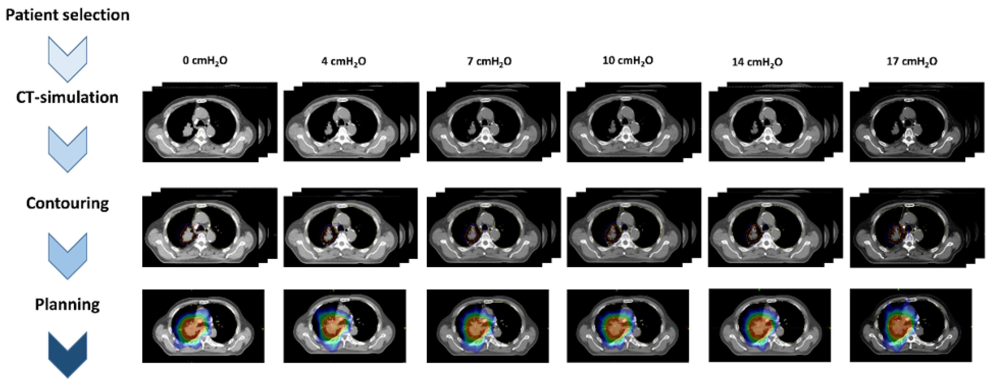
2. Materials and Methods
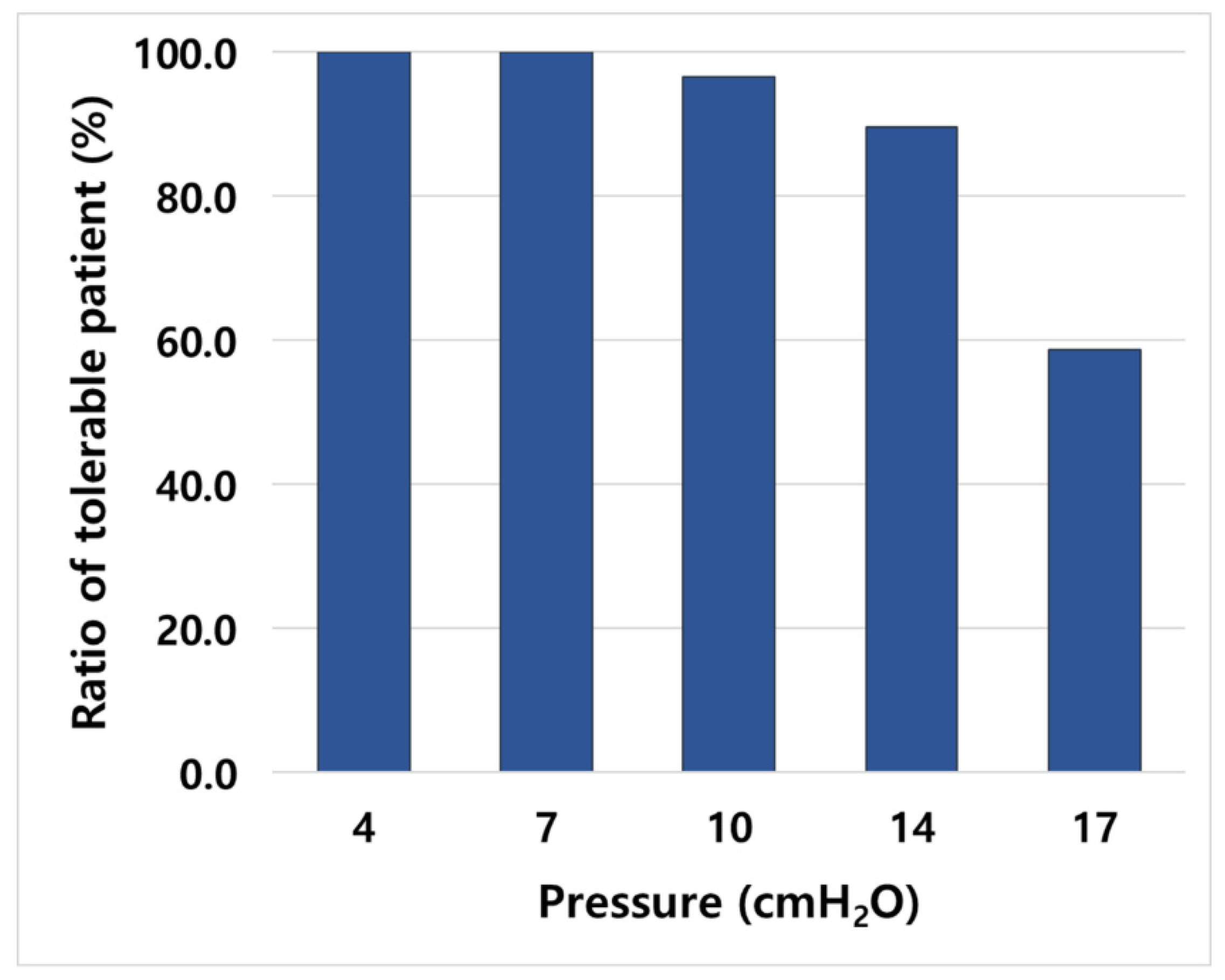
2.1. Patients

This study was approved by the Institutional Review Board (2021-08-058). The progress of this study is summarized in Figure 1. We selected patients with locally advanced lung cancer who underwent CPAP during computed tomography (CT) simulation between July 2019 and December 2020. A total of 29 patients were included in the study. Table 1 shows the patient characteristics. The median age was 67 years (range, 51–82). There were 22 patients with NSCLC (76%) and 7 patients with SCLC (24%). Most tumors (59%) were stage IIIA according to the American Joint Committee on Cancer (AJCC) 8th edition. The most common lesions were in the upper lobe; 14 patients had lesions in the RUL, and 7 in the LUL. Thirteen patients (44%) had respiratory comorbidities.

2.2. Application of CPAP on CT-Simulation

We used AirSenseTM10 (ResMed, San Diego, CA, USA) as the CPAP machine, and all the patients wore CPAP masks that fitted their face sizes. These were worn for 5–10 min at a pressure of 4 cmH2O for adaptation to the CPAP before the CT simulation. All patients were simulated in the supine position with the arm up and immobilized with a Vac-lok bag and wing board. The air pressure was raised step-by-step to the patient’s individual tolerable pressure in five steps of 4, 7, 10, 14, and 17 cmH2O. We used the Brilliance Big Bore CT simulator (Philips Inc., Cleveland, OH) and a CT scan with a 2.5-mm slice thickness was performed at the baseline and each pressure step.

2.3. Contouring and RT Planning

All CT datasets were transferred to a commercial treatment planning system (Varian Eclipse TPS version 15.6.05, Palo Alto, CA, USA). The delineation of the target volume and normal organs, including the esophagus and heart, was conducted by an experienced radiation oncologist (J.P.). The lung contouring was performed using auto-segmentation of the TPS. The gross target volume (GTV) was defined as primary lesions and lymph nodes that showed positive findings on the positron emission tomography-CT and contrast-enhanced chest CT. This was expanded with a 5-mm margin to generate the clinical target volume (CTV). The planning target volume (PTV) was obtained by adding a craniocaudal 7-mm and radial 5-mm margin to the CTV.
A treatment plan was generated for each CT set using an anisotropic analytic algorithm. The prescribed radiation dose was 66 Gy in 33 fractions with a fraction size of 2 Gy delivered to the PTV, with 95% of the PTV being covered by the prescription dose. The dose constraints for the organs at risk (OAR) are summarized in Table S1. Volumetric modulated arc therapy was used in 27 patients, and nine-field static intensity-modulated radiotherapy was used in 2 patients.

2.4. Statistics

Paired t- and Wilcoxon signed rank tests were used to measure meaningful changes in the dosimetric outcomes, including lung volumes, lung-dose parameters (Dmean, V5, V10, V15, and V20), and heart dose parameters (Dmean, V5, and V50).
The difference in the parameters according to pressure was defined as follows:
Relative   change   % = P x P 0 P 0 × 100
where Px was the parameter in pressure x, and P0 the parameter at baseline. The difference in the parameter between the interpressure was defined as follows:
Relative   change   % = P x + 1 P x P x × 100
where Px+1 was a parameter in pressure step x + 1 and Px a parameter in pressure x. All the statistical analyses were performed using SPSS ver. 25.0 (SPSS Inc., Chicago, IL, USA).

3. Results

A total of 158 CT datasets were obtained with 1, 2, 9, and 17 patients who tolerated up to 7, 10, 14, and 17 cmH2O, respectively. Cumulatively, approximately 90% of the patients tolerated up to 14 cmH2O, and only 59% of the patients tolerated up to 17 cmH2O (Figure 2).
The volume of GTV, CTV and PTV at baseline were 77.3 ± 48.4 cm3, 161.7 ± 78.3 cm3 and 292.3 ± 129.0 cm3, respectively. There were no statistically significant differences in the target volumes according to the pressure (Table 2).

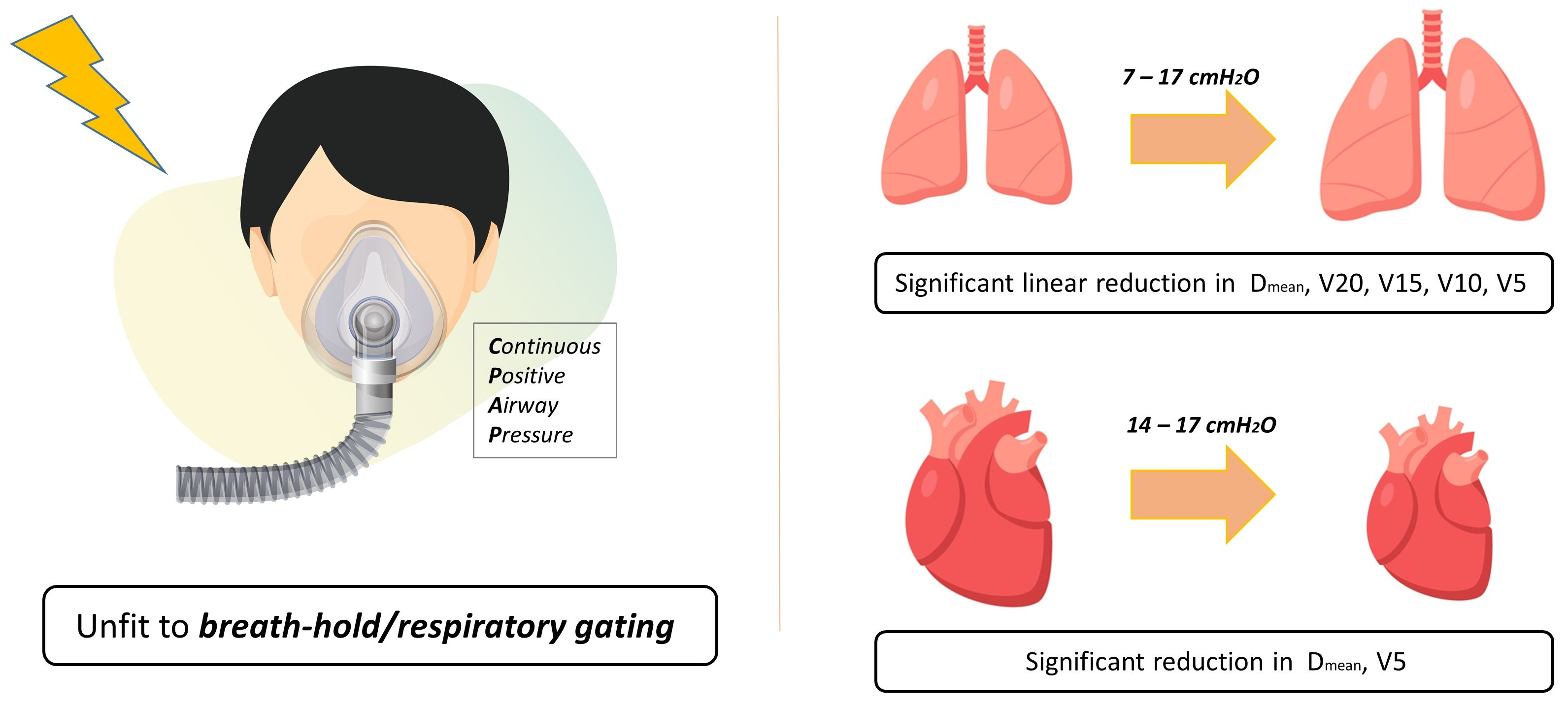
3.1. Change in Lung Volume and Dose Parameters

The volumes of the lung were 3318.0 ± 742.4 cm3, 3575.4 ± 762.1 cm3, 3840.1 ± 764.5 cm3, 4148.9 ± 820.5 cm3, 44529.5 ± 905.9 cm3 and 4883.2 ± 799.4 cm3 from baseline in the order of 4, 7, 10, 14 and 17 cmH2O (Table 2). The lung volume increased significantly at all pressures, and the increase was linear (Figure 3A). It increased by approximately 8% with each increase in pressure step; however, the degree of increase decreased to 3% when increasing from 14 to 17 cmH2O (Figure S1(A1)).
The Dmean for the lung were 1573.5 ± 314.0 cGy, 1545.9 ± 327.7 cGy, 1495.3 ± 330.8 cGy, 1455.9 ± 334.6 cGy, 1392.8 ± 304.4 cGy and 1443.4 ± 268.7 cGy from baseline in the order of 4, 7, 10, 14 and 17 cmH2O (Table 2). It decreased significantly from 7 cmH2O and decreased by 5.1–11.9% as the pressure increased (Figure 3B). Significant decreases were observed when the interpressure was increased from 4 to 7 cmH2O and from 10 to 14 cmH2O (Figure S1(A2)).
V5, V10, V15, and V20 also showed a significant decrease from 7 cmH2O with decreases of 3.2–12.6%, 4.8–12.7%, 6.3–13.8%, and 5.6–11.9%, respectively, with increasing pressure (Figure 3C–F). With the change in the interpressure, V5 decreased significantly only when increasing from 10 to 14 cmH2O, and V10, V15, and V20 decreased significantly only when increasing from 4 to 7 cmH2O (Figure S1(A3–A6)).

3.2. Change in Heart Volume and Dose Parameters

The volumes of the heart were 725.6 ± 155.4 cm3, 719.3 ± 162.0 cm3, 702.6 ± 167.5 cm3, 683.8 ± 134.5 cm3, 679.0 ± 109.3 cm3, and 622.8 ± 102.4 cm3 from baseline in the order of 4, 7, 10, 14, and 17 cmH2O (Table 2). The heart volume decreased significantly at 14 and 17 cmH2O.
Dmean and V5 showed significant decreases from 14 cmH2O, with decreases of 5.4–11.1% and 7.4–9.2%, respectively, with increasing pressure (Figure 4A,B). V50 showed no significant differences at any pressure (Figure 4C). In the analysis of the interpressure, there were no intervals showing significant differences in Dmean, V5, or V50 (Figure S2(B1–B3)).

4. Discussion

In this study, changes in the lung volumes and dosimetric parameters according to pressure were confirmed using conventional RT for lung cancer. To control the effect of the target volume on the RT plan, one radiation oncologist performed the contouring, and there were no significant differences in the pressure. The lung volume increased linearly with pressure, and the dose parameters, including Dmean, V5, V10, V15, and V20, showed a linear tendency to decrease. While the change in lung volumes was significant at all pressures, all the dose parameters showed a significant change from 7 cmH2O. The volume increase in the interpressure was approximately 8%, but when the pressure was increased from 14 to 17 cmH2O, the increase decreased to 3%. In addition, the proportion of tolerable patients was reduced from 90% to 59%. Based on these results, we suggest that in conventional RT for lung cancer, an appropriate pressure range is 7–14 cmH2O.
The incidence of RP was reported to be 9.1% higher with the combination of durvalumab and CCRT compared to CCRT alone in the PACIFIC trial [14], raising interest in RP in conventional RT for lung cancer. Wang et al. investigated the factors associated with RP in NSCLC patients after CCRT [15]. Lung volume, mean lung dose, V5 through V65, and target volume were all significant factors for RP in the univariate analysis, but only V5 was significant in the multivariate analysis. The 1-year incidence of grade 3 ≥ RP with V5 ≤ 42% and V5 > 42% was 3% and 38%, respectively (p < 0.01). Palma et al. also reported predictive factors of RP in patients undergoing CCRT for NSCLC through an individual patient data meta-analysis [3]. They suggested that the V20 of the lung, carboplatin/paclitaxel chemotherapy, and age were predictive factors.
Since the lung cancer tumor is located in the lungs and it moves according to respiration, it is necessary to manage its movement to reduce the irradiation volume of normal lungs. RGRT is commonly used for motion management. RGRT is a method of irradiating only a specific respiratory window, so the target volume can be reduced compared to free breathing [16]. Rouabhi et al. reported that RGRT could reduce the mean lung dose and V20 by 6–16% and 7–20%, respectively, compared to the non-gated plans [17]. However, this method requires patient collaboration and a regular breathing pattern. Irregular breathing causes geometric variations, which have a significant impact on the accuracy of target contouring and treatment [18]. In addition, end-expiration is preferred to end-inspiration for the gating window because of the stability and smallness of tumor movement [19]. Expiration is the densest state of the lungs and the benefit of RGRT may be dampened, due to the large amount of lung parenchyma per volume.
In contrast, DIBH, another commonly used technique, is a method of sparing the lungs using lung expansion at deep inspiration and tumor immobilization by breath-holding. This method has been studied mainly in breast cancer to reduce the heart dose [20,21,22]. Swanson et al. reported that DIBH also reduced ipsilateral lung parameters, such as mean lung dose, V5, V10, V15, and V20 by 5–16%, significantly [21]. The efficacy of DIBH has also been confirmed in lung cancer. Rosenzweig et al. showed that the normal tissue complication probability (NTCP) of radiation pneumonitis decreased in patients with DIBH, compared to patients with free breathing, in conventional RT for NSCLC [23]. Hanley et al. reported high reproducibility of DIBH with 1 mm and 2.5 mm in the deviation of intra breath-hold and inter breath-hold, respectively, as well as improved dose parameters of lung in conventional RT for lung cancer [24]. However, patient cooperation is essential for stable and reproducible treatment. In the INHALE trial, 28% of the patients were unable to perform DIBH throughout the treatment course [25].
Most of the patients enrolled in our study were elderly, and 44% of patients tolerated CPAP well, even though they had respiratory comorbidities. CPAP supplies positive pressure to keep the airways open, allowing the lungs to be inflated continuously. Thus, a similar effect to DIBH can be expected. Kil et al. used CPAP in patients with breast cancer who had difficulty in performing DIBH [12,13]. A pressure of 8–15 cmH2O was used. Compared with free breathing, the total lung volume was increased by 30% (p < 0.01), and the Dmean and V20 of the ipsilateral lung decreased by 20% and 25%, respectively (p = 0.03, both). The Dmean and V25 of the heart also decreased by 47% and 87%, respectively (p < 0.01).
Another advantage of using CPAP in RT is that it is economical. In using CPAP in RT, no special equipment is required, except for the CPAP machine and a mask for each patient. The CPAP machine we used was approximately USD 1500 and a mask was USD 250, which is a very low cost compared to a real-time position management system or the surface guided radiation therapy for DIBH or RGRT, which costs hundreds of thousands of dollars to equip. Therefore, it can be used as an effective method for protecting the lung and heart in radiation oncology departments in a financially constrained environment.
Goldstein et al. conducted a controlled pilot study to confirm the effect of CPAP on SBRT [26]. They used a pressure of 10–15 cmH2O, and most of the patients tolerated CPAP. CPAP significantly decreased tumor movement in all directions (superior–inferior, 0.5 ± 0.8 cm; right–left, 0.4 ± 0.7 cm; anterior–posterior, 0.6 ± 0.8 cm). A relative increase of 32% in lung volume was observed. Our study showed that the relative increases in the lungs at 10 and 14 cmH2O were 26% and 36%, respectively, considering the pressure range they used, and the degree of lung expansion appeared to be consistent. The Dmean of the lung and heart also showed relative reductions of 22% and 29%, respectively, which were larger than those of approximately 12% and 11% at 17 cmH2O in our study. This difference was due to the large target volume because our study investigated conventional RT for stages II-III.
The heart-sparing effect of CPAP was also observed in the present study. The dosimetric parameters of the heart are known to be associated with lymphopenia, which is an important prognostic factor in lung cancer [27]. The mean dose and low-dose exposure area of the heart also decreased significantly at 14 cmH2O or higher. However, V50, a high-dose exposure area of the heart, did not show significant improvement even at high pressure. This result is presumably because most of the patients included in this study had upper lobe lesions.
Recently, Wiezman et al. evaluated the volume, position, and motion of the heart at free-breathing and 15 cmH2O to determine the mechanism of dosimetric improvement from CPAP [28]. They reported a 6% reduction in heart volume (p < 0.008), 1 cm of caudal displacement (p < 0.008), and a 49% reduction in movement (p < 0.01) with CPAP. Among them, the caudal displacement of the heart was correlated with increased lung volume. In our study, there was also a significant decrease in heart volume at pressures above 14 cmH2O. We did not expect a significant change in the heart volume by the airway pressure, since it depends on the blood volume according to the cardiac cycle. It may have been caused by a reduction in the amount of blood flow into the heart, due to an increase in intrathoracic pressure.
There were some limitations to this study. First, the selection of the tumor location was biased. Since the patients were selected retrospectively, 76% of the patients had tumors in the upper lobe. Therefore, we were unable to analyze how the effects of CPAP differed according to tumor location. Second, four-dimensional CT (4D-CT) was not used for the CT simulation. Four-dimensional CT can determine the extent of tumor movement during respiration. The degree of reduction in tumor movement according to pressure makes it possible to set the PTV margin for each pressure, which can potentially further reduce the irradiation dose to normal organs, such as the lungs and heart. Third, this study focused on the changes in lung volume and dosimetric parameters according to pressure in the simulation. It was not clear whether the lung volume increased, similar to the simulation, when the same pressure is applied during treatment. For practical application, further studies are needed to confirm whether the lung volume increases consistently when the same pressure is applied during treatment.

5. Conclusions

This is the first study to demonstrate the relationship of pressure with lung volume and dosimetric parameters. Considering the increase in lung volume, the decrease in the dosimetric parameters, the proportion of tolerable patients, and the change in lung volume between pressures, we recommend using at least 7 cmH2O. In conclusion, a pressure of 14 cmH2O, which can even achieve the effect of preserving the heart, can be considered optimal.

Supplementary Materials

The following supporting information can be downloaded at: https://www.mdpi.com/article/10.3390/cancers14174308/s1, Figure S1: Lung volume and dose parameter change between interpressure; (A1) volume, (A2) Dmean, (A3) V5, (A4) V10, (A5) V15, and (A6) V20; Figure S2: Heart dose parameter change between interpressure; (B1) Dmean, (B2) V5, and (B3) V50; Table S1: Dose constraints for the organs at risk.

Author Contributions

Conceptualization, J.P. (Jaehyeon Park) and J.W.P.; methodology, J.P. (Jaehyeon Park), J.W.Y. and J.W.P.; validation, S.A.O. and J.P. (Jongmoo Park); formal analysis, J.P. (Jaehyeon Park); investigation, J.P. (Jaehyeon Park), S.A.O., J.W.Y. and J.W.P.; data curation, J.P. (Jongmoo Park); writing—original draft preparation, J.P. (Jaehyeon Park); writing—review and editing, J.W.P. and J.E.L.; supervision, J.W.P. and J.E.L. All authors have read and agreed to the published version of the manuscript.

Funding

The work was supported by the 2021 Yenungnam University Research Grant [221A580039].

Institutional Review Board Statement

The study was conducted in accordance with the Declaration of Helsinki, and approved by the Institutional Review Board of Yenungnam University Medical Center (2021-08-058).

Informed Consent Statement

Patient consent was waived due to the retrospective nature of the analysis.

Data Availability Statement

The data presented in this study are available upon request from the corresponding author.

Conflicts of Interest

The authors declare no conflict of interest.

References

  1. Bray, F.; Ferlay, J.; Soerjomataram, I.; Siegel, R.L.; Torre, L.A.; Jemal, A. Global cancer statistics 2018: GLOBOCAN estimates of incidence and mortality worldwide for 36 cancers in 185 countries. CA Cancer J. Clin. 2018, 68, 394–424. [Google Scholar] [CrossRef] [PubMed]
  2. Rodrigues, G.; Lock, M.; D’Souza, D.; Yu, E.; Van Dyk, J. Prediction of radiation pneumonitis by dose-volume histogram parameters in lung cancer—a systematic review. Radiother. Oncol. 2004, 71, 127–138. [Google Scholar] [CrossRef] [PubMed]
  3. Palma, D.A.; Senan, S.; Tsujino, K.; Barriger, R.B.; Rengan, R.; Moreno, M.; Bradley, J.D.; Kim, T.H.; Ramella, S.; Marks, L.B.; et al. Predicting radiation pneumonitis after chemoradiation therapy for lung cancer: An international individual patient data meta-analysis. Int. J. Radiat. Oncol. Biol. Phys. 2013, 85, 444–450. [Google Scholar] [CrossRef] [PubMed]
  4. Shaverdian, N.; Lisberg, A.E.; Bornazyan, K.; Veruttipong, D.; Goldman, J.W.; Formenti, S.C.; Garon, E.B.; Lee, P. Previous radiotherapy and the clinical activity and toxicity of pembrolizumab in the treatment of non-small-cell lung cancer: A secondary analysis of the KEYNOTE-001 phase 1 trial. Lancet Oncol. 2017, 18, 895–903. [Google Scholar] [CrossRef]
  5. Theelen, W.; Peulen, H.M.U.; Lalezari, F.; van der Noort, V.; de Vries, J.F.; Aerts, J.; Dumoulin, D.W.; Bahce, I.; Niemeijer, A.N.; de Langen, A.J.; et al. Effect of Pembrolizumab After Stereotactic Body Radiotherapy vs. Pembrolizumab Alone on Tumor Response in Patients with Advanced Non-Small Cell Lung Cancer: Results of the PEMBRO-RT Phase 2 Randomized Clinical Trial. JAMA Oncol. 2019, 5, 1276–1282. [Google Scholar] [CrossRef]
  6. Marks, L.B.; Bentzen, S.M.; Deasy, J.O.; Kong, F.M.; Bradley, J.D.; Vogelius, I.S.; El Naqa, I.; Hubbs, J.L.; Lebesque, J.V.; Timmerman, R.D.; et al. Radiation dose-volume effects in the lung. Int. J. Radiat. Oncol. Biol. Phys. 2010, 76, S70–S76. [Google Scholar] [CrossRef]
  7. Becker, H.F.; Jerrentrup, A.; Ploch, T.; Grote, L.; Penzel, T.; Sullivan, C.E.; Peter, J.H. Effect of nasal continuous positive airway pressure treatment on blood pressure in patients with obstructive sleep apnea. Circulation 2003, 107, 68–73. [Google Scholar] [CrossRef]
  8. Epstein, L.J.; Kristo, D.; Strollo, P.J., Jr.; Friedman, N.; Malhotra, A.; Patil, S.P.; Ramar, K.; Rogers, R.; Schwab, R.J.; Weaver, E.M.; et al. Clinical guideline for the evaluation, management and long-term care of obstructive sleep apnea in adults. J. Clin. Sleep Med. 2009, 5, 263–276. [Google Scholar]
  9. O’Donoghue, F.J.; Catcheside, P.G.; Jordan, A.S.; Bersten, A.D.; McEvoy, R.D. Effect of CPAP on intrinsic PEEP, inspiratory effort, and lung volume in severe stable COPD. Thorax 2002, 57, 533–539. [Google Scholar] [CrossRef]
  10. Ferreyra, G.P.; Baussano, I.; Squadrone, V.; Richiardi, L.; Marchiaro, G.; Del Sorbo, L.; Mascia, L.; Merletti, F.; Ranieri, V.M. Continuous positive airway pressure for treatment of respiratory complications after abdominal surgery: A systematic review and meta-analysis. Ann. Surg. 2008, 247, 617–626. [Google Scholar] [CrossRef]
  11. Kil, W.J. Novel Use of Continuous Positive Airway Pressure Permits Heart- and Lung-Sparing Mediastinal Involved-Site Radiation Therapy for Patient with Hodgkin Lymphoma in Community Clinic When Deep Inspiration Breath-Hold Technique Is Not Available. Pract. Radiat. Oncol. 2019, 9, 142–146. [Google Scholar] [CrossRef] [PubMed]
  12. Kil, W.J.; Pham, T.; Hossain, S.; Casaigne, J.; Jones, K.; Khalil, M. The impact of continuous positive airway pressure on radiation dose to heart and lung during left-sided postmastectomy radiotherapy when deep inspiration breath hold technique is not applicable: A case report. Radiat. Oncol. J. 2018, 36, 79–84. [Google Scholar] [CrossRef] [PubMed]
  13. Kil, W.J.; Lee, I.J.; Pham, T.; Cho, I.H. Practical Heart Sparing Breast Cancer Radiation Therapy Using Continuous Positive Airway Pressure (CPAP) in Resource-Limited Radiation Oncology Clinics. Am. J. Clin. Oncol. 2019, 42, 797–801. [Google Scholar] [CrossRef] [PubMed]
  14. Antonia, S.J.; Villegas, A.; Daniel, D.; Vicente, D.; Murakami, S.; Hui, R.; Yokoi, T.; Chiappori, A.; Lee, K.H.; de Wit, M.; et al. Durvalumab after Chemoradiotherapy in Stage III Non-Small-Cell Lung Cancer. N. Engl. J. Med. 2017, 377, 1919–1929. [Google Scholar] [CrossRef]
  15. Wang, S.; Liao, Z.; Wei, X.; Liu, H.H.; Tucker, S.L.; Hu, C.S.; Mohan, R.; Cox, J.D.; Komaki, R. Analysis of clinical and dosimetric factors associated with treatment-related pneumonitis (TRP) in patients with non-small-cell lung cancer (NSCLC) treated with concurrent chemotherapy and three-dimensional conformal radiotherapy (3D-CRT). Int. J. Radiat. Oncol. Biol. Phys. 2006, 66, 1399–1407. [Google Scholar] [CrossRef]
  16. Lin, H.; Lu, H.; Shu, L.; Huang, H.; Chen, H.; Chen, J.; Cheng, J.; Pang, Q.; Peng, L.; Gu, J.; et al. Dosimetric study of a respiratory gating technique based on four-dimensional computed tomography in non-small-cell lung cancer. J. Radiat. Res. 2014, 55, 583–588. [Google Scholar] [CrossRef]
  17. Rouabhi, O.; Gross, B.; Bayouth, J.; Xia, J. The Dosimetric and Temporal Effects of Respiratory-Gated, High-Dose-Rate Radiation Therapy in Patients with Lung Cancer. Technol. Cancer Res. Treat. 2019, 18, 1533033818816072. [Google Scholar] [CrossRef]
  18. Pan, C.H.; Shiau, A.C.; Li, K.C.; Hsu, S.H.; Liang, J.A. The irregular breathing effect on target volume and coverage for lung stereotactic body radiotherapy. J. Appl. Clin. Med. Phys. 2019, 20, 109–120. [Google Scholar] [CrossRef]
  19. Berbeco, R.I.; Nishioka, S.; Shirato, H.; Jiang, S.B. Residual motion of lung tumors in end-of-inhale respiratory gated radiotherapy based on external surrogates. Med. Phys. 2006, 33, 4149–4156. [Google Scholar] [CrossRef]
  20. Chen, M.H.; Chuang, M.L.; Bornstein, B.A.; Gelman, R.; Harris, J.R.; Manning, W.J. Impact of respiratory maneuvers on cardiac volume within left-breast radiation portals. Circulation 1997, 96, 3269–3272. [Google Scholar] [CrossRef]
  21. Swanson, T.; Grills, I.S.; Ye, H.; Entwistle, A.; Teahan, M.; Letts, N.; Yan, D.; Duquette, J.; Vicini, F.A. Six-year experience routinely using moderate deep inspiration breath-hold for the reduction of cardiac dose in left-sided breast irradiation for patients with early-stage or locally advanced breast cancer. Am J. Clin. Oncol. 2013, 36, 24–30. [Google Scholar] [CrossRef] [Green Version]
  22. Lu, H.M.; Cash, E.; Chen, M.H.; Chin, L.; Manning, W.J.; Harris, J.; Bornstein, B. Reduction of cardiac volume in left-breast treatment fields by respiratory maneuvers: A CT study. Int. J. Radiat. Oncol. Biol. Phys. 2000, 47, 895–904. [Google Scholar] [CrossRef]
  23. Rosenzweig, K.E.; Hanley, J.; Mah, D.; Mageras, G.; Hunt, M.; Toner, S.; Burman, C.; Ling, C.C.; Mychalczak, B.; Fuks, Z.; et al. The deep inspiration breath-hold technique in the treatment of inoperable non-small-cell lung cancer. Int. J. Radiat. Oncol. Biol. Phys. 2000, 48, 81–87. [Google Scholar] [CrossRef]
  24. Hanley, J.; Debois, M.M.; Mah, D.; Mageras, G.S.; Raben, A.; Rosenzweig, K.; Mychalczak, B.; Schwartz, L.H.; Gloeggler, P.J.; Lutz, W.; et al. Deep inspiration breath-hold technique for lung tumors: The potential value of target immobilization and reduced lung density in dose escalation. Int. J. Radiat. Oncol. Biol. Phys. 1999, 45, 603–611. [Google Scholar] [CrossRef]
  25. Josipovic, M.; Aznar, M.C.; Thomsen, J.B.; Scherman, J.; Damkjaer, S.M.; Nygård, L.; Specht, L.; Pøhl, M.; Persson, G.F. Deep inspiration breath hold in locally advanced lung cancer radiotherapy: Validation of intrafractional geometric uncertainties in the INHALE trial. Br. J. Radiol 2019, 92, 569. [Google Scholar] [CrossRef] [PubMed]
  26. Goldstein, J.D.; Lawrence, Y.R.; Appel, S.; Landau, E.; Ben-David, M.A.; Rabin, T.; Benayun, M.; Dubinski, S.; Weizman, N.; Alezra, D.; et al. Continuous Positive Airway Pressure for Motion Management in Stereotactic Body Radiation Therapy to the Lung: A Controlled Pilot Study. Int. J. Radiat. Oncol. Biol. Phys. 2015, 93, 391–399. [Google Scholar] [CrossRef]
  27. Upadhyay, R.; Venkatesulu, B.P.; Giridhar, P.; Kim, B.K.; Sharma, A.; Elghazawy, H.; Dhanireddy, B.; Elumalai, T.; Mallick, S.; Harkenrider, M. Risk and impact of radiation related lymphopenia in lung cancer: A systematic review and meta-analysis. Radiother. Oncol. 2021, 157, 225–233. [Google Scholar] [CrossRef]
  28. Weizman, N.; Baidun, K.; Goldstein, A.; Amit, U.; Saad, A.; Lawrence, Y.R.; Appel, S.; Orion, I.; Alezra, D.; Abrams, R.; et al. Initial estimates of continuous positive airway pressure (CPAP) on heart volume, position and motion in patients receiving chest radiation. Med. Dosim. 2022, 47, 191–196. [Google Scholar] [CrossRef]
Figure 1. Study workflow chart.
Cancers 14 04308 g001
Figure 2. Cumulative proportion of tolerable patients at each pressure.
Cancers 14 04308 g002
Figure 3. Lung volume and dose parameter change comparison with baseline; (A) volume, (B) Dmean, (C) V5, (D) V10, (E) V15, and (F) V20. Statistically significant, *.
Cancers 14 04308 g003
Figure 4. Heart dose parameter change; comparison with baseline; (A) Dmean, (B) V5, and (C) V50.
Cancers 14 04308 g004
Table 1. Summary of patient characteristics.
VariableNo. of Patients
Age (yr)
        Median (range)67 (51–82)
Gender
        Male28
        Female1
Histology
        Non-small cell lung cancer22
                Squamous cell carcinoma15
                Adenocarcinoma5
                NOS 2
        Small cell lung cancer7
Staging (AJCC 8th edition)
        IIB3
        IIIA17
        IIIB6
        IIIC3
Tumor location
        Right upper lobe14
        Right middle lobe1
        Right lower lobe5
        Left upper lobe7
        Left lower lobe1
        Both upper lobe1
Respiratory comorbidity
        Chronic obstructive pulmonary disease11
        Emphysema1
        Idiopathic pulmonary fibrosis 1
Table 2. Comparison of structure volume and dosimetric parameters at each pressure.
StructureBaseline 4 cmH2Op7 cmH2Op10 cmH2Op14 cmH2Op17 cmH2Op
GTV
Volume (cm3)77.3 ± 48.477.4 ± 47.90.8576.7 ± 49.20.7473.8 ± 44.80.3775.5 ± 44.30.9469.1 ± 39.80.69
CTV
Volume (cm3)161.7 ± 78.3162.9 ± 77.90.57157.6 ± 81.60.32156.3 ± 70.60.15158.7 ± 70.60.11151.4 ± 66.50.95
PTV
Volume (cm3)292.3 ± 129.0286.9 ± 137.20.47291.0 ± 127.10.72284.1 ± 118.30.12287.8 ± 118.30.03 *284.3 ± 114.70.79
Lung
Volume (cm3)3318.0 ± 742.43575.4 ± 762.1<0.01 *3840.1 ± 764.5<0.01 *4148.9 ± 820.5<0.01 *44529.5 ± 905.9<0.01 *4883.2 ± 799.4<0.01 *
Dmean (cGy)1573.5 ± 314.01545.9 ± 327.70.081495.3 ± 330.8<0.01 *1455.9 ± 334.6<0.01 *1392.8 ± 304.4<0.01 *1443.4 ± 268.7<0.01 *
V5 (%)65.1 ± 13.764.0 ± 13.60.0762.7 ± 13.1<0.01 *61.1 ± 13.5<0.01 *58.6 ± 13.2<0.01 *59.6 ± 12.3<0.01 *
V10 (%)50.7 ± 13.349.5 ± 13.40.03 *48.2 ± 13.2<0.01 *47.1 ± 13.1<0.01 *44.8 ± 12.7<0.01 *46.8 ± 11.2<0.01 *
V15 (%)38.5 ± 10.737.5 ± 10.40.1036.0 ± 10.2<0.01 *35.4 ± 10.1<0.01 *33.8 ± 9.6<0.01 *34.9 ± 8.0<0.01 *
V20 (%)28.1 ± 7.427.7 ± 7.50.2426.6 ± 7.6<0.01 *26.0 ± 7.4<0.01 *25.1 ± 6.9<0.01 *26.0 ± 5.6<0.01 *
Heart
Volume (cm3)725.6 ± 155.4719.3 ± 162.00.63702.6 ± 167.50.06683.8 ± 134.50.05679.0 ± 109.3<0.01 *622.8 ± 102.4<0.01 *
Dmean (cGy)1277.4 ± 601.11174.7 ± 566.50.271238.1 ± 612.10.141243.1 ± 607.70.131212.2 ± 653.40.02 *1139.6 ± 651.90.01 *
V5 (%)55.5 ± 25.854.0 ± 26.40.1553.5 ± 27.20.0853.8 ± 27.40.0752.1 ± 27.8<0.01 *49.7 ± 25.8<0.01 *
V50 (%)3.6 ± 2.63.6 ± 2.50.883.3 ± 2.40.283.3 ± 2.30.423.1 ± 2.40.333.1 ± 2.40.50
(Mean ± standard deviation), statistically significant, *. Abbreviations: GTV, gloss target volume; CTV, clinical target volume; PTV, planning target volume, Dmean, mean dose.
Publisher’s Note: MDPI stays neutral with regard to jurisdictional claims in published maps and institutional affiliations.

