
The Immune Landscape of Papillary Thyroid Cancer in the Context of Autoimmune Thyroiditis
 , , , ,
Abstract
:Simple Summary
Abstract
1. Autoimmune Thyroiditis and Papillary Thyroid Cancer: New Insights
1.1. Human Data: A Long-Standing Debate

| Scheme | Study Method (Thyroiditis Diagnosis) | State, Country | Sample Size (n. Patients) | Prevalence of PTC in Thyroiditis % | Reference |
|---|---|---|---|---|---|
| Dailey, 1955 | Retrospective (histology) | USA | 278 | 12.6 | [7] |
| Okayasu, 1995 | Retrospective (histology) | USA, JAPAN | 1046 | 46.2–76.0 | [29] |
| Buyukasik, 2011 | Retrospective (histology) | TURKEY | 917 | 19.5 | [16] |
| Cipolla, 2005 | Retrospective (serum; histology) | ITALY | 225 | 27.6 | [27] |
| Larson, 2007 | Retrospective (histology) | USA | 812 | 37.7 | [21] |
| Bradly, 2009 | Retrospective (histology) | USA | 678 | 12.0 | [22] |
| Siriweera, 2010 | Retrospective (histology) | SRI LANKA | 5357 | 9.46 | [26] |
| Jancovic, 2013 | Review | USA | 9431 (8 studies) | 27.56 (mean) | [23] |
| Chen, 2013 | Retrospective (* NA) | TAIWAN | 7605 | NA | [18] |
| Castagna, 2014 | Retrospective (Serum, FNAC) | ITALY | 2054 | 4.5 | [12] |
| Azizi, 2014 | Prospective (serum, FNAB, Histology) | TURKEY | 2023 | 11 | [19] |
| Moon, 2018 | Review | SOUTH KOREA | 44,034 (71 studies) | 39.2 (mean) | [28] |
| Radetti, 2019 | Retrospective (serum, FNAB, Histology) | ITALY | 904 | 5.7 | [31] |
| Boi, 2018 | Retrospective (FNAB, serum) | ITALY | 484 | 31 | [10] |
| Pilli, 2019 | Retrospective (histology) | ITALY | 375 | 20 | [25] |
| Rotondi, 2021 | Retrospective (histology) | ITALY | 189 | 24.3 | [24] |
| McLeod, 2022. | Case/control (Serum, histology) | USA | 952 (451 cases and matched controls) | 58 | [32] |
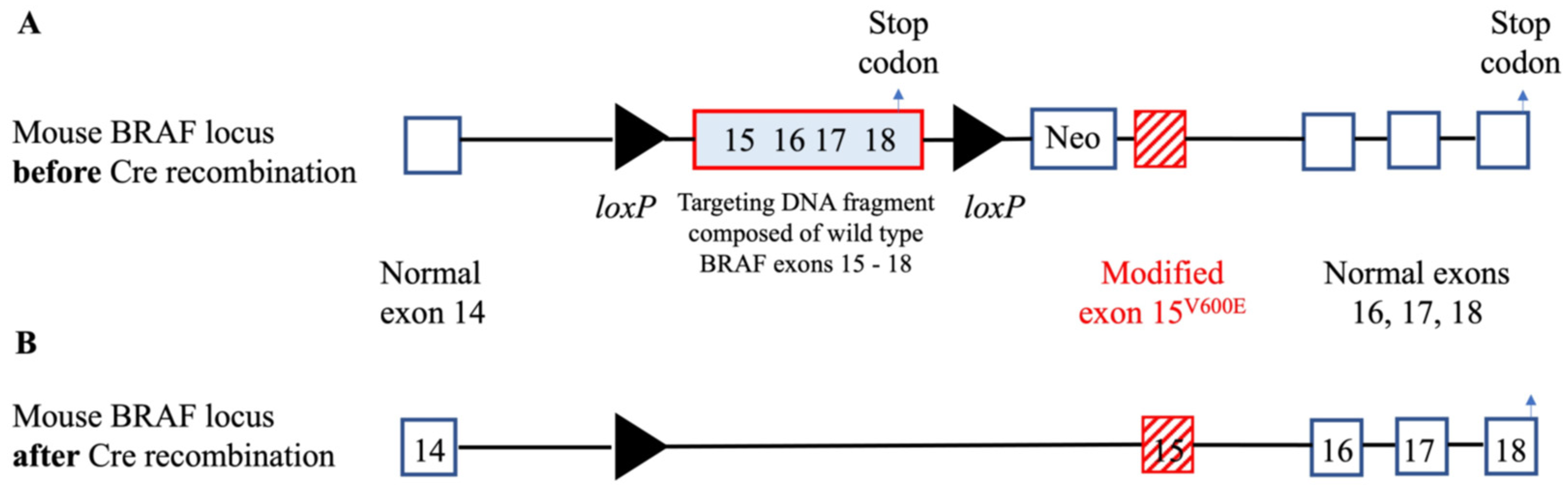
1.2. Experimental Data: Animal Models
| Mouse Line/Rat | Promoter/Expression | Original Strain | Pathology | Thyroid Function | References |
|---|---|---|---|---|---|
| RET/PTC1 | Rat Tg, thyroid | C57BL/6J | 4/18 (22%) diagnosed with * PTC starting at 8 months | NA | [37] |
| RET/PTC1 | Bovine Tg, thyroid | FVB /N | 100% mice with multifocal * PTC | Congenital hypothyroidism | [38] |
| RET/PTC3 | Bovine Tg, thyroid | CH3/He | 4/13 (31%) * PTC by 3 months and 6 /11 (55%) over 3 months | Normal | [39] |
| RET/PTC3 | Bovine Tg, thyroid | FVB/N | 7/12 (58%) * PTC-like neoplasia over 5 months | Primary hypothyroidism | [39,40] |
| TBP-3743 | TPO, BRAFV600E/WT, Trp53 | B6129SF1/J | 100% mice with anaplastic thyroid cancer | More than ~1000-fold elevation of TSH | [41,42] |
| TPOCREER/BRAFV600E | TPO, BRAFV600E, Trp53 | C57BL/6J | 100% mice with multifocal * PTC 12 weeks post-TAM injection’s | Less than ~10-fold elevation of TSH | [45] |
| TPO KRASG12D, PTENLL | TPO | 129Sv | 100 % with follicular thyroid carcinoma | TSH autonomy (TSH drastically reduced with increased T4) | [47] |
| NODH2H4_TPO-CRE-ER_BRAFV600E | TPO, BRAF V600E under CRE expression | NODH2H4/C57BL/6J | Autoimmune thyroiditis and small/unifocal * PTC | Severe hypothyroidism in concomitant and no thyroiditis | [54] |
1.3. PTC and Thyroiditis: In Vitro Study
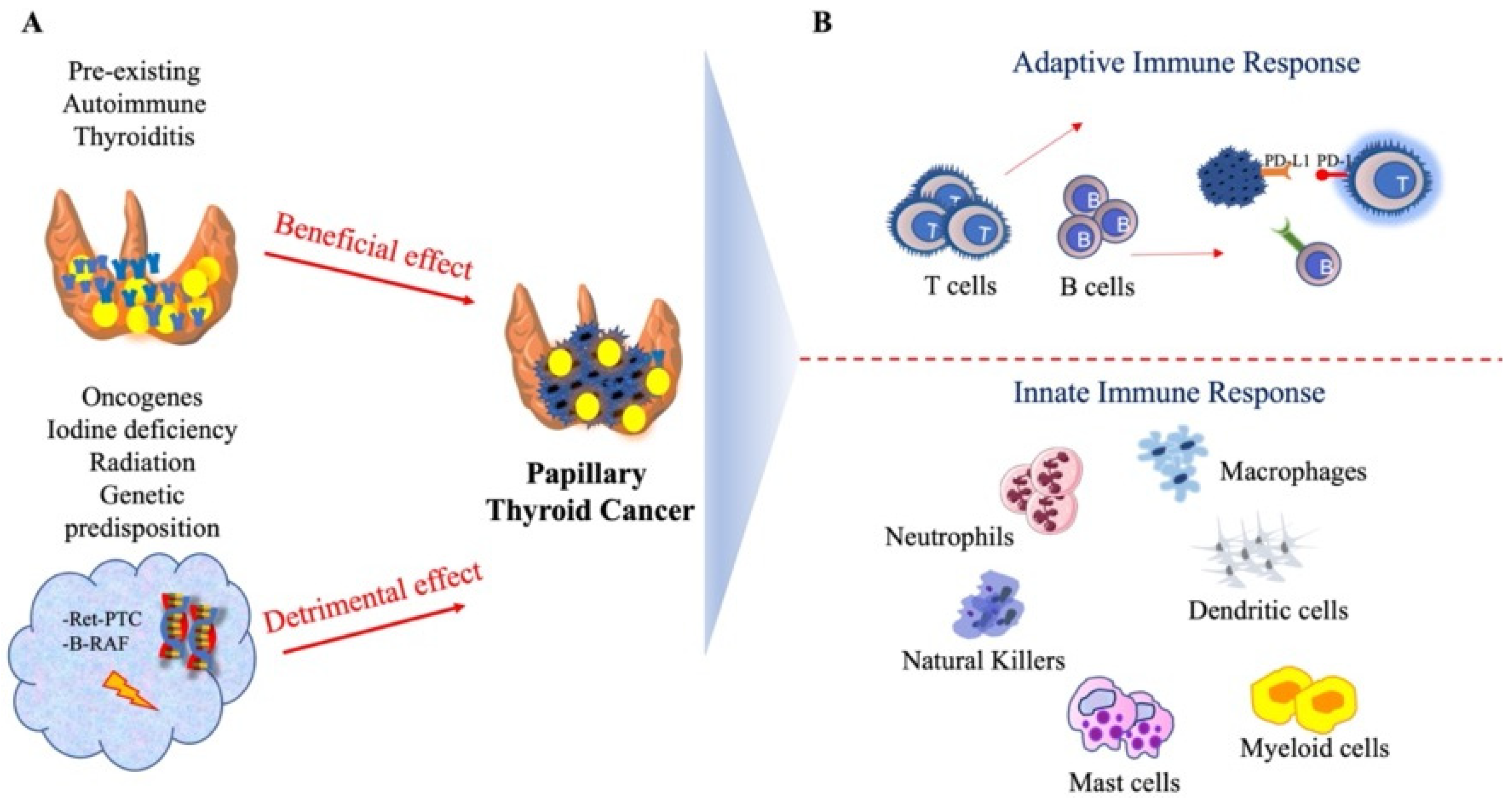
2. Adaptive and Innate Immune Response Surrounding PTC in the Context of Autoimmune Thyroiditis
2.1. T Lymphocytes and Tumor-Infiltrating Lymphocytes (TILs): General and New Concepts
2.2. B Lymphocytes and Tumor-Infiltrated B Cells: A Novel and Emerging Role in Anti-Tumor Immunity
2.3. Mast Cells, Natural Killer Cells, Macrophages, Dendritic Cells, Myeloid Cells, and Neutrophils: The Innate Immune Response
2.4. Single-Cell Transcriptome Signature of PTC and Autoimmune Thyroiditis
2.5. Antigen Specificity in Autoimmune Thyroiditis and Correlation with PTC Outcome
3. Molecular Biomarkers of the Pathogenic Link between Thyroiditis and PTC
4. Thyroiditis and PTC: Implications for Cancer Immunotherapy
5. Conclusions
Author Contributions
Funding
Conflicts of Interest
References
- McLeod, D.S.A.; Zhang, L.; Durante, C.; Cooper, D.S. Contemporary Debates in Adult Papillary Thyroid Cancer Management. Endocr. Rev. 2019, 40, 1481–1499. [Google Scholar] [CrossRef]
- Davies, L.; Morris, L.G.; Haymart, M.; Chen, A.Y.; Goldenberg, D.; Morris, J.; Ogilvie, J.B.; Terris, D.J.; Netterville, J.; Wong, R.J.; et al. American association of clinical endocrinologists and american college of endocrinology disease state clinical review: The increasing incidence of thyroid cancer. Endocr. Pract. 2015, 21, 686–696. [Google Scholar] [CrossRef]
- Cancer Genome Atlas Research Network. Integrated genomic characterization of papillary thyroid carcinoma. Cell 2014, 159, 676–690. [Google Scholar] [CrossRef]
- Blomberg, M.; Feldt-Rasmussen, U.; Andersen, K.K.; Kjaer, S.K. Thyroid cancer in Denmark 1943–2008, before and after iodine supplementation. Int. J. Cancer 2012, 131, 2360–2366. [Google Scholar] [CrossRef]
- Feldt-Rasmussen, U.; Rasmussen, A.K. Autoimmunity in differentiated thyroid cancer: Significance and related clinical problems. Hormones 2010, 9, 109–117. [Google Scholar] [CrossRef]
- Vaccarella, S.; Franceschi, S.; Bray, F.; Wild, C.P.; Plummer, M.; Dal Maso, L. Worldwide Thyroid-Cancer Epidemic? The Increasing Impact of Overdiagnosis. N. Engl. J. Med. 2016, 375, 614–617. [Google Scholar] [CrossRef]
- Dailey, M.E.; Lindsay, S.; Skahen, R. Relation of thyroid neoplasms to Hashimoto disease of the thyroid gland. AMA Arch. Surg. 1955, 70, 291–297. [Google Scholar] [CrossRef]
- Xu, L.; Li, G.; Wei, Q.; El-Naggar, A.K.; Sturgis, E.M. Family history of cancer and risk of sporadic differentiated thyroid carcinoma. Cancer 2012, 118, 1228–1235. [Google Scholar] [CrossRef]
- Fiore, E.; Latrofa, F.; Vitti, P. Iodine, thyroid autoimmunity and cancer. Eur. Thyroid J. 2015, 4, 26–35. [Google Scholar] [CrossRef]
- Boi, F.; Pani, F.; Calò, P.G.; Lai, M.L.; Mariotti, S. High prevalence of papillary thyroid carcinoma in nodular Hashimoto’s thyroiditis at the first diagnosis and during the follow-up. J. Endocrinol. Investig. 2018, 41, 395–402. [Google Scholar] [CrossRef]
- Boi, F.; Pani, F.; Mariotti, S. Thyroid Autoimmunity and Thyroid Cancer: Review Focused on Cytological Studies. Eur. Thyroid J. 2017, 6, 178–186. [Google Scholar] [CrossRef] [PubMed]
- Castagna, M.G.; Belardini, V.; Memmo, S.; Maino, F.; Di Santo, A.; Toti, P.; Carli, A.F.; Caruso, G.; Pacini, F. Nodules in autoimmune thyroiditis are associated with increased risk of thyroid cancer in surgical series but not in cytological series: Evidence for selection bias. J. Clin. Endocrinol. Metab. 2014, 99, 3193–3198. [Google Scholar] [CrossRef] [PubMed]
- Rotondi, M.; Groppelli, G.; Croce, L.; Latrofa, F.; Ancona, G.; Coperchini, F.; Pasquali, D.; Cappelli, C.; Fugazza, A.; Guazzoni, V.; et al. Patients with chronic autoimmune thyroiditis are not at higher risk for developing clinically overt thyroid cancer: A 10-year follow-up study. Eur. J. Endocrinol. 2020, 183, 317–323. [Google Scholar] [CrossRef] [PubMed]
- Sun, J.; Shi, R.; Zhang, X.; Fang, D.; Rauch, J.; Lu, S.; Wang, X.; Käsmann, L.; Ma, J.; Belka, C.; et al. Characterization of immune landscape in papillary thyroid cancer reveals distinct tumor immunogenicity and implications for immunotherapy. Oncoimmunology 2021, 10, e1964189. [Google Scholar] [CrossRef]
- Menicali, E.; Guzzetti, M.; Morelli, S.; Moretti, S.; Puxeddu, E. Immune Landscape of Thyroid Cancers: New Insights. Front. Endocrinol. 2020, 11, 637826. [Google Scholar] [CrossRef]
- Büyükaşık, O.; Hasdemir, A.O.; Yalçın, E.; Celep, B.; Sengül, S.; Yandakçı, K.; Tunç, G.; Küçükpınar, T.; Alkoy, S.; Cöl, C. The association between thyroid malignancy and chronic lymphocytic thyroiditis: Should it alter the surgical approach? Endokrynol. Pol. 2011, 62, 303–308. [Google Scholar] [PubMed]
- Yoon, Y.H.; Kim, H.J.; Lee, J.W.; Kim, J.M.; Koo, B.S. The clinicopathologic differences in papillary thyroid carcinoma with or without co-existing chronic lymphocytic thyroiditis. Eur. Arch. Otorhinolaryngol. 2012, 269, 1013–1017. [Google Scholar] [CrossRef]
- Chen, Y.K.; Lin, C.L.; Cheng, F.T.; Sung, F.C.; Kao, C.H. Cancer risk in patients with Hashimoto’s thyroiditis: A nationwide cohort study. Br. J. Cancer 2013, 109, 2496–2501. [Google Scholar] [CrossRef]
- Azizi, G.; Keller, J.M.; Lewis, M.; Piper, K.; Puett, D.; Rivenbark, K.M.; Malchoff, C.D. Association of Hashimoto’s thyroiditis with thyroid cancer. Endocr. Relat. Cancer 2014, 21, 845–852. [Google Scholar] [CrossRef]
- Resende de Paiva, C.; Grønhøj, C.; Feldt-Rasmussen, U.; von Buchwald, C. Association between Hashimoto’s Thyroiditis and Thyroid Cancer in 64,628 Patients. Front. Oncol. 2017, 7, 53. [Google Scholar] [CrossRef] [Green Version]
- Larson, S.D.; Jackson, L.N.; Riall, T.S.; Uchida, T.; Thomas, R.P.; Qiu, S.; Evers, B.M. Increased incidence of well-differentiated thyroid cancer associated with Hashimoto thyroiditis and the role of the PI3k/Akt pathway. J. Am. Coll. Surg. 2007, 204, 764–773; discussion 773–765. [Google Scholar] [CrossRef]
- Bradly, D.P.; Reddy, V.; Prinz, R.A.; Gattuso, P. Incidental papillary carcinoma in patients treated surgically for benign thyroid diseases. Surgery 2009, 146, 1099–1104. [Google Scholar] [CrossRef] [PubMed]
- Jankovic, B.; Le, K.T.; Hershman, J.M. Clinical Review: Hashimoto’s thyroiditis and papillary thyroid carcinoma: Is there a correlation? J. Clin. Endocrinol. Metab. 2013, 98, 474–482. [Google Scholar] [CrossRef] [PubMed]
- Rotondi, M.; Molteni, M.; Cappelli, C.; Croce, L.; Caputo, A.; Groppelli, G.; Liboà, F.; Guazzoni, V.; Villani, L.; Zeppa, P.; et al. The diagnostic accuracy of fine-needle aspiration cytology for thyroid nodules is not affected by coexistent chronic autoimmune thyroiditis: Results from a cyto-histological series of patients with indeterminate cytology. Eur. J. Endocrinol. 2021, 185, 201–208. [Google Scholar] [CrossRef] [PubMed]
- Pilli, T.; Toti, P.; Occhini, R.; Castagna, M.G.; Cantara, S.; Caselli, M.; Cardinale, S.; Barbagli, L.; Pacini, F. Chronic lymphocytic thyroiditis (CLT) has a positive prognostic value in papillary thyroid cancer (PTC) patients: The potential key role of Foxp3+ T lymphocytes. J. Endocrinol. Investig. 2018, 41, 703–709. [Google Scholar] [CrossRef]
- Siriweera, E.H.; Ratnatunga, N.V. Profile of Hashimoto’s Thyroiditis in Sri Lankans: Is There an Increased Risk of Ancillary Pathologies in Hashimoto’s Thyroiditis? J. Thyroid Res. 2010, 2010, 124264. [Google Scholar] [CrossRef]
- Cipolla, C.; Sandonato, L.; Graceffa, G.; Fricano, S.; Torcivia, A.; Vieni, S.; Latteri, S.; Latteri, M.A. Hashimoto thyroiditis coexistent with papillary thyroid carcinoma. Am. Surg. 2005, 71, 874–878. [Google Scholar] [CrossRef]
- Moon, S.; Chung, H.S.; Yu, J.M.; Yoo, H.J.; Park, J.H.; Kim, D.S.; Park, Y.J. Associations between Hashimoto Thyroiditis and Clinical Outcomes of Papillary Thyroid Cancer: A Meta-Analysis of Observational Studies. Endocrinol. Metab. 2018, 33, 473–484. [Google Scholar] [CrossRef]
- Okayasu, I.; Fujiwara, M.; Hara, Y.; Tanaka, Y.; Rose, N.R. Association of chronic lymphocytic thyroiditis and thyroid papillary carcinoma. A study of surgical cases among Japanese, and white and African Americans. Cancer 1995, 76, 2312–2318. [Google Scholar] [CrossRef]
- Okayasu, I. The Relationship of Lymphocytic Thyroiditis to the Development of Thyroid Carcinoma. Endocr. Pathol. 1997, 8, 225–230. [Google Scholar] [CrossRef]
- Radetti, G.; Loche, S.; D’Antonio, V.; Salerno, M.; Guzzetti, C.; Aversa, T.; Cassio, A.; Cappa, M.; Gastaldi, R.; Deluca, F.; et al. Influence of Hashimoto Thyroiditis on the Development of Thyroid Nodules and Cancer in Children and Adolescents. J. Endocr. Soc. 2019, 3, 607–616. [Google Scholar] [CrossRef]
- McLeod, D.S.A.; Bedno, S.A.; Cooper, D.S.; Hutfless, S.M.; Ippolito, S.; Jordan, S.J.; Matos, P.G.; Neale, R.E.; Sabini, E.; Whiteman, D.C.; et al. Pre-existing Thyroid Autoimmunity and Risk of Papillary Thyroid Cancer: A Nested Case-Control Study of US Active-Duty Personnel. J. Clin. Oncol. 2022, 40, 2578–2587. [Google Scholar] [CrossRef]
- Kim, C.S.; Zhu, X. Lessons from mouse models of thyroid cancer. Thyroid 2009, 19, 1317–1331. [Google Scholar] [CrossRef] [PubMed]
- Cheon, D.J.; Orsulic, S. Mouse models of cancer. Annu. Rev. Pathol. 2011, 6, 95–119. [Google Scholar] [CrossRef] [PubMed]
- Jhiang, S.M.; Sagartz, J.E.; Tong, Q.; Parker-Thornburg, J.; Capen, C.C.; Cho, J.Y.; Xing, S.; Ledent, C. Targeted expression of the ret/PTC1 oncogene induces papillary thyroid carcinomas. Endocrinology 1996, 137, 375–378. [Google Scholar] [CrossRef] [PubMed]
- Powell, D.J., Jr.; Russell, J.; Nibu, K.; Li, G.; Rhee, E.; Liao, M.; Goldstein, M.; Keane, W.M.; Santoro, M.; Fusco, A.; et al. The RET/PTC3 oncogene: Metastatic solid-type papillary carcinomas in murine thyroids. Cancer Res. 1998, 58, 5523–5528. [Google Scholar] [PubMed]
- Capen, C.C. Mechanistic data and risk assessment of selected toxic end points of the thyroid gland. Toxicol. Pathol. 1997, 25, 39–48. [Google Scholar] [CrossRef] [PubMed]
- Bellelli, R.; Vitagliano, D.; Federico, G.; Marotta, P.; Tamburrino, A.; Salerno, P.; Paciello, O.; Papparella, S.; Knauf, J.A.; Fagin, J.A.; et al. Oncogene-induced senescence and its evasion in a mouse model of thyroid neoplasia. Mol. Cell. Endocrinol. 2018, 460, 24–35. [Google Scholar] [CrossRef]
- Gunda, V.; Gigliotti, B.; Ndishabandi, D.; Ashry, T.; McCarthy, M.; Zhou, Z.; Amin, S.; Freeman, G.J.; Alessandrini, A.; Parangi, S. Combinations of BRAF inhibitor and anti-PD-1/PD-L1 antibody improve survival and tumour immunity in an immunocompetent model of orthotopic murine anaplastic thyroid cancer. Br. J. Cancer 2018, 119, 1223–1232. [Google Scholar] [CrossRef]
- Gunda, V.; Gigliotti, B.; Ashry, T.; Ndishabandi, D.; McCarthy, M.; Zhou, Z.; Amin, S.; Lee, K.E.; Stork, T.; Wirth, L.; et al. Anti-PD-1/PD-L1 therapy augments lenvatinib’s efficacy by favorably altering the immune microenvironment of murine anaplastic thyroid cancer. Int. J. Cancer 2019, 144, 2266–2278. [Google Scholar] [CrossRef]
- Dankort, D.; Filenova, E.; Collado, M.; Serrano, M.; Jones, K.; McMahon, M. A new mouse model to explore the initiation, progression, and therapy of BRAFV600E-induced lung tumors. Genes Dev. 2007, 21, 379–384. [Google Scholar] [CrossRef] [PubMed]
- McFadden, D.G.; Vernon, A.; Santiago, P.M.; Martinez-McFaline, R.; Bhutkar, A.; Crowley, D.M.; McMahon, M.; Sadow, P.M.; Jacks, T. p53 constrains progression to anaplastic thyroid carcinoma in a Braf-mutant mouse model of papillary thyroid cancer. Proc. Natl. Acad. Sci. USA 2014, 111, E1600–E1609. [Google Scholar] [CrossRef] [PubMed]
- Kusakabe, T.; Kawaguchi, A.; Kawaguchi, R.; Feigenbaum, L.; Kimura, S. Thyrocyte-specific expression of Cre recombinase in transgenic mice. Genesis 2004, 39, 212–216. [Google Scholar] [CrossRef] [PubMed]
- Knauf, J.A.; Ma, X.; Smith, E.P.; Zhang, L.; Mitsutake, N.; Liao, X.H.; Refetoff, S.; Nikiforov, Y.E.; Fagin, J.A. Targeted expression of BRAFV600E in thyroid cells of transgenic mice results in papillary thyroid cancers that undergo dedifferentiation. Cancer Res. 2005, 65, 4238–4245. [Google Scholar] [CrossRef] [PubMed]
- Miller, K.A.; Yeager, N.; Baker, K.; Liao, X.H.; Refetoff, S.; Di Cristofano, A. Oncogenic Kras requires simultaneous PI3K signaling to induce ERK activation and transform thyroid epithelial cells in vivo. Cancer Res. 2009, 69, 3689–3694. [Google Scholar] [CrossRef]
- Rose, N.R.; Witebsky, E. Studies on organ specificity. V. Changes in the thyroid glands of rabbits following active immunization with rabbit thyroid extracts. J. Immunol. 1956, 76, 417–427. [Google Scholar]
- Jones, H.E.; Roitt, I.M. Experimental auto-immune thyroiditis in the rat. Br. J. Exp. Pathol. 1961, 42, 546–557. [Google Scholar]
- Ng, H.P.; Kung, A.W. Induction of autoimmune thyroiditis and hypothyroidism by immunization of immunoactive T cell epitope of thyroid peroxidase. Endocrinology 2006, 147, 3085–3092. [Google Scholar] [CrossRef]
- Podolin, P.L.; Pressey, A.; DeLarato, N.H.; Fischer, P.A.; Peterson, L.B.; Wicker, L.S. I-E+ nonobese diabetic mice develop insulitis and diabetes. J. Exp. Med. 1993, 178, 793–803. [Google Scholar] [CrossRef]
- Braley-Mullen, H.; Yu, S. NOD.H-2h4 mice: An important and underutilized animal model of autoimmune thyroiditis and Sjogren’s syndrome. Adv. Immunol. 2015, 126, 1–43. [Google Scholar] [CrossRef]
- Kolypetri, P.; King, J.; Larijani, M.; Carayanniotis, G. Genes and environment as predisposing factors in autoimmunity: Acceleration of spontaneous thyroiditis by dietary iodide in NOD.H2(h4) mice. Int. Rev. Immunol. 2015, 34, 542–556. [Google Scholar] [CrossRef] [PubMed]
- Bonita, R.E.; Rose, N.R.; Rasooly, L.; Caturegli, P.; Burek, C.L. Kinetics of mononuclear cell infiltration and cytokine expression in iodine-induced thyroiditis in the NOD-H2h4 mouse. Exp. Mol. Pathol. 2003, 74, 1–12. [Google Scholar] [CrossRef]
- Aubin, A.M.; Lombard-Vadnais, F.; Collin, R.; Aliesky, H.A.; McLachlan, S.M.; Lesage, S. The NOD Mouse Beyond Autoimmune Diabetes. Front. Immunol. 2022, 13, 874769. [Google Scholar] [CrossRef] [PubMed]
- Pani, F.; Yasuda, Y.; Di Dalmazi, G.; Chalan, P.; Gabrielson, K.; Adamo, L.; Sabini, E.; Mariotti, S.; Caturegli, P. Pre-existing Thyroiditis Ameliorates Papillary Thyroid Cancer: Insights From a New Mouse Model. Endocrinology 2021, 162, bqab144. [Google Scholar] [CrossRef]
- Payne, S.; De Val, S.; Neal, A. Endothelial-Specific Cre Mouse Models. Arterioscler. Thromb. Vasc. Biol. 2018, 38, 2550–2561. [Google Scholar] [CrossRef]
- Mueller, S.N.; Gebhardt, T.; Carbone, F.R.; Heath, W.R. Memory T cell subsets, migration patterns, and tissue residence. Annu. Rev. Immunol. 2013, 31, 137–161. [Google Scholar] [CrossRef]
- Wieland, A.; Patel, M.R.; Cardenas, M.A.; Eberhardt, C.S.; Hudson, W.H.; Obeng, R.C.; Griffith, C.C.; Wang, X.; Chen, Z.G.; Kissick, H.T.; et al. Defining HPV-specific B cell responses in patients with head and neck cancer. Nature 2021, 597, 274–278. [Google Scholar] [CrossRef]
- Martinez-Pacheco, S.; O’Driscoll, L. Pre-Clinical In Vitro Models Used in Cancer Research: Results of a Worldwide Survey. Cancers 2021, 13, 6033. [Google Scholar] [CrossRef]
- Costa, E.C.; Moreira, A.F.; de Melo-Diogo, D.; Gaspar, V.M.; Carvalho, M.P.; Correia, I.J. 3D tumor spheroids: An overview on the tools and techniques used for their analysis. Biotechnol. Adv. 2016, 34, 1427–1441. [Google Scholar] [CrossRef]
- Ravi, M.; Paramesh, V.; Kaviya, S.R.; Anuradha, E.; Solomon, F.D. 3D cell culture systems: Advantages and applications. J. Cell. Physiol. 2015, 230, 16–26. [Google Scholar] [CrossRef]
- Chew, D.; Green, V.; Riley, A.; England, R.J.; Greenman, J. The Changing Face of in vitro Culture Models for Thyroid Cancer Research: A Systematic Literature Review. Front. Surg. 2020, 7, 43. [Google Scholar] [CrossRef] [PubMed]
- Maric, I.; Viaggi, S.; Caria, P.; Frau, D.V.; Degan, P.; Vanni, R. Centrosomal and mitotic abnormalities in cell lines derived from papillary thyroid cancer harboring specific gene alterations. Mol. Cytogenet. 2011, 4, 26. [Google Scholar] [CrossRef] [Green Version]
- Schweppe, R.E. Thyroid cancer cell lines: Critical models to study thyroid cancer biology and new therapeutic targets. Front. Endocrinol. 2012, 3, 81. [Google Scholar] [CrossRef] [PubMed]
- Saiselet, M.; Floor, S.; Tarabichi, M.; Dom, G.; Hébrant, A.; van Staveren, W.C.; Maenhaut, C. Thyroid cancer cell lines: An overview. Front. Endocrinol. 2012, 3, 133. [Google Scholar] [CrossRef]
- Caria, P.; Pillai, R.; Dettori, T.; Frau, D.V.; Zavattari, P.; Riva, G.; Romano, G.; Pani, F.; Bentivegna, A.; Giovannoni, R.; et al. Thyrospheres from B-CPAP Cell Line with BRAF and TERT Promoter Mutations have Different Functional and Molecular Features than Parental Cells. J. Cancer 2017, 8, 1629–1639. [Google Scholar] [CrossRef] [PubMed]
- Caria, P.; Tronci, L.; Dettori, T.; Murgia, F.; Santoru, M.L.; Griffin, J.L.; Vanni, R.; Atzori, L. Metabolomic Alterations in Thyrospheres and Adherent Parental Cells in Papillary Thyroid Carcinoma Cell Lines: A Pilot Study. Int. J. Mol. Sci. 2018, 19, 2948. [Google Scholar] [CrossRef]
- Caria, P.; Dettori, T.; Frau, D.V.; Lichtenzstejn, D.; Pani, F.; Vanni, R.; Mai, S. Characterizing the three-dimensional organization of telomeres in papillary thyroid carcinoma cells. J. Cell. Physiol. 2019, 234, 5175–5185. [Google Scholar] [CrossRef]
- Tronci, L.; Caria, P.; Frau, D.V.; Liggi, S.; Piras, C.; Murgia, F.; Santoru, M.L.; Pibiri, M.; Deiana, M.; Griffin, J.L.; et al. Crosstalk between Metabolic Alterations and Altered Redox Balance in PTC-Derived Cell Lines. Metabolites 2019, 9, 23. [Google Scholar] [CrossRef]
- Tronci, L.; Serreli, G.; Piras, C.; Frau, D.V.; Dettori, T.; Deiana, M.; Murgia, F.; Santoru, M.L.; Spada, M.; Leoni, V.P.; et al. Vitamin C Cytotoxicity and Its Effects in Redox Homeostasis and Energetic Metabolism in Papillary Thyroid Carcinoma Cell Lines. Antioxidants 2021, 10, 809. [Google Scholar] [CrossRef]
- Denning, K.; Smyth, P.; Cahill, S.; Li, J.; Flavin, R.; Aherne, S.; JJ, O.L.; Sheils, O. ret/PTC-1 expression alters the immunoprofile of thyroid follicular cells. Mol. Cancer 2008, 7, 44. [Google Scholar] [CrossRef]
- Russell, J.P.; Shinohara, S.; Melillo, R.M.; Castellone, M.D.; Santoro, M.; Rothstein, J.L. Tyrosine kinase oncoprotein, RET/PTC3, induces the secretion of myeloid growth and chemotactic factors. Oncogene 2003, 22, 4569–4577. [Google Scholar] [CrossRef] [PubMed]
- Puxeddu, E.; Mitsutake, N.; Knauf, J.A.; Moretti, S.; Kim, H.W.; Seta, K.A.; Brockman, D.; Myatt, L.; Millhorn, D.E.; Fagin, J.A. Microsomal prostaglandin E2 synthase-1 is induced by conditional expression of RET/PTC in thyroid PCCL3 cells through the activation of the MEK-ERK pathway. J. Biol. Chem. 2003, 278, 52131–52138. [Google Scholar] [CrossRef] [PubMed] [Green Version]
- Puxeddu, E.; Moretti, S.; Elisei, R.; Romei, C.; Pascucci, R.; Martinelli, M.; Marino, C.; Avenia, N.; Rossi, E.D.; Fadda, G.; et al. BRAF(V599E) mutation is the leading genetic event in adult sporadic papillary thyroid carcinomas. J. Clin. Endocrinol. Metab. 2004, 89, 2414–2420. [Google Scholar] [CrossRef] [PubMed]
- Han, L.T.; Hu, J.Q.; Ma, B.; Wen, D.; Zhang, T.T.; Lu, Z.W.; Wei, W.J.; Wang, Y.L.; Wang, Y.; Liao, T.; et al. IL-17A increases MHC class I expression and promotes T cell activation in papillary thyroid cancer patients with coexistent Hashimoto’s thyroiditis. Diagn. Pathol. 2019, 14, 52. [Google Scholar] [CrossRef]
- Lu, Z.W.; Hu, J.Q.; Liu, W.L.; Wen, D.; Wei, W.J.; Wang, Y.L.; Wang, Y.; Liao, T.; Ji, Q.H. IL-10 Restores MHC Class I Expression and Interferes With Immunity in Papillary Thyroid Cancer With Hashimoto Thyroiditis. Endocrinology 2020, 161, bqaa062. [Google Scholar] [CrossRef]
- Xiao, H.; Liang, J.; Liu, S.; Zhang, Q.; Xie, F.; Kong, X.; Guo, S.; Wang, R.; Fu, R.; Ye, Z.; et al. Proteomics and Organoid Culture Reveal the Underlying Pathogenesis of Hashimoto’s Thyroiditis. Front. Immunol. 2021, 12, 784975. [Google Scholar] [CrossRef]
- Ragusa, F.; Fallahi, P.; Elia, G.; Gonnella, D.; Paparo, S.R.; Giusti, C.; Churilov, L.P.; Ferrari, S.M.; Antonelli, A. Hashimotos’ thyroiditis: Epidemiology, pathogenesis, clinic and therapy. Best Pract. Res. Clin. Endocrinol. Metab. 2019, 33, 101367. [Google Scholar] [CrossRef]
- Deng, Q.; Luo, Y.; Chang, C.; Wu, H.; Ding, Y.; Xiao, R. The Emerging Epigenetic Role of CD8+T Cells in Autoimmune Diseases: A Systematic Review. Front. Immunol. 2019, 10, 856. [Google Scholar] [CrossRef]
- Bonilla, F.A.; Oettgen, H.C. Adaptive immunity. J. Allergy Clin. Immunol. 2010, 125, S33–S40. [Google Scholar] [CrossRef]
- Walter, U.; Santamaria, P. CD8+ T cells in autoimmunity. Curr. Opin. Immunol. 2005, 17, 624–631. [Google Scholar] [CrossRef]
- Tandon, N.; Weetman, A.P. T cells and thyroid autoimmunity. J. R. Coll. Physicians Lond. 1994, 28, 10–18. [Google Scholar] [PubMed]
- Wu, Z.; Podack, E.R.; McKenzie, J.M.; Olsen, K.J.; Zakarija, M. Perforin expression by thyroid-infiltrating T cells in autoimmune thyroid disease. Clin. Exp. Immunol. 1994, 98, 470–477. [Google Scholar] [CrossRef] [PubMed]
- Maecker, H.T.; McCoy, J.P.; Nussenblatt, R. Standardizing immunophenotyping for the Human Immunology Project. Nat. Rev. Immunol. 2012, 12, 191–200. [Google Scholar] [CrossRef] [PubMed]
- Jameson, S.C.; Masopust, D. Understanding Subset Diversity in T Cell Memory. Immunity 2018, 48, 214–226. [Google Scholar] [CrossRef]
- Nagataki, S.; Eguchi, K. Cytokines and immune regulation in thyroid autoimmunity. Autoimmunity 1992, 13, 27–34. [Google Scholar] [CrossRef]
- Martin, M.D.; Badovinac, V.P. Defining Memory CD8 T Cell. Front. Immunol. 2018, 9, 2692. [Google Scholar] [CrossRef]
- Kallies, A.; Zehn, D.; Utzschneider, D.T. Precursor exhausted T cells: Key to successful immunotherapy? Nat. Rev. Immunol. 2020, 20, 128–136. [Google Scholar] [CrossRef]
- Wherry, E.J. T cell exhaustion. Nat. Immunol. 2011, 12, 492–499. [Google Scholar] [CrossRef]
- Mami-Chouaib, F.; Tartour, E. Editorial: Tissue Resident Memory T Cells. Front. Immunol. 2019, 10, 1018. [Google Scholar] [CrossRef]
- Enamorado, M.; Iborra, S.; Priego, E.; Cueto, F.J.; Quintana, J.A.; Martínez-Cano, S.; Mejías-Pérez, E.; Esteban, M.; Melero, I.; Hidalgo, A.; et al. Enhanced anti-tumour immunity requires the interplay between resident and circulating memory CD8(+) T cells. Nat. Commun. 2017, 8, 16073. [Google Scholar] [CrossRef]
- Glick, A.B.; Wodzinski, A.; Fu, P.; Levine, A.D.; Wald, D.N. Impairment of regulatory T-cell function in autoimmune thyroid disease. Thyroid 2013, 23, 871–878. [Google Scholar] [CrossRef]
- Mohr, A.; Trésallet, C.; Monot, N.; Bauvois, A.; Abiven, D.; Atif, M.; Claër, L.; Malhotra, R.; Mayer, G.; Balderas, R.; et al. Tissue Infiltrating LTi-Like Group 3 Innate Lymphoid Cells and T Follicular Helper Cells in Graves’ and Hashimoto’s Thyroiditis. Front. Immunol. 2020, 11, 601. [Google Scholar] [CrossRef] [PubMed]
- Coussens, L.M.; Werb, Z. Inflammation and cancer. Nature 2002, 420, 860–867. [Google Scholar] [CrossRef] [PubMed]
- Guarino, V.; Castellone, M.D.; Avilla, E.; Melillo, R.M. Thyroid cancer and inflammation. Mol. Cell. Endocrinol. 2010, 321, 94–102. [Google Scholar] [CrossRef] [PubMed]
- Bergdorf, K.; Ferguson, D.C.; Mehrad, M.; Ely, K.; Stricker, T.; Weiss, V.L. Papillary thyroid carcinoma behavior: Clues in the tumor microenvironment. Endocr. Relat. Cancer 2019, 26, 601–614. [Google Scholar] [CrossRef]
- Gentles, A.J.; Newman, A.M.; Liu, C.L.; Bratman, S.V.; Feng, W.; Kim, D.; Nair, V.S.; Xu, Y.; Khuong, A.; Hoang, C.D.; et al. The prognostic landscape of genes and infiltrating immune cells across human cancers. Nat. Med. 2015, 21, 938–945. [Google Scholar] [CrossRef]
- Gooden, M.J.; de Bock, G.H.; Leffers, N.; Daemen, T.; Nijman, H.W. The prognostic influence of tumour-infiltrating lymphocytes in cancer: A systematic review with meta-analysis. Br. J. Cancer 2011, 105, 93–103. [Google Scholar] [CrossRef]
- Clemente, C.G.; Mihm, M.C., Jr.; Bufalino, R.; Zurrida, S.; Collini, P.; Cascinelli, N. Prognostic value of tumor infiltrating lymphocytes in the vertical growth phase of primary cutaneous melanoma. Cancer 1996, 77, 1303–1310. [Google Scholar] [CrossRef]
- Zhang, L.; Conejo-Garcia, J.R.; Katsaros, D.; Gimotty, P.A.; Massobrio, M.; Regnani, G.; Makrigiannakis, A.; Gray, H.; Schlienger, K.; Liebman, M.N.; et al. Intratumoral T cells, recurrence, and survival in epithelial ovarian cancer. N. Engl. J. Med. 2003, 348, 203–213. [Google Scholar] [CrossRef]
- Galon, J.; Costes, A.; Sanchez-Cabo, F.; Kirilovsky, A.; Mlecnik, B.; Lagorce-Pagès, C.; Tosolini, M.; Camus, M.; Berger, A.; Wind, P.; et al. Type, density, and location of immune cells within human colorectal tumors predict clinical outcome. Science 2006, 313, 1960–1964. [Google Scholar] [CrossRef]
- El Bairi, K.; Haynes, H.R.; Blackley, E.; Fineberg, S.; Shear, J.; Turner, S.; de Freitas, J.R.; Sur, D.; Amendola, L.C.; Gharib, M.; et al. The tale of TILs in breast cancer: A report from The International Immuno-Oncology Biomarker Working Group. NPJ Breast Cancer 2021, 7, 150. [Google Scholar] [CrossRef] [PubMed]
- Galdiero, M.R.; Varricchi, G.; Marone, G. The immune network in thyroid cancer. Oncoimmunology 2016, 5, e1168556. [Google Scholar] [CrossRef] [PubMed] [Green Version]
- Cunha, L.L.; Morari, E.C.; Guihen, A.C.; Razolli, D.; Gerhard, R.; Nonogaki, S.; Soares, F.A.; Vassallo, J.; Ward, L.S. Infiltration of a mixture of immune cells may be related to good prognosis in patients with differentiated thyroid carcinoma. Clin. Endocrinol. 2012, 77, 918–925. [Google Scholar] [CrossRef]
- Cunha, L.L.; Marcello, M.A.; Ward, L.S. The role of the inflammatory microenvironment in thyroid carcinogenesis. Endocr. Relat. Cancer 2014, 21, R85–R103. [Google Scholar] [CrossRef] [PubMed]
- Villagelin, D.G.; Santos, R.B.; Romaldini, J.H. Is diffuse and peritumoral lymphocyte infiltration in papillary thyroid cancer a marker of good prognosis? J. Endocrinol. Investig. 2011, 34, e403–e408. [Google Scholar] [CrossRef]
- Bastman, J.J.; Serracino, H.S.; Zhu, Y.; Koenig, M.R.; Mateescu, V.; Sams, S.B.; Davies, K.D.; Raeburn, C.D.; McIntyre, R.C., Jr.; Haugen, B.R.; et al. Tumor-Infiltrating T Cells and the PD-1 Checkpoint Pathway in Advanced Differentiated and Anaplastic Thyroid Cancer. J. Clin. Endocrinol. Metab. 2016, 101, 2863–2873. [Google Scholar] [CrossRef]
- Modi, J.; Patel, A.; Terrell, R.; Tuttle, R.M.; Francis, G.L. Papillary thyroid carcinomas from young adults and children contain a mixture of lymphocytes. J. Clin. Endocrinol. Metab. 2003, 88, 4418–4425. [Google Scholar] [CrossRef]
- French, J.D.; Weber, Z.J.; Fretwell, D.L.; Said, S.; Klopper, J.P.; Haugen, B.R. Tumor-associated lymphocytes and increased FoxP3+ regulatory T cell frequency correlate with more aggressive papillary thyroid cancer. J. Clin. Endocrinol. Metab. 2010, 95, 2325–2333. [Google Scholar] [CrossRef]
- Bagnasco, M.; Venuti, D.; Paolieri, F.; Torre, G.; Ferrini, S.; Canonica, G.W. Phenotypic and functional analysis at the clonal level of infiltrating T lymphocytes in papillary carcinoma of the thyroid: Prevalence of cytolytic T cells with natural killer-like or lymphokine-activated killer activity. J. Clin. Endocrinol. Metab. 1989, 69, 832–836. [Google Scholar] [CrossRef]
- Lee, R.S.; Schlumberger, M.; Caillou, B.; Pages, F.; Fridman, W.H.; Tartour, E. Phenotypic and functional characterisation of tumour-infiltrating lymphocytes derived from thyroid tumours. Eur. J. Cancer 1996, 32a, 1233–1239. [Google Scholar] [CrossRef]
- Ozaki, O.; Ito, K.; Mimura, T.; Sugino, K.; Hosoda, Y. Papillary carcinoma of the thyroid. Tall-cell variant with extensive lymphocyte infiltration. Am. J. Surg. Pathol. 1996, 20, 695–698. [Google Scholar] [CrossRef] [PubMed]
- Wang, T.; Shi, J.; Li, L.; Zhou, X.; Zhang, H.; Zhang, X.; Wang, Y.; Liu, L.; Sheng, L. Single-Cell Transcriptome Analysis Reveals Inter-Tumor Heterogeneity in Bilateral Papillary Thyroid Carcinoma. Front. Immunol. 2022, 13, 840811. [Google Scholar] [CrossRef] [PubMed]
- Xie, Z.; Li, X.; He, Y.; Wu, S.; Wang, S.; Sun, J.; He, Y.; Lun, Y.; Zhang, J. Immune Cell Confrontation in the Papillary Thyroid Carcinoma Microenvironment. Front. Endocrinol. 2020, 11, 570604. [Google Scholar] [CrossRef] [PubMed]
- Proietti, A.; Ugolini, C.; Melillo, R.M.; Crisman, G.; Elisei, R.; Santoro, M.; Minuto, M.; Vitti, P.; Miccoli, P.; Basolo, F. Higher intratumoral expression of CD1a, tryptase, and CD68 in a follicular variant of papillary thyroid carcinoma compared to adenomas: Correlation with clinical and pathological parameters. Thyroid 2011, 21, 1209–1215. [Google Scholar] [CrossRef]
- Gogali, F.; Paterakis, G.; Rassidakis, G.Z.; Kaltsas, G.; Liakou, C.I.; Gousis, P.; Neonakis, E.; Manoussakis, M.N.; Liapi, C. Phenotypical analysis of lymphocytes with suppressive and regulatory properties (Tregs) and NK cells in the papillary carcinoma of thyroid. J. Clin. Endocrinol. Metab. 2012, 97, 1474–1482. [Google Scholar] [CrossRef]
- Caturegli, P.; De Remigis, A.; Rose, N.R. Hashimoto thyroiditis: Clinical and diagnostic criteria. Autoimmun. Rev. 2014, 13, 391–397. [Google Scholar] [CrossRef]
- Kang, S.; Kang, J.; Shen, H.; Wu, N. Advances in regulatory B cells in autoimmune thyroid diseases. Int. Immunopharmacol. 2021, 96, 107770. [Google Scholar] [CrossRef]
- Álvarez-Sierra, D.; Marín-Sánchez, A.; Gómez-Brey, A.; Bello, I.; Caubet, E.; Moreno-Llorente, P.; Petit, A.; Zafón, C.; Iglesias, C.; González, Ó.; et al. Lymphocytic Thyroiditis Transcriptomic Profiles Support the Role of Checkpoint Pathways and B Cells in Pathogenesis. Thyroid 2022, 32, 682–693. [Google Scholar] [CrossRef]
- Pieper, K.; Grimbacher, B.; Eibel, H. B-cell biology and development. J. Allergy Clin. Immunol. 2013, 131, 959–971. [Google Scholar] [CrossRef]
- French, J.D.; Bible, K.; Spitzweg, C.; Haugen, B.R.; Ryder, M. Leveraging the immune system to treat advanced thyroid cancers. Lancet Diabetes Endocrinol. 2017, 5, 469–481. [Google Scholar] [CrossRef]
- Song, J.; Deng, Z.; Su, J.; Yuan, D.; Liu, J.; Zhu, J. Patterns of Immune Infiltration in HNC and Their Clinical Implications: A Gene Expression-Based Study. Front. Oncol. 2019, 9, 1285. [Google Scholar] [CrossRef] [PubMed]
- Lee-Chang, C.; Rashidi, A.; Miska, J.; Zhang, P.; Pituch, K.C.; Hou, D.; Xiao, T.; Fischietti, M.; Kang, S.J.; Appin, C.L.; et al. Myeloid-Derived Suppressive Cells Promote B cell-Mediated Immunosuppression via Transfer of PD-L1 in Glioblastoma. Cancer Immunol. Res. 2019, 7, 1928–1943. [Google Scholar] [CrossRef] [PubMed]
- Pinto, R.; Petriella, D.; Lacalamita, R.; Montrone, M.; Catino, A.; Pizzutilo, P.; Botticella, M.A.; Zito, F.A.; Del Bene, G.; Zonno, A.; et al. KRAS-Driven Lung Adenocarcinoma and B Cell Infiltration: Novel Insights for Immunotherapy. Cancers 2019, 11, 1145. [Google Scholar] [CrossRef]
- Yang, Z.; Yin, L.; Zeng, Y.; Li, Y.; Chen, H.; Yin, S.; Zhang, F.; Yang, W. Diagnostic and prognostic value of tumor-infiltrating B cells in lymph node metastases of papillary thyroid carcinoma. Virchows Arch. 2021, 479, 947–959. [Google Scholar] [CrossRef]
- Pan, J.; Ye, F.; Yu, C.; Zhu, Q.; Li, J.; Zhang, Y.; Tian, H.; Yao, Y.; Zhu, M.; Shen, Y.; et al. Papillary Thyroid Carcinoma Landscape and Its Immunological Link With Hashimoto Thyroiditis at Single-Cell Resolution. Front. Cell Dev. Biol. 2021, 9, 758339. [Google Scholar] [CrossRef] [PubMed]
- Wu, Z.; Zhou, J.; Xiao, Y.; Ming, J.; Zhou, J.; Dong, F.; Zhou, X.; Xu, Z.; Zhao, X.; Lei, P.; et al. CD20(+)CD22(+)ADAM28(+) B Cells in Tertiary Lymphoid Structures Promote Immunotherapy Response. Front. Immunol. 2022, 13, 865596. [Google Scholar] [CrossRef]
- Melillo, R.M.; Guarino, V.; Avilla, E.; Galdiero, M.R.; Liotti, F.; Prevete, N.; Rossi, F.W.; Basolo, F.; Ugolini, C.; de Paulis, A.; et al. Mast cells have a protumorigenic role in human thyroid cancer. Oncogene 2010, 29, 6203–6215. [Google Scholar] [CrossRef]
- Visciano, C.; Liotti, F.; Prevete, N.; Cali, G.; Franco, R.; Collina, F.; de Paulis, A.; Marone, G.; Santoro, M.; Melillo, R.M. Mast cells induce epithelial-to-mesenchymal transition and stem cell features in human thyroid cancer cells through an IL-8-Akt-Slug pathway. Oncogene 2015, 34, 5175–5186. [Google Scholar] [CrossRef]
- Xu, X.; Rao, G.; Gaffud, M.J.; Ding, H.G.; Maki, G.; Klingemann, H.G.; Groh, V.; Spies, T.; Caillat-Zucman, S.; Gattuso, P.; et al. Clinicopathological significance of major histocompatibility complex class I-related chain a and B expression in thyroid cancer. J. Clin. Endocrinol. Metab. 2006, 91, 2704–2712. [Google Scholar] [CrossRef]
- Angell, T.E.; Lechner, M.G.; Jang, J.K.; LoPresti, J.S.; Epstein, A.L. MHC class I loss is a frequent mechanism of immune escape in papillary thyroid cancer that is reversed by interferon and selumetinib treatment in vitro. Clin. Cancer Res. 2014, 20, 6034–6044. [Google Scholar] [CrossRef]
- Qing, W.; Fang, W.Y.; Ye, L.; Shen, L.Y.; Zhang, X.F.; Fei, X.C.; Chen, X.; Wang, W.Q.; Li, X.Y.; Xiao, J.C.; et al. Density of tumor-associated macrophages correlates with lymph node metastasis in papillary thyroid carcinoma. Thyroid 2012, 22, 905–910. [Google Scholar] [CrossRef] [PubMed]
- Ryder, M.; Ghossein, R.A.; Ricarte-Filho, J.C.; Knauf, J.A.; Fagin, J.A. Increased density of tumor-associated macrophages is associated with decreased survival in advanced thyroid cancer. Endocr. Relat. Cancer 2008, 15, 1069–1074. [Google Scholar] [CrossRef] [PubMed] [Green Version]
- Scarpino, S.; Stoppacciaro, A.; Ballerini, F.; Marchesi, M.; Prat, M.; Stella, M.C.; Sozzani, S.; Allavena, P.; Mantovani, A.; Ruco, L.P. Papillary carcinoma of the thyroid: Hepatocyte growth factor (HGF) stimulates tumor cells to release chemokines active in recruiting dendritic cells. Am. J. Pathol. 2000, 156, 831–837. [Google Scholar] [CrossRef]
- Galdiero, M.R.; Varricchi, G.; Loffredo, S.; Mantovani, A.; Marone, G. Roles of neutrophils in cancer growth and progression. J. Leukoc. Biol. 2018, 103, 457–464. [Google Scholar] [CrossRef] [PubMed]
- Jaillon, S.; Galdiero, M.R.; Del Prete, D.; Cassatella, M.A.; Garlanda, C.; Mantovani, A. Neutrophils in innate and adaptive immunity. Semin. Immunopathol. 2013, 35, 377–394. [Google Scholar] [CrossRef]
- Galdiero, M.R.; Bianchi, P.; Grizzi, F.; Di Caro, G.; Basso, G.; Ponzetta, A.; Bonavita, E.; Barbagallo, M.; Tartari, S.; Polentarutti, N.; et al. Occurrence and significance of tumor-associated neutrophils in patients with colorectal cancer. Int. J. Cancer 2016, 139, 446–456. [Google Scholar] [CrossRef]
- Donskov, F. Immunomonitoring and prognostic relevance of neutrophils in clinical trials. Semin. Cancer Biol. 2013, 23, 200–207. [Google Scholar] [CrossRef]
- Lee, J.J.; Jacobsen, E.A.; Ochkur, S.I.; McGarry, M.P.; Condjella, R.M.; Doyle, A.D.; Luo, H.; Zellner, K.R.; Protheroe, C.A.; Willetts, L.; et al. Human versus mouse eosinophils: “that which we call an eosinophil, by any other name would stain as red”. J. Allergy Clin. Immunol. 2012, 130, 572–584. [Google Scholar] [CrossRef]
- Taylor, M.H.; Takahashi, S.; Capdevila, J.; Tahara, M.; Leboulleux, S.; Kiyota, N.; Dutcus, C.E.; Xie, R.; Robinson, B.; Sherman, S.; et al. Correlation of Performance Status and Neutrophil-Lymphocyte Ratio with Efficacy in Radioiodine-Refractory Differentiated Thyroid Cancer Treated with Lenvatinib. Thyroid 2021, 31, 1226–1234. [Google Scholar] [CrossRef]
- Guthrie, G.J.; Charles, K.A.; Roxburgh, C.S.; Horgan, P.G.; McMillan, D.C.; Clarke, S.J. The systemic inflammation-based neutrophil-lymphocyte ratio: Experience in patients with cancer. Crit. Rev. Oncol. Hematol. 2013, 88, 218–230. [Google Scholar] [CrossRef]
- Lee, F.; Yang, P.S.; Chien, M.N.; Lee, J.J.; Leung, C.H.; Cheng, S.P. An Increased Neutrophil-to-Lymphocyte Ratio Predicts Incomplete Response to Therapy in Differentiated Thyroid Cancer. Int. J. Med. Sci. 2018, 15, 1757–1763. [Google Scholar] [CrossRef] [PubMed]
- Wu, P.; Sun, W.; Zhang, H. An immune-related prognostic signature for thyroid carcinoma to predict survival and response to immune checkpoint inhibitors. Cancer Immunol. Immunother. 2022, 71, 747–759. [Google Scholar] [CrossRef] [PubMed]
- Hornburg, M.; Desbois, M.; Lu, S.; Guan, Y.; Lo, A.A.; Kaufman, S.; Elrod, A.; Lotstein, A.; DesRochers, T.M.; Munoz-Rodriguez, J.L.; et al. Single-cell dissection of cellular components and interactions shaping the tumor immune phenotypes in ovarian cancer. Cancer Cell 2021, 39, 928–944.e6. [Google Scholar] [CrossRef] [PubMed]
- Baslan, T.; Hicks, J. Unravelling biology and shifting paradigms in cancer with single-cell sequencing. Nat. Rev. Cancer 2017, 17, 557–569. [Google Scholar] [CrossRef]
- Peng, M.; Wei, G.; Zhang, Y.; Li, H.; Lai, Y.; Guo, Y.; Chen, Y.; Liu, L.; Xiao, H.; Guan, H.; et al. Single-cell transcriptomic landscape reveals the differences in cell differentiation and immune microenvironment of papillary thyroid carcinoma between genders. Cell Biosci. 2021, 11, 39. [Google Scholar] [CrossRef]
- Luo, H.; Xia, X.; Kim, G.D.; Liu, Y.; Xue, Z.; Zhang, L.; Shu, Y.; Yang, T.; Chen, Y.; Zhang, S.; et al. Characterizing dedifferentiation of thyroid cancer by integrated analysis. Sci. Adv. 2021, 7, eabf3657. [Google Scholar] [CrossRef]
- Ferraro, A.; Schepis, F.; Leone, V.; Federico, A.; Borbone, E.; Pallante, P.; Berlingieri, M.T.; Chiappetta, G.; Monaco, M.; Palmieri, D.; et al. Tumor suppressor role of the CL2/DRO1/CCDC80 gene in thyroid carcinogenesis. J. Clin. Endocrinol. Metab. 2013, 98, 2834–2843. [Google Scholar] [CrossRef]
- Ehlers, M.; Thiel, A.; Bernecker, C.; Porwol, D.; Papewalis, C.; Willenberg, H.S.; Schinner, S.; Hautzel, H.; Scherbaum, W.A.; Schott, M. Evidence of a combined cytotoxic thyroglobulin and thyroperoxidase epitope-specific cellular immunity in Hashimoto’s thyroiditis. J. Clin. Endocrinol. Metab. 2012, 97, 1347–1354. [Google Scholar] [CrossRef]
- Spencer, C.A.; Takeuchi, M.; Kazarosyan, M.; Wang, C.C.; Guttler, R.B.; Singer, P.A.; Fatemi, S.; LoPresti, J.S.; Nicoloff, J.T. Serum thyroglobulin autoantibodies: Prevalence, influence on serum thyroglobulin measurement, and prognostic significance in patients with differentiated thyroid carcinoma. J. Clin. Endocrinol. Metab. 1998, 83, 1121–1127. [Google Scholar] [CrossRef]
- Latrofa, F.; Ricci, D.; Montanelli, L.; Rocchi, R.; Piaggi, P.; Sisti, E.; Grasso, L.; Basolo, F.; Ugolini, C.; Pinchera, A.; et al. Thyroglobulin autoantibodies in patients with papillary thyroid carcinoma: Comparison of different assays and evaluation of causes of discrepancies. J. Clin. Endocrinol. Metab. 2012, 97, 3974–3982. [Google Scholar] [CrossRef]
- Latrofa, F.; Ricci, D.; Vitti, P.; Prinzis, A.; Cambuli, V.M.; Ghiani, M.; Pilia, S.; Carta, D.; Loche, S.; Pinchera, A.; et al. Characterization of thyroglobulin epitopes in Sardinian adults and juveniles with Hashimoto’s thyroiditis: Evidence against a major effect of age and genetic background on B-cell epitopes. Clin. Endocrinol. 2010, 73, 110–113. [Google Scholar] [CrossRef] [PubMed]
- Shuxian, J.; Xiaoyun, C.; Zhihui, F.; Xiaohua, L.; Zhanhui, D.; Bin, H.; Lin, Z. Association of HLA-B*51:01 with papillary thyroid carcinoma in the Chinese Han population of the Shandong coastal areas. Thyroid 2014, 24, 867–871. [Google Scholar] [CrossRef] [PubMed] [Green Version]
- Lupoli, G.A.; Okosieme, O.E.; Evans, C.; Clark, P.M.; Pickett, A.J.; Premawardhana, L.D.; Lupoli, G.; Lazarus, J.H. Prognostic significance of thyroglobulin antibody epitopes in differentiated thyroid cancer. J. Clin. Endocrinol. Metab. 2015, 100, 100–108. [Google Scholar] [CrossRef] [PubMed]
- Ehlers, M.; Kuebart, A.; Hautzel, H.; Enczmann, J.; Reis, A.C.; Haase, M.; Allelein, S.; Dringenberg, T.; Schmid, C.; Schott, M. Epitope-Specific Antitumor Immunity Suppresses Tumor Spread in Papillary Thyroid Cancer. J. Clin. Endocrinol. Metab. 2017, 102, 2154–2161. [Google Scholar] [CrossRef]
- Fagin, J.A.; Wells, S.A., Jr. Biologic and Clinical Perspectives on Thyroid Cancer. N. Engl. J. Med. 2016, 375, 1054–1067. [Google Scholar] [CrossRef]
- Wirtschafter, A.; Schmidt, R.; Rosen, D.; Kundu, N.; Santoro, M.; Fusco, A.; Multhaupt, H.; Atkins, J.P.; Rosen, M.R.; Keane, W.M.; et al. Expression of the RET/PTC fusion gene as a marker for papillary carcinoma in Hashimoto’s thyroiditis. Laryngoscope 1997, 107, 95–100. [Google Scholar] [CrossRef] [PubMed]
- Landa, I.; Pozdeyev, N.; Korch, C.; Marlow, L.A.; Smallridge, R.C.; Copland, J.A.; Henderson, Y.C.; Lai, S.Y.; Clayman, G.L.; Onoda, N.; et al. Comprehensive Genetic Characterization of Human Thyroid Cancer Cell Lines: A Validated Panel for Preclinical Studies. Clin. Cancer Res. 2019, 25, 3141–3151. [Google Scholar] [CrossRef]
- Muzza, M.; Degl’Innocenti, D.; Colombo, C.; Perrino, M.; Ravasi, E.; Rossi, S.; Cirello, V.; Beck-Peccoz, P.; Borrello, M.G.; Fugazzola, L. The tight relationship between papillary thyroid cancer, autoimmunity and inflammation: Clinical and molecular studies. Clin. Endocrinol. 2010, 72, 702–708. [Google Scholar] [CrossRef]
- Mechler, C.; Bounacer, A.; Suarez, H.; Saint Frison, M.; Magois, C.; Aillet, G.; Gaulier, A. Papillary thyroid carcinoma: 6 cases from 2 families with associated lymphocytic thyroiditis harbouring RET/PTC rearrangements. Br. J. Cancer 2001, 85, 1831–1837. [Google Scholar] [CrossRef]
- Kang, D.Y.; Kim, K.H.; Kim, J.M.; Kim, S.H.; Kim, J.Y.; Baik, H.W.; Kim, Y.S. High prevalence of RET, RAS, and ERK expression in Hashimoto’s thyroiditis and in papillary thyroid carcinoma in the Korean population. Thyroid 2007, 17, 1031–1038. [Google Scholar] [CrossRef]
- Nikiforova, M.N.; Caudill, C.M.; Biddinger, P.; Nikiforov, Y.E. Prevalence of RET/PTC rearrangements in Hashimoto’s thyroiditis and papillary thyroid carcinomas. Int. J. Surg. Pathol. 2002, 10, 15–22. [Google Scholar] [CrossRef]
- Sadow, P.M.; Heinrich, M.C.; Corless, C.L.; Fletcher, J.A.; Nosé, V. Absence of BRAF, NRAS, KRAS, HRAS mutations, and RET/PTC gene rearrangements distinguishes dominant nodules in Hashimoto thyroiditis from papillary thyroid carcinomas. Endocr. Pathol. 2010, 21, 73–79. [Google Scholar] [CrossRef] [PubMed]
- Xu, J.; Ding, K.; Mu, L.; Huang, J.; Ye, F.; Peng, Y.; Guo, C.; Ren, C. Hashimoto’s Thyroiditis: A “Double-Edged Sword” in Thyroid Carcinoma. Front. Endocrinol. 2022, 13, 801925. [Google Scholar] [CrossRef] [PubMed]
- Lim, J.Y.; Hong, S.W.; Lee, Y.S.; Kim, B.W.; Park, C.S.; Chang, H.S.; Cho, J.Y. Clinicopathologic implications of the BRAF(V600E) mutation in papillary thyroid cancer: A subgroup analysis of 3130 cases in a single center. Thyroid 2013, 23, 1423–1430. [Google Scholar] [CrossRef] [PubMed]
- Lang, B.H.; Chai, Y.J.; Cowling, B.J.; Min, H.S.; Lee, K.E.; Youn, Y.K. Is BRAFV600E mutation a marker for central nodal metastasis in small papillary thyroid carcinoma? Endocr. Relat. Cancer 2014, 21, 285–295. [Google Scholar] [CrossRef] [PubMed]
- Kwak, H.Y.; Chae, B.J.; Eom, Y.H.; Hong, Y.R.; Seo, J.B.; Lee, S.H.; Song, B.J.; Jung, S.S.; Bae, J.S. Does papillary thyroid carcinoma have a better prognosis with or without Hashimoto thyroiditis? Int. J. Clin. Oncol. 2015, 20, 463–473. [Google Scholar] [CrossRef]
- Kim, S.J.; Myong, J.P.; Jee, H.G.; Chai, Y.J.; Choi, J.Y.; Min, H.S.; Lee, K.E.; Youn, Y.K. Combined effect of Hashimoto’s thyroiditis and BRAF(V600E) mutation status on aggressiveness in papillary thyroid cancer. Head Neck 2016, 38, 95–101. [Google Scholar] [CrossRef] [PubMed]
- Molnár, C.; Molnár, S.; Bedekovics, J.; Mokánszki, A.; Győry, F.; Nagy, E.; Méhes, G. Thyroid Carcinoma Coexisting with Hashimoto’s Thyreoiditis: Clinicopathological and Molecular Characteristics Clue up Pathogenesis. Pathol. Oncol. Res. 2019, 25, 1191–1197. [Google Scholar] [CrossRef] [PubMed]
- Zeng, R.C.; Jin, L.P.; Chen, E.D.; Dong, S.Y.; Cai, Y.F.; Huang, G.L.; Li, Q.; Jin, C.; Zhang, X.H.; Wang, O.C. Potential relationship between Hashimoto’s thyroiditis and BRAF(V600E) mutation status in papillary thyroid cancer. Head Neck 2016, 38 (Suppl. 1), E1019–E1025. [Google Scholar] [CrossRef]
- Dobrinja, C.; Makovac, P.; Pastoricchio, M.; Cipolat Mis, T.; Bernardi, S.; Fabris, B.; Piscopello, L.; de Manzini, N. Coexistence of chronic lymphocytic thyroiditis and papillary thyroid carcinoma. Impact on presentation, management, and outcome. Int. J. Surg. 2016, 28 (Suppl. 1), S70–S74. [Google Scholar] [CrossRef]
- Unger, P.; Ewart, M.; Wang, B.Y.; Gan, L.; Kohtz, D.S.; Burstein, D.E. Expression of p63 in papillary thyroid carcinoma and in Hashimoto’s thyroiditis: A pathobiologic link? Hum. Pathol. 2003, 34, 764–769. [Google Scholar] [CrossRef]
- Ciampi, R.; Nikiforov, Y.E. RET/PTC rearrangements and BRAF mutations in thyroid tumorigenesis. Endocrinology 2007, 148, 936–941. [Google Scholar] [CrossRef] [PubMed]
- de la Fouchardière, C.; Wassermann, J.; Calcagno, F.; Bardet, S.; Al Ghuzlan, A.; Borget, I.; Borson Chazot, F.; Do Cao, C.; Buffet, C.; Zerdoud, S.; et al. Molecular genotyping in refractory thyroid cancers in 2021: When, how and why? A review from the TUTHYREF network. Bull. Cancer 2021, 108, 1044–1056. [Google Scholar] [CrossRef]
- Busaidy, N.; Konda, B.; Wei, L.; Wirth, L.J.; Devine, C.; Daniels, G.A.; DeSouza, J.A.; Poi, M.; Seligson, N.D.; Cabanillas, M.; et al. Dabrafenib vs dabrafenib + trametinib in BRAF-mutated radioactive iodine refractory differentiated thyroid cancer—Results of a randomized, phase 2, open-label, multicenter trial. Thyroid 2022. [Google Scholar] [CrossRef]
- Pani, F.; Macerola, E.; Basolo, F.; Boi, F.; Scartozzi, M.; Mariotti, S. Aggressive differentiated thyroid cancer with multiple metastases and NRAS and TERT promoter mutations: A case report. Oncol. Lett. 2017, 14, 2186–2190. [Google Scholar] [CrossRef] [PubMed]
- Cable, J.; Greenbaum, B.; Pe’er, D.; Bollard, C.M.; Bruni, S.; Griffin, M.E.; Allison, J.P.; Wu, C.J.; Subudhi, S.K.; Mardis, E.R.; et al. Frontiers in cancer immunotherapy-a symposium report. Ann. N. Y. Acad. Sci. 2021, 1489, 30–47. [Google Scholar] [CrossRef]
- Topalian, S.L.; Taube, J.M.; Pardoll, D.M. Neoadjuvant checkpoint blockade for cancer immunotherapy. Science 2020, 367, eaax0182. [Google Scholar] [CrossRef]
- Topalian, S.L.; Hodi, F.S.; Brahmer, J.R.; Gettinger, S.N.; Smith, D.C.; McDermott, D.F.; Powderly, J.D.; Carvajal, R.D.; Sosman, J.A.; Atkins, M.B.; et al. Safety, activity, and immune correlates of anti-PD-1 antibody in cancer. N. Engl. J. Med. 2012, 366, 2443–2454. [Google Scholar] [CrossRef]
- Pedicord, V.A.; Montalvo, W.; Leiner, I.M.; Allison, J.P. Single dose of anti-CTLA-4 enhances CD8+ T-cell memory formation, function, and maintenance. Proc. Natl. Acad. Sci. USA 2011, 108, 266–271. [Google Scholar] [CrossRef]
- Leach, D.R.; Krummel, M.F.; Allison, J.P. Enhancement of antitumor immunity by CTLA-4 blockade. Science 1996, 271, 1734–1736. [Google Scholar] [CrossRef]
- Shiravand, Y.; Khodadadi, F.; Kashani, S.M.A.; Hosseini-Fard, S.R.; Hosseini, S.; Sadeghirad, H.; Ladwa, R.; O’Byrne, K.; Kulasinghe, A. Immune Checkpoint Inhibitors in Cancer Therapy. Curr. Oncol. 2022, 29, 3044–3060. [Google Scholar] [CrossRef] [PubMed]
- French, J.D.; Kotnis, G.R.; Said, S.; Raeburn, C.D.; McIntyre, R.C., Jr.; Klopper, J.P.; Haugen, B.R. Programmed death-1+ T cells and regulatory T cells are enriched in tumor-involved lymph nodes and associated with aggressive features in papillary thyroid cancer. J. Clin. Endocrinol. Metab. 2012, 97, E934–E943. [Google Scholar] [CrossRef] [PubMed]
- Severson, J.J.; Serracino, H.S.; Mateescu, V.; Raeburn, C.D.; McIntyre, R.C., Jr.; Sams, S.B.; Haugen, B.R.; French, J.D. PD-1+Tim-3+ CD8+ T Lymphocytes Display Varied Degrees of Functional Exhaustion in Patients with Regionally Metastatic Differentiated Thyroid Cancer. Cancer Immunol. Res. 2015, 3, 620–630. [Google Scholar] [CrossRef]
- French, J.D. Immunotherapy for advanced thyroid cancers—Rationale, current advances and future strategies. Nat. Rev. Endocrinol. 2020, 16, 629–641. [Google Scholar] [CrossRef] [PubMed]
- Moretti, S.; Menicali, E.; Nucci, N.; Guzzetti, M.; Morelli, S.; Puxeddu, E. THERAPY OF ENDOCRINE DISEASE Immunotherapy of advanced thyroid cancer: From bench to bedside. Eur. J. Endocrinol. 2020, 183, R41–R55. [Google Scholar] [CrossRef] [PubMed]
- Newton, J.M.; Hanoteau, A.; Liu, H.C.; Gaspero, A.; Parikh, F.; Gartrell-Corrado, R.D.; Hart, T.D.; Laoui, D.; Van Ginderachter, J.A.; Dharmaraj, N.; et al. Immune microenvironment modulation unmasks therapeutic benefit of radiotherapy and checkpoint inhibition. J. Immunother. Cancer 2019, 7, 216. [Google Scholar] [CrossRef]
- Routy, B.; Le Chatelier, E.; Derosa, L.; Duong, C.P.M.; Alou, M.T.; Daillère, R.; Fluckiger, A.; Messaoudene, M.; Rauber, C.; Roberti, M.P.; et al. Gut microbiome influences efficacy of PD-1-based immunotherapy against epithelial tumors. Science 2018, 359, 91–97. [Google Scholar] [CrossRef]
- Chat, V.; Ferguson, R.; Simpson, D.; Kazlow, E.; Lax, R.; Moran, U.; Pavlick, A.; Frederick, D.; Boland, G.; Sullivan, R.; et al. Autoimmune genetic risk variants as germline biomarkers of response to melanoma immune-checkpoint inhibition. Cancer Immunol. Immunother. 2019, 68, 897–905. [Google Scholar] [CrossRef] [PubMed]
- He, H.; Jazdzewski, K.; Li, W.; Liyanarachchi, S.; Nagy, R.; Volinia, S.; Calin, G.A.; Liu, C.G.; Franssila, K.; Suster, S.; et al. The role of microRNA genes in papillary thyroid carcinoma. Proc. Natl. Acad. Sci. USA 2005, 102, 19075–19080. [Google Scholar] [CrossRef] [Green Version]

Publisher’s Note: MDPI stays neutral with regard to jurisdictional claims in published maps and institutional affiliations. |
© 2022 by the authors. Licensee MDPI, Basel, Switzerland. This article is an open access article distributed under the terms and conditions of the Creative Commons Attribution (CC BY) license (https://creativecommons.org/licenses/by/4.0/).
Share and Cite
Pani, F.; Caria, P.; Yasuda, Y.; Makoto, M.; Mariotti, S.; Leenhardt, L.; Roshanmehr, S.; Caturegli, P.; Buffet, C. The Immune Landscape of Papillary Thyroid Cancer in the Context of Autoimmune Thyroiditis. Cancers 2022, 14, 4287. https://doi.org/10.3390/cancers14174287
Pani F, Caria P, Yasuda Y, Makoto M, Mariotti S, Leenhardt L, Roshanmehr S, Caturegli P, Buffet C. The Immune Landscape of Papillary Thyroid Cancer in the Context of Autoimmune Thyroiditis. Cancers. 2022; 14(17):4287. https://doi.org/10.3390/cancers14174287
Chicago/Turabian StylePani, Fabiana, Paola Caria, Yoshinori Yasuda, Miyara Makoto, Stefano Mariotti, Laurence Leenhardt, Solmaz Roshanmehr, Patrizio Caturegli, and Camille Buffet. 2022. "The Immune Landscape of Papillary Thyroid Cancer in the Context of Autoimmune Thyroiditis" Cancers 14, no. 17: 4287. https://doi.org/10.3390/cancers14174287
APA StylePani, F., Caria, P., Yasuda, Y., Makoto, M., Mariotti, S., Leenhardt, L., Roshanmehr, S., Caturegli, P., & Buffet, C. (2022). The Immune Landscape of Papillary Thyroid Cancer in the Context of Autoimmune Thyroiditis. Cancers, 14(17), 4287. https://doi.org/10.3390/cancers14174287