Adding Concomitant Chemotherapy to Postoperative Radiotherapy in Oral Cavity Carcinoma with Minor Risk Factors: Systematic Review of the Literature and Meta-Analysis
Abstract
Simple Summary
Abstract
1. Introduction
2. Materials and Methods
2.1. Inclusion and Exclusion Criteria
2.2. Review of the Trials
3. Results
3.1. The Site, Dose, and Interval of Radiotherapy
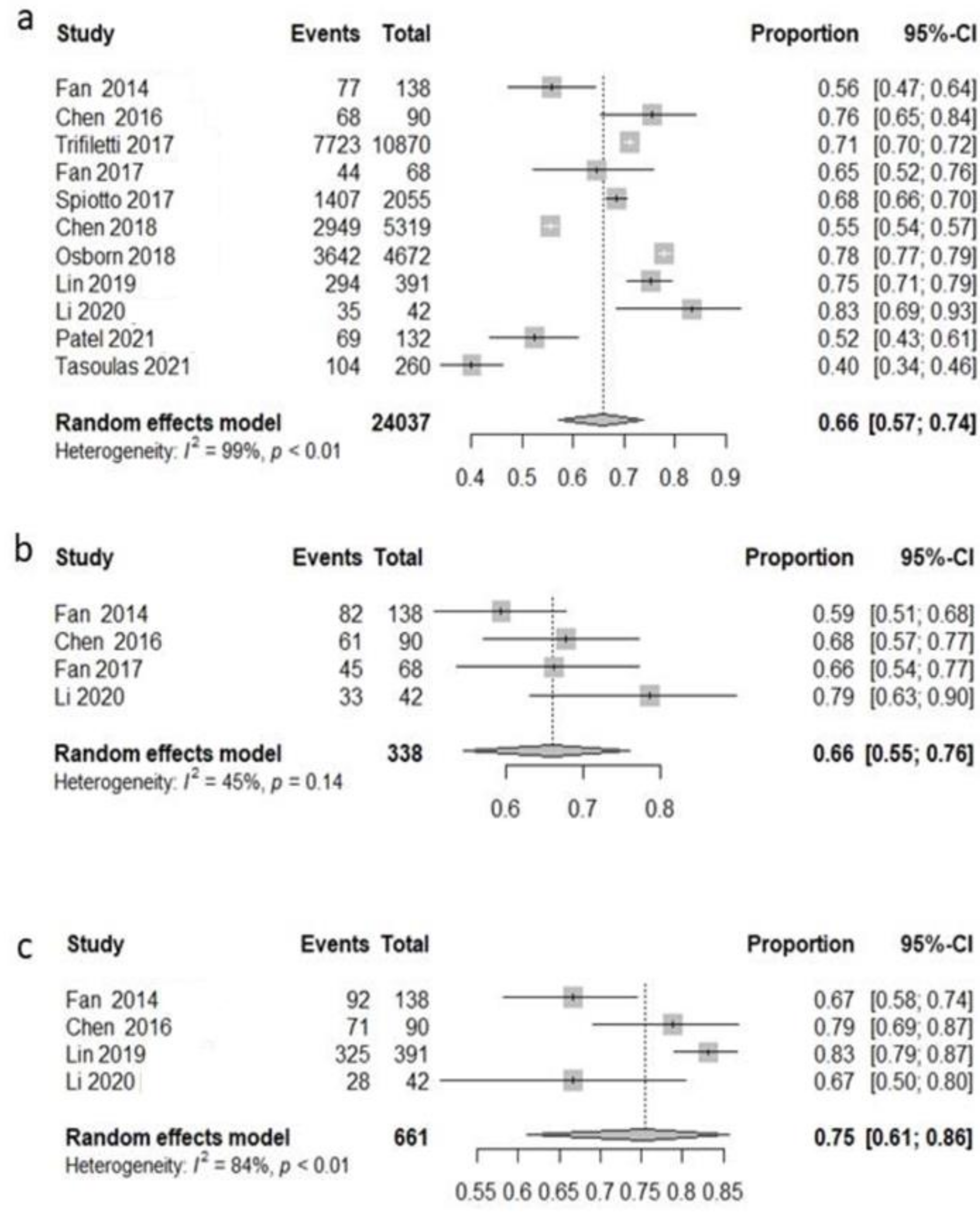
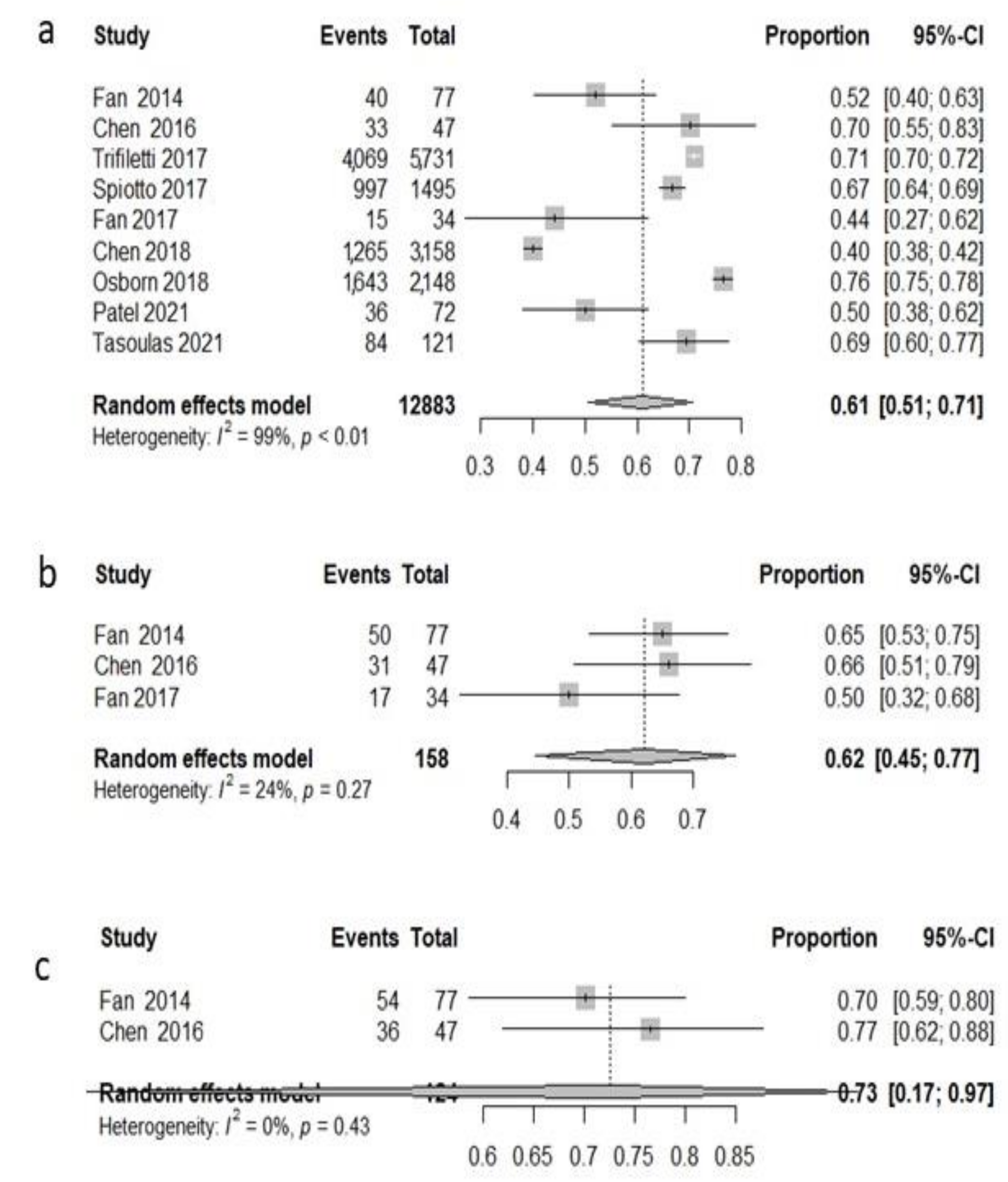
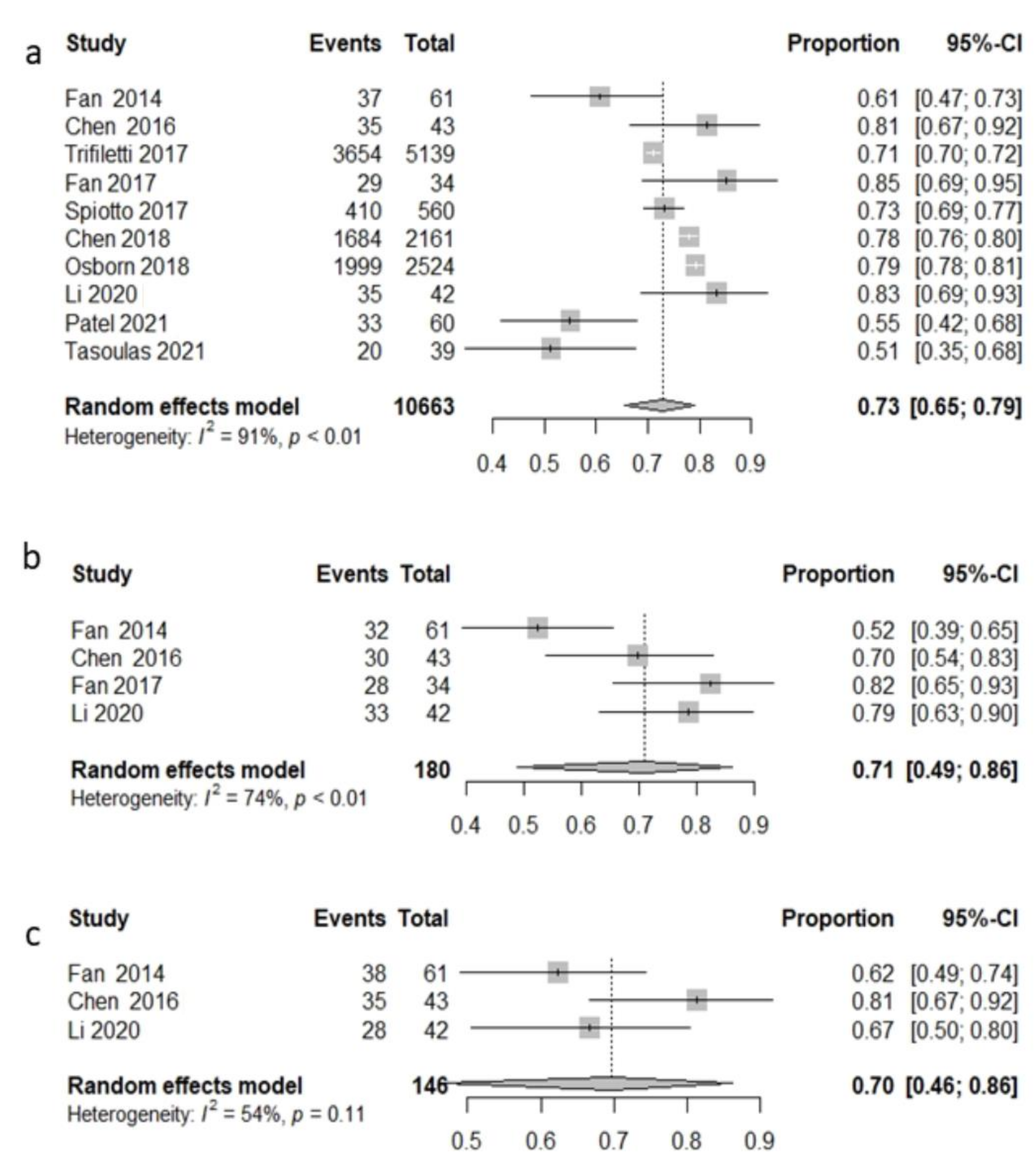
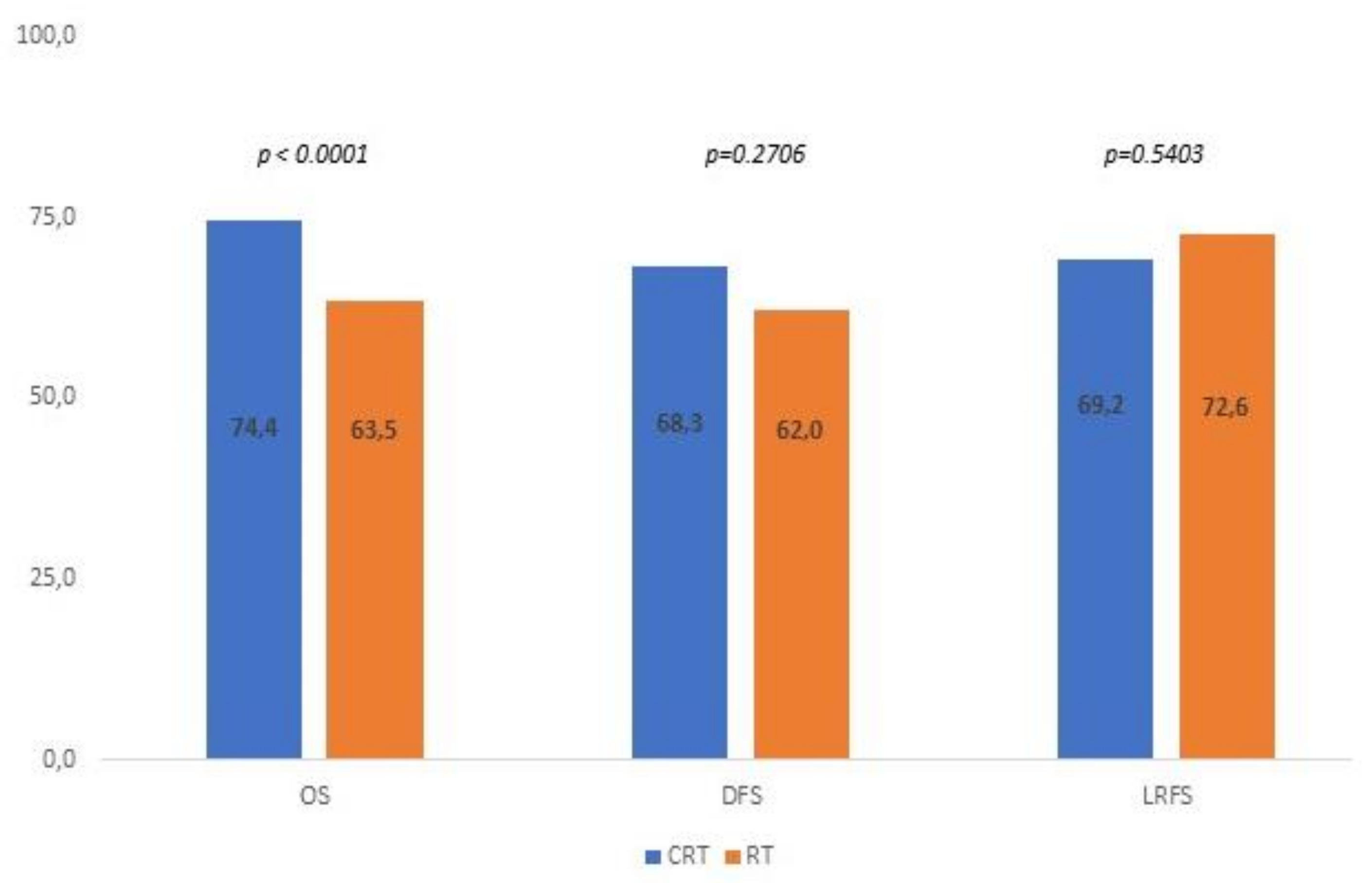
3.2. Survival Analysis
4. Discussion
4.1. Analysis of Minor Risk Factors
4.1.1. Depth of Invasion (DOI)
4.1.2. Perineural Invasion (PNI)
4.1.3. Lymph Vascular Invasion (LVI)
4.1.4. pN1
4.1.5. Multiple Positive Nodes
4.1.6. pT3–pT4 Tumors
4.1.7. Low Neck Positive Nodes (Levels IV–V)
4.1.8. Close Surgical Margins
4.1.9. Other Minor Risk Factors
4.2. Main Issues and Limitations
5. Conclusions
Supplementary Materials
Author Contributions
Funding
Conflicts of Interest
References
- International Agency for Research on Cancer. Available online: http://gco.iarc.fr/ (accessed on 1 November 2021).
- Daly, M.E.; Le, Q.T.; Kozak, M.M.; Maxim, P.G.; Murphy, J.D.; Hsu, A.; Loo, B.W., Jr.; Kaplan, M.J.; Fischbein, N.J.; Chang, D.T. Intensity-modulated radiotherapy for oral cavity saquemos cell carcinoma: Patterns of failure and predictors of local control. Int. J. Radiat. Oncol. Biol. Phys. 2011, 80, 1412–1422. [Google Scholar] [CrossRef] [PubMed]
- Bernier, J.; Domenge, C.; Ozsahin, M.; Matuszewska, K.; Lefèbvre, J.L.; Greiner, R.H.; Giralt, J.; Maingon, P.; Rolland, F.; Bolla, M.; et al. Postoperative irradiation with or without concomitant chemotherapy for locally advanced head and neck cancer. N. Engl. J. Med. 2004, 350, 1945–1952. [Google Scholar] [CrossRef] [PubMed]
- Cooper, J.S.; Pajak, T.F.; Forastiere, A.A.; Jacobs, J.; Campbell, B.H.; Saxman, S.B.; Kish, J.A.; Kim, H.E.; Cmelak, A.J.; Rotman, M.; et al. Postoperative concurrent radiotherapy and chemotherapy for high-risk squamous-cell carcinoma of the head and neck. N. Engl. J. Med. 2004, 350, 1937–1944. [Google Scholar] [CrossRef]
- Bernier, J.; Cooper, J.S.; Pajak, T.F.; van Glabbeke, M.; Bourhis, J.; Forastiere, A.; Ozsahin, E.M.; Jacobs, J.R.; Jassem, J.; Ang, K.K.; et al. Defining risk levels in locally advanced head and neck cancers: A comparative analysis of concurrent postoperative radiation plus chemotherapy trials of the EORTC (#22931) and RTOG (#9501). Head Neck 2005, 27, 843–850. [Google Scholar] [CrossRef] [PubMed]
- Available online: https://www.nccn.org/professionals/physician_gls/pdf/head-and-neck.pdf (accessed on 1 November 2021).
- Fan, K.-H.; Wang, H.-M.; Kang, C.-J.; Lee, L.-Y.; Huang, S.-F.; Lin, C.-Y.; Chen, E.Y.-C.; Chen, I.H.; Liao, C.-T.; Chang, J.T.-C. Treatment Results of Postoperative Radiotherapy on Squamous Cell Carcinoma of the Oral Cavity: Coexistence of Multiple Minor Risk Factors Results in Higher Recurrence Rates. Int. J. Radiat. Oncol. Biol. Phys. 2010, 77, 1024–1029. [Google Scholar] [CrossRef] [PubMed]
- Moher, D.; Liberati, A.; Tetzlaff, J.; Altman, D.G. Preferred Reporting Items for Systematic Reviews and Meta-Analyses: The PRISMA Statement. J. Clin. Epidemiol. 2009, 62, 1006–1012. [Google Scholar] [CrossRef] [PubMed]
- Yanamoto, S.; Japan Oral Oncology Group; Ota, Y.; Okura, M.; Aikawa, T.; Kurita, H.; Kamata, T.; Kirita, T.; Yamakawa, N.; Otsuru, M.; et al. Multicenter Retrospective Study of Adjuvant Therapy for Patients with Pathologically Lymph Node-Positive Oral Squamous Cell Carcinoma: Analysis of Covariance Using Propensity Score. Ann. Surg. Oncol. 2015, 22, 992–999. [Google Scholar] [CrossRef]
- Takahashi, H.; Yanamoto, S.; Yamada, S.; Umeda, M.; Shigeta, T.; Minamikawa, T.; Shibuya, Y.; Komori, T.; Shiraishi, T.; Asahina, I.; et al. Effects of postoperative chemotherapy and radiotherapy on patients with squamous cell carcinoma of the oral cavity and multiple regional lymph node metastases. Int. J. Oral Maxillofac. Surg. 2014, 43, 680–685. [Google Scholar] [CrossRef]
- De Almeida, J.R.; Truong, T.; Khan, N.M.; Su, J.S.; Irish, J.; Gilbert, R.; Goldstein, D.; Huang, S.H.; O’Sullivan, B.; Hosni, A.; et al. Treatment implications of postoperative chemoradiotherapy for squamous cell carcinoma of the oral cavity with minor and major extranodal extension. Oral Oncol. 2020, 110, 104845. [Google Scholar] [CrossRef]
- Fan, K.-H.; Lin, C.-Y.; Kang, C.-J.; Huang, S.-F.; Wang, H.-M.; Chen, E.Y.-C.; Chen, I.-H.; Liao, C.-T.; Cheng, A.-J.; Chang, J.T.-C. Combined-modality treatment for advanced oral tongue squamous cell carcinoma. Int. J. Radiat. Oncol. 2007, 67, 453–461. [Google Scholar] [CrossRef]
- Yamada, S.-I.; Kondo, E.; Hashidume, M.; Sakurai, A.; Otagiri, H.; Matsumura, N.; Kubo, K.; Hakoyama, Y.; Yajima, J.; Morioka, M.; et al. A retrospective investigation of minor risk factors as prognostic predictors and treatment indications in oral squamous cell carcinoma. J. Dent. Sci. 2020, 16, 445–452. [Google Scholar] [CrossRef]
- Lee, L.-Y.; Lin, C.-Y.; Cheng, N.-M.; Tsai, C.-Y.; Hsueh, C.; Fan, K.-H.; Wang, H.-M.; Hsieh, C.-H.; Ng, S.-H.; Yeh, C.-H.; et al. Poor tumor differentiation is an independent adverse prognostic variable in pa-tients with locally advanced oral cavity cancer—Comparison with pathological risk factors according to the NCCN guidelines. Cancer Med. 2021, 10, 6627–6641. [Google Scholar] [CrossRef]
- Kim, T.H.; Cha, I.-H.; Choi, E.C.; Kim, H.R.; Kim, H.J.; Kim, S.-H.; Keum, K.C.; Lee, C.G. Postoperative Concurrent Chemoradiotherapy Versus Radiotherapy Alone for Advanced Oral Cavity Cancer in the Era of Modern Radiation Techniques. Front. Oncol. 2021, 11, 619372. [Google Scholar] [CrossRef]
- Anand, A.K.; Agarwal, P.; Gulia, A.; Goel, V.; Jain, J.; Chaturvedi, H.; Hazarika, B.; Mukherjee, U.; Arora, D.; Bansal, A.K. Significance of perineural invasion in locally advanced bucco alveolar complex carcinomas treated with surgery and postoperative radiation ± concurrent chemotherapy. Head Neck 2017, 39, 1446–1453. [Google Scholar] [CrossRef]
- Chen, C.C.; Lin, J.-C.; Chen, K.-W. Comparison cisplatin with cisplatin plus 5FU in head and neck cancer patients received postoperative chemoradiotherapy. Oral Oncol. 2017, 69, 11–14. [Google Scholar] [CrossRef]
- Hasegawa, T.; Yanamoto, S.; Otsuru, M.; Kakei, Y.; Okura, M.; Yamakawa, N.; Yamada, S.I.; Ota, Y.; Umeda, M.; Kirita, T.; et al. Multi-center retrospective study of the prognosis and treatment outcomes of Japanese oral squamous cell carcinoma patients with single lymph node metastasis and extra nodal extension. J. Surg. Oncol. 2018, 117, 1736–1743. [Google Scholar] [CrossRef]
- Hasegawa, T.; Yanamoto, S.; Otsuru, M.; Kakei, Y.; Okura, M.; Yamakawa, N.; Dds, S.Y.; Ota, Y.; Umeda, M.; Kirita, T.; et al. Multicenter retrospective study of the prognosis and treatment outcomes of Japanese oral squamous cell carcinoma patients with level IV/V metastasis. Head Neck 2019, 41, 2256–2263. [Google Scholar] [CrossRef]
- Brooker, R.C.; Hobkirk, A.; Cashman, H.; Sato, T.; Broderick, D.; Wong, H.; Kyzas, P.; Haridass, A.; Sacco, J.J.; Schache, A.G. Adjuvant management of locally advanced oral squamous cell carcinoma—Real-world challenges and opportunities. Br. J. Oral Maxillofac. Surg. 2021, 59, 952–958. [Google Scholar] [CrossRef]
- Chan, A.K.; Huang, S.H.; Le, L.W.; Yu, E.; Dawson, L.A.; Kim, J.J.; Cho, B.C.; Bayley, A.J.; Ringash, J.; Goldstein, D.; et al. Postoperative intensity-modulated radiotherapy following surgery for oral cavity squamous cell carcinoma: Patterns of failure. Oral Oncol. 2013, 49, 255–260. [Google Scholar] [CrossRef]
- Harari, P.M.; Harris, J.; Kies, M.S.; Myers, J.N.; Jordan, R.C.; Gillison, M.L.; Foote, R.L.; Machtay, M.; Rotman, M.; Khuntia, D.; et al. Postoperative chemoradiotherapy and cetuximab for high-risk squamous cell carcinoma of the head and neck: Radiation Therapy Oncology Group RTOG-0234. J. Clin. Oncol. 2014, 32, 2486–2495. [Google Scholar] [CrossRef]
- Hingsammer, L.; Seier, T.; Ikenberg, J.; Schumann, P.; Zweifel, D.; Rücker, M.; Bredell, M.; Lanzer, M. The influence of lymph node ratio on survival and disease recurrence in squamous cell carcinoma of the tongue. Int. J. Oral Maxillofac. Surg. 2019, 48, 851–856. [Google Scholar] [CrossRef] [PubMed]
- Hinerman, R.W.; Mendenhall, W.M.; Morris, C.G.; Amdur, R.J.; Werning, J.W.; Villaret, D.B. Postoperative irradiation for squamous cell carcinoma of the oral cavity: 35-year experience. Head Neck 2004, 26, 984–994. [Google Scholar] [CrossRef] [PubMed]
- Hasegawa, T.; Yanamoto, S.; Otsuru, M.; Yamada, S.I.; Minamikawa, T.; Shigeta, T.; Naruse, T.; Suzuki, T.; Sasaki, M.; Ota, Y.; et al. Retrospective study of treatment outcomes after postoperative chemoradi-otherapy in Japanese oral squamous cell carcinoma patients with risk factors of recurrence. Oral Surg. Oral Med. Oral Pathol. Oral Radiol. 2017, 123, 524–530. [Google Scholar] [CrossRef] [PubMed]
- Jardim, J.F.; Francisco, A.L.N.; Gondak, R.; Damascena, A.; Kowalski, L.P. Prognostic impact of perineural invasion and lymphovascular invasion in advanced stage oral squamous cell carcinoma. Int. J. Oral Maxillofac. Surg. 2015, 44, 23–28. [Google Scholar] [CrossRef]
- Kademani, D.; Bell, R.B.; Bagheri, S.; Holmgren, E.; Dierks, E.; Potter, B.; Homer, L. Prognostic factors in intraoral squamous cell carcinoma: The influence of histologic grade. J. Oral Maxillofac. Surg. 2005, 63, 1599–1605. [Google Scholar] [CrossRef]
- Kang, C.-J.; Lin, C.-Y.; Wang, H.-M.; Fan, K.-H.; Ng, S.-H.; Lee, L.-Y.; Chen, I.-H.; Huang, S.-F.; Liao, C.-T.; Yen, T.-C. The number of pathologically positive lymph nodes and pathological tumor depth predicts prognosis in patients with poorly differentiated squamous cell carcinoma of the oral cavity. Int. J. Radiat. Oncol. Biol. Phys. 2011, 81, e223–e230. [Google Scholar] [CrossRef]
- Kreppel, M.; Drebber, U.; Eich, H.T.; Dreiseidler, T.; Zöller, J.E.; Müller, R.P.; Scheer, M. Combined-modality treatment in advanced oral squamous cell carcinoma: Primary surgery followed by adjuvant concomitant radiochemotherapy. Strahlenther Onkol. 2011, 187, 555–560. [Google Scholar] [CrossRef]
- Lee, J.H.; Song, J.H.; Lee, S.N.; Kang, J.H.; Kim, M.S.; Sun, D.I.; Kim, Y.S. Adjuvant postoperative radiotherapy with or without chemotherapy for locally ad-vanced squamous cell carcinoma of the head and neck: The importance of patient selection for the postoperative chemoradiotherapy. Cancer Res. Treat. 2013, 45, 31–39. [Google Scholar] [CrossRef]
- Liao, C.-T.; Chang, J.T.-C.; Wang, H.-M.; Ng, S.-H.; Hsueh, C.; Lee, L.-Y.; Lin, C.H.; Chen, I.-H.; Kang, C.-J.; Huang, S.-F.; et al. Surgical outcome of T4a and resected T4b oral cavity cancer. Cancer 2006, 107, 337–344. [Google Scholar] [CrossRef]
- Liao, C.-T.; Hsueh, C.; Lee, L.-Y.; Lin, C.-Y.; Fan, K.-H.; Wang, H.-M.; Huang, S.-F.; Chen, I.-H.; Kang, C.-J.; Ng, S.-H.; et al. Neck dissection field and lymph node density predict prognosis in patients with oral cavity cancer and pathological node metastases treated with adjuvant therapy. Oral Oncol. 2011, 48, 329–336. [Google Scholar] [CrossRef]
- Liao, C.-T.; Lee, L.-Y.; Hsueh, C.; Lin, C.; Fan, K.; Wang, H.; Hsieh, C.; Ng, S.; Lin, C.; Tsao, C.; et al. Pathological risk factors stratification in pN3b oral cavity squamous cell carcinoma: Focus on the number of positive nodes and extranodal extension. Oral Oncol. 2018, 86, 188–194. [Google Scholar] [CrossRef]
- Lin, Y.-W.; Chen, Y.-F.; Yang, C.-C.; Ho, C.-H.; Wu, T.-C.; Yen, C.-Y.; Lin, L.-C.; Lee, S.P.; Lee, C.-C.; Tai, M.-H. Patterns of failure after postoperative intensity-modulated radiotherapy for locally advanced buccal cancer: Initial masticator space involvement is the key factor of recurrence. Head Neck 2018, 40, 2621–2632. [Google Scholar] [CrossRef]
- Maihoefer, C.; Schüttrumpf, L.; Macht, C.; Pflugradt, U.; Hess, J.; Schneider, L.; Woischke, C.; Walch, A.; Baumeister, P.; Kirchner, T.; et al. Postoperative (chemo) radiation in patients with squamous cell cancers of the head and neck—Clinical results from the cohort of the clinical cooperation group “Personalized Radiotherapy in Head and Neck Cancer”. Radiat Oncol 2018, 13, 123. [Google Scholar] [CrossRef]
- Mair, M.D.; Shetty, R.; Nair, D.; Mathur, Y.; Nair, S.; Deshmukh, A.; Thiagarajan, S.; Pantvaidya, G.; Lashkar, S.; Prabhash, K.; et al. Depth of invasion, size and number of metastatic nodes predicts extracapsular spread in early oral cancers with occult metastases. Oral Oncol. 2018, 81, 95–99. [Google Scholar] [CrossRef]
- Lee, A.; Givi, B.; Roden, D.F.; Tam, M.M.; Wu, S.P.; Gerber, N.K.; Hu, K.S.; Schreiber, D. Utilization and Survival of Postoperative Radiation or Chemoradiation for pT1-2N1M0 Head and Neck Cancer. Otolaryngol. Neck Surg. 2017, 158, 677–684. [Google Scholar] [CrossRef]
- Lee, Y.G.; Kang, E.J.; Keam, B.; Choi, J.H.; Kim, J.S.; Park, K.U.; Lee, K.E.; Kwon, J.H.; Lee, K.W.; Kim, M.K.; et al. Treatment strategy and outcomes in locally advanced head and neck squamous cell carcinoma: A nationwide retrospective cohort study (KCSG HN13-01). BMC Cancer 2020, 20, 813. [Google Scholar] [CrossRef]
- Leeman, J.E.; Li, J.-G.; Pei, X.; Venigalla, P.; Zumsteg, Z.S.; Katsoulakis, E.; Lupovitch, E.; McBride, S.M.; Tsai, C.J.; Boyle, J.O.; et al. Patterns of Treatment Failure and Postrecurrence Outcomes Among Patients With Locally Advanced Head and Neck Squamous Cell Carcinoma After Chemoradiotherapy Using Modern Radiation Techniques. JAMA Oncol. 2017, 3, 1487–1494. [Google Scholar] [CrossRef]
- Lin, C.-S.; Jen, Y.-M.; Cheng, M.-F.; Lin, Y.-S.; Su, W.-F.; Hwang, J.-M.; Chang, L.-P.; Chao, H.-L.; Liu, D.-W.; Lin, H.-Y.; et al. Squamous cell carcinoma of the buccal mucosa: An aggressive cancer requiring multimodality treatment. Head Neck 2005, 28, 150–157. [Google Scholar] [CrossRef]
- Liu, T.; David, M.; Batstone, M.; Clark, J.; Low, T.-H.; Goldstein, D.; Hope, A.; Hosni, A.; Chua, B. The utility of postoperative radiotherapy in intermediate-risk oral squamous cell carcinoma. Int. J. Oral Maxillofac. Surg. 2021, 50, 143–150. [Google Scholar] [CrossRef]
- Lo, W.-L.; Kao, S.-Y.; Chi, L.-Y.; Wong, Y.K.; Chang, R.C.S. Outcomes of oral squamous cell carcinoma in Taiwan after surgical therapy: Factors affecting survival. J. Oral Maxillofac. Surg. 2003, 61, 751–758. [Google Scholar] [CrossRef]
- Luukkaa, M.; Minn, H.; Aitasalo, K.; Kronqvist, P.; Kulmala, J.; Pyrhönen, S.; Grenman, R. Treatment of squamous cell carcinoma of the oral cavity, oropharynx and hypopharynx--an analysis of 174 patients in south western Finland. Acta Oncol. 2003, 42, 756–762. [Google Scholar] [CrossRef]
- Ma’aita, J.K. Oral cancer in Jordan: A retrospective study of 118 patients. Croat. Med. J. 2000, 41, 64–69. [Google Scholar]
- Mair, M.D.; Sawarkar, N.; Nikam, S.; Sarin, R.; Nair, D.; Gupta, T.; Chaturvedi, P.; D’cruz, A.; Nair, S. Impact of radical treatments on survival in locally advanced T4a and T4b buccal mucosa cancers: Selected surgically treated T4b cancers have similar control rates as T4a. Oral Oncol. 2018, 82, 17–22. [Google Scholar] [CrossRef]
- Marra, A.; Violati, M.; Broggio, F.; Codecà, C.; Blasi, M.; Luciani, A.; Zonato, S.; Rabbiosi, D.; Moneghini, L.; Saibene, A.; et al. Long-term disease-free survival in surgically-resected oral tongue cancer: A 10-year retrospective study. Acta Otorhinolaryngol. Ital. 2019, 39, 84–91. [Google Scholar] [CrossRef]
- Martínez Carrillo, M.; Tovar Martín, I.; Martínez Lara, I.; Ruiz de Almodóvar Rivera, J.M.; Del Moral Ávila, R. Selective use of postoperative neck radiotherapy in oral cavity and oropharynx cancer: A prospective clinical study. Radiat. Oncol. 2013, 8, 103. [Google Scholar] [CrossRef]
- McMahon, J.D.; Robertson, G.A.J.; Liew, C.; McManners, J.; Mackenzie, F.R.; Hislop, W.S.; Morley, S.E.; Devine, J.; Carton, A.T.M.; Harvey, S.; et al. Oral and oropharyngeal cancer in the West of Scotland-long-term outcome data of a prospective audit 1999–2001. Br. J. Oral Maxillofac. Surg. 2011, 49, 92–98. [Google Scholar] [CrossRef]
- Metcalfe, E.; Aspin, L.; Speight, R.; Ermiş, E.; Ramasamy, S.; Cardale, K.; Dyker, K.; Sen, M.; Prestwich, R. Postoperative (Chemo)Radiotherapy for Oral Cavity Squamous Cell Carcinomas: Outcomes and Patterns of Failure. Clin. Oncol. 2016, 29, 51–59. [Google Scholar] [CrossRef]
- Mroueh, R.; Haapaniemi, A.; Grénman, R.; Laranne, J.; Pukkila, M.; Almangush, A.; Salo, T.; Mäkitie, A. Improved outcomes with oral tongue squamous cell carcinoma in Finland. Head Neck 2017, 39, 1306–1312. [Google Scholar] [CrossRef] [PubMed]
- Murthy, V.; Agarwal, J.; Laskar, S.G.; Gupta, T.; Budrukkar, A.; Pai, P.; Chaturvedi, P.; Chaukar, D.; D′cruz, A. Analysis of prognostic factors in 1180 patients with oral cavity primary cancer treated with definitive or adjuvant radiotherapy. J. Cancer Res. Ther. 2010, 6, 282–289. [Google Scholar] [CrossRef]
- Nair, D.; Mair, M.; Singhvi, H.; Mishra, A.; Nair, S.V.; Agrawal, J.; Chaturvedi, P. Perineural invasion: Independent prognostic factor in oral cancer that warrants adjuvant treatment. Head Neck 2018, 40, 1780–1787. [Google Scholar] [CrossRef]
- Newman, M.; Dziegielewski, P.; Nguyen, N.; Seikaly, H.; Xie, M.; O’Connell, D.; Harris, J.; Biron, V.; Gupta, M.; Archibald, S.; et al. Relationship of depth of invasion to survival outcomes and patterns of recurrence for T3 oral tongue squamous cell carcinoma. Oral Oncol. 2021, 116, 105195. [Google Scholar] [CrossRef] [PubMed]
- Noble, A.R.; Greskovich, J.F.; Han, J.; Reddy, C.A.; Nwizu, T.I.; Khan, M.F.; Scharpf, J.; Adelstein, D.J.; Burkey, B.B.; Koyfman, S.A.; et al. Risk Factors Associated with Disease Recurrence in Patients with Stage III/IV Squamous Cell Carcinoma of the Oral Cavity Treated with Surgery and Postoperative Radiotherapy. Anticancer Res. 2016, 36, 785–792. [Google Scholar] [PubMed]
- Osman, N.; Elamin, Y.Y.; Rafee, S.; O’Brien, C.; Stassen, L.F.A.; Timon, C.; Kinsella, J.; Brennan, S.; O’Byrne, K.J. Weekly cisplatin concurrently with radiotherapy in head and neck squamous cell cancer: A retrospective analysis of a tertiary institute experience. Head Neck 2013, 271, 2253–2259. [Google Scholar] [CrossRef] [PubMed]
- Palazzi, M.; Alterio, D.; Tonoli, S.; Caspiani, O.; Bolner, A.; Colombo, S.; Dall’Oglio, S.; Lastrucci, L.; Bunkheila, F.; Cianciulli, M.; et al. Patterns of postoperative radiotherapy for head and neck cancer in Italy: A prospective, observational study by the head and neck group of the Italian Association for Radiation Oncology (AIRO). Tumori J. 2011, 97, 170–176. [Google Scholar] [CrossRef]
- Pillai, V.; Yadav, V.; Kekatpure, V.; Trivedi, N.; Chandrashekar, N.H.; Shetty, V.; Rangappa, V.; Subramaniam, N.; Bhat, V.; Raghavan, N.; et al. Prognostic determinants of locally advanced buccal mucosa cancer: Do we need to relook the current staging criteria? Oral Oncol. 2019, 95, 43–51. [Google Scholar] [CrossRef]
- Quinsan, I.D.C.M.; Costa, G.C.; Priante, A.V.M.; Cardoso, C.A.; Nunes, C.L.S. Functional outcomes and survival of patients with oral and oropharyngeal cancer after total glossectomy. Braz. J. Otorhinolaryngol. 2019, 86, 545–551. [Google Scholar] [CrossRef]
- Rades, D.; Ulbricht, T.; Hakim, S.; Schild, S. Cisplatin superior to carboplatin in adjuvant radiochemotherapy for locally advanced cancers of the oropharynx and oral cavity. Strahlenther. und Onkol. 2011, 188, 42–48. [Google Scholar] [CrossRef]
- Dos Santos, F.M.; Viani, G.A.; Pavoni, J.F. Evaluation of survival of patients with locally advanced head and neck cancer treated in a single center. Braz. J. Otorhinolaryngol. 2019, 87, 3–10. [Google Scholar] [CrossRef]
- Jensen, J.S.; Jakobsen, K.K.; Mirian, C.; Christensen, J.T.; Schneider, K.; Nahavandipour, A.; Wingstrand, V.L.L.; Wessel, I.; Tvedskov, J.F.; Frisch, T.; et al. The Copenhagen Oral Cavity Squamous Cell Carcinoma database: Protocol and report on establishing a comprehensive oral cavity cancer database. Clin. Epidemiol. 2019, 11, 733–741. [Google Scholar] [CrossRef]
- Sher, D.J.; Thotakura, V.; Balboni, T.A.; Norris, C.M.; Haddad, R.I.; Posner, M.R.; Lorch, J.; Goguen, L.A.; Annino, D.J.; Tishler, R.B. Treatment of Oral Cavity Squamous Cell Carcinoma With Adjuvant or Definitive Intensity-Modulated Radiation Therapy. Int. J. Radiat. Oncol. 2011, 81, e215–e222. [Google Scholar] [CrossRef]
- Shia, B.-C.; Qin, L.; Lin, K.-C.; Fang, C.-Y.; Tsai, L.-L.; Kao, Y.-W.; Wu, S.-Y. Outcomes for Elderly Patients Aged 70 to 80 Years or Older with Locally Advanced Oral Cavity Squamous Cell Carcinoma: A Propensity Score–Matched, Nationwide, Oldest Old Patient–Based Cohort Study. Cancers 2020, 12, 258. [Google Scholar] [CrossRef]
- Silva, P.; Lemos, J.; Borges, M.; Rêgo, T.D.; Dantas, T.; Leite, C.; Lima, M.; Cunha, M.; Sousa, F. Prognostic factors on surgically and non-surgically treated oral squamous cell carcinoma: Advances in survival in fifteen years of follow up. J. Clin. Exp. Dent. 2021, 13, e240–e249. [Google Scholar] [CrossRef]
- Spiotto, M.T.; Jefferson, G.; Wenig, B.; Markiewicz, M.; Weichselbaum, R.R.; Koshy, M. Differences in Survival With Surgery and Postoperative Radiotherapy Com-pared With Definitive Chemoradiotherapy for Oral Cavity Cancer: A National Cancer Database Analysis. JAMA Oto-laryngol. Head Neck Surg. 2017, 143, 691–699. [Google Scholar] [CrossRef]
- Suzuki, M.; Nishimura, Y.; Nakamatsu, K.; Kanamori, S.; Koike, R.; Kawamoto, M.; Mori, K. Phase I study of weekly docetaxel infusion and concurrent radiation therapy for head and neck cancer. Jpn. J. Clin. Oncol. 2003, 33, 297–301. [Google Scholar] [CrossRef][Green Version]
- Tangthongkum, M.; Kirtsreesakul, V.; Supanimitjaroenporn, P.; Leelasawatsuk, P. Treatment outcome of advance staged oral cavity cancer: Concurrent chemoradiotherapy compared with primary surgery. Head Neck 2017, 40, 1879–2572. [Google Scholar] [CrossRef]
- Thakar, A.; Thakur, R.; Kakkar, A.; Malhotra, R.K.; Ms, C.A.S.; Sikka, K.; Kumar, R.; Pramanik, R.; Biswas, A.; Bhalla, A.S.; et al. Oral Cancer in the Indian Subcontinent-Survival Outcomes and Risk Factors with Primary Surgery. Laryngoscope 2021, 131, 2254–2261. [Google Scholar] [CrossRef]
- Wan, X.C.; Egloff, A.M.; Johnson, J. Histological assessment of cervical lymph node identifies patients with head and neck squamous cell carcinoma (HNSCC): Who would benefit from chemoradiation after surgery? Laryngoscope 2012, 122, 2712–2722. [Google Scholar] [CrossRef]
- Yokota, T.; Iida, Y.; Ogawa, H.; Kamijo, T.; Onozawa, Y.; Todaka, A.; Hamauchi, S.; Onoe, T.; Nakagawa, M.; Yurikusa, T.; et al. Prognostic Factors and Multidisciplinary Postoperative Chemoradiotherapy for Clinical T4a Tongue Cancer. Oncology 2016, 91, 78–84. [Google Scholar] [CrossRef]
- Zanoni, D.K.; Montero, P.H.; Migliacci, J.C.; Shah, J.P.; Wong, R.J.; Ganly, I.; Patel, S.G. Survival outcomes after treatment of cancer of the oral cavity (1985–2015). Oral Oncol. 2019, 90, 115–121. [Google Scholar] [CrossRef]
- Zumsteg, Z.; Luu, M.; Kim, S.; Tighiouart, M.; Mita, A.; Scher, K.; Lu, D.; Shiao, S.; Clair, J.M.-S.; Ho, A. Quantitative lymph node burden as a ‘very-high-risk’ factor identifying head and neck cancer patients benefiting from postoperative chemoradiation. Ann. Oncol. 2019, 30, 1669. [Google Scholar] [CrossRef]
- Available online: https://www.ihe.ca/research-programs/rmd/cssqac/cssqac-about (accessed on 1 November 2021).
- Higgins, J.P.; Thompson, S.G.; Deeks, J.J.; Altman, D.G. Measuring inconsistency in meta-analyses. BMJ 2003, 327, 557–560. [Google Scholar] [CrossRef] [PubMed]
- Li, R.; Jiang, W.; Dou, S.; Zhong, L.; Sun, J.; Zhang, C.; Zhu, G. A Phase 2 Trial of Chemoradiation Therapy Using Weekly Docetaxel for High-Risk Postoperative Oral Squamous Cell Carcinoma Patients. Int. J. Radiat. Oncol. Biol. Phys. 2020, 107, 462–468. [Google Scholar] [CrossRef] [PubMed]
- Spiotto, M.T.; Jefferson, G.D.; Wenig, B.; Markiewicz, M.R.; Weichselbaum, R.R.; Koshy, M. Survival outcomes for postoperative chemoradiation in intermediate-risk oral tongue cancers. Head Neck 2017, 39, 2537–2548. [Google Scholar] [CrossRef] [PubMed]
- Trifiletti, D.M.; Smith, A.; Mitra, N.; Grover, S.; Lukens, J.N.; Cohen, R.B.; Read, P.; Mendenhall, W.M.; Lin, A.; Swisher-McClure, S. Beyond Positive Margins and Extracapsular Extension: Evaluating the Utilization and Clinical Impact of Postoperative Chemoradiotherapy in Resected Locally Advanced Head and Neck Cancer. J. Clin. Oncol. 2017, 35, 1550–1560. [Google Scholar] [CrossRef] [PubMed]
- Chen, W.C.; Lai, C.H.; Fang, C.C.; Yang, Y.H.; Chen, P.C.; Lee, C.P.; Chen, M.F. Identification of High-Risk Subgroups of Patients With Oral Cavity Cancer in Need of Postoperative Adjuvant Radiotherapy or Chemo-Radiotherapy. Medicine 2016, 95, e3770. [Google Scholar] [CrossRef]
- Fan, K.H.; Chen, Y.C.; Lin, C.Y.; Kang, C.J.; Lee, L.Y.; Huang, S.F.; Liao, C.T.; Ng, S.H.; Wang, H.M.; Chang, J.T. Postoperative radiotherapy with or without concurrent chemotherapy for oral squamous cell carcinoma in patients with three or more minor risk factors: A propensity score matching analysis. Radiat. Oncol. 2017, 12, 184. [Google Scholar] [CrossRef]
- Feng, Z.; Xu, Q.S.; Wang, C.; Li, J.Z.; Mao, M.H.; Li, H.; Qin, L.Z.; Han, Z. Lymph node ratio is associated with adverse clinicopathological features and is a crucial nodal parameter for oral and oropharyngeal cancer. Sci. Rep. 2017, 7, 6708. [Google Scholar] [CrossRef]
- Chen, M.M.; Colevas, A.D.; Megwalu, U.; Divi, V. Survival benefit of post-operative chemotherapy for intermediate-risk advanced stage head and neck cancer differs with patient age. Oral Oncol. 2018, 84, 71–75. [Google Scholar] [CrossRef]
- Fan, K.H.; Lin, C.Y.; Kang, C.J.; Lee, L.Y.; Huang, S.F.; Liao, C.T.; Chen, I.H.; Ng, S.H.; Wang, H.M.; Chang, J.T. Postoperative concomitant chemoradiotherapy improved treatment outcomes of patients with oral cavity cancer with multiple-node metastases but no other major risk factors. PLoS ONE 2014, 9, e86922. [Google Scholar] [CrossRef]
- Patel, E.J.; Oliver, J.R.; Vaezi, A.; Li, Z.; Persky, M.; Tam, M.; Hu, K.S.; Jacobson, A.S.; Givi, B. Primary Surgical Treatment in Very Advanced (T4b) Oral Cavity Squamous Cell Carcinomas. Otolaryngol. Head Neck Surg. 2021, 165, 431–437. [Google Scholar] [CrossRef]
- Osborn, V.W.; Givi, B.; Rineer, J.; Roden, D.; Sheth, N.; Lederman, A.; Katsoulakis, E.; Hu, K.; Schreiber, D. Patterns of care and outcomes of adjuvant therapy for high-risk head and neck cancer after surgery. Head Neck 2018, 40, 1254–1262. [Google Scholar] [CrossRef]
- Lin, C.Y.; Fan, K.H.; Lee, L.Y.; Hsueh, C.; Yang, L.Y.; Ng, S.H.; Wang, H.M.; Hsieh, C.H.; Lin, C.H.; Tsao, C.K.; et al. Precision Adjuvant Therapy Based on Detailed Pathologic Risk Factors for Resected Oral Cavity Squamous Cell Carcinoma: Long-Term Outcome Comparison of CGMH and NCCN Guidelines. Int. J. Radiat. Oncol. Biol. Phys. 2019, 106, 916–925. [Google Scholar] [CrossRef]
- Tasoulas, J.; Lenze, N.R.; Farquhar, D.; Schrank, T.P.; Shen, C.; Shazib, M.A.; Singer, B.; Patel, S.; Grilley Olson, J.E.; Hayes, D.N.; et al. The addition of chemotherapy to adjuvant radiation is associated with inferior survival outcomes in intermediate-risk HPV-negative HNSCC. Cancer. Med. 2021, 10, 3231–3239. [Google Scholar] [CrossRef]
- Ebrahimi, A.; Gil, Z.; Amit, M.; Yen, T.C.; Liao, C.T.; Chaturvedi, P.; Agarwal, J.P.; Kowalski, L.P.; Köhler, H.F.; Kreppel, M.; et al. Depth of invasion alone as an indication for postoperative radiotherapy in small oral squamous cell carcinomas: An International Collaborative Study. Head Neck 2019, 41, 1935–1942. [Google Scholar] [CrossRef]
- Lee, N.C.J.; Eskander, A.; Park, H.S.; Mehra, S.; Burtness, B.A.; Husain, Z. Pathologic staging changes in oral cavity squamous cell carcinoma: Stage migration and implications for adjuvant treatment. Cancer 2019, 125, 2975–2983. [Google Scholar] [CrossRef]
- Shinn, J.R.; Wood, C.B.; Colazo, J.M.; Harrell, F.E., Jr.; Rohde, S.L.; Mannion, K. Cumulative incidence of neck recurrence with increasing depth of invasion. Oral Oncol. 2018, 87, 36–42. [Google Scholar] [CrossRef]
- Alterio, D.; D’Urso, P.; Volpe, S.; Tagliabue, M.; De Berardinis, R.; Augugliaro, M.; Gandini, S.; Maffini, F.A.; Bruschini, R.; Turturici, I.; et al. The Impact of Post-Operative Radiotherapy in Early Stage (pT1-pT2N0M0) Oral Tongue Squamous Cell Carcinoma in Era of DOI. Cancers 2021, 13, 4851. [Google Scholar] [CrossRef]
- Caldeira, P.C.; Soto, A.M.L.; de Aguiar, M.C.F.; Martins, C.C. Tumor depth of invasion and prognosis of early-stage oral squamous cell carcinoma: A meta-analysis. Oral Dis. 2020, 26, 1357–1365. [Google Scholar] [CrossRef]
- Carter, R.L.; Foster, C.S.; Dinsdale, E.A.; Pittam, M.R. Perineural spread by squamous carcinomas of the head and neck: A morphological study using antiaxonal and antimyelin monoclonal antibodies. J. Clin. Pathol. 1983, 36, 269–275. [Google Scholar] [CrossRef]
- Soo, K.C.; Carter, R.L.; O’Brien, C.J.; Barr, L.; Bliss, J.M.; Shaw, H.J. Prognostic implications of perineural spread in squamous carcinomas of the head and neck. Laryngoscope 1986, 96, 1145–1148. [Google Scholar] [CrossRef]
- Kurtz, K.A.; Hoffman, H.T.; Zimmerman, M.B.; Robinson, R.A. Perineural and vascular invasion in oral cavity squamous carcinoma: Increased incidence on re-review of slides and by using immunohistochemical enhancement. Arch. Pathol. Lab. Med. 2005, 129, 354–359. [Google Scholar] [CrossRef]
- Alkhadar, H.; Macluskey, M.; White, S.; Ellis, I. Perineural invasion in oral squamous cell carcinoma: Incidence, prognostic impact and molecular insight. J. Oral Pathol. Med. 2020, 49, 994–1003. [Google Scholar] [CrossRef]
- Schmitd, L.B.; Scanlon, C.S.; D’Silva, N.J. Perineural Invasion in Head and Neck Cancer. J. Dent. Res. 2018, 97, 742–750. [Google Scholar] [CrossRef]
- Tai, S.K.; Li, W.Y.; Yang, M.H.; Chang, S.Y.; Chu, P.Y.; Tsai, T.L.; Wang, Y.F.; Chang, P.M. Treatment for T1-2 oral squamous cell carcinoma with or without perineural invasion: Neck dissection and postoperative adjuvant therapy. Ann. Surg. Oncol. 2012, 19, 1995–2002. [Google Scholar] [CrossRef]
- Brown, B.; Barnes, L.; Mazariegos, J.; Taylor, F.; Johnson, J.; Wagner, R.L. Prognostic factors in mobile tongue and floor of mouth carcinoma. Cancer 1989, 64, 1195–1202. [Google Scholar] [CrossRef]
- Aivazian, K.; Ebrahimi, A.; Low, T.H.; Gao, K.; Clifford, A.; Shannon, K.; Clark, J.R.; Gupta, R. Perineural invasion in oral squamous cell carcinoma: Quantitative subcategorisation of perineural invasion and prognostication. J. Surg. Oncol. 2015, 111, 352–358. [Google Scholar] [CrossRef]
- Fagan, J.J.; Collins, B.; Barnes, L.; D’Amico, F.; Myers, E.N.; Johnson, J.T. Perineural invasion in squamous cell carcinoma of the head and neck. Arch. Otolaryngol. Head Neck Surg. 1998, 124, 637–640. [Google Scholar] [CrossRef]
- Kim, R.Y.; Helman, J.I.; Braun, T.M.; Ward, B.B. Increased Presence of Perineural Invasion in the Tongue and Floor of the Mouth: Could It Represent a More Aggressive Oral Squamous Cell Carcinoma, or Do Larger Aggressive Tumors Cause Perineural Invasion? J. Oral Maxillofac. Surg. 2019, 77, 852–858. [Google Scholar] [CrossRef] [PubMed]
- Babar, A.; Woody, N.M.; Ghanem, A.I.; Tsai, J.; Dunlap, N.E.; Schymick, M.; Liu, H.Y.; Burkey, B.B.; Lamarre, E.D.; Ku, J.A.; et al. Outcomes of Post-Operative Treatment with Concurrent Chemoradiotherapy (CRT) in High-Risk Resected Oral Cavity Squamous Cell Carcinoma (OCSCC): A Multi-Institutional Collaboration. Curr. Oncol. 2021, 28, 2409–2419. [Google Scholar] [CrossRef] [PubMed]
- Azam, S.H.; Pecot, C.V. Cancer’s got nerve: Schwann cells drive perineural invasion. J. Clin. Investig. 2016, 126, 1242–1244. [Google Scholar] [CrossRef] [PubMed]
- Ein, L.; Bracho, O.; Mei, C.; Patel, J.; Boyle, T.; Monje, P.; Fernandez-Valle, C.; Bas, E.; Thomas, G.; Weed, D.; et al. Inhibition of tropomyosine receptor kinase B on the migration of human Schwann cell and dispersion of oral tongue squamous cell carcinoma in vitro. Head Neck 2019, 41, 4069–4075. [Google Scholar] [CrossRef]
- Ein, L.; Mei, C.; Bracho, O.; Bas, E.; Monje, P.; Weed, D.; Sargi, Z.; Thomas, G.; Dinh, C. Modulation of BDNF-TRKB Interactions on Schwann Cell-induced Oral Squamous Cell Carcinoma Dispersion In Vitro. Anticancer Res. 2019, 39, 5933–5942. [Google Scholar] [CrossRef]
- Amin, M.B.; Greene, F.L.; Edge, S.B.; Compton, C.C.; Gershenwald, J.E.; Brookland, R.K.; Meyer, L.; Gress, D.M.; Byrd, D.R.; Winchester, D.P. The Eighth Edition AJCC Cancer Staging Manual: Continuing to build a bridge from a population-based to a more “personalized” approach to cancer staging. CA Cancer J. Clin. 2017, 67, 93–99. [Google Scholar] [CrossRef]
- Huang, S.; Zhu, Y.; Cai, H.; Zhang, Y.; Hou, J. Impact of lymphovascular invasion in oral squamous cell carcinoma: A meta-analysis. Oral Surg. Oral Med. Oral Pathol. Oral Radiol. 2021, 131, 319–328.e1. [Google Scholar] [CrossRef]
- Dolens, E.d.S.; Dourado, M.R.; Almangush, A.; Salo, T.A.; Gurgel Rocha, C.A.; da Silva, S.D.; Brennan, P.A.; Coletta, R.D. The Impact of Histopathological Features on the Prognosis of Oral Squamous Cell Carcinoma: A Comprehensive Review and Meta-Analysis. Front. Oncol. 2021, 11, 784924. [Google Scholar] [CrossRef]
- Fives, C.; Feeley, L.; O’Leary, G.; Sheahan, P. Importance of lymphovascular invasion and invasive front on survival in floor of mouth cancer. Head Neck 2016, 38 (Suppl. 1), E1528–E1534. [Google Scholar] [CrossRef]
- Mascitti, M.; Tempesta, A.; Togni, L.; Capodiferro, S.; Troiano, G.; Rubini, C.; Maiorano, E.; Santarelli, A.; Favia, G.; Limongelli, L. Histological features and survival in young patients with HPV-negative oral squamous cell carcinoma. Oral Dis. 2020, 26, 1640–1648. [Google Scholar] [CrossRef]
- Greenberg, J.S.; El Naggar, A.K.; Mo, V.; Roberts, D.; Myers, J.N. Disparity in pathologic and clinical lymph node staging in oral tongue carcinoma. Implication for therapeutic decision making. Cancer 2003, 98, 508–515. [Google Scholar] [CrossRef]
- Ho, A.S.; Kim, S.; Tighiouart, M.; Gudino, C.; Mita, A.; Scher, K.S.; Laury, A.; Prasad, R.; Shiao, S.L.; Van Eyk, J.E.; et al. Metastatic Lymph Node Burden and Survival in Oral Cavity Cancer. J. Clin. Oncol. 2017, 35, 3601–3609. [Google Scholar] [CrossRef]
- Zumsteg, Z.S.; Kim, S.; David, J.M.; Yoshida, E.J.; Tighiouart, M.; Shiao, S.L.; Scher, K.; Mita, A.; Sherman, E.J.; Lee, N.Y.; et al. Impact of concomitant chemoradiation on survival for patients with T1-2N1 head and neck cancer. Cancer 2017, 123, 1555–1565. [Google Scholar] [CrossRef]
- Ang, K.K.; Harris, J.; Wheeler, R.; Weber, R.; Rosenthal, D.I.; Nguyen-Tân, P.F.; Westra, W.H.; Chung, C.H.; Jordan, R.C.; Lu, C.; et al. Human Papillomavirus and Survival of Patients with Oropharyngeal Cancer. N. Engl. J. Med. 2010, 363, 24–35. [Google Scholar] [CrossRef]
- Compton, C.C.; Byrd, D.R.; Garcia-Aguilar, J.; Kurtzman, S.H.; Olawaiye, A.; Washington, M.K. AJCC Cancer Staging Atlas; Springer: New York, NY, USA, 2012. [Google Scholar] [CrossRef]
- Amin, M.B.; Edge, S.B.; Greene, F.L.; Byrd, D.R.; Brookland, R.K.; Washington, M.K.; Gershenwald, J.E.; Compton, C.C.; Hess, K.R.; Sullivan, D.C.; et al. AJCC Cancer Staging Manual; Springer: Cham, Switzerland, 2017. [Google Scholar]
- Kano, S.; Sakashita, T.; Tsushima, N.; Mizumachi, T.; Nakazono, A.; Suzuki, T.; Yasukawa, S.; Homma, A. Validation of the 8th edition of the AJCC/UICC TNM staging system for tongue squamous cell carcinoma. Int. J. Clin. Oncol. 2018, 23, 844–850. [Google Scholar] [CrossRef]
- Jung, J.; Cho, N.H.; Kim, J.; Choi, E.C.; Lee, S.Y.; Byeon, H.K.; Park, Y.M.; Yang, W.S.; Kim, S.H. Significant invasion depth of early oral tongue cancer originated from the lateral border to predict regional metastases and prognosis. Int. J. Oral. Maxillofac. Surg. 2009, 38, 653–660. [Google Scholar] [CrossRef]
- Liao, C.T.; Chang, J.T.; Wang, H.M.; Ng, S.H.; Hsueh, C.; Lee, L.Y.; Lin, C.H.; Chen, I.H.; Huang, S.F.; Cheng, A.J.; et al. Survival in squamous cell carcinoma of the oral cavity: Differences between pT4 N0 and other stage IVA categories. Cancer 2007, 110, 564–571. [Google Scholar] [CrossRef]
- Liao, C.T.; Wang, H.M.; Ng, S.H.; Yen, T.C.; Lee, L.Y.; Hsueh, C.; Wei, F.C.; Chen, I.H.; Kang, C.J.; Huang, S.F.; et al. Good tumor control and survivals of squamous cell carcinoma of buccal mucosa treated with radical surgery with or without neck dissection in Taiwan. Oral Oncol. 2006, 42, 800–809. [Google Scholar] [CrossRef]
- Liao, C.T.; Ng, S.H.; Chang, J.T.; Wang, H.M.; Hsueh, C.; Lee, L.Y.; Tsao, C.K.; Chen, W.H.; Chen, I.H.; Kang, C.J.; et al. T4b oral cavity cancer below the mandibular notch is resectable with a favorable outcome. Oral Oncol. 2007, 43, 570–579. [Google Scholar] [CrossRef]
- Köhler, H.F.; Kowalski, L.P. Prognostic impact of the level of neck metastasis in oral cancer patients. Braz. J. Otorhinolaryngol. 2012, 78, 15–20. [Google Scholar] [CrossRef] [PubMed]
- Byers, R.M. Modified neck dissection. A study of 967 cases from 1970 to 1980. Am. J. Surg. 1985, 150, 414–421. [Google Scholar] [CrossRef]
- Marchiano, E.; Patel, T.D.; Eloy, J.A.; Baredes, S.; Park, R.C. Impact of Nodal Level Distribution on Survival in Oral Cavity Squamous Cell Carcinoma: A Population-Based Study. Otolaryngol. Head Neck Surg. 2016, 155, 99–105. [Google Scholar] [CrossRef] [PubMed]
- Jones, A.S.; Roland, N.J.; Field, J.K.; Phillips, D.E. The level of cervical lymph node metastases: Their prognostic relevance and relationship with head and neck squamous carcinoma primary sites. Clin. Otolaryngol. Allied Sci. 1994, 19, 63–69. [Google Scholar] [CrossRef] [PubMed]
- Liao, C.T.; Huang, S.F.; Chen, I.H.; Kang, C.J.; Lin, C.Y.; Fan, K.H.; Wang, H.M.; Ng, S.H.; Hsueh, C.; Lee, L.Y.; et al. Outcome analysis of patients with pN2 oral cavity cancer. Ann. Surg. Oncol. 2010, 17, 1118–1126. [Google Scholar] [CrossRef]
- Barroso, E.M.; Aaboubout, Y.; van der Sar, L.C.; Mast, H.; Sewnaik, A.; Hardillo, J.A.; ten Hove, I.; Nunes Soares, M.R.; Ottevanger, L.; Bakker Schut, T.C.; et al. Performance of Intraoperative Assessment of Resection Margins in Oral Cancer Surgery: A Review of Literature. Front. Oncol. 2021, 11, 628297. [Google Scholar] [CrossRef]
- Tasche, K.K.; Buchakjian, M.R.; Pagedar, N.A.; Sperry, S.M. Definition of “Close Margin” in Oral Cancer Surgery and Association of Margin Distance With Local Recurrence Rate. JAMA Otolaryngol. Head Neck Surg. 2017, 143, 1166–1172. [Google Scholar] [CrossRef]
- Solomon, J.; Hinther, A.; Matthews, T.W.; Nakoneshny, S.C.; Hart, R.; Dort, J.C.; Chandarana, S.P. The impact of close surgical margins on recurrence in oral squamous cell carcinoma. J. Otolaryngol. Head Neck Surg. 2021, 50, 9. [Google Scholar] [CrossRef]
- Margalit, D.N.; Sacco, A.G.; Cooper, J.S.; Ridge, J.A.; Bakst, R.L.; Beadle, B.M.; Beitler, J.J.; Chang, S.S.; Chen, A.M.; Galloway, T.J.; et al. Systematic review of postoperative therapy for resected squamous cell carcinoma of the head and neck: Executive summary of the American Radium Society appropriate use criteria. Head Neck 2021, 43, 367–391. [Google Scholar] [CrossRef]
- Hakim, S.G.; von Bialy, R.; Falougy, M.; Steller, D.; Tharun, L.; Rades, D.; Sieg, P.; Alsharif, U. Impact of stratified resection margin classification on local tumor control and survival in patients with oral squamous cell carcinoma. J. Surg. Oncol. 2021, 124, 1284–1295. [Google Scholar] [CrossRef]
- Burns, C.; Gorina Faz, M. An Analysis of Tumor Margin Shrinkage in the Surgical Resection of Squamous Cell Carcinoma of the Oral Cavity. Cureus 2021, 13, e15329. [Google Scholar] [CrossRef]
- Farhood, Z.; Simpson, M.; Ward, G.M.; Walker, R.J.; Osazuwa-Peters, N. Does anatomic subsite influence oral cavity cancer mortality? A SEER database analysis. Laryngoscope 2019, 129, 1400–1406. [Google Scholar] [CrossRef]
- Yamamoto, E.; Kohama, G.; Sunakawa, H.; Iwai, M.; Hiratsuka, H. Mode of invasion, bleomycin sensitivity, and clinical course in squamous cell carcinoma of the oral cavity. Cancer 1983, 51, 2175–2180. [Google Scholar] [CrossRef]
- Parsons, J.T.; Mendenhall, W.M.; Stringer, S.P.; Cassisi, N.J.; Million, R.R. An analysis of factors influencing the outcome of postoperative irradiation for squamous cell carcinoma of the oral cavity. Int. J. Radiat. Oncol. Biol. Phys. 1997, 39, 137–148. [Google Scholar] [CrossRef]
- Nguyen, N.P.; Moltz, C.C.; Frank, C.; Karlsson, U.; Nguyen, P.D.; Vos, P.; Smith, H.J.; Dutta, S.; Nguyen, L.M.; Lemanski, C.; et al. Dysphagia severity following chemoradiation and postoperative radiation for head and neck cancer. Eur. J. Radiol. 2006, 59, 453–459. [Google Scholar] [CrossRef]
- Kucha, N.; Soni, T.P.; Jakhotia, N.; Patni, N.; Singh, D.K.; Gupta, A.K.; Sharma, L.M.; Goyal, J. A prospective, comparative analysis of acute toxicity profile between three-dimensional conformal radiotherapy (3DCRT) and intensity-modulated radiotherapy (IMRT) in locally advanced head and neck cancer patients. Cancer Treat. Res. Commun. 2020, 25, 100223. [Google Scholar] [CrossRef]


| Selection Criteria | Inclusion Criteria | Exclusion Criteria |
|---|---|---|
| P Population | Adults (age > 18 years) with resected non-metastatic squamous OCC | Pediatric patients (age < 18) and histology other than SCC |
| I Intervention | Postoperative radiotherapy alone (PORT) | Post-operative chemo-radiotherapy (POCRT) |
| C Comparison | Squamous OCC with minor/intermediate pathological risk factors | Squamous OCC with major pathological risk factors (positive margins and/or ECE) |
| O Outcome | OS, DFS, LRFS | |
| T Timing | 2000–2021 |
| Author, Year | Oral Cavity Subsite | Number of Patients | Accrual Period | Types of Minor Pathological Risk Factors | Presence of Major Pathological Risk Factor | Number of Patients in CRT Arm/Total | Median FU (Months) | Disease-Free Survival (DFS) | Local Recurrence-Free Survival (LRFS) | Overall Survival (OS) |
|---|---|---|---|---|---|---|---|---|---|---|
| Spiotto M. (2017) [76] | Oral tongue | 2803 | 2004–2012 | LVI, DOI ≥ 5 mm, pT3 or pT4, multiple lymph nodes without ENE | No | 1308/2803 | 33 | / | / | 73.3% HR: 0.78 (95%CI: 0.64–0.96) |
| Trifiletti (2017) [77] | Oral cavity and other H and N sites (oropharynx, larynx, etc.) | 5094/10870 (2899 RT, 2195 CTRT) | 2004–2012 | Positive node at level IV or V, multiple lymph nodes without ENE | No | 2195/10870 | 38,4 | / | / | 3 y OS: 74.2% 5 y OS: 65.3% HR: 0.902 (95%CI: 0.861 to 0.944) |
| Chen W.C. (2016) [78] | OSCC | 567 | 2002–2013 | PNI, LVI, DOI ≥ 5 mm (10 mm), close margin (< 2–5 mm), pT3 or pT4, multiple lymph nodes without ENE | 1 (positive margins in 28 patients, ENE 83 patients) | 127/567 | 42 | 50.2% HR: 0.38 (95%CI: 0.21–0.68) | 74.5% HR: 0.33 (95%CI: 0.14–0.78) | 59.8% HR: 0.37 (95%CI: 0.19–0.72) |
| Fan K.H. (2017) [79] | Buccal mucosa, tongue, gums, retromolar trigon, mouth floor, hard palate | 68 of 109 initially selected (34 CRT, 34 RT) | 1999–2009 | PNI, LVI, DOI ≥ 5 mm, close margin (<2–5 mm), pT3 or pT4 | No | 34/68 | 86.4 | 75.4% | 75.4% HR: 0.248 (95%CI: 0.103–0.596) | 67.2% HR: 0.426 (95%CI: 0.212–0.858) |
| Feng (2017) [80] | Tongue, gingiva, buccal mucosa, mouth floor, hard palate | 809 (14% oropharynx) | / | PNI, LVI, pT3, or pT4, multiple lymph nodes without ENE | Yes, ENE + | 114/809 | Not reported | 51.4% | / | / |
| Chen M.M. (2018) [81] | Lips, oral cavity | 5319 total H and N patients. Oral cavity: pRT group 1571, pCRT 956 | 2010–2013 | LVI, pT3, or pT4, multiple lymph nodes without ENE | No | 956/1571 | Not reported | / | / | For T1–4 N2–3, HR: 0.73 (95%CI: 0.58–0.93). For T3–4 N0–1, HR: 0.92 (95%CI: 0.71–1.19) |
| Fan K.H. (2014) [82] | Tongue, buccal mucosa, gums, retromolar trigone, mouth floor, hard palate, lips | 138 | 1998–2008 | PNI, LVI, DOI ≥ 5 mm, close margin (<2–5 mm), positive nodes level IV or V | No | 77/138 | 35 | 60% | 70% | 60% |
| Li R. (2020) [75] | Tongue, gingiva, buccal mucosa, mouth floor, retromolar trigone, palate, lip | 91 | 2016–2018 | pT3 or pT4, multiple lymph nodes without ENE | Yes, positive margins and ENE + | 91/91 | 24 | 75.3% (95%CI: 65.7–84.2%) | 79.0% | 82.4% (95%CI, 73.0–89.6%) |
| Patel (2021) [83] | Retromolar trigone, gum, cheek mucosa, mouth floor and NOS, tongue, vestibule, lip | 1338 | 2004–2017 | pT3 or pT4 | Yes, positive margins and ENE + | 163/1338 (the other 23 patients received neoadjuvant CT then surgery +RT) | 24 | / | / | 64.6% |
| Osborn (2018) [84] | OCSCC | 2303 | 2004–2012 | pT3 or pT4 | Yes, positive margins and ENE + | 1381/2303 | 47,7 | / | / | 67.4% |
| Lin C (2019) [85] | OCSCC | 1200 | 2004–2016 | pT4, DOI > 5 mm (5 mm), positive nodes level IV or V, PNI, LVI | Yes, positive margins and ENE + | 411/1200 | 61 | 75% | / | 83% |
| Tasoulas (2021) [86] | OCSCC | 616, 167 for OC | 2002–2006 | LVI, PNI, T3 or T4, multiple lymph nodes without ENE | Yes, ENE + | 92/616 45 high-risk patients | Not reported | / | / | HR: 0.30 (95%CI: 0.15–0.61) for high-risk patients |
| Study | Comorbidities | PNI | LVI | Multiple Nodes | pT3–T4 | DOI | Close Margins | pN1 | Low Neck Nodes |
|---|---|---|---|---|---|---|---|---|---|
| Fan 2014 [82] | NE | 50% POCRT 25% PORT | 17% POCRT 8% PORT | 100% POCRT 100% PORT | 53% POCRT 26% PORT | ≥10 mm in 65% POCRT 67% PORT | 32% POCRT 23% PORT | No patients | 4% POCRT 5% PORT |
| Spiotto 2017 [76] | Charlson index Well-balanced in POCRT vs. PORT | NE | 10% POCRT 9% PORT | 56% POCRT 36% PORT | 25% POCRT 18% PORT | ≥5 mm in 4% POCRT vs. 8% PORT | NE | 30% POCRT 58% PORT | NE |
| Fan 2017 [79] | ECOG 0–1 in 97% POCRT 94% PORT | 62% POCRT 65% PORT | 15% POCRT 6% PORT | No patients | pT4: 62% POCRT, 65% PORT | ≥10 mm in 75% POCRT 94% PORT | 56% POCRT 74% PORT | 53% POCRT 26% PORT | NE |
| Chen WC 2016 (subgroup analysis) [78] | NE | NPE for patients with only minor RFs | NPE for patients with only minor RFs | NPE for patients with only minor RFs | NPE for patients with only minor RFs | NPE for patients with only minor RFs | NPE for patients with only minor RFs | NPE for patients with only minor RFs | NPE for patients with only minor RFs |
| Patel 2021 [83] | No comorbidity in 78% of patients | NE | NE | 53% of patients | 100% pT4b | NE | NE | 11% of patients | NE |
| Li 2020 [75] | Not specified in the study | NPE for patients with only pN2 | NPE for patients with only pN2 | NPE for patients with only pN2 | NPE for patients with only pN2 | NPE for patients with only pN2 | NPE for patients with only pN2 | NPE for patients with only pN2 | NPE for patients with only pN2 |
| Lin 2019 [85] | NE | 49% patients with minor RFs | 6% patients with minor RFs | 13% patients with minor RF | 28% pT3 44% pT4 patients with minor RFs | ≥10 mm, 64% patients with minor RFs | 13% patients with minor RFs | 18% patients with minor RFs | 0.7%patients with minor RF |
| Trifiletti 2017 [77] | Charlson Index: 0: 78% PORT, 82%POCRT; 1: 17%PORT, 15% POCRT; 2: 4% PORT, 3% POCRT | NPE for studies with mixed populations of HN cancers | NPE for studies with mixed populations of HN cancers | NPE for studies with mixed populations of HN cancers | NPE for studies with mixed populations of HN cancers | NPE for studies with mixed populations of HN cancers | NPE for studies with mixed populations of HN cancers | NPE for studies with mixed populations of HN cancers | NPE for studies with mixed populations of HN cancers |
| Tasoulas 2021 [86] | NE | NPE for studies with mixed populations of HN cancers | NPE for studies with mixed popultions of HN cancers | NPE for studies with mixed popu- lations of HN cancers | NPE for studies with mixed populations of HN cancers | NPE for studies with mixed populations of HN cancers | NPE for studies with mixed populations of HN cancers | NPE for studies with mixed populations of HN cancers | NPE for studies with mixed populations of HN cancers |
| Feng 2017 [80] | NE | NPE for studies with mixed populations of HN cancers | NPE for studies with mixed populations of HN cancers | NPE for studies with mixed populations of HN cancers | NPE for studies with mixed populations of HN cancers | NPE for studies with mixed populations of HN cancers | NPE for studies with mixed populations of HN cancers | NPE for studies with mixed populations of HN cancers | NPE for studies with mixed populations of HN cancers |
| Osborn 2018 [84] | Charlson Index: 0: 79% PORT, 81% POCRT; 1: 16% PORT, 15% POCRT; 2: 5% PORT, 3% POCRT | NPE for studies with mixed populations of HN cancers | NPE for studies with mixed populations of HN cancers | NPE for studies with mixed populations of HN cancers | NPE for studies with mixed populations of HN cancers | NPE for studies with mixed populations of HN cancers | NPE for studies with mixed populations of HN cancers | NPE for studies with mixed populations of HN cancers | NPE for studies with mixed populations of HN cancers |
| Chen MM 2018 [81] | Comorbidities: 0: 37% PORT, 39% POCRT; 1: 10% PORT, 9% POCRT; 2 or +: 2.5% PORT, 2% POCRT | NPE for studies with mixed populations of HN cancers | NPE for studies with mixed populations of HN cancers | NPE for studies with mixed populations of HN cancers | NPE for studies with mixed populations of HN cancers | NPE for studies with mixed populations of HN cancers | NPE for studies with mixed populations of HN cancers | NPE for studies with mixed populations of HN cancers | NPE for studies with mixed populations of HN cancers |
Publisher’s Note: MDPI stays neutral with regard to jurisdictional claims in published maps and institutional affiliations. |
© 2022 by the authors. Licensee MDPI, Basel, Switzerland. This article is an open access article distributed under the terms and conditions of the Creative Commons Attribution (CC BY) license (https://creativecommons.org/licenses/by/4.0/).
Share and Cite
Di Rito, A.; Fiorica, F.; Carbonara, R.; Di Pressa, F.; Bertolini, F.; Mannavola, F.; Lohr, F.; Sardaro, A.; D’Angelo, E. Adding Concomitant Chemotherapy to Postoperative Radiotherapy in Oral Cavity Carcinoma with Minor Risk Factors: Systematic Review of the Literature and Meta-Analysis. Cancers 2022, 14, 3704. https://doi.org/10.3390/cancers14153704
Di Rito A, Fiorica F, Carbonara R, Di Pressa F, Bertolini F, Mannavola F, Lohr F, Sardaro A, D’Angelo E. Adding Concomitant Chemotherapy to Postoperative Radiotherapy in Oral Cavity Carcinoma with Minor Risk Factors: Systematic Review of the Literature and Meta-Analysis. Cancers. 2022; 14(15):3704. https://doi.org/10.3390/cancers14153704
Chicago/Turabian StyleDi Rito, Alessia, Francesco Fiorica, Roberta Carbonara, Francesca Di Pressa, Federica Bertolini, Francesco Mannavola, Frank Lohr, Angela Sardaro, and Elisa D’Angelo. 2022. "Adding Concomitant Chemotherapy to Postoperative Radiotherapy in Oral Cavity Carcinoma with Minor Risk Factors: Systematic Review of the Literature and Meta-Analysis" Cancers 14, no. 15: 3704. https://doi.org/10.3390/cancers14153704
APA StyleDi Rito, A., Fiorica, F., Carbonara, R., Di Pressa, F., Bertolini, F., Mannavola, F., Lohr, F., Sardaro, A., & D’Angelo, E. (2022). Adding Concomitant Chemotherapy to Postoperative Radiotherapy in Oral Cavity Carcinoma with Minor Risk Factors: Systematic Review of the Literature and Meta-Analysis. Cancers, 14(15), 3704. https://doi.org/10.3390/cancers14153704


