Review

How the Potassium Channel Response of T Lymphocytes to the Tumor Microenvironment Shapes Antitumor Immunity

by
Martina Chirra
1,2,†,
Hannah S. Newton
1,†,
Vaibhavkumar S. Gawali
1,
Trisha M. Wise-Draper
2,
Ameet A. Chimote
1 and
Laura Conforti
1,*
1
Division of Nephrology, Department of Internal Medicine, University of Cincinnati, Cincinnati, OH 45267, USA
2
Division of Hematology-Oncology, Department of Internal Medicine, University of Cincinnati, Cincinnati, OH 45267, USA
*
Author to whom correspondence should be addressed.
These authors contributed equally to this work.
Cancers 2022, 14(15), 3564; https://doi.org/10.3390/cancers14153564
Submission received: 6 July 2022 / Revised: 18 July 2022 / Accepted: 20 July 2022 / Published: 22 July 2022
(This article belongs to the Collection Ion Channels in Cancer Therapies)

Abstract

:

Simple Summary

Ion channels are proteins that control the movement of ions across the membranes of cells, thus regulating their physiological functions. In T lymphocytes, ion channels control the Ca2+ influx that, in turn, promotes proliferation and effector functions. In the context of cancer, T cells have the role of fighting tumor cells. However, the tumor microenvironment negatively regulates T cell antitumor capabilities. Therefore, it is of utmost importance to understand the relationship between the tumor microenvironment and the ion channel apparatus of T cells to overcome the tumors’ immunosuppressive capabilities.

Abstract

Competent antitumor immune cells are fundamental for tumor surveillance and combating active cancers. Once established, tumors generate a tumor microenvironment (TME) consisting of complex cellular and metabolic elements that serve to suppress the function of antitumor immune cells. T lymphocytes are key cellular elements of the TME. In this review, we explore the role of ion channels, particularly K+ channels, in mediating the suppressive effects of the TME on T cells. First, we will review the complex network of ion channels that mediate Ca2+ influx and control effector functions in T cells. Then, we will discuss how multiple features of the TME influence the antitumor capabilities of T cells via ion channels. We will focus on hypoxia, adenosine, and ionic imbalances in the TME, as well as overexpression of programmed cell death ligand 1 by cancer cells that either suppress K+ channels in T cells and/or benefit from regulating these channels’ activity, ultimately shaping the immune response. Finally, we will review some of the cancer treatment implications related to ion channels. A better understanding of the effects of the TME on ion channels in T lymphocytes could promote the development of more effective immunotherapies, especially for resistant solid malignancies.

1. Introduction

The immune system is essential for cancer surveillance. However, it is recognized that the unique milieu associated with tumors (known as the tumor microenvironment—TME) is responsible for the functional abnormalities of the immune system that impair cancer elimination [1,2]. The cellular components of the TME (which include fibroblasts, vascular, and immune cells) together with extracellular, soluble, and metabolic features inhibit the functions of a complex network of antitumor immune cells that would otherwise prevent tumor growth and metastasis [3,4]. Effector T cells are key players in the fight against cancer, and the presence of cytotoxic CD8+ and Th1 CD4+ T cells in the tumor is associated with a favorable prognosis and response to immunotherapies. However, the aforementioned alterations in the TME negatively affect the T cell capacity to infiltrate the tumors and kill cancer cells, which, to this day, remain the rate-limiting steps towards an effective antitumor response [5]. In order for T cells to infiltrate the tumor and exert their antitumor functions, it is imperative that the appropriate movements of ions across plasma and intracellular membranes occur, as T cells are critically dependent on ion channel regulated-Ca2+ signaling for their function [6].
This review focuses on the role that ion channels, particularly of the potassium (K+) class, play in mediating the immunosuppressive effects of the TME on T cells. Following a brief overview of the current understanding of the role of ion channels in normal T cell function, we will discuss the negative impact of the metabolic (hypoxia, adenosine, and electrolyte imbalance) and cellular (programmed cell death ligand 1, PD-L1) components of the TME on T cell immune surveillance and their dependence on K+ channels. Furthermore, we will highlight the important role that K+ channels play in response to immune checkpoint inhibitors and the potential benefits of developing new single agent or combination cancer immunotherapies targeting K+ channels.

2. Ion Channel Network in T lymphocytes

Ion channels and transporters form an intricate network responsible for intracellular ion homeostasis and Ca2+ levels necessary for effector functions in T lymphocytes (Figure 1) [7,8]. Intracellular Ca2+, in fact, regulates the activation of Ca2+-dependent transcription factors, the expression of most activation genes in T cells, and mechanical functions such as exocytosis and motility [9,10,11]. The expression profile and role of different ion channels in T cell function have been described in detail by others [7,8,12]. Briefly, there are five major ion channels in human T cells, with each playing a unique and key role in ion homeostasis: Ca2+ release-activated Ca2+ (CRAC) channels; two K+ channels, the voltage-gated K+ channel Kv1.3 and the Ca2+ dependent K+ channel KCa3.1; and transient receptor potential (TRP) channels (e.g., TRPM4 and TRPM7).
The first of these ion channels that play a direct role in controlling intracellular Ca2+ levels are the CRAC channels. CRAC channels allow the influx of Ca2+ after T cell receptor (TCR) stimulation [8] (Figure 1). These channels are formed by two different subunits, ORAI1 (ORAI calcium release-activated calcium modulator 1)—and/or its homologs ORAI2 and ORAI3—and STIM1/STIM2 (stromal interaction molecule 1/2) accessory proteins [6]. ORAI subunits represent the pore-forming subunits of the CRAC channel and are localized in the plasma membrane. Conversely, STIM proteins are on the endoplasmic reticulum (ER) membrane and play a key role as Ca2+-sensors. The contribution of these channels to the T cell response to antigen presentation is the following: antigen presentation and recognition at the TCR/CD3 complex activates phospholipase Cγ (PLCγ), which cleaves phosphatidylinositol 4,5-bisphosphate (PIP2) into inositol (1,4,5)—trisphosphate (IP3) and diacylglycerol (DAG) (Figure 1). IP3 then binds to its receptor located on the ER, resulting in the release of Ca2+ from the ER store. The STIM subunits detect the Ca2+ depletion from the ER, oligomerize and migrate next to the plasma membrane, where they interact with ORAI, leading to its opening and, consequently, Ca2+ influx into the T cell (store-operated calcium entry—SOCE) [7,8,13,14]. Early studies in human and murine T cells revealed that the production of cytokines, such as interleukin-2 (IL-2), interleukin-4 (IL-4), and interferon-γ (IFN-γ), and granule exocytosis required SOCE through STIM1 and ORAI1 [11,15,16]. Furthermore, Weidinger and colleagues (2013) showed that the genetic deletion of STIM1 and STIM2 in cytotoxic T cells in mice reduced cancer cell killing [17]. An increase in intracellular Ca2+ regulates important transcription factors such as nuclear factor of activated T cells (NF-AT) and nuclear factor kappa B (NF-κB), which control the transcription of multiple genes. It has been estimated that more than 80% of T cell activation genes are regulated by Ca2+ [9]. The specificity of the transcription factor activated by Ca2+ influx depends on the shape and amplitude of the Ca2+ response [18].
Kv1.3 and KCa3.1, the principal K+ channels in human T cells, play a significant role in Ca2+ influx by maintaining the electrochemical driving force necessary for Ca2+ entry into the cell. The depolarization of the cell following CRAC-mediated Ca2+ influx activates the voltage-dependent Kv1.3, while the increase in intracellular Ca2+ levels activates KCa3.1, a Ca2+-activated voltage-independent K+ channel. The subsequent increase in K+ efflux through these two K+ channels hyperpolarizes the plasma membrane and sustains the influx of the positively charged Ca2+ ions through the CRAC channels [7,8,19]. Conversely, activation of Na+ influx and Cl efflux, and the inhibition of K+ efflux, all of which lead to membrane depolarization, negatively regulate CRAC-mediated Ca2+ fluxes [7].
Lastly, TRP channels represent non-selective cation channels and are categorized into six subfamilies (TRPA, TRPC, TRPV, TRPM, TRPP, TRPML) [20]. In T cells, TRPM7 channels allow Ca2+ and Mg2+ influx through the plasma membrane and are involved in cell development and motility. In contrast, TRPM4 allows Na+ influx, thus contributing to the depolarization of the membrane potential and reducing the driving force for Ca2+ entry [20,21].
Overall, each ion channel plays a significant role in a complex network that ultimately shapes the effector functions of T cells.

3. Potassium Channels and T Cell Functions

Ion channels mediate T cell differentiation and T cell functions such as proliferation, motility, cytokine release and cytotoxicity, and are implicated in multiple pathological conditions including infections [22,23], autoimmunity [24,25,26,27,28,29,30,31], and cancer [32,33].
Specific to K+ channels, their quantitative expression, and, consequently, their functional role, varies depending on the T cell activation status (resting versus activated) and subset (naïve—Tnaïve, central memory—TCM, and effector memory—TEM T cells) [7,8,19,31] In general, resting human T cells predominantly express Kv1.3 compared to KCa3.1 channels. Upon activation, the type of K+ channel that is preferentially upregulated depends on the T cell subset. Activated TEM cells (CD45RO+CCR7) express high levels of Kv1.3 channels, while KCa3.1 channels are more abundant in activated naïve (CD45ROCCR7+) and TCM cells (CD45RO+CCR7+) [7,8,19,31]. Functionally, a seminal work by Wulff et al. in 2003 showed that autoantigen-specific TEM cells overexpressed Kv1.3, and their proliferative capacity was selectively suppressed by Kv1.3 blockers [34]. A potential therapeutic role for Kv1.3 channel blockers has been reported for many diseases where autoreactive TEM cells contribute to disease pathology including rheumatoid arthritis, type-1 diabetes mellitus and systemic lupus erythematosus (SLE) [27,35]. The functional capabilities of Tnaïve and TCM are instead suppressed by KCa3.1 blockers which have been shown to be effective in animal models of inflammatory bowel disease and asthma [36]. Interestingly, while repeatedly stimulated antigen-specific T cells that acquire a TEM phenotype progressively overexpress Kv1.3 and rely on it, and not KCa3.1, for their effector functions, in case of loss of Kv1.3 function, KCa3.1 has been shown to compensate, maintaining the proliferation and the effector responses of the T cells, though not to the full extent [37].
Overall, both K+ channels are implicated in proliferation and cytokine production. Kv1.3 and KCa3.1 blockade suppress the proliferation of human T cells [7,38]. Conversely, highly proliferative tumor-infiltrating lymphocytes (TILs), characterized by the high expression of the proliferation marker Ki67, express high levels of Kv1.3 [32]. Blockade of Kv1.3 and KCa3.1 channels also suppresses the production of cytokines, including those with antitumor functions such as IFN-γ, tumor necrosis factor- α (TNF-α) and IL-2, in different subsets of effector T cells [39,40]. Kv1.3 channels are also implicated in T cell differentiation and cytotoxicity. Kv1.3 channels regulate the differentiation of CD8+ T cells, as evidenced by the fact that the expression of a dominant-negative Kv1.3 mutant decreased the differentiation of activated CD8+ T cells into TEM cells and led to the reversion of TEM cells into the TCM phenotype [29]. This is true also for CD4+ T cells, in which Kv1.3 loss-of-function mutations have shown to (a) negatively impact CD4+ TCM conversion into TEM cells, (b) lead to reversion of CD4+ TEM into TCM cells, and c) slow down CD4+ TEM cell proliferation and differentiation by blocking the G2/M phase of the cell cycle [41]. Knockdown of Kv1.3 in T cells has also been reported to shift the T cell population from a memory to a naïve phenotype [39]. Furthermore, Kv1.3 channels regulate T cell cytotoxicity. Hu and colleagues (2013) showed that Kv1.3 channels regulate granzyme B (GrB) release early upon activation [29]. However, at later time points, KCa3.1 blockade also suppressed GrB levels [29]. Studies of Kv1.3 channels in CD8+ TILs of head and neck cancer patients showed that Kv1.3 expression correlated with GrB levels and marked functionally competent T cells [32]. The inhibitory effect of K+ channel blockade on cytokine production/release and cytotoxicity is a consequence of their regulatory function on SOCE.
Ion channels also play a major role in the homing and migration of effector T cells. Effective leukocyte migration is fundamental to combatting infections, as shown in primary immunodeficiency caused by defects in leukocyte motility and trafficking, and cancer [42]. In cancer, the close proximity between effector cytotoxic CD8+ T cells and cancer cells allows physical contact between the two cells and facilitates the direct killing of tumor cells. K+ and TRP channels have emerged as crucial components as they are involved in the control of cell motility regulators such as membrane potential and cellular volume [21,43].
In migrating human T cells, KCa3.1, together with TRPM7 channels, compartmentalize at the uropod, and their interplay controls the forward motion of these cells, possibly through the concerted regulation of membrane potential, Ca2+ influx, and actomyosin contractility (Figure 2) [21]. Oscillations in intracellular Ca2+ levels were reported to occur exclusively at the uropod, revealing an intracellular Ca2+ gradient that may be necessary to support opposing cellular events occurring at the two poles of a migrating cell. In contrast to KCa3.1 and TRPM7, Kv1.3 and ORAI1 are confined to the leading-edge, and their blockade does not influence T cell migration [21]. However, the position of Kv1.3 and CRAC channels at the leading-edge may facilitate the interaction with the antigen-presenting cells and the activation process itself [21]. Others have reported in an in vivo mouse model that CRAC channels oversee activated human T lymphocytes homing to lymph nodes [44]. The abolishment of CRAC activity suppressed CCL12—a chemokine responsible for T cell homing—activation of integrins required for trans-endothelial migration and lymph node entry of T lymphocytes [44]. However, in naïve CD4+ T cells, genetic inactivation of STIM1 (or STIM1/STIM2) did not impair homing of T cells to secondary lymphoid organs in an in vivo model [45]. CRAC-mediated Ca2+ influxes also controlled the timing of the “stop and go” behavior of T cells during the search for the cognate antigen, and the decrease in velocity upon cognate antigen stimulation, an advantageous feature for effective contact with the antigen presenting cell (APC) [45,46]. Indeed, a dominant-negative ORAI1 mutant and CRAC blockade caused an increase in cell velocity, an inefficient strategy during the search for the cognate antigen that could affect antigen recognition at the tumor site [21,46].
While KCa3.1 channels play an important role in integrin- and chemokine-driven migration, they do not seem to regulate trans-endothelial migration [21,33,40,47]. Indeed, Sim and colleagues (2017) showed that a KCa3.1 inhibitor reduced the migration of human CD8+ TEM cells that express high levels of interleukin (IL)-7 receptor alpha (IL-7Rα) chain on intercellular adhesion molecule 1 (ICAM1) surfaces, but not trans-endothelial migration [40]. There is also evidence that Kv1.3 plays a role in the motility of rat T cells [48]. Using a rat model of skin delayed-type hypersensitivity, the authors showed that, while the Kv1.3 blockade did not prevent the accumulation of activated antigen-specific CD4+ TEM cells at the site of inflammation, once in the inflamed area it caused the TEM cells to have unproductive contact with antigen-bearing APCs, insufficient re-activation, and inefficient Ca2+-dependent activation of ß1 integrin, thus decreasing motility and velocity [48].
Overall, K+ channels are regulators of key T cell functions that are necessary for an effective antitumor response. These include the ability of the cells to chemotax and infiltrate the tumor, make contact with cancer cells, as well as deliver the “lethal hit” and produce antitumor cytokines. However, T cells fail to perform these functions in cancer as they encounter a hostile TME.

4. Tumor Microenvironment and Ion Channels

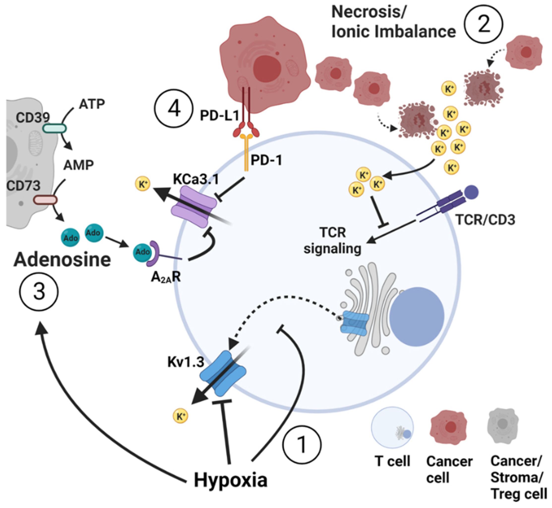
Multiple elements of the TME affect T cell antitumor capabilities through alterations of K+ homeostasis (Figure 3).

4.1. Hypoxia

Proliferating tumor cells require and consume more oxygen (O2) than that provided by pre-existing blood vessels, decreasing the O2 availability for other cell types. While the tumor may stimulate increased vascularization, this adaptation is typically dysfunctional to sustain adequate O2 concentrations with newly formed blood vessels that are leaky, with blind ends and temporary occlusions [49]. The resulting hypoxia affects both cancer and immune cell function. Levels of O2 tension as low as ≤2.5 mmHg have been measured in tumors [50]. The effects of hypoxia occur both transcriptionally (through the stabilization of the well-described transcription factor HIF1α, hypoxia-inducible factor 1α) and post-transcriptionally. The induction of HIF1α causes cancer cells to undergo metabolic reprogramming, which leads to an increased glucose uptake with the production of lactate, even in the presence of O2 and functional mitochondria (termed the “Warburg effect”) [51,52]. These metabolic changes support cancer cell growth in hypoxic regions and drive tumor advantage for migration, invasion, metastases, and resistance to treatments [49,53]. Hypoxic effects have also been shown to occur through alterations in the function of voltage-dependent K+ channels in cancer cells such as PC12 pheochromocytoma cells, and other cells of tissues that respond quickly to change in O2 availability such as carotid body cells and pulmonary artery smooth muscle cells [54].
Likewise, hypoxia alters the immune system. Hypoxia decreases T cell activation, cytotoxicity, and cytokine release [55]. Hypoxia delays cytotoxic T cell development in vitro and decreases T cell activation in vivo [55,56]. In CD4+ T cells, hypoxia induces the differentiation of CD4+ T cells into regulatory T (Treg) or T helper 17 cells [52]. While hypoxia has these effects on immune cells, it additionally alters the TME by promoting the up-regulation of immune checkpoint inhibitors, such as T-lymphocyte–associated antigen 4 (CTLA-4) and PD-L1 [52] and inducing acidosis and the production of adenosine [4].
Overall, the presence of hypoxia in tumors has been associated with poor prognosis and response to therapies. The overall detrimental effects of hypoxia in cancer are exemplified by the results of a study on the effects of hyperoxia in mice [57]. In this study, the authors showed that a reduction in intratumoral hypoxia via the delivery of O2 to tumor-bearing mice converted an immunosuppressive TME into an immunopermissive one [57]. Increasing O2 availability in the tumor reduced extracellular adenosine, increased T cell infiltration and release of immune stimulating cytokines and chemokines, and decreased tumor growth factor β (TGFβ). These effects were entirely dependent on T and NK cells. It also reduced Treg accumulation in the tumor and CTLA-4 expression in these cells. Ultimately, it improved the regression of tumors, prevented spontaneous metastasis, and enhanced long-term survival. Furthermore, it improved the efficacy of adoptively transferred tumor-reactive CD8+ T cells [57].
Kv1.3 channels play an important role as transducers of O2 deprivation in T cells. Indeed, hypoxia inhibits Kv1.3 channels in T cells in both the acute (minutes) and chronic (days) setting [58,59]. These inhibitions occurred post-transcriptionally, with no changes in Kv1.3 mRNA. Acute hypoxia inhibited Kv1.3 currents in human T cells and Jurkat T cells [58] caused membrane depolarization and, consequently, the suppression of TCR-mediated Ca2+ influx (Figure 3) [59]. These effects of hypoxia on Ca2+ fluxes were mainly attributable to Kv1.3, because there was no change in CRAC and Ca2+-activated K+ channel function [59]. The src protein tyrosine kinase p56Lck was required for Kv1.3 response to hypoxia [60]. Supporting this conclusion, p56Lck-deficient cell lines and human T lymphocytes pre-treated with a src protein tyrosine kinase inhibitor lost Kv1.3-mediated response to hypoxia [60]. Chronic hypoxia instead induced a decrease in the pore-forming Kv1.3α subunit protein expression in Jurkat T cells and, consequently, a decrease in functional Kv1.3 channels [58]. Notably, in sustained O2 depletion, Kv1.3 current inhibition was also associated with reduced T cell proliferation induced by TCR cross-linking agents [58]. Experiments focused on the underlying mechanisms that led to this decrease in Kv1.3 channel expression during chronic exposure to hypoxia in T cells [61] and revealed that chronic hypoxia decreased Kv1.3 surface expression by inhibiting the forward trafficking of Kv1.3 from the trans-Golgi to the plasma membrane, which physiologically involved clathrin-coated vesicle formation initiated via the AP1 adaptor protein (Figure 3). Under hypoxic conditions, gene and protein expression of the γ subunit of AP1 is decreased with the consequent inhibition of clathrin-coated vesicle formation and impaired forward trafficking of newly synthesized Kv1.3 to the plasma membrane [61]. Together, these results provide evidence of a role of Kv1.3 channels in the T cell response to changes in O2 tension, thus explaining the linkage between hypoxia, T cell immunosuppression, and the ion channel apparatus.

4.2. Necrosis and Ionic Imbalance

Despite hypoxic conditions and the inadequate provision of nutrients, cancer cells continue to rapidly divide, thus creating conditions for irregular areas of necrosis within the tumor mass. Eil and colleagues (2016) demonstrated that these regions of tumor necrosis lead to extracellular ionic imbalance as the dying cells release their intracellular contents and, thus, high levels of K+ ions [62]. The intracellular [K+] is 154 mM, much higher than the extracellular [K+] (5.4 mM). This increase in extracellular [K+] in the necrotic tumor areas ultimately leads to increased intracellular [K+] in the TILs through pump or leak channels, due to the attenuation of K+ chemical gradient (Figure 3) [62]. No alteration in membrane potential was reported. However, this ionic imbalance inhibited the serine/threonine kinases Akt and mTOR (mammalian target of rapamycin) in the TCR-signaling pathway, ultimately suppressing T cell transcriptional programs required for cytokine production (Figure 3) [62]. This finding is in line with the fact that necrotic areas represent a marker of poor prognosis in a variety of solid malignant tumors [63,64]. Interestingly, Kv1.3 overexpression corrected the immunosuppressive K+ accumulation in tumor necrotic areas, increased IL-2 and IFN-γ production, reduced tumor burden and prolonged survival of melanoma bearing mice [62]. KCa3.1 activation via specific pharmacological activators also rescued T cell function in high extracellular [K+] in vitro [62]. The effect of KCa3.1 activation in vivo has yet to be determined. Recently, Ong and colleagues (2019) showed that human peripheral blood T cells were negatively influenced by high extracellular [K+] [65]. Indeed, high extracellular [K+] and the consequent high intracellular [K+] led to impaired T cell proliferation in TCM and TEM, but not in T memory stem (TSCM) cells. Moreover, high extracellular [K+] suppressed T cell cytokine production and tumor cell killing ability and enhanced programmed cell death protein-1 (PD-1) expression [65]. This suppression of T cell function in high extracellular [K+] was accelerated by the blockade of KCa3.1 and Kv1.3, while the activation of KCa3.1 restored T cell function [65].
Therefore, it may be a fair assumption to predict that the immunosuppressive effects of K+ accumulation in necrotic areas could be exacerbated by the concomitant presence of hypoxia, adenosine and PD-L1 (see below) in the TME that suppress K+ channels.
Overall, these data show the importance of K+ channels in correcting the immunosuppressive effects induced by necrosis. However, we should also take into account that in a later study, Vodnala and colleagues (2019) showed that the increase in extracellular [K+] within the tumor has an additional impact on T cell stemness [66]. They demonstrated that extracellular [K+] leads to T cell starvation and metabolic and epigenetic reprogramming that, while limiting T cell effector functions, promote stem cell-like programs [66].

4.3. Adenosine

Adenosine, a catabolic product of adenosine triphosphate (ATP), is an anti-inflammatory nucleoside that represents one of the predominant negative immune regulators of the TME. Tumors accumulate extracellular ATP through passive loss from dead cells and inflammatory- or hypoxia-induced release from stressed cells [67,68]. This accumulation of ATP leads to increased levels of adenosine as the ecto-nucleotidases CD39 and CD73 metabolize ATP to adenosine monophosphate (AMP) and AMP to adenosine, respectively (Figure 3) [67,68]. These enzymes are present on the surface of adenosine-producing cells such as stromal cells, tumor cells, or Treg that, in addition, can also directly secrete adenosine through bidirectional nucleoside transporters [68]. Extracellular levels of adenosine in solid tumors can be as high as 10 μM; >300 times higher than those found in healthy tissues (30 nM) [69,70]. Adenosine can then bind to its receptors on the surface of T cells and exert its anti-inflammatory functions, which include the inhibition of effector cells and the promotion of suppressive Treg [68,71]. Extracellular adenosine is decreased through degradation via adenosine deaminase or transport into cells by the cell membrane nucleoside transporters. However, in the TME, salvage pathways can be inhibited, thereby adding to the extracellular adenosine concentration [68,69]. Hypoxia facilitates the accumulation of adenosine in the TME by increasing the expression of adenosine-producing enzymes and reducing the expression of adenosine-degrading enzymes via HIF1α [4,69].
There is strong evidence that the immunosuppressive effects of adenosine on effector T cells are partly mediated through the inhibition of KCa3.1 channels. In activated human T cells, adenosine binds to A2A receptor (A2AR), increases cAMP production, and, consequently, induces protein kinase A1 (PKAI) activation. These changes serve to inhibit KCa3.1 and cause decreased T cell integrin-dependent motility, chemotaxis, and cytokine release (Figure 3) [33,47]. Additionally, KCa3.1 channels mediate the sensitivity to adenosine of circulating T cells from head and neck squamous cell carcinoma (HNSCC) patients [33]. It is well established that the immunosuppressive effects of adenosine are more pronounced in T cells from cancer patients than healthy donors [33,71]. More specifically and relevant to KCa3.1, chemotaxis was significantly more inhibited by adenosine in HNSCC CD8+ T cells than in CD8+ T cells from healthy controls [33]. This effect of adenosine in HNSCC CD8+ T cells was neither mediated by an increase in A2AR expression nor proximal cAMP-PKAI signaling. Rather, a reduction in KCa3.1 functionality (and not expression) in HNSCC CD8+ T cells as compared to healthy CD8+ T cells accounted for the increased sensitivity to adenosine and translated into reduced infiltration into the adenosine-rich HNSCC tumors [33]. Indeed, patients whose circulating CD8+ chemotaxis was most severely inhibited by adenosine were also those who had the lowest infiltration of CD8+ T cells into the tumor [33]. Important from a therapeutic perspective, positive modulators of KCa3.1 restored the chemotaxis of HNSCC CD8+ T cells in the presence of adenosine [33]. More recently, follow-up studies showed that the reduction in KCa3.1 functionality in HNSCC CD8+ T cells was secondary to a defect in plasma-membrane localized calmodulin, a signaling molecule tethered to the channels’ C-terminal domain and responsible for its Ca2+-sensitivity [72]. Binding of Ca2+ to calmodulin translates into a conformational change in KCa3.1 and its opening [73]. Knockdown of calmodulin in healthy CD8+ T cells reproduced HNSCC T cell dysfunction, namely reducing KCa3.1 activity and chemotactic capabilities in the presence of adenosine [33,72]. In contrast, pre-incubation of calmodulin knocked-down healthy CD8+ T cells with a specific KCa3.1 activator (1-EBIO) rescued the cells’ capability to chemotax in the presence of adenosine [72]. Therefore, KCa3.1 activators could be used to overcome the immunosuppressive action of adenosine and improve the antitumor functions of T cells.
The blockade of the adenosine pathway and the subsequent increase in antitumor immune functions may be also achieved by targeting different points along the adenosine pathway [68]. These include inhibiting adenosine synthesis [74,75], increasing adenosine degradation through increased adenosine deaminase activity [71], or preventing the interaction with specific receptors on the surface of target cells [76]. We have shown that targeted silencing of A2AR in memory CD45RO+CD8+ T cell of HNSCC patients via lipid nanoparticles abrogated the inhibitory effect of adenosine on the chemotaxis of these cells [77]. In vivo studies provide conclusive evidence of the importance of adenosine in cancer [76,78]. A phase I clinical trial of treatment-refractory renal cell cancer patients demonstrated antitumor activity of an A2AR antagonist alone and in combination with checkpoint inhibitors and established the safety and feasibility of targeting this pathway [76]. However, the advantage of KCa3.1 activators over conventional adenosine receptor blockers is that this treatment approach may overcome the actions of prostaglandin E2 (PGE2), another immunosuppressive molecule that accumulates in the TME and inhibits KCa3.1. It has been reported that PGE2 inhibits KCa3.1 in mast cells [79].
Overall, the development of immunotherapies targeting KCa3.1 on the adenosine pathway may serve as promising therapeutics [76,80].

4.4. Programmed Death Ligand 1

Overexpression of PD-L1 is utilized by cancer cells to suppress the antitumor immune response. The effect of PD-L1 on T lymphocytes is mediated by engagement with its cognate receptor PD-1 and, consequently, inhibition of TCR-dependent effector functions, such as Ca2+ fluxing, secretion of cytokines and cytotoxicity [81]. KCa3.1 channels provide a link between PD-1 stimulation by PD-L1 and reduced Ca2+-related functions [82]. PD-L1 reduces KCa3.1 activity and Ca2+ fluxes in CD8+ T cells; this effect is rescued by treatment with anti-PD-1 blocking antibodies. KCa3.1 function is controlled by multiple mechanisms, including the regulation of the channel Ca2+ sensitivity by calmodulin (described before) and histidine phosphorylation [83,84]. Histidine phosphorylation of KCa3.1 has been shown to be triggered by the phosphoinositide 3-kinase (PI3K): the phosphatidylinositol-3 phosphatase (PI3P) signaling pathway [84,85]. In T cells, ligation of PD-1 by PD-L1 results in a decrease in TCR proximal signaling through inhibition of PI3K activity [86]. A decrease in PI3K suppresses the production of PI3P from phosphatidylinositol (PI). PI3P is known to activate the nucleoside diphosphate kinase B (NDPK-B), which, ultimately, increases KCa3.1 activity via histidine phosphorylation [84,85]. Thus, the reduction in PI3K triggered by PD-1 stimulation suppresses KCa3.1 (Figure 3). Indeed, we showed that the response of KCa3.1 channels to PD-1 ligation is mediated by PI3K-PI3P signaling after short exposure to PD-L1. Longer exposures to PD-L1 show a role for calmodulin. Five-day exposure to PD-L1 reduced calmodulin expression in T cells by 40% while the contribution of PI3K was reduced compared to early time points [82]. The involvement of KCa3.1 in mediating the effect of the PD-L1/PD-1 response is further supported by in vitro studies showing that PD-1 and PD-L1 blocking antibodies increased KCa3.1 activity [82]. An increase in Kv1.3 activity was also reported. However, there may be a difference in sensitivity to PD-L1 between these two channels; a higher concentration of anti-PD-L1 blocking antibodies was necessary to unleash Kv1.3 activity in T cells from cancer patients compared to KCa3.1 [82]. Further studies are necessary to dissect the mechanisms mediating the effect of PD-L1 on Kv1.3 channels. Ex vivo studies of K+ channel activity in CD8+ T lymphocytes of HNSCC patients treated with anti-PD-1 blocking antibodies also showed an increase in KCa3.1 and Kv1.3 activities [87]. These studies position K+ channels downstream to immune checkpoint inhibitors.
Immune checkpoint inhibitors have arisen as new effective treatments in solid malignancies as they have shown remarkable improvements in treatment outcomes including long-lasting remissions after discontinuation of therapy [88,89]. However, the majority of patients do not respond or eventually relapse [90]. Activation of K+ channels may be critical to expanding the efficacy of these revolutionary treatments.
Kv1.3 channels have also been recently implicated as biomarkers of response to immunotherapy. Goggi and colleagues (2022) developed a new Kv1.3 targeting radiopharmaceutical ([18F]AlF-NOTA-KCNA3P) and showed that it could be used to differentiate tumors responding to immune checkpoint inhibitors (anti-PD-1 antibodies alone or in combination with anti-CTLA-4 antibodies) in a syngeneic colon cancer model [91]. This radiolabeled ligand of Kv1.3 allowed the quantification of TEM cells infiltrating the tumor, which were increased in responding tumors, and raised the possibility that a similar strategy could be used to develop new biomarkers of response to immune checkpoint inhibitors.

5. K+ Channels as a Target for Cancer Therapy

We and others have examined the effects of the TME on the ion channel apparatus of antitumor immune cells (Table 1) and have explored the potential of restoring impaired channel function for cancer treatment. As multiple elements of the TME affect K+ channels in T cells as described above, the anticipated repercussion of this would be reduced K+ channel activity and Ca2+ fluxes in TILs. Indeed, the Ca2+ response of TILs is severely dampened compared to circulating T cells [32,92]. In HNSCC patients, TILs exhibit suppressed Kv1.3 and KCa3.1 function compared to circulating T cells. Additionally, we showed that the number of Kv1.3 channels per membrane unit surface (described as Kv1.3 current density) was lower in HNSCC TILs compared to HNSCC PBT. Hence, TILs had a significantly lower number of channels. [32,87]. Moreover, TILs were not only “functionally” but also “spatially” impaired. The expression of Kv1.3 was shown to be higher in stromal than epithelial CD8+ TILs, and highly proliferating-cytotoxic TILs were physically excluded from the tumor and trapped in the stroma [32]. In this scenario, ion channels represent a fascinating target to counteract the immunosuppressive effects of the TME on immune cells and to restore antitumor immunity. Proof of the benefits of targeting Kv1.3 in cancer in vivo have already been provided, while the benefits of KCa3.1 are still indirect, as they are supported only by in vitro evidence [33,62]. On the other side, ion channels are currently investigated as therapeutic strategies in autoimmune diseases, the opposite situation compared to cancer, whereas the immune system is hyperactive rather than suppressed. As a consequence, the immune system’s hyperactivity in autoimmune disorders may be prevented by the inhibition of ion fluxes, which are responsible for the functionality of disease-provoking immune cells [93,94]. In proof of this, our group has showed that silencing RNA against Kv1.3 in memory T cells improved survival in a mouse model of SLE [35]. Thus, upregulating K+ channel function in T cells, and particularly Kv1.3 in the context of cancer, while very promising, has to take into consideration the potential development of immune-related adverse events that are already reported for the immune checkpoint inhibitors currently in use [95].
Recently, we have investigated the effects of the immune checkpoint inhibitor pembrolizumab (a monoclonal antibody against PD-1) on the ion channel functionality in T cells of HNSCC patients [87]. These studies revealed that patients who responded to neoadjuvant pembrolizumab (where the response was defined according to the % of viable tumor at resection, namely less than 80%) had a specific ion channel signature in circulating T cells. In more detail, while in TILs pembrolizumab induced an increase in Kv1.3 activity and Ca2+ fluxes regardless of the response, a characteristic response was identified in circulating T cells [87]. Interestingly, all patients (responders and non-responders) experienced an immediate increase in KCa3.1 activity in circulating CD8+ T cells after pembrolizumab administration. However, circulating CD8+ T cells of patients who responded to pembrolizumab also had a marked increase in Kv1.3 activity compared with non-responders. This was accompanied by an increase in Ca2+ fluxes and chemotactic ability in the presence of the immunosuppressive adenosine [87]. Hence, K+ channels are significantly implicated in the response to pembrolizumab in HNSCC patients, where responders are characterized by a typical ion channel signature (Kv1.3high KCa3.1high Ca2+high) in circulating cytotoxic T cells that may also elucidate strategies to overcome resistance.
However, the network of ion channels that contribute to Ca2+ fluxes in T cells is complex. As discussed above, ion channels are differentially expressed in resting and activated T cells, and their expression depends on the T cell subset. Moreover, many different settings (e.g., tumors, autoimmune and infectious diseases) and models (e.g., rat, mouse, and human) were used to study ion channel properties and, therefore, the translatability between fields may not be completely compatible and, thus, may add to the heterogeneity of results. This is true particularly for studies on Kv1.3 channels that utilize mouse T cells, as the expression and functional role of Kv1.3 channels is different in mice compared to human T cells [96]. Additionally, the ion channel network is a very sensitive and highly regulated system. For example, T cells operate efficiently towards cancer elimination in a defined and relatively small range of extracellular [Ca2+]: between 23 and 625 µM, which allows the release of an elevated number of cytotoxic granules per cell [97].
Nevertheless, K+ channels contribute to the reduction in the antitumor function of T cells and targeting these channels has shown promising therapeutic value. As K+ channels are downstream to multiple immunosuppressive elements of the TME, targeting these channels would ultimately allow to simultaneously counteract the multidirectional attacks employed by the tumor to suppress antitumor immunity.

6. Conclusions

Ion channels play an integral role in Ca2+ signaling and downstream T cell effector functions, and, therefore, are, when fully functional, critically important for the immune system role in detecting and eliminating cancer cells. Although this ion channel network is complex, it is clear that many factors within the TME converge to impair ion channel function (Figure 3). Further understanding the effect of the TME on ion channels could help to develop effective treatments, especially for patients resistant to conventional immunotherapies.

Author Contributions

Conceptualization, M.C., H.S.N. and L.C.; writing—original draft preparation, M.C., H.S.N., V.S.G. and L.C.; writing—review and editing, M.C., H.S.N., T.M.W.-D., A.A.C. and L.C.; visualization, M.C., H.S.N., A.A.C. and L.C.; supervision, L.C. All authors have read and agreed to the published version of the manuscript.

Funding

This research was supported by the Office of the Assistant Secretary of Defense for Health Affairs, through the Peer Reviewed Cancer Research Program, under Award No W81XWH-17-1-0374 (Laura Conforti, Trisha M. Wise-Draper, Edith M. Janssen). Opinions, interpretations, conclusions and recommendations are those of the authors and are not necessarily endorsed by the Department of Defense. The United States Army Medical Research Acquisition Activity, Fort Detrick, Maryland is the awarding and administering acquisition office. Additional funding was provided by the National Institutes of Health (grant R01-CA95286) to Laura Conforti. Hannah S. Newton was supported by NIH training grant T32 CA117846. Martina Chirra was supported by NIH grant R38HL155775 01.

Acknowledgments

We would like to acknowledge Eric Smith, for his help in editing the manuscript. Figures were created with BioRender.com, accessed on 7 July 2022.

Conflicts of Interest

The authors declare no conflict of interest.

References

  1. Speiser, D.E.; Ho, P.C.; Verdeil, G. Regulatory circuits of T cell function in cancer. Nat. Rev. Immunol. 2016, 16, 599–611. [Google Scholar] [CrossRef] [PubMed]
  2. Thommen, D.S.; Schumacher, T.N. T Cell Dysfunction in Cancer. Cancer Cell 2018, 33, 547–562. [Google Scholar] [CrossRef] [PubMed] [Green Version]
  3. Belli, C.; Trapani, D.; Viale, G.; D’Amico, P.; Duso, B.A.; Della Vigna, P.; Orsi, F.; Curigliano, G. Targeting the microenvironment in solid tumors. Cancer Treat. Rev. 2018, 65, 22–32. [Google Scholar] [CrossRef] [PubMed]
  4. Vaupel, P.; Multhoff, G. Accomplices of the Hypoxic Tumor Microenvironment Compromising Antitumor Immunity: Adenosine, Lactate, Acidosis, Vascular Endothelial Growth Factor, Potassium Ions, and Phosphatidylserine. Front. Immunol. 2017, 8, 1887. [Google Scholar] [CrossRef] [PubMed] [Green Version]
  5. Joyce, J.A.; Fearon, D.T. T cell exclusion, immune privilege, and the tumor microenvironment. Science 2015, 348, 74–80. [Google Scholar] [CrossRef] [PubMed] [Green Version]
  6. Vaeth, M.; Kahlfuss, S.; Feske, S. CRAC Channels and Calcium Signaling in T Cell-Mediated Immunity. Trends Immunol. 2020, 41, 878–901. [Google Scholar] [CrossRef]
  7. Feske, S.; Wulff, H.; Skolnik, E.Y. Ion channels in innate and adaptive immunity. Annu. Rev. Immunol. 2015, 33, 291–353. [Google Scholar] [CrossRef] [Green Version]
  8. Cahalan, M.D.; Chandy, K.G. The functional network of ion channels in T lymphocytes. Immunol. Rev. 2009, 231, 59–87. [Google Scholar] [CrossRef] [Green Version]
  9. Feske, S.; Giltnane, J.; Dolmetsch, R.; Staudt, L.M.; Rao, A. Gene regulation mediated by calcium signals in T lymphocytes. Nat. Immunol. 2001, 2, 316–324. [Google Scholar] [CrossRef]
  10. Feske, S. Calcium signalling in lymphocyte activation and disease. Nat. Rev. Immunol. 2007, 7, 690–702. [Google Scholar] [CrossRef]
  11. Maul-Pavicic, A.; Chiang, S.C.; Rensing-Ehl, A.; Jessen, B.; Fauriat, C.; Wood, S.M.; Sjöqvist, S.; Hufnagel, M.; Schulze, I.; Bass, T.; et al. ORAI1-mediated calcium influx is required for human cytotoxic lymphocyte degranulation and target cell lysis. Proc. Natl. Acad. Sci. USA 2011, 108, 3324–3329. [Google Scholar] [CrossRef] [PubMed] [Green Version]
  12. Panyi, G.; Beeton, C.; Felipe, A. Ion channels and anti-cancer immunity. Philos. Trans. R. Soc. B Biol. Sci. 2014, 369, 20130106. [Google Scholar] [CrossRef] [PubMed] [Green Version]
  13. Vaeth, M.; Feske, S. Ion channelopathies of the immune system. Curr. Opin. Immunol. 2018, 52, 39–50. [Google Scholar] [CrossRef]
  14. Nohara, L.L.; Stanwood, S.R.; Omilusik, K.D.; Jefferies, W.A. Tweeters, Woofers and Horns: The Complex Orchestration of Calcium Currents in T Lymphocytes. Front. Immunol. 2015, 6, 234. [Google Scholar] [CrossRef] [PubMed]
  15. Gwack, Y.; Srikanth, S.; Oh-Hora, M.; Hogan, P.G.; Lamperti, E.D.; Yamashita, M.; Gelinas, C.; Neems, D.S.; Sasaki, Y.; Feske, S.; et al. Hair loss and defective T- and B-cell function in mice lacking ORAI1. Mol. Cell. Biol. 2008, 28, 5209–5222. [Google Scholar] [CrossRef] [Green Version]
  16. Oh-Hora, M.; Yamashita, M.; Hogan, P.G.; Sharma, S.; Lamperti, E.; Chung, W.; Prakriya, M.; Feske, S.; Rao, A. Dual functions for the endoplasmic reticulum calcium sensors STIM1 and STIM2 in T cell activation and tolerance. Nat. Immunol. 2008, 9, 432–443. [Google Scholar] [CrossRef] [Green Version]
  17. Weidinger, C.; Shaw, P.J.; Feske, S. STIM1 and STIM2-mediated Ca2+ influx regulates antitumour immunity by CD8+ T cells. EMBO Mol. Med. 2013, 5, 1311–1321. [Google Scholar] [CrossRef]
  18. Berry, C.T.; May, M.J.; Freedman, B.D. STIM- and Orai-mediated calcium entry controls NF-κB activity and function in lymphocytes. Cell Calcium 2018, 74, 131–143. [Google Scholar] [CrossRef]
  19. Pérez-García, M.T.; Cidad, P.; López-López, J.R. The secret life of ion channels: Kv1.3 potassium channels and proliferation. Am. J. Physiol. Cell Physiol. 2018, 314, C27–C42. [Google Scholar] [CrossRef] [Green Version]
  20. Bertin, S.; Raz, E. Transient Receptor Potential (TRP) channels in T cells. Semin. Immunopathol. 2016, 38, 309–319. [Google Scholar] [CrossRef]
  21. Kuras, Z.; Yun, Y.H.; Chimote, A.A.; Neumeier, L.; Conforti, L. KCa3.1 and TRPM7 channels at the uropod regulate migration of activated human T cells. PLoS ONE 2012, 7, e43859. [Google Scholar] [CrossRef] [PubMed] [Green Version]
  22. Shaw, P.J.; Weidinger, C.; Vaeth, M.; Luethy, K.; Kaech, S.M.; Feske, S. CD4+ and CD8+ T cell-dependent antiviral immunity requires STIM1 and STIM2. J. Clin. Investig. 2014, 124, 4549–4563. [Google Scholar] [CrossRef] [PubMed] [Green Version]
  23. Desvignes, L.; Weidinger, C.; Shaw, P.; Vaeth, M.; Ribierre, T.; Liu, M.; Fergus, T.; Kozhaya, L.; McVoy, L.; Unutmaz, D.; et al. STIM1 controls T cell-mediated immune regulation and inflammation in chronic infection. J. Clin. Investig. 2015, 125, 2347–2362. [Google Scholar] [CrossRef] [PubMed] [Green Version]
  24. Ma, J.; McCarl, C.A.; Khalil, S.; Lüthy, K.; Feske, S. T-cell-specific deletion of STIM1 and STIM2 protects mice from EAE by impairing the effector functions of Th1 and Th17 cells. Eur. J. Immunol. 2010, 40, 3028–3042. [Google Scholar] [CrossRef] [PubMed]
  25. Tanner, M.R.; Tajhya, R.B.; Huq, R.; Gehrmann, E.J.; Rodarte, K.E.; Atik, M.A.; Norton, R.S.; Pennington, M.W.; Beeton, C. Prolonged immunomodulation in inflammatory arthritis using the selective Kv1.3 channel blocker HsTX1[R14A] and its PEGylated analog. Clin. Immunol. 2017, 180, 45–57. [Google Scholar] [CrossRef]
  26. Beeton, C.; Wulff, H.; Barbaria, J.; Clot-Faybesse, O.; Pennington, M.; Bernard, D.; Cahalan, M.D.; Chandy, K.G.; Béraud, E. Selective blockade of T lymphocyte K+ channels ameliorates experimental autoimmune encephalomyelitis, a model for multiple sclerosis. Proc. Natl. Acad. Sci. USA 2001, 98, 13942–13947. [Google Scholar] [CrossRef] [Green Version]
  27. Beeton, C.; Wulff, H.; Standifer, N.E.; Azam, P.; Mullen, K.M.; Pennington, M.W.; Kolski-Andreaco, A.; Wei, E.; Grino, A.; Counts, D.R.; et al. Kv1.3 channels are a therapeutic target for T cell-mediated autoimmune diseases. Proc. Natl. Acad. Sci. USA 2006, 103, 17414–17419. [Google Scholar] [CrossRef] [Green Version]
  28. Koch Hansen, L.; Sevelsted-Møller, L.; Rabjerg, M.; Larsen, D.; Hansen, T.P.; Klinge, L.; Wulff, H.; Knudsen, T.; Kjeldsen, J.; Köhler, R. Expression of T-cell KV1.3 potassium channel correlates with pro-inflammatory cytokines and disease activity in ulcerative colitis. J. Crohn’s Colitis 2014, 8, 1378–1391. [Google Scholar] [CrossRef]
  29. Hu, L.; Wang, T.; Gocke, A.R.; Nath, A.; Zhang, H.; Margolick, J.B.; Whartenby, K.A.; Calabresi, P.A. Blockade of Kv1.3 potassium channels inhibits differentiation and granzyme B secretion of human CD8+ T effector memory lymphocytes. PLoS ONE 2013, 8, e54267. [Google Scholar] [CrossRef] [Green Version]
  30. Di, L.; Srivastava, S.; Zhdanova, O.; Ding, Y.; Li, Z.; Wulff, H.; Lafaille, M.; Skolnik, E.Y. Inhibition of the K+ channel KCa3.1 ameliorates T cell-mediated colitis. Proc. Natl. Acad. Sci. USA 2010, 107, 1541–1546. [Google Scholar] [CrossRef] [Green Version]
  31. Beeton, C.; Chandy, K.G. Potassium channels, memory T cells, and multiple sclerosis. Neuroscientist 2005, 11, 550–562. [Google Scholar] [CrossRef] [PubMed]
  32. Chimote, A.A.; Hajdu, P.; Sfyris, A.M.; Gleich, B.N.; Wise-Draper, T.; Casper, K.A.; Conforti, L. Kv1. 3 channels mark functionally competent CD8+ tumor-infiltrating lymphocytes in head and neck cancer. Cancer Res. 2017, 77, 53–61. [Google Scholar] [CrossRef] [Green Version]
  33. Chimote, A.A.; Balajthy, A.; Arnold, M.J.; Newton, H.S.; Hajdu, P.; Qualtieri, J.; Wise-Draper, T.; Conforti, L. A defect in KCa3.1 channel activity limits the ability of CD8+ T cells from cancer patients to infiltrate an adenosine-rich microenvironment. Sci. Signal. 2018, 11, eaaq1616. [Google Scholar] [CrossRef] [PubMed] [Green Version]
  34. Wulff, H.; Calabresi, P.A.; Allie, R.; Yun, S.; Pennington, M.; Beeton, C.; Chandy, K.G. The voltage-gated Kv1.3 K+ channel in effector memory T cells as new target for MS. J. Clin. Investig. 2003, 111, 1703–1713. [Google Scholar] [CrossRef] [Green Version]
  35. Khodoun, M.; Chimote, A.A.; Ilyas, F.Z.; Duncan, H.J.; Moncrieffe, H.; Kant, K.S.; Conforti, L. Targeted knockdown of Kv1.3 channels in T lymphocytes corrects the disease manifestations associated with systemic lupus erythematosus. Sci. Adv. 2020, 6, eabd1471. [Google Scholar] [CrossRef]
  36. Wulff, H.; Castle, N.A. Therapeutic potential of KCa3.1 blockers: Recent advances and promising trends. Expert Rev. Clin. Pharmacol. 2010, 3, 385–396. [Google Scholar] [CrossRef] [PubMed] [Green Version]
  37. Chiang, E.Y.; Li, T.; Jeet, S.; Peng, I.; Zhang, J.; Lee, W.P.; DeVoss, J.; Caplazi, P.; Chen, J.; Warming, S.; et al. Potassium channels Kv1.3 and KCa3.1 cooperatively and compensatorily regulate antigen-specific memory T cell functions. Nat. Commun. 2017, 8, 14644. [Google Scholar] [CrossRef] [PubMed]
  38. Ghanshani, S.; Wulff, H.; Miller, M.J.; Rohm, H.; Neben, A.; Gutman, G.A.; Cahalan, M.D.; Chandy, K.G. Up-regulation of the IKCa1 potassium channel during T-cell activation. Molecular mechanism and functional consequences. J. Biol. Chem. 2000, 275, 37137–37149. [Google Scholar] [CrossRef] [Green Version]
  39. Chimote, A.A.; Hajdu, P.; Kottyan, L.C.; Harley, J.B.; Yun, Y.; Conforti, L. Nanovesicle-targeted Kv1.3 knockdown in memory T cells suppresses CD40L expression and memory phenotype. J. Autoimmun. 2016, 69, 86–93. [Google Scholar] [CrossRef] [Green Version]
  40. Sim, J.H.; Kim, K.S.; Park, H.; Kim, K.J.; Lin, H.; Kim, T.J.; Shin, H.M.; Kim, G.; Lee, D.S.; Park, C.W.; et al. Differentially Expressed Potassium Channels Are Associated with Function of Human Effector Memory CD8+ T Cells. Front. Immunol. 2017, 8, 859. [Google Scholar] [CrossRef] [Green Version]
  41. Hu, L.; Gocke, A.R.; Knapp, E.; Rosenzweig, J.M.; Grishkan, I.V.; Baxi, E.G.; Zhang, H.; Margolick, J.B.; Whartenby, K.A.; Calabresi, P.A. Functional blockade of the voltage-gated potassium channel Kv1. 3 mediates reversion of T effector to central memory lymphocytes through SMAD3/p21cip1 signaling. J. Biol. Chem. 2012, 287, 1261–1268. [Google Scholar] [CrossRef] [PubMed] [Green Version]
  42. Badolato, R. Defects of leukocyte migration in primary immunodeficiencies. Eur. J. Immunol. 2013, 43, 1436–1440. [Google Scholar] [CrossRef] [PubMed]
  43. Schwab, A.; Hanley, P.; Fabian, A.; Stock, C. Potassium channels keep mobile cells on the go. Physiology 2008, 23, 212–220. [Google Scholar] [CrossRef] [Green Version]
  44. Greenberg, M.L.; Yu, Y.; Leverrier, S.; Zhang, S.L.; Parker, I.; Cahalan, M.D. Orai1 function is essential for T cell homing to lymph nodes. J. Immunol. 2013, 190, 3197–3206. [Google Scholar] [CrossRef] [PubMed] [Green Version]
  45. Waite, J.C.; Vardhana, S.; Shaw, P.J.; Jang, J.E.; McCarl, C.A.; Cameron, T.O.; Feske, S.; Dustin, M.L. Interference with Ca2+ release activated Ca2+ (CRAC) channel function delays T-cell arrest in vivo. Eur. J. Immunol. 2013, 43, 3343–3354. [Google Scholar] [CrossRef] [Green Version]
  46. Dong, T.X.; Othy, S.; Greenberg, M.L.; Jairaman, A.; Akunwafo, C.; Leverrier, S.; Yu, Y.; Parker, I.; Dynes, J.L.; Cahalan, M.D. Intermittent Ca2+ signals mediated by Orai1 regulate basal T cell motility. eLife 2017, 6, e27827. [Google Scholar] [CrossRef]
  47. Chimote, A.A.; Hajdu, P.; Kucher, V.; Boiko, N.; Kuras, Z.; Szilagyi, O.; Yun, Y.H.; Conforti, L. Selective inhibition of KCa3.1 channels mediates adenosine regulation of the motility of human T cells. J. Immunol. 2013, 191, 6273–6280. [Google Scholar] [CrossRef] [Green Version]
  48. Matheu, M.P.; Beeton, C.; Garcia, A.; Chi, V.; Rangaraju, S.; Safrina, O.; Monaghan, K.; Uemura, M.I.; Li, D.; Pal, S.; et al. Imaging of effector memory T cells during a delayed-type hypersensitivity reaction and suppression by Kv1.3 channel block. Immunity 2008, 29, 602–614. [Google Scholar] [CrossRef] [Green Version]
  49. McDonald, P.C.; Chafe, S.C.; Dedhar, S. Overcoming Hypoxia-Mediated Tumor Progression: Combinatorial Approaches Targeting pH Regulation, Angiogenesis and Immune Dysfunction. Front. Cell Dev. Biol. 2016, 4, 27. [Google Scholar] [CrossRef] [Green Version]
  50. Vaupel, P.; Mayer, A. Hypoxia in cancer: Significance and impact on clinical outcome. Cancer Metastasis Rev. 2007, 26, 225–239. [Google Scholar] [CrossRef]
  51. Liberti, M.V.; Locasale, J.W. The Warburg Effect: How Does it Benefit Cancer Cells? Trends Biochem. Sci. 2016, 41, 211–218. [Google Scholar] [CrossRef] [PubMed] [Green Version]
  52. Petrova, V.; Annicchiarico-Petruzzelli, M.; Melino, G.; Amelio, I. The hypoxic tumour microenvironment. Oncogenesis 2018, 7, 10. [Google Scholar] [CrossRef] [PubMed]
  53. Parks, S.K.; Cormerais, Y.; Marchiq, I.; Pouyssegur, J. Hypoxia optimises tumour growth by controlling nutrient import and acidic metabolite export. Mol. Asp. Med. 2016, 47–48, 3–14. [Google Scholar] [CrossRef] [PubMed]
  54. Lopez-Barneo, J.; Pardal, R.; Ortega-Sáenz, P. Cellular mechanism of oxygen sensing. Annu. Rev. Physiol. 2001, 63, 259–287. [Google Scholar] [CrossRef] [PubMed]
  55. Ohta, A.; Diwanji, R.; Kini, R.; Subramanian, M.; Ohta, A.; Sitkovsky, M. In vivo T cell activation in lymphoid tissues is inhibited in the oxygen-poor microenvironment. Front. Immunol. 2011, 2, 27. [Google Scholar] [CrossRef] [Green Version]
  56. Caldwell, C.C.; Kojima, H.; Lukashev, D.; Armstrong, J.; Farber, M.; Apasov, S.G.; Sitkovsky, M.V. Differential effects of physiologically relevant hypoxic conditions on T lymphocyte development and effector functions. J. Immunol. 2001, 167, 6140–6149. [Google Scholar] [CrossRef]
  57. Hatfield, S.M.; Kjaergaard, J.; Lukashev, D.; Schreiber, T.H.; Belikoff, B.; Abbott, R.; Sethumadhavan, S.; Philbrook, P.; Ko, K.; Cannici, R.; et al. Immunological mechanisms of the antitumor effects of supplemental oxygenation. Sci. Transl. Med. 2015, 7, 277ra30. [Google Scholar] [CrossRef] [Green Version]
  58. Conforti, L.; Petrovic, M.; Mohammad, D.; Lee, S.; Ma, Q.; Barone, S.; Filipovich, A.H. Hypoxia regulates expression and activity of Kv1.3 channels in T lymphocytes: A possible role in T cell proliferation. J. Immunol. 2003, 170, 695–702. [Google Scholar] [CrossRef] [Green Version]
  59. Robbins, J.R.; Lee, S.M.; Filipovich, A.H.; Szigligeti, P.; Neumeier, L.; Petrovic, M.; Conforti, L. Hypoxia modulates early events in T cell receptor-mediated activation in human T lymphocytes via Kv1.3 channels. J. Physiol. 2005, 564, 131–143. [Google Scholar] [CrossRef]
  60. Szigligeti, P.; Neumeier, L.; Duke, E.; Chougnet, C.; Takimoto, K.; Lee, S.M.; Filipovich, A.H.; Conforti, L. Signalling during hypoxia in human T lymphocytes—Critical role of the src protein tyrosine kinase p56Lck in the O2 sensitivity of Kv1.3 channels. J. Physiol. 2006, 573, 357–370. [Google Scholar] [CrossRef]
  61. Chimote, A.A.; Kuras, Z.; Conforti, L. Disruption of kv1.3 channel forward vesicular trafficking by hypoxia in human T lymphocytes. J. Biol. Chem. 2012, 287, 2055–2067. [Google Scholar] [CrossRef] [PubMed] [Green Version]
  62. Eil, R.; Vodnala, S.K.; Clever, D.; Klebanoff, C.A.; Sukumar, M.; Pan, J.H.; Palmer, D.C.; Gros, A.; Yamamoto, T.N.; Patel, S.J.; et al. Ionic immune suppression within the tumour microenvironment limits T cell effector function. Nature 2016, 537, 539–543. [Google Scholar] [CrossRef] [PubMed] [Green Version]
  63. Pollheimer, M.J.; Kornprat, P.; Lindtner, R.A.; Harbaum, L.; Schlemmer, A.; Rehak, P.; Langner, C. Tumor necrosis is a new promising prognostic factor in colorectal cancer. Hum. Pathol. 2010, 41, 1749–1757. [Google Scholar] [CrossRef] [PubMed]
  64. Zhang, L.; Zha, Z.; Qu, W.; Zhao, H.; Yuan, J.; Feng, Y.; Wu, B. Tumor necrosis as a prognostic variable for the clinical outcome in patients with renal cell carcinoma: A systematic review and meta-analysis. BMC Cancer 2018, 18, 870. [Google Scholar] [CrossRef]
  65. Ong, S.T.; Ng, A.S.; Ng, X.R.; Zhuang, Z.; Wong, B.H.S.; Prasannan, P.; Kok, Y.J.; Bi, X.; Shim, H.; Wulff, H. Extracellular K+ dampens T cell functions: Implications for immune suppression in the tumor microenvironment. Bioelectricity 2019, 1, 169–179. [Google Scholar] [CrossRef] [PubMed] [Green Version]
  66. Vodnala, S.K.; Eil, R.; Kishton, R.J.; Sukumar, M.; Yamamoto, T.N.; Ha, N.H.; Lee, P.H.; Shin, M.; Patel, S.J.; Yu, Z.; et al. T cell stemness and dysfunction in tumors are triggered by a common mechanism. Science 2019, 363, eaau0135. [Google Scholar] [CrossRef]
  67. Allard, B.; Longhi, M.S.; Robson, S.C.; Stagg, J. The ectonucleotidases CD 39 and CD 73: Novel checkpoint inhibitor targets. Immunol. Rev. 2017, 276, 121–144. [Google Scholar] [CrossRef] [Green Version]
  68. Whiteside, T.L. Targeting adenosine in cancer immunotherapy: A review of recent progress. Expert Rev. Anticancer Ther. 2017, 17, 527–535. [Google Scholar] [CrossRef]
  69. Sitkovsky, M.V.; Lukashev, D.; Apasov, S.; Kojima, H.; Koshiba, M.; Caldwell, C.; Ohta, A.; Thiel, M. Physiological control of immune response and inflammatory tissue damage by hypoxia-inducible factors and adenosine A2A receptors. Annu. Rev. Immunol. 2004, 22, 657–682. [Google Scholar] [CrossRef]
  70. Blay, J.; White, T.D.; Hoskin, D.W. The extracellular fluid of solid carcinomas contains immunosuppressive concentrations of adenosine. Cancer Res. 1997, 57, 2602–2605. [Google Scholar]
  71. Mandapathil, M.; Szczepanski, M.; Harasymczuk, M.; Ren, J.; Cheng, D.; Jackson, E.K.; Gorelik, E.; Johnson, J.; Lang, S.; Whiteside, T.L. CD26 expression and adenosine deaminase activity in regulatory T cells (Treg) and CD4+ T effector cells in patients with head and neck squamous cell carcinoma. Oncoimmunology 2012, 1, 659–669. [Google Scholar] [CrossRef] [PubMed] [Green Version]
  72. Chimote, A.A.; Gawali, V.S.; Newton, H.S.; Wise-Draper, T.M.; Conforti, L. A Compartmentalized Reduction in Membrane-Proximal Calmodulin Reduces the Immune Surveillance Capabilities of CD8+ T Cells in Head and Neck Cancer. Front. Pharmacol. 2020, 11, 143. [Google Scholar] [CrossRef] [PubMed] [Green Version]
  73. Lee, C.H.; MacKinnon, R. Activation mechanism of a human SK-calmodulin channel complex elucidated by cryo-EM structures. Science 2018, 360, 508–513. [Google Scholar] [CrossRef] [PubMed] [Green Version]
  74. Hay, C.M.; Sult, E.; Huang, Q.; Mulgrew, K.; Fuhrmann, S.R.; McGlinchey, K.A.; Hammond, S.A.; Rothstein, R.; Rios-Doria, J.; Poon, E.; et al. Targeting CD73 in the tumor microenvironment with MEDI9447. Oncoimmunology 2016, 5, e1208875. [Google Scholar] [CrossRef]
  75. Bonnefoy, N.; Bastid, J.; Alberici, G.; Bensussan, A.; Eliaou, J.F. CD39: A complementary target to immune checkpoints to counteract tumor-mediated immunosuppression. Oncoimmunology 2015, 4, e1003015. [Google Scholar] [CrossRef] [Green Version]
  76. Fong, L.; Hotson, A.; Powderly, J.D.; Sznol, M.; Heist, R.S.; Choueiri, T.K.; George, S.; Hughes, B.G.M.; Hellmann, M.D.; Shepard, D.R.; et al. Adenosine 2A Receptor Blockade as an Immunotherapy for Treatment-Refractory Renal Cell Cancer. Cancer Discov. 2020, 10, 40–53. [Google Scholar] [CrossRef] [Green Version]
  77. Newton, H.S.; Chimote, A.A.; Arnold, M.J.; Wise-Draper, T.M.; Conforti, L. Targeted knockdown of the adenosine A(2A) receptor by lipid NPs rescues the chemotaxis of head and neck cancer memory T cells. Mol. Methods Clin. Dev. 2021, 21, 133–143. [Google Scholar] [CrossRef]
  78. Beavis, P.A.; Henderson, M.A.; Giuffrida, L.; Mills, J.K.; Sek, K.; Cross, R.S.; Davenport, A.J.; John, L.B.; Mardiana, S.; Slaney, C.Y.; et al. Targeting the adenosine 2A receptor enhances chimeric antigen receptor T cell efficacy. J. Clin. Investig. 2017, 127, 929–941. [Google Scholar] [CrossRef] [Green Version]
  79. Duffy, S.M.; Cruse, G.; Cockerill, S.L.; Brightling, C.E.; Bradding, P. Engagement of the EP2 prostanoid receptor closes the K+ channel KCa3.1 in human lung mast cells and attenuates their migration. Eur. J. Immunol. 2008, 38, 2548–2556. [Google Scholar] [CrossRef]
  80. Sitkovsky, M.V. Lessons from the A2A Adenosine Receptor Antagonist–Enabled Tumor Regression and Survival in Patients with Treatment-Refractory Renal Cell Cancer. Cancer Discov. 2020, 10, 16–19. [Google Scholar] [CrossRef]
  81. Wei, F.; Zhong, S.; Ma, Z.; Kong, H.; Medvec, A.; Ahmed, R.; Freeman, G.J.; Krogsgaard, M.; Riley, J.L. Strength of PD-1 signaling differentially affects T-cell effector functions. Proc. Natl. Acad. Sci. USA 2013, 110, E2480–E2489. [Google Scholar] [CrossRef] [PubMed] [Green Version]
  82. Gawali, V.S.; Chimote, A.A.; Newton, H.S.; Feria-Garzón, M.G.; Chirra, M.; Janssen, E.M.; Wise-Draper, T.M.; Conforti, L. Immune Checkpoint Inhibitors Regulate K+ Channel Activity in Cytotoxic T Lymphocytes of Head and Neck Cancer Patients. Front. Pharmacol. 2021, 12, 742862. [Google Scholar] [CrossRef] [PubMed]
  83. Ohya, S.; Kito, H. Ca2+-Activated K+ Channel K(Ca)3.1 as a Therapeutic Target for Immune Disorders. Biol. Pharm. Bull. 2018, 41, 1158–1163. [Google Scholar] [CrossRef] [PubMed] [Green Version]
  84. Srivastava, S.; Li, Z.; Lin, L.; Liu, G.; Ko, K.; Coetzee, W.A.; Skolnik, E.Y. The phosphatidylinositol 3-phosphate phosphatase myotubularin- related protein 6 (MTMR6) is a negative regulator of the Ca2+-activated K+ channel KCa3.1. Mol. Cell. Biol. 2005, 25, 3630–3638. [Google Scholar] [CrossRef] [Green Version]
  85. Srivastava, S.; Li, Z.; Ko, K.; Choudhury, P.; Albaqumi, M.; Johnson, A.K.; Yan, Y.; Backer, J.M.; Unutmaz, D.; Coetzee, W.A.; et al. Histidine phosphorylation of the potassium channel KCa3.1 by nucleoside diphosphate kinase B is required for activation of KCa3.1 and CD4 T cells. Mol. Cell 2006, 24, 665–675. [Google Scholar] [CrossRef]
  86. Seidel, J.A.; Otsuka, A.; Kabashima, K. Anti-PD-1 and Anti-CTLA-4 Therapies in Cancer: Mechanisms of Action, Efficacy, and Limitations. Front. Oncol. 2018, 8, 86. [Google Scholar] [CrossRef]
  87. Newton, H.S.; Gawali, V.S.; Chimote, A.A.; Lehn, M.A.; Palackdharry, S.M.; Hinrichs, B.H.; Jandarov, R.; Hildeman, D.; Janssen, E.M.; Wise-Draper, T.M.; et al. PD1 blockade enhances K+ channel activity, Ca2+ signaling, and migratory ability in cytotoxic T lymphocytes of patients with head and neck cancer. J. Immunother. Cancer 2020, 8, e000844. [Google Scholar] [CrossRef]
  88. McCusker, M.G.; Orkoulas-Razis, D.; Mehra, R. Potential of Pembrolizumab in Metastatic or Recurrent Head and Neck Cancer: Evidence to Date. OncoTargets Ther. 2020, 13, 3047–3059. [Google Scholar] [CrossRef] [Green Version]
  89. Cohen, E.E.W.; Bell, R.B.; Bifulco, C.B.; Burtness, B.; Gillison, M.L.; Harrington, K.J.; Le, Q.T.; Lee, N.Y.; Leidner, R.; Lewis, R.L.; et al. The Society for Immunotherapy of Cancer consensus statement on immunotherapy for the treatment of squamous cell carcinoma of the head and neck (HNSCC). J. Immunother. Cancer 2019, 7, 184. [Google Scholar] [CrossRef] [Green Version]
  90. Lee, M.Y.; Allen, C.T. Mechanisms of resistance to T cell-based immunotherapy in head and neck cancer. Head Neck 2020, 42, 2722–2733. [Google Scholar] [CrossRef]
  91. Goggi, J.L.; Khanapur, S.; Ramasamy, B.; Hartimath, S.V.; Rong, T.J.; Cheng, P.; Tan, Y.X.; Yeo, X.Y.; Jung, S.; Goay, S.S.M.; et al. Imaging Kv1.3 Expressing Memory T Cells as a Marker of Immunotherapy Response. Cancers 2022, 14, 1217. [Google Scholar] [CrossRef] [PubMed]
  92. Wang, S.-F.; Fouquet, S.; Chapon, M.; Salmon, H.; Regnier, F.; Labroquère, K.; Badoual, C.; Damotte, D.; Validire, P.; Maubec, E.; et al. Early T Cell Signalling Is Reversibly Altered in PD-1+ T Lymphocytes Infiltrating Human Tumors. PLoS ONE 2011, 6, e17621. [Google Scholar] [CrossRef] [PubMed]
  93. Lam, J.; Wulff, H. The Lymphocyte Potassium Channels Kv1.3 and KCa3.1 as Targets for Immunosuppression. Drug Dev. Res. 2011, 72, 573–584. [Google Scholar] [CrossRef] [PubMed] [Green Version]
  94. Serrano-Albarrás, A.; Cirera-Rocosa, S.; Sastre, D.; Estadella, I.; Felipe, A. Fighting rheumatoid arthritis: Kv1.3 as a therapeutic target. Biochem. Pharmacol. 2019, 165, 214–220. [Google Scholar] [CrossRef] [Green Version]
  95. Postow, M.A.; Sidlow, R.; Hellmann, M.D. Immune-Related Adverse Events Associated with Immune Checkpoint Blockade. N. Engl. J. Med. 2018, 378, 158–168. [Google Scholar] [CrossRef]
  96. Tanner, M.R.; Beeton, C. Differences in ion channel phenotype and function between humans and animal models. Front. Biosci. (Landmark Ed.) 2018, 23, 43–64. [Google Scholar] [CrossRef] [Green Version]
  97. Zhou, X.; Friedmann, K.S.; Lyrmann, H.; Zhou, Y.; Schoppmeyer, R.; Knorck, A.; Mang, S.; Hoxha, C.; Angenendt, A.; Backes, C.S.; et al. A calcium optimum for cytotoxic T lymphocyte and natural killer cell cytotoxicity. J. Physiol. 2018, 596, 2681–2698. [Google Scholar] [CrossRef] [Green Version]
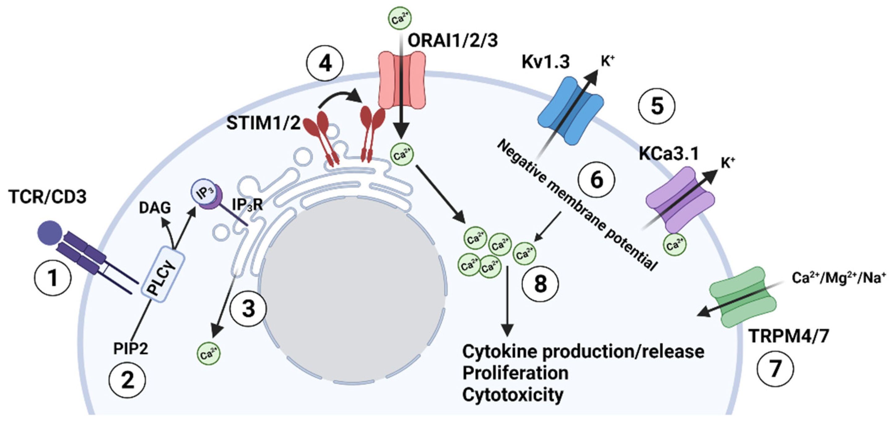
Figure 1. TCR-mediated regulation of the ion channel network in T lymphocytes. (1) Antigen stimulates the T cell receptor (TCR); (2) TCR stimulation activates phospholipase C (PLCγ), which cleaves phosphatidylinositol 4,5-bisphosphate (PIP2) to inositol (1,4,5)-trisphosphate (IP3) and diacylglycerol (DAG); (3) IP3 binds to its cognate receptor (IP3R) on the endoplasmic reticulum (ER) and depletes the ER Ca2+ store; (4) The depletion of Ca2+ in the ER causes the STIM protein (located on the ER membrane) to oligomerize and associate with the ORAI subunits (located on the plasma membrane) forming a functional CRAC channel (Ca2+ release-activated Ca2+ channel) that allows the influx of Ca2+ into the cell; (5) The increase in intracellular Ca2+ depolarizes the cell thus activating the voltage-gated K+ channel Kv1.3, which allows the efflux of K+ ions. While at the same time, the increase in intracellular Ca2+ activates the Ca2+ dependent K+ channel KCa3.1, which also allows the efflux of potassium ions; (6) The efflux of K+ ions hyperpolarizes the membrane; (7) TRP channels can also play a role in Ca2+ influx directly (like TRPM7) or by regulating the membrane potential (like TRPM4); (8) The negative membrane potential ultimately supports the electrochemical gradient necessary for the sustained influx of Ca2+ through the CRAC channels and, consequently, the increase intracellular Ca2+ levels needed for downstream functions such as cytokine production or release, T cell proliferation and cytotoxicity.
Cancers 14 03564 g001
Figure 2. Ion channel regulation of human T cell motility. Ion channels acquire a distinct polarization in migrating human T cells: KCa3.1 and TRPM7 localize at the uropod, while Kv1.3 and CRAC (ORAI1) are at the leading-edge. Intracellular Ca2+ levels ([Ca2+]i) are higher at the uropod than other cell compartments, which may serve to support the Ca2+ requirements for cell motility. The blue line indicates intracellular Ca2+ oscillations; the red arrow indicates directionality of movement.
Cancers 14 03564 g002
Figure 3. Effect of the tumor microenvironment (TME) on K+ homeostasis in T lymphocytes. (1) Hypoxia inhibits Kv1.3 channels via a dual mechanism. Short-term exposure to hypoxia directly inhibits Kv1.3 activity by changing the channel’s phosphorylation state. Prolonged exposure to low O2 tension reduces the forward trafficking of newly synthesized channel’s pore-forming subunits from the trans-Golgi to the plasma membrane. (2) Necrosis or cell death leads to ionic imbalances and increased K+ ions in the extracellular space. The consequent increase in intracellular K+ inhibits TCR activation via blockade of TCR signaling. (3) Adenosine (Ado) accumulates in the extracellular space via consecutive degradation of ATP and AMP by the nucleotidase CD39 and CD73 present on the membrane of cancer, stroma and Treg cells. Adenosine binds to its receptor on T cells and triggers KCa3.1 inhibition via cAMP and PKA1 (4) PD-L1 presented by cancer cells binds to the PD-1 receptor on the T cells and triggers KCa3.1 inhibition through PI3K signaling and calmodulin. Inhibition of Kv1.3 and KCa3.1 channels by hypoxia, adenosine and PD-L1 (1,3,4) decreases overall T cell effector functions. Accumulation of K+ in the cells and its deleterious effects on T cell function can be exacerbated by the concomitant presence of hypoxia, adenosine and PD-L1 in the TME. Conversely, overexpression and/or activation of Kv1.3 and KCa3.1 channels can simultaneously counteract all these immunosuppressive elements of the TME.
Cancers 14 03564 g003
Table 1. Effects of the tumor microenvironment (TME) characteristics on K+ channel network of T cells. Abbreviations: TILs, tumor-infiltrating lymphocytes; PD-L1, programmed cell death ligand 1; PD-1, programmed cell death protein-1.
TME FeatureEffect on Ion ChannelsReferences
Hypoxia
Acute
  • Inhibition of Kv1.3 currents
[58,59]
Chronic
  • Decrease in Kv1.3 expression
[58,61]
Tumor necrosis and increase in intra-tumoral K+ concentration
  • Responsible for the increase in intracellular [K+] in TILs that leads to inhibition of T cell transcriptional programs required for cytokine production. Kv1.3 overexpression (in vivo) and overexpression/pharmacological activation of KCa3.1 (in vitro) rescue T cell function
[62]
Adenosine
  • Adenosine binds to A2A receptor on T cells triggering KCa3.1 inhibition
[33,47,72]
PD-L1
  • PD-L1 binds to PD-1 receptor on T cells triggering KCa3.1 inhibition
[82]
Publisher’s Note: MDPI stays neutral with regard to jurisdictional claims in published maps and institutional affiliations.