Article

NF2 Alteration/22q Loss Is Associated with Recurrence in WHO Grade 1 Sphenoid Wing Meningiomas

1
Departments of Neurosurgery, Faculty of Medicine, The University of Tokyo, Tokyo 113-8655, Japan
2
Department of Neurosurgery, Faculty of Medicine, Kyorin University, Tokyo 113-8655, Japan
3
Department of Molecular Neurology, Graduate School of Medicine, The University of Tokyo, Tokyo 113-8655, Japan
*
Author to whom correspondence should be addressed.
Cancers 2022, 14(13), 3183; https://doi.org/10.3390/cancers14133183
Submission received: 4 May 2022 / Revised: 17 June 2022 / Accepted: 24 June 2022 / Published: 29 June 2022
(This article belongs to the Special Issue State-of-the-Art Research in Meningioma)

Abstract

:

Simple Summary

Sphenoid wing meningiomas account for 11–20% of all intracranial meningiomas and have a higher recurrence rate than those at other sites. The molecular background of meningiomas has been clarified in recent years; therefore, in this study, we aimed to analyze factors associated with the recurrence of sphenoid wing meningiomas. We found that NF2 alteration/22q loss is associated with recurrence in WHO grade 1 sphenoid wing meningiomas, as well as some radiological findings. Together, our study suggests that genotyping meningiomas could help to inform treatment strategies and play an important role in precision medicine, while the preoperative prediction of genotype from radiological features could influence surgical strategies. Furthermore, appropriate follow-up planning could improve the appropriate distribution of medical resources and provide both medical and economic benefits.

Abstract

Sphenoid wing meningiomas account for 11–20% of all intracranial meningiomas and have a higher recurrence rate than those at other sites. Recent molecular biological analyses of meningiomas have proposed new subgroups; however, the correlation between genetic background and recurrence in sphenoid wing meningiomas has not yet been fully elucidated. In this study, we evaluated the clinical characteristics, pathological diagnosis, and molecular background of 47 patients with sphenoid wing meningiomas. Variants of NF2, AKT1, KLF4, SMO, POLR2A, PIK3CA, TRAF7, and TERT were determined using Sanger sequencing, and 22q loss was detected using multiplex ligation-dependent probe amplification. Alterations were localized at NF2 in 11 cases, had other genotypes in 17 cases, and were not detected in 12 cases. Interestingly, WHO grade 1 meningiomas with NF2 alteration/22q loss (p = 0.008) and a MIB-1 labeling index > 4 (p = 0.03) were associated with a significantly shorter recurrence-free survival, and multivariate analysis revealed that NF2 alteration/22q loss was associated with recurrence (hazard ratio, 13.1). The duration of recurrence was significantly shorter for meningiomas with NF2 alteration/22q loss (p = 0.0007) even if gross-total resection was achieved. Together, these findings suggest that NF2 alteration/22q loss is associated with recurrence in WHO grade 1 sphenoid wing meningiomas.

1. Introduction

Sphenoid wing meningiomas account for 11–20% of all intracranial meningiomas [1] and are adjacent to important neurovascular structures, including the internal carotid artery, anterior choroidal artery, cavernous sinus, optic nerve, and cranial oculomotor nerves. These structures can limit gross-total resection (GTR), which is a key determinant of recurrence [2,3,4]. Indeed, it has been reported that the extent of resection (EOR) is associated with the recurrence of sphenoid wing meningiomas [5], which have a higher rate of recurrence than meningiomas in other locations [6].
Molecular genetic studies have revealed that NF2 gene variants and 22q loss occur in approximately 40–60% and 60–70% of sporadic meningiomas, respectively [7,8,9,10]. Recent studies have also reported that TRAF7, KLF4, AKT1, SMO, PIK3CA, and POLR2A variants are all mutually exclusive of NF2 variants [11,12,13,14]. In addition, TERT promoter (TERTp) variants have been associated with a shorter time to progression in meningiomas [15] and have been included as an independent criterion for WHO grade 3 meningiomas in the 2021 WHO Classifications of Tumors of the Central Nervous System [16]. In the last decade, a series of reports have been published on meningiomas related to copy number variants (CNVs), methylomes, and transcriptomes, and new subgroups have been proposed based on several molecular biological perspectives [17,18,19,20,21,22]. However, the association between the genetic background of sphenoid wing meningiomas and their recurrence has not yet been fully elucidated. In this study, we aimed to analyze factors associated with the recurrence of sphenoid wing meningiomas.

2. Materials and Methods

2.1. Study Cohort

We retrospectively analyzed the data of patients who underwent sphenoid wing meningioma resection at the University of Tokyo Hospital between April 2002 and December 2020. Sphenoid wing meningiomas were defined as those with dural attachment of the lesser wing of the sphenoid bone [23]. Patients with neurofibromatosis type 2, multiple meningiomas, and craniofacial meningiomas were excluded. If the patient underwent multiple surgeries, only data from the first surgery were used.
This study included 47 patients with sphenoid wing meningiomas whose characteristics are listed in Table 1. The cohort included 15 men and 32 women, with a median age of 57 (49–64) years and a median follow-up month of 27 (12–61.5). Of the 47 patients, 43 had WHO grade 1 meningiomas, three had WHO grade 2 meningiomas, one had a WHO grade 3 meningioma, and none had en plaque meningiomas. Although the grade 3 tumor was histologically classified as a meningothelial meningioma, it had a TERTp variant and was therefore classed as grade 3 according to the 2021 WHO Classifications of Tumors of the Central Nervous System. All grade 2 tumors were histologically classified as atypical meningiomas. The histology distribution is listed in Table S3.

2.2. Data Collection

Clinical charts, surgical records, radiological findings, and pathological findings were reviewed to evaluate the following patient parameters: gender, age, preoperative symptoms, radiological features, the EOR (GTR or sub-total resection [STR]), time to tumor recurrence, and pathological diagnosis.
The meningioma location was classified as described previously [23]. Briefly, the sphenoid wing was divided into three roughly equal portions. The “deep, inner or clinoidal” inner third of the sphenoid wing was classified as medial, while the “middle or alar” and “outer or pterional” outer two-thirds were classified as lateral. The localization of each tumor was defined according to the position of the center of its attachment in the sphenoid bone. Bone invasion was defined as the presence of enhancing soft tissue within the sphenoid bone on a Magnetic Resonance Imaging (MRI) scan, similar in appearance to the soft tissue enhancement and intensity of intracranial tumors [24]. Hyperostosis was defined as sphenoid wing bone adjacent to the tumor that was thicker than normal cortical bone in Computed Tomography (CT) and MRI scans, with similar CT attenuation. Tumors with an irregular shape were defined as those with margin irregularity. Tumor volumetric analysis was performed using the volumetric function of Horos version 3.3.6 (http://www.horosproject.org accessed on 1 April 2022).
On the second day, the sixth month, and the first year after surgery, patients underwent contrast-enhanced magnetic resonance imaging (CE-MRI) for follow-up. If no tumor recurrence was observed, annual CE-MRI follow-ups were performed. All CE-MRIs were subjected to a central review. When the apparent enlargement of residual tumors was detected on CE-MRI scans, tumor recurrence was defined through inter-observer agreement between the neuro-radiologist and two neurosurgeons who were blind to the clinical or genetic data. Recurrence-free survival (RFS) was defined as the time between surgery and recurrence or final follow-up. Cases in which radiotherapy was administered before recurrence were considered censored at that time. The EOR was categorized as either GTR (Simpson grades I, II, and III) or STR (Simpson grade IV) based on postoperative imaging and surgical records [25].
A central review was conducted for all pathological diagnoses according to the 2021 WHO Classifications of Tumors of the Central Nervous System [26]. The MIB-1 labeling index (LI) was determined by using the highest labeling index values in the areas of maximum density, as identified through visual analysis [27]. Clinical and genetic information results were not yet available to the reference neuropathologists.

2.3. DNA Extraction and Sanger Sequencing

DNA was extracted from fresh-frozen or FFPE (formalin-fixed paraffin-embedded) tumors using a QIAamp DNA Mini Kit (QIAGEN; Venlo, The Netherlands) or a QIAamp DNA FFPE Tissue Kit according to the manufacturer’s instructions. DNA quality was evaluated using a spectrophotometer.
Based on previous reports [12,15,28,29,30], hotspot variants were evaluated in AKT1 (NM_001382430, c.49G>A [p.Glu17Lys]), KLF4 (NM_4235.6, c.1228A>C [p.Lys409Gln]), SMO (NM_005631, c.1234C>T [p.Leu412Phe] and c.1604G>T [p.Trp535Leu]), POLR2A (NM_000937.5, c.1207C>A [p.Gln403Lys], and c.1310–1315delACCTTC [p.Leu438_His439del]), PIK3CA (NM_006218, c.1624G>A [p.Glu542Lys], c.1633G>A [p.Glu545Lys/Ala], and c.3140A>G [p.His1047Arg]), and TERTp (NM_1014431, C228A(T) and C250T). Direct Sanger sequencing was performed for all exons in NF2 (NM_000268.4) and for exons 12–21 (covering the WD40 domain) in TRAF7 (NM_032271).
A total of 50 ng of DNA and KOD FX NEO (Applied Biosystems; Foster City, CA, USA) were used for polymerase chain reaction (PCR) in a 20 μL reaction mixture with the following reaction cycle: initial denaturation at 94 °C for 2 min, followed by 32 cycles of denaturation at 98 °C for 10 s, annealing at 58–60 °C for 30 s, and extension at 68 °C for 30 s, followed by final extension at 68 °C for 7 min (Table S2). Sequences were determined using an ABI 3130xl Genetic Analyzer (Applied Biosystems). NF2, AKT1, KLF4, SMO, POLR2A, and TERT primer alignment is described in Table S2. The PCR and primer alignment procedures for PIK3CA and TRAF7 were performed as described previously [12,29,30].

2.4. Multiplex Ligation-Dependent Probe Amplification (MLPA)

The SALSA P044-C1 NF2 probemix (MRC Holland; Amsterdam, The Netherlands) was used for MLPA analyses according to the manufacturer’s instructions. The P044-C1 NF2 probemix contained 21 probes for the NF2 gene; five probes for chromosome 22q upstream of NF2 (4 of which target SMARCB1 and LZTR1); four probes for chromosome 22q downstream of NF2, and 13 reference probes for sequences on other chromosomes and some control fragments. DNA dosage was determined using Coffalyser.Net v210226.1433 (MRC Holland): 0.8–1.2 dosage quotients was considered normal, 0.4–0.7 dosage quotients indicated heterozygous or single exon deletions [31].

2.5. Statistical Analysis

All statistical analyses were performed using R v.4.1.1 (R Core Team, http://www.R-project.org accessed on 1 April 2022). Numerical variables were expressed as the median (interquartile range [IQR]) and compared using the Mann–Whitney U test. Categorical data were expressed as the count and percentage, and compared using Fisher’s exact test or the chi-square test. All reported p values were two-sided and values of < 0.05 were considered significant. Kaplan–Meier survival curves were plotted and differences in RFS between groups were compared using the log-rank test. Cutoff scores were determined according to the median score and clinical applicability. Multivariate analysis was performed using the Cox proportional hazard model.

3. Results

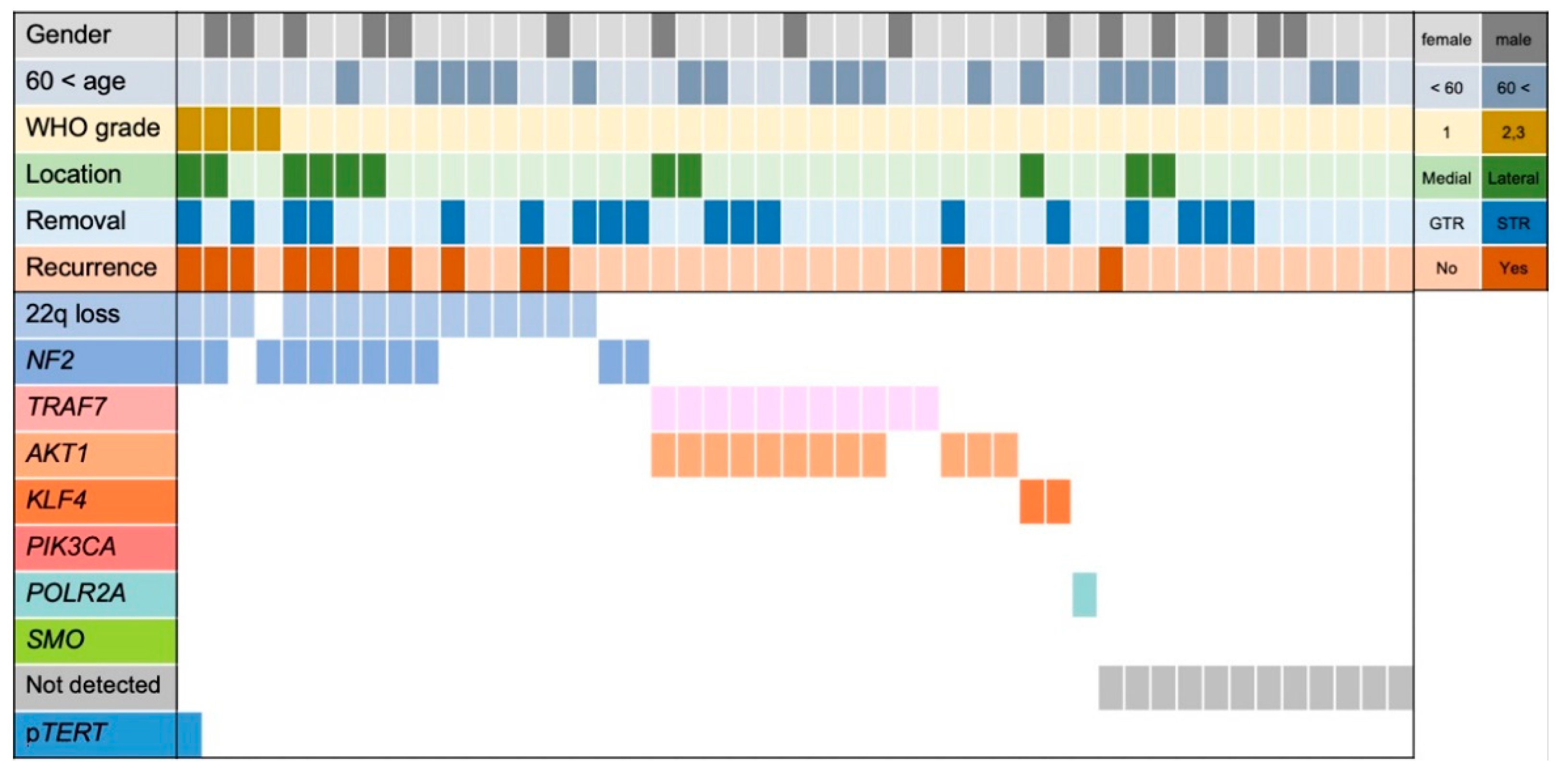
3.1. Tumor Characteristics of This Cohort

In the study cohort, genetic alterations were detected in NF2 in 11 cases (23.4%), AKT1 in 12 cases (25.5%), TRAF7 in 11 cases (23.4%), KLF4 in two cases (4.3%), POLR2A in one case (4.3%), TERTp in one case (4.3%), and as 22q loss in 15 cases (31.9%) (Figure 1). Details of the genetic information are provided in Table S4. Among the 11 cases with TRAF7 variants, nine (81.8%) also had AKT1 variants. Among the cases with NF2 alterations, variants were detected by Sanger sequencing in nine cases, and exon deletions were detected by MLPA in two cases. No variants were detected at PIK3CA or SMO. In 12 cases (25.5%), no gene variants could be identified, and these cases were designated as “not detected”.
Notably, all grade 2 and 3 meningiomas had NF2 alterations and/or 22q loss (Figure 1). Among the four patients with grade 2 and 3 meningiomas, three experienced recurrence and one underwent irradiation therapy before recurrence and was considered censored at that time. WHO grade 1 and grade 2–3 meningiomas had the following characteristics: NF2 alteration and/or 22q loss (grade 1, 32.6%; grade 2–3, 100%), bone invasion (grade 1, 9.3%; grade 2–3, 75%), irregular shape (grade 1, 20.9%; grade 2–3, 75%), MIB-1 LI > 4 (grade 1, 20.9%; grade 2–3, 50%), and GTR (grade 1, 62.8%; grade 2–3, 50%; Table 1).

3.2. Tumor Characteristics of WHO Grade 1 Meningiomas

A total of 43 patients in this study had WHO grade 1 sphenoid wing meningiomas, including 13 men and 30 women, with a median age of 57 (50–65.5) years, and a median follow-up months of 30 (14.5–70) (Table 2). Of the nine patients that experienced recurrence, seven had NF2 alteration/22q loss, one had AKT1 variation, and the genotype of the last case was not detected. In recurrent cases with NF2 alteration/22q loss, GTR was achieved in three cases, and STR was achieved in four. When comparing the 14 patients with NF2 alteration/22q loss and the 29 patients with wild-type NF2/22q, recurrence (p = 0.0026) and bone invasion (p = 0.008) differed significantly between the two groups.

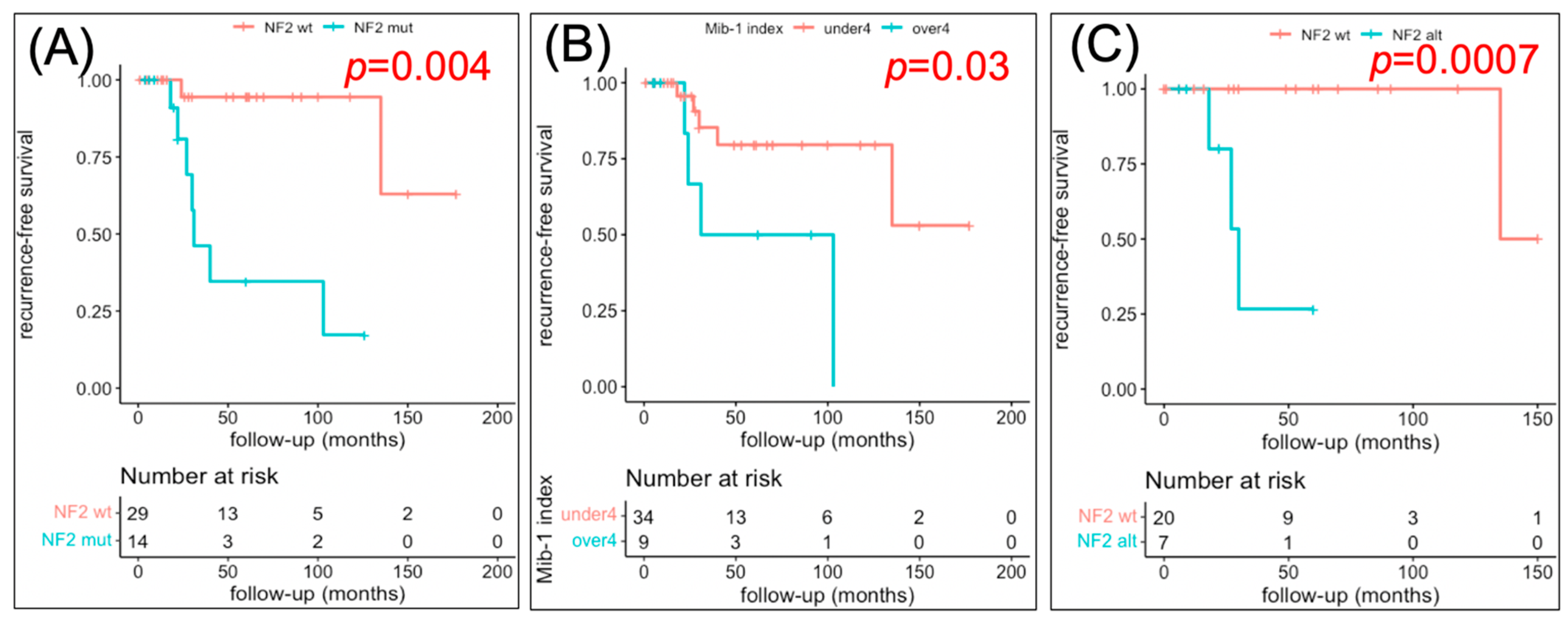
3.3. Factors Related to RFS

Next, we performed a Kaplan–Meier analysis of RFS (Figure 2), finding that NF2 alteration and/or 22q loss (p = 0.008) and MIB-1 LI > 4 (p = 0.03) were associated with a statistically significantly shorter time to recurrence, whereas gender and resection rate were not significantly associated with RFS (p = 0.07 and 0.3, respectively, Figure S1). In patients where GTR was achieved, NF2 alteration and/or 22q loss were associated with a statistically significantly shorter time to recurrence (Figure 2C). Univariate Cox proportional hazards analysis (Table 3) revealed that NF2 alteration/22q loss (hazard ratio [HR], 16.8; 95% confidence interval [CI], 2.1–137.0; p = 0.009) and MIB-1 LI > 4 (HR, 4.2; 95% CI, 1.05–16.9; p = 0.04) were significantly associated with tumor recurrence. Multivariate analysis performed using factors with p < 0.25 in the univariate analysis further indicated that NF2 alteration/22q loss was associated with tumor recurrence (HR, 13.1; 95% CI, 1.5–111; p = 0.019).

4. Discussion

Sphenoid wing meningiomas have a higher rate of recurrence than meningiomas in other locations; however, the association between the genetic background of sphenoid wing meningiomas and their recurrence has not yet been fully elucidated. In this study, we found that WHO grade 1 sphenoid wing meningiomas with NF2 alteration/22q loss are associated with a higher rate of recurrence than those with wild-type NF2/22q (p = 0.0026), even though the rate of GTR did not differ significantly between the two groups (p = 0.23). In addition, we found that the period of RFS was significantly shorter for patients with NF2 alteration/22q loss (p = 0.008) and that NF2 alteration/22q loss was significantly associated with recurrence (HR 13.1 [1.5–111]). Together, these results suggest that NF2 alteration/22q loss is an independent factor that affects recurrence.
As for CNVs, it has been reported that cases with 22q loss have a HR of 12 (2–68, 95% CI) in recurrence and a complex karyotype HR of 32 (3–296, 95% CI) [32]. Moreover, cases with 22q loss but without NF2 variants and complex karyotypes are reported to have a significantly poor prognosis for recurrence [33]. It may be worthwhile to evaluate CNVs other than 22q. Also, in recent years, several classifications based on integrated molecular analyses have been proposed and have indicated that the prognosis of patients with meningiomas with NF2 variant/22q loss ranges from good to poor. In addition, these studies have suggested that a more accurate prognosis can be determined by analyzing CNVs, the methylome, and the transcriptome in addition to driver variants [17,18,34,35,36,37]. Nassiri et al. [18] divided meningiomas into four groups by combining DNA somatic CNVs, DNA somatic point mutations, DNA methylation, and messenger RNA abundance in a unified analysis. Based on this analysis, cases with NF2 alteration/22q loss were classified into molecular groups (MGs) 1, 3, and 4, among which the prognosis for recurrence in MG3 and 4 meningiomas is reported to be poor. Sahm et al. [17] divided meningiomas into six groups mainly based on the methylome and classified WHO grade 1 meningiomas with NF2 alteration/22q loss into methylation clusters (MCs) ben-1, ben-3, or int-A, among which int-A meningiomas have a relatively short RFS. The findings of these analyses suggest that the ability of NF2 to predict recurrence also depends on CNVs, the methylome, and the transcriptome. Interestingly, our study suggested that, in sphenoid wing meningiomas, NF2 alteration/22q loss can significantly predict recurrence; however, additional CNV, methylome, and transcriptome analysis could allow for more accurate prognostic analysis. Further research is required to elucidate the significance of driver variants in specific tumor locations.
Interestingly, we found that three patients with NF2 alteration/22q loss (3/7: 42.9%) experienced recurrence even after GTR was achieved (Figure 1), suggesting that sphenoid wing meningiomas with NF2 alteration/22q loss require close postoperative follow-up even if GTR is achieved (Figure 2F). Conversely, only one patient with AKT1/TRAF7 alteration (1/14:7.1%, STR case) experienced recurrence, and no recurrence was observed among patients with AKT1/TRAF7 alteration who achieved GTR (Figure 1). Therefore, conventional follow-up is considered necessary in patients with AKT1/TRAF7 alteration that achieve STR, but the likelihood of recurrence is likely to be low in patients that achieve GTR. Meanwhile, no recurrence was observed in patients with KLF4 and POLR2A variants. Taken together, these findings indicate that adjusting postoperative follow-up intensity according to genotype might be justified in patients with sphenoid wing meningiomas, and could improve the distribution of medical resources and provide medical and economic benefits. Genotyping meningiomas could also play a significant role in the consideration of different treatment strategies and in precision medicine; however, further studies are required to determine the modality and timing of treatments for recurrent meningiomas.
In this study, many of the meningiomas with NF2 alteration/22q loss displayed bone invasion (Table 2), suggesting that meningiomas with bone invasion could harbor NF2 variants preoperatively. Previously, Jin et al. reported that the radiographic presence of tumor invasion into the sphenoid wing and its association with underlying NF2 variants can provide insights into the biological aggression potential of a tumor [24]. Therefore, future studies should investigate the molecular mechanisms that relate NF2 variants with bone invasion. We found that oculomotor dysfunction was more common in patients with NF2 alteration/22q loss, which may be confounded by bone invasion. The preoperative prediction of genotype from radiological features could therefore influence surgical strategy and tactics. However, even if it is presumed that the patient has NF2 alteration/22q loss preoperatively, GTR should remain the main aim within safe limits. As such, future reports should aim to explore the association between genotype and clinical findings such as radiological features.
Our study had several limitations that should be addressed in future investigations. First, our study suffers from a small sample size and a retrospective, single-institution design. This small sample size could be the reason for the discovery of fewer cases with PIK3CA or KLF4 variants. Therefore, the significance of these driver genes in the recurrence of WHO 1 sphenoid wing meningiomas could not be clarified in this study.
Second, our study did not analyze all driver genes, CNVs except 22q, the methylome, or the transcriptome. The driver genes evaluated in this study did not include all genes that had not been reported to be associated with meningiomas before. A more accurate prognostic analysis may be possible if the number of target genes is expanded. Especially in cases with NF2 alteration/22q loss, adding CNVs, the methylome, or the transcriptome to that of this study may allow for more accurate prognostic analysis.
Third, since reference genetic material was not used in the genetic screening, it was not possible to confirm the somatic/germline nature of the variants identified, which was another limitation.

5. Conclusions

In this study, we found that in WHO grade 1 sphenoid wing meningiomas, NF2 alteration/22q loss is an independent factor that is associated with recurrence and that patients with these meningiomas may require close postoperative follow-up even if GTR is achieved. Further studies are required to elucidate the significance of driver variants in specific tumor locations.

Supplementary Materials

The following supporting information can be downloaded at https://www.mdpi.com/article/10.3390/cancers14133183/s1: Table S1: Primer alignment; Table S2: Composition of PCR reaction mixtures; Table S3: Histological distribution of the cases in this stud; Table S4: Patients characteristics; Figure S1: Kaplan-Meier plots of time to recurrence.

Author Contributions

Conceptualization, Y.S., S.M., Y.T. and A.O.; methodology, Y.S., S.M., Y.T. and A.O.; validation, S.M. and Y.T.; formal analysis, Y.S.; investigation, Y.S., Y.T. and A.O.; resources, Y.S., S.M. and J.M.; data curation, Y.S., Y.T. and A.O.; writing—original draft preparation, Y.S.; writing—review and editing, S.M., Y.T., A.O., K.O., H.H., D.I., D.S., J.M., H.N. and N.S.; visualization, Y.S.; supervision, S.M., H.N., and N.S.; project administration, S.M., Y.T. and A.O.; funding acquisition, S.M., H.N. and N.S. All authors have read and agreed to the published version of the manuscript.

Funding

This research was funded by Grants-in-aid for Scientific Research from the Japan Society for the Promotion of Science (B, No. 21H03041 to N.S.; C, No. 19K09499 to H.N.; and C, No. 19K09473 to S.M.) and a research grant from the Takeda Science Foundation (to S.M.).

Institutional Review Board Statement

The Institutional Review Board approved the study protocol of The University of Tokyo Hospital (G10028). Informed consent was obtained from all patients.

Informed Consent Statement

Informed consent was obtained from all subjects involved in the study.

Data Availability Statement

Data are available on reasonable request. The authors confirm that the data supporting the findings of this study will be shared by request from any qualified investigator.

Acknowledgments

The authors are grateful to Reiko Matsuura (Departments of Neurosurgery, Faculty of Medicine, The University of Tokyo, Tokyo, Japan) for providing technical assistance.

Conflicts of Interest

The authors declare no conflict of interest.

References

  1. Güdük, M.; Özduman, K.; Pamir, M.N. Sphenoid Wing Meningiomas: Surgical Outcomes in a Series of 141 Cases and Proposal of a Scoring System Predicting Extent of Resection. World Neurosurg. 2019, 125, e48–e59. [Google Scholar] [CrossRef] [PubMed]
  2. Al-Mefty, O. Clinoidal meningiomas. J. Neurosurg. 1990, 73, 840–849. [Google Scholar] [CrossRef] [PubMed]
  3. Mirimanoff, R.O.; Dosoretz, D.E.; Linggood, R.M.; Ojemann, R.G.; Martuza, R.L. Meningioma: Analysis of recurrence and progression following neurosurgical resection. J. Neurosurg. 1985, 62, 18–24. [Google Scholar] [CrossRef] [PubMed] [Green Version]
  4. Bonnal, J.; Thibaut, A.; Brotchi, J.; Born, J. Invading meningiomas of the sphenoid ridge. J. Neurosurg. 1980, 53, 587–599. [Google Scholar] [CrossRef] [Green Version]
  5. Mathiesen, T.; Lindquist, C.; Kihlström, L.; Karlsson, B. Recurrence of Cranial Base Meningiomas. Neurosurgery 1996, 39, 2–9. [Google Scholar] [CrossRef]
  6. Nakamura, M.; Roser, F.; Jacobs, C.; Vorkapic, P.; Samii, M. Medial Sphenoid Wing Meningiomas: Clinical Outcome and Recurrence Rate. Neurosurgery 2006, 58, 626–639. [Google Scholar] [CrossRef]
  7. Kros, J.; de Greve, K.; van Tilborg, A.; Hop, W.; Pieterman, H.; Avezaat, C.; Lekanne Dit Deprez, R.; Zwarthoff, E. NF2 status of meningiomas is associated with tumour localization and histology. J. Pathol. 2001, 194, 367–372. [Google Scholar] [CrossRef]
  8. Riemenschneider, M.J.; Perry, A.; Reifenberger, G. Histological classification and molecular genetics of meningiomas. Lancet Neurol. 2006, 5, 1045–1054. [Google Scholar] [CrossRef]
  9. Zang, K.D.; Singer, H. Chromosomal Constitution of Meningiomas. Nature 1967, 216, 84–85. [Google Scholar] [CrossRef]
  10. Suppiah, S.; Nassiri, F.; Bi, W.L.; Dunn, I.F.; Hanemann, C.O.; Horbinski, C.M.; Hashizume, R.; James, C.D.; Mawrin, C.; Noushmehr, H.; et al. Molecular and translational advances in meningiomas. Neuro-Oncology 2019, 21, i4–i17. [Google Scholar] [CrossRef]
  11. Clark, V.E.; Erson-Omay, E.Z.; Serin, A.; Yin, J.; Cotney, J.; Özduman, K.; Avşar, T.; Li, J.; Murray, P.B.; Henegariu, O.; et al. Genomic Analysis of Non- NF2 Meningiomas Reveals Mutations in TRAF7, KLF4, AKT1, and SMO. Science 2013, 339, 1077–1080. [Google Scholar] [CrossRef] [PubMed] [Green Version]
  12. Reuss, D.E.; Piro, R.M.; Jones, D.T.; Simon, M.; Ketter, R.; Kool, M.; Becker, A.; Sahm, F.; Pusch, S.; Meyer, J.; et al. Secretory meningiomas are defined by combined KLF4 K409Q and TRAF7 mutations. Acta Neuropathol. 2013, 125, 351–358. [Google Scholar] [CrossRef] [PubMed]
  13. Clark, V.E.; Harmanci, A.S.; Bai, H.; Youngblood, M.W.; Lee, T.I.; Baranoski, J.F.; Ercan-Sencicek, A.G.; Abraham, B.J.; Weintraub, A.S.; Hnisz, D.; et al. Recurrent somatic mutations in POLR2A define a distinct subset of meningiomas. Nat. Genet. 2016, 48, 1253–1259. [Google Scholar] [CrossRef] [PubMed] [Green Version]
  14. Pang, J.C.-S.; Chung, N.Y.F.; Chan, N.H.L.; Poon, W.S.; Thomas, T.; Ng, H.-K. Rare mutation of PIK3CA in meningiomas. Acta Neuropathol. 2006, 111, 284–285. [Google Scholar] [CrossRef] [PubMed]
  15. Sahm, F.; Schrimpf, D.; Olar, A.; Koelsche, C.; Reuss, D.; Bissel, J.; Kratz, A.; Capper, D.; Schefzyk, S.; Hielscher, T.; et al. TERT Promoter Mutations and Risk of Recurrence in Meningioma. J. Natl. Cancer Inst. 2016, 108, djv377. [Google Scholar] [CrossRef]
  16. WHO Classification of Tumours Editorial Board. World Health Organization Classification of Tumours of the Central Nervous System, 5th ed.; International Agency for Research on Cancer: Lyon, France, 2021. [Google Scholar]
  17. Sahm, F.; Schrimpf, D.; Stichel, D.; Jones, D.T.W.; Hielscher, T.; Schefzyk, S.; Okonechnikov, K.; Koelsche, C.; Reuss, D.E.; Capper, D.; et al. DNA methylation-based classification and grading system for meningioma: A multicentre, retrospective analysis. Lancet Oncol. 2017, 18, 682–694. [Google Scholar] [CrossRef] [Green Version]
  18. Nassiri, F.; Liu, J.; Patil, V.; Mamatjan, Y.; Wang, J.Z.; Hugh-White, R.; Macklin, A.M.; Khan, S.; Singh, O.; Karimi, S.; et al. A clinically applicable integrative molecular classification of meningiomas. Nature 2021, 597, 119–125. [Google Scholar] [CrossRef]
  19. Maas, S. Integrated molecular-morphologic meningiomas classification: A multicenter retrospective analysis, retrospectively and prospetively validated. J. Clin. Oncol. 2021, 39, 3839–3852. [Google Scholar] [CrossRef]
  20. Nassiri, F.; Mamatjan, Y.; Suppiah, S.; Badhiwala, J.H.; Mansouri, S.; Karimi, S.; Saarela, O.; Poisson, L.M.; Gepfner-Tuma, I.; Schittenhelm, J.; et al. DNA methylation profiling to predict recurrence risk in meningioma: Development and validation of a nomogram to optimize clinical management. Neuro-Oncology 2019, 21, 901–910. [Google Scholar] [CrossRef] [Green Version]
  21. Paramasivam, N.; Hübschmann, D.; Toprak, U.H.; Ishaque, N.; Neidert, M.; Schrimpf, D.; Stichel, D.; Reuss, D.; Sievers, P.; Reinhardt, A.; et al. Mutational patterns and regulatory networks in epigenetic subgroups of meningioma. Acta Neuropathol. 2019, 138, 295–308. [Google Scholar] [CrossRef]
  22. Youngblood, M.W.; Miyagishima, D.F.; Jin, L.; Gupte, T.; Li, C.; Duran, D.; Montejo, J.D.; Zhao, A.; Sheth, A.; Tyrtova, E.; et al. Associations of meningioma molecular subgroup and tumor recurrence. Neuro-Oncology 2021, 23, 783–794. [Google Scholar] [CrossRef] [PubMed]
  23. Cushing, H.W.; Eisenhardt, L. Meningiomas: Their Classification, Regional Behavior Life History, and Surgical End Results; Hafner Publishing Company: New York, NY, USA, 1938; pp. 298–341. [Google Scholar]
  24. Jin, L.; Youngblood, M.W.; Gupte, T.P.; Vetsa, S.; Nadar, A.; Barak, T.; Yalcin, K.; Aguilera, S.M.; Mishra-Gorur, K.; Blondin, N.A.; et al. Type of bony involvement predicts genomic subgroup in sphenoid wing meningiomas. J. Neuro-Oncol. 2021, 154, 237–246. [Google Scholar] [CrossRef] [PubMed]
  25. Simpson, D. The Recurrence of Intracranial Meningiomas after Surgical Treatment. J. Neurol. Neurosurg. Psychiatry 1957, 20, 22–39. [Google Scholar] [CrossRef] [Green Version]
  26. Louis, D.N.; Perry, A.; Wesseling, P.; Brat, D.J.; Cree, I.A.; Figarella-Branger, D.; Hawkins, C.; Ng, H.K.; Pfister, S.M.; Reifenberger, G.; et al. The 2021 WHO Classification of Tumors of the Central Nervous System: A summary. Neuro-Oncology 2021, 23, 1231–1251. [Google Scholar] [CrossRef] [PubMed]
  27. Oya, S.; Kawai, K.; Nakatomi, H.; Saito, N. Significance of Simpson grading system in modern meningioma surgery: Integration of the grade with MIB-1 labeling index as a key to predict the recurrence of WHO Grade I meningiomas. J. Neurosurg. 2012, 117, 121–128. [Google Scholar] [CrossRef] [Green Version]
  28. Okano, A.; Miyawaki, S.; Hongo, H.; Dofuku, S.; Teranishi, Y.; Mitsui, J.; Tanaka, M.; Shin, M.; Nakatomi, H.; Saito, N. Associations of pathological diagnosis and genetic abnormalities in meningiomas with the embryological origins of the meninges. Sci. Rep. 2021, 11, 6987. [Google Scholar] [CrossRef]
  29. Korkolopoulou, P.; Levidou, G.; Trigka, E.-A.; Prekete, N.; Karlou, M.; Thymara, I.; Sakellariou, S.; Fragkou, P.; Isaiadis, D.; Pavlopoulos, P.; et al. A comprehensive immunohistochemical and molecular approach to the PI3K/AKT/mTOR (phosphoinositide 3-kinase/v-akt murine thymoma viral oncogene/mammalian target of rapamycin) pathway in bladder urothelial carcinoma. Br. J. Urol. 2012, 110, E1237–E1248. [Google Scholar] [CrossRef] [Green Version]
  30. El-Habr, E.; Levidou, G.; Trigka, E.-A.; Sakalidou, J.; Piperi, C.; Chatziandreou, I.; Spyropoulou, A.; Soldatos, R.; Tomara, G.; Petraki, K.; et al. Complex interactions between the components of the PI3K/AKT/mTOR pathway, and with components of MAPK, JAK/STAT and Notch-1 pathways, indicate their involvement in meningioma development. Virchows Arch. 2014, 465, 473–485. [Google Scholar] [CrossRef]
  31. Chen, H.; Xue, L.; Wang, H.; Wang, Z.; Wu, H. Differential NF2 Gene Status in Sporadic Vestibular Schwannomas and its Prognostic Impact on Tumour Growth Patterns. Sci. Rep. 2017, 7, 5470. [Google Scholar] [CrossRef] [Green Version]
  32. Domingues, P.H.; Sousa, P.; Otero, A.; Gonçalves, J.M.; Ruiz, L.; De Oliveira, C.; Lopes, M.C.; Orfao, A.; Tabernero, M.D. Proposal for a new risk stratification classification for meningioma based on patient age, WHO tumor grade, size, localization, and karyotype. Neuro-Oncology 2014, 16, 735–747. [Google Scholar] [CrossRef] [Green Version]
  33. González-Tablas, M.; Prieto, C.; Arandia, D.; Jara-Acevedo, M.; Otero, Á.; Pascual, D.; Ruíz, L.; Álvarez-Twose, I.; García-Montero, A.C.; Orfao, A. Whole-Exome Sequencing Reveals Recurrent but Heterogeneous Mutational Profiles in Sporadic WHO Grade 1 Meningiomas. Front. Oncol. 2021, 11, 740782. [Google Scholar] [CrossRef] [PubMed]
  34. Patel, A.J.; Wan, Y.W.; Al-Ouran, R.; Revelli, J.P.; Cardenas, M.F.; Oneissi, M.; Xi, L.; Jalali, A.; Magnotti, J.F.; Muzny, D.M.; et al. Molecular profiling predicts meningioma recurrence and reveals loss of DREAM complex repression in aggressive tumors. Proc. Natl. Acad. Sci. USA 2019, 116, 21715–21726. [Google Scholar] [CrossRef] [PubMed] [Green Version]
  35. Sanson, M.; Kalamarides, M. Epigenetics: A new tool for meningioma management? Lancet Oncol. 2017, 18, 569–570. [Google Scholar] [CrossRef]
  36. Blume, C. Integrated phspho-proteogenomic and single-cell transcriptomic analysis of menigiomas establishes robust subtyping and reveals subtype-specific immune invasion. bioRxiv 2021. [Google Scholar] [CrossRef]
  37. Choudhury, A.; Magill, S.T.; Eaton, C.D.; Prager, B.C.; Chen, W.C.; Cady, M.A.; Seo, K.; Lucas, C.-H.G.; Casey-Clyde, T.J.; Vasudevan, H.N.; et al. Meningioma DNA methylation groups identify biologic drivers and therapeutic vulnerabilities. Nat Genet. 2022, 54, 649–659. [Google Scholar] [CrossRef]
Figure 1. Clinical characteristics and genetic status of patients with sphenoid wing meningiomas. Besides one case, NF2 alteration and/or 22q loss, AKT1/TRAF7, KLF4, and POLR2A were mutually exclusive. A total of 12 cases were defined as “not detected”. GTR: gross-total resection. STR: sub-total resection.
Cancers 14 03183 g001
Figure 2. Kaplan–Meier plots of time to recurrence. (A) NF2 alteration/22q loss vs. wild-type NF2/22q. RFS was significantly shorter in patients with NF2 alteration/22q loss (p = 0.004). (B) MIB-1 LI > 4 vs. < 4. RFS was significantly shorter in patients with MIB-1 LI > 4 (p = 0.03). (C) NF2 alteration/22q loss vs. wild-type NF2/22q in patients with GTR. RFS was significantly shorter in patients with NF2 alteration/22q loss (p = 0.0007). GTR: gross-total resection.
Cancers 14 03183 g002
Table 1. Patient characteristics.
VariablesWHO Grade 1–3 (47)WHO Grade 1 (43)WHO Grade 2/3 (4)
General
Age (years)57 (49–64)57 (50–65.5)50.5 (48.5–53)
Male15 (31.9%)13 (30.2%)2 (50%))
Recurrence12 (25.5%)9 (20.9%)3 (75%)
Follow-up months27 (12–61.5)28 (13–64)16 (8.3–26.5)
Clinical symptoms
Eye field symptom22 (46.8%)19 (44.2%)3 (75%)
Vision loss14 (29.8%)12 (27.9%)2 (50%)
Oculomotor dysfunction4 (8.5%)3 (7.0%)1 (25%)
Radiological features
Medial36 (76.6%)34 (79.1%)2 (50%)
Bone invasion7 (14.9%)4 (9.3%)3 (75%)
Hyperostosis10 (21.3%)8 (18.6%)2 (50%)
Brain edema17 (36.2%)14 (32.6%)3 (75%)
CS invasion8 (17.0%)6 (14.0%)2 (50%)
Irregular shape12 (25.5%)9 (20.9%)3 (75%)
Volume (cm3)14.0 (6.0–33.1)11.7 (5.8–28.5)79.7 (42.8–125.6)
Histological analysis
MIB-1 LI ≧ 411 (23.4%)9 (20.9%)2 (50%)
Genetic analysis
NF2 alt./22q loss18 (38.3%)14 (32.6%)4 (100%)
Surgery
GTR29 (61.7%)27 (62.8%)2 (50%)
CS: cavernous sinus; LI: labeling index; GTR: gross-total resection.
Table 2. Characteristics of patients with WHO grade 1 meningiomas.
VariablesAll (43)NF2 alt./22q Loss (14)NF2/22q wt. (29)p Value
General
Age (years)57 (50–65.5)57 (47.8–63.8)58 (51–67)0.64
Male13 (30.2%)4 (28.6%))9 (31.0%)0.87
Recurrence9 (20.9%)7 (50%)2 (6.9%)0.0026
Follow-up months30 (14.5–70)25 (18.5–37.8)30 (13–70)0.30
Clinical symptom
Eye field symptom19 (44.2%)7 (50.0%)12 (41.4%)0.59
Vision loss12 (27.9%)4 (28.6%)8 (27.6%)0.95
Oculomotor dysfunction3 (7.0%)3 (14.3%)0 (0%)0.030
Radiological features
Medial34 (79.1%)10 (71.4%)24 (82.8%)0.39
Bone invasion4 (9.3%)4 (28.6%)0 (0%)0.008
Hyperostosis8 (18.6%)4 (28.6%)4 (13.8%)0.24
Brain edema14 (32.6%)3 (21.4%)11 (37.9%)0.28
CS invasion6 (14.0%)1 (7.1%)5 (17.2%)0.37
Irregular shape9 (20.9%)4 (28.6%)5 (17.2%)0.39
Volume (cm3)11.7 (5.8–28.5)10.2 (5.2–52.6)11.7 (6.9–24.0)0.21
Histological analysis
MIB-1 LI ≧ 49 (20.9%)4 (28.6%)5 (17.2%)0.39
Surgery
GTR27 (62.8%)7 (50%)20 (69.0%)0.23
CS: cavernous sinus; LI: labeling index; GTR: gross-total resection.
Table 3. Multivariate analysis of tumor recurrence.
VariablesUnivariateMultivariate
HR95% CIp ValueHR95% CIp Value
NF2/22q loss16.82.1–137.00.00913.11.5–1110.019
MIB-1 LI ≥ 44.21.05–16.90.042.20.5–9.30.28
Lateral2.30.5–9.50.27
Male2.20.6–8.80.25
Age < 602.30.6–9.50.231.60.4–6.60.53
STR2.00.5–7.50.31
HR: hazard risk; CI: confidence interval; LI: labeling index; STR: sub-total resection.
Publisher’s Note: MDPI stays neutral with regard to jurisdictional claims in published maps and institutional affiliations.

