Article

The High Proportion of Discordant EGFR Mutations among Multiple Lung Tumors

1
Department of Pathology and Translational Genomics, Samsung Medical Center, Sungkyunkwan University School of Medicine, Seoul 06351, Korea
2
Department of Pathology, Konkuk University Medical Center, Konkuk University School of Medicine, Seoul 05030, Korea
3
Department of Thoracic and Cardiovascular Surgery, Samsung Medical Center, Sungkyunkwan University School of Medicine, Seoul 06351, Korea
4
Department of Health Science and Technology, Samsung Advanced Institute for Health Sciences and Technology, Sungkyunkwan University, Seoul 06351, Korea
*
Authors to whom correspondence should be addressed.
Cancers 2022, 14(12), 3011; https://doi.org/10.3390/cancers14123011
Submission received: 5 May 2022 / Revised: 17 June 2022 / Accepted: 17 June 2022 / Published: 18 June 2022
(This article belongs to the Special Issue Lung Cancer - Molecular Insights and Targeted Therapies)

Abstract

:

Simple Summary

Lung cancer is one of the leading causes of cancer-related mortality worldwide. The incidence of multiple primary lung cancers has been increasing. In addition to the identification of epidermal growth factor receptor (EGFR) tyrosine kinase inhibitors, the evaluation of the EGFR mutation status in lung cancer is important to devise optimal treatment strategies. In this study, the EGFR mutation status in multiple primary lung cancers was examined, and its discordance rate in individual tumors was determined to be high. Our findings reveal the importance of EGFR mutation analysis in individual tumors of multiple primary lung cancers.

Abstract

The prevalence of multiple lung cancers has been increasing recently. Molecular analysis of epidermal growth factor receptor (EGFR) mutations in individual tumors of multiple lung cancers is essential for devising an optimal therapeutic strategy. The EGFR mutation status in multiple lung cancers was evaluated to determine its therapeutic implications. In total, 208 tumors from 101 patients who underwent surgery for multiple lung cancers were analyzed. Individual tumors were subjected to histological evaluation and EGFR analysis using a real-time polymerase chain reaction. Additionally, EGFR-wildtype tumors were subjected to next-generation sequencing (NGS). EGFR mutations were detected in 113 tumors from 72 patients, predominantly in females (p < 0.001) and non-smokers (p < 0.001). Among patients with at least one EGFR-mutant tumor, approximately 72% of patients (52/72) had different EGFR mutations in individual tumors. NGS analysis of EGFR-wildtype tumors from 12 patients revealed four and eight cases with concordant and discordant molecular alterations, respectively. These findings revealed a high proportion of discordant EGFR mutations among multiple lung tumors. Hence, EGFR analysis of individual tumors of multiple lung tumors is essential for the evaluation of clonality and the development of an optimal treatment strategy.

1. Introduction

Lung cancer is one of the leading causes of mortality worldwide [1]. Primary lung cancer, which is associated with multifocal presentations, accounts for 0.2–8.1% of all lung cancer cases [2,3,4,5]. The incidence of multiple lung cancers, which are classified as synchronous (simultaneous detection of cancerous lesions) and metachronous (a new cancer lesion is detected six months post-first-cancer lesion detection), and multiple primary lung cancers, has markedly increased. The pathogenesis of adenocarcinoma has been widely examined. However, the mechanisms underlying the pathogenesis of multiple adenocarcinomas have not been elucidated. The multifocal presentations of squamous cell carcinoma, which is closely linked to smoking, can be explained by ‘field cancerization’ [6]. However, the mechanism of adenocarcinoma development, which involves a multi-step process from atypical adenomatous hyperplasia (AAH) to the occurrence of invasive cancerous lesions [7], cannot efficiently explain the mechanism of multifocal presentations.
Several studies have suggested the potential mechanisms underlying the development of multiple adenocarcinomas. The detection of EGFR mutations in the non-neoplastic epithelium around the adenocarcinoma indicates that EGFR mutations occur in the early stages of carcinogenesis [8]. Alternatively, independently occurring EGFR mutations play an important role in multifocal carcinogenesis as evidenced by cancerous lesions, such as microinvasive adenocarcinoma (MIA) or invasive adenocarcinoma exhibiting an increased frequency of EGFR mutations when compared with AAH lesions [9]. Irrespective of the timing of the occurrence of mutations, analysis of EGFR mutations has been a crucial step for the application of tyrosine-kinase-inhibitor (TKI)-based targeted therapy in patients with lung cancer harboring EGFR mutations [10].
Distinguishing multifocal primary lung cancers (MPLC) from intrapulmonary metastasis (IPM) has been a long-standing clinical challenge in multiple lung cancers. Traditionally, MPLC and IPM are distinguished based on the clinical stage and histological features [11,12,13,14]. However, the histological characteristics of the primary tumor may be discordant with those of metastatic foci. Recent studies have suggested that the genetic similarity between a primary tumor and metastatic foci must be determined using next-generation sequencing (NGS) [15,16,17]. However, performing NGS on all tumors is not feasible in general clinical settings. In cases of multiple cancers, only one typical cancerous lesion is subjected to EGFR analysis. Thus, the response of the tumors to EGFR-TKI treatment may be different from the expected response.
Adenocarcinoma accounts for 40.3–91.3% of all multiple primary lung cancer cases [5,18,19]. The frequency of EGFR mutation occurrence in adenocarcinomas is 12–48% [20,21]. Therefore, the actual prevalence of EGFR mutation in MPLC can be higher than the expected prevalence. Recently, the US Food and Drug Administration approved osimertinib, a third-generation TKI, as an adjuvant treatment for early-stage, non-small cell lung cancer (NSCLC). To establish appropriate treatment guidelines for MPLC, the prevalence of EGFR mutation and the potential for discordant EGFR status in individual tumors of MPLC must be evaluated. This study aimed to examine the diverse EGFR mutation statuses in multifocal cancer lesions. Based on the EGFR status, the optimal treatment for patients can be determined. Additionally, the applicability of the criteria used to distinguish tumors from metastases was confirmed.

2. Materials and Methods

2.1. Case Selection and Clinicopathologic Review

All patients who underwent surgery for multiple lung cancers between 2017 and 2020 at the Samsung Medical Center (Seoul, Korea) were enrolled in this study. The results of patients with multiple lung cancers who underwent EGFR analysis for at least two tumors were reviewed. All histopathological findings of hematoxylin-and-eosin (H&E) stained sections were evaluated by two pathologists (Y.-L.C. and H.L.). Cases with obvious IPM, as determined based on both histological and clinical assessments, were excluded. The cases were classified as MPLC or IPM based on the standard Martini–Melamed criteria (Figure S1) for multiple lung cancers [11], definitions and guidelines for MPLC proposed by the American College of Chest Physicians (ACCP) (Figure S2) [12,13], and the comprehensive histological assessment proposed by Girard et al. (Figure S3) [14].
This study was approved by the Institutional Review Board (IRB) of Samsung Medical Center (IRB No. 2020-04-071-001, 2022-01-009-001). The written informed consent was waived due to anonymous data analysis.

2.2. EGFR Analysis

EGFR mutation analysis was performed using the cobas® EGFR mutation test v2 (Roche Molecular Systems, Inc., Pleasanton, CA, USA). A list of detectable EGFR mutations is summarized in Table S1. Formalin-fixed paraffin-embedded (FFPE) tissues were sectioned into a thickness of 5 μm. The sections were deparaffinized and subjected to genomic DNA extraction using a cobas® DNA sample preparation kit (Roche Molecular Systems, Inc.). The isolated genomic DNA was quantified using spectrophotometry, following the manufacturer’s instructions. Next, the genomic DNA (150 ng) was amplified and detected using a cobas® z480 analyzer (Roche Molecular Systems, Inc.). All results were automatically described and reported as positive, negative, or invalid using the cobas® 4800 software (Roche Molecular Systems, Inc.).

2.3. Sample Preparation and DNA Extraction for NGS

After the histological assessment of H&E-stained sections by a pathologist to confirm tumor cell contents (tumor purity), the tumor areas of the FFPE sections were macro-dissected. The tumor cellularity for NGS was set at 10%. The FFPE samples were sectioned into 4 μm thick sections, and 5–10 slides of unstained tissue were prepared. The sample was deparaffinized using xylene and 100% ethanol. Genomic DNA (20 ng per sample) and RNA (20 ng per sample) were extracted using a RecoverAll total nucleic acid isolation kit (Thermo Fisher Scientific, Waltham, MA, USA), following the manufacturer’s instructions. DNA and RNA concentrations were examined using a Qubit 3.0 Fluorometer (Thermo Fisher Scientific) with a Qubit dsDNA HS assay kit and a Qubit RNA HS assay kit (Thermo Fisher Scientific), respectively.

2.4. Library Preparation and Sequencing

The Oncomine comprehensive assay v1 (Thermo Fisher Scientific), which examines 143 genes, was performed to detect single nucleotide variants (SNVs), copy number alterations (CNAs), insertions and deletions (indels), and fusions. DNA and RNA were amplified using the Ion AmpliSeq library kit 2.0 (Thermo Fisher Scientific). To prepare the barcoded library, the Ion Xpress Barcode Adapter 1–96 kit (Thermo Fisher Scientific) was combined with the non-barcoded adapter mix in the Ion AmpliSeq library kit. The resulting amplicons were purified using the Agencourt AMPure XP reagent (Beckman Coulter, Brea, CA, USA) and 70% ethanol, following the manufacturer’s instructions. The concentration of the final library was 50 pM. Libraries were quantified using the Ion Library TaqMan quantitation kit (Thermo Fisher Scientific). The final libraries were transferred to the Ion Chef System (Thermo Fisher Scientific) for automated template preparation. Sequencing was performed on the Ion Torrent S5XL Machine platform (Thermo Fisher Scientific) with an Ion 540 Chip kit (Thermo Fisher Scientific).

2.5. Data Analysis

The sequencing data were analyzed using the Ion Torrent software (Torrent Suite 5.10.0 with Ion Reporter 5.2) with a default configuration (Thermo Fisher Scientific) using a medium sensitivity setting for CNA detection. The Ion Torrent software was also used for the alignment of reads to the reference genome (GRCh37-Hg19). Briefly, the criteria of the variant allele frequency for SNVs and indels were ≥4% and ≥7% (hotspot ≥ 3%), respectively. Average CNAs ≥ 4 and <1 were interpreted as a gain (amplification) and loss (deletion), respectively. For translocations, read counts ≥ 20 and total valid mapped reads ≥ 50,000 were interpreted as positive results. The analysis results of most tumor samples were within the standards of sequencing results, such as mapped reads > 5,000,000, an on-target rate > 90%, a mean depth > 1200, and a uniformity > 90%. Results with poor quality and suspected errors were filtered out based on the following criteria: variant allele frequency < 5%; coverage < 100×; variants in the intron region.

2.6. Assay Validation

To verify the workflow of the Ion S5XL system, a verification test was performed using commercially available control reference agents of OncoSpan gDNA (Horizon Diagnostics, Cambridge, UK) and the 5-Fusion RNA multiplex positive/negative control (Horizon Diagnostics). NGS reactions were performed using two replicates to validate control testing and two replicates to demonstrate the limit of detection testing.

2.7. Statistical Analysis

All statistical analyses were performed using the IBM SPSS statistical software package, version 27 (IBM Corp., Armonk, NY, USA). The significance of the correlation between clinicopathological parameters and EGFR status was analyzed using the paired t-test, Pearson’s chi-square test, and Fisher’s exact test. Survival data were analyzed using the Kaplan–Meier method and compared using the log-rank test. Differences were considered significant at p < 0.05.

3. Results

3.1. Case Selection and Clinical Parameters

The results of the analysis of 208 tumors from 101 patients with multiple lung cancers diagnosed between 2017 and 2020 were retrieved (Figure S4). The mean age of the patients was 65.89 years (range, 44–82 years; median, 67 years). The number of female patients (n = 59; 58.4%) was higher than that of male patients (n = 42; 41.6%). Sixty-six patients (65.3%) did not have a smoking history. Multiple lung cancers located in the same lobe were identified in 33 patients (32.7%), whereas bilateral lung tumors were identified in 35 patients (34.7%). The size of the largest tumor was ≤3 cm (<pT2) in 81 patients (80.2%). Lymph node metastasis was identified in 11 patients (10.9%). Nine patients had metachronous multiple lung tumors, and the interval between the tumor appearances was in the range of 7–48 months (mean, 19.6 months). Other demographic characteristics are summarized in Table 1.

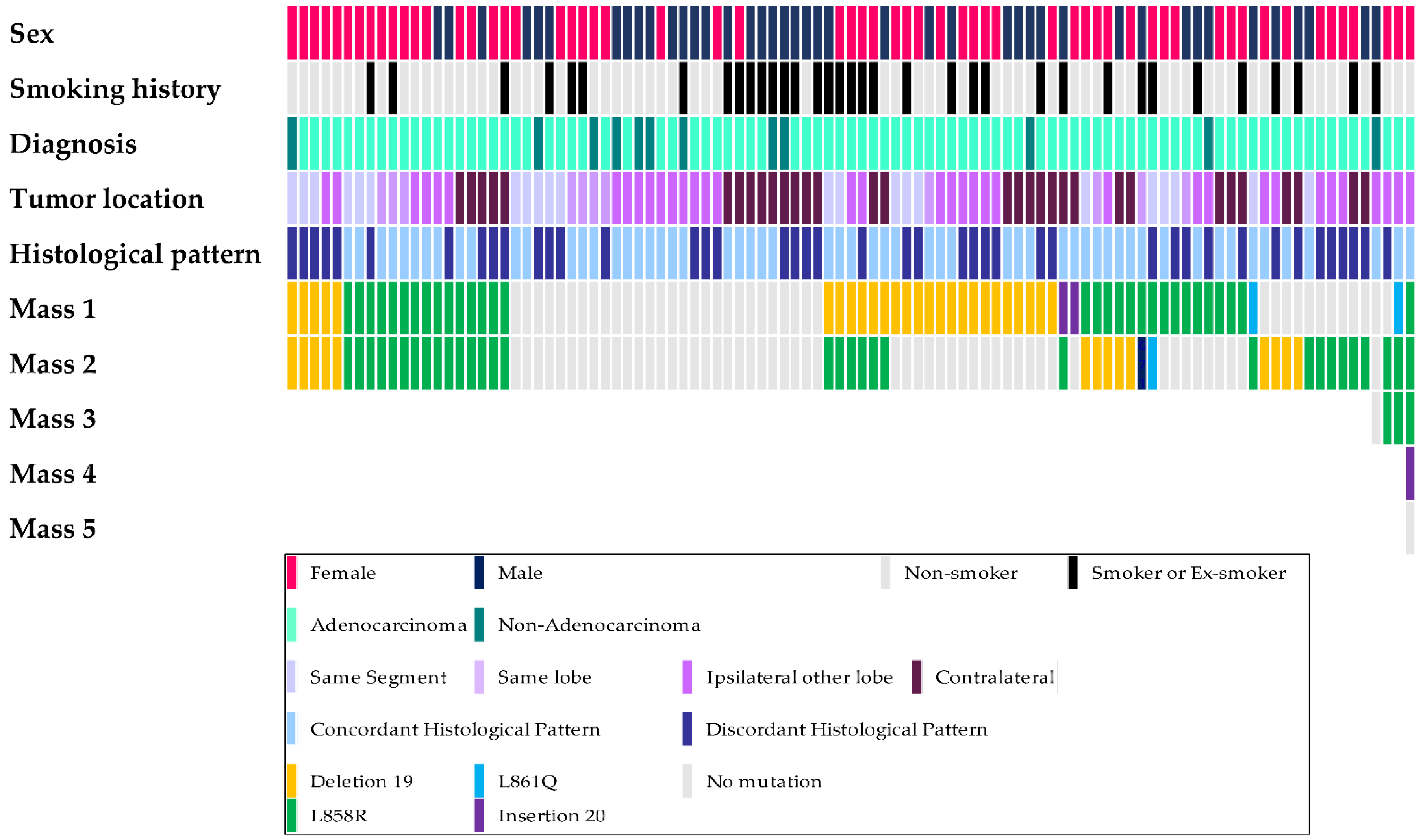
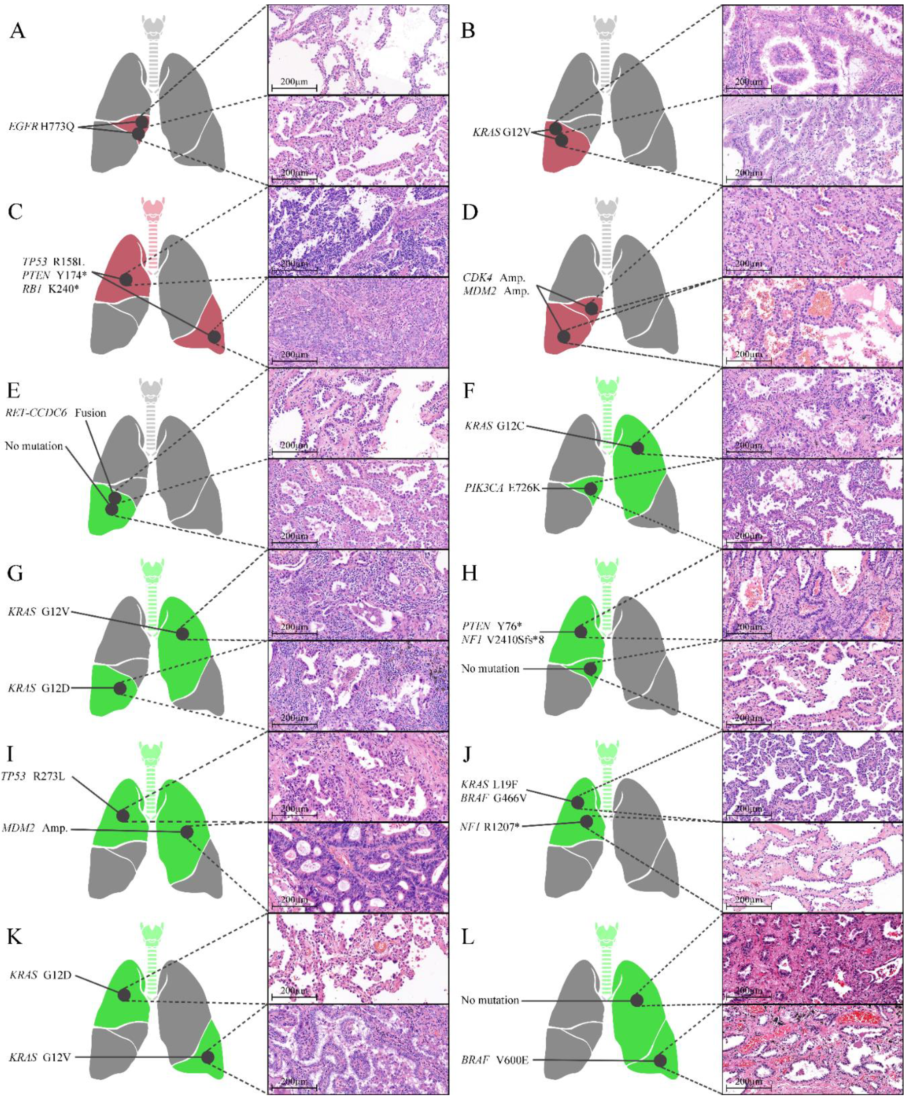
3.2. EGFR Mutation Status of Tumors

Table 2 and Table S2 summarize the analysis of the EGFR mutation status and clinicopathological parameters. The incidence of EGFR mutations was high in women (p < 0.001) and never-smokers (p < 0.001). The duration of smoking was significantly low in patients with EGFR-mutant tumors who were ex-smokers or current smokers (p < 0.001). Among patients having at least one EGFR-mutant tumor, the demographic parameters were not significantly different between patients having all EGFR-mutant tumors and those having an EGFR-mutant tumor and EGFR-wildtype tumor. Additionally, the frequency of EGFR mutations was high in cancers occurring in the upper portion of the lung (right upper lobe, right middle lobe, or left upper lobe) (p = 0.002).
Comprehensive histopathological and molecular evaluation (Figure S5) revealed that EGFR mutations were frequent in adenocarcinomas (p < 0.001) with a predominant acinar pattern (p < 0.001). In contrast, adenocarcinomas with a predominant lepidic pattern were associated with significantly less frequency of EGFR mutations (p = 0.017).

3.3. Concordance of EGFR Mutation Status

In this study, 52 patients (51.5%) had different EGFR mutation statuses (16 patients had different mutations, and 36 patients had at least one EGFR-wildtype tumor), 20 patients had the same EGFR mutations (19.8%), and 29 patients (28.7%) had only EGFR-wildtype tumors. The clinicopathological data and EGFR mutation status are schematized in Figure 1. The most frequently detected EGFR mutation among the patients with the same EGFR mutation was L858R (detected in 15 patients), followed by an exon 19 deletion (detected in 5 patients). The results of comparative analysis of clinicopathological parameters between patients with the same mutation and those with different mutations among patients with EGFR-mutant tumors only are summarized in Table 3. Lymph node metastasis was detected in 5 patients with the same EGFR mutation, although this was not significant (p = 0.053). Other clinicopathological parameters were not significantly different.
As EGFR-wildtype NSCLC can exhibit various molecular alterations, all EGFR-wildtypes in the same patient may not exhibit the same molecular profile. For an accurate comparison, 24 EGFR tumors from 12 patients with EGFR-wildtype tumors only, and 9 EGFR-wildtype tumors from 9 patients with both EGFR-mutant and EGFR-wildtype tumors were randomly selected and subjected to NGS analysis. NGS analysis of 33 EGFR-wildtype tumors revealed two rare EGFR mutations (H773Q in two tumors from one patient and A289V in one tumor from other patient), which could not be detected using the cobas® EGFR mutation test v2. The location, histological diagnosis, and molecular alterations of each tumor for 12 pairs of EGFR-wildtype tumors are shown in Figure 2. The results of NGS analysis of tumors from nine patients who had at least one EGFR-wildtype tumor are summarized in Table S3.
We excluded 17 patients with all EGFR-wildtype tumors and who were not subjected to NGS analysis. The clinicopathological parameters between molecular-concordant patients and molecular-discordant patients were comparatively analyzed (Table S4). The discordance rate was 71.4% (60/84). Additionally, the parameters were not significantly different between the molecular-concordant and molecular-discordant patients. Furthermore, the Martini–Melamed criteria [11], ACCP guidelines [12,13], and comprehensive histological assessment proposed by Girard et al. [14] were applied to evaluate the clinicopathological criteria for determining MPLC and IPM. The discriminatory power of these parameters was not significantly different between the concordant and discordant groups.

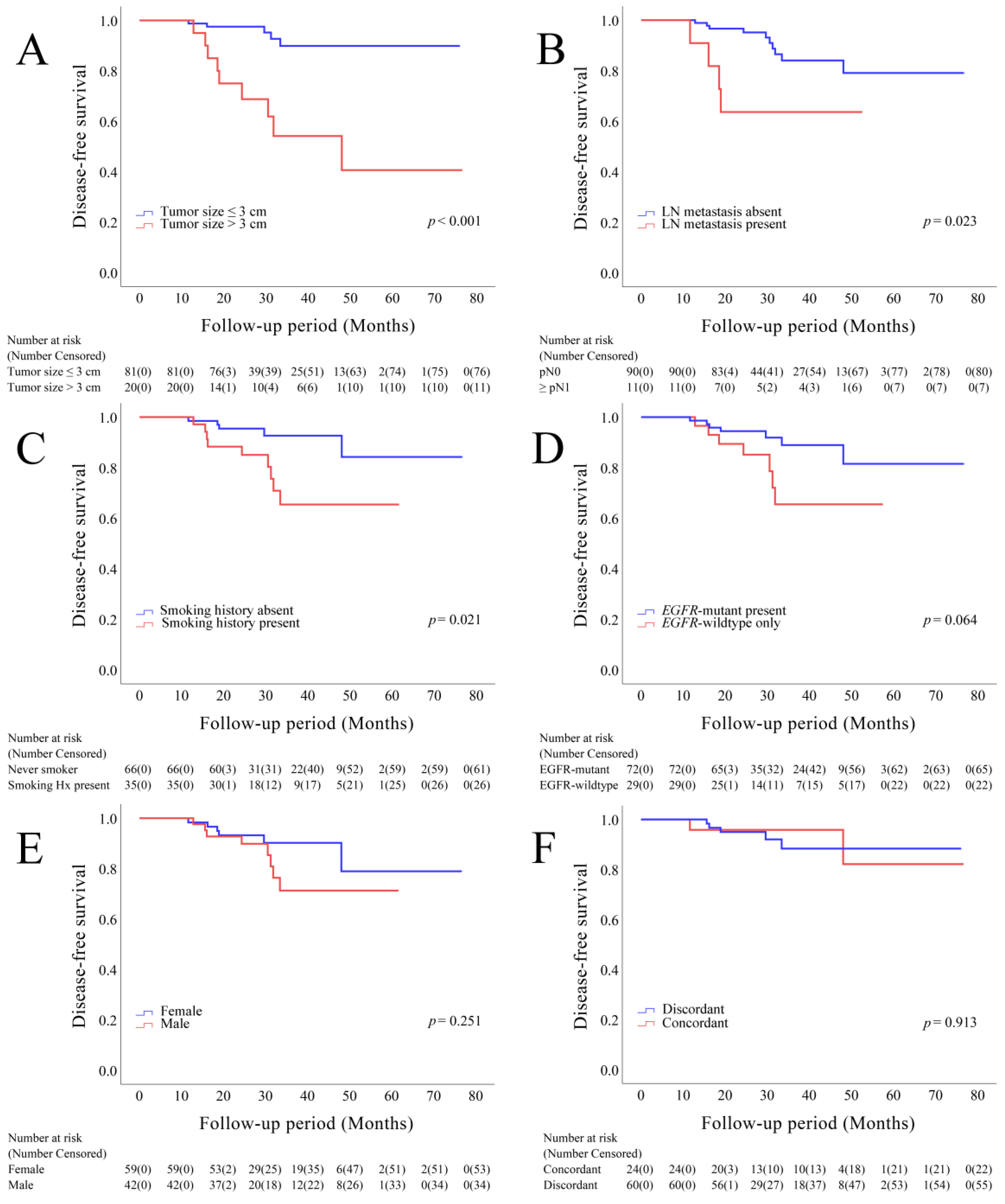
3.4. Survival Analysis

The mean follow-up period was 34.49 months (range, 13.70–76.60 months; median, 30.30 months). During the follow-up period, tumor recurrence was observed in 14 (13.9%) patients, and 4 (4.0%) patients died. The presence of tumors with a size of >3 cm, lymph node metastasis, and smoking history were negatively correlated with disease-free survival (DFS) (Figure 3A–C). The presence of an EGFR-mutant tumor was negatively correlated with DFS, although this correlation was not significant (Figure 3D). Sex and the concordance of molecular alterations of multiple cancers did not influence DFS (Figure 3E–F).

4. Discussion

In this study, 72 (71.3%) of the 101 patients with multiple lung cancers had EGFR mutations in at least one tumor. Consistent with the previous findings, EGFR mutations were frequently detected in women (51/72, 69.4%) and never-smokers (58/72, 80.6%) [22,23,24]. Additionally, patients with EGFR-mutant/wildtype tumors exhibited similar characteristics to patients with EGFR-mutant/mutant tumors when compared with patients with EGFR-wildtype/wildtype tumors. The demographic parameters were not significantly different between patients with EGFR-mutant/wildtype tumors and those with EGFR-mutant/mutant tumors. These findings suggest that patients with multiple lung cancers, at least one EGFR, and those with multiple lung cancers with all EGFR-wildtype tumors had exhibited different demographic characteristics.
EGFR mutation was frequently detected in tumors located in the upper portion of the lung (79/113; 69.9%) [25] and was common in adenocarcinoma (111/113; 98.2%), especially in tumors with an acinar-predominant pattern (85/113; 76.6%) [26]. In particular, EGFR mutations were less frequently detected in lepidic-predominant tumors (13/113; 11.7%). Among the lepidic-predominant tumors included in this study, the proportion of less progressed cancers, including MIA, was high. This finding was consistent with the hypothesis that EGFR mutations were acquired during the development of invasive adenocarcinoma from pre-invasive lesions, such as AAH, and that they accelerated the progression of tumors rather than initiating tumorigenesis [27]. Additionally, EGFR mutations were acquired independently in precancerous lesions with multifocal presentations [9], even though the same patient exhibited different EGFR statuses in individual tumors.
This study demonstrated that more than 50% of patients had tumors with different EGFR mutation statuses. After excluding patients with EGFR-wildtype/wildtype tumors who exhibited demographically diverse characteristics when compared with other patients, the rate of discordance was 72%. The number of patients with both EGFR-mutant and EGFR-wildtype tumors was 36 (36/72; 50%). Therefore, subjecting only one cancerous lesion to EGFR analysis cannot conclusively determine the EGFR mutation status in the remaining tumors.
The pathogenic mechanism of multiple lung cancers was explained through the development of metachronous or synchronous tumors in the squamous epithelium of the oral cavity of smokers (‘field cancerization’) [6,28,29]. However, never-smokers account for approximately 25% of lung cancer cases [30]. History of tobacco usage cannot explain the occurrence of cancer in these patients. Previous studies have reported that 70–76% of patients with lung cancer in Korea have a history of smoking [31,32]. In contrast, 65.3% of patients with lung cancer were never-smokers in this study. Recently, the incidence of lung cancer has been increasing in never-smokers [33]. Adenocarcinoma accounts for 40.3–91.3% of all synchronous MPLC cases [5,18,19]. The study cohort comprised a high proportion of Asian female patients with MPLC who were never-smokers [24]. Thus, these findings indicate the importance of screening programs in the never-smoker subgroup [34].
Various studies have examined the pathogenesis and driver mutation of lung cancer in never-smokers. The most common driver mutation of lung cancer in East Asian never-smokers is EGFR mutation [24,35,36,37]. Determining the timing of EGFR mutations during carcinogenesis can predict whether multiple lung cancers share the same EGFR mutation or have different EGFR mutations. The development of diagnostic imaging technology has enabled the detection of multiple ground-glass opacities (GGOs) and increased the detection rate of synchronous lung cancer. In particular, TNM classification [38] can distinguish between MPLC and IPM. Therefore, determining if multiple lung cancers develop from MPLC or IPM is critical for accurate diagnosis and the determination of optimal treatment plans.
In the last 35 years, several studies have aimed to accurately diagnose multiple lung cancers [11,12,13,14]. However, differentiating MPLC or IPM based on histological features has been challenging, owing to the heterogeneous histological features of lung cancer. Consistently, differentiating between MPLC and IPM based only on histological features and clinical findings was challenging in this study. The results of this study suggested that the rate of the discordant histological pattern in patients with EGFR-mutant/wildtype tumors was higher than that in patients with EGFR-mutant/mutant tumors. These findings can be attributed to the differences in histological features between EGFR-mutant tumors and EGFR-wildtype tumors [24,39]. The concordance of the histological patterns was not significantly different between the molecular-concordant and the molecular-discordant groups. In particular, the discriminatory power of the currently used criteria to distinguish MPLC and IPM was not significantly different between the two groups. Thus, distinguishing tumors based only on clinicopathological findings without molecular findings is a major hurdle. Therefore, a combined histomolecular approach [16] must be utilized for differentiating MPLC and IPM in real-world clinical settings. Additionally, individual tumors of multiple lung cancers must be subjected to molecular analysis.
To evaluate the multiple lung tumors using the combined histomolecular approach, molecular markers other than EGFR mutations must be identified. Although the cost effectiveness of NGS is superior compared to the quantity of information provided by NGS, and the turnaround time is reduced due to the development of technology [40,41], the implementation of NGS in all patients with NSCLC, including those with early-stage tumors, is difficult in real-world clinical settings. The administration of adjuvant EGFR-TKI increased DFS in patients with resected EGFR-mutant NSCLCs [42,43]. Hence, EGFR analysis in all NSCLC lesions is critical for the management of patients with NSCLC in real-world clinical settings. Analysis for other molecular markers or NGS of all NSCLC lesions should be carefully deliberated and validated considering the cost-effectiveness and therapeutic benefits to the patients. Future studies must establish comprehensive histomolecular criteria that can be used as a gold standard in real-world clinical settings.
In patients with multiple lung cancers who underwent surgery for major lesions harboring EGFR mutations, postoperative EGFR-TKI treatment for unresected GGO lesions decreased the size of residual GGOs, especially in patients with large or residual GGOs or advanced stage tumors. However, the size of residual GGOs in most patients was unchanged [44]. In our study, only 50% of patients with EGFR-mutant tumors had another EGFR-mutant tumor. These findings suggest the importance of determining the mutation status of each residual lesion when considering adjuvant EGFR-TKI treatment for patients with multiple lung cancers.
Advances in diagnostic technology have provided various methods to detect EGFR mutations, including circulating tumor DNA (ctDNA) analysis (also called liquid biopsy analysis) [45]. Liquid biopsy can confirm the EGFR mutation status without the need for a surgical resection of the tumor, which is useful for analyzing EGFR mutations in unresectable MPLC cases. However, some studies including our previous study have reported that ctDNA analysis detects EGFR mutations in only 54–57% of advanced NSCLC cases with EGFR mutations, which was confirmed using tissue DNA analysis [46,47]. Additionally, the sensitivity of ctDNA analysis to detect EGFR mutations is low in localized NSCLC [48]. Therefore, the probability of the presence of EGFR mutations should be considered even when liquid biopsy analysis does not detect EGFR mutations in MPLC. EGFR mutation analysis must be performed using all obtainable tumor tissues of MPLC.
This study has some limitations. Only data from patients with multiple lung cancers at the operable stage were examined in this study. Hence, the pathological and clinical stages of lung cancer were relatively low. Consequently, the concordance of EGFR mutations in advanced-stage multiple lung cancers has not been verified. Additionally, the follow-up period was not sufficient. In this study, the concordance of EGFR mutations did not affect the prognosis; however, the follow-up period was not sufficient to determine whether concordant EGFR mutations indicate metastasis or tumors that share the same EGFR mutation. These limitations must be addressed in future studies.

5. Conclusions

This study revealed the independent occurrence of EGFR mutations in patients with multifocal lung cancers. The discordance of EGFR mutations in individual tumors was observed in more than half of the patients. Additionally, histological or clinical findings did not successfully predict the presence of concordance. Therefore, the presence of different EGFR mutations in multiple lung cancers with the same or different histological characteristics must be considered, except when they are highly predicted to share the same molecular profiles, such as those of IPM.

Supplementary Materials

The following are available online at https://www.mdpi.com/article/10.3390/cancers14123011/s1. Figure S1: Algorithm for determining the nature of multifocal lung cancer based on the Martini and Melamed criteria. Figure S2: Algorithm for determining the nature of multifocal lung cancer based on the definition of satellite nodules, multifocal primary lung cancer, and metastasis proposed by the American College of Chest Physicians. Figure S3: Algorithm for determining the nature of multifocal lung cancer based on the comprehensive histological assessment methodology proposed by Girard et al. Figure S4: Workflow for patient selection. Figure S5: Examples of histological features and radiological findings of adenocarcinomas with different (A–C) and same histological characteristics (D–F). Table S1: List of detectable EGFR mutations using cobas® EGFR mutation test v2. Table S2: Comparison of clinicopathological parameters between EGFR-wildtype tumors and EGFR-mutant tumors. Table S3: Molecular alterations analyzed using next-generation sequencing and cobas® EGFR mutation test v2 in patients with EGFR-mutant/wildtype tumors. Table S4: Comparison of clinicopathological parameters between patients with molecular-concordant and molecular-discordant tumors.

Author Contributions

Conceptualization, H.L. and Y.-L.C.; methodology, H.L., J.H.P., Y.-L.C. and W.-S.K.; software, H.L., Y.-L.C. and W.-S.K.; validation, H.L. and Y.-L.C.; formal analysis, H.L., Y.-L.C. and W.-S.K.; investigation, H.L., Y.-L.C. and W.-S.K.; resources, H.L., J.H.P., J.H., Y.M.S., J.K., Y.S.C., H.K.K., J.H.C., Y.-L.C. and W.-S.K.; data curation, H.L.; writing—original draft preparation, H.L. and Y.-L.C.; writing—review and editing, Y.-L.C. and W.-S.K.; visualization, H.L.; supervision, Y.-L.C. and W.-S.K.; project administration, H.L., Y.-L.C. and W.-S.K.; funding acquisition, Y.-L.C. and W.-S.K. All authors have read and agreed to the published version of the manuscript.

Funding

This work was supported by the National Research Foundation of Korea (NRF) grants funded by the Korean government (Ministry of Science, ICT, and Future Planning) (2022R1A2C2006322 and NRF: 2016R1A5A2945889).

Institutional Review Board Statement

This study was approved by the IRB of Samsung Medical Center (IRB No. 2020-04-071-001, 2022-01-009-001).

Informed Consent Statement

The written informed consent was waived due to anonymous data analysis.

Data Availability Statement

All data and materials are available from Y.-L.C. ([email protected]) upon reasonable request.

Conflicts of Interest

The authors declare no conflict of interest.

References

  1. Siegel, R.L.; Miller, K.D.; Jemal, A. Cancer statistics, 2020. CA Cancer J. Clin. 2020, 70, 7–30. [Google Scholar] [CrossRef]
  2. Nakata, M.; Sawada, S.; Yamashita, M.; Saeki, H.; Kurita, A.; Takashima, S.; Tanemoto, K. Surgical treatments for multiple primary adenocarcinoma of the lung. Ann. Thorac. Surg. 2004, 78, 1194–1199. [Google Scholar] [CrossRef] [PubMed]
  3. Rostad, H.; Strand, T.E.; Naalsund, A.; Norstein, J. Resected synchronous primary malignant lung tumors: A population-based study. Ann. Thorac. Surg. 2008, 85, 204–209. [Google Scholar] [CrossRef] [PubMed]
  4. Pommier, R.F.; Vetto, J.T.; Lee, J.T.; Johnston, K.M. Synchronous non-small cell lung cancers. Am. J. Surg. 1996, 171, 521–524. [Google Scholar] [CrossRef]
  5. Chang, Y.-L.; Wu, C.-T.; Lee, Y.-C. Surgical treatment of synchronous multiple primary lung cancers: Experience of 92 patients. J. Thorac. Cardiovasc. Surg. 2007, 134, 630–637. [Google Scholar] [CrossRef] [PubMed] [Green Version]
  6. Slaughter, D.P.; Southwick, H.W.; Smejkal, W. Field cancerization in oral stratified squamous epithelium; clinical implications of multicentric origin. Cancer 1953, 6, 963–968. [Google Scholar] [CrossRef]
  7. Kitamura, H.; Kameda, Y.; Ito, T.; Hayashi, H. Atypical adenomatous hyperplasia of the lung. Implications for the pathogenesis of peripheral lung adenocarcinoma. Am. J. Clin. Pathol. 1999, 111, 610–622. [Google Scholar] [CrossRef] [Green Version]
  8. Tang, X.; Shigematsu, H.; Bekele, B.N.; Roth, J.A.; Minna, J.D.; Hong, W.K.; Gazdar, A.F.; Wistuba, I.I. EGFR tyrosine kinase domain mutations are detected in histologically normal respiratory epithelium in lung cancer patients. Cancer Res. 2005, 65, 7568–7572. [Google Scholar] [CrossRef] [Green Version]
  9. Ikeda, K.; Nomori, H.; Ohba, Y.; Shibata, H.; Mori, T.; Honda, Y.; Iyama, K.; Kobayashi, T. Epidermal growth factor receptor mutations in multicentric lung adenocarcinomas and atypical adenomatous hyperplasias. J. Thorac. Oncol. 2008, 3, 467–471. [Google Scholar] [CrossRef] [PubMed] [Green Version]
  10. Mok, T.S.; Wu, Y.L.; Thongprasert, S.; Yang, C.H.; Chu, D.T.; Saijo, N.; Sunpaweravong, P.; Han, B.; Margono, B.; Ichinose, Y.; et al. Gefitinib or carboplatin-paclitaxel in pulmonary adenocarcinoma. N. Engl. J. Med. 2009, 361, 947–957. [Google Scholar] [CrossRef] [PubMed]
  11. Martini, N.; Melamed, M.R. Multiple primary lung cancers. J. Thorac. Cardiovasc. Surg. 1975, 70, 606–612. [Google Scholar] [CrossRef]
  12. Alberts, W.M. Diagnosis and management of lung cancer executive summary: ACCP evidence-based clinical practice guidelines (2nd edition). Chest 2007, 132, 1S–19S. [Google Scholar] [CrossRef] [PubMed]
  13. Shen, K.R.; Meyers, B.F.; Larner, J.M.; Jones, D.R. Special treatment issues in lung cancer: ACCP evidence-based clinical practice guidelines (2nd edition). Chest 2007, 132, 290S–305S. [Google Scholar] [CrossRef] [PubMed] [Green Version]
  14. Girard, N.; Deshpande, C.; Lau, C.; Finley, D.; Rusch, V.; Pao, W.; Travis, W.D. Comprehensive histologic assessment helps to differentiate multiple lung primary nonsmall cell carcinomas from metastases. Am. J. Surg. Pathol. 2009, 33, 1752–1764. [Google Scholar] [CrossRef] [Green Version]
  15. Zheng, R.; Shen, Q.; Mardekian, S.; Solomides, C.; Wang, Z.-X.; Evans III, N.R. Molecular profiling of key driver genes improves staging accuracy in multifocal non–small cell lung cancer. J. Thorac. Cardiovasc. Surg. 2020, 160, e71–e79. [Google Scholar] [CrossRef]
  16. Mansuet-Lupo, A.; Barritault, M.; Alifano, M.; Janet-Vendroux, A.; Zarmaev, M.; Biton, J.; Velut, Y.; Le Hay, C.; Cremer, I.; Regnard, J.F.; et al. Proposal for a Combined Histomolecular Algorithm to Distinguish Multiple Primary Adenocarcinomas from Intrapulmonary Metastasis in Patients with Multiple Lung Tumors. J. Thorac. Oncol. 2019, 14, 844–856. [Google Scholar] [CrossRef]
  17. Liu, C.; Liu, C.; Zou, X.; Shao, L.; Sun, Y.; Guo, Y. Next-generation sequencing facilitates differentiating between multiple primary lung cancer and intrapulmonary metastasis: A case series. Diagn. Pathol. 2021, 16, 21. [Google Scholar] [CrossRef]
  18. Wang, H.; Hou, J.; Zhang, G.; Zhang, M.; Li, P.; Yan, X.; Ma, Z. Clinical characteristics and prognostic analysis of multiple primary malignant neoplasms in patients with lung cancer. Cancer Gene Ther. 2019, 26, 419–426. [Google Scholar] [CrossRef]
  19. Ishikawa, Y.; Nakayama, H.; Ito, H.; Yokose, T.; Tsuboi, M.; Nishii, T.; Masuda, M. Surgical Treatment for Synchronous Primary Lung Adenocarcinomas. Ann. Thorac. Surg. 2014, 98, 1983–1988. [Google Scholar] [CrossRef]
  20. Midha, A.; Dearden, S.; McCormack, R. EGFR mutation incidence in non-small-cell lung cancer of adenocarcinoma histology: A systematic review and global map by ethnicity (mutMapII). Am. J. Cancer Res. 2015, 5, 2892–2911. [Google Scholar]
  21. Dearden, S.; Stevens, J.; Wu, Y.L.; Blowers, D. Mutation incidence and coincidence in non small-cell lung cancer: Meta-analyses by ethnicity and histology (mutMap). Ann. Oncol. 2013, 24, 2371–2376. [Google Scholar] [CrossRef] [PubMed]
  22. Kosaka, T.; Yatabe, Y.; Endoh, H.; Kuwano, H.; Takahashi, T.; Mitsudomi, T. Mutations of the epidermal growth factor receptor gene in lung cancer: Biological and clinical implications. Cancer Res. 2004, 64, 8919–8923. [Google Scholar] [CrossRef] [PubMed] [Green Version]
  23. Shigematsu, H.; Lin, L.; Takahashi, T.; Nomura, M.; Suzuki, M.; Wistuba, I.I.; Fong, K.M.; Lee, H.; Toyooka, S.; Shimizu, N.; et al. Clinical and biological features associated with epidermal growth factor receptor gene mutations in lung cancers. J. Natl. Cancer Inst. 2005, 97, 339–346. [Google Scholar] [CrossRef] [PubMed] [Green Version]
  24. Ha, S.Y.; Choi, S.-J.; Cho, J.H.; Choi, H.J.; Lee, J.; Jung, K.; Irwin, D.; Liu, X.; Lira, M.E.; Mao, M.; et al. Lung cancer in never-smoker Asian females is driven by oncogenic mutations, most often involving EGFR. Oncotarget 2015, 6, 5465–5474. [Google Scholar] [CrossRef] [PubMed] [Green Version]
  25. Tseng, C.H.; Chen, K.C.; Hsu, K.H.; Tseng, J.S.; Ho, C.C.; Hsia, T.C.; Su, K.Y.; Wu, M.F.; Chiu, K.L.; Liu, C.M.; et al. EGFR mutation and lobar location of lung adenocarcinoma. Carcinogenesis 2016, 37, 157–162. [Google Scholar] [CrossRef] [PubMed] [Green Version]
  26. Zhang, Y.; Sun, Y.; Pan, Y.; Li, C.; Shen, L.; Li, Y.; Luo, X.; Ye, T.; Wang, R.; Hu, H.; et al. Frequency of driver mutations in lung adenocarcinoma from female never-smokers varies with histologic subtypes and age at diagnosis. Clin. Cancer Res. 2012, 18, 1947–1953. [Google Scholar] [CrossRef] [Green Version]
  27. Yoshida, Y.; Shibata, T.; Kokubu, A.; Tsuta, K.; Matsuno, Y.; Kanai, Y.; Asamura, H.; Tsuchiya, R.; Hirohashi, S. Mutations of the epidermal growth factor receptor gene in atypical adenomatous hyperplasia and bronchioloalveolar carcinoma of the lung. Lung Cancer 2005, 50, 1–8. [Google Scholar] [CrossRef]
  28. Gazdar, A.F.; Minna, J.D. Multifocal lung cancers—Clonality vs field cancerization and does it matter? J. Natl. Cancer Inst. 2009, 101, 541–543. [Google Scholar] [CrossRef] [Green Version]
  29. Strong, M.S.; Incze, J.; Vaughan, C.W. Field cancerization in the aerodigestive tract--its etiology, manifestation, and significance. J. Otolaryngol. 1984, 13, 1–6. [Google Scholar]
  30. Sun, S.; Schiller, J.H.; Gazdar, A.F. Lung cancer in never smokers—A different disease. Nat. Rev. Cancer 2007, 7, 778–790. [Google Scholar] [CrossRef]
  31. Park, J.Y.; Jang, S.H. Epidemiology of Lung Cancer in Korea: Recent Trends. Tuberc. Respir. Dis. 2016, 79, 58–69. [Google Scholar] [CrossRef] [PubMed]
  32. Kim, Y.-C.; Won, Y.-J. The Development of the Korean Lung Cancer Registry (KALC-R). Tuberc. Respir. Dis. 2019, 82, 91–93. [Google Scholar] [CrossRef]
  33. Kang, H.; Park, C.-W.; Kim, W.; Song, S.-Y.; Na, K.-J.; Jeong, J.U.; Yoon, M.S.; Ahn, S.-J.; Choi, Y.-D.; Choi, C.; et al. Never-Smoker Lung Cancer Is Increasing. J. Lung Cancer 2012, 11, 89–93. [Google Scholar] [CrossRef] [Green Version]
  34. Kerpel-Fronius, A.; Tammemägi, M.; Cavic, M.; Henschke, C.; Jiang, L.; Kazerooni, E.; Lee, C.T.; Ventura, L.; Yang, D.; Lam, S.; et al. Screening for Lung Cancer in Individuals Who Never Smoked: An International Association for the Study of Lung Cancer Early Detection and Screening Committee Report. J. Thorac. Oncol. 2022, 17, 56–66. [Google Scholar] [CrossRef]
  35. Zhou, F.; Zhou, C. Lung cancer in never smokers-the East Asian experience. Transl. Lung Cancer Res. 2018, 7, 450–463. [Google Scholar] [CrossRef]
  36. Sun, Y.; Ren, Y.; Fang, Z.; Li, C.; Fang, R.; Gao, B.; Han, X.; Tian, W.; Pao, W.; Chen, H.; et al. Lung adenocarcinoma from East Asian never-smokers is a disease largely defined by targetable oncogenic mutant kinases. J. Clin. Oncol. 2010, 28, 4616–4620. [Google Scholar] [CrossRef] [Green Version]
  37. Li, S.; Choi, Y.L.; Gong, Z.; Liu, X.; Lira, M.; Kan, Z.; Oh, E.; Wang, J.; Ting, J.C.; Ye, X.; et al. Comprehensive Characterization of Oncogenic Drivers in Asian Lung Adenocarcinoma. J. Thorac. Oncol. 2016, 11, 2129–2140. [Google Scholar] [CrossRef] [Green Version]
  38. Amin, M.B.; Edge, S.B.; Greene, F.L.; Byrd, D.R.; Brookland, R.K.; Washington, M.K.; Gershenwald, J.E.; Compton, C.C.; Hess, K.R.; Sullivan, D.C.; et al. The AJCC Cancer Staging Manual, 8th ed.; Springer: New York, NY, USA, 2017; pp. 431–456. [Google Scholar]
  39. Sun, P.L.; Seol, H.; Lee, H.J.; Yoo, S.B.; Kim, H.; Xu, X.; Jheon, S.; Lee, C.T.; Lee, J.S.; Chung, J.H. High incidence of EGFR mutations in Korean men smokers with no intratumoral heterogeneity of lung adenocarcinomas: Correlation with histologic subtypes, EGFR/TTF-1 expressions, and clinical features. J. Thorac. Oncol. 2012, 7, 323–330. [Google Scholar] [CrossRef] [Green Version]
  40. De Alava, E.; Pareja, M.J.; Carcedo, D.; Arrabal, N.; García, J.-F.; Bernabé-Caro, R. Cost-effectiveness analysis of molecular diagnosis by next-generation sequencing versus sequential single testing in metastatic non-small cell lung cancer patients from a south Spanish hospital perspective. Expert Rev. Pharmacoecon. Outcomes Res. 2022, 1–10. [Google Scholar] [CrossRef]
  41. Christofyllakis, K.; Bittenbring, J.T.; Thurner, L.; Ahlgrimm, M.; Stilgenbauer, S.; Bewarder, M.; Kaddu-Mulindwa, D. Cost-effectiveness of precision cancer medicine-current challenges in the use of next generation sequencing for comprehensive tumour genomic profiling and the role of clinical utility frameworks. Mol. Clin. Oncol. 2022, 16, 21. [Google Scholar] [CrossRef]
  42. Chen, R.L.; Sun, L.L.; Cao, Y.; Chen, H.R.; Zhou, J.X.; Gu, C.Y.; Zhang, Y.; Wang, S.Y.; Hou, W.; Lin, L.Z. Adjuvant EGFR-TKIs for Patients With Resected EGFR-Mutant Non-Small Cell Lung Cancer: A Meta-Analysis of 1283 Patients. Front. Oncol. 2021, 11, 629394. [Google Scholar] [CrossRef]
  43. Cheng, H.; Li, X.J.; Wang, X.J.; Chen, Z.W.; Wang, R.Q.; Zhong, H.C.; Wu, T.C.; Cao, Q.D. A meta-analysis of adjuvant EGFR-TKIs for patients with resected non-small cell lung cancer. Lung Cancer 2019, 137, 7–13. [Google Scholar] [CrossRef]
  44. Cheng, B.; Li, C.; Zhao, Y.; Li, J.; Xiong, S.; Liang, H.; Liu, Z.; Zeng, W.; Liang, W.; He, J. The impact of postoperative EGFR-TKIs treatment on residual GGO lesions after resection for lung cancer. Signal Transduct. Target. Ther. 2021, 6, 73. [Google Scholar] [CrossRef]
  45. Fenizia, F.; De Luca, A.; Pasquale, R.; Sacco, A.; Forgione, L.; Lambiase, M.; Iannaccone, A.; Chicchinelli, N.; Franco, R.; Rossi, A. EGFR mutations in lung cancer: From tissue testing to liquid biopsy. Future Oncol. 2015, 11, 1611–1623. [Google Scholar] [CrossRef]
  46. Lee, H.; Han, J.; Choi, Y.L. Real-World Analysis of the EGFR Mutation Test in Tissue and Plasma Samples from Non-Small Cell Lung Cancer. Diagnostics 2021, 11, 1695. [Google Scholar] [CrossRef]
  47. Liang, X.; Zhang, W.; Li, J.; Zhu, J.; Shao, J.; Wang, J.; Wu, H.; Dai, J.; Li, J.; Xu, J.; et al. Clinical implications of ctDNA for EGFR-TKIs as first-line treatment in NSCLC. J. Cancer. Res. Clin. Oncol. 2022. [Google Scholar] [CrossRef]
  48. Szpechcinski, A.; Bryl, M.; Wojcik, P.; Czyzewicz, G.; Wojda, E.; Rudzinski, P.; Duk, K.; Moes-Sosnowska, J.; Maszkowska-Kopij, K.; Langfort, R.; et al. Detection of EGFR mutations in liquid biopsy samples using allele-specific quantitative PCR: A comparative real-world evaluation of two popular diagnostic systems. Adv. Med. Sci. 2021, 66, 336–342. [Google Scholar] [CrossRef]
Figure 1. Schematic demonstration of the clinicopathological parameters and EGFR mutation.
Cancers 14 03011 g001
Figure 2. Locations, histological characteristics (H&E-stained sections), and molecular alterations of individual tumors of 12 pairs of EGFR-wildtype tumors. Four tumors (AD) shared the same molecular alterations (red), while eight tumors (EL) exhibited different molecular alterations (green). * stop-gain mutation.
Cancers 14 03011 g002
Figure 3. Kaplan–Meier survival curves depicting disease-free survival, stratified based on the size of the largest tumor (A), lymph node metastasis (B), smoking history (C), EGFR status (D), sex (E), and concordance of molecular alterations in individual tumors (F).
Cancers 14 03011 g003
Table 1. Demographic characteristics of patients.
ParametersNumber of Patients (%)
Age (years; mean ± SD)65.89 ± 8.30
<6546 (45.6)
≥6555 (54.5)
Sex
Male42 (41.6)
Female59 (58.4)
Tumor location
Unilateral side, single lobe33 (32.7)
Same segment22 (21.8)
Multiple segments11 (10.9)
Unilateral side, multiple lobes33 (32.7)
Bilateral side35 (34.6)
Number of masses
297 (96.0)
33 (2.9)
51 (0.1)
Largest tumor size
≤3 cm81 (80.2)
>3 cm20 (19.8)
Lymph node metastasis
Absent90 (89.1)
Present11 (10.9)
Synchronous vs. Metachronous
Synchronous92 (91.1)
Metachronous9 (8.9)
Smoking history
Absent66 (65.3)
Present35 (34.7)
Present smoking status
Never-smoker66 (65.3)
Ex-smoker20 (19.8)
Current smoker15 (14.9)
Patient status
Alive97 (96.0)
Dead4 (4.0)
Abbreviation: SD, standard deviation
Table 2. Comparison of demographic parameters between patients with EGFR-wildtype tumors and EGFR-mutant tumors.
ParametersEGFR-Mutant (n = 72)p-ValueEGFR-Wildtype (n = 29)p-Value *
EGFR-
Mutant/Wildtype
(n = 36)
EGFR-
Mutant/Mutant
(n = 36)
Sex
Male13 (36.1)9 (25.0)0.30620 (69.0)<0.001
Female23 (63.9)27 (75.0)9 (31.0)
Age (years; mean ± SD)64.50 ± 7.5465.58 ± 8.300.56468.00 ± 9.040.130
<6519 (52.8)16 (44.4)0.47911 (37.9)0.652
≥6517 (47.2)20 (55.6)18 (62.1)
Smoking history
Absent28 (77.8)30 (83.3)0.5518 (27.6)<0.001
Present8 (22.2)6 (16.7)21 (72.4)
Smoking duration
(pack–years)
22.13 ± 12.3613.00 ± 14.570.24537.81 ± 12.78<0.001
Present smoking status
Never-smoker28 (77.8)30 (83.3)-8 (27.6)<0.001
Ex-smoker5 (13.9)5 (13.9)10 (34.5)
Current smoker3 (8.3)1 (2.8)11 (37.9)
Data are presented as number of patients (%) unless otherwise noted. Abbreviation: SD, standard deviation. * p-value represents differences between the EGFR-mutant group and EGFR-wildtype group. Smoking duration was calculated only in patients with a history of smoking.
Table 3. Comparison of demographic parameters between patients with same EGFR mutation and those with different EGFR mutations in the EGFR-mutant/mutant group.
ParameterSame (n = 20)Different (n = 16)p-Value
Sex 0.146
Male3 (15.0)6 (37.5)
Female17 (85.0)10 (62.5)
Age (years) 0.940
<659 (45.0)7 (43.8)
≥6511 (55.0)9 (56.3)
History of smoking
No18 (90.0)12 (75.0)
Yes2 (10.0)4 (25.0)0.374
Present smoking status
Never-smoker18 (90.0)12 (75.0)
Ex-smoker2 (10.0)3 (18.8)
Current smoker0 (0.0)1 (6.3)0.175
Largest tumor size 0.455
≤3 cm16 (80.0)14 (87.5)
>3 cm4 (20.0)2 (12.5)
Lymph node metastasis 0.053
Absent15 (75.0)16 (100.0)
Present5 (25.0)0 (0.0)
Tumor location
Unilateral side, same lobe9 (45.0)7 (43.8)0.762
Same segment6 (30.0)5 (31.3)
Different segment3 (15.0)2 (12.5)
Unilateral side, different lobe6 (30.0)3 (18.8)
Bilateral side5 (25.0)6 (37.5)
Data are presented as number of patients (%).
Publisher’s Note: MDPI stays neutral with regard to jurisdictional claims in published maps and institutional affiliations.

