Article

Immune Response in Vitamin D Deficient Metastatic Colorectal Cancer Patients: A Player That Should Be Considered for Targeted Vitamin D Supplementation

by
Cristina Morelli
1,†,
Michela Rofei
1,†,
Silvia Riondino
1,
Daniela Fraboni
2,
Francesco Torino
1,
Augusto Orlandi
3,
Manfredi Tesauro
4,
Giovanna Del Vecchio Blanco
5,
Massimo Federici
6,
Hendrik-Tobias Arkenau
7,
Vincenzo Formica
1,*,‡ and
Mario Roselli
1,‡
1
Medical Oncology Unit, Department of Systems Medicine, Tor Vergata University Hospital, 00133 Rome, Italy
2
Department of Biomedicine and Prevention, University of Rome “Tor Vergata”, 00133 Rome, Italy
3
Anatomic Pathology, Department of Biomedicine and Prevention, University of Rome “Tor Vergata”, 00133 Rome, Italy
4
Department of Internal Medicine, University of Rome “Tor Vergata”, 00133 Rome, Italy
5
Gastroenterology Unit, Department of Systems Medicine, University of Rome “Tor Vergata”, 00133 Rome, Italy
6
Department of Systems Medicine, University of Rome “Tor Vergata”, 00133 Rome, Italy
7
Sarah Cannon Research Institute, Cancer Institute, University College London, London W1G 6AD, UK
*
Author to whom correspondence should be addressed.
These authors contributed equally to this work.
Cancers 2022, 14(11), 2594; https://doi.org/10.3390/cancers14112594
Submission received: 25 April 2022 / Revised: 22 May 2022 / Accepted: 23 May 2022 / Published: 24 May 2022
(This article belongs to the Special Issue Targeted Therapies in Colorectal Cancer: What’s New?)

Abstract

:

Simple Summary

In this study, we investigated the role of vitamin D levels and the impact on immune response in patients with mCRC and propose a vitamin D cut-off level. Among several hematological, biochemical and immunological variables, we identified the neutrophils-to-lymphocytes ratio, CD4+ T lymphocytes and B lymphocytes as being closely related to vitamin D status with an impact on survival.

Abstract

Background: Vitamin D deficiency is a poor prognostic factor in metastatic colorectal cancer (mCRC); however, targeted supplementation trials have so far yielded limited results. We investigated clinical-laboratory parameters influencing vitamin D deficiency, with a particular focus on immune response, and the effect on survival. These parameters could help optimize targeted supplementation therapy. Methods: Association of plasma 25-hydroxyvitamin D (25(OH])D) with overall survival (OS) was assessed with the Hazard Ratio Smoothed Curve with Restricted Cubic Splines (HRSC-RCS) and maximally selected rank statistics (MSRS) in mCRC patients who underwent first-line chemotherapy. Several hematobiochemical variables were evaluated as predictors of vitamin D deficiency by means of Least Absolute Shrinkage and Selection Operator (LASSO) analysis. In a patient subset, peripheral lymphocyte subpopulations were also analyzed. Results: One hundred thirty-three mCRC patients were included. The median(m) baseline 25(OH)D was 10.8 ng/mL (range 3–53.4). HRSC-RCS revealed a linear association between 25(OH)D and OS. MSRS found 10 ng/mL as the optimal 25(OH)D cut-off. The median OS for 25(OH)D < 10 (n = 60) vs. > 10 ng/mL (n = 73) was 12.3 and 24.5 months, respectively (p = 0.002). The LASSO analysis identified high neutrophil-to-lymphocyte ratio (NLR > 3.5) as the strongest predictor of vitamin D deficiency (Odds Ratio 3.35, p 0.0009). Moreover, patients with low 25(OH)D levels (< 10 ng/mL) and high NLR (>3.5) had the shortest survival and patients with 25(OH)D >10 ng/mL and NLR <3.5 had the longest: mOS 8.1 and 28.1 months, respectively, HR 3.40 (1.76–6.59), p 0.0004. Besides the significant difference in NLR between 25(OH)D < and > 10 ng/mL patients (mNLR 3.6 vs. 2.9, p 0.03), the lymphocyte subpopulation analysis revealed that vitamin D deficiency was associated with high T- CD4+ (p = 0.04) and low B (p = 0.03) lymphocyte frequency. Conclusions: NLR is a powerful predictor of Vitamin D deficiency and can further help in stratifying prognosis. Vitamin D deficiency was associated with significant variations in peripheral immune cells. We hypothesize that integrated targeted interventions to both vitamin D and immune system would improve the prognosis of mCRC patients.

1. Introduction

Colorectal cancer (CRC) is the second cause of cancer-related death in the United States and in Europe [1,2]. Around 20% of the cases present with stage IV disease at the diagnosis and this is associated with a 5-year survival rate lower than 15% [3]. Although about 20% of cases arises on a hereditary-family basis, more than 50% of the CRCs are caused by potentially modifiable risk factors, such as physical inactivity, cigarettes smoking, poor dietary calcium and fiber intake [4].
The correlation between vitamin D3 (cholecalciferol) and cancer has been extensively explored. Previous in vitro studies on several tumor types, including CRC, have shown that the vitamin D active form, 1α,25-dihydroxyvitamin D3 [1,25(OH)2D3] (calcitriol), is able to promote cell differentiation and inhibit tumor invasion and proliferation upon binding to its receptor VDR [5]. Furthermore, plasma concentration of 25-Hydroxyvitamin D3 (25(OH)D) has been found to correlate with risk of developing colorectal adenoma and adenocarcinoma and lower expression of vitamin D receptor (VDR) has been observed in colorectal adenocarcinoma cells as compared to adjacent normal tissue [6].
Besides a direct effect on cancer cells, vitamin D might also indirectly affect cancer growth by modulating innate and adaptive immunity. Previous genome-wide analyses have explored all the genes regulated by vitamin D in immune cells such as macrophages, monocytes, dendritic cells, B and T cells. Overall vitamin D target genes in these cells have anti-inflammatory and immune tolerance actions as well as anti-tumor immunity effects [7,8,9].
A protective role of vitamin D has been suggested for a number of pathological conditions other than cancer, such as cardiovascular, metabolic and immunologic diseases [10,11,12,13].
Despite the consistent evidence that low plasma 25(OH)D levels represent a risk factor for many diseases and illnesses, randomized trials investigating the preventive and curative role of targeted therapy with oral vitamin D3 administration have been largely disappointing [14,15,16] and doubts have been raised on the utility of the screening for vitamin D deficiency in adults [17].
In colorectal cancer, a correlation between low concentrations of circulating 25(OH)D and increased incidence of CRC has been found in many epidemiological studies [18], and a correlation with impaired prognosis has been demonstrated in patients with advanced CRC [19]. However, no specific cut-off value of 25(OH)D level has been identified to precisely predict risk or prognosis of CRC.
More recently, a randomized study comparing oral targeted therapy with vitamin D3 at two different doses (4000 vs. 400 UI/day) in combination with standard first-line chemotherapy in metastatic CRC (mCRC) found that higher dose vitamin D was associated with a non-significant improvement in progression free survival [15].
The purpose of the present study was to identify a CRC-specific vitamin D cut-off correlated with survival in patients with advanced CRC approaching a standard first-line chemotherapy. Moreover, we tried to identify potential determinants of vitamin D deficiency especially among immune-related variables. Finally, we also attempted to further refine survival in vitamin D deficient patients in order to generate suggestions on integrated targeted approaches for patient outcome improvement.

2. Materials and Methods

The patients under study described in the present paper were part of the population afferent to the study NCT01533740 and the REVERT trial (IRB approval n° 149.20 23 July 2020).
Three hundred forty-three patients with histologically confirmed CRC and measurable metastatic disease treated at the Tor Vergata University Hospital of Rome, Italy, with a standard first-line regimen between December 2010 and April 2021 were retrospectively evaluated. First-line regimens administered were either FOLFIRI (fluorouracil/irinotecan) or FOLFOX (fluorouracil/oxaliplatin) plus either an anti-epidermal growth factor (EGFR) agent (panitumumab or cetuximab) or an anti-vascular endothelial growth factor (VEGF) agent (bevacizumab) according to the RAS/BRAF mutational status (wild-type or mutant, respectively). Only patients whose plasma 25(OH)D was measured at baseline (within one week before treatment start) were included in the present analysis.

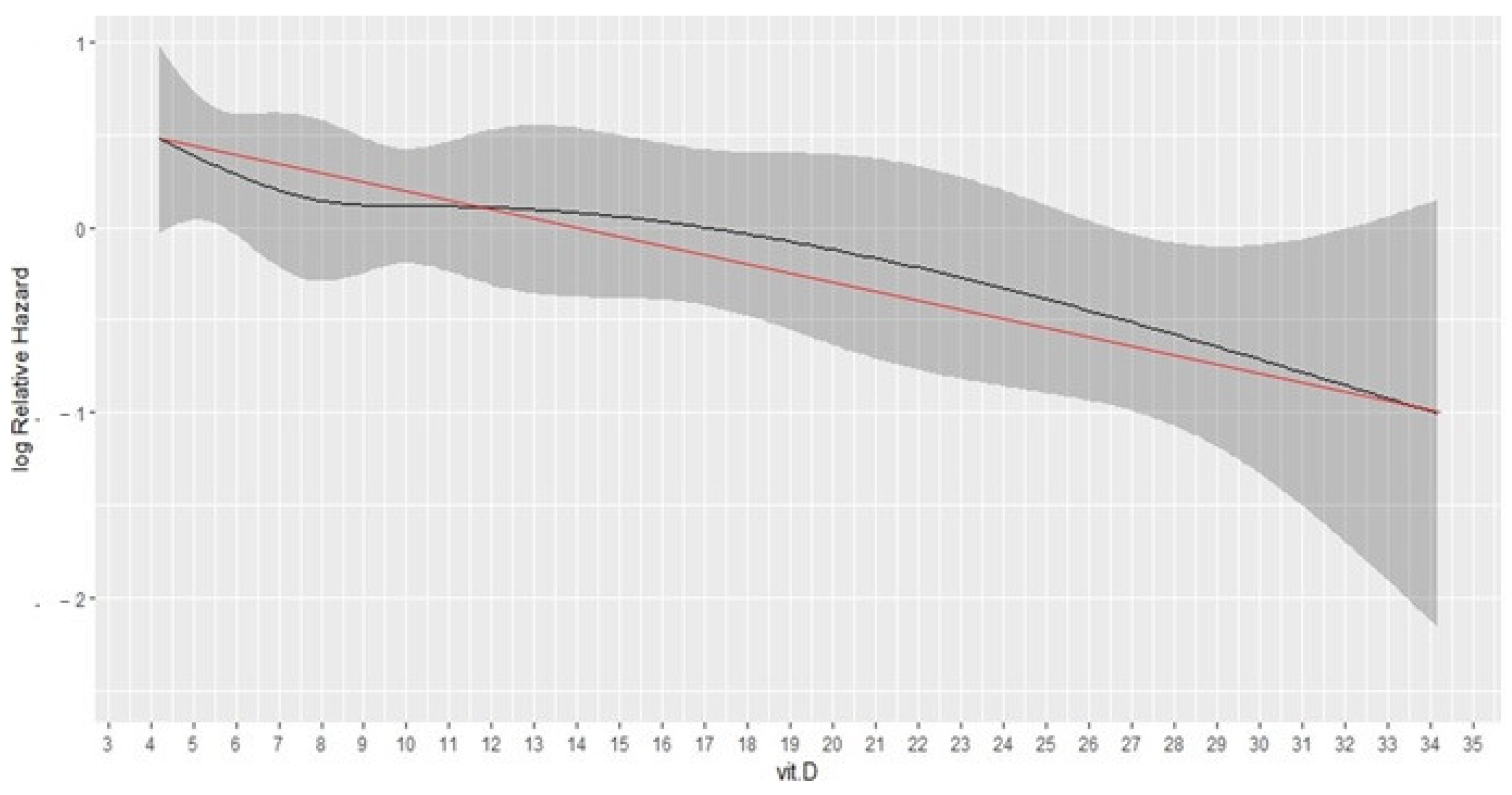
2.1. Optimal Vitamin D Deficiency Cut-Off Identification

First, we attempted to identify the optimal plasma 25(OH)D cut-off value predictive of overall survival (OS) in the study patients. For this purpose, a Hazard Ratio Smoothed curve with Restricted Cubic Splines (HRSC-RCS) [20] with 5 knots was initially used to unravel non-linear correlation between 25(OH)D and OS. Hazard Ratio curves represent the computation of pointwise estimates of the Hazard Ratio for increasing values of a continuous prognostic factor and unravel possible non-linear associations between the factor and the hazard. Then, a maximally selected rank statistics (MSRS) analysis was carried out to define the optimal cut-off value to predict poor survival in vitamin D deficient patients [21]. MSRS allows to identify the cut-point of a continuous variable with the optimal log-rank test when the variable is dichotomized to assess the impact on a survival outcome. Kaplan–Meier and Cox-regression analyses were performed to confirm the survival effect of plasma 25(OH)D, either by using the identified cut-off value or 25(OH)D as continuous variable. Given the importance of KRAS, NRAS and BRAF mutations for the outcome of mCRC patients, a subgroup analysis of the vitamin D survival effect according to the mutational status together with the interaction test was carried out as a meta-analysis and presented as a Forest plot.
After the cut-off value identification, an analysis of candidate predictors of vitamin D deficiency was performed. Thirty-three baseline epidemiologic and biochemical parameters were retrospectively collected: time since diagnosis, primary removal, sidedness, synchronous vs. metachronous metastasis onset, liver vs. not-liver metastasis, rat sarcoma virus (RAS)/BRAF status, Karnofsky Performance status (KPS), age, gender, body mass index (BMI), hemoglobin (g/dL) (Hb), platelets (m3/µL) (PLT), monocytes (m3/µL) (Mono), lymphocytes (m3/µL) (lymph), neutrophils (m3/µL) (Neu), lymph/white blood cells (WBC) (%), Neu/WBC (%), neutrophils-to-lymphocyte ratio (NLR), platelet-to-lymphocytes ratio (PLR), systemic inflammatory index (SII) (plt*neu/lymph), d-dimer (ng/mL) (DD), carcinoembryonic antigen (ng/mL) (CEA), CA 19.9 (U/mL), creatinine (mg/dL) (Cre), alanine aminotransferase (ALT) (U/L), aspartate aminotransferase (AST) (U/L), alkaline phosphatase (UI/L) (ALP), total bilirubin (mg/dL) (BilT), gamma glutamyl transferase (UI/L) (γGT), C-reactive protein (mg/L) (CRP), glycemia (mg/dL) (Glu), albumin (g/dL) (Alb) and lactate dehydrogenase (U/L) (LDH).
Among these, the continuous ones were first optimally dichotomized by means of ROC curve analysis with vitamin D deficiency obtained as classification variable before proceeding with a multivariable logistic regression model.
A multivariable logistic regression analysis with Least Absolute Shrinkage and Selection Operator (LASSO) was performed, including all the 33 covariates described above to identify the one with the highest predictive power for vitamin D deficiency and odds ratio (OR), with relevant confidence intervals being estimated [22]. Distribution of the identified predictor among vitamin D groups was assessed by means of chi square test and Mann–Whitney–Wilcoxon test. Additionally, the impact of the predictor on survival according to vitamin D strata was assessed by means of Kaplan–Meier and Cox-regression analyses.

2.2. Inflammatory/Immune System Status Evaluation

In order to assess the interplay between the immune cell system and vitamin D deficiency, nine peripheral blood immune variables were analyzed in a subset of patients: frequency of CD4+ among lymphocytes (CD4/lymph), CD4+ among WBC (CD4/WBC), CD8+ among lymphocytes (CD8/Lymph), CD8+ among WBC (CD8/WBC), CD4/CD8 ratio, B-lymphocytes among lymphocytes (B/lymph), B-lymphocytes among WBC (B/WBC), NK cells among lymphocytes (NK/lymph) and NK cells among WBC (NK/WBC).
The Mann–Whitney–Wilcoxon test was used to analyze for differences of immune variables between vitamin D deficient and non-deficient patients.
All analyses were performed with the R software v.4.0.3 and MedCalc software version 20.006. All tests were considered statistically significant for two tail p values < 0.05.

3. Results

3.1. Optimal Vitamin D Deficiency Cut-Off Identification

One hundred thirty-three patients were included in the study (60 males and 73 females). Median age was 64 years (range 30–84 years). A total of 47 patients had liver metastases, and 36 had no liver metastases. The median 25(OH)D level was 10.8 ng/mL (range 3–53.4 ng/mL), with 60 deficient (<10 ng/mL), 37 insufficient (10–20 ng/mL) and 36 within normal range (>20 ng/mL) levels, according to the standard cut-offs used for osteoporosis diagnosis [23] (Table S1). Patients were treated with first-line FOLFOX-panitumumab RAS/BRAF wild-type (56%) or FOLFOX-bevacizumab if RAS/BRAF mutated (44%). Ten percent of patients received 400 UI/day of vitamin D supplementation on a regular basis, with no apparent meaningful impact on plasma 25(OH)D levels.
The HRSC-RCS curve revealed an approximately linear association between vitamin D values and risk of death (Figure 1). According to a Cox-regression analysis using plasma 25(OH)D as the continuous variable, 1 unit increase in 25(OH)D was associated to a 4% reduction in the risk of death (HR 0.96, 95%CI 0.93–0.99, p = 0.007).
The maximally selected rank test statistics was used to identify the most prognostic cut-off of 25(OH)D. We found that the cut-off of 10 ng/mL, that is also the cut-off universally used for osteoporosis, was the most significant (Figure S1).
mOS for 25(OH)D < 10 vs. > 10 ng/mL was 12.3 vs. 24.5 months, respectively, HR 2.03 [95% confidence interval (CI) 1.29 to 3.26], p = 0.002 (Figure 2).
No interaction was found between 25(OH)D and tumor mutational status for the effect on survival, with increased risk of death associated to vitamin D deficiency both in RAS/BRAF wild-type tumors (treated with anti-EGFR based first-line therapy) and in RAS or BRAF mutated tumors (treated with anti-VEGF based regimen), HR 2.15 and 1.87, respectively, p for interaction = 0.773 (Figure 3).

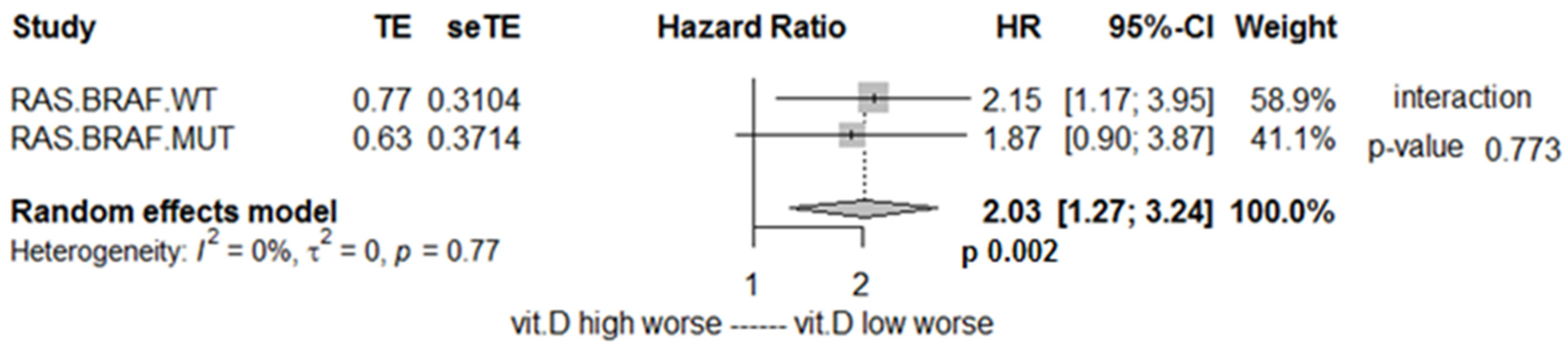
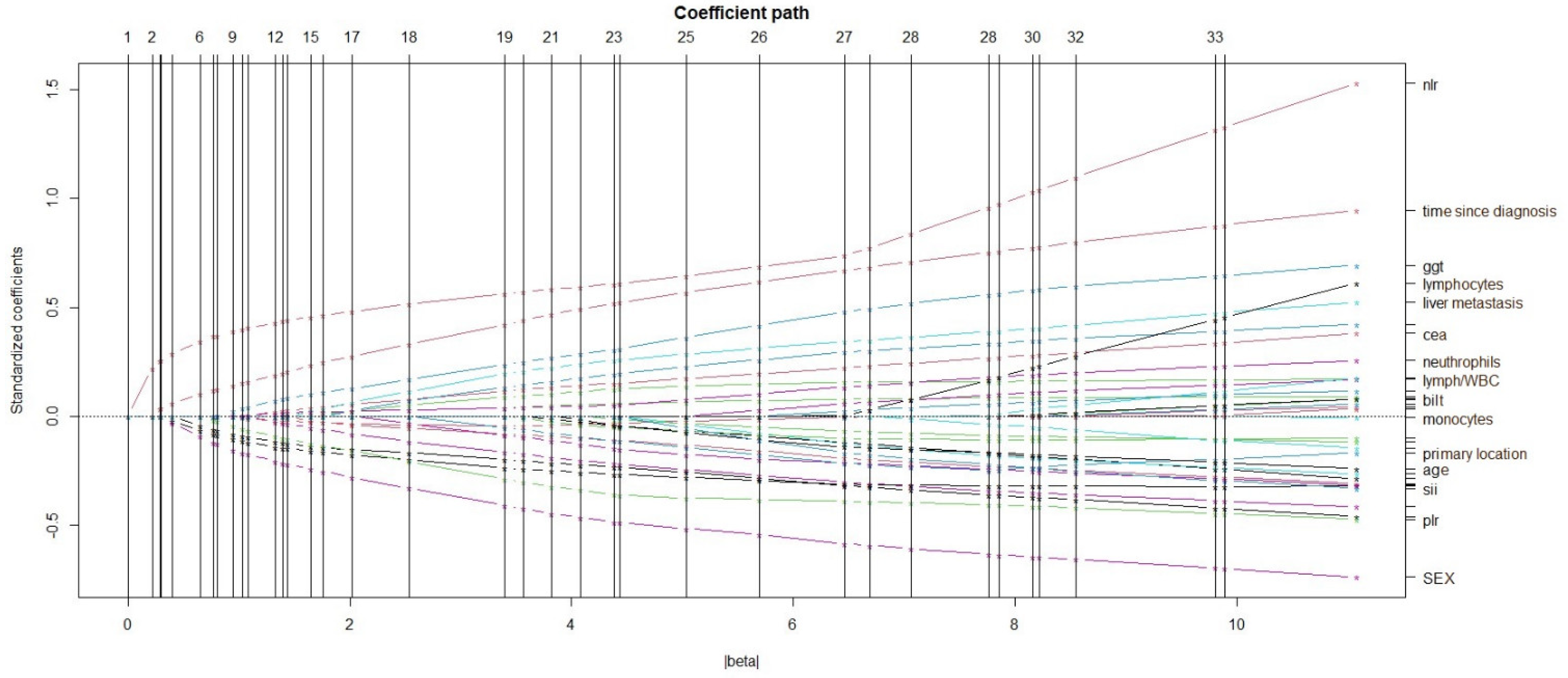
Vitamin D Deficiency Predictors

Thirty-three, clinical and hematochemical variables were investigated as potential predictors of Vitamin D deficiency in a multivariable logistic regression model.
Continuous covariates were conveniently dichotomized, using ROC curve analyses with vitamin D deficiency as classification factor (data not shown). Categories for the 33 covariates were therefore set as follows: time since diagnosis < vs. > 18 months, resection of the primary (yes vs. no), primary location rectum vs. colon, synchronous vs. metachronous metastasis onset, liver vs. not-liver metastasis, RAS/BRAF status (mutant vs. wild-type), KPS > vs. ≤ 80, age < vs. > 60 years, gender male vs. female, BMI < vs. > 20, Hb > vs. <11 g/dL, plt > vs. < 250 m3/µL, Mono > vs. < 0.6 m3/µL, lymph > vs. < 2 m3/µL, Neu > vs. < 5 m3/µL, lymph/WBC < vs. > 20%, Neu/WBC < vs. > 70%, NLR > vs. < 3.5, PLR > vs. < 250, SII > vs. < 1200, DD > vs. < 700 ng/mL, CEA > vs. < 15 ng/mL, CA19.9 > vs. < 15 U/mL, Cre > vs. < 0.6 mg/dL, ALT > vs. < 20 UI/L, AST > vs. < 15 UI/L, ALP > vs. < 80 UI/L, BilT > vs. < 0.7 mg/dL, γGT > vs. < 100 UI/L, CRP > vs. < 30 mg/L, Glu > vs. < 110 mg/dL, Alb > vs. < 3.5 g/dL and LDH > vs. < 200 U/L.
At a multivariate logistic regression with LASSO, NLR > 3.5 was found to be the most powerful predictor of vitamin D deficiency with an OR of 3.35, 95% CI 1.63 to 6.92, p value = 0.0009 (Figure 4).
Proportion of NLR > 3.5 among vitamin D deficient vs. non deficient patients was 56% vs. 27%, chi-square p value = 0.0009 (Figure S2). The median NLR among vitamin D deficient and non-deficient patients was 3.6 vs. 2.9, respectively, Mann–Whitney–Wilcoxon test p value = 0.03.
NLR was also investigated as a prognostic factor in conjunction with 25(OH)D level. NLR was able to further stratify prognosis among patients with vitamin D deficiency: mOS in patients with 25(OH)D < 10 ng/mL for NLR < 3.5 vs. >3.5 was 19.1 vs. 8.1 months, respectively, HR 2.10 (95% CI 1.10 to 3.99), p = 0.02 (Figure 5A).
Survival analysis stratified by NLR and 25(OH)D level demonstrated that patients with NLR > 3.5 plus 25(OH)D < 10 ng/mL and patients with NLR < 3.5 plus 25(OH)D > 10 ng/mL had the shortest and longest mOS, respectively, 8.1 vs. 28.1 months, HR 3.40 (1.76–6.59), p = 0.0004 (Figure 5B).

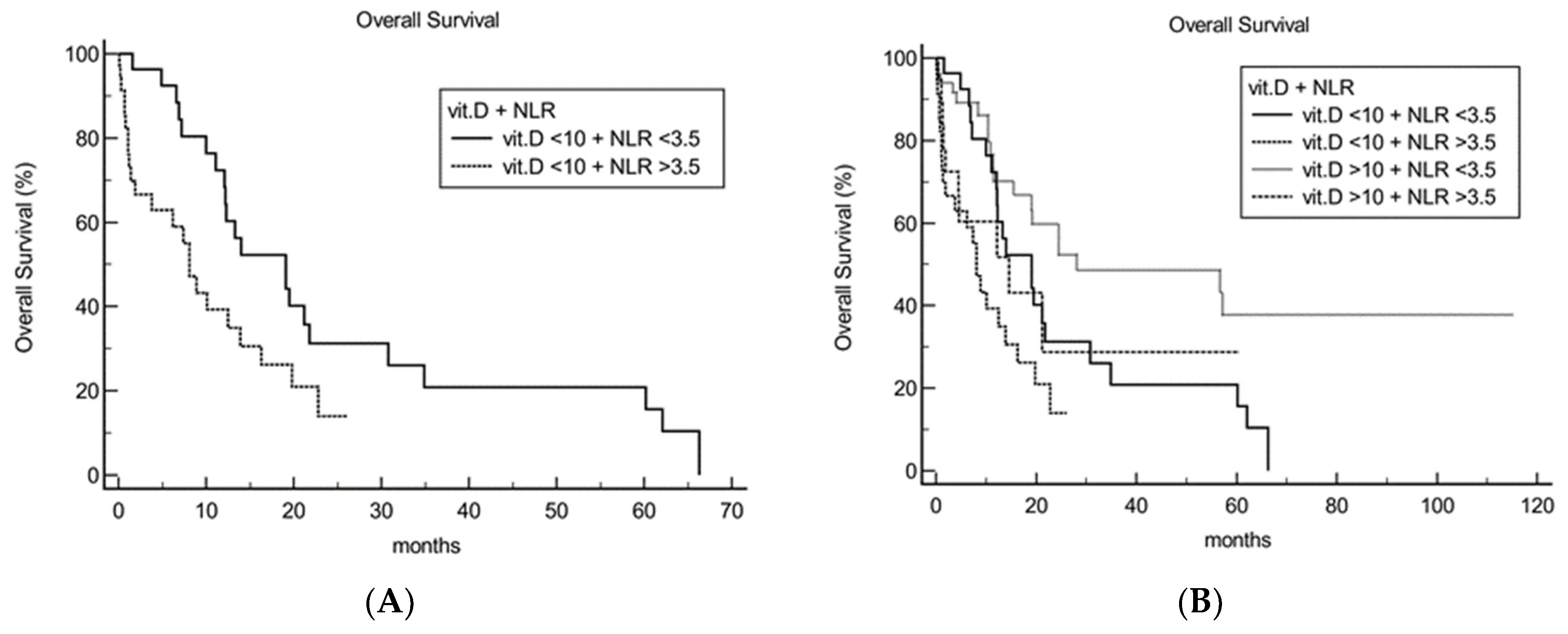
3.2. Immune System Status Evaluation

Since NLR was found to be an important predictive factor of vitamin D deficiency, we investigated potential immune changes in peripheral blood between vitamin D deficient vs. non deficient patients, in a subset of patients with available circulating lymphocyte subpopulation frequency. CD4/lymph, CD4/WBC, CD8/lymph, CD8/WBC, CD4/CD8 ratio, B/lymph, B/WBC, NK/lymph, NK/WBC frequencies were available for 32 out of the 133 patients. The Mann–Whitney–Wilcoxon test was performed to compare the differences in the nine above reported variables between vitamin D deficient (<10 ng/dL) and non-deficient (>10 ng/dL) patients (Table 1). Deficient patients had statistically significant higher CD4+ T cells (median 48% vs. 40%, p 0.04) and lower B-lymphocytes (median 4% vs. 7%, p = 0.03) frequency among lymphocytes, as compared to non-deficient patients.

4. Discussion

In the present study, we analyzed the effect of plasma 25(OH)D levels on survival of metastatic colorectal cancer patients starting a standard first-line chemotherapy [24]. The prognostic value for vitamin D in cancer patients, including colorectal cancer, has been widely documented in recent years [19,25,26]. However, a mCRC-specific plasma 25(OH)D cut-off predictive of overall survival has not been investigated so far.
We found an approximately linear association between 25(OH)D and survival; however, the best cut-off value to predict prognosis was <10 ng/mL (25 nmol/L), which is remarkably close to the risk threshold for other skeletal and extra-skeletal conditions and has been unanimously recommended by international nutritional guidelines [27,28,29]. Importantly, the 10 ng/mL cut-off has been used as a reference value in most interventional studies regardless of other influencing factors such as age, sex, BMI and renal function. In our study, all these covariates were considered in the multivariable logistic regression model.
Notably, almost half of the patients (60 out of 133, 47.4%) in our cohort had a baseline plasma 25(OH)D level < 10 ng/mL and median level in the entire cohort was 10.8 ng/mL. This is in line with previously reported data in mCRC patients with median levels ranging from 10 to 18 ng/mL [15,29]. Tumor intrinsic features, such as KRAS, NRAS and BRAF mutational status, did not interact with the survival effect of 25(OH)D levels (p = 0.773) and this was in line with a recently published review [30]. A further analysis with a larger sample size taking into account the distinct subtypes of KRAS mutations would be of interest given a possible interaction with specific KRAS variants [31].
The optimal plasma 25(OH)D level that should be achieved is a matter of controversy, with many experts suggesting concentrations > 20 ng/mL (50 nmol/L) [14]. Given the high depletion observed in metastatic colorectal cancer patients, it is not surprising that vitamin D targeted therapy requires extensive doses of exogenous supplementation to demonstrate an impact on outcome in this setting.
In the recent SUNSHINE randomized trial, 139 mCRC patient candidates for a standard first-line chemotherapy were treated with mFOLFOX plus bevacizumab plus either standard dose of oral vitamin D3 (400 IU/d) or 10-fold higher dose of vitamin D3 (4000 IU/d). The high-dose group demonstrated a longer median progression-free survival as compared to the standard dose (13 vs. 11 months) but this was of borderline statistical significance according to the log-rank test (p = 0.07) [15].
Based on the pleiotropic effects of vitamin D, hypovitaminosis D has also been implicated in many other skeletal, cardiovascular, infectious, metabolic and pulmonary diseases. However, trials investigating the efficacy of targeted vitamin D3 supplementation at doses of 2000–4400 IU/d were not able to demonstrate an improvement in the outcomes in settings such as tuberculosis, asthma, diabetes and arterial thromboembolic events. [32,33,34,35].
The disappointing results obtained with targeted vitamin D supplementation might rely on the insufficient doses administered, especially if no stratification by baseline serum 25(OH)D is performed, or on the presence of concomitant factors that might have an influence in patients with vitamin D deficiency.
We scrutinized 33 common clinical and hematochemical variables, including those notably associated with 25(OH)D variations (i.e., BMI, age, creatinine, gender), and found that the well-known inflammatory index, neutrophil/lymphocyte ratio (NLR), displayed the most powerful association with vitamin D deficiency. Moreover, NLR significantly influenced the outcome of patients in conjunction with plasma 25(OH)D, as patients with 25(OH)D < 10 ng/mL plus NLR > 3.5 had a significantly shorter survival as compared to patients with 25(OH)D < 10 ng/mL plus NLR < 3.5 (p = 0.0004). Our results are in agreement with previously reported studies that correlated 25(OH)D with NLR in cardiovascular disease and diabetes [36,37,38,39].
Inflammation is notoriously involved in all phases of cancer growth, with a number of cytokines, chemokines and immune cells of the tumor microenvironment involved [39].
Moreover, systemic inflammation has consistently been reported as associated with decreased 25(OH)D concentrations. Furthermore, an anti-inflammatory property of exogenous vitamin D has been suggested [40,41].
Vitamin D as anti-inflammatory/immunomodulatory agent relies on the modulation of several pathways such as prostaglandin, nuclear factor kappa-light-chain-enhancer of activated B cells (NF-KB) and P38 mitogen-activated protein kinase (MAPK) [42,43]. It is exerted through its action on both innate and adaptive immunity since its receptor, VDR, is expressed and activated in many immune cells such as CD4 and CD8 T cells, B cells, neutrophils, macrophages, and dendritic cells [44]. It has been demonstrated that vitamin D silences CD4+ T helper 1 cells [45] and induces IL4, IL5, IL9 and IL13 release [46], which boost B cell proliferation and promote humoral immune system. Recent evidence has highlighted the role of vitamin D in restraining autoimmune diseases by reducing T helper 1 mediated response [45].
In vitamin D deficiency, an opposite scenario might be observed as evidenced in our study, with high CD4+ cells and low lymphocyte B [47,48,49,50]. The effect of these immune changes on patient prognosis is yet to be determined.
Hamada et al. evaluated the peri-tumoral lymphocyte reaction in conjunction with plasma 25(OH)D levels in more than 800 CRC cases. They observed that 25(OH)D significantly influenced cancer-specific risk of death only in tumors with low or null peri-tumoral lymphocyte reaction, thus reinforcing the hypothesis of an interaction between 25(OH)D levels and immune response [8]. In this study however, no specific analysis on B or T helper cells was performed.
Our results might provide suggestions on how to optimize future trials of vitamin D supplementation. We first encourage the design of specific trials for patients with plasma 25(OH)D < 10 ng/mL. Moreover, patient stratification according to baseline NLR would be desirable.
Patients with 25(OH)D < 10 ng/mL and NLR > 3.5 would possibly require ‘super-high’ vitamin D supplementation doses that have never been tested. Given the anti-inflammatory property of vitamin D, an extra-amount of exogenous vitamin D might be necessary to initially sedate inflammation in this setting of patients, before having an anticancer effect. Trials with very high doses of vitamin D (i.e., >4000 IU/d) should therefore be taken into consideration in our opinion.
Moreover, it would be of particular interest to investigate the effect of targeted combination treatment with vitamin D supplementation and immune-modulating agents in the setting of mCRC patients, especially in the case of hypovitaminosis D and high systemic inflammation.
Immunotherapy, namely anti-PD-1/PD-L1 antibodies, has extensively demonstrated efficacy in many solid tumors, but proved of limited success in colorectal cancer with both low vitamin D and high NLR being associated with inferior drug efficacy [51,52,53,54]. Since both exogenous vitamin D and anti-PD-1/PD-L1 agents might reduce systemic inflammation and favor antigen-specific anticancer immunity (as in the case of head and neck cancer patients [55]), we would encourage the design of trials investigating combination strategies of immunotherapy plus exogenous vitamin D.
Finally, a synergism has been observed with the combined use of corticosteroids, particularly dexamethasone, and vitamin D supplementation, for the beneficial impact on specific immune cells in diseases other than cancer (e.g., multiple sclerosis and asthma) [56,57]. It would be interesting to evaluate, in a randomized trial, the therapeutic effect of this combination in mCRC patients with hypovitaminosis D and high systemic inflammation.
We are acknowledging several limitations of our study. First, it is retrospective in nature and the sample size is limited. In addition, questionnaires on food intake were not administered to study patients; therefore, nutritional amount of vitamin D intake could not be considered. Only a minority of our patients were on regular oral vitamin D supplementation, and this did not change the overall final results.
Furthermore, it would be desirable to study a larger lymphocyte subpopulation analysis, also taking into consideration T and B-regulatory cells as well as the analysis of the complement system, given their possible role on vitamin D activation [45].
The dynamic evaluation of immune variables and plasma 25(OH)D during the course of the first-line treatment has not been performed. The longitudinal assessment of the variables would better define their interaction with the effect of the anticancer treatment and the evolution of the disease. The assessment of plasma 25(OH)D at different time-points (after 1 month, 3 months, 6 months and at disease progression of first-line) is currently underway in our center and we suggest including this longitudinal evaluation in future trials on vitamin D supplementation in metastatic cancer patients.

5. Conclusions

In conclusion, in the present study we present an mCRC-specific plasma 25(OH)D cut-off and implications of vitamin D deficiency on the immune system.
Collectively, these data suggest attempting an improvement of oral vitamin D targeted therapy either by further increasing the treatment doses, thus potentiating its anti-inflammatory properties (especially in patients with vitamin D deficiency and concomitant high systemic inflammation) or by integrating vitamin D supplementation with anti-inflammatory or immune-modulating agents.

Supplementary Materials

The following supporting information can be downloaded at: https://www.mdpi.com/article/10.3390/cancers14112594/s1. Table S1: Patients’ characteristics; Figure S1: Maximally selected rank statistics analysis for overall survival defining the best cut-off value of vitamin D to stratify patients between good prognosis (longer survival) and poor prognosis (shorter survival); Figure S2: NLR < or >3.5 prevalence according to Vit.D levels.

Author Contributions

Conceptualization, V.F., C.M. and M.R. (Michela Rofei); methodology, V.F. and C.M.; software, V.F.; validation, D.F.; formal analysis, V.F.; investigation, C.M. and M.R. (Michela Rofei); resources, V.F., C.M., M.R. (Michela Rofei), M.R. (Mario Roselli), A.O. and G.D.V.B.; data curation, V.F. and C.M.; writing—original draft preparation, C.M. and V.F.; writing—review and editing, C.M., V.F. and S.R.; visualization, F.T., M.T. and M.F.; supervision, H.-T.A.; funding acquisition, M.R. (Mario Roselli). All authors have read and agreed to the published version of the manuscript.

Funding

The research was partially supported by the European Union’s Horizon 2020 research and innovation program under grant agreement n°848098.

Institutional Review Board Statement

The study was conducted according to the guidelines of the Declaration of Helsinki and approved by the Ethics Committee of Tor Vergata University Hospital as part of NCT01533740 trial and REVERT trial (IRB approval n° 149.20 23 July 2020).

Informed Consent Statement

Informed consent was obtained from all subjects involved in the study.

Data Availability Statement

The data presented in this study are available on request from the corresponding author. The data are not publicly available due to privacy regulations in force at our hospital.

Acknowledgments

The present study was carried out within the Program on Experimental System and Medicine (XXXV cycle) at the University of Rome “Tor Vergata”.

Conflicts of Interest

The authors declare no conflict of interest.

References

  1. Siegel, R.L.; Miller, K.D.; Goding Sauer, A.; Fedewa, S.A.; Butterly, L.F.; Anderson, J.C.; Cercek, A.; Smith, R.A.; Jemal, A. Colorectal cancer statistics, 2020. CA Cancer J. Clin. 2020, 70, 145–164. [Google Scholar] [CrossRef] [PubMed] [Green Version]
  2. Cardoso, R.; Guo, F.; Heisser, T.; Hackl, M.; Ihle, P.; De Schutter, H.; Van Damme, N.; Valerianova, Z.; Atanasov, T.; Májek, O.; et al. Colorectal cancer incidence, mortality, and stage distribution in European countries in the colorectal cancer screening era, an international population-based study. Lancet Oncol. 2021, 22, 1002–1013. [Google Scholar] [CrossRef]
  3. National Cancer Institute Surveillance, Epidemiology, and End Results Program. Cancer Stat Facts, Colorectal Cancer. Available online: https://seer.cancer.gov/statfacts/html/colorect.html (accessed on 2 September 2021).
  4. Islami, F.; Goding Sauer, A.; Miller, K.D.; Siegel, R.L.; Fedewa, S.A.; Jacobs, E.J.; McCullough, M.L.; Patel, A.V.; Ma, J.; Soerjomataram, I.; et al. Proportion and number of cancer cases and deaths attributable to potentially modifiable risk factors in the United States. CA Cancer J. Clin. 2018, 68, 31–54. [Google Scholar] [CrossRef] [PubMed]
  5. Carlberg, C.; Muñoz, A. An update on vitamin D signaling and cancer. Semin. Cancer Biol. 2022, 79, 217–230. [Google Scholar] [CrossRef]
  6. Fang, Y.; Song, H.; Huang, J.; Zhou, J.; Ding, X. The clinical significance of vitamin D levels and vitamin D receptor mRNA expression in colorectal neoplasms. J. Clin. Lab. Anal. 2021, 35, e23988. [Google Scholar] [CrossRef]
  7. Meza-Meza, M.R.; Ruiz-Ballesteros, A.I.; de la Cruz-Mosso, U. Functional effects of vitamin D, From nutrient to immunomodulator. Crit. Rev. Food Sci. Nutr. 2020, 62, 3042–3062. [Google Scholar] [CrossRef]
  8. Hamada, T.; Liu, L.; Nowak, J.A.; Mima, K.; Cao, Y.; Ng, K.; Twombly, T.S.; Song, M.; Jung, S.; Dou, R.; et al. Vitamin D status after colorectal cancer diagnosis and patient survival according to immune response to tumour. Eur. J. Cancer 2018, 103, 98–107. [Google Scholar] [CrossRef]
  9. Minton, K. Vitamin D shuts down T cell-mediated inflammation. Nat. Rev. Immunol. 2022, 22, 1. [Google Scholar] [CrossRef]
  10. Emerging Risk Factors Collaboration/EPIC-CVD/Vitamin D Studies Collaboration. Estimating dose-response relationships for vitamin D with coronary heart disease, stroke, and all-cause mortality: Observational and Mendelian randomisation analyses. Lancet Diabetes Endocrinol. 2021, 9, 837–846. [Google Scholar] [CrossRef]
  11. Lopez-Caleya, J.F.; Ortega-Valín, L.; Fernández-Villa, T.; Delgado-Rodríguez, M.; Martín-Sánchez, V.; Molina, A.J. The role of calcium and vitamin D dietary intake on risk of colorectal cancer, systematic review and meta-analysis of case-control studies. Cancer Causes Control 2022, 33, 167–182. [Google Scholar] [CrossRef]
  12. Maalmi, H.; Ordóñez-Mena, J.M.; Schöttker, B.; Brenner, H. Serum 25-hydroxyvitamin D levels and survival in colorectal and breast cancer patients, systematic review and meta-analysis of prospective cohort studies. Eur. J. Cancer 2014, 50, 1510–1521. [Google Scholar] [CrossRef] [PubMed]
  13. Bouillon, R.; Marcocci, C.; Carmeliet, G.; Bikle, D.; White, J.H.; Dawson-Hughes, B.; Lips, P.; Munns, C.F.; Lazaretti-Castro, M.; Giustina, A.; et al. Skeletal and Extraskeletal Actions of Vitamin D: Current Evidence and Outstanding Questions. Endocr. Rev. 2019, 40, 1109–1151. [Google Scholar] [CrossRef] [PubMed] [Green Version]
  14. Bouillon, R.; Manousaki, D.; Rosen, C.; Trajanoska, K.; Rivadeneira, F.; Richards, J.B. The health effects of vitamin D supplementation: Evidence from human studies. Nat. Rev. Endocrinol. 2022, 18, 96–110. [Google Scholar] [CrossRef] [PubMed]
  15. Ng, K.; Nimeiri, H.S.; McCleary, N.J.; Abrams, T.A.; Yurgelun, M.B.; Cleary, J.M.; Rubinson, D.A.; Schrag, D.; Miksad, R.; Bullock, A.J.; et al. Effect of High-Dose vs Standard-Dose Vitamin D3 Supplementation on Progression-Free Survival Among Patients With Advanced or Metastatic Colorectal Cancer: The SUNSHINE Randomized Clinical Trial. JAMA 2019, 321, 1370–1379. [Google Scholar] [CrossRef] [PubMed] [Green Version]
  16. Urashima, M.; Ohdaira, H.; Akutsu, T.; Okada, S.; Yoshida, M.; Kitajima, M.; Suzuki, Y. Effect of Vitamin D Supplementation on Relapse-Free Survival Among Patients With Digestive Tract Cancers, The AMATERASU Randomized Clinical Trial. JAMA 2019, 321, 1361–1369. [Google Scholar] [CrossRef]
  17. Aung, K.; Htay, T. USPSTF found insufficient evidence on benefits and harms of screening for vitamin D deficiency in asymptomatic adults. Ann. Intern. Med. 2021, 174, JC100. [Google Scholar] [CrossRef]
  18. Hernández-Alonso, P.; Boughanem, H.; Canudas, S.; Becerra-Tomás, N.; Fernández de la Puente, M.; Babio, N.; Macias-Gonzalez, M.; Salas-Salvadó, J. Circulating vitamin D levels and colorectal cancer risk: A meta-analysis and systematic review of case-control and prospective cohort studies. Crit. Rev. Food Sci. Nutr. 2021, 5, 1–17. [Google Scholar] [CrossRef]
  19. Yuan, C.; Sato, K.; Hollis, B.W.; Zhang, S.; Niedzwiecki, D.; Ou, F.S.; Chang, I.W.; O’Neil, B.H.; Innocenti, F.; Lenz, H.J.; et al. Plasma 25-Hydroxyvitamin D Levels and Survival in Patients with Advanced or Metastatic Colorectal Cancer, Findings from CALGB/SWOG 80405 (Alliance). Clin. Cancer Res. 2019, 25, 7497–7505. [Google Scholar] [CrossRef] [Green Version]
  20. Lambert, P.C.; Wilkes, S.R.; Crowther, M.J. Flexible parametric modelling of the cause-specific cumulative incidence function. Stat. Med. 2017, 36, 1429–1446. [Google Scholar] [CrossRef]
  21. Hothorn, T.; Zeileis, A. Generalized maximally selected statistics. Biometrics 2008, 64, 1263–1269. [Google Scholar] [CrossRef] [Green Version]
  22. Tibshirani, R. Regression shrinkage and selection via the Lasso. J. R Statist. Soc. B 1996, 58, 267–288. [Google Scholar] [CrossRef]
  23. Holick, M.F.; Binkley, N.C.; Bischoff-Ferrari, H.A.; Gordon, C.M.; Hanley, D.A.; Heaney, R.P.; Murad, M.H.; Weaver, C.M.; Endocrine Society. Evaluation, treatment, and prevention of vitamin D deficiency: An Endocrine Society clinical practice guideline. J. Clin. Endocrinol. Metab. 2011, 96, 1911–1930. [Google Scholar] [CrossRef] [PubMed] [Green Version]
  24. Busch, E.L. Cut points and contexts. Cancer 2021, 127, 4348–4355. [Google Scholar] [CrossRef] [PubMed]
  25. Mondul, A.M.; Weinstein, S.J.; Layne, T.M.; Albanes, D. Vitamin D and Cancer Risk and Mortality, State of the Science, Gaps, and Challenges. Epidemiol. Rev. 2017, 39, 28–48. [Google Scholar] [CrossRef] [PubMed]
  26. Vaughan-Shaw, P.G.; Zgaga, L.; Ooi, L.Y.; Theodoratou, E.; Timofeeva, M.; Svinti, V.; Walker, M.; O’Sullivan, F.; Ewing, A.; Johnston, S.; et al. Low plasma vitamin D is associated with adverse colorectal cancer survival after surgical resection, independent of systemic inflammatory response. Gut 2020, 69, 103–111. [Google Scholar] [CrossRef]
  27. Bouillon, R. Comparative analysis of nutritional guidelines for vitamin D. Nat. Rev. Endocrinol. 2017, 13, 466–479. [Google Scholar] [CrossRef]
  28. Ross, A.C.; Manson, J.E.; Abrams, S.A.; Aloia, J.F.; Brannon, P.M.; Clinton, S.K.; Durazo-Arvizu, R.A.; Gallagher, J.C.; Gallo, R.L.; Jones, G.; et al. The 2011 report on dietary reference intakes for calcium and vitamin D from the Institute of Medicine, what clinicians need to know. J. Clin. Endocrinol. Metab. 2011, 96, 53–58. [Google Scholar] [CrossRef]
  29. Thacher, T.D.; Clarke, B.L. Vitamin D insufficiency. Mayo Clin. Proc. 2011, 86, 50–60. [Google Scholar] [CrossRef] [Green Version]
  30. El Asri, A.; Zarrouq, B.; El Kinany, K.; Bouguenouch, L.; Ouldim, K.; El Rhazi, K. Associations between nutritional factors and KRAS mutations in colorectal cancer: A systematic review. BMC Cancer 2020, 20, 696. [Google Scholar] [CrossRef]
  31. Laso, N.; Mas, S.; Jose Lafuente, M.; Casterad, X.; Trias, M.; Ballesta, A.; Molina, R.; Salas, J.; Ascaso, C.; Zheng, S.; et al. Decrease in specific micronutrient intake in colorectal cancer patients with tumors presenting Ki-ras mutation. Anticancer. Res. 2004, 24, 2011–2020. [Google Scholar]
  32. Ganmaa, D.; Uyanga, B.; Zhou, X.; Gantsetseg, G.; Delgerekh, B.; Enkhmaa, D.; Khulan, D.; Ariunzaya, S.; Sumiya, E.; Bolortuya, B.; et al. Vitamin D Supplements for Prevention of Tuberculosis Infection and Disease. N. Engl. J. Med. 2020, 383, 359–368. [Google Scholar] [CrossRef] [PubMed]
  33. Litonjua, A.A.; Carey, V.J.; Laranjo, N.; Stubbs, B.J.; Mirzakhani, H.; O’Connor, G.T.; Sandel, M.; Beigelman, A.; Bacharier, L.B.; Zeiger, R.S.; et al. Six-Year Follow-up of a Trial of Antenatal Vitamin D for Asthma Reduction. N. Engl. J. Med. 2020, 382, 525–533. [Google Scholar] [CrossRef] [PubMed]
  34. Pittas, A.G.; Dawson-Hughes, B.; Sheehan, P.; Ware, J.H.; Knowler, W.C.; Aroda, V.R.; Brodsky, I.; Ceglia, L.; Chadha, C.; Chatterjee, R.; et al. Vitamin D Supplementation and Prevention of Type 2 Diabetes. N. Engl. J. Med. 2019, 381, 520–530. [Google Scholar] [CrossRef] [PubMed] [Green Version]
  35. Manson, J.E.; Cook, N.R.; Lee, I.M.; Christen, W.; Bassuk, S.S.; Mora, S.; Gibson, H.; Gordon, D.; Copeland, T.; D’Agostino, D.; et al. Vitamin D Supplements and Prevention of Cancer and Cardiovascular Disease. N. Engl. J. Med. 2019, 380, 33–44. [Google Scholar] [CrossRef]
  36. Akbas, E.M.; Gungor, A.; Ozcicek, A.; Akbas, N.; Askin, S.; Polat, M. Vitamin D and inflammation: Evaluation with neutrophil-to-lymphocyte ratio and platelet-to-lymphocyte ratio. Arch. Med. Sci. 2016, 12, 721–727. [Google Scholar] [CrossRef]
  37. Wang, S.Y.; Shen, T.T.; Xi, B.L.; Shen, Z.; Zhang, X. Vitamin D affects the neutrophil-to-lymphocyte ratio in patients with type 2 diabetes mellitus. J. Diabetes Invest. 2021, 12, 254–265. [Google Scholar] [CrossRef]
  38. Verdoia, M.; Nardin, M.; Rolla, R.; Negro, F.; Gioscia, R.; Saghir Afifeh, A.M.; Viglione, F.; Suryapranata, H.; Marcolongo, M.; De Luca, G. Cholecalciferol levels, inflammation and leukocytes parameters: Results from a large single-centre cohort of patients. Clin. Nutr. 2021, 40, 2228–2236. [Google Scholar] [CrossRef]
  39. Hou, J.; Karin, M.; Sun, B. Targeting cancer-promoting inflammation-have anti-inflammatory therapies come of age? Nat. Rev. Clin. Oncol. 2021, 18, 261–279. [Google Scholar] [CrossRef]
  40. Thorand, B.; Zierer, A.; Huth, C.; Linseisen, J.; Meisinger, C.; Roden, M.; Peters, A.; Koenig, W.; Herder, C. Effect of serum 25-hydroxyvitamin D on risk for type 2 diabetes may be partially mediated by subclinical inflammation: Results from the MONICA/KORA Augsburg study. Diabetes Care 2011, 34, 2320–2322. [Google Scholar] [CrossRef] [Green Version]
  41. Filgueiras, M.S.; Rocha, N.P.; Novaes, J.F.; Bressan, J. Vitamin D status, oxidative stress, and inflammation in children and adolescents: A systematic review. Crit. Rev. Food Sci. Nutr. 2020, 60, 660–669. [Google Scholar] [CrossRef]
  42. Chen, J.; Tang, Z.; Slominski, A.T.; Li, W.; Żmijewski, M.A.; Liu, Y.; Chen, J. Vitamin D and its analogs as anticancer and anti-inflammatory agents. Eur. J. Med. Chem. 2020, 207, 112738. [Google Scholar] [CrossRef] [PubMed]
  43. Hahn, J.; Cook, N.R.; Alexander, E.K.; Friedman, S.; Walter, J.; Bubes, V.; Kotler, G.; Lee, I.M.; Manson, J.E.; Costenbader, K.H. Vitamin D and marine omega 3 fatty acid supplementation and incident autoimmune disease: VITAL randomized controlled trial. BMJ 2022, 376, e066452. [Google Scholar] [CrossRef]
  44. Malaguarnera, L. Vitamin D and microbiota, two sides of the same coin in the immunomodulatory aspects. Int. Immunopharmacol. 2020, 79, 106112. [Google Scholar] [CrossRef] [PubMed]
  45. Chauss, D.; Freiwald, T.; McGregor, R.; Yan, B.; Wang, L.; Nova-Lamperti, E.; Kumar, D.; Zhang, Z.; Teague, H.; West, E.E.; et al. Autocrine vitamin D signaling switches off pro-inflammatory programs of TH1 cells. Nat. Immunol. 2022, 23, 62–74. [Google Scholar] [CrossRef]
  46. Charoenngam, N.; Holick, M.F. Immunologic Effects of Vitamin D on Human Health and Disease. Nutrients 2020, 12, 2097. [Google Scholar] [CrossRef] [PubMed]
  47. Murdaca, G.; Tonacci, A.; Negrini, S.; Greco, M.; Borro, M.; Puppo, F.; Gangemi, S. Emerging role of vitamin D in autoimmune diseases: An update on evidence and therapeutic implications. Autoimmun. Rev. 2019, 18, 102350. [Google Scholar] [CrossRef]
  48. Rolf, L.; Muris, A.H.; Hupperts, R.; Damoiseaux, J. Vitamin D effects on B cell function in autoimmunity. Ann. N. Y. Acad. Sci. 2014, 1317, 84–91. [Google Scholar] [CrossRef]
  49. Drozdenko, G.; Heine, G.; Worm, M. Oral vitamin D increases the frequencies of CD38+ human B cells and ameliorates IL-17-producing T cells. Exp. Dermatol. 2014, 23, 107–112. [Google Scholar] [CrossRef]
  50. Malihi, Z.; Wu, Z.; Stewart, A.W.; Lawes, C.M.; Scragg, R. Hypercalcemia, hypercalciuria, and kidney stones in long-term studies of vitamin D supplementation, a systematic review and meta-analysis. Am. J. Clin. Nutr. 2016, 104, 1039–1051. [Google Scholar] [CrossRef] [Green Version]
  51. Cusato, J.; Genova, C.; Tomasello, C.; Carrega, P.; Ottonello, S.; Pietra, G.; Mingari, M.C.; Cossu, I.; Rijavec, E.; Leggieri, A.; et al. Influence of Vitamin D in Advanced Non-Small Cell Lung Cancer Patients Treated with Nivolumab. Cancers 2019, 11, 125. [Google Scholar] [CrossRef] [Green Version]
  52. Formica, V.; Morelli, C.; Patrikidou, A.; Murias, C.; Butt, S.; Nardecchia, A.; Lucchetti, J.; Renzi, N.; Shiu, K.K.; Roselli, M.; et al. Gastric Inflammatory Prognostic Index (GIPI) in Patients with Metastatic Gastro-Esophageal Junction/Gastric Cancer Treated with PD-1/PD-L1 Immune Checkpoint Inhibitors. Target. Oncol. 2020, 15, 327–336. [Google Scholar] [CrossRef] [PubMed]
  53. Alessi, J.V.; Ricciuti, B.; Alden, S.L.; Bertram, A.A.; Lin, J.J.; Sakhi, M.; Nishino, M.; Vaz, V.R.; Lindsay, J.; Turner, M.M.; et al. Low peripheral blood derived neutrophil-to-lymphocyte ratio (dNLR) is associated with increased tumor T-cell infiltration and favorable outcomes to first-line pembrolizumab in non-small cell lung cancer. J. Immunother. Cancer 2021, 9, e003536. [Google Scholar] [CrossRef] [PubMed]
  54. De Giorgi, U.; Procopio, G.; Giannarelli, D.; Sabbatini, R.; Bearz, A.; Buti, S.; Basso, U.; Mitterer, M.; Ortega, C.; Bidoli, P.; et al. Association of Systemic Inflammation Index and Body Mass Index with Survival in Patients with Renal Cell Cancer Treated with Nivolumab. Clin. Cancer Res. 2019, 25, 3839–3846. [Google Scholar] [CrossRef] [PubMed] [Green Version]
  55. Bochen, F.; Balensiefer, B.; Körner, S.; Bittenbring, J.T.; Neumann, F.; Koch, A.; Bumm, K.; Marx, A.; Wemmert, S.; Papaspyrou, G.; et al. Vitamin D deficiency in head and neck cancer patients-prevalence, prognostic value and impact on immune function. Oncoimmunology 2018, 7, e1476817. [Google Scholar] [CrossRef]
  56. Cho, E.B.; Shin, J.H.; Kwon, S.; Kim, J.; Seok, J.M.; Kim, B.J.; Min, J.H. Effects of Vitamin D and Dexamethasone on Lymphocyte Proportions and Their Associations With Serum Concentrations of 25-Hydroxyvitamin D3In Vitro in Patients With Multiple Sclerosis or Neuromyelitis Optica Spectrum Disorder. Front. Immunol. 2021, 12, 677041. [Google Scholar] [CrossRef]
  57. Mehta, A.A.; Agrawal, A.D.; Appanna, V.; Chaudagar, K.K. Vitamin D improves corticosteroid efficacy and attenuates its side-effects in an animal model of asthma. Can. J. Physiol. Pharmacol. 2015, 93, 53–61. [Google Scholar] [CrossRef]
Figure 1. Hazard Ratio Smoothed Curve with Restricted Cubic Splines for the effect of continuous value of plasma 25(OH)D on overall survival. The graph demonstrates an approximately linear association with vitamin D (simplified by red straight-line). Vit.D: vitamin D; 25(OH)D: plasma 25-hydroxyvitamin D.
Cancers 14 02594 g001
Figure 2. Survival analysis evidenced different survival according to vitamin level (< vs. >10 ng/dL). Vit.D: vitamin D.
Cancers 14 02594 g002
Figure 3. Impact of vitamin D on overall survival according to RAS and BRAF mutational status presented as meta-analysis. RAS.BRAF.WT: patients with RAS and BRAF wild-type. RAS.BRAF.MUT.: patients with either RAS or BRAF mutation; vit.D: Vitamin D.
Cancers 14 02594 g003
Figure 4. Path of coefficients for a multivariable logistic regression with Least Absolute Shrinkage and Selection Operator (LASSO) analysis of 33 candidate predictors. NLR was identified as the most powerful predictor of vitamin D deficiency.
Cancers 14 02594 g004
Figure 5. Survival of vitamin D low (25(OH)D <10 ng/mL) (A) and high (25(OH)D >10 ng/mL) (B) patients could be further stratified by NLR level (< vs. >3.5). NLR: Neutrophil-to-lymphocyte ratio; vit.D: Vitamin D.
Cancers 14 02594 g005
Table 1. Mann–Whitney–Wilcoxon test analysis of 9 immune/inflammatory variables frequencies among circulating lymphocyte subpopulation and among WBC in Vit D < and > 10 ng/mL paired cohorts (statistically significant values are evidenced in bold).
ParameterVit.D < 10 ng/dLVit.D > 10 ng/dLp Value
Median Values
CD4/lymph48%40%0.04
CD4/WBC10%9%0.22
CD8/lymph28%27%0.54
CD8/WBC6%6%0.83
CD4/CD8 ratio1.811.340.13
B/lymph4%7%0.03
B/WBC1%1%0.41
NK/lymph16%17%0.82
NK/WBC4%3%0.98
Vit.D: vitamin D; WBC: white blood cells; lymph: lymphocytes; NK: natural killer lymphocytes.
Publisher’s Note: MDPI stays neutral with regard to jurisdictional claims in published maps and institutional affiliations.

