
Fertility after Cancer: Risks and Successes

 , and
, and
Simple Summary
Abstract

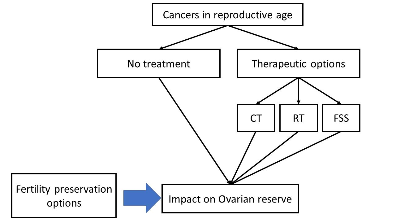
1. Introduction
Hematological Malignancies
2. Ovarian Reserve in Female Cancer Survivors
3. Influence of Cancer on Ovarian Function
4. Effects of Cancer Treatment on Female Reproductive Function
4.1. Ovarian Damage
4.1.1. Impact of Chemotherapy
4.1.2. Impact of Radiotherapy
4.2. Uterine Damage
4.2.1. Impact of Chemotherapy
4.2.2. Impact of Radiotherapy
5. Oncofertility
5.1. Fertility Preservation Options
- Hormone Protection by Suppressing Ovaries
- Oophoropexy
- Embryo Storage, Oocyte Storage
- Ovarian Tissue Storage
- Fertility Sparing Surgery
5.1.1. GnRH-Analogues
5.1.2. Oophoropexy
5.1.3. Embryo and Oocyte Cryopreservation
5.1.4. New Strategies
5.1.5. Ovarian Tissue Cryopreservation
5.2. Success Rates
6. Fertility Sparing Surgery for Gynecologic Cancer and Reproductive Outcomes
6.1. Cervical Cancer
Reproductive Outcomes
6.2. Endometrial Cancer
Reproductive Outcomes
6.3. Ovarian Cancer
Reproductive Outcomes
6.4. Vulvar Cancer
7. Special Categories
7.1. Fertility Preservation Post Cancer Diagnosis in Young Women
7.2. Fertility Preservation in Women with Hereditary Cancer Syndromes
8. Conclusions
Author Contributions
Funding
Data Availability Statement
Conflicts of Interest
References
- You, L.; Lv, Z.; Li, C.; Ye, W.; Zhou, Y.; Jin, J.; Han, Q. Worldwide cancer statistics of adolescents and young adults in 2019: A systematic analysis of the Global Burden of Disease Study 2019. ESMO Open 2021, 6, 100255. [Google Scholar] [CrossRef] [PubMed]
- Mahajan, N. Fertility preservation in female cancer patients: An overview. J. Hum. Reprod. Sci. 2015, 8, 3–13. [Google Scholar] [CrossRef] [PubMed]
- Rossi, L.; Mazzara, C.; Pagani, O. Diagnosis and Treatment of Breast Cancer in Young Women. Curr. Treat. Options Oncol. 2019, 20, 86. [Google Scholar] [CrossRef] [PubMed]
- Francis, P.A.; Regan, M.M.; Fleming, G.F.; Láng, I.; Ciruelos, E.; Bellet, M.; Bonnefoi, H.R.; Climent, M.A.; Da Prada, G.A.; Burstein, H.J.; et al. Adjuvant ovarian suppression in premenopausal breast cancer. N. Engl. J. Med. 2015, 372, 436–446. [Google Scholar] [CrossRef]
- Smallridge, R.C.; Copland, J.A. Anaplastic thyroid carcinoma: Pathogenesis and emerging therapies. Clin. Oncol. 2010, 22, 486–497. [Google Scholar] [CrossRef]
- DiSano, J.A.; Schaefer, E.W.; Kjerulff, K.; Hollenbeak, C.S.; Pameijer, C.R. Pregnancy after a melanoma diagnosis in women in the United States. J. Surg. Res. 2018, 231, 133–139. [Google Scholar] [CrossRef]
- Arbyn, M.; Weiderpass, E.; Bruni, L.; de Sanjosé, S.; Saraiya, M.; Ferlay, J.; Bray, F. Estimates of incidence and mortality of cervical cancer in 2018: A worldwide analysis. Lancet Glob. Health 2020, 8, e191–e203. [Google Scholar] [CrossRef]
- Guillon, S.; Popescu, N.; Phelippeau, J.; Koskas, M. A systematic review and meta-analysis of prognostic factors for remission in fertility-sparing management of endometrial atypical hyperplasia and adenocarcinoma. Int. J. Gynaecol. Obstet. 2019, 146, 277–288. [Google Scholar] [CrossRef]
- Shandley, L.M.; McKenzie, L.J. Recent Advances in Fertility Preservation and Counseling for Reproductive-Aged Women with Colorectal Cancer: A Systematic Review. Dis. Colon Rectum 2019, 62, 762–771. [Google Scholar] [CrossRef]
- Salama, M.; Anazodo, A.; Woodruff, T.K. Preserving fertility in female patients with hematological malignancies: A multidisciplinary oncofertility approach. Ann. Oncol. 2019, 30, 1760–1775. [Google Scholar] [CrossRef]
- Barton, S.E.; Najita, J.S.; Ginsburg, E.S.; Leisenring, W.M.; Stovall, M.; Weathers, R.E.; Sklar, C.A.; Robison, L.L.; Diller, L. Infertility, infertility treatment, and achievement of pregnancy in female survivors of childhood cancer: A report from the Childhood Cancer Survivor Study cohort. Lancet Oncol. 2013, 14, 873–881. [Google Scholar] [CrossRef]
- Overbeek, A.; van den Berg, M.; Louwé, L.; Wendel, E.; Ter Kuile, M.; Kaspers, G.; Stiggelbout, A.; van Dulmen-Den Broeder, E.; Hilders, C. Practice, attitude and knowledge of Dutch paediatric oncologists regarding female fertility. Neth. J. Med. 2014, 72, 264–270. [Google Scholar] [PubMed]
- De Lambert, G.; Poirot, C.; Guérin, F.; Brugières, L.; Martelli, H. Preservation of fertility in children with cancer. Bull. Cancer 2015, 102, 436–442. [Google Scholar] [CrossRef] [PubMed]
- Keegan, T.H.; Ries, L.A.; Barr, R.D.; Geiger, A.M.; Dahlke, D.V.; Pollock, B.H.; Bleyer, W.A.; National Cancer Institute Next Steps for Adolescent and Young Adult Oncology Epidemiology Working Group. Comparison of cancer survival trends in the United States of adolescents and young adults with those in children and older adults. Cancer 2016, 122, 1009–1016. [Google Scholar] [CrossRef]
- De Melo, A.S.; de Paula, C.T.V.; Rufato, M.A.F.; Rufato, M.C.A.C.; Rodrigues, J.K.; Ferriani, R.A.; Barreto, J. Fertility optimization in women with cancer: From preservation to contraception. JBRA Assist. Reprod. 2019, 23, 418–429. [Google Scholar]
- Peccatori, F.A.; Azim, H.A., Jr.; Orecchia, R.; Hoekstra, H.J.; Pavlidis, N.; Kesic, V.; Pentheroudakis, G.; ESMO Guidelines Working Group. Cancer, pregnancy and fertility: ESMO Clinical Practice Guidelines for diagnosis, treatment and follow-up. Ann. Oncol. 2013, 24, 160–170. [Google Scholar] [CrossRef]
- Barton, S.E.; Missmer, S.A.; Berry, K.F.; Ginsburg, E.S. Female cancer survivors are low responders and have reduced success compared with other patients undergoing assisted reproductive technologies. Fertil. Steril. 2012, 97, 381–386. [Google Scholar] [CrossRef] [PubMed]
- Boyle, K.E.; Vlahos, N.; Jarow, J.P. Assisted reproductive technology in the new millennium: Part II. Urology 2004, 63, 217–224. [Google Scholar] [CrossRef]
- Howell, S.; Shalet, S. Gonadal damage from chemotherapy and radiotherapy. Endocrinol. Metab. Clin. N. Am. 1998, 27, 927–943. [Google Scholar] [CrossRef]
- Gelber, S.; Coates, A.S.; Goldhirsch, A.; Castiglione-Gertsch, M.; Marini, G.; Lindtner, J.; Edelmann, D.Z.; Gudgeon, A.; Harvey, V.; Gelber, R.D. International Breast Cancer Study Group. Effect of pregnancy on overall survival after the diagnosis of early-stage breast cancer. J. Clin. Oncol. 2001, 19, 1671–1675. [Google Scholar] [CrossRef]
- Córdoba, O.; Bellet, M.; Vidal, X.; Cortés, J.; Llurba, E.; Rubio, I.T.; Xercavins, J. Pregnancy after treatment of breast cancer in young women does not adversely affect the prognosis. Breast 2012, 21, 272–275. [Google Scholar] [CrossRef] [PubMed]
- Azim Jr, H.A.; Santoro, L.; Pavlidis, N.; Gelber, S.; Kroman, N.; Azim, H.; Peccatori, F.A. Safety of pregnancy following breast cancer diagnosis: A meta-analysis of 14 studies. Eur. J. Cancer 2011, 47, 74–83. [Google Scholar] [CrossRef] [PubMed]
- Azim Jr, H.A.; Kroman, N.; Paesmans, M.; Gelber, S.; Rotmensz, N.; Ameye, L.; De Mattos-Arruda, L.; Pistilli, B.; Pinto, A.; Jensen, M.B. Prognostic impact of pregnancy after breast cancer according to estrogen receptor status: A multicenter retrospective study. J. Clin. Oncol. 2013, 31, 73–79. [Google Scholar] [CrossRef]
- Leader, A.; Lishner, M.; Michaeli, J.; Revel, A. Fertility considerations and preservation in haemato-oncology patients undergoing treatment. Br. J. Haematol. 2011, 153, 291–308. [Google Scholar] [CrossRef] [PubMed]
- Donnez, J.; Dolmans, M.M. Preservation of fertility in females with haematological malignancy. Br. J. Haematol. 2011, 154, 175–184. [Google Scholar] [CrossRef]
- Shapira, M.; Raanani, H.; Cohen, Y.; Meirow, D. Fertility preservation in young females with hematological malignancies. Acta Haematol. 2014, 132, 400–413. [Google Scholar] [CrossRef]
- Loren, A.W. Fertility issues in patients with hematologic malignancies. Hematol. Am. Soc. Hematol. Educ. Program 2015, 2015, 138–145. [Google Scholar] [CrossRef]
- Anderson, R.A.; Remedios, R.; Kirkwood, A.A.; Patrick, P.; Stevens, L.; Clifton-Hadley, L.; Roberts, T.; Hatton, C.; Kalakonda, N.; Milligan, D.W.; et al. Determinants of ovarian function after response-adapted therapy in patients with advanced Hodgkin’s lymphoma (RATHL): A secondary analysis of a randomized phase 3 trial. Lancet Oncol. 2018, 19, 1328–1337. [Google Scholar] [CrossRef]
- Salama, M.; Winkler, K.; Murach, K.F.; Seeber, B.; Ziehr, S.C.; Wildt, L. Female fertility loss and preservation: Threats and opportunities. Ann. Oncol. 2013, 24, 598–608. [Google Scholar] [CrossRef]
- De Vos, M.; Smitz, J.; Woodruff, T.K. Fertility preservation in women with cancer. Lancet 2014, 384, 1302–1310. [Google Scholar] [CrossRef]
- Metzger, M.L.; Meacham, L.R.; Patterson, B.; Casillas, J.S.; Constine, L.S.; Hijiya, N.; Kenney, L.B.; Leonard, M.; Lockart, B.A.; Likes, W.; et al. Female reproductive health after childhood, adolescent, and young adult cancers: Guidelines for the assessment and management of female reproductive complications. J. Clin. Oncol. 2013, 31, 1239–1247. [Google Scholar] [CrossRef] [PubMed]
- Hirshfeld-Cytron, J.; Gracia, C.; Woodruff, T.K. Nonmalignant diseases and treatments associated with primary ovarian failure: An expanded role for fertility preservation. J. Womens Health 2011, 20, 1467–1477. [Google Scholar] [CrossRef] [PubMed]
- West, E.R.; Zelinski, M.B.; Kondapalli, L.A.; Gracia, C.; Chang, J.; Coutifaris, C.; Critser, J.; Stouffer, R.L.; Shea, L.D.; Woodruff, T.K. Preserving female fertility following cancer treatment: Current options and future possibilities. Pediatr. Blood Cancer 2009, 53, 289–295. [Google Scholar] [CrossRef] [PubMed]
- Roberts, J.; Ronn, R.; Tallon, N.; Holzer, H. Fertility preservation in reproductive-age women facing gonadotoxic treatments. Curr. Oncol. 2015, 22, e294–e304. [Google Scholar] [CrossRef] [PubMed]
- Donnez, J.; Dolmans, M.M. Fertility preservation in women. N. Engl. J. Med. 2017, 377, 1657–1665. [Google Scholar] [CrossRef]
- Dolmans, M.M.; Lambertini, M.; Macklon, K.T.; Almeida Santos, T.; Ruiz-Casado, A.; Borini, A.; Bordes, V.; Frith, L.; Van Moer, E.; Germeyer, A. EUropean REcommendations for female FERtility preservation (EU-REFER): A joint collaboration between oncologists and fertility specialists. Crit. Rev. Oncol. Hematol. 2019, 138, 233–240. [Google Scholar] [CrossRef]
- Gardino, S.L.; Emanuel, L.L. Choosing life when facing death: Understanding fertility preservation decision-making for cancer patients. Cancer Treat. Res. 2010, 156, 447–458. [Google Scholar]
- Li, N.; Jayasinghe, Y.; Kemertzis, M.A.; Moore, P.; Peate, M. Fertility preservation in pediatric and adolescent oncology patients: The decision-making process of parents. J. Adolesc. Young Adult Oncol. 2017, 6, 213–222. [Google Scholar] [CrossRef]
- Cohn, F. Oncofertility and informed consent: Addressing beliefs, values, and future decision making. Cancer Treat. Res. 2010, 156, 249–258. [Google Scholar]
- Harada, M.; Osuga, Y. Where are oncofertility and fertility preservation treatments heading in 2016? Future Oncol. 2016, 12, 2313–2321. [Google Scholar] [CrossRef]
- Salama, M.; Anazodo, A.; Woodruff, T.K. Preserving fertility in female patients with hematological malignancies: The key points. Expert Rev. Hematol. 2019, 12, 375–377. [Google Scholar] [CrossRef] [PubMed]
- Pfeifer, S.; Goldberg, I.; McClure, R.; Lobo, R.; Thomas, M.; Widra, E.; Licht, M.; Collins, I.; Cedars, M.; Racowsky, C.; et al. Mature oocyte cryopreservation: A guideline. Fertil. Steril. 2013, 99, 37–43. [Google Scholar]
- Thomas-Teinturier, C.; Allodji, R.S.; Svetlova, E.; Frey, M.A.; Oberlin, O.; Millischer, A.E.; Epelboin, S.; Decanter, C.; Pacquement, H.; Tabone, M.D.; et al. Ovarian reserve after treatment with alkylating agents during childhood. Hum. Reprod. 2015, 30, 1437–1446. [Google Scholar] [CrossRef] [PubMed]
- Chow, E.J.; Stratton, K.L.; Leisenring, W.M.; Oeffinger, K.C.; Sklar, C.A.; Donaldson, S.S.; Ginsberg, J.P.; Kenney, L.B.; Levine, J.M.; Robison, L.L.; et al. Pregnancy after chemotherapy in male and female survivors of childhood cancer treated between 1970 and 1999: A report from the Childhood Cancer Survivor Study cohort. Lancet Oncol. 2016, 17, 567–576. [Google Scholar] [CrossRef]
- Velez, M.P.; Richardson, H.; Baxter, N.N.; McClintock, C.; Greenblatt, E.; Barr, R.; Green, M. Risk of infertility in female adolescents and young adults with cancer: A population-based cohort study. Hum. Reprod. 2021, 36, 1981–1988. [Google Scholar] [CrossRef]
- Mackie, E.J.; Radford, M.; Shalet, S.M. Gonadal function following chemotherapy for childhood Hodgkin’s disease. Med. Pediatr. Oncol. 1996, 27, 74–78. [Google Scholar] [CrossRef]
- Bath, L.E.; Wallace, W.H.; Critchley, H.O. Late effects of the treatment of childhood cancer on the female reproductive system and the potential for fertility preservation. BJOG 2002, 109, 107–114. [Google Scholar] [CrossRef]
- Whitehead, E.; Shalet, S.M.; Blackledge, G.; Todd, I.; Crowther, D.; Beardwell, C.G. The effect of combination chemotherapy on ovarian function in women treated for Hodgkin’s disease. Cancer 1983, 52, 988–993. [Google Scholar] [CrossRef]
- Johnson, L.N.; Sammel, M.D.; Dillon, K.E.; Lechtenberg, L.; Schanne, A.; Gracia, C.R. Antimüllerian hormone and antral follicle count are lower in female cancer survivors and healthy women taking hormonal contraception. Fertil. Steril. 2014, 102, 774–781.e3. [Google Scholar] [CrossRef]
- Dillon, K.E.; Sammel, M.D.; Prewitt, M.; Ginsberg, J.P.; Walker, D.; Mersereau, J.E.; Gosiengfiao, Y.; Gracia, C.R. Pretreatment antimüllerian hormone levels determine rate of posttherapy ovarian reserve recovery: Acute changes in ovarian reserve during and after chemotherapy. Fertil. Steril. 2013, 99, 477–483.e1. [Google Scholar] [CrossRef]
- Anderson, R.A.; Wallace, W.H. Antimullerian hormone, the assessment of the ovarian reserve, and the reproductive outcome of the young patient with cancer. Fertil. Steril. 2013, 99, 1469–1475. [Google Scholar] [CrossRef] [PubMed]
- Broer, S.L.; Broekmans, F.J.; Laven, J.S.; Fauser, B.C. Anti-Müllerian hormone: Ovarian reserve testing and its potential clinical implications. Hum. Reprod. Update 2014, 20, 688–701. [Google Scholar] [CrossRef] [PubMed]
- Gracia, C.R.; Sammel, M.D.; Freeman, E.; Prewitt, M.; Carlson, C.; Ray, A.; Vance, A.; Ginsberg, J.P. Impact of cancer therapies on ovarian reserve. Fertil. Steril. 2012, 97, 134–140.e1. [Google Scholar] [CrossRef]
- Nielsen, S.N.; Andersen, A.N.; Schmidt, K.T.; Rechnitzer, C.; Schmiegelow, K.; Bentzen, J.G.; Larsen, E.C. A 10-year follow up of reproductive function in women treated for childhood cancer. Reprod. Biomed. Online 2013, 27, 192–200. [Google Scholar] [CrossRef]
- Wenners, A.; Grambach, J.; Koss, J.; Maass, N.; Jonat, W.; Schmutzler, A.; Mundhenke, C. Reduced ovarian reserve in young early breast cancer patients: Preliminary data from a prospective cohort trial. BMC Cancer 2017, 17, 632. [Google Scholar] [CrossRef] [PubMed]
- Van den Berg, M.H.; Overbeek, A.; Lambalk, C.B.; Kaspers, G.; Bresters, D.; van den Heuvel-Eibrink, M.M.; Kremer, L.C.; Loonen, J.J.; van der Pal, H.J.; Ronckers, C.M.; et al. Long-term effects of childhood cancer treatment on hormonal and ultrasound markers of ovarian reserve. Hum. Reprod. 2018, 33, 1474–1488. [Google Scholar] [CrossRef] [PubMed]
- Friedler, S.; Koc, O.; Gidoni, Y.; Raziel, A.; Ron-El, R. Ovarian response to stimulation for fertility preservation in women with malignant disease: A systematic review and meta-analysis. Fertil. Steril. 2012, 97, 125–133. [Google Scholar] [CrossRef]
- Garcia-Velasco, J.A.; Domingo, J.; Cobo, A.; Martinez, M.; Carmona, L.; Pellicer, A. Five years’ experience using oocyte vitrification to pre- serve fertility for medical and nonmedical indications. Fertil. Steril. 2013, 99, 1994–1999. [Google Scholar] [CrossRef]
- Pal, L.; Leykin, L.; Schifren, J.L.; Isaacson, K.B.; Chang, Y.C.; Nikruil, N.; Chen, Z.; Toth, T.L. Malignancy may adversely influence the quality and behaviour of oocytes. Hum. Reprod. 1998, 13, 1837–1840. [Google Scholar] [CrossRef]
- Domingo, J.; Guillen, V.; Ayllon, Y.; Martınez, M.; Munoz, E.; Pellicer, A.; Garcia-Velasco, J.A. Ovarian response to controlled ovarian hyperstimulation in cancer patients is diminished even before oncological treatment. Fertil. Steril. 2012, 97, 930–934. [Google Scholar] [CrossRef]
- Alvarez, R.M.; Ramanathan, P. Fertility preservation in female oncology patients: The influence of the type of cancer on ovarian stimulation response. Hum. Reprod. 2018, 33, 2051–2059. [Google Scholar] [CrossRef] [PubMed]
- Volodarsky-Perel, A.; Cohen, Y.; Arab, S.; Son, W.Y.; Suarthana, E.; Dahan, M.H.; Tulandi, T.; Buckett, W. Effects of cancer stage and grade on fertility preservation outcome and ovarian stimulation response. Hum. Reprod. 2019, 34, 530–538. [Google Scholar] [CrossRef] [PubMed]
- Decanter, C.; Robin, G.; Mailliez, A.; Sigala, J.; Morschhauser, F.; Ramdane, N.; Devos, P.; Dewailly, D.; Leroy-Martin, B.; Keller, L. Prospective assessment of follicular growth and the oocyte cohort after ovarian stimulation for fertility preservation in 90 cancer patients versus 180 matched controls. Reprod. Biomed. Online 2018, 36, 543–551. [Google Scholar] [CrossRef] [PubMed]
- Moria, A.; Das, M.; Shehata, F.; Holzer, H.; Son, W.Y.; Tulandi, T. Ovarian reserve and oocyte maturity in women with malignancy undergoing in vitro maturation treatment. Fertil. Steril. 2011, 95, 1621–1623. [Google Scholar] [CrossRef]
- Johnson, L.N.; Dillon, K.E.; Sammel, M.D.; Efymow, B.L.; Mainigi, M.A.; Dokras, A.; Gracia, C.R. Response to ovarian stimulation in patients facing gonadotoxic therapy. Reprod. Biomed. Online 2013, 26, 337–344. [Google Scholar] [CrossRef]
- Levin, I.; Almog, B. Effect of cancer on ovarian function in patients undergoing in vitro fertilization for fertility preservation: A reappraisal. Curr. Oncol. 2013, 20, e1–e33. [Google Scholar] [CrossRef]
- Nurudeen, S.K.; Douglas, N.C.; Mahany, E.L.; Sauer, M.V.; Choi, J.M. Fertility preservation decisions among newly diagnosed oncology patients: A single-center experience. Am. J. Clin. Oncol. 2016, 39, 154–159. [Google Scholar] [CrossRef]
- Cardozo, E.R.; Thomson, A.P.; Karmon, A.E.; Dickinson, K.A.; Wright, D.L.; Sabatini, M.E. Ovarian stimulation and in-vitro fertilization outcomes of cancer patients undergoing fertility preservation compared to age matched controls: A 17-year experience. J. Assist. Reprod. Genet. 2015, 32, 587–596. [Google Scholar] [CrossRef]
- Almog, B.; Azem, F.; Gordon, D.; Pauzner, D.; Amit, A.; Barkan, G.; Levin, I. Effects of cancer on ovarian response in controlled ovarian stimulation for fertility preservation. Fertil. Steril. 2012, 98, 957–960. [Google Scholar] [CrossRef]
- Liu, D.; Yan, J.; Qiao, J. Effects of malignancies on fertility preservation outcomes and relevant cryobiological advances. Sci. China Life Sci. 2020, 63, 217–227. [Google Scholar] [CrossRef]
- Roxburgh, C.S.; McMillan, D.C. Cancer and systemic inflammation: Treat the tumour and treat the host. Br. J. Cancer 2014, 110, 1409–1412. [Google Scholar] [CrossRef] [PubMed]
- Hanahan, D.; Weinberg, R.A. Hallmarks of cancer: The next generation. Cell 2011, 144, 646–674. [Google Scholar] [CrossRef] [PubMed]
- Ingman, W.V.; Robertson, S.A. Defining the actions of transforming growth factor beta in reproduction. Bioessays 2002, 24, 904–914. [Google Scholar] [CrossRef]
- Derks-Smeets, I.A.P.; Van Tilborg, T.C.; Van Montfoort, A.; Smits, L.; Torrance, H.L.; Meijer-Hoogeveen, M.; Broekmans, F.; Dreesen, J.C.F.M.; Paulussen, A.D.C.; Tjan-Heijnen, V.C.G. BRCA1 mutation carriers have a lower number of mature oocytes after ovarian stimulation for IVF/PGD. J. Assist. Reprod. Genet. 2017, 34, 1475–1482. [Google Scholar] [CrossRef] [PubMed]
- Lekovich, J.; Lobel, A.L.S.; Stewart, J.D.; Pereira, N.; Kligman, I.; Rosenwaks, Z. Female patients with lymphoma demonstrate diminished ovarian reserve even before initiation of chemotherapy when compared with healthy controls and patients with other malignancies. J. Assist. Reprod. Genet. 2016, 33, 657–662. [Google Scholar] [CrossRef] [PubMed]
- Porcu, E.; Cillo, G.M.; Cipriani, L.; Sacilotto, F.; Notarangelo, L.; Damiano, G.; Dirodi, M.; Roncarati, I. Impact of BRCA1 and BRCA2 mutations on ovarian reserve and fertility preservation outcomes in young women with breast cancer. J. Assist. Reprod. Genet. 2020, 37, 709–715. [Google Scholar] [CrossRef] [PubMed]
- Balachandren, N.; Davies, M. Fertility, ovarian reserve and cancer. Maturitas 2017, 105, 64–68. [Google Scholar] [CrossRef]
- Spears, N.; Lopes, F.; Stefansdottir, A.; Rossi, V.; De Felici, M.; Anderson, R.A.; Klinger, F.G. Ovarian damage from chemotherapy and current approaches to its protection. Hum. Reprod. Update 2019, 25, 673–693. [Google Scholar] [CrossRef]
- Lee, S.J.; Schover, L.R.; Partridge, A.H.; Patrizio, P.; Wallace, W.H.; Hagerty, K.; Beck, L.N.; Brennan, L.V.; Oktay, K. American Society of Clinical Oncology recommendations on fertility preservation in cancer patients. J. Clin. Oncol. 2006, 24, 2917–2931. [Google Scholar] [CrossRef]
- Jayasinghe, J.L.; Wallace, W.H.B.; Anderson, R.A. Expert Review of Endocrinology & Metabolism Ovarian function, fertility and reproductive lifespan in cancer patients. Expert Rev. Endocrinol. Metab. 2018, 13, 125–136. [Google Scholar]
- Tauchmanova, L.; Selleri, C.; De Rosa, G.; Esposito, M.; Orio, J.F.; Palomba, S.; Bifulco, G.; Nappi, C.; Lombardi, G.; Rotoli, B.; et al. Gonadal status in reproductive age women after haematopoietic stem cell transplantation for haematological malignancies. Hum. Reprod. 2003, 18, 1410–1416. [Google Scholar] [CrossRef] [PubMed]
- Vatanen, A.; Wilhelmsson, M.; Borgström, B.; Gustafsson, B.; Taskinen, M.; Saarinen-Pihkala, U.M.; Winiarski, J.; Jahnukainen, K. Ovarian function after allogeneic hematopoietic stem cell transplantation in childhood and adolescence. Eur. J. Endocrinol. 2014, 170, 211–218. [Google Scholar] [CrossRef] [PubMed]
- Szymanska, K.J.; Tan, X.; Oktay, K. Unraveling the mechanisms of chemotherapy-induced damage to human primordial follicle reserve: Road to developing therapeutics for fertility preservation and reversing ovarian aging. Mol. Hum. Reprod. 2020, 26, 553–566. [Google Scholar] [CrossRef] [PubMed]
- Bedoschi, G.; Navarro, P.A.; Oktay, K. Chemotherapy-induced damage to ovary: Mechanisms and clinical impact. Future Oncol. 2016, 12, 2333–2344. [Google Scholar] [CrossRef] [PubMed]
- Cho, H.W.; Lee, S.; Min, K.J.; Hong, J.H.; Song, J.Y.; Lee, J.K.; Lee, N.W.; Kim, T. Advances in the Treatment and Prevention of Chemotherapy-Induced Ovarian Toxicity. Int. J. Mol. Sci. 2020, 21, 7792. [Google Scholar] [CrossRef]
- Kalich-Philosoph, L.; Roness, H.; Carmely, A.; Fishel-Bartal, M.; Ligumsky, H.; Paglin, S.; Wolf, I.; Kanety, H.; Sredni, B.; Meirow, D. Cyclophosphamide triggers follicle activation and “burnout”; AS101 prevents follicle loss and preserves fertility. Sci. Transl. Med. 2013, 5, 185ra162. [Google Scholar] [CrossRef]
- Gavish, Z.; Peer, G.; Roness, H.; Cohen, Y.; Meirow, D. Follicle activation and ‘burn-out’ contribute to post-transplantation follicle loss in ovarian tissue grafts: The effect of graft thickness. Hum. Reprod. 2014, 29, 989–996. [Google Scholar] [CrossRef]
- Wallace, W.H.; Shalet, S.M.; Hendry, J.H.; Morris-Jones, P.H.; Gattamaneni, H.R. Ovarian failure following abdominal irradiation in childhood: The radiosensitivity of the human oocyte. Br. Inst. Radiol. 2014, 62, 995–998. [Google Scholar] [CrossRef]
- Kim, S.; Kim, S.W.; Han, S.J.; Lee, S.; Park, H.T.; Song, J.Y.; Kim, T. Molecular Mechanism and Prevention Strategy of Chemotherapy-and Radiotherapy-Induced Ovarian Damage. Int. J. Mol. Sci. 2021, 22, 7484. [Google Scholar] [CrossRef]
- Beerendonk, C.C.M.; Braat, D.D.M. Present and future options for the preservation of fertility in female adolescents with cancer. Endocr. Dev. 2005, 8, 166–175. [Google Scholar]
- Stroud, J.S.; Mutch, D.; Rader, J.; Powell, M.; Thaker, P.H.; Grigsby, P.W. Effects of cancer treatment on ovarian function. Fertil. Steril. 2009, 92, 417–427. [Google Scholar] [CrossRef] [PubMed]
- Devine, P.J.; Perreault, S.D.; Luderer, U. Roles of reactive oxygen species and antioxidants in ovarian toxicity. Biol. Reprod. 2012, 86, 27. [Google Scholar] [CrossRef] [PubMed]
- Griffiths, M.J.; Winship, A.L.; Hutt, K.J. Do cancer therapies damage the uterus and compromise fertility? Hum. Reprod. Update 2020, 26, 161–173. [Google Scholar] [CrossRef] [PubMed]
- Fujimoto, A.; Ichinose, M.; Harada, M.; Hirata, T.; Osuga, Y.; Fujii, T. The outcome of infertility treatment in patients undergoing assisted reproductive technology after conservative therapy. J. Assist. Reprod. Genet. 2014, 31, 1189–1194. [Google Scholar] [CrossRef]
- Beneventi, F.; Locatelli, E.; Giorgiani, G.; Zecca, M.; Mina, T.; Simonetta, M.; Cavagnoli, C.; Albanese, M.; Spinillo, A. Adolescent and adult uterine volume and uterine artery Doppler blood flow among subjects treated with bone marrow transplantation or chemotherapy in pediatric age: A case-control study. Fertil. Steril. 2015, 103, 455–461. [Google Scholar] [CrossRef]
- Van de Loo, L.; Van den Berg, M.H.; Overbeek, A.; van Dijk, M.; Damen, L.; Lambalk, C.B.; Ronckers, C.M.; van den Heuvel-Eibrink, M.M.; Kremer, L.; van der Pal, H.J.; et al. Uterine function, pregnancy complications, and pregnancy outcomes among female childhood cancer survivors. Fertil. Steril. 2019, 111, 372–380. [Google Scholar] [CrossRef]
- Massarotti, C.; Scaruffi, P.; Lambertini, M.; Sozzi, F.; Remorgida, V.; Anserini, P. Beyond fertility preservation: Role of the oncofertility unit in the reproductive and gynecological follow-up of young cancer patients. Hum. Reprod. 2019, 34, 1462–1469. [Google Scholar] [CrossRef]
- Lambertini, M.; Del Mastro, L.; Pescio, M.C.; Andersen, C.Y.; Azim, H.A.; Peccatori, F.A.; Costa, M.; Revelli, A.; Salvagno, F.; Gennari, A.; et al. Cancer and fertility preservation: International recommendations from an expert meeting. BMC Med. 2016, 14, 1. [Google Scholar] [CrossRef]
- Linkeviciute, A.; Boniolo, G.; Chiavari, L.; Peccatori, F.A. Fertility preservation in cancer patients: The global framework. Cancer Treat. Rev. 2014, 40, 1019–1027. [Google Scholar] [CrossRef]
- Van den Berg, M.; Baysal, Ö.; Nelen, W.L.D.M.; Braat, D.D.M.; Beerendonk, C.C.M.; Hermens, R.P.M.G. Professionals’ barriers in female oncofertility care and strategies for improvement. Hum. Reprod. 2019, 34, 1074–1082. [Google Scholar] [CrossRef]
- Goodman, L.R.; Balthazar, U.; Kim, J.; Mersereau, J.E. Trends of socioeconomic disparities in referral patterns for fertility preservation consultation. Hum. Reprod. 2012, 27, 2076–2081. [Google Scholar] [CrossRef] [PubMed]
- Vindrola-Padros, C.; Dyer, K.E.; Cyrus, J.; Lubker, I.M. Healthcare professionals’ views on discussing fertility preservation with young cancer patients: A mixed method systematic review of the literature. Psychooncology 2017, 26, 4–14. [Google Scholar] [CrossRef] [PubMed]
- Lewin, J.; Ma, J.M.Z.; Mitchell, L.; Tam, S.; Puri, N.; Stephens, D.; Srikanthan, A.; Bedard, P.; Razak, A.; Crump, M.; et al. The positive effect of a dedicated adolescent and young adult fertility program on the rates of documentation of therapy-associated infertility risk and fertility preservation options. Support. Care Cancer 2017, 25, 1915–1922. [Google Scholar] [CrossRef] [PubMed]
- Lambertini, M.; Cinquini, M.; Moschetti, I.; Peccatori, F.A.; Anserini, P.; Menada, M.V.; Tomirotti, M.; Del Mastro, L. Temporary ovarian suppression during chemotherapy to preserve ovarian function and fertility in breast cancer patients: A GRADE approach for evidence evaluation and recommendations by the Italian Association of Medical Oncology. Eur. J. Cancer 2017, 71, 25–33. [Google Scholar] [CrossRef]
- Lee, J.H.; Choi, Y.S. The role of gonadotropin-releasing hormone agonists in female fertility preservation. Clin. Exp. Reprod. Med. 2021, 48, 11–16. [Google Scholar] [CrossRef]
- Roberts, J.E.; Oktay, K. Fertility preservation: A comprehensive approach to the young woman with cancer. JNCI Monogr. 2005, 2005, 57–59. [Google Scholar] [CrossRef]
- Ajala, T.; Rafi, J.; Larsen-Disney, P.; Howell, R. Fertility preservation for cancer patients: A review. Obstet. Gynecol. Int. 2010, 2010, 160386. [Google Scholar] [CrossRef]
- Lambertini, M.; Boni, L.; Michelotti, A.; Magnolfi, E.; Cogoni, A.A.; Mosconi, A.M.; Giordano, M.; Garrone, O.; Arpino, G.; Poggio, F.; et al. Long-term outcomes with pharmacological ovarian suppression during chemotherapy in premenopausal early breast cancer patients. J. Natl. Cancer. Inst. 2021, 114, 400–408. [Google Scholar] [CrossRef]
- Lambertini, M.; Moore, H.C.; Leonard, R.C.; Loibl, S.; Munster, P.; Bruzzone, M.; Boni, L.; Unger, J.M.; Anderson, R.A.; Mehta, K.; et al. Gonadotropin-releasing hormone agonists during chemotherapy for preservation of ovarian function and fertility in premenopausal patients with early breast cancer: A systematic review and meta-analysis of individual patient–level data. J. Clin. Oncol. 2018, 36, 1981–1990. [Google Scholar] [CrossRef]
- Urruticoechea, A.; Arnedos, M.; Walsh, G.; Dowsett, M.; Smith, I.E. Ovarian protection with goserelin during adjuvant chemotherapy for pre-menopausal women with early breast cancer (EBC). Breast Cancer Res. Treat. 2008, 110, 411–416. [Google Scholar] [CrossRef]
- Moore, H.C.; Unger, J.M.; Albain, K.S. Ovatian protection during adjuvant chemotherapy. N. Eng. J. Med. 2015, 372, 2268–2269. [Google Scholar] [CrossRef] [PubMed]
- Leonard, R.C.F.; Adamson, D.J.A.; Bertelli, G.; Mansi, J.; Yellowlees, A.; Dunlop, J.; Thomas, A.; Coleman, R.E.; Anderson, R.A. GnRH agonist for protection against ovarian toxicity during chemotherapy for early breast cancer: The Anglo Celtic Group OPTION trial. Ann. Oncol. 2017, 28, 1811–1816. [Google Scholar] [CrossRef] [PubMed]
- Huang, K.G.; Lee, C.L.; Tsai, C.S.; Han, C.M.; Hwang, L.L. A new approach for laparoscopic ovarian transposition before pelvic irradiation. Gynecol. Oncol. 2007, 105, 234–237. [Google Scholar] [CrossRef] [PubMed]
- Irtan, S.; Orbach, D.; Helfre, S.; Sarnacki, S. Ovarian transposition in prepubescent and adolescent girls with cancer. Lancet Oncol. 2013, 14, e601–e608. [Google Scholar] [CrossRef]
- Laios, A.; Portela, S.D.; Papadopoulou, A.; Gallos, I.D.; Otify, M.; Ind, T. Ovarian transposition and cervical cancer. Best Pract. Res. Clin. Obstet. Gynaecol. 2021, 75, 37–53. [Google Scholar] [CrossRef]
- Corney, R.H.; Swinglehurst, A.J. Young childless women with breast cancer in the UK: A qualitative study of their fertility-related experiences, options, and the information given by health professionals. Psychooncology 2014, 23, 20–26. [Google Scholar] [CrossRef]
- Jones, G.; Hughes, J.; Mahmoodi, N.; Smith, E.; Skull, J.; Ledger, W. What factors hinder the decision-making process for women with cancer and contemplating fertility preservation treatment? Hum. Reprod. Update 2017, 23, 433–457. [Google Scholar] [CrossRef]
- Dolmans, M.M.; Donnez, J. Fertility preservation in women for medical and social reasons: Oocytes vs ovarian tissue. Best Pract. Res. Clin. Obstet. Gynaecol. 2021, 70, 63–80. [Google Scholar] [CrossRef]
- Vu, J.V.; Llarena, N.C.; Estevez, S.L.; Moravek, M.B.; Jeruss, J.S. Oncofertility program implementation increases access to fertility preservation options and assisted reproductive procedures for breast cancer patients. J. Surg. Oncol. 2017, 115, 116–121. [Google Scholar] [CrossRef]
- Silvestris, E.; De Palma, G.; Canosa, S.; Palini, S.; Dellino, M.; Revelli, A.; Paradiso, A.V. Human ovarian cortex biobanking: A fascinating resource for fertility preservation in cancer. Int. J. Mol. Sci. 2020, 21, 3245. [Google Scholar] [CrossRef]
- Kasum, M.; Beketić-Orešković, L.; Peddi, P.F.; Orešković, S.; Johnson, R.H. Fertility after breast cancer treatment. Eur. J. Obstet. Gynecol. Reprod. Biol. 2014, 173, 13–18. [Google Scholar] [CrossRef] [PubMed]
- Creux, H.; Monnier, P.; Son, W.Y.; Buckett, W. Thirteen years’ experience in fertility preservation for cancer patients after in vitro fertilization and in vitro maturation treatments. J. Assist. Reprod. Genet. 2018, 35, 583–592. [Google Scholar] [CrossRef] [PubMed]
- Segers, I.; Bardhi, E.; Mateizel, I.; Van Moer, E.; Schots, R.; Verheyen, G.; Tournaye, H.; De Vos, M. Live births following fertility preservation using in-vitro maturation of ovarian tissue oocytes. Hum. Reprod. 2020, 35, 2026–2036. [Google Scholar] [CrossRef] [PubMed]
- Wallace, W.H.B.; Smith, A.G.; Kelsey, T.W.; Edgar, A.E.; Anderson, R.A. Fertility preservation for girls and young women with cancer: Population-based validation of criteria for ovarian tissue cryopreservation. Lancet Oncol. 2014, 15, 1129–1136. [Google Scholar] [CrossRef]
- Levine, J.; Stern, C.J. Fertility preservation in adolescents and young adults with cancer. J. Clin. Oncol. 2010, 28, 4831–4841. [Google Scholar] [CrossRef] [PubMed]
- Van der Perk, M.M.; Van der Kooi, A.L.L.; Van de Wetering, M.D.; IJgosse, I.M.; Van Dulmen-den Broeder, E.; Broer, S.L.; Klijn, A.J.; Versluys, A.B.; Arends, B.; Oude Ophuis, R.J.A. Oncofertility care for newly diagnosed girls with cancer in a national pediatric oncology setting, the first full year experience from the Princess Máxima Center, the PEARL study. PLoS ONE 2021, 16, e0246344. [Google Scholar] [CrossRef] [PubMed]
- Donnez, J.; Dolmans, M.M. Fertility preservation in women. Nat. Rev. Endocrinol. 2013, 9, 735–749. [Google Scholar] [CrossRef]
- Oktay, K. Ovarian tissue cryopreservation and transplantation: Preliminary findings and implications for cancer patients. Hum. Reprod. Update 2001, 7, 526–534. [Google Scholar] [CrossRef]
- Seshadri, T.; Gook, D.; Lade, S.; Spencer, A.; Grigg, A.; Tiedemann, K.; McKedrick, J.; Mitchell, J.; Stern, C.; Seymour, J.F. Lack of evidence of disease contamination in ovarian tissue harvested for cryopreservation from patients with Hodgkin lymphoma and analysis of factors predictive of oocyte yield. Br. J. Cancer 2006, 94, 1007–1010. [Google Scholar] [CrossRef]
- Dolmans, M.M.; Donnez, J.; Cacciottola, L. Fertility preservation: The challenge of freezing and transplanting ovarian tissue. Trends Mol. Med. 2021, 27, 777–791. [Google Scholar] [CrossRef]
- Donnez, J.; Dolmans, M.M. Ovarian cortex transplantation: 60 reported live births brings the success and worldwide expansion of the technique towards routine clinical practice. J. Assist. Reprod. Genet. 2015, 32, 1167–1170. [Google Scholar] [CrossRef] [PubMed]
- Donnez, J.; Dolmans, M.M.; Demylle, D.; Jadoul, P.; Pirard, C.; Squifflet, J. Livebirth after orthotopic transplantation of cryopreserved ovarian tissue. Lancet 2004, 364, 1405–1410. [Google Scholar] [CrossRef]
- Janse, F.; Donnez, J.; Anckaert, E.; De Jong, F.H.; Fauser, B.C.; Dolmans, M.M. Limited value of ovarian function markers following orthotopic transplantation of ovarian tissue after gonadotoxic treatment. J. Clin. Endocrinol. Metab. 2011, 96, 1136–1144. [Google Scholar] [CrossRef] [PubMed]
- Dolmans, M.M.; De Ouderaen, S.H.; Demylle, D.; Pirard, C. Utilization rates and results of long-term embryo cryopreservation before gonadotoxic treatment. J. Assist. Reprod. Genet. 2015, 32, 1233–1237. [Google Scholar] [CrossRef] [PubMed]
- Oktay, K.; Turan, V.; Bedoschi, G.; Pacheco, F.S.; Moy, F. Fertility preservation success subsequent to concurrent aromatase inhibitor treatment and ovarian stimulation in women with breast cancer. J. Clin. Oncol. 2015, 33, 2424. [Google Scholar] [CrossRef]
- Cobo, A.; García-Velasco, J.A.; Coello, A.; Domingo, J.; Pellicer, A.; Remohí, J. Oocyte vitrification as an efficient option for elective fertility preservation. Fertil. Steril. 2016, 105, 755–764. [Google Scholar] [CrossRef]
- Diaz-Garcia, C.; Domingo, J.; Garcia-Velasco, J.A.; Herraiz, S.; Mirabet, V.; Iniesta, I.; Cobo, A.; Remohí, J.; Pellicer, A. Oocyte vitrification versus ovarian cortex transplantation in fertility preservation for adult women undergoing gonadotoxic treatments: A prospective cohort study. Fertil. Steril. 2018, 109, 478–485. [Google Scholar] [CrossRef]
- Specchia, C.; Baggiani, A.; Immediata, V.; Ronchetti, C.; Cesana, A.; Smeraldi, A.; Scaravelli, G.; Levi-Setti, P.E. Oocyte cryopreservation in oncological patients: Eighteen years experience of a tertiary care referral center. Front. Endocrinol. 2019, 10, 600. [Google Scholar] [CrossRef]
- Donnez, J.; Dolmans, M.M.; Diaz, C.; Pellicer, A. Ovarian cortex transplantation: Time to move on from experimental studies to open clinical application. Fertil. Steril. 2015, 104, 1097–1098. [Google Scholar] [CrossRef]
- Van der Ven, H.; Liebenthron, J.; Beckmann, M.; Toth, B.; Korell, M.; Krüssel, J.; Frambach, T.; Kupka, M.; Hohl, M.K.; Winkler-Crepaz, K.; et al. Ninety-five orthotopic transplantations in 74 women of ovarian tissue after cytotoxic treatment in a fertility preservation network: Tissue activity, pregnancy and delivery rates. Hum. Reprod. 2016, 31, 2031–2041. [Google Scholar] [CrossRef]
- Meirow, D.; Ra’anani, H.; Shapira, M.; Brenghausen, M.; Chaim, S.D.; Aviel-Ronen, S. Transplantations of frozen-thawed ovarian tissue demonstrate high reproductive performance and the need to revise restrictive criteria. Fertil. Steril. 2016, 106, 467–474. [Google Scholar] [CrossRef] [PubMed]
- Jensen, A.K.; Macklon, K.T.; Fedder, J.; Ernst, E.; Humaidan, P.; Andersen, C.Y. 86 successful births and 9 ongoing pregnancies worldwide in women transplanted with frozen-thawed ovarian tissue: Focus on birth and perinatal outcome in 40 of these children. J. Assist. Reprod. Genet. 2017, 34, 325–336. [Google Scholar] [CrossRef] [PubMed]
- Shapira, M.; Dolmans, M.M.; Silber, S.; Meirow, D. Evaluation of ovarian tissue transplantation: Results from three clinical centers. Fertil. Steril. 2020, 114, 388–397. [Google Scholar] [CrossRef]
- Terenziani, M.; Piva, L.; Meazza, C.; Gandola, L.; Cefalo, G.; Merola, M. Oophoropexy: A relevant role in preservation of ovarian function after pelvic irradiation. Fertil. Steril. 2009, 91, 935.e15–935.e16. [Google Scholar] [CrossRef] [PubMed]
- Abu-Rustum, N.R.; Yashar, C.M.; Bean, S.; Bradley, K.; Campos, S.M.; Chon, H.S.; Chu, C.; Cohn, D.; Crispens, M.A.; Damast, S.; et al. NCCN Guidelines Insights: Cervical Cancer, Version 1. 2020. J. Natl. Compr. Cancer Netw. 2020, 18, 660–666. [Google Scholar] [CrossRef] [PubMed]
- Plante, M.; Van Trommel, N.; Lheureux, S.; Oza, A.M.; Wang, L.; Sikorska, K.; Ferguson, S.E.; Han, K.; Amant, F. Cervical cancer treated with Neo-adjuvant chemotherapy followed by fertility Sparing Surgery (CONTESSA); Neo-Adjuvant Chemotherapy and Conservative Surgery in Cervical Cancer to Preserve Fertility (NEOCON-F). A PMHC, DGOG, GCIG/CCRN and multicenter study. Int. J. Gynecol. Cancer 2019, 29, 969–975. [Google Scholar] [CrossRef] [PubMed]
- Cao, D.Y.; Yang, J.X.; Wu, X.H.; Chen, Y.L.; Li, L.; Liu, K.J.; Cui, M.H.; Xie, X.; Wu, Y.M.; Kong, B.H.; et al. Comparisons of vaginal and abdominal radical trachelectomy for early-stage cervical cancer: Preliminary results of a multi-center research in China. Br. J. Cancer 2013, 109, 2778–2782. [Google Scholar] [CrossRef]
- Tesfai, F.M.; Kroep, J.R.; Gaarenstroom, K.; De Kroon, C.; Van Loenhout, R.; Smit, V.; Trimbos, B.; Nout, R.A.; Van Poelgeest, M.I.E.; Beltman, J.J. Fertility-sparing surgery of cervical cancer >2 cm (International Federation of Gynecology and Obstetrics 2009 stage IB1-IIA) after neoadjuvant chemotherapy. Int. J. Gynecol. Cancer. 2020, 30, 115–121. [Google Scholar] [CrossRef]
- Bentivegna, E.; Gouy, S.; Maulard, A.; Chargari, C.; Leary, A.; Morice, P. Oncological outcomes after fertility-sparing surgery for cervical cancer: A systematic review. Lancet Oncol. 2016, 17, e240–e253. [Google Scholar] [CrossRef]
- Bentivegna, E.; Maulard, A.; Pautier, P.; Chargari, C.; Gouy, S.; Morice, P. Fertility results and pregnancy outcomes after conservative treatment of cervical cancer: A systematic review of the literature. Fertil. Steril. 2016, 106, 1195–1211. [Google Scholar] [CrossRef]
- Floyd, J.L.; Campbell, S.; Rauh-Hain, J.A.; Woodard, T. Fertility preservation in women with early-stage gynecologic cancer: Optimizing oncologic and reproductive outcomes. Int. J. Gynecol. Cancer 2021, 31, 345–351. [Google Scholar] [CrossRef] [PubMed]
- Nezhat, C.; Roman, R.A.; Rambhatla, A.; Nezhat, F. Reproductive and oncologic outcomes after fertility-sparing surgery for early stage cervical cancer: A systematic review. Fertil. Steril. 2020, 113, 685–703. [Google Scholar] [CrossRef] [PubMed]
- Schuurman, T.; Zilver, S.; Samuels, S.; Schats, W.; Amant, F.; van Trommel, N.; Lok, C. Fertility-Sparing Surgery in Gynecologic Cancer: A Systematic Review. Cancers 2021, 28, 1008. [Google Scholar] [CrossRef] [PubMed]
- Tamauchi, S.; Kajiyama, H.; Osuka, S.; Moriyama, Y.; Yoshihara, M.; Kikkawa, F. Reduced response to controlled ovarian stimulation after radical trachelectomy: A pitfall of fertility-sparing surgery for cervical cancer. Int. J. Gynaecol. Obstet. 2021, 154, 162–168. [Google Scholar] [CrossRef]
- Li, J. Improving pregnancy outcomes in fertility preserved cervical cancer patients: Big challenge after radical trachelectomy. J. Gynecol. Oncol. 2019, 30, 1–3. [Google Scholar] [CrossRef]
- Obermair, A.; Baxter, E.; Brennan, D.J.; McAlpine, J.N.; Muellerer, J.J.; Amant, F.; van Gent, M.D.J.M.; Coleman, R.L.; Westin, S.N.; Yates, M.S.; et al. Fertility-sparing treatment in early endometrial cancer: Current state and future strategies. Obstet. Gynecol. Sci. 2020, 63, 417–431. [Google Scholar] [CrossRef]
- SGO Clinical Practice Endometrial Cancer Working Group; Burke, W.M.; Orr, J.; Leitao, M.; Salom, E.; Gehrig, P.; Olawaiye, A.B.; Brewer, M.; Boruta, D.; Villella, J.; et al. Endometrial cancer: A review and current management strategies: Part I. Gynecol. Oncol. 2014, 134, 385–392. [Google Scholar] [CrossRef]
- Concin, N.; Matias-Guiu, X.; Vergote, I.; Cibula, D.; Mirza, M.R.; Marnitz, S.; Ledermann, J.; Bosse, T.; Chargari, C.; Fagotti, A.; et al. ESGO/ESTRO/ESP guidelines for the management of patients with endometrial carcinoma. Int. J. Gynecol. Cancer. 2021, 31, 12–39. [Google Scholar] [CrossRef]
- SGO Clinical Practice Endometrial Cancer Working Group; Burke, W.M.; Orr, J.; Leitao, M.; Salom, E.; Gehrig, P.; Olawaiye, A.B.; Brewer, M.; Boruta, D.; Villella, J.; et al. Endometrial cancer: A review and current management strategies: Part II. Gynecol. Oncol. 2014, 134, 393–402. [Google Scholar] [CrossRef]
- Zhang, Q.; Qi, G.; Kanis, M.J.; Dong, R.; Cui, B.; Yang, X.; Kong, B. Comparison among fertility-sparing therapies for well differentiated early-stage endometrial carcinoma and complex atypical hyperplasia. Oncotarget 2017, 8, 57642–57653. [Google Scholar] [CrossRef]
- Lucchini, S.M.; Esteban, A.; Nigra, M.A.; Palacios, A.T.; Alzate-Granados, J.P.; Borla, H.F. Updates on conservative management of endometrial cancer in patients younger than 45 years. Gynecol. Oncol. 2021, 161, 802–809. [Google Scholar] [CrossRef] [PubMed]
- Wei, J.; Zhang, W.; Feng, L.; Gao, W. Comparison of fertility-sparing treatments in patients with early endometrial cancer and atypical complex hyperplasia: A meta-analysis and systematic review. Medicine 2017, 96, e8034. [Google Scholar] [CrossRef] [PubMed]
- Chen, J.; Cao, D.; Yang, J.; Yu, M.; Zhou, H.; Cheng, N.; Wang, J.; Zhang, Y.; Peng, P.; Shen, K. Management of Recurrent Endometrial Cancer or Atypical Endometrial Hyperplasia Patients After Primary Fertility-Sparing Therapy. Front. Oncol. 2021, 11, 3470. [Google Scholar] [CrossRef] [PubMed]
- Brannstrom, M.; Kahler, P.D. Uterus transplantation and fertility preservation. Best. Pract. Res. Clin. Obstet. Gynaecol. 2019, 55, 109–116. [Google Scholar] [CrossRef] [PubMed]
- Park, J.Y.; Seong, S.J.; Kim, T.J.; Kim, J.W.; Kim, S.M.; Bae, D.S.; Nam, J.H. Pregnancy outcomes after fertility-sparing management in young women with early endometrial cancer. Obstet. Gynecol. 2013, 121, 136–142. [Google Scholar] [CrossRef] [PubMed]
- Roberti, L.; Maggiore, U.; Martinelli, F.; Dondi, G.; Bogani, G.; Chiappa, V.; Evangelista, M.T.; Liberale, V.; Ditto, A.; Ferrero, S.; et al. Efficacy and fertility outcomes of levonorgestrel-releasing intra-uterine system treatment for patients with atypical complex hyperplasia or endometrial cancer: A retrospective study. J. Gynecol. Oncol. 2019, 30, e57. [Google Scholar]
- Harrison, R.F.; He, W.; Fu, S.; Zhao, H.; Sun, C.C.; Suidan, R.S.; Woodard, T.L.; Rauh-Hain, J.A.; Westin, S.N.; Giordano, S.H.; et al. National patterns of care and fertility outcomes for reproductive-aged women with endometrial cancer or atypical hyperplasia. Am. J. Obstet. Gynecol. 2019, 221, 474.e1–474.e11. [Google Scholar] [CrossRef]
- Ng, J.S.; Low, J.J.H.; Ilancheran, A. Epithelial ovarian cancer. Best Pract. Res. Clin. Obstet. Gynaecol. 2012, 26, 337–345. [Google Scholar] [CrossRef]
- Bentivegna, E.; Morice, P.; Uzan, C.; Gouy, S. Fertility-sparing surgery in epithelial ovarian cancer. Future Oncol. 2016, 12, 389–398. [Google Scholar] [CrossRef]
- Bentivegna, E.; Gouy, S.; Maulard, A.; Pautier, P.; Leary, A.; Colombo, N.; Morice, P. Fertility-sparing surgery in epithelial ovarian cancer: A systematic review of oncological issues. Ann. Oncol. 2016, 27, 1994–2004. [Google Scholar] [CrossRef]
- Kajiyama, H.; Shibata, K.; Mizuno, M.; Umezu, T.; Suzuki, S.; Nawa, A.; Kawai, M.; Nagasaka, T.; Kikkawa, F. Long-term survival of young women receiving fertility-sparing surgery for ovarian cancer in comparison with those undergoing radical surgery. Br. J. Cancer 2011, 105, 1288–1294. [Google Scholar] [CrossRef] [PubMed]
- Fruscio, R.; Ceppi, L.; Corso, S.; Galli, F.; Dell’Anna, T.; Dell’Orto, F.; Giuliani, D.; Garbi, A.; Chiari, S.; Mangioni, C.; et al. Long-term results of fertility-sparing treatment compared with standard radical surgery for early-stage epithelial ovarian cancer. Br. J. Cancer 2016, 115, 641–648. [Google Scholar] [CrossRef] [PubMed]
- Morice, P.; Leblanc, E.; Rey, A.; Baron, M.; Querleu, D.; Blanchot, J.; Duvillard, P.; Lhommé, C.; Castaigne, D.; Classe, J.M.; et al. Conservative treatment in epithelial ovarian cancer: Results of a multicentre study of the GCCLCC (Groupe des Chirurgiens de centre de Lutte Contre Le cancer) and SFOG (Société Francaise d’Oncologie Gynécologique). Hum. Reprod. 2005, 20, 1379–1385. [Google Scholar] [CrossRef] [PubMed]
- Plett, H.; Harter, P.; Ataseven, B.; Heitz, F.; Prader, S.; Schneider, S.; Heikaus, S.; Fisseler-Eckhoff, A.; Kommoss, F.; Lax, S.F.; et al. Fertility-sparing surgery and reproductive-outcomes in patients with borderline ovarian tumors. Gynecol. Oncol. 2020, 157, 411–417. [Google Scholar] [CrossRef] [PubMed]
- Palomba, S.; Falbo, A.; Del Negro, S.; Rocca, M.; Russo, T.; Cariati, F.; Annunziata, G.; Tolino, A.; Tagliaferri, P.; Zullo, F. Ultra-conservative fertility-sparing strategy for bilateral borderline ovarian tumours: An 11-year follow-up. Hum. Reprod. 2010, 25, 1966–1972. [Google Scholar] [CrossRef]
- Ray-Coquard, I.; Morice, P.; Lorusso, D.; Prat, J.; Oaknin, A.; Pautier, P.; Colombo, N. Non-epithelial ovarian cancer: ESMO clinical practice guidelines for diagnosis, treatment and follow-up. Ann. Oncol. 2018, 29, iv1–iv18. [Google Scholar] [CrossRef]
- Tamauchi, S.; Kajiyama, H.; Yoshihara, M.; Ikeda, Y.; Yoshikawa, N.; Nishino, K.; Utsumi, F.; Niimi, K.; Suzuki, S.; Kikkawa, F. Reproductive outcomes of 105 malignant ovarian germ cell tumor survivors: A multicenter study. Am. J. Obstet. Gynecol. 2018, 219, 385.e1–385.e7. [Google Scholar] [CrossRef]
- Di Tucci, C.; Casorelli, A.; Morrocchi, E.; Palaia, I.; Muzii, L.; Panici, P.B. Fertility management for malignant ovarian germ cell tumors patients. Crit. Rev. Oncol. Hematol. 2017, 120, 34–42. [Google Scholar] [CrossRef]
- Nasioudis, D.; Frey, M.K.; Chapman-Davis, E.; Witkin, S.S.; Holcomb, K. Safety of Fertility-Sparing Surgery for Premenopausal Women with Sex Cord-Stromal Tumors Confined to the Ovary. Int. J. Gynecol. Cancer 2017, 27, 1826–1832. [Google Scholar] [CrossRef]
- Bergamini, A.; Fais, M.L.; Dellino, M.; Silvestri, E.; Loizzi, V.; Bocciolone, L.; Rabaiotti, E.; Cioffi, R.; Sabetta, G.; Cormio, G.; et al. Fertility sparing surgery in sex-cord stromal tumors: Oncological and reproductive outcomes. Int. J. Gynecol. Cancer 2022, 1–8. [Google Scholar] [CrossRef]
- Vasta, F.M.; Dellino, M.; Bergamini, A.; Gargano, G.; Paradiso, A.; Loizzi, V.; Bocciolone, L.; Silvestri, E.; Petrone, M.; Cormio, G.; et al. Reproductive Outcomes and Fertility Preservation Strategies in Women with Malignant Ovarian Germ Cell Tumors after Fertility Sparing Surgery. Biomedicines 2020, 8, 554. [Google Scholar]
- Bercow, A.; Nitecki, R.; Brady, P.C.; Rauh-Hain, J.A. Outcomes after Fertility-sparing Surgery for Women with Ovarian Cancer: A Systematic Review of the Literature. J. Minim. Invasive Gynecol. 2021, 28, 527–536. [Google Scholar] [CrossRef] [PubMed]
- Tomao, F.; Di Tucci, C.; Marchetti, C.; Perniola, G.; Bellati, F.; Benedetti Panici, P. Role of chemotherapy in the management of vulvar carcinoma. Crit. Rev. Oncol. Hematol. 2012, 82, 25–39. [Google Scholar] [CrossRef]
- Steiner, A.Z.; Pritchard, D.; Stanczyk, F.Z.; Kesner, J.S.; Meadows, J.W.; Herring, A.H.; Baird, D.D. Association between biomarkers of ovarian reserve and infertility among older women of reproductive age. JAMA 2017, 318, 1367–1376. [Google Scholar] [CrossRef]
- Lehmann, V.; Kutteh, W.H.; Sparrow, C.K.; Bjornard, K.L.; Klosky, J.L. Fertility-related services in pediatric oncology across the cancer continuum: A clinic overview. Support. Care Cancer 2020, 28, 3955–3964. [Google Scholar] [CrossRef] [PubMed]
- Filippi, F.; Meazza, C.; Somigliana, E.; Podda, M.; Dallagiovanna, C.; Massimino, M.; Raspagliesi, F.; Terenziani, M. Fertility preservation in childhood and adolescent female tumor survivors. Fertil. Steril. 2021, 116, 1087–1095. [Google Scholar] [CrossRef]
- Somigliana, E.; Costantini, M.P.; Filippi, F.; Terenziani, M.; Riccaboni, A.; Nicotra, V.; Rago, R.; Paffoni, A.; Mencaglia, L.; Magnolfi, S.; et al. Fertility counseling in women with hereditary cancer syndromes. Crit. Rev. Oncol. Hematol. 2022, 171, 103604. [Google Scholar] [CrossRef]
- Chen, L.; Blank, S.V.; Burton, E.; Glass, K.; Penick, E.; Woodard, T. Reproductive and hormonal considerations in women at increased risk for hereditary gynecologic cancers: Society of Gynecologic Oncology and American Society for Reproductive Medicine Evidence-Based Review. Fertil. Steril. 2019, 112, 1034–1042. [Google Scholar] [CrossRef]


| Damage Risk | Cancer Treatment |
|---|---|
| High risk | CMF |
| Cyclophosphamide | |
| Melphalan | |
| Busulfan | |
| Intermediate risk | Cisplatin |
| Carboplatin | |
| Oxaliplatin | |
| Doxorubicin | |
| Low/very low | CHOP |
| Vinblastine | |
| Vincristine | |
| Methotrexate | |
| 5-fluorouracil |
| Techniques | Authors | LBR |
|---|---|---|
| Embryos-cryopreservation | Dolmans et al. 2015 | 20–40% |
| Oktay et al. 2015 | ||
| Oocytes cryopreservation | Cobo et al. 2016 | 20–50% |
| Diaz-Garcia et al. 2018 | ||
| Specchia et al. 2019 | ||
| Ovarian tissue cryopreservation and reimplantation | Donnez et al. 2015 | 18.2–41.6% |
| Van der Ven et al. 2016 | ||
| Diaz-Garcia et al. 2018 | ||
| Meirow et al. 2016 | ||
| Donnez et al. 2017 | ||
| Jensen et al. 2017 | ||
| Shapira et al. 2020 | ||
| In vitro maturation | Silvestris et al. 2020 | 8.9% |
| In vitro activation | Silvestris et al. 2020 | 7% |
| Oophoropexis | Terenziani et al. 2009 | Rare |
| Irten et al. 2013 |
| Type of Cancer | Author | Treatment | Patients | Reproductive Outcomes | Oncological Outcomes | ||||
|---|---|---|---|---|---|---|---|---|---|
| Total | TTC (%) | N° Pregnancies | ART Pregnancies | LBR (%) | Recurrence (%) | Death (%) | |||
| Cervical cancer | Bentivegna et al. 2016 | ST | 212 | NA | 103 | NA | 74.0 | NA | NA |
| VRT | 1355 | NA | 499 | NA | 67.0 | NA | NA | ||
| RT (lptm) | 735 | NA | 175 | NA | 68.0 | NA | NA | ||
| RT(MIS) | 314 | NA | 74 | NA | 78.0 | NA | NA | ||
| NACT | 161 | NA | 93 | NA | 76.0 | NA | NA | ||
| Willows et al. 2016 | RT (a-b: lesion </> 2 cm) | 1238 (a) | 44 | 469 | NA | 66.7 | 4.5 | 1.7 | |
| 134 (b) | 30 | 10 | 70.0 | 11.1 | 4.2 | ||||
| Non-radical FSS | 124 | NA | 71 | NA | 67.6 | 2.7 | 0.5 | ||
| NACT | 62 | NA | 36 | NA | 72.2 | 6.3 | 1.3 | ||
| Nezhat et al. 2020 | ST/CKC | 283 | 29.3 | 131 | 8 | 86.4 | 1.4 | 0.2 | |
| VRT | 1387 | 43.8 | 606 | 78 | 63.4 | 3.7 | 1.1 | ||
| AbRT | 1060 | 43 | 229 | 104 | 65.7 | 3.6 | 0.7 | ||
| RT (MIS) | 314 | 22.6 | 81 | 16 | 56.5 | 3.3 | 0.1 | ||
| Schuurman et al. 2021 | LLETZ/CKC/ST | 612 | 48.5 | 241 | NA | 77.4 | 3.6 | 0.8 | |
| VRT | 1539 | 55 | 707 | NA | 70.6 | 4.2 | 1.7 | ||
| AbRT (lptm) | 1635 | 48 | 353 | NA | 58 | 3.1 | 1.5 | ||
| RT (MIS) | 344 | 35.7 | 81 | NA | 71.6 | 4.5 | 1.5 | ||
| Endometrial cancer | Park et al. 2013 | Progestin therapy (PG) | 141 | 49.6 | 51 | 44 | 66.0 | 31.9 | 0 |
| Harrison et al. 2019 | PG: alone (1); followed by hysterectomy (2) | 421 (1) | NA | 131 | 65 | 11.6 | NA | NA | |
| 397 (2) | NA | 34 | 216 | 52.0 | NA | NA | |||
| Leone et al. 2019 | LNG-IUD | 44 | 43.1 | 14 | 8 | 23.4 | 41.5 | NA | |
| Schuurman et al. 2021 | FSS/PG/LNG | 505 | 62.6 | 256 | NA | 72 | 34.7 | 0.8 | |
| Ovarian cancer | Bentivegna et al. 2016 | Conservative surgery * | 651 (EOC) | 42.8 | 323 | NA | 69.0 | 12 | 6 |
| Tamauchi et al. 2018 | Conservative surgery | 105 (OGCT) | 42.8 | 65 | 7 | 64.6 | 10.4 | 3.8 | |
| Plett et al. 2020 | Conservative surgery | 95 (BOT) | 43.1 | 48 | NA | 79.0 | 13 | NA | |
| Bercow et al. 2021 | Conservative surgery | 614 (EOC) | 50 | 242 | NA | 76–96 | 5–18 | 8.8 | |
| 992 (BOT) | NA | 657 | NA | 23–100 | 11.6 | 2.3 | |||
| Schuurman et al. 2021 | Conservative surgery | 750 | 44.2 | 280 | NA | 89.4 | 15.7 | 14.7 | |
Publisher’s Note: MDPI stays neutral with regard to jurisdictional claims in published maps and institutional affiliations. |
© 2022 by the authors. Licensee MDPI, Basel, Switzerland. This article is an open access article distributed under the terms and conditions of the Creative Commons Attribution (CC BY) license (https://creativecommons.org/licenses/by/4.0/).
Share and Cite
Di Tucci, C.; Galati, G.; Mattei, G.; Chinè, A.; Fracassi, A.; Muzii, L. Fertility after Cancer: Risks and Successes. Cancers 2022, 14, 2500. https://doi.org/10.3390/cancers14102500
Di Tucci C, Galati G, Mattei G, Chinè A, Fracassi A, Muzii L. Fertility after Cancer: Risks and Successes. Cancers. 2022; 14(10):2500. https://doi.org/10.3390/cancers14102500
Chicago/Turabian StyleDi Tucci, Chiara, Giulia Galati, Giulia Mattei, Alessandra Chinè, Alice Fracassi, and Ludovico Muzii. 2022. "Fertility after Cancer: Risks and Successes" Cancers 14, no. 10: 2500. https://doi.org/10.3390/cancers14102500
APA StyleDi Tucci, C., Galati, G., Mattei, G., Chinè, A., Fracassi, A., & Muzii, L. (2022). Fertility after Cancer: Risks and Successes. Cancers, 14(10), 2500. https://doi.org/10.3390/cancers14102500