Article

High-Grade Gliomas in Children—A Multi-Institutional Polish Study

by
Aleksandra Napieralska
1,*,
Aleksandra Krzywon
2,
Agnieszka Mizia-Malarz
3,
Joanna Sosna-Zielińska
3,
Ewa Pawłowska
4,
Małgorzata A. Krawczyk
5,
Katarzyna Konat-Bąska
6,7,
Aneta Kaczorowska
8,
Anna Dąbrowska
9 and
Maciej Harat
10
1
Radiotherapy Department, Maria Sklodowska-Curie National Research Institute of Oncology Gliwice Branch, 44-101 Gliwice, Poland
2
Department of Biostatistics and Bioinformatics, Maria Sklodowska-Curie National Research Institute of Oncology, Gliwice Branch, 44-101 Gliwice, Poland
3
Department of Pediatrics, Medical University of Silesia, 40-752 Katowice, Poland
4
Department of Oncology and Radiotherapy, Faculty of Medicine, Medical University of Gdansk, 80-210 Gdansk, Poland
5
Department of Pediatrics, Hematology and Oncology, Medical University of Gdansk, 80-210 Gdansk, Poland
6
Wroclaw Comprehensive Cancer Center, 53-413 Wrocław, Poland
7
Department of Oncology, Wroclaw Medical University, 53-413 Wrocław, Poland
8
Department of Children Oncology and Haematology, Wroclaw Medical University, 53-413 Wrocław, Poland
9
Department of Paediatrics, Haematology and Oncology Nicolaus Copernicus University in Toruń Collegium Medicum in Bydgoszcz, 85-094 Bydgoszcz, Poland
10
Department of Neurooncology and Radiosurgery, Franciszek Lukaszczyk Memorial Oncology Center, 85-796 Bydgoszcz, Poland
*
Author to whom correspondence should be addressed.
Cancers 2021, 13(9), 2062; https://doi.org/10.3390/cancers13092062
Submission received: 26 March 2021 / Revised: 7 April 2021 / Accepted: 22 April 2021 / Published: 24 April 2021
(This article belongs to the Special Issue Pediatric Brain Tumors)

Abstract

:

Simple Summary

High-grade gliomas constitute less than 5% of pediatric brain tumors. Due to the rarity of such a diagnosis, the lack of consensus about the best therapeutic approach, and the difficulty in conducting prospective trials; a retrospective multi-institutional analysis, such as the one presented in this article, is needed. We carried out the survival analysis of children diagnosed and treated with high-grade gliomas in seven major polish institutions. The assessment of the outcome of 82 consecutive patients with grade III and grade IV tumors was performed and showed a 5-year overall survival of only 30%. The extent of resection, immediate temozolomide-based chemotherapy, and radical radiotherapy were found as factors positively influencing survival.

Abstract

Due to the rarity of high-grade gliomas (HGG) in children, data on this topic are scarce. The study aimed to investigate the long-term results of treatment of children with HGG and to identify factors related to better survival. We performed a retrospective analysis of patients treated for HGG who had the main tumor located outside the brainstem. The evaluation of factors that correlated with better survival was performed with the Cox proportional-hazard model. Survival was estimated with the Kaplan–Meier method. The study group consisted of 82 consecutive patients. All of them underwent surgery as primary treatment. Chemotherapy was applied in 93% of children with one third treated with temozolomide. After or during the systemic treatment, 79% of them received radiotherapy with a median dose of 54 Gy. Median follow-up was 122 months, and during that time, 59 patients died. One-, 2-, 5-, and 10-year overall survival was 78%, 48%, 30% and 17%, respectively. Patients with radical (R0) resection and temozolomide-based chemotherapy had better overall survival. Progression-free survival was better in patients after R0 resection and radical radiotherapy. The best outcome in HGG patients was observed in patients after R0 resection with immediate postoperative temozolomide-based chemotherapy and radical radiotherapy.

Graphical Abstract

1. Introduction

Glioblastoma multiforme (GBM), anaplastic astrocytoma (AA), and anaplastic oligodendroglioma (AO) are the most common high-grade gliomas (HGG) diagnosed in the adult population. In pediatric patients, HGG are much rarer and constitute less than 5% of brain tumors [1,2]. The 5-year overall survival (OS) of adult patients with grade III tumors is less than 50%, while less than 10% of grade IV patients live more than 5 years [2]. Childhood central nervous system cancers, while rare, contribute substantially to cancer-related mortality in this population, surpassing other cancers as the main reason for cancer mortality [2,3]. While in adult patients, treatment approach is well established and based on the tumor mutation profile and age of the patient, treatment in children varies between institutions, and there is lack of consensus about the best therapeutic schema. To date, maximal safe resection combined with chemotherapy (CTH) and local radiotherapy (RT) is the mainstay of treatment, even though the timing of the irradiation and the choice of systemic agent is still a matter of debate [4,5]. With only one prospective trial and few retrospective studies proving the benefit of temozolomide-based CTH, the value of this therapeutic agent is still a subject of discussion [6,7,8,9,10]. The use of adjuvant CTH after RT or radiochemotherapy (RCTH) and the sequence of treatment or total dose of RT also varied between the studies [1,6,7,8,9,10,11,12,13,14,15,16,17,18,19,20,21,22,23,24,25,26,27,28,29,30,31]. The above-mentioned, together with the rarity of HGG diagnosis in the pediatric population, strongly calls for a multi-institutional analysis, such as the one presented in this article [32,33,34,35].
The aim of our study is to assess the long-term results of treatment of children with HGG in the last 25 years and to identify factors related to better survival. Hence, we report the results of the multi-institutional cohort analysis.

2. Materials and Methods

A retrospective analysis of children with primary HGG (GBM, AA, and AO), treated in seven major institutions (two hospitals in Bydgoszcz, one in Gdańsk, one in Gliwice, one in Katowice, and two in Wrocław) in Poland between 1994 and 2019, was performed. Cooperation between RT centers and children oncologic departments was established in order to conduct this study. We included all consecutive patients younger than 18 years old who were diagnosed with HHG during the study period, had main tumor located outside the brainstem, and received at least 2 months of follow-up from the date of diagnosis. In all the cases, the diagnosis was based on diagnostic imaging (computed tomography (CT) and, in later years of the study, magnetic resonance, (MR)) and pathologic examination of the tumor tissue samples obtained during a biopsy or surgery. The presence of previous neoplasm was not an exclusion criterion. Medical records were screened individually in each participating center and all of the records were verified by the first author. The study was approved by the ethical committee in MSC National Research Institute of Oncology in Gliwice (number: KB/430-05/20) and performed according to the Helsinki Declaration.
Overall survival (OS) was calculated from the date of diagnosis to the date of death or last follow-up. Progression-free survival (PFS) was calculated from the date of diagnosis to the date of progression of the tumor or death of the patient. Eastern Cooperative Oncology Group (ECOG) scale was used to classify patients’ performance status during RT. Progression was assessed with diagnostic imaging unless observed clinically apparent.
Parameters included in analysis were: age, tumor histopathological grade, the date of primary diagnosis, given curative treatment, patients’ performance status, type of CTH, extent of resection, total dose of RT, disease symptoms, tumor location (supra/infratentorial), time between the surgery and RT, time between the surgery and CTH, response to RT, the date of progression, and death. Missing dates of deaths were obtained from the Polish National Cancer Registry. All the various patients’ characteristics were included in the univariate and multivariate analysis to identify their impact on OS and PFS.
In the statistical analysis, OS and PFS were estimated using the Kaplan–Meier method. Median follow-up was estimated by the Kaplan–Meier analysis with the reversed meaning of the status indicator. Comparisons were made with the use of the log-rank test. The Cox proportional-hazards model for the univariate and multivariate analyses of the prognostic factors was applied. Variables with p-value of <0.05 in the univariate Cox analysis were used in the multivariate Cox analysis. The Fisher’s exact test was used to determine the association between EOR and grade. Two-sided p values of <0.05 were considered statistically significant. Statistical analysis was performed using Statistica software (version 13.3.721.1, Stasoft Polska TIBICO Software Inc. Palo Alto, CA, USA) and R statistical software package version 4.0.1. released in June 2020 (R Foundation for Statistical Computing, Vienna, Austria, http://www.r-project.org).

3. Results

3.1. Patients Characteristics

The study group consisted of 82 consecutive patients. The patients’ and treatment characteristics are presented in Table 1. Family history of brain tumor was positive in the cases of five patients. Eight patients had the history of previous oncologic treatment—three were previously diagnosed with astrocytoma grade II, two with rhabdomyosarcoma, two with acute lymphoblastic leukemia (ALL), and one patient was treated for retinoblastoma a few years earlier. The majority of patients had the diagnosis based on MR, except for those treated in early years of the study. In one patient, the tumor was bifocal; in another, dissemination to meninges was diagnosed; and all other patients were diagnosed with a single brain tumor. Mean tumor dimensions were 48.6 × 44.5 × 44.8 mm.
The most common symptoms were headaches (47.7%) and vomiting (41.5%). The mean and median time of duration of the symptoms was 81 and 30 days, respectively. In one patient, tumor was diagnosed in prenatal examination; one patient had diagnostic imaging during routine ALL follow-up; and in two patients, head injury was the reason to perform diagnostic imaging. All those children were lacking any symptoms.

3.2. Primary Treatment

All patients had surgery as the first step of primary treatment. Only 14 patients had radical (R0) resection confirmed with MR. Among them, six had Grade III tumors and eight had Grade IV tumors. The second and third surgery was performed in 18 and 2 patients, respectively. CTH was applied in 93% of children. In Poland, the majority of pediatric oncologic centers applied CTH according to national protocols. However, varied treatment protocols were employed due to the long study period. At least 40% received one (usually four) cycle of irinotecan plus carboplatin, 30% received systemic treatment based on temozolomide +/− cisplatin, and 39% received ifosfamid with adriamycin or vincristine. Among patients who received temozodomide-based CTH, 12 had WHO G III tumors and 13 had WHO GIV tumors. In nine patients, precise information about the systemic treatment was not available. The time from surgery to the beginning of CTH ranged from 0 to 153 days with mean and median of 30 and 22 days, respectively. Some children received combined treatment (RT with temozolomide), especially in the latter years of the study. The response to systemic treatment was assessed in 68 patients, with complete regression observed in one patient, partial regression in nine, stable disease in 17 and progression of the lesion in 41 patients.
After or during systemic treatment, 65 (79%) children received RT. The time between the first surgery and the beginning of RT ranged from 12 to 511 days (median and mean of 132 and 143, respectively). Mean and median time to the start of irradiation was usually longer in children younger than 5 years old (mean and median of 259 and 178, respectively) compared to the older ones (mean and median of 128 and 132, respectively). The difference in delay of the offset of RT was the most pronounced in the group of children younger than 3 years old, with mean and median of 341 and 398 days, respectively. Radical RT was applied in 59 patients and palliative in six. Four patients did not complete RT due to the deterioration of performance status and finished irradiation after total dose of 3.6 Gy, 12 Gy, 37.8 Gy, and 46.8 Gy, respectively.
Total dose used in radical and palliative RT ranged from 46.8 to 70 Gy (median 54, mean 55.9) and from 3.6 to 39.6 Gy (median 30, mean 25.5), respectively. In all of the patients, treatment planning was based on 3D imaging. A thermoplastic mask was used for each patient for treatment position reproducibility. A CT scan was done in the treatment position for treatment planning. CT (and MR images in the later years of the study) were used to define target volumes and normal structures. The gross tumor volume (GTV) included the operative bed and residual tumor as defined by contrast-enhanced T1-weighted, T2-weighted, and fluid attenuated inversion recovery (FLAIR) sequences. In pre-MR era, preoperative and postoperative contrast-enhanced tumor and tumor bed visible on CT was considered GTV. The GTV was expanded by an anatomically constrained margin from the clinical target volume (CTV). An additional geometric expansion was added to the CTV to create the planning target volume (PTV). The PTV was meant to account for set-up errors and intrafraction motion. Two-stage RT was applied in 23 children, one-stage in 42. In all of the patients, after non-radical surgery in the first stage, residual tumor +/− tumor bed was irradiated, but in five patients, due to the extent of the disease, the irradiation field was larger and also covered the ventricular system (one patient), cerebellum and brainstem (one patient), craniospinal axis (one patient), middle cranial fossa (one patient), or whole brain (one patient). In patients, after radical resection, tumor bed with margin was irradiated. In the second stage of RT (boost field, total dose ranged from 5.4 to 18 Gy) tumor bed +/− residual tumor was irradiated unless total resection was performed. Total dose over 54 Gy was used in 24 patients. Mean GTV, CTV, and PTV was 76 ± 83 mm2, 239 ± 244 cm2, and 336 ± 280 mm2, respectively. Margin between GTV/CTV and PTV ranged from 5 to 45 mm (mean 20). RT was delivered with the use of 6–23 MV X photon beams. Response to RT was assessed in 16 patients with good response (partial or complete regression) observed in 14 of them.

3.3. Recurrence of the Disease

Progression of the disease was observed in 43 patients with median PFS of 122 months. ECOG performance status at the time of recurrence diagnosis of 0, 1, or 2 and more was observed in 4, 21, and 18 patients, respectively. In 12 patients, ECOG at the time of recurrence diagnosis was worse than at the time of primary tumor diagnosis. A recurrence location description was available in 34 out of 43 patients, and tumor bed was the most common place of disease progression (in 31 patients), with only three cases of the progression outside the previous lesion location. Metastases were diagnosed in 10 patients during the follow-up.
Twenty-eight (65%) among 43 patients diagnosed with recurrence received treatment. A deterioration in the performance status was the most common reason for therapy discontinuation. Surgery, RT, and CTH were implemented in 18, 14, and 23 patients, respectively. Among those patients who received irradiation, the conventional RT was used in nine patients (in one combined with stereotactic boost of 5 Gy), the stereotactic RT in six, and in one case, the RT description was unavailable. Total and fraction dose used in conventional irradiation ranged from 16.2 to 60 Gy (median 54) and 1.8 to 3 Gy (median 1.8), respectively. In three patients, it was the second course of RT. Stereotactic RT was delivered with the median fraction dose of 8 Gy (range 5 to 12) to median total dose of 12 Gy (range 5 to 16). In five patients, stereotactic treatment was the second course of RT, and in two of them, it was the third. Only one patient had the deterioration of performance status during RT and finished irradiation at the dose of 16.8 Gy.

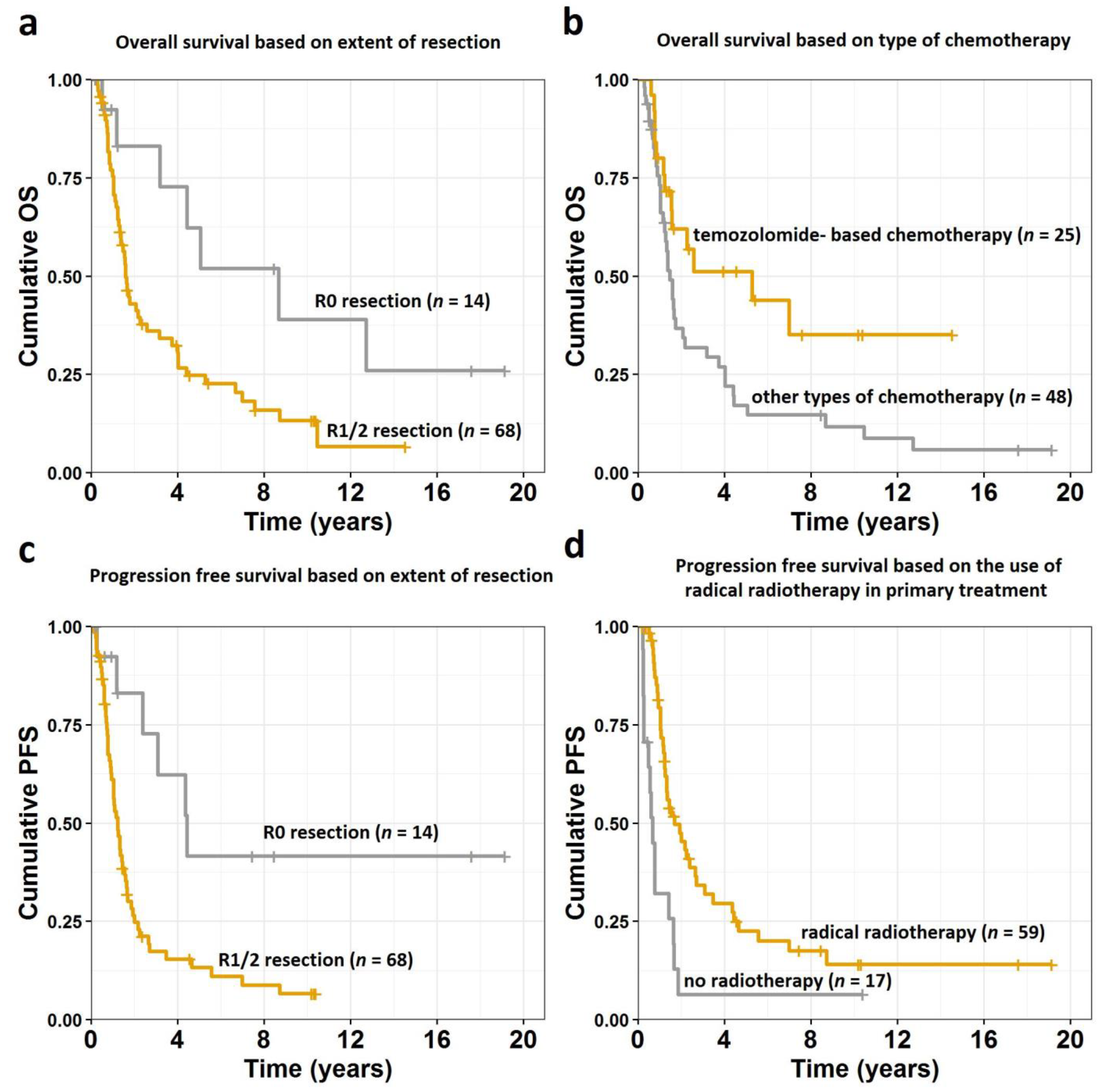
3.4. Survival Analysis

Median follow-up was 122 months. During that time, 59 patients died (51 died with disease, eight due to other reasons), 11 are alive with disease, and 12 alive with no signs of disease. One, 2-, 5-, and 10-year OS from the date of diagnosis was 78%, 48%, 30%, and 17%, respectively. Factors that had statistically significant impact on OS in univariate analysis are presented in Table 2. All variables with p values of < 0.05 (tumor grade, R0 extent of resection, radical RT, temozolomide-based CTH) were included in the multivariate analysis, which showed that only radical (R0) resection and temozolomide-based CTH are factors independently affecting OS with p = 0.005 and p = 0.004, respectively (Figure 1a,b, Figure 2).
Table 3 shows factors that had statistically significant impact on PFS in univariate analysis. One, 2-, 5-, and 10-year PFS was 65%, 35%, 17%, and 12%, respectively. Multivariate analysis of factors that had p value below 0.05 in univariate analysis (tumor grade IV, ECOG performance status equal or higher than 2, R0 extent of resection, radical RT) showed that only radical (R0) resection and radical RT are factors independently affecting PFS with p = 0.040 and p = 0.007, respectively (Figure 1c,d, Figure 2).
Among those who were diagnosed with recurrence of the disease, additional survival analysis was performed. The timing of adjuvant treatment (120 days for RT and 22 days for CTH) was statistically significant in multivariate analysis—with p = 0.041 and 0.023, respectively. Treatment of the recurrence and the use of RT in recurrence treatment were also found as statistically significant in terms of survival in this group.

4. Discussion

4.1. Study Limitations

The limitations of our study are the same as in all of the studies on rare tumors—a retrospective study covering a long time period with different treatment modalities. However, due to the rarity of this tumor in the pediatric population, studies like ours are the only way to collect experience in this topic. Additionally, no unified imaging modality was used for evaluation of surgical outcome, and some patients in early years of the study did not have MR. Furthermore, we did not perform the review of the pathologic diagnosis. Mutations or MGMT methylation are not routinely evaluated in pediatric population and we did not assess its possible impact on survival. What is more, the adjuvant RT and CTH schema varied depending on the institutional protocol at the time of the treatment. Nevertheless, this is one of the biggest reports on HGG, as conducted in seven major Polish hospitals, during the 25 years of the study period.

4.2. Clinical Presentation

The diagnosis of HGG, despite improvements in neurosurgery, RT, and systemic therapies, still carries poor prognosis in children and adults. For a long time, a younger age has been believed to be a favorable prognostic factor in HGG, which is in accordance with SEER analyses but in contrast with one report by Finlay J.L. [22,25,33,35]. The impact of age on OS or PFS was not observed in our study. The mean age at the time of disease diagnosis in our group of 12 years old is comparable to the mean age reported by other authors—within the range of 10 to 14 years old [25,33,34]. Looking at the impact of gender on the treatment results, two studies identified male sex as a better prognostic factor, which was in contrast with Finlay J.L.’s observations, but the number of patients included in those analyses was much smaller than in the CCG-945 study [19,22,24]. In accordance with the previous reports and SEER analyses, male sex predilection was observed in our group, but we did not find it statistically significant in terms of OS or PFS [25,33,34]. Prognostic factors for OS found by other authors are presented in Table 4.
Patient’s reported symptoms often are described as signs of increased intracranial pressure, seizures, or paresis, depending on tumor location [1,9,10,14,25]. Clinical presentations of the disease observed in our patients were similar to those described in previous reports. The presence of symptoms such as seizures, hemiparesis, headaches, or others was not found as statistically significant in terms of OS in our group or in the studies of other authors. The duration of symptoms before the diagnosis was described to be shorter for HGG than for lower-grade lesions [36,37]. Roulecke B.C. reported median and average intervals of 24 and 59 days from symptoms onset to the tumor diagnosis with the length being significantly shorter in patients with HGG [36]. This difference was not observed in our group with the same (30 days) median duration of disease symptoms in patients with grade III and grade IV tumors, which is in accordance with the Bilginer B. study, which reported the majority of patients being diagnosed in less than 3 months [14]. In contrast, much longer time intervals with a median of 4 months were reported in other studies [7,9,25,26].

4.3. Histopathological Diagnosis

HGG are a histologically heterogeneous group of tumors and are further classified according to the cell of origin as astrocytic tumors (AA, GBM, giant cell GBM, gliosarcoma), oligodendroglial tumors (AO), or oligoastrocytic (mixed) tumors (anaplastic oligoastrocytoma) [38]. Special categories of HGG include diffuse intrinsic pontine gliomas (DIPG); however, such a diagnosis requires a different treatment approach so patients with main tumor location in the brainstem were not included in our analysis. Mathew R.K. reported 5-year OS of 18.9% (United Kingdom, UK) and 30.1% (United States, US) for patients with AA and 8.5% (UK) and 22.3% (US) for GBM [32]. Results observed in our study showed much better OS in AA group (5-year OS of 72%) and comparable OS in GBM patients (5-year OS of 17%). Results observed by other authors are shown in Table 4. Grade IV histopathology was found as statistically significant in terms of OS and PFS in our study in the univariate analysis, as well as in the reports of other authors (Table 4) [6,8,10,19,22,23,28].

4.4. Treatment—Surgery

Maximal safe resection is the first step of HHG treatment. Many retrospective series reported extent of resection (EOR) as the only one or one of the main prognostic factors influencing OS (Table 4) [1,6,8,9,10,11,12,13,14,17,19,22,25,27,29,30]. Additionally, SEER analysis performed by Adams H. showed that gross total resection is independently associated with improved OS in GBM patients [34]. Additionally, in our study, the patients who had R0 resection tended to have better OS and PFS compared to those with R1/R2 surgery (p = 0.005 and p = 0.04, respectively). The Fisher’s exact test was used to determine the association between EOR and grade and showed that EOR is an independent factor influencing survival, irrespective of tumor grade, with p = 0.765. These findings are in accordance with the reports of other authors and demonstrate a survival advantage provided by radical resection. Based on those data, an aggressive surgical therapy for malignant pediatric astrocytomas should be considered.

4.5. Treatment—Chemotherapy

The current protocol in HGG in children over 3 years old in Poland (which is recommended, but not standard procedure in all Polish hospitals) includes: surgery (maximal safe resection), two to four cycles of CTH (irinotecan + carboplatin), RT (+/− temozolomide), and adjuvant CTH—four cycles of cisplatin/ vincristine/ lomustine and four cycles of temozolomide/cisplatin alternately. However, due to the long study period, varied treatment protocols were employed. Many authors evaluated the role of the systemic treatment and its impact on OS. In the multi-institutional analysis conducted by Walston S., EOR and use of CTH after RT/RCTH were statistically significant for OS and PFS. Furthermore, in patients with subtotal resection/biopsy only, total RT dose of more than 59.4 Gy and adjuvant CTH had a positive impact on OS [11]. Yazici G. found that those who received temozolomide had better OS in GBM group—median OS of 32 vs 12 months [10]. Similar observations were found by Yang T.; however, the difference did not reach statistical significance with p = 0.075, and authors did not correlate better survival with the use of this systemic agent but rather with better intraoperative imaging guidance techniques and improved diagnostic imaging [1]. CCG-943 is the only randomized study to date that demonstrated a clear survival advantage associated with adjuvant CTH in children. Patients who were treated with CTH fared significantly better than children treated with RT alone; although, in this trial, unintentional inclusion of low-grade glioma (LGG) as HGG occurred, which resulted in better outcomes [6]. Among other trials, CCG-9933, German HIT-GBM, or French Society of Pediatric Oncology, improvement in OS was shown when adjuvant CTH was implemented before RT, but EOR was still the most important factor affecting survival [15,16,17,18,19,20,21]. Interestingly, in 2015, Mallick S. and Walston S., and in 2018, Azizi A.A., in retrospective analyses, described better OS in group receiving temozolomide [7,8,11]. Their results are in accordance with our observations. Furthermore, better survival observed in the subgroup of patients with disease progression indicate that the delay of over 3 weeks in the beginning of CTH should be omitted. The decision about the therapeutic agent used should be made with caution in patients with grade III tumors, because long administration of temozolomide may lead to mutations in MMR genes and, subsequently, mutations in pathways leading to progression from grade III to grade IV.

4.6. Treatment—Radiotherapy

RT as adjuvant treatment (combined with CTH in some patients) was used in almost all of the studies. Doses prescribed were usually within the range of 54 to 60 Gy, given in 1.8 to 2 Gy fractions, and covered tumor (GTV), along with tumor bed and additional margins of 2 to 2.5 cm (CTV,PTV) [7,8,9,10,11,12,14,17,19,22,24,25,28]. Some authors limited the dose to 54 Gy in cases of children 6 years of age or younger, as well as included patients irradiated to only 45 Gy [6,17,27,30]. In the study by Perkins S.M., children received smaller doses (50.4-54 Gy), and in seven patients, RT was delivered to the whole brain. However, her study covered a very long period of time (from 1970 to 2008), which could be the reason for including such patients [13]. Extensive fields and low doses (of 45 to 52.5 Gy) were also used in patients treated in the Sposto R. study, conducted in 1989 [6]. In the Lopez-Aguilar E. study, children underwent hyperfractionated RT to the total dose of 54 Gy, given in two fractions of 1.1 Gy per day, but the authors did not focus on RT outcome and nobody repeated their RT schema [23]. RT as a prognostic factor for better OS was found by Wolff J.E. when doses over 54 Gy were applied [19]. Boudaouara O. found that postoperative RT to the total dose of 60 Gy was significantly associated with better OS [26]. Liu M. found that the use of RT improved OS in patients older than 11 years old [35]. We also observed that the use of radical RT had positive impact on OS and PFS in the univariate analysis, but the multivariate Cox model showed its impact on PFS only. The correlation between the total dose used and the survival was not observed. Given the outcome observed in our patients and the best results observed in studies with RCTH, such adjuvant treatment should be considered in all patients in good performance status after surgery. As shown in the disease progression subgroup, RT should be started sooner than 4 months after the surgery. Furthermore, since 60% of our patients evaluated during systemic treatment showed disease progression, the offset of RT should not be delayed due to extended CTH (except in children younger than 3 years old for whom the decision should be made individually).

4.7. Secondary Neoplasms

There are a few inherited syndromes that increase the risk of HGG development, such as Li–Fraumeni syndrome, neurofibromatosis type 1, or Turcot syndrome [26,39]. A family history of brain tumor was present in the cases of five patients in our group. Furthermore, eight patients had the history of previous oncologic treatment—three were previously treated for LGG, two for rhabdomyosarcoma, two for ALL and one for retinoblastoma. Other authors also found that their patients had previous oncologic treatment or tumor-predisposing conditions [40,41]. In Yang T.’s study, five patients received cranial RT during ALL treatment, and three had neurofibromatosis [1]. Additionally, Wolf J.E. described previous neoplasms: one ALL, one ALL and PNET and then HGG, and one craniopharyngioma [17]. LGG were also observed in four patients in Boudaouara O.’s report [26]. Additionally, in Bilginer B.’s study, in two patients, HGG progressed from LGG [14]. The same study reported HGG in patients with: neurofibromatosis type 1, family history of brain tumor, and cranial RT during ALL treatment [14]. Patients with previous history of oncologic treatment, especially those with LGG and ALL, should be considered as high risk for developing secondary high-grade neoplasms, and careful follow-up is recommended [41].

5. Conclusions

The treatment outcome of children with HGG is poor. The best results are observed in patients after MR-confirmed R0 resection with immediate postoperative temozolomide-based CTH and RT. Further multi-institutional studies are needed to establish the optimal treatment sequence or more effective treatment regimens.

Author Contributions

Conceptualization, A.N.; methodology, A.N.; software, A.N. and A.K. (Aleksandra Krzywon); validation, A.N. and A.K. (Aleksandra Krzywon); formal analysis, A.N. and A.K. (Aleksandra Krzywon); investigation, A.N.; resources, A.N., J.S.-Z., A.M.-M., E.P., M.A.K., K.K.-B., A.K. (Aneta Kaczorowska), A.D. and M.H.; data curation, A.N.; writing—original draft preparation, A.N.; writing—review and editing, A.N., A.K. (Aleksandra Krzywon), M.A.K. and E.P.; visualization, A.N. and A.K.; supervision, M.H.; project administration, A.N. All authors have read and agreed to the published version of the manuscript.

Funding

This research did not receive any specific grant from funding agencies in the public, commercial, or not-for-profit sectors.

Institutional Review Board Statement

The study was conducted according to the guidelines of the Declaration of Helsinki, and approved by the Institutional Review Board committee in MSC National Research Institute of Oncology in Gliwice (number: KB/430-05/20).

Informed Consent Statement

Patient consent was waived due to retrospective character of the analysis. The paper does not report on the use of experimental or new protocols. Ethical review and approval were waived for this study. In our analysis, we used routinely collected anonymous data; therefore, ethical approval was not required according to the national law.

Data Availability Statement

The data presented in this study are available in this article.

Conflicts of Interest

The authors declare that the research was conducted in the absence of any commercial or financial relationships that could be construed as a potential conflict of interest.

References

  1. Yang, T.; Temkin, N.; Barber, J.; Geyer, J.R.; Leary, S.; Browd, S.; Ojemann, J.G.; Ellenbogen, R.G. Gross Total Resection Correlates with Long-Term Survival in Pediatric Patients with Glioblastoma. World Neurosurg. 2013, 79, 537–544. [Google Scholar] [CrossRef] [PubMed]
  2. Ostrom, Q.T.; Cioffi, G.; Gittleman, H.; Patil, N.; Waite, K.; Kruchko, C.; Barnholtz-Sloan, J.S. CBTRUS Statistical Report: Primary Brain and Other Central Nervous System Tumors Diagnosed in the United States in 2012–2016. Neuro Oncology. 2019, 21, v1–v100. [Google Scholar] [CrossRef] [PubMed]
  3. de Blank, P.M.; Ostrom, Q.T.; Rouse, C.; Wolinsky, Y.; Kruchko, C.; Salcido, J.; Barnholtz-Sloan, J.S. Years of life lived with disease and years of potential life lost in children who die of cancer in the United States, 2009. Cancer Med. 2015, 4, 608–619. [Google Scholar] [CrossRef]
  4. Vanan, M.I.; Eisenstat, D.D. Management of high-grade gliomas in the pediatric patient: Past, present, and future. Neuro Oncol. Pr. 2014, 1, 145–157. [Google Scholar] [CrossRef] [PubMed]
  5. Funakoshi, Y.; Hata, N.; Kuga, D.; Hatae, R.; Sangatsuda, Y.; Fujioka, Y.; Takigawa, K.; Mizoguchi, M. Pediatric Glioma: An Update of Diagnosis, Biology and Treatment. Cancers 2021, 13, 758. [Google Scholar] [CrossRef] [PubMed]
  6. Sposto, R.; Ertel, I.J.; Jenkin, R.D.; Boesel, C.P.; Venes, J.L.; Ortega, J.A.; Evans, A.E.; Waral, W.; Hammond, D. The effectiveness of chemotherapy for treatment of high grade astrocytoma in children: Results of a randomized trial—A report from the Childrens Cancer Study Group. J. Neuro Oncol. 1989, 7, 165–177. [Google Scholar] [CrossRef] [Green Version]
  7. Mallick, S.; Gandhi, A.K.; Joshi, N.P.; Kumar, A.; Puri, T.; Sharma, D.N.; Haresh, K.P.; Gupta, S.; Julka, P.K.; Rath, G.K.; et al. Outcomes of pediatric glioblastoma treated with adjuvant chemoradiation with temozolomide and correlation with prognostic factors. Indian J. Med. Paediatr. Oncol. 2015, 36, 99–104. [Google Scholar] [CrossRef] [Green Version]
  8. Azizi, A.A.; Paur, S.; Kaider, A.; Dieckmann, K.; Peyrl, A.; Chocholous, M.; Czech, T.; Slavc, I. Does the interval from tumour surgery to radiotherapy influence survival in paediatric high grade glioma? Strahlenther. Onkol. 2018, 194, 552–559. [Google Scholar] [CrossRef] [PubMed] [Green Version]
  9. Gupta, S.; Mallick, S.; Benson, R.; Haresh, K.P.; Julka, P.K.; Rath, G.K. Extent of surgical resection and adjuvant temozolomide improves survival in pediatric GBM: A single center experience. Childs Nerv. Syst. 2017, 33, 951–956. [Google Scholar] [CrossRef]
  10. Yazici, G.; Zorlu, F.; Cengiz, M.; Ozyigit, G.; Eren, G.; Yüce, D.; Varan, A.; Akyuz, C.; Akalan, N.; Gurkaynak, M. High-grade glioma in children and adolescents: A single-center experience. Childs Nerv. Syst. 2016, 32, 291–297. [Google Scholar] [CrossRef]
  11. Walston, S.; Hamstra, D.A.; Oh, K.; Woods, G.; Guiou, M.; Olshefski, R.S.; Chakravarti, A.; Williams, T.A. A Multi-Institutional Experience in Pediatric High-Grade Glioma. Front. Oncol. 2015, 5, 28. [Google Scholar] [CrossRef]
  12. Jalali, R.; Rishi, A.; Goda, J.S.; Sridhar, E.; Gurav, M.; Sharma, P.; Moiyadi, A.; Shetty, P.; Gupta, T. Clinical outcome and molecular characterization of pediatric glioblastoma treated with postoperative radiotherapy with concurrent and adjuvant temozolomide: A single institutional study of 66 children. Neuro Oncol. Pr. 2016, 3, 39–47. [Google Scholar] [CrossRef]
  13. Perkins, S.M.; Rubin, J.B.; Leonard, J.R.; Smyth, M.D.; El Naqa, I.; Michalski, J.M.; Simpson, J.R.; Limbrick, D.L.; Park, T.S.; Mansur, D.B. Glioblastoma in Children: A Single-Institution Experience. Int. J. Radiat. Oncol. Biol. Phys. 2011, 80, 1117–1121. [Google Scholar] [CrossRef]
  14. Bilginer, B.; Hanalioglu, S.; Turk, C.C.; Narin, F.; Oguz, K.K.; Soylemezoglu, F.; Akalan, N. Is the knowledge pertaining to adult glioblastomas enough for pediatric cases? Prognostic factors in childhood. Turk. Neurosurg. 2017, 27, 279–288. [Google Scholar] [CrossRef] [Green Version]
  15. Wolff, J.E.; Molenkamp, G.; Westphal, S.; Pietsch, T.; Gnekow, A.; Kortmann, R.D.; Kuehl, J. Oral trosfosfamide and etoposide in pediatric patients with glioblastoma multiforme. Cancer 2000, 89, 2131–2137. [Google Scholar] [CrossRef]
  16. Wolff, J.E.; Wagner, S.; Reinert, C.; Gnekow, A.; Kortmann, R.D.; Kühl, J.; Van Gool, S.W. Maintenance treatment with interferon-gamma and low-dose cyclophosphamide for pediatric high-grade gliomas. J. Neuro Oncol. 2006, 79, 315–321. [Google Scholar] [CrossRef]
  17. Wolff, J.E.; Driever, P.H.; Erdlenbruch, B.; Kortmann, R.D.; Rutkowski, S.; Pietsch, T.; Parker, C.; Metz, M.W.; Gnekow, A.; Kramm, C.M. Intensive chemotherapy improves survival in pediatric high-grade glioma after gross total resection: Results of the HIT-GBM-C protocol. Cancer 2010, 116, 705–712. [Google Scholar] [CrossRef]
  18. Wolff, J.E.; Kortmann, R.D.; Wolff, B.; Pietsch, T.; Peters, O.; Schmid, H.-J.; Rutkowski, S.; Warmuth-Metz, M.; Kramm, C. High dose methotrexate for pediatric high grade glioma: Results of the HIT-GBM-D pilot study. J. Neuro Oncol. 2011, 102, 433–442. [Google Scholar] [CrossRef] [PubMed]
  19. Wolff, J.E.; Gnekow, A.K.; Kortmann, R.D.; Pietsch, T.; Urban, C.; Graf, N.; Kühl, J. Preradiation chemotherapy for pediatric patients with high-grade glioma. Cancer 2002, 94, 264–271. [Google Scholar] [CrossRef] [PubMed]
  20. Chastagner, P.; Kalifa, C.; Doz, F.; Bouffet, E.; Gentet, J.C.; Ruchoux, M.M.; Bracard, S.; Desandes, E.; Frappaz, D. Outcome of children treated with preradiation chemotherapy for a high-grade glioma: Results of a French Society of Pediatric Oncology (SFOP) pilot study. Pediatr. Blood Cancer 2007, 49, 803–880. [Google Scholar] [CrossRef] [PubMed]
  21. McDonald, T.J.; Arenson, E.B.; Ater, J.; Sposto, R.; Bevan, H.H.; Bruner, J.; Deutsch, M.; Kurczynski, E.; Luerssen, T.; McGuire-Cullen, P.; et al. Phase II study of high dose chemotherapy before radiation in children with newly diagnosed high-grade astrocytomas: Final analysis of Children’s Cancer Group Study 9933. Cancer 2005, 104, 2862–2871. [Google Scholar] [CrossRef]
  22. Finlay, J.L.; Boyett, J.M.; Yates, A.J.; Wisoff, J.H.; Milstein, J.M.; Geyer, J.R.; Bertolone, S.J.; McGuire, P.; Cherlow, J.M.; Tefft, M. Randomized phase III trial in childhood high-grade astrocytoma comparing vincristine, lomustine, and prednisone with the eight-drugs-in-1-day regimen. Childrens Cancer Group. J. Clin. Oncol. 1995, 13, 112–123. [Google Scholar] [CrossRef]
  23. López-Aguilar, E.; Sepúlveda-Vildósola, A.C.; Rivera-Márquez, H.; Cerecedo-Díaz, F.; Valdés-Sánchez, M.; Delgado-Huerta, S.; Wanzke-del Angel, V.; Ramón-García, G.; Rodríguez-Jiménez, H.; Hernández-Contreras, I.; et al. Preirradiation ifosfamide, carboplatin and etoposide (ICE) for the treatment of high-grade astrocytomas in children. Childs Nerv. Syst. 2003, 19, 818–823. [Google Scholar] [CrossRef]
  24. Espinoza, J.C.; Haley, K.; Patel, N.; Dhall, G.; Gardner, S.; Allen, J.; Torkildson, J.; Cornelius, A.; Rassekh, R.; Bedros, A.; et al. Outcome of young children with high-grade glioma treated with irradiation-avoiding intensive chemotherapy regimens: Final report of the Head Start II and III trials. Pediatr. Blood Cancer 2016, 63, 1806–1813. [Google Scholar] [CrossRef] [Green Version]
  25. Das, K.K.; Mehrotra, A.; Nair, A.P.; Kumar, S.; Srivastava, A.K.; Sahu, R.N.; Kumar, R. Pediatric glioblastoma: Clinico-radiological profile and factors affecting the outcome. Childs Nerv. Syst. 2012, 28, 2055–2062. [Google Scholar] [CrossRef]
  26. Boudaouara, O.; Charfi, S.; Bahri, M.; Daoud, J.; Boudawara, M.Z.; Gouiaa, N.; Boudawara, S.T. Pediatric high grade gliomas: Clinico-pathological profile, therapeutic approaches and factors affecting overall survival. Neurochirurgie 2019, 65, 63–68. [Google Scholar] [CrossRef] [PubMed]
  27. Jung, T.-Y.; Lee, J.Y.; Kim, D.-S.; Park, H.J.; Kim, C.-Y.; Ra, Y.-S.; Lee, M.-J.; Kim, S.-H.; Baek, H.-J.; Kim, I.H.; et al. Pediatric supratentorial high-grade glioma: Multicenter retrospective observational study of the Korean Society for Pediatric Neuro-Oncology. J. Neuro Oncol. 2015, 121, 413–419. [Google Scholar] [CrossRef] [PubMed]
  28. Lucas, J.T.; Cooper, D.A.; Hwang, S.; Tinkle, C.; Li, X.; Li, Y.; Orr, B.; Merchant, T.E.; Broniscer, A. Prognostic Relevance of Treatment Failure Patterns in Pediatric High-Grade Glioma: Is There a Role for a Revised Failure Classification System? Int. J. Radiat. Oncol. Biol. Phys. 2017, 99, 450–458. [Google Scholar] [CrossRef] [PubMed]
  29. Lee, J.W.; Lim, D.H.; Sung, K.W.; Lee, H.J.; Yi, E.S.; Yoo, K.H.; Koo, H.H.; Suh, Y.L.; Shin, H.J. Tandem High-Dose Chemotherapy and Autologous Stem Cell Transplantation for High-Grade Gliomas in Children and Adolescents. J. Korean Med. Sci. 2017, 32, 195–203. [Google Scholar] [CrossRef] [Green Version]
  30. Cohen, K.J.; Pollack, I.F.; Zhou, T.; Buxton, A.; Holmes, E.J.; Burger, P.C.; Brat, D.J.; Rosenblum, M.K.; Hamilton, R.L.; Lavey, R.S.; et al. Temozolomide in the treatment of high-grade gliomas in children: A report from the Children’s Oncology Group. Neuro Oncol. 2011, 13, 317–323. [Google Scholar] [CrossRef]
  31. Cabanas, R.; Saurez, G.; Rios, M.; Alert, J.; Reyes, A.; Valdes, J.; Gonzalez, M.C.; Pedrayes, J.L.; Avila, M.; Herrera, R.; et al. Treatment of children with high grade glioma with nimotuzumab: A 5-year institutional experience. MAB 2013, 5, 202–207. [Google Scholar] [CrossRef] [Green Version]
  32. Mathew, R.K.; O’Kane, R.; Parslow, R.; Stiller, C.; Kenny, T.; Picton, S.; Chumas, P.D. Comparison of survival between the UK and US after surgery for most common pediatric CNS tumors. Neuro Oncol. 2014, 16, 1137–1145. [Google Scholar] [CrossRef] [PubMed] [Green Version]
  33. Lam, S.; Lin, Y.; Zinn, P.; Su, J.; Pan, I.-W. Patient and treatment factors associated with survival among pediatric glioblastoma patients: A Surveillance, Epidemiology, and End Results study. J. Clin. Neurosci. 2018, 47, 285–293. [Google Scholar] [CrossRef]
  34. Adams, H.; Adams, H.H.H.; Jackson, C.; Rincon-Torroella, J.; Jallo, G.I.; Quiñones-Hinojosa, A. Evaluating extent of resection in pediatric glioblastoma: A multiple propensity score-adjusted population-based analysis. Childs Nerv. Syst. 2016, 32, 493–503. [Google Scholar] [CrossRef] [PubMed]
  35. Liu, M.; Thakkar, J.P.; Garcia, C.R.; Dolecek, T.A.; Wagner, L.M.; Dressler, E.V.M.; Villano, J.L. National cancer database analysis of outcomes in pediatric glioblastoma. Cancer Med. 2018, 7, 1151–1159. [Google Scholar] [CrossRef] [PubMed]
  36. Reulecke, B.C.; Erker, C.G.; Fiedler, B.J.; Niederstadt, T.-U.; Kurlemann, G. Brain Tumors in Children: Initial Symptoms and Their Influence on the Time Span Between Symptom Onset and Diagnosis. J. Child Neurol. 2008, 23, 178–183. [Google Scholar] [CrossRef] [PubMed]
  37. Mehta, V.; Chapman, A.; McNeely, P.D.; Walling, S.; Howes, W.J. Latency between Symptom Onset and Diagnosis of Pediatric Brain Tumors: An Eastern Canadian Geographic Study. Neurosurgery 2002, 51, 365–372. [Google Scholar] [CrossRef]
  38. Louis, D.N.; Ohgaki, H.; Wiestler, O.D.; Cavenee, W.K.; Burger, P.C.; Jouvet, A.; Scheithauer, B.W.; Kleihues, P. The 2007 WHO Classification of Tumours of the Central Nervous System. Acta Neuropathol. 2007, 114, 97–109. [Google Scholar] [CrossRef] [Green Version]
  39. Braunstein, S.; Raleigh, D.; Bindra, R.; Mueller, S.; Daphne Haas-Kogan, D. High-grade glioma: Current molecular landscape and therapeutic approaches. J. Neuro Oncol. 2017, 134, 541–549. [Google Scholar] [CrossRef]
  40. Pettorini, B.L.; Park, Y.-S.; Caldarelli, M.; Massimi, L.; Tamburrini, G.; Di Rocco, C. Radiation-induced brain tumours after central nervous system irradiation in childhood: A review. Childs Nerv. Syst. 2008, 24, 793–805. [Google Scholar] [CrossRef]
  41. Carret, A.-S.; Tabori, U.; Crooks, B.; Hukin, J.; Odame, I.; Johnston, D.L.; Keene, D.L.; Freeman, C.; Bouffet, E. Outcome of secondary high-grade glioma in children previously treated for a malignant condition: A study of the Canadian Pediatric Brain Tumour Consortium. Radiother. Oncol. 2006, 81, 33–38. [Google Scholar] [CrossRef] [PubMed]
Figure 1. Overall survival curves for all analyzed patients divided into subgroups based on (a) extent of resection and (b) type of chemotherapy used in primary treatment and progression free survival for all analyzed patients divided into subgroups based on (c) extent of resection and (d) use of radical radiotherapy in primary treatment. Abbreviations: OS—overall survival; PFS—progression free survival; R0—radical resection (macro- and microscopically); R1—macroscopically radical resection (but not microscopically); R2—macroscopically not-radical resection.
Cancers 13 02062 g001
Figure 2. Hazard ratios for overall survival (a) and progression free survival (b) with 95% confidence intervals and p-values calculated from a multivariate Cox proportional-hazards model. Abbreviations: ECOG—Eastern Cooperative Oncology Group; R0—radical resection (macro- and microscopically); R1—macroscopically radical resection (but not microscopically); R2—macroscopically not-radical resection.
Cancers 13 02062 g002
Table 1. The patients’ and treatment characteristics.
Characteristic Value
Age at diagnosisMean (range)12 (1–18)
Number of patients (%)
SexFemale35 (42.7)
Male47 (57.3)
Pathological subtypeGlioblastoma multiforme G IV51 (62.2)
Anaplastic astrocytoma G III19 (23.2)
Anaplastic oligodendroglioma G III12 (14.6)
ECOG performance status019 (23.2)
148 (58.5)
211 (13.4)
33 (3.7)
41 (1.2)
Primary siteSupratentorial68 (82.9)
Infratentorial12 (14.6)
No data2 (2.5)
SymptomsSeizures17 (20.7)
Paresis18 (21.9)
Headaches39 (47.7)
Vomiting/nausea34 (41.5)
Visual deficits9 (11.0)
Balance disorders10 (12.2)
Disturbances of consciousness16 (19.5)
Speech problems
Hearing deficits
5 (6.1)
4 (4.9)
Weakness17 (20.7)
Other *29 (35.4)
Underwent surgeryYes82 (100)
R014 (17.1)
R116 (19.5)
R2/biopsy52 (63.4)
Underwent radiotherapy in primary treatmentYes65 (79.3)
Radical59
Palliative6
No17 (20.7)
Underwent chemotherapy in primary treatmentYes76 (92.7)
No6 (7.3)
Abbreviations: ECOG—Eastern Cooperative Oncology Group; G—grade; R0—radical resection (macro- and microscopically); R1—macroscopically radical resection (but not microscopically); R2—macroscopically not-radical resection. * Other observed symptoms were (the number of symptoms does not reflect the number of patients, as one patient can present with more than one symptom): facial nerve compression symptoms (eight patients), behavior change (six patients), impaired concentration (five patients), somnolence (two patients), memory impairment (two patients), psychotic symptoms (two patients), nervous tics (one patient), numbness of the foot (one patient), dizziness (one patient), aphasia (one patient), forced position of the head (one patient), breathing difficulties (one patient), taste disturbances (one patient), paleness (one patient), asymmetrical positive Babinski symptom (one patient), and tachycardia (one patient).
Table 2. Prognostic factors: univariate analysis of overall survival for the whole cohort.
Variable HR95% CIp-Value
Age (years) 10.96–1.050.906
Sexmaleref.
female0.950.57–1.600.855
Tumor gradeG IIIref.
G IV2.321.31–4.110.004
ECOG performance status0ref.
11.360.73–2.560.332
2 or more1.760.83–3.730.138
Tumor locationinfratentorialref.
supratentorial0.870.44–1.720.679
Extent of resectionR1/2ref.
R00.410.18–0.920.030
Radiotherapyno radiotherapyref.
radical radiotherapy0.520.29–0.960.036
palliative radiotherapy1.120.41–3.110.821
Total dose over 54 Gynoref.
yes0.920.50–1.700.790
Chemotherapyother than temozolomideref.
temozolomide-based0.490.26–0.910.023
no chemotherapy0.460.16–1.300.144
Abbreviations: ECOG—Eastern Cooperative Oncology Group; G—grade; R0—radical resection (macro- and microscopically); R1—macroscopically radical resection (but not microscopically); R2—macroscopically not-radical resection. The p-value of statistically significant (below 0.05) factors were marked in bold to make it more visible for readers.
Table 3. Prognostic factors: univariate analysis of progression-free survival for the whole cohort.
Variable HR95% CIp-Value
Age (years) 0.990.95–1.040.706
Sexmaleref.
female1.060.64–1.750.822
Tumor gradeG IIIref.
G IV1.801.05–3.120.032
ECOG performance status0ref.
11.720.92–3.230.091
2 or more2.241.05–4.790.037
Tumor locationinfratentorialref.
supratentorial1.110.55–2.260.773
Extent of resectionR1/2ref.
R00.310.13–0.720.006
Radiotherapyno radiotherapyref.
radical radiotherapy0.360.20–0.66<0.001
palliative radiotherapy1.750.67– 4.570.253
Total dose over 54 Gynoref.
yes0.840.46–1.520.558
Chemotherapyother than temozolomideref.
temozolomide-based0.720.41–1.260.247
no chemotherapy0.510.18–1.450.210
Abbreviations: ECOG—Eastern Cooperative Oncology Group; G—grade; R0—radical resection (macro- and microscopically); R1—macroscopically radical resection (but not microscopically); R2—macroscopically not-radical resection. The p-value of statistically significant (below 0.05) factors were marked in bold to make it more visible for readers.
Table 4. Pediatric high-grade gliomas-prognostic factors.
Author (Year of Publication) [Reference Number]Number of PatientsTreatment SchemaOSPrognostic Factors for OS
Sposto R. et al. (1989) [6]58 pts
18 G III
40 G IV
S + RT + CTH or
RT alone
1,2,5-y OS G III 65%, 60%, 60%
1,2,5-y OS G IV 65%, 35%, 15%
AA histology, lack of necrosis, location outside basal ganglia, EOR, CTH
Finlay J.L. et al. (1995) [22]172 pts
82 G III
57 G IV
33 other
S + RT + CTH or
CTH-RT-CTH
1,2,5-y OS 80%, 50%, 40%White race, older age, EOR, AA histology, female sex
Wolf J.E. et al. (2002) [19]52 pts
25 G III
27 G IV
S + RT + CTH or
CTH + RT + CTH (sandwich)
1,2,5-y OS G III 100%, 100%, 100%
1,2,5-y OS G IV 100%, 45%, 40%
EOR, male sex, AA histology, residual tumor > 2 cm, RT ≥ 54 Gy, sandwich CTH
López-Aguilar E. et al. (2003) [23]25 pts
20 G III
5 G IV
S + CTH + hyperRT + CTH1,2,5-y OS 67%, 67%, 67%AA histology, supratentorial location
Wolf J.E. et al. (2010) [17]97 pts
30 G III
41 G IV
26 no data
S + RT + CTH1,2,5-y OS 56%, 30%, 19%EOR
Perkins M. et al. (2011) [13]24 pts G IVS + RT +/− CTH1-2-5-y OS 57%, 32%, 10%EOR
Cohen K.J. et al. (2011) [30]90 pts
31 G III
55 G IV
4 other
S + RCTH +CTH1,2,5-y OS 70%, 35%, 15%EOR
Das K.K. et al. (2012)
[25]
65 pts G IVS + RT + CTH1,2,5-y OS 55%, 30%, 15%EOR
Yang T et al. (2013)
[1]
37 pts G IVS + RT + CTH1,2,5-y OS 64%, 45%, 18%EOR
Cabanas R. et al. (2013) [31]23 pts
19 G III
4 G IV
S + RT + CTH + nimotuzumab1,2,5-y OS 64%, 54%, not reportedNot reported
Walston S. et al. (2015)
[11]
51 pts,
23 G III
28 G IV
S +/- RT/RCTH +/− CTH1,2,5-y OS 87%, 59%, 37%
1,2,5-y OS GIII 90%, 75%, 50%
1,2,5-y OS GIV 90%, 30%, 20%
EOR, CTH after RT/RCTH
Jung T.-J. et al. (2015)
[27]
62 pts
28 G III
34 G IV
S + RCTH or RT+CTH or RT1,2,5-y OS 76%, 35%, not reportedEOR, cerebrospinal fluid dissemination
Mallick S. et al. (2015)
[7]
23 pts G IVS + RCTH +CTH1,2,5-y OS 70%, 61%, 40%CTH-TMX
Yazici G. et al. (2016)
[10]
63 pts,
26 G III
37 G IV
S + RT +/− CTH1,2,5-y OS G III 79%, 63%, 35%
1,2,5-y OS G IV 48%, 30%, 22%
AA histology, better PS, EOR, CTH-TMX
Espinoza J.C. et al. (2016) [24]32 pts
19 G III
11 G IV
2 other
S + CTH + AuHPCR +/− RT (50%)1,2,5-y OS 70%, 50%, 36%Male sex
Jalali R. et al. (2016)
[12]
66 pts G IVS + RCTH +CTH1,2,5-y OS 62%, 30%, 20%EOR, thalamic location
Gupta S. et al. (2017)
[9]
51 pts G IVS + RCTH +CTH1,2,5-y OS 60%, 40%, 20%EOR, CTH-TMX
Bilginer B. et al. (2017)
[14]
42 pts G IVS + RT + CTH1,2,5-y OS 50%, 19%, 10%EOR, lack of seeding metastases
Lucas J.T. et al. (2017)
[28]
56 pts
29 G III
27 G IV
S + RT + erlotynib1,2,5-y OS 45%, 25%, 20%AA histology, lack of brainstem/cerebellar involvement
Lee J.W. et al. (2017)
[29]
30 pts
7 G III
16 G IV
7 other
S + RT or CTH +/− HDCT/auto-SCT3-y OS 32%EOR
Azizi AA. et al. (2018)
[8]
38 pts
17 G III
21 G IV
S + RCTH +CTH1,2,5-y OS 68%, 29%, 21%EOR, lower grade, CTH-TMX
Boudaouara O. et al. (2019) [26]30 pts
3 G III
27 G IV
S + RT +/− CTHMedian OS 13 months; 1,2,5-y OS described only in subgroupsPostoperative RT, complete response
AA—anaplastic astrocytoma; AuHPCR—autologous hematopoietic progenitor cell rescue; CTH—chemotherapy; EOR—extent of resection; G—grade, HDCT/auto-SCT—high-dose chemotherapy and autologous stem cell transplantation; hyperRT—hyperfractionated radiotherapy; OS—overall survival; PS—performance status; pts—patients; RCTH—radiochemotherapy; RT—radiotherapy; S—surgery; TMX—temozolomide; y—year.
Publisher’s Note: MDPI stays neutral with regard to jurisdictional claims in published maps and institutional affiliations.

